EP0666756B1 - Method of producing analgesia using neurotrophins - Google Patents
Method of producing analgesia using neurotrophins Download PDFInfo
- Publication number
- EP0666756B1 EP0666756B1 EP93921561A EP93921561A EP0666756B1 EP 0666756 B1 EP0666756 B1 EP 0666756B1 EP 93921561 A EP93921561 A EP 93921561A EP 93921561 A EP93921561 A EP 93921561A EP 0666756 B1 EP0666756 B1 EP 0666756B1
- Authority
- EP
- European Patent Office
- Prior art keywords
- use according
- bdnf
- active ingredient
- 5hiaa
- day
- Prior art date
- Legal status (The legal status is an assumption and is not a legal conclusion. Google has not performed a legal analysis and makes no representation as to the accuracy of the status listed.)
- Expired - Lifetime
Links
- 108010025020 Nerve Growth Factor Proteins 0.000 title claims abstract description 42
- 102000007072 Nerve Growth Factors Human genes 0.000 title claims abstract description 39
- 230000036592 analgesia Effects 0.000 title claims abstract description 26
- 238000000034 method Methods 0.000 title description 11
- 102000004219 Brain-derived neurotrophic factor Human genes 0.000 claims abstract description 61
- 108090000715 Brain-derived neurotrophic factor Proteins 0.000 claims abstract description 61
- 238000001802 infusion Methods 0.000 claims abstract description 61
- 108090000742 Neurotrophin 3 Proteins 0.000 claims abstract description 54
- QZAYGJVTTNCVMB-UHFFFAOYSA-N serotonin Chemical compound C1=C(O)C=C2C(CCN)=CNC2=C1 QZAYGJVTTNCVMB-UHFFFAOYSA-N 0.000 claims abstract description 48
- 102000003683 Neurotrophin-4 Human genes 0.000 claims abstract description 29
- 108090000099 Neurotrophin-4 Proteins 0.000 claims abstract description 29
- 229940076279 serotonin Drugs 0.000 claims abstract description 23
- 210000001609 raphe nuclei Anatomy 0.000 claims description 27
- 241001465754 Metazoa Species 0.000 claims description 21
- 210000000278 spinal cord Anatomy 0.000 claims description 16
- 239000004480 active ingredient Substances 0.000 claims description 15
- 210000004556 brain Anatomy 0.000 claims description 13
- 210000001259 mesencephalon Anatomy 0.000 claims description 12
- 210000001320 hippocampus Anatomy 0.000 claims description 9
- 239000003814 drug Substances 0.000 claims description 7
- 238000007913 intrathecal administration Methods 0.000 claims description 6
- 108090000765 processed proteins & peptides Proteins 0.000 claims description 4
- 230000001174 ascending effect Effects 0.000 claims description 3
- 210000000133 brain stem Anatomy 0.000 claims description 3
- 239000012634 fragment Substances 0.000 claims description 3
- 238000004519 manufacturing process Methods 0.000 claims description 3
- 102000015534 trkB Receptor Human genes 0.000 claims description 3
- 102000047459 trkC Receptor Human genes 0.000 claims description 3
- 108010064892 trkC Receptor Proteins 0.000 claims description 3
- 239000002775 capsule Substances 0.000 claims description 2
- 210000001638 cerebellum Anatomy 0.000 claims description 2
- 239000003405 delayed action preparation Substances 0.000 claims description 2
- 239000002502 liposome Substances 0.000 claims description 2
- 239000003094 microcapsule Substances 0.000 claims description 2
- 239000006187 pill Substances 0.000 claims description 2
- 229920000642 polymer Polymers 0.000 claims description 2
- 239000003826 tablet Substances 0.000 claims description 2
- 108010064880 trkB Receptor Proteins 0.000 claims description 2
- 229940077737 brain-derived neurotrophic factor Drugs 0.000 abstract description 58
- 229940032018 neurotrophin 3 Drugs 0.000 abstract description 52
- 229940097998 neurotrophin 4 Drugs 0.000 abstract description 28
- 241000124008 Mammalia Species 0.000 abstract description 5
- 230000001404 mediated effect Effects 0.000 abstract description 2
- 208000037265 diseases, disorders, signs and symptoms Diseases 0.000 abstract 2
- 102100029268 Neurotrophin-3 Human genes 0.000 abstract 1
- 201000010099 disease Diseases 0.000 abstract 1
- 208000035475 disorder Diseases 0.000 abstract 1
- DUUGKQCEGZLZNO-UHFFFAOYSA-N 5-hydroxyindoleacetic acid Chemical compound C1=C(O)C=C2C(CC(=O)O)=CNC2=C1 DUUGKQCEGZLZNO-UHFFFAOYSA-N 0.000 description 106
- RVWZUOPFHTYIEO-UHFFFAOYSA-N 5-hydroxyindoleacetic acid Natural products C1=C(O)C=C2C(C(=O)O)=CNC2=C1 RVWZUOPFHTYIEO-UHFFFAOYSA-N 0.000 description 52
- 239000003310 5-hydroxyindoleacetic acid Substances 0.000 description 52
- 102000004230 Neurotrophin 3 Human genes 0.000 description 52
- 241000700159 Rattus Species 0.000 description 43
- 230000000694 effects Effects 0.000 description 34
- 239000003981 vehicle Substances 0.000 description 27
- 239000002953 phosphate buffered saline Substances 0.000 description 18
- 108090000623 proteins and genes Proteins 0.000 description 17
- 230000004044 response Effects 0.000 description 17
- 230000000202 analgesic effect Effects 0.000 description 16
- BQJCRHHNABKAKU-KBQPJGBKSA-N morphine Chemical compound O([C@H]1[C@H](C=C[C@H]23)O)C4=C5[C@@]12CCN(C)[C@@H]3CC5=CC=C4O BQJCRHHNABKAKU-KBQPJGBKSA-N 0.000 description 16
- 210000002569 neuron Anatomy 0.000 description 16
- 208000002193 Pain Diseases 0.000 description 15
- 210000001009 nucleus accumben Anatomy 0.000 description 15
- 230000036407 pain Effects 0.000 description 15
- 102000004169 proteins and genes Human genes 0.000 description 12
- 238000012360 testing method Methods 0.000 description 11
- 210000004027 cell Anatomy 0.000 description 10
- 238000001543 one-way ANOVA Methods 0.000 description 10
- 210000002509 periaqueductal gray Anatomy 0.000 description 10
- VYFYYTLLBUKUHU-UHFFFAOYSA-N dopamine Chemical compound NCCC1=CC=C(O)C(O)=C1 VYFYYTLLBUKUHU-UHFFFAOYSA-N 0.000 description 8
- 229960005181 morphine Drugs 0.000 description 8
- 229940005483 opioid analgesics Drugs 0.000 description 8
- 230000000638 stimulation Effects 0.000 description 8
- 102000010909 Monoamine Oxidase Human genes 0.000 description 7
- 108010062431 Monoamine oxidase Proteins 0.000 description 7
- LOKCTEFSRHRXRJ-UHFFFAOYSA-I dipotassium trisodium dihydrogen phosphate hydrogen phosphate dichloride Chemical compound P(=O)(O)(O)[O-].[K+].P(=O)(O)([O-])[O-].[Na+].[Na+].[Cl-].[K+].[Cl-].[Na+] LOKCTEFSRHRXRJ-UHFFFAOYSA-I 0.000 description 7
- 210000005230 lumbar spinal cord Anatomy 0.000 description 7
- 210000003594 spinal ganglia Anatomy 0.000 description 7
- 230000002146 bilateral effect Effects 0.000 description 6
- 238000005259 measurement Methods 0.000 description 6
- 210000001577 neostriatum Anatomy 0.000 description 6
- 230000003389 potentiating effect Effects 0.000 description 6
- 238000001356 surgical procedure Methods 0.000 description 6
- VREFGVBLTWBCJP-UHFFFAOYSA-N alprazolam Chemical compound C12=CC(Cl)=CC=C2N2C(C)=NN=C2CN=C1C1=CC=CC=C1 VREFGVBLTWBCJP-UHFFFAOYSA-N 0.000 description 5
- 238000011161 development Methods 0.000 description 5
- 210000003140 lateral ventricle Anatomy 0.000 description 5
- 239000000463 material Substances 0.000 description 5
- 230000003204 osmotic effect Effects 0.000 description 5
- 238000010149 post-hoc-test Methods 0.000 description 5
- 230000004083 survival effect Effects 0.000 description 5
- 238000012353 t test Methods 0.000 description 5
- 238000000692 Student's t-test Methods 0.000 description 4
- 230000009471 action Effects 0.000 description 4
- 230000015572 biosynthetic process Effects 0.000 description 4
- 229960003638 dopamine Drugs 0.000 description 4
- 238000002347 injection Methods 0.000 description 4
- 239000007924 injection Substances 0.000 description 4
- 230000001722 neurochemical effect Effects 0.000 description 4
- 239000002858 neurotransmitter agent Substances 0.000 description 4
- 210000003625 skull Anatomy 0.000 description 4
- 238000007492 two-way ANOVA Methods 0.000 description 4
- 108091000117 Tyrosine 3-Monooxygenase Proteins 0.000 description 3
- 102000048218 Tyrosine 3-monooxygenases Human genes 0.000 description 3
- 238000003556 assay Methods 0.000 description 3
- 210000003050 axon Anatomy 0.000 description 3
- 230000004071 biological effect Effects 0.000 description 3
- 210000004978 chinese hamster ovary cell Anatomy 0.000 description 3
- 229940079593 drug Drugs 0.000 description 3
- 238000000338 in vitro Methods 0.000 description 3
- 238000001727 in vivo Methods 0.000 description 3
- 230000005764 inhibitory process Effects 0.000 description 3
- 230000007246 mechanism Effects 0.000 description 3
- 239000002207 metabolite Substances 0.000 description 3
- 210000000929 nociceptor Anatomy 0.000 description 3
- 229940021182 non-steroidal anti-inflammatory drug Drugs 0.000 description 3
- 230000001473 noxious effect Effects 0.000 description 3
- -1 or a hybrid Proteins 0.000 description 3
- 230000008058 pain sensation Effects 0.000 description 3
- 238000011160 research Methods 0.000 description 3
- 238000012552 review Methods 0.000 description 3
- 230000000862 serotonergic effect Effects 0.000 description 3
- 210000003523 substantia nigra Anatomy 0.000 description 3
- 239000000758 substrate Substances 0.000 description 3
- IHWDSEPNZDYMNF-UHFFFAOYSA-N 1H-indol-2-amine Chemical compound C1=CC=C2NC(N)=CC2=C1 IHWDSEPNZDYMNF-UHFFFAOYSA-N 0.000 description 2
- CIWBSHSKHKDKBQ-JLAZNSOCSA-N Ascorbic acid Chemical compound OC[C@H](O)[C@H]1OC(=O)C(O)=C1O CIWBSHSKHKDKBQ-JLAZNSOCSA-N 0.000 description 2
- 102000004127 Cytokines Human genes 0.000 description 2
- 108090000695 Cytokines Proteins 0.000 description 2
- 241000588724 Escherichia coli Species 0.000 description 2
- 101000739876 Homo sapiens Brain-derived neurotrophic factor Proteins 0.000 description 2
- 102000015336 Nerve Growth Factor Human genes 0.000 description 2
- 108010093625 Opioid Peptides Proteins 0.000 description 2
- 102000001490 Opioid Peptides Human genes 0.000 description 2
- 102000003840 Opioid Receptors Human genes 0.000 description 2
- 108090000137 Opioid Receptors Proteins 0.000 description 2
- BHHGXPLMPWCGHP-UHFFFAOYSA-N Phenethylamine Chemical compound NCCC1=CC=CC=C1 BHHGXPLMPWCGHP-UHFFFAOYSA-N 0.000 description 2
- PXIPVTKHYLBLMZ-UHFFFAOYSA-N Sodium azide Chemical compound [Na+].[N-]=[N+]=[N-] PXIPVTKHYLBLMZ-UHFFFAOYSA-N 0.000 description 2
- 239000004830 Super Glue Substances 0.000 description 2
- 238000004458 analytical method Methods 0.000 description 2
- 238000010171 animal model Methods 0.000 description 2
- 230000003502 anti-nociceptive effect Effects 0.000 description 2
- 230000001580 bacterial effect Effects 0.000 description 2
- 230000006399 behavior Effects 0.000 description 2
- 239000003795 chemical substances by application Substances 0.000 description 2
- 230000001684 chronic effect Effects 0.000 description 2
- 230000000375 enkephalinergic effect Effects 0.000 description 2
- FGBJXOREULPLGL-UHFFFAOYSA-N ethyl cyanoacrylate Chemical compound CCOC(=O)C(=C)C#N FGBJXOREULPLGL-UHFFFAOYSA-N 0.000 description 2
- 238000009472 formulation Methods 0.000 description 2
- 230000006870 function Effects 0.000 description 2
- 230000000971 hippocampal effect Effects 0.000 description 2
- QRMZSPFSDQBLIX-UHFFFAOYSA-N homovanillic acid Chemical compound COC1=CC(CC(O)=O)=CC=C1O QRMZSPFSDQBLIX-UHFFFAOYSA-N 0.000 description 2
- 102000051542 human BDNF Human genes 0.000 description 2
- 229940077456 human brain-derived neurotrophic factor Drugs 0.000 description 2
- 239000007943 implant Substances 0.000 description 2
- 230000002401 inhibitory effect Effects 0.000 description 2
- 238000000185 intracerebroventricular administration Methods 0.000 description 2
- 238000007912 intraperitoneal administration Methods 0.000 description 2
- 239000000203 mixture Substances 0.000 description 2
- RPUFERHPLAVBJN-UHFFFAOYSA-N n,n-dihydroxy-1-phenylmethanamine Chemical compound ON(O)CC1=CC=CC=C1 RPUFERHPLAVBJN-UHFFFAOYSA-N 0.000 description 2
- 239000004081 narcotic agent Substances 0.000 description 2
- 229940053128 nerve growth factor Drugs 0.000 description 2
- 239000003399 opiate peptide Substances 0.000 description 2
- 239000002243 precursor Substances 0.000 description 2
- 210000004761 scalp Anatomy 0.000 description 2
- 239000000126 substance Substances 0.000 description 2
- 230000005062 synaptic transmission Effects 0.000 description 2
- 238000003786 synthesis reaction Methods 0.000 description 2
- 210000001519 tissue Anatomy 0.000 description 2
- 230000000451 tissue damage Effects 0.000 description 2
- 231100000827 tissue damage Toxicity 0.000 description 2
- 230000007306 turnover Effects 0.000 description 2
- 210000001170 unmyelinated nerve fiber Anatomy 0.000 description 2
- 238000012795 verification Methods 0.000 description 2
- 206010002091 Anaesthesia Diseases 0.000 description 1
- 241000894006 Bacteria Species 0.000 description 1
- 102400000748 Beta-endorphin Human genes 0.000 description 1
- 101800005049 Beta-endorphin Proteins 0.000 description 1
- 108060001064 Calcitonin Proteins 0.000 description 1
- 102000055006 Calcitonin Human genes 0.000 description 1
- 102100038518 Calcitonin Human genes 0.000 description 1
- VEXZGXHMUGYJMC-UHFFFAOYSA-M Chloride anion Chemical compound [Cl-] VEXZGXHMUGYJMC-UHFFFAOYSA-M 0.000 description 1
- 241001573498 Compacta Species 0.000 description 1
- 206010010774 Constipation Diseases 0.000 description 1
- 241000699802 Cricetulus griseus Species 0.000 description 1
- 206010052804 Drug tolerance Diseases 0.000 description 1
- 102000004190 Enzymes Human genes 0.000 description 1
- 108090000790 Enzymes Proteins 0.000 description 1
- 101100127166 Escherichia coli (strain K12) kefB gene Proteins 0.000 description 1
- 241000287828 Gallus gallus Species 0.000 description 1
- 101000634196 Homo sapiens Neurotrophin-3 Proteins 0.000 description 1
- 206010022714 Intestinal ulcer Diseases 0.000 description 1
- WHUUTDBJXJRKMK-VKHMYHEASA-N L-glutamic acid Chemical compound OC(=O)[C@@H](N)CCC(O)=O WHUUTDBJXJRKMK-VKHMYHEASA-N 0.000 description 1
- URLZCHNOLZSCCA-VABKMULXSA-N Leu-enkephalin Chemical compound C([C@@H](C(=O)N[C@@H](CC(C)C)C(O)=O)NC(=O)CNC(=O)CNC(=O)[C@@H](N)CC=1C=CC(O)=CC=1)C1=CC=CC=C1 URLZCHNOLZSCCA-VABKMULXSA-N 0.000 description 1
- VVQNEPGJFQJSBK-UHFFFAOYSA-N Methyl methacrylate Chemical compound COC(=O)C(C)=C VVQNEPGJFQJSBK-UHFFFAOYSA-N 0.000 description 1
- 125000001429 N-terminal alpha-amino-acid group Chemical group 0.000 description 1
- 206010028980 Neoplasm Diseases 0.000 description 1
- 108090000189 Neuropeptides Proteins 0.000 description 1
- 208000000114 Pain Threshold Diseases 0.000 description 1
- 102000007079 Peptide Fragments Human genes 0.000 description 1
- 108010033276 Peptide Fragments Proteins 0.000 description 1
- 229920005372 Plexiglas® Polymers 0.000 description 1
- 239000004743 Polypropylene Substances 0.000 description 1
- 101710171573 Primary amine oxidase Proteins 0.000 description 1
- 102000004005 Prostaglandin-endoperoxide synthases Human genes 0.000 description 1
- 108090000459 Prostaglandin-endoperoxide synthases Proteins 0.000 description 1
- 101100517385 Rattus norvegicus Ntrk2 gene Proteins 0.000 description 1
- 208000004756 Respiratory Insufficiency Diseases 0.000 description 1
- 206010038678 Respiratory depression Diseases 0.000 description 1
- FAPWRFPIFSIZLT-UHFFFAOYSA-M Sodium chloride Chemical compound [Na+].[Cl-] FAPWRFPIFSIZLT-UHFFFAOYSA-M 0.000 description 1
- 102000005157 Somatostatin Human genes 0.000 description 1
- 108010056088 Somatostatin Proteins 0.000 description 1
- 229930006000 Sucrose Natural products 0.000 description 1
- CZMRCDWAGMRECN-UGDNZRGBSA-N Sucrose Chemical compound O[C@H]1[C@H](O)[C@@H](CO)O[C@@]1(CO)O[C@@H]1[C@H](O)[C@@H](O)[C@H](O)[C@@H](CO)O1 CZMRCDWAGMRECN-UGDNZRGBSA-N 0.000 description 1
- 208000003443 Unconsciousness Diseases 0.000 description 1
- 241000251539 Vertebrata <Metazoa> Species 0.000 description 1
- 208000027418 Wounds and injury Diseases 0.000 description 1
- 238000010521 absorption reaction Methods 0.000 description 1
- 238000009825 accumulation Methods 0.000 description 1
- 230000004913 activation Effects 0.000 description 1
- 210000003766 afferent neuron Anatomy 0.000 description 1
- 230000004075 alteration Effects 0.000 description 1
- 125000000539 amino acid group Chemical group 0.000 description 1
- 150000001413 amino acids Chemical class 0.000 description 1
- 210000004727 amygdala Anatomy 0.000 description 1
- 230000037005 anaesthesia Effects 0.000 description 1
- 229940035676 analgesics Drugs 0.000 description 1
- 238000000540 analysis of variance Methods 0.000 description 1
- 239000000730 antalgic agent Substances 0.000 description 1
- 229940072107 ascorbate Drugs 0.000 description 1
- 235000010323 ascorbic acid Nutrition 0.000 description 1
- 239000011668 ascorbic acid Substances 0.000 description 1
- WOPZMFQRCBYPJU-NTXHZHDSSA-N beta-endorphin Chemical compound C([C@@H](C(=O)N[C@@H](CCCCN)C(=O)N[C@@H](CC(N)=O)C(=O)N[C@@H](C)C(=O)N[C@@H]([C@@H](C)CC)C(=O)N[C@@H]([C@@H](C)CC)C(=O)N[C@@H](CCCCN)C(=O)N[C@@H](CC(N)=O)C(=O)N[C@@H](C)C(=O)N[C@@H](CC=1C=CC(O)=CC=1)C(=O)N[C@@H](CCCCN)C(=O)N[C@@H](CCCCN)C(=O)NCC(=O)N[C@@H](CCC(N)=O)C(O)=O)NC(=O)[C@H](CC(C)C)NC(=O)[C@@H](NC(=O)[C@@H](NC(=O)[C@H](CC(C)C)NC(=O)[C@H]1N(CCC1)C(=O)[C@@H](NC(=O)[C@H](CCC(N)=O)NC(=O)[C@H](CO)NC(=O)[C@H](CCCCN)NC(=O)[C@H](CCC(N)=O)NC(=O)[C@H](CO)NC(=O)[C@@H](NC(=O)[C@H](CCSC)NC(=O)[C@H](CC=1C=CC=CC=1)NC(=O)CNC(=O)CNC(=O)[C@@H](N)CC=1C=CC(O)=CC=1)[C@@H](C)O)[C@@H](C)O)C(C)C)[C@@H](C)O)C1=CC=CC=C1 WOPZMFQRCBYPJU-NTXHZHDSSA-N 0.000 description 1
- 238000004166 bioassay Methods 0.000 description 1
- 230000005540 biological transmission Effects 0.000 description 1
- 230000000903 blocking effect Effects 0.000 description 1
- BBBFJLBPOGFECG-VJVYQDLKSA-N calcitonin Chemical compound N([C@H](C(=O)N[C@@H](CC(C)C)C(=O)NCC(=O)N[C@@H](CCCCN)C(=O)N[C@@H](CC(C)C)C(=O)N[C@@H](CO)C(=O)N[C@@H](CCC(N)=O)C(=O)N[C@@H](CCC(O)=O)C(=O)N[C@@H](CC(C)C)C(=O)N[C@@H](CC=1NC=NC=1)C(=O)N[C@@H](CCCCN)C(=O)N[C@@H](CC(C)C)C(=O)N[C@@H](CCC(N)=O)C(=O)N[C@@H]([C@@H](C)O)C(=O)N[C@@H](CC=1C=CC(O)=CC=1)C(=O)N1[C@@H](CCC1)C(=O)N[C@@H](CCCNC(N)=N)C(=O)N[C@@H]([C@@H](C)O)C(=O)N[C@@H](CC(N)=O)C(=O)N[C@@H]([C@@H](C)O)C(=O)NCC(=O)N[C@@H](CO)C(=O)NCC(=O)N[C@@H]([C@@H](C)O)C(=O)N1[C@@H](CCC1)C(N)=O)C(C)C)C(=O)[C@@H]1CSSC[C@H](N)C(=O)N[C@@H](CO)C(=O)N[C@@H](CC(N)=O)C(=O)N[C@@H](CC(C)C)C(=O)N[C@@H](CO)C(=O)N[C@@H]([C@@H](C)O)C(=O)N1 BBBFJLBPOGFECG-VJVYQDLKSA-N 0.000 description 1
- 229960004015 calcitonin Drugs 0.000 description 1
- 201000011510 cancer Diseases 0.000 description 1
- 230000015556 catabolic process Effects 0.000 description 1
- 210000005056 cell body Anatomy 0.000 description 1
- 230000001413 cellular effect Effects 0.000 description 1
- 210000003169 central nervous system Anatomy 0.000 description 1
- 210000003037 cerebral aqueduct Anatomy 0.000 description 1
- 210000003710 cerebral cortex Anatomy 0.000 description 1
- 230000002490 cerebral effect Effects 0.000 description 1
- 230000008859 change Effects 0.000 description 1
- 238000012512 characterization method Methods 0.000 description 1
- 239000003153 chemical reaction reagent Substances 0.000 description 1
- 230000001713 cholinergic effect Effects 0.000 description 1
- 210000002932 cholinergic neuron Anatomy 0.000 description 1
- 238000004587 chromatography analysis Methods 0.000 description 1
- 238000010367 cloning Methods 0.000 description 1
- 238000003869 coulometry Methods 0.000 description 1
- 230000006378 damage Effects 0.000 description 1
- 230000002939 deleterious effect Effects 0.000 description 1
- 210000001947 dentate gyrus Anatomy 0.000 description 1
- 238000000502 dialysis Methods 0.000 description 1
- 230000003291 dopaminomimetic effect Effects 0.000 description 1
- 231100000673 dose–response relationship Toxicity 0.000 description 1
- 239000003937 drug carrier Substances 0.000 description 1
- 238000009510 drug design Methods 0.000 description 1
- 238000000835 electrochemical detection Methods 0.000 description 1
- 230000003028 elevating effect Effects 0.000 description 1
- 238000010828 elution Methods 0.000 description 1
- 210000002257 embryonic structure Anatomy 0.000 description 1
- 210000001353 entorhinal cortex Anatomy 0.000 description 1
- 210000000647 epithalamus Anatomy 0.000 description 1
- 210000003527 eukaryotic cell Anatomy 0.000 description 1
- 238000002474 experimental method Methods 0.000 description 1
- 239000013604 expression vector Substances 0.000 description 1
- 238000000605 extraction Methods 0.000 description 1
- 239000000835 fiber Substances 0.000 description 1
- 210000002950 fibroblast Anatomy 0.000 description 1
- 210000004055 fourth ventricle Anatomy 0.000 description 1
- 210000001222 gaba-ergic neuron Anatomy 0.000 description 1
- 230000002496 gastric effect Effects 0.000 description 1
- 238000002523 gelfiltration Methods 0.000 description 1
- 239000011521 glass Substances 0.000 description 1
- 229930195712 glutamate Natural products 0.000 description 1
- 210000004884 grey matter Anatomy 0.000 description 1
- 238000004128 high performance liquid chromatography Methods 0.000 description 1
- 210000000548 hind-foot Anatomy 0.000 description 1
- 102000057714 human NTF3 Human genes 0.000 description 1
- 238000004191 hydrophobic interaction chromatography Methods 0.000 description 1
- 210000003016 hypothalamus Anatomy 0.000 description 1
- 238000010874 in vitro model Methods 0.000 description 1
- 238000011065 in-situ storage Methods 0.000 description 1
- 210000003000 inclusion body Anatomy 0.000 description 1
- 230000001939 inductive effect Effects 0.000 description 1
- 239000003112 inhibitor Substances 0.000 description 1
- 210000004347 intestinal mucosa Anatomy 0.000 description 1
- 238000007918 intramuscular administration Methods 0.000 description 1
- 238000001990 intravenous administration Methods 0.000 description 1
- 238000004255 ion exchange chromatography Methods 0.000 description 1
- 230000003447 ipsilateral effect Effects 0.000 description 1
- 230000003902 lesion Effects 0.000 description 1
- 239000003446 ligand Substances 0.000 description 1
- 239000007788 liquid Substances 0.000 description 1
- 238000011068 loading method Methods 0.000 description 1
- 230000005923 long-lasting effect Effects 0.000 description 1
- FKHVEXUWEQFKCJ-UHFFFAOYSA-L magnesium;sodium;5-ethyl-5-pentan-2-yl-1,3-diazinane-2,4,6-trione;2,2,2-trichloroethane-1,1-diol;sulfate Chemical compound [Na+].[Mg+2].[O-]S([O-])(=O)=O.OC(O)C(Cl)(Cl)Cl.CCCC(C)C1(CC)C(=O)NC(=O)NC1=O FKHVEXUWEQFKCJ-UHFFFAOYSA-L 0.000 description 1
- 108020004999 messenger RNA Proteins 0.000 description 1
- 230000004060 metabolic process Effects 0.000 description 1
- 229960002455 methoxyflurane Drugs 0.000 description 1
- RFKMCNOHBTXSMU-UHFFFAOYSA-N methoxyflurane Chemical compound COC(F)(F)C(Cl)Cl RFKMCNOHBTXSMU-UHFFFAOYSA-N 0.000 description 1
- CWWARWOPSKGELM-SARDKLJWSA-N methyl (2s)-2-[[(2s)-2-[[2-[[(2s)-2-[[(2s)-2-[[(2s)-5-amino-2-[[(2s)-5-amino-2-[[(2s)-1-[(2s)-6-amino-2-[[(2s)-1-[(2s)-2-amino-5-(diaminomethylideneamino)pentanoyl]pyrrolidine-2-carbonyl]amino]hexanoyl]pyrrolidine-2-carbonyl]amino]-5-oxopentanoyl]amino]-5 Chemical compound C([C@@H](C(=O)NCC(=O)N[C@@H](CC(C)C)C(=O)N[C@@H](CCSC)C(=O)OC)NC(=O)[C@H](CC=1C=CC=CC=1)NC(=O)[C@H](CCC(N)=O)NC(=O)[C@H](CCC(N)=O)NC(=O)[C@H]1N(CCC1)C(=O)[C@H](CCCCN)NC(=O)[C@H]1N(CCC1)C(=O)[C@@H](N)CCCN=C(N)N)C1=CC=CC=C1 CWWARWOPSKGELM-SARDKLJWSA-N 0.000 description 1
- 210000002200 mouth mucosa Anatomy 0.000 description 1
- UZHSEJADLWPNLE-GRGSLBFTSA-N naloxone Chemical compound O=C([C@@H]1O2)CC[C@@]3(O)[C@H]4CC5=CC=C(O)C2=C5[C@@]13CCN4CC=C UZHSEJADLWPNLE-GRGSLBFTSA-N 0.000 description 1
- 229960004127 naloxone Drugs 0.000 description 1
- 239000003887 narcotic antagonist Substances 0.000 description 1
- 210000000653 nervous system Anatomy 0.000 description 1
- 230000001537 neural effect Effects 0.000 description 1
- 230000014511 neuron projection development Effects 0.000 description 1
- 230000003955 neuronal function Effects 0.000 description 1
- 230000006576 neuronal survival Effects 0.000 description 1
- 108091008700 nociceptors Proteins 0.000 description 1
- 210000000956 olfactory bulb Anatomy 0.000 description 1
- 239000000014 opioid analgesic Substances 0.000 description 1
- 210000001672 ovary Anatomy 0.000 description 1
- 229940094443 oxytocics prostaglandins Drugs 0.000 description 1
- 230000008533 pain sensitivity Effects 0.000 description 1
- 230000037040 pain threshold Effects 0.000 description 1
- 238000007427 paired t-test Methods 0.000 description 1
- 230000037361 pathway Effects 0.000 description 1
- 229960001412 pentobarbital Drugs 0.000 description 1
- WEXRUCMBJFQVBZ-UHFFFAOYSA-N pentobarbital Chemical compound CCCC(C)C1(CC)C(=O)NC(=O)NC1=O WEXRUCMBJFQVBZ-UHFFFAOYSA-N 0.000 description 1
- VLTRZXGMWDSKGL-UHFFFAOYSA-M perchlorate Inorganic materials [O-]Cl(=O)(=O)=O VLTRZXGMWDSKGL-UHFFFAOYSA-M 0.000 description 1
- VLTRZXGMWDSKGL-UHFFFAOYSA-N perchloric acid Chemical compound OCl(=O)(=O)=O VLTRZXGMWDSKGL-UHFFFAOYSA-N 0.000 description 1
- 210000001871 perforant pathway Anatomy 0.000 description 1
- 210000005223 peripheral sensory neuron Anatomy 0.000 description 1
- 229940117803 phenethylamine Drugs 0.000 description 1
- 230000036470 plasma concentration Effects 0.000 description 1
- 229920003023 plastic Polymers 0.000 description 1
- 239000004033 plastic Substances 0.000 description 1
- 229920001155 polypropylene Polymers 0.000 description 1
- 230000001242 postsynaptic effect Effects 0.000 description 1
- 230000003518 presynaptic effect Effects 0.000 description 1
- 230000008569 process Effects 0.000 description 1
- 102000004196 processed proteins & peptides Human genes 0.000 description 1
- 238000012545 processing Methods 0.000 description 1
- 150000003180 prostaglandins Chemical class 0.000 description 1
- 210000002637 putamen Anatomy 0.000 description 1
- 108020003175 receptors Proteins 0.000 description 1
- 102000005962 receptors Human genes 0.000 description 1
- 230000011514 reflex Effects 0.000 description 1
- 230000001105 regulatory effect Effects 0.000 description 1
- 230000000717 retained effect Effects 0.000 description 1
- 210000004189 reticular formation Anatomy 0.000 description 1
- 230000007441 retrograde transport Effects 0.000 description 1
- 238000004366 reverse phase liquid chromatography Methods 0.000 description 1
- 230000035945 sensitivity Effects 0.000 description 1
- 230000001953 sensory effect Effects 0.000 description 1
- 210000001044 sensory neuron Anatomy 0.000 description 1
- 238000012163 sequencing technique Methods 0.000 description 1
- 239000012679 serum free medium Substances 0.000 description 1
- 239000004017 serum-free culture medium Substances 0.000 description 1
- 239000011780 sodium chloride Substances 0.000 description 1
- 239000000243 solution Substances 0.000 description 1
- NHXLMOGPVYXJNR-ATOGVRKGSA-N somatostatin Chemical compound C([C@H]1C(=O)N[C@H](C(N[C@@H](CO)C(=O)N[C@@H](CSSC[C@@H](C(=O)N[C@@H](CCCCN)C(=O)N[C@@H](CC(N)=O)C(=O)N[C@@H](CC=2C=CC=CC=2)C(=O)N[C@@H](CC=2C=CC=CC=2)C(=O)N[C@@H](CC=2C3=CC=CC=C3NC=2)C(=O)N[C@@H](CCCCN)C(=O)N[C@H](C(=O)N1)[C@@H](C)O)NC(=O)CNC(=O)[C@H](C)N)C(O)=O)=O)[C@H](O)C)C1=CC=CC=C1 NHXLMOGPVYXJNR-ATOGVRKGSA-N 0.000 description 1
- 229960000553 somatostatin Drugs 0.000 description 1
- 238000013222 sprague-dawley male rat Methods 0.000 description 1
- 238000010186 staining Methods 0.000 description 1
- 239000012086 standard solution Substances 0.000 description 1
- 238000007619 statistical method Methods 0.000 description 1
- 238000007920 subcutaneous administration Methods 0.000 description 1
- 210000001055 substantia gelatinosa Anatomy 0.000 description 1
- 239000005720 sucrose Substances 0.000 description 1
- 238000013268 sustained release Methods 0.000 description 1
- 239000012730 sustained-release form Substances 0.000 description 1
- 210000001587 telencephalon Anatomy 0.000 description 1
- 210000001103 thalamus Anatomy 0.000 description 1
- 238000002560 therapeutic procedure Methods 0.000 description 1
- 210000000211 third ventricle Anatomy 0.000 description 1
- 210000003901 trigeminal nerve Anatomy 0.000 description 1
- 210000004515 ventral tegmental area Anatomy 0.000 description 1
- 230000002861 ventricular Effects 0.000 description 1
- 125000000391 vinyl group Chemical group [H]C([*])=C([H])[H] 0.000 description 1
- 229920002554 vinyl polymer Polymers 0.000 description 1
Images


Classifications
-
- A—HUMAN NECESSITIES
- A61—MEDICAL OR VETERINARY SCIENCE; HYGIENE
- A61K—PREPARATIONS FOR MEDICAL, DENTAL OR TOILETRY PURPOSES
- A61K38/00—Medicinal preparations containing peptides
- A61K38/16—Peptides having more than 20 amino acids; Gastrins; Somatostatins; Melanotropins; Derivatives thereof
- A61K38/17—Peptides having more than 20 amino acids; Gastrins; Somatostatins; Melanotropins; Derivatives thereof from animals; from humans
- A61K38/18—Growth factors; Growth regulators
- A61K38/185—Nerve growth factor [NGF]; Brain derived neurotrophic factor [BDNF]; Ciliary neurotrophic factor [CNTF]; Glial derived neurotrophic factor [GDNF]; Neurotrophins, e.g. NT-3
-
- A—HUMAN NECESSITIES
- A61—MEDICAL OR VETERINARY SCIENCE; HYGIENE
- A61P—SPECIFIC THERAPEUTIC ACTIVITY OF CHEMICAL COMPOUNDS OR MEDICINAL PREPARATIONS
- A61P25/00—Drugs for disorders of the nervous system
- A61P25/04—Centrally acting analgesics, e.g. opioids
Definitions
- a ⁇ nociceptive neurons contain myelinated axons and are aroused primarily by noxious heat and mechanical stimuli.
- the second set of nociceptive neurons which possess unmyelinated axons and are known as C fibers, are activated by high intensity, mechanical, chemical and thermal stimulation.
- Each of these sets of neurons have their cell bodies in the dorsal root ganglia. Their processes are pseudounipolar, with one axon that terminates in the periphery and one that terminates on neurons in the dorsal horn of the spinal cord.
- Analgesia is the loss of sensitivity to pain without loss of consciousness.
- SPA stimulation-produced analgesia
- analgesia caused by exogenous opioids such as morphine and endogenous opioids
- the first means known to man for inducing analgesia was through the use of plant-derived opioid narcotics such as morphine.
- plant-derived opioid narcotics such as morphine.
- the recent characterization of postsynaptic opioid receptors has lead to the discovery of naturally occurring, endogenous opioids which similarly bind the opioid receptor and thereby produce analgesia.
- the endogenous opioids include the met- and leu-enkephalins, as well as ⁇ -endorphin.
- Analgesia can also be produced by electrical stimulation of the gray matter that surrounds the third ventricle, cerebral aqueduct, and fourth ventricle, in particular, the raphe nuclei or the periaqueductal gray region of the brain. Such stimulation results in complete and fairly long lasting analgesia. Stimulation induced analgesia is not increased by destruction of the periaqueductal gray, indicating that this region is not responsible for pain relay, but rather its effect is through active involvement in the inhibition of afferent input in the dorsal horn of the spinal cord and the trigeminal nerve nucleus. In fact, electrical stimulation appears to exert inhibition on spinal cord sensory neurons in a manner which is very similar to that observed using opioid peptides. The narcotic antagonist naloxone blocks both morphine and stimulation induced analgesia.
- the monoamine neurotransmitter serotonin (5 hydroxytryptamine; 5HT) appears to be the common link in the analgesic mechanism in stimulation- as well as both endogenously- and exogenously-induced opioid analgesia.
- Neurons in the periventricular and periaqueductal gray matter excite those in the raphe nuclei in the medullary reticular formation.
- Serotonergic fibers descend in the dorsolateral funiculus of the spinal cord to terminate in the substantia gelatinosa, where they are believed to activate enkephalinergic inhibitor neurons. Stimulation of C-fiber primary afferent neurons associated with pain results in the release of the potent neuropeptides substance P, calcitonin, gene related peptide (CGRP) and somatostatin, as well as the "fast" neurotransmitter glutamate. The activated enkephalinergic inhibitory neurons in turn exert presynaptic inhibitory control over the release of these neurotransmitters, thus blocking the sensation of pain.
- CGRP gene related peptide
- Non-steroidal anti-inflammatory drugs provide an alternative therapy for the treatment of pain. Their mode of action is believed to be through inhibition of cyclooxygenase, the enzyme responsible for biosynthesis of the prostaglandins.
- the NSAIDs lack many of the side effects on the CNS that are associated with the opioids and they do not result in the development of dependence. They are only effective, however, on low to moderate intensity pain, and are not generally useful for intense pain. In addition, they have undesirable side effects, including the propensity to induce gastric or intestinal ulceration as well as disturbances of platelet function.
- the Neurotrophins The Neurotrophins .
- BDNF Brain-derived neurotrophic factor
- neurotrophins in particular the neurotrophin BDNF
- BDNF neurotrophin BDNF
- Chronic intraseptal infusions of BDNF can prevent most of the axotomy-induced loss of cholinergic neuron staining in the medial septum [Morse et al, in press].
- High affinity binding sites for [1251]NT-3 are found within the medial substantia nigra and ventral tegmental area, nucleus accumbens, caudate-putamen, and raphe nuclei, and the binding to these sites is potently displaced by BDNF [Altar, et al., Am. Acad. Neurol. San Diego, CA., (1992)]. BDNF mRNA is also present in these areas and appears to overlap with tyrosine hydroxylase (TOH)-positive cells [Gall, et al., (1992), unpublished observations].
- TOH tyrosine hydroxylase
- Intrastriatal injections of [1251]-labeled NT-3 or BDNF result in a retrograde transport and accumulation of radioactivity within TOH-positive cells in these same regions [Wiegand, et al., Soc. Neurosci. AB., 17 : 1121, (1991)].
- NT-4 binding has been found to be widely distributed throughout the brain including the cortex, striatum, hippocampus, cerebellum, olfactory bulbs, periaqueductal gray, and raphe nuclei.
- the invention relates to a method of providing analgesia by delivering cytokines, preferably neurotrophins, into a mammal to alleviate pain.
- brain-derived neurotrophic factor is delivered in the vicinity of the raphe nuclei or periaqueductal gray region of the midbrain of a mammal to inhibit pain.
- the present invention relates to a method of producing analgesia in mammals by administering a therapeutically effective dose of an active ingredient as defined above. It is based, in part, on the discovery by applicants that neurotrophins, in particular the neurotrophins BDNF, NT-3 and NT-4 can induce analgesia when delivered in the vicinity of the raphe nuclei or periaqueductal gray of the brain.
- neurotrophins in particular the neurotrophins BDNF, NT-3 and NT-4 can induce analgesia when delivered in the vicinity of the raphe nuclei or periaqueductal gray of the brain.
- the neurotrophin family includes brain-derived neurotrophic factor (BDNF), neurotrophin-3 (NT-3) and neurotrophin-4 (NT-4), all of which have recently been molecularly cloned and shown to be members of the nerve growth factor (NGF) family by virtue of their sequence homology [Leibrock, et al., Nature 341 : 149-152, (1989); Hohn, et al., Nature, 344 : 339-341, (1990); Maisonpierre, et al., Science, 247 : 1446-1451, (1990a); Rosenthal, et al., Neuron, 4 : 767-773, (1990); Ernfors, et al., Proc. Natl. Acad.
- BDNF brain-derived neurotrophic factor
- NT-3 neurotrophin-3
- NT-4 neurotrophin-4
- the cytokines which are useful in practicing the present invention include, but are not limited to, those neurotrophins that bind to the trkB or the trkC receptor.
- BDNF and NT-4 primarily use the trkB receptor [Squinto, et al., Cell 65 : 885-893 (1991); Ip, et al., Proc. Natl. Acad. Sci.,U.SA., 89 : 3060-3064 (1992)] and NT-3 is the preferred ligand for trkC [Lamballe, et a!., Cell 66 : 967-979 (1991)].
- chimeras, or peptides or fragments derived from the neurotrophins will be useful to practice the present invention.
- neurotrophins useful for practicing the present invention may be prepared by first cloning and sequencing a gene encoding each respective protein. Each cloned gene may then be expressed in a prokaryotic or eukaryotic expression system. Any of a number of protocols available to one skilled in the art may be utilized to clone and sequence the neurotrophins. The recombinant neurotrophin gene may be expressed and purified utilizing any number of methods.
- each factor may be prepared from bacterial cells or eukaryotic cells that express recombinant factor. The gene encoding the factor may be subcloned into a bacterial expression vector, such as for example, but not by way of limitation, pCP110.
- the recombinant factors may be purified by any technique which allows for the subsequent formation of a stable, biologically active protein.
- the factors produced in prokaryotes may be recovered from cells as inclusion bodies, followed by quantitative extraction in 8M guanadinium chloride and dialysis.
- conventional ion exchange chromatography, hydrophobic interaction chromatography, reverse phase chromatography or gel filtration may be used.
- the active ingredient which may comprise one purified neurotrophin, or a hybrid, peptide fragment or mutant thereof, a combination of several neurotrophins, or a combination of neurotrophin and opioid peptide, should be formulated in a suitable pharmaceutical carrier for administration in vivo by any appropriate route including, but not limited to injection (e.g. , intravenous, intraperitoneal, intraparenchymal, intramuscular, subcutaneous, endoneural, perineural, intraspinal, intracerebroventricular, intrathecal etc.), by absorption through epithelial or mucocutaneous linings ( e.g. , oral mucosa, rectal and intestinal mucosa, etc.); or by a sustained release implant, including a cellular or tissue implant.
- injection e.g. , intravenous, intraperitoneal, intraparenchymal, intramuscular, subcutaneous, endoneural, perineural, intraspinal, intracerebroventricular, intrathecal etc.
- the concentration of the active ingredient used in the formulation will depend upon the effective dose required and the mode of administration used.
- the dose used should be sufficient to achieve circulating plasma concentrations of active ingredient that are efficacious.
- Effective doses may be extrapolated from dose-response curves derived from in vitro or animal model test systems.
- neurotrophins are delivered in the vicinity of the raphe nuclei or periaqueductal gray.
- the raphe nuclei include any of the midline group of neurons that are associated with the midsagittal seam of the brain stem.
- a characteristic feature of the raphe nuclei which include the caudal linear nucleus, the dorsal raphe nucleus, the median raphe nucleus, the raphe pontis nucleus, the raphe magnus nucleus, the raphe obscurus nucleus, and the raphe pallidus nucleus, is their high proportion of serotonergic neurons.
- neurotrophins are delivered along the ascending and descending projections of the serotonergic neurons of the raphe nuclei, including, but not limited to the thalamus, hypothalamus and epithalamus, the basal telencephalon, the cerebral cortex, the brain stem, cerebellar projections and spinal projections.
- neurotrophins such as BDNF
- BDNF neurotrophins
- infusions of neurotrophins such as, for example, BDNF, cause an alteration in the level of serotonin in the spinal cord.
- Infusion of such neurotrophins directly into the spinal cord via, for example, intrathecal delivery may provide an effective analgesic alternative to morphine without the concomitant side effects associated with the opioid analgesics.
- infusions are made into one of the main target areas of the ascending projections from the dorsal and median raphe nuclei, the hippocampus. This area receives afferents from both the dorsal and median raphe, and the smaller dorsal raphe projections reach the dentate gyrus through the entorhinal cortex and perforant path [Azmitia and Segal, J. Comp. Neurol. 179 , 641-668 (1978)].
- the active ingredient may be formulated in a liquid carrier such as saline, incorporated into liposomes, microcapsules, polymer or wax-based and controlled release preparations, or formulated into tablet, pill or capsule forms.
- a liquid carrier such as saline
- neurotrophins may be produced in situ by transfected cells which are implanted or infused into the target site.
- analgesic properties of the neurotrophins described herein may be due to their ability to alter serotonin neurotransmission within the brain or spinal cord. This is based on the findings, by applicants, in studies in which changes in the levels of serotonin and its principle metabolite, 5-hydroxyindoleacetic acid (5HIAA), following infusions of BDNF and NT-3 in the vicinity of the raphe nuclei were monitored (see Examples 2 and 3 below).
- 5HIAA 5-hydroxyindoleacetic acid
- BDNF Production and Purity of BDNF, NT-3 and NT-4.
- Recombinant human BDNF [Leibrock, et al., Nature 341 :149-152 (1989)] was produced by expression of the gene, complete with its precursor sequence, in Chinese hamster ovary (CHO) cells grown in serum free media.
- the recombinant human BDNF was at least as active as BDNF isolated from pig brain using bioassays of cultured dorsal root ganglion explants or dissociated neurons
- Recombinant human NT-3 (119 amino acid residues) was produced in CHO cells as described [Radziejewski, et al., Biochemistry 31 : 4431-4436 (1992)].
- BDNF and NT-3 were monitored for biological activity using dissociated lumbar dorsal root ganglia (DRG) cells from E8 chicken embryos maintained 2 days in vitro [Lindsay, et a., Neurosci. 12 : 45-51 (1984)].
- DRG dissociated lumbar dorsal root ganglia
- NT-4 can be produced in CHO cells and assayed as set forth in Ip, et a., Proc. Natl. Acad. Sci. USA 89 :3060-3064 (1992) or be produced in E. coli (see Panayotatos and Fandl, PCT application WO/92/22665 published on December 23, 1992).
- NT-4 produced and purified from E. coli showed greater than 95% purity and, like NT-4 which was produced in CHO cells, promoted the survival in serum free medium of MG87 fibroblasts that express the rat TrkB receptor and the survival and neurite growth of rat E15/16 DRG cells.
- BDNF and NT-3 were diluted in PBS to 1.0 mg/ml (BDNF) or 0.5 mg/ml (NT-3,) and loaded into Alzet 2002 osmotic pumps (Alza Corp., Palo Alto, CA; flow rate 0.5 ⁇ l/hr). Each pump was connected to 11.2 cm of vinyl tubing (Biolab Products, Lake Havasu City, AZ) filled with the same solution contained in the pumps, immersed in a glass vial that contained PBS and 0.002% sodium azide and maintained at 37° C for 2 weeks. The effluent was collected into siliconized 1.5 ml polypropylene tubes that contained 400 ⁇ l PBS.
- the collection tubes were replaced with new tubes every 4 days, and the aliquots were measured for biological activity using the dorsal root ganglia outgrowth assay. Both factors retained full biological activity over the 14 days compared to the activity obtained with the same material stored at 2° C.
- the flow moderators of the osmotic pumps were fitted with a 2 cm piece of silated PE50 tubing (Micro-Renathane, Braintree Scientific, Braintree, MA) and a 28G osmotic pump connector cannula (Plastics One, Inc., Roanoke, VA) that was 5.5, 6.0, and 6.8 mm long for intrastriatal, intra-accumbens and supranigral, or raphe infusions, respectively.
- the pumps and flow moderators were filled with vehicle (sterile PBS), NT-3 or BDNF at a concentration of 1 mg/ml to deliver 12 ⁇ g of either factor each day. All tubing joints were sealed with cyanoacrylate adhesive (Wonder Bond Plus, Borden, Inc., Columbus, OH) and the flow moderator was sealed to the pump with melted dental wax. Each rat was anesthetized with an i.p. injection of chloropent (149 mg/kg chlorohydrate and 30.8 mg/kg sodium pentobarbital) and mounted in a small animal stereotaxic apparatus (Kopf, Tijunga, CA).
- Either cannula system was inserted through the skull hole, attached to the skull with cyanoacrylate adhesive, and the scalp incision was closed with wound clips.
- Several animals sacrificed for histological verification of cannula placement following injection of 1.5 ⁇ l of dye revealed cannula terminations mostly above the middle or medial aspect of the pars compacta, in the central caudate-putamen, in the nucleus accumbens just dorsal to the anterior commissure, or about 0.5 mm ventral to the midpoint between the medial and dorsal raphe nuclei. Verification of cannula placements in experimental animals took place at the time of sacrifice. Animals with incorrect cannula placements were removed from the study and the data was not used for statistical analysis.
- the tail flick test follows the procedure derived from [D'Amour and Smith, J. Pharmacol. Exp. Ther. 72 : 74-79 (1941)].
- a noxious stimulus, produced by a beam a high intensity light was focused on the tail.
- the response time was measured automatically and defined as the interval between the onset of the heat stimulus and the abrupt flick of the tail.
- the average of three consecutive determinations, taken in immediate succession, was taken as the tail flick response latency.
- a different patch of tail skin was stimulated on each trial. Animals not responding after 14 seconds were removed from the apparatus and assigned a response latency of 14 seconds.
- Tail flick latencies of unoperated rats (control) or rats infused for either 5, 7, 8, or 11 days with phosphate buffered saline vehicle or BDNF (12 ug/day) or NT-3 (12 ug/day) near the dorsal and median raphe nuclei are shown in Figure 1.
- post-hoc tests indicated that BDNF, NT-3 and NT-4 infusions produced a significant analgesic effect.
- tail flick response can be modulated by activation of pathways descending from the brain to the spinal cord [for reviews see Willis, et al., Sensory Mechanisms of the Spinal Cord, R.E., Plenum Press, New York (1978); Yaksh, Spinal Afferent Processing , Plenum Press, New York (1986)].
- the tail flick test is often used to assay the effectiveness of various agents which require spinal and/or supraspinal structures for their expression.
- Several investigators have observed the tail flick reflex in anesthetized rats [Banks, et al., Phar. Biochem. Behav. 31 : 397-403 (1988); Brink, et al., Brain Behav. Evol.
- Tail flick testing on infusion day 8 was performed in metofane-anesthetized rats.
- Tail flick testing in unanesthetized rats was repeated on infusion day 11 with identical results to both days 5 and 7.
- NT-3 infusion in the lateral ventricle results in analgesia as early as 24 hours after the onset of infusion, whereas midbrain infusion of NT-3 has never shown significant increases in tail flick latencies this soon.
- day 5 tail flick latencies were consistently and significantly higher than day 1.
- the analgesia obtained from midbrain infusions was greater than that seen following i.c.v. infusion, despite the fact that the concentration used was lower (midbrain infusions used 12 ug/day, while the i.c.v. experiments used 24 ug/day for BDNF and NT-3).
- Example 2 The Neurotrophins Alter Serotonin Levels in the Dorsal and Median Raphe Nucleus Hippocampus and Spinal Cord
- 5HT AND 5HIAA CONCENTRATIONS AND THE 5HIAA/5HT RATIO IN DORSAL AND MEDIAN RAPHE MICROPUNCHES 5HT AND 5HIAA CONCENTRATIONS AND THE 5HIAA/5HT RATIO IN DORSAL AND MEDIAN RAPHE MICROPUNCHES .
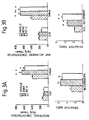
- 5HT and 5HIAA concentrations and the 5HIAA/5HT ratio in the neostriatum and nucleus accumbens were elevated consistently in the nucleus accumbens following 18 days of continuous bilateral supranigral infusion of BDNF or NT-3 (12 ⁇ g/day; Figure 3).
- 5-HIAA levels were also elevated by bilateral infusion of BDNF or NT-3 into the nucleus accumbens ( Figure 4). Serotonin levels were undetectable in tissues obtained from animals receiving nucleus accumbens infusions.
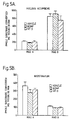
- the MAO A and MAO B activities of the nucleus accumbens and neostriatum following 2 week bilateral nucleus accumbens infusions of BDNF or NT-3 were unchanged from vehicle-infused animals ( Figure 5) indicating that increased levels of serotonin metabolites following neurotrophin infusions were not due to increased MAO A activity, for which serotonin is a preferred substrate, or MAO B activity.
- the hot-plate tests are conducted with an IITC model 39D Analgesia Meter.
- a rat is placed on a heated surface (54° C). The surface is surrounded by a cylinder of clear plexiglass (10 in high). The latency between the time the rat is placed on the surface and the time it licks its hindpaw or attempts to escape is the hot plate latency.
- the animal is immediately removed from the apparatus at that time. One determination is recorded. To prevent tissue damage, the trial is terminated after 40 seconds if no response is observed.
- BDNF did not produce significant increases in the response latency. It seems likely that if the dose of NT-4 was increased to 24 ⁇ g/day (equal to the BDNF dose used), significant changes would also be seen for NT-4. Again, NT-3 is less potent than BDNF.
Landscapes
- Health & Medical Sciences (AREA)
- Life Sciences & Earth Sciences (AREA)
- Engineering & Computer Science (AREA)
- Chemical & Material Sciences (AREA)
- General Health & Medical Sciences (AREA)
- Neurology (AREA)
- Neurosurgery (AREA)
- Bioinformatics & Cheminformatics (AREA)
- Veterinary Medicine (AREA)
- Public Health (AREA)
- Medicinal Chemistry (AREA)
- Biomedical Technology (AREA)
- Animal Behavior & Ethology (AREA)
- Pharmacology & Pharmacy (AREA)
- Proteomics, Peptides & Aminoacids (AREA)
- Gastroenterology & Hepatology (AREA)
- Immunology (AREA)
- Zoology (AREA)
- Epidemiology (AREA)
- Psychology (AREA)
- Organic Chemistry (AREA)
- Chemical Kinetics & Catalysis (AREA)
- General Chemical & Material Sciences (AREA)
- Pain & Pain Management (AREA)
- Nuclear Medicine, Radiotherapy & Molecular Imaging (AREA)
- Medicines That Contain Protein Lipid Enzymes And Other Medicines (AREA)
- Medicinal Preparation (AREA)
- Pharmaceuticals Containing Other Organic And Inorganic Compounds (AREA)
- Enzymes And Modification Thereof (AREA)
Abstract
Description
- Although drugs useful for the treatment of pain have been available to mankind since at least as early as the 3rd century B.C., the search continues for therapeutics that alleviate the sensation of pain without deleterious or undesirable side effects. As the sciences of molecular biology and neurobiology advance, rational drug design of such therapeutics becomes a realistic possibility.
- In mammals, the receptors that respond selectively to noxious stimuli are known as nociceptors. Two distinct sets of peripheral sensory neurons are primarily responsible for the sensation of pain. The first, Aδ nociceptive neurons, contain myelinated axons and are aroused primarily by noxious heat and mechanical stimuli. The second set of nociceptive neurons, which possess unmyelinated axons and are known as C fibers, are activated by high intensity, mechanical, chemical and thermal stimulation. Each of these sets of neurons have their cell bodies in the dorsal root ganglia. Their processes are pseudounipolar, with one axon that terminates in the periphery and one that terminates on neurons in the dorsal horn of the spinal cord.
- Analgesia is the loss of sensitivity to pain without loss of consciousness. In recent years, the convergence of various lines of research involving stimulation-produced analgesia (SPA), as well as analgesia caused by exogenous opioids such as morphine and endogenous opioids, have resulted in a model which explains the mechanism whereby pain is inhibited. [For a comprehensive review, see Kelly, Dennis, (1985) "Central Representations Of Pain and Analgesia", in Principals of Neural Science, Kandel and Schwartz, eds].
- The first means known to man for inducing analgesia was through the use of plant-derived opioid narcotics such as morphine. The recent characterization of postsynaptic opioid receptors has lead to the discovery of naturally occurring, endogenous opioids which similarly bind the opioid receptor and thereby produce analgesia. The endogenous opioids include the met- and leu-enkephalins, as well as β-endorphin.
- Analgesia can also be produced by electrical stimulation of the gray matter that surrounds the third ventricle, cerebral aqueduct, and fourth ventricle, in particular, the raphe nuclei or the periaqueductal gray region of the brain. Such stimulation results in complete and fairly long lasting analgesia. Stimulation induced analgesia is not increased by destruction of the periaqueductal gray, indicating that this region is not responsible for pain relay, but rather its effect is through active involvement in the inhibition of afferent input in the dorsal horn of the spinal cord and the trigeminal nerve nucleus. In fact, electrical stimulation appears to exert inhibition on spinal cord sensory neurons in a manner which is very similar to that observed using opioid peptides. The narcotic antagonist naloxone blocks both morphine and stimulation induced analgesia.
- The monoamine neurotransmitter serotonin (5 hydroxytryptamine; 5HT) appears to be the common link in the analgesic mechanism in stimulation- as well as both endogenously- and exogenously-induced opioid analgesia. Depletion of serotonin by agents which block its synthesis, or by lesions of the serotoninrich dorsal raphe nuclei, block the ability of both electrical stimulation and narcotics to diminish pain. Neurons in the periventricular and periaqueductal gray matter excite those in the raphe nuclei in the medullary reticular formation. Serotonergic fibers descend in the dorsolateral funiculus of the spinal cord to terminate in the substantia gelatinosa, where they are believed to activate enkephalinergic inhibitor neurons. Stimulation of C-fiber primary afferent neurons associated with pain results in the release of the potent neuropeptides substance P, calcitonin, gene related peptide (CGRP) and somatostatin, as well as the "fast" neurotransmitter glutamate. The activated enkephalinergic inhibitory neurons in turn exert presynaptic inhibitory control over the release of these neurotransmitters, thus blocking the sensation of pain.
- Despite the initial, intense analgesic effect of the opioids, the development of drug tolerance, as well as the development of dependence on such drugs, remains a major limitation to their use. In addition, they produce a wide variety of unwanted side effects, including respiratory depression and constipation.
- Non-steroidal anti-inflammatory drugs (NSAIDs) provide an alternative therapy for the treatment of pain. Their mode of action is believed to be through inhibition of cyclooxygenase, the enzyme responsible for biosynthesis of the prostaglandins.
- As analgesics, the NSAIDs lack many of the side effects on the CNS that are associated with the opioids and they do not result in the development of dependence. They are only effective, however, on low to moderate intensity pain, and are not generally useful for intense pain. In addition, they have undesirable side effects, including the propensity to induce gastric or intestinal ulceration as well as disturbances of platelet function.
- Despite the wide range of analgesic substances available, still lacking are drugs that are effective without undesirable side effects.
- Brain-derived neurotrophic factor (BDNF); [Barde, et al., (1987); Leibrock, et al., Nature 341: 149-152 (1989)], neurotrophin-3 (NT-3); [Maisonpierre, et al., Science, 247: 1446-1451, (1990)] and NT-4 [Ip, ,et al., Proc. Natl. Acad. Sci., U.S.A., 89: 3060-3064 (1992)] each potentiate the in vitro survival or phenotypic expression of distinct classes of neurons including central cholinergic [Alderson, et al., Neuron 5: 297-306, (1990)], dopaminergic [Hyman, et al., Nature 350: 230-232, (1991a); Kunsel, et al., (1991)], and GABAergic neurons [Hyman, et al., Soc. Neurosci. Ab. 17: 908, (1991b)].
- Studies involving the in vivo actions of the neurotrophins, in particular the neurotrophin BDNF, have confirmed their actions in maintaining the survival and regulating the function or phenotype of various neuronal cells. Chronic intraseptal infusions of BDNF can prevent most of the axotomy-induced loss of cholinergic neuron staining in the medial septum [Morse et al, in press]. In otherwise intact rats, chronic infusions of BDNF above the substantia nigra produces marked behavioral responses and elevated dopamine metabolism, the latter determined by increases in homovanillic acid (a breakdown product of dopamine; HVA) in the ipsilateral caudate-putamen and even larger elevations in the dihydroxyphenylacetic/dopamine and HVA/dopamine ratios [Altar, et al., Proc. Natl. Acad. Sci., (USA) (in press)].
- High affinity binding sites for [1251]NT-3 are found within the medial substantia nigra and ventral tegmental area, nucleus accumbens, caudate-putamen, and raphe nuclei, and the binding to these sites is potently displaced by BDNF [Altar, et al., Am. Acad. Neurol. San Diego, CA., (1992)]. BDNF mRNA is also present in these areas and appears to overlap with tyrosine hydroxylase (TOH)-positive cells [Gall, et al., (1992), unpublished observations]. Intrastriatal injections of [1251]-labeled NT-3 or BDNF result in a retrograde transport and accumulation of radioactivity within TOH-positive cells in these same regions [Wiegand, et al., Soc. Neurosci. AB., 17: 1121, (1991)]. In brain sections, NT-4 binding has been found to be widely distributed throughout the brain including the cortex, striatum, hippocampus, cerebellum, olfactory bulbs, periaqueductal gray, and raphe nuclei.
- Based on the in vitro activities as well as in vivo binding data, it is expected that the actions of BDNF, NT-3 and NT-4 will extend to brain regions containing or innervated by these neurons.
- In general, the invention relates to a method of providing analgesia by delivering cytokines, preferably neurotrophins, into a mammal to alleviate pain.
- The use, in the manufacture of a medicament for the treatment of the human or animal body to produce analgesia, of an active ingredient selected from a neurotrophin; a chimera, peptide or fragment derived therefrom.
- In a preferred embodiment, brain-derived neurotrophic factor (BDNF) is delivered in the vicinity of the raphe nuclei or periaqueductal gray region of the midbrain of a mammal to inhibit pain.
-
- Figure 1. Tail flick latencies of unoperated rats (control) or rats infused for 5, 7, 8, or 11 days with BDNF or NT-3 (12 µg/day) or PBS vehicle (12 µl/day) in the midbrain near the PAG/DR. Values are mean ± sem, n = 6 - 8/group. Day 8 anesthesia was obtained with metofane inhalation.
- FIGURE 2: Tail flick latencies of rats receiving i.c.v. infusions of PBS vehicle (12 µl/day) or BDNF (24 µg/day), NT-3 (24 µg/day) or NT-4 (11.5 µg/day) for 1, 5 or 11 days. Values shown are mean ± SEM for 7-8 rats per group.
- Figure 3. 5HT and 5HIAA concentrations and the 5HIAA/5HT ratio in the neostriatum (Figure 3A) and nucleus accumbens (Figure 3B) following 18 days of continuous bilateral supranigral infusions of vehicle, BDNF or NT-3 (12 µg/day). *p < 0.05 and **p < 0.01 vs vehicle, Dunnett's t-test. n = 2 - 7 per group.
- Figure 4. Elevated 5HIAA concentrations in the nucleus accumbens and neostriatum following 17 days of continuous bilateral nucleus accumbens infusions of vehicle, BDNF or NT-3 (12 µg/day). *p < 0.05 and **p < 0.01 vs vehicle, Dunnett's t-test; + p < 0.05, ++ p < 0.02 versus vehicle, paired t-test. n = 3 - 6 per group.
- Figure 5. MAO A (monoamine oxidase A; serotonin substrate) and MAO B (monamine oxidase B; phenethylamine substrate) activities in the nucleus accumbens (Figure 5A) and neostriatum (Figure 5B) following 2 week bilateral nucleus accumbens infusions of BDNF or NT-3 were unchanged from vehicle-infused animals. Values are means ± SEM, n = 3 - 6/group.
- FIGURE 6: Hot-plate latencies of rats receiving i.c.v. infusion of PBS vehicle (12 µl/day) or BDNF (24 µg/day), NT-3 (24 µg/day) or NT-4 (11.5 µg/day) for 1, 5 or 11 days. Values shown are mean ± SEM for 7-8 rats per group.
-
- The present invention relates to a method of producing analgesia in mammals by administering a therapeutically effective dose of an active ingredient as defined above. It is based, in part, on the discovery by applicants that neurotrophins, in particular the neurotrophins BDNF, NT-3 and NT-4 can induce analgesia when delivered in the vicinity of the raphe nuclei or periaqueductal gray of the brain.
- The neurotrophin family includes brain-derived neurotrophic factor (BDNF), neurotrophin-3 (NT-3) and neurotrophin-4 (NT-4), all of which have recently been molecularly cloned and shown to be members of the nerve growth factor (NGF) family by virtue of their sequence homology [Leibrock, et al., Nature 341: 149-152, (1989); Hohn, et al., Nature, 344: 339-341, (1990); Maisonpierre, et al., Science, 247: 1446-1451, (1990a); Rosenthal, et al., Neuron, 4: 767-773, (1990); Ernfors, et al., Proc. Natl. Acad. Sci., U.S.A., 87: 5454-5458 (1990): Jones, et al., Proc. Natl. Acad. Sci. U.S.A., 87:8060-8064 (1990); Hallbook, et al., Neuron 6: 845-858, (1991); Ip, et al., Proc. Natl. Acad. Sci., U.S.A., 89: 3060-3064 (1992)]. This family of proteins plays an important role in both the developing and the adult vertebrate nervous system, where these proteins support neuronal survival, and regulate neuronal function such as neurotransmitter synthesis.
- The cytokines which are useful in practicing the present invention include, but are not limited to, those neurotrophins that bind to the trkB or the trkC receptor. Definitive studies have indicated that BDNF and NT-4 primarily use the trkB receptor [Squinto, et al., Cell 65: 885-893 (1991); Ip, et al., Proc. Natl. Acad. Sci.,U.SA., 89: 3060-3064 (1992)] and NT-3 is the preferred ligand for trkC [Lamballe, et a!., Cell 66: 967-979 (1991)]. In addition to the native neurotrophins, it is further contemplated that chimeras, or peptides or fragments derived from the neurotrophins, will be useful to practice the present invention.
- For example, neurotrophins useful for practicing the present invention may be prepared by first cloning and sequencing a gene encoding each respective protein. Each cloned gene may then be expressed in a prokaryotic or eukaryotic expression system. Any of a number of protocols available to one skilled in the art may be utilized to clone and sequence the neurotrophins. The recombinant neurotrophin gene may be expressed and purified utilizing any number of methods. In a preferred, nonlimiting embodiment of the invention, each factor may be prepared from bacterial cells or eukaryotic cells that express recombinant factor. The gene encoding the factor may be subcloned into a bacterial expression vector, such as for example, but not by way of limitation, pCP110.
- The recombinant factors may be purified by any technique which allows for the subsequent formation of a stable, biologically active protein. For example, and not by way of limitation, the factors produced in prokaryotes may be recovered from cells as inclusion bodies, followed by quantitative extraction in 8M guanadinium chloride and dialysis. In order to further purify the factors, conventional ion exchange chromatography, hydrophobic interaction chromatography, reverse phase chromatography or gel filtration may be used.
- The active ingredient, which may comprise one purified neurotrophin, or a hybrid, peptide fragment or mutant thereof, a combination of several neurotrophins, or a combination of neurotrophin and opioid peptide, should be formulated in a suitable pharmaceutical carrier for administration in vivo by any appropriate route including, but not limited to injection (e.g., intravenous, intraperitoneal, intraparenchymal, intramuscular, subcutaneous, endoneural, perineural, intraspinal, intracerebroventricular, intrathecal etc.), by absorption through epithelial or mucocutaneous linings (e.g., oral mucosa, rectal and intestinal mucosa, etc.); or by a sustained release implant, including a cellular or tissue implant.
- The concentration of the active ingredient used in the formulation will depend upon the effective dose required and the mode of administration used. The dose used should be sufficient to achieve circulating plasma concentrations of active ingredient that are efficacious. Effective doses may be extrapolated from dose-response curves derived from in vitro or animal model test systems.
- In a preferred embodiment, neurotrophins are delivered in the vicinity of the raphe nuclei or periaqueductal gray. The raphe nuclei include any of the midline group of neurons that are associated with the midsagittal seam of the brain stem. A characteristic feature of the raphe nuclei, which include the caudal linear nucleus, the dorsal raphe nucleus, the median raphe nucleus, the raphe pontis nucleus, the raphe magnus nucleus, the raphe obscurus nucleus, and the raphe pallidus nucleus, is their high proportion of serotonergic neurons.
- In another embodiment, neurotrophins are delivered along the ascending and descending projections of the serotonergic neurons of the raphe nuclei, including, but not limited to the thalamus, hypothalamus and epithalamus, the basal telencephalon, the cerebral cortex, the brain stem, cerebellar projections and spinal projections.
- In a preferred embodiment, neurotrophins, such as BDNF, are delivered to the dorsal horn of the spinal cord, a descending projection of the raphe nuclei associated with pain. As described herein in Example 2, infusions of neurotrophins such as, for example, BDNF, cause an alteration in the level of serotonin in the spinal cord. Infusion of such neurotrophins directly into the spinal cord via, for example, intrathecal delivery, may provide an effective analgesic alternative to morphine without the concomitant side effects associated with the opioid analgesics.
- In another embodiment, infusions are made into one of the main target areas of the ascending projections from the dorsal and median raphe nuclei, the hippocampus. This area receives afferents from both the dorsal and median raphe, and the smaller dorsal raphe projections reach the dentate gyrus through the entorhinal cortex and perforant path [Azmitia and Segal, J. Comp. Neurol. 179, 641-668 (1978)]. Projections from the median raphe to the hippocampal region are more numerous than from the dorsal raphe, and these median raphe projections reach the hippocampal region via two routes: the cingulum bundle, and the fornix [Azmitia and Segal, J. Comp. Neurol. 179, 641-668 (1978)]. Additional projections reach the hippocampus through the amygdala, which in turn receives its raphe projection through the ansa peduncularis-ventral amygdaloid bundle [Moore, in B.L. Jacobs and A. Gelperin (eds) Serotonin neurotransmission and behaviors, 35-71 (1981); Moore and Halaris, J. Comp. Neurol. 164: 171-184 (1975)].
- Depending upon the mode of administration, the active ingredient may be formulated in a liquid carrier such as saline, incorporated into liposomes, microcapsules, polymer or wax-based and controlled release preparations, or formulated into tablet, pill or capsule forms. Alternatively, neurotrophins may be produced in situ by transfected cells which are implanted or infused into the target site.
- Although not intending to be limited by theory, it is postulated that the analgesic properties of the neurotrophins described herein may be due to their ability to alter serotonin neurotransmission within the brain or spinal cord. This is based on the findings, by applicants, in studies in which changes in the levels of serotonin and its principle metabolite, 5-hydroxyindoleacetic acid (5HIAA), following infusions of BDNF and NT-3 in the vicinity of the raphe nuclei were monitored (see Examples 2 and 3 below). Such neurochemical data appears to confirm that infusions of BDNF and NT-3 in this area result in elevated levels and turnover of serotonin, which may subsequently effect release of enkephalin or other naturally occurring opioids, thus causing the analgesia observed following their infusion. Selective serotonin depletion studies have shown that serotonin mediates the analgesic property of morphine and that serotonin is required for normal pain sensitivity [for review, see Messing and Lytle, Pain 4: 1-21, (1977)]. Increases in serotonin turnover in such lesioned animals by precursor loading can restore pain thresholds to normal levels and potentiate the analgesic properties of morphine. Accordingly, intrathecal, intracerebroventricular or intraparencymal delivery of neurotrophin could substitute for intrathecal morphine or potentiate the analgesic effects of intrathecal morphine that is given to cancer patients.
- Production and Purity of BDNF, NT-3 and NT-4. Recombinant human BDNF [Leibrock, et al., Nature 341:149-152 (1989)] was produced by expression of the gene, complete with its precursor sequence, in Chinese hamster ovary (CHO) cells grown in serum free media. The recombinant human BDNF was at least as active as BDNF isolated from pig brain using bioassays of cultured dorsal root ganglion explants or dissociated neurons Recombinant human NT-3 (119 amino acid residues) was produced in CHO cells as described [Radziejewski, et al., Biochemistry 31: 4431-4436 (1992)]. The eluted material was monitored for protein content by amino acid analysis and found to be > 95% NT-3 protein, of which 50% was comprised of intact protein and 50% of cleaved product in which the 5 N-terminal amino acids were missing. BDNF and NT-3 was monitored for biological activity using dissociated lumbar dorsal root ganglia (DRG) cells from E8 chicken embryos maintained 2 days in vitro [Lindsay, et a., Neurosci. 12: 45-51 (1984)]. BDNF and NT-3 each mediated survival of 30% and 35% subpopulations, respectively, of DRG neurons with EC50 values of approximately 0.01-0.1 ng/ml. NT-4 can be produced in CHO cells and assayed as set forth in Ip, et a., Proc. Natl. Acad. Sci. USA 89:3060-3064 (1992) or be produced in E. coli (see Panayotatos and Fandl, PCT application WO/92/22665 published on December 23, 1992). NT-4 produced and purified from E. coli showed greater than 95% purity and, like NT-4 which was produced in CHO cells, promoted the survival in serum free medium of MG87 fibroblasts that express the rat TrkB receptor and the survival and neurite growth of rat E15/16 DRG cells.
- Formulation, Stability, and Delivery of BDNF and NT-3 from Osmotic Pumps. Each factor was diluted in PBS to 1.0 mg/ml (BDNF) or 0.5 mg/ml (NT-3,) and loaded into Alzet 2002 osmotic pumps (Alza Corp., Palo Alto, CA; flow rate 0.5 µl/hr). Each pump was connected to 11.2 cm of vinyl tubing (Biolab Products, Lake Havasu City, AZ) filled with the same solution contained in the pumps, immersed in a glass vial that contained PBS and 0.002% sodium azide and maintained at 37° C for 2 weeks. The effluent was collected into siliconized 1.5 ml polypropylene tubes that contained 400 µl PBS. The collection tubes were replaced with new tubes every 4 days, and the aliquots were measured for biological activity using the dorsal root ganglia outgrowth assay. Both factors retained full biological activity over the 14 days compared to the activity obtained with the same material stored at 2° C.
- Animal Surgery. Male Sprague-Dawley rats (200-240 g; n = 6-12/group) were housed and treated in compliance with AALAC guidelines. One day prior to surgery, the flow moderators of the osmotic pumps were fitted with a 2 cm piece of silated PE50 tubing (Micro-Renathane, Braintree Scientific, Braintree, MA) and a 28G osmotic pump connector cannula (Plastics One, Inc., Roanoke, VA) that was 5.5, 6.0, and 6.8 mm long for intrastriatal, intra-accumbens and supranigral, or raphe infusions, respectively. The pumps and flow moderators were filled with vehicle (sterile PBS), NT-3 or BDNF at a concentration of 1 mg/ml to deliver 12 µg of either factor each day. All tubing joints were sealed with cyanoacrylate adhesive (Wonder Bond Plus, Borden, Inc., Columbus, OH) and the flow moderator was sealed to the pump with melted dental wax. Each rat was anesthetized with an i.p. injection of chloropent (149 mg/kg chlorohydrate and 30.8 mg/kg sodium pentobarbital) and mounted in a small animal stereotaxic apparatus (Kopf, Tijunga, CA). A 2 cm long incision was made on the scalp, through which the osmotic pump was inserted and subcutaneously implanted between the shoulder blades. A 1 mm diameter hole was drilled in the calvarium either above the center of either the right caudate-putamen [at the skull surface co-ordinates: anterior 0.3 mm; lateral ± 3.0 mm], left and right nucleus accumbens [anterior 1.0 mm; lateral ± 2.0 mm], above left and right substantia nigra [at the interaural co-ordinates: anterior 2.5 mm; lateral from midline ± 2.7 mm], in the midbrain, adjacent to the periaqueductal grey and dorsal raphe nuclei [7.6 mm posterior to bregma, 1 mm lateral to the sagittal suture] or intracerebral ventricular (i.c.v.) infusion aimed at the lateral ventricle [0.4 mm posterior to bregma, 1.8 mm lateral to the sagittal suture]. Either cannula system was inserted through the skull hole, attached to the skull with cyanoacrylate adhesive, and the scalp incision was closed with wound clips. Several animals sacrificed for histological verification of cannula placement following injection of 1.5 µl of dye revealed cannula terminations mostly above the middle or medial aspect of the pars compacta, in the central caudate-putamen, in the nucleus accumbens just dorsal to the anterior commissure, or about 0.5 mm ventral to the midpoint between the medial and dorsal raphe nuclei. Verification of cannula placements in experimental animals took place at the time of sacrifice. Animals with incorrect cannula placements were removed from the study and the data was not used for statistical analysis.
- Analgesiometric Test. The tail flick test follows the procedure derived from [D'Amour and Smith, J. Pharmacol. Exp. Ther. 72: 74-79 (1941)]. A noxious stimulus, produced by a beam a high intensity light was focused on the tail. The response time was measured automatically and defined as the interval between the onset of the heat stimulus and the abrupt flick of the tail. The average of three consecutive determinations, taken in immediate succession, was taken as the tail flick response latency. In order to minimize possible tissue damage, a different patch of tail skin was stimulated on each trial. Animals not responding after 14 seconds were removed from the apparatus and assigned a response latency of 14 seconds.
- Tail flick latencies of unoperated rats (control) or rats infused for either 5, 7, 8, or 11 days with phosphate buffered saline vehicle or BDNF (12 ug/day) or NT-3 (12 ug/day) near the dorsal and median raphe nuclei are shown in Figure 1. A one-way analysis of variance confirmed that there was a significant effect of treatment after 5 days of infusion of BDNF( F2,20 = 10, p < 0.001). A post-hoc Dunnett's t test (t = 4.3, k = 3, df = 19) revealed a significant analgesic effect of the BDNF-infused rats versus vehicle infused rats. Although the effect of the NT-3 infusion did not reach significance when using the post-hoc tests (Dunnett: t = 1.4, k = 3, df = 21, p > 0.05), a Student's t test comparing the vehicle and NT-3 infused groups showed a significant analgesic effect of the NT-3 treatment (NT-3 vs. VEH, df = 14, t = 2.32, p < 0.046).
- Tail flick testing for the BDNF or NT-3 treated animals was repeated on
infusion day 7 with identical results (Figure 1). At this time, an unoperated group of weight matched rats was also tested on the tail flick for comparison. A one-way ANOVA confirmed a significant effect of treatment (F3,28 = 8.8, p < 0.001). No difference was found between the unoperated control or vehicle- infused rats (Dunnett: t < 0.04, k = 3, df = 26). The BDNF infusion produced a significant level of analgesia (Dunnett: t = 4.33, k = 3, df = 26; p < 0.01) when compared to the control group. Again, the NT-3 tail flick response latencies did not reach significance in the post-hoc tests (Dunnett's: t = 1.41, k = 3, df = 26, p > 0.05) but did show a significant analgesic level when a Student's t test was used for comparison (NT-3 vs. VEH, t = 2.25, df = 14, p < 0.04). A two-way ANOVA comparing tail flick response latencies onday - Tail flick latencies of rats infused for either 1, 5, or 11 days with phosphate buffered saline vehicle or neurotrophin (BDNF or NT-3 = 24 µg/day or NT-4 = 11.5 µg/day) into the lateral ventricle are shown in Figure 2. A on-way ANOVA showed a significant effect of treatment after only 1 day of infusion for all three neurotrophins (F3,30 = 9.7, p < 0.001). A post-hoc Dunnett's t test revealed a significant analgesic effect of the BDNF (t = 5.1, p < 0.01), NT-3 (t = 3.5, p < 0.01) and NT-4 (t = 3.4, 0 < 0.01) rats versus vehicle infused rats. Tail flick testing was repeated 5 days after the onset of infusion. Again a significant effect of treatment was seen (F3,29 = 7.3, p < 0.001). Again, post-hoc tests indicated that BDNF, NT-3 and NT-4 infusions produced a significant analgesic effect. Identical results were found 11 days after the onset of infusion (F3,30 = 19.0, p < 0.001) with BDNF, NT-3 and NT-4 infusions all significantly elevating the tail flick latency.
- A two-way ANOVA comparing tail flick response latencies on
days day 1, and no tolerance was observed (F2,91 = 1.5, p = 0.2, not significant). - It has been clearly demonstrated that the tail flick response can be modulated by activation of pathways descending from the brain to the spinal cord [for reviews see Willis, et al., Sensory Mechanisms of the Spinal Cord, R.E., Plenum Press, New York (1978); Yaksh, Spinal Afferent Processing, Plenum Press, New York (1986)]. For this reason, the tail flick test is often used to assay the effectiveness of various agents which require spinal and/or supraspinal structures for their expression. Several investigators have observed the tail flick reflex in anesthetized rats [Banks, et al., Phar. Biochem. Behav. 31: 397-403 (1988); Brink, et al., Brain Behav. Evol. 17: 1-47 (1980); Grossman, et al., J. Comp. Neurol. 206: 9-16 (1982); Sandkuhler, et al., Brain Research 305: 67-77, p. 77-87 (1984); Zorman et al., Brain Research 236: 77-84 (1981)]. Tail flick testing on infusion day 8 was performed in metofane-anesthetized rats. A one-way ANOVA indicated a significant effect of treatment (F3,28 = 19.49, p < 0.001). Post-hoc tests found no difference between unoperated control and vehicle-infused rats (Dunnett: k = 4, df = 28, t = 0.09, non-significant). However, significant antinociceptive effects of both NT-3 (Dunnett: t = 5.69, p < 0.01); and BDNF (Dunnett: t = 5.1, p < 0.01) were seen. A two-way ANOVA comparing tail flick response latencies on day 7 (unanesthetized) and 8 (anesthetized) of infusion again indicated a significant effect of treatment (F3,57 = 26.8, p < 0.001). Post-hoc comparisons found a significant difference in response latencies between the awake and metofane-anesthetized rats (F1,57 = 4, p < 0.046) but no changes between anesthetized rats in the control and vehicle-infusion groups. Although increased tail flick response latencies were observed for both the BDNF (day 7: 8.3 ± 1.1 sec vs. day 8: 10.5 ± 1.1) and NT-3 (day 7: 5.9 ± 0.53 sec vs. day 8: 9.2 ± 1.7 sec) infused groups, post-hoc comparisons indicated only the antinociceptive effect of NT-3-infusion was significantly different between awake and anesthetized animals.
- Tail flick testing in unanesthetized rats was repeated on
infusion day 11 with identical results to bothdays day - The midbrain and i.c.v. infusion paradigms show several distinct differences. First, NT-3 infusion in the lateral ventricle results in analgesia as early as 24 hours after the onset of infusion, whereas midbrain infusion of NT-3 has never shown significant increases in tail flick latencies this soon. Secondly, all neurotrophins reached their maximum analgesic effect by
day 1, i.e.day 1 =day 5 tail flick latencies, following i.c.v. infusion. Following midbrain infusion,day 5 tail flick latencies were consistently and significantly higher thanday 1. Finally, the analgesia obtained from midbrain infusions was greater than that seen following i.c.v. infusion, despite the fact that the concentration used was lower (midbrain infusions used 12 ug/day, while the i.c.v. experiments used 24 ug/day for BDNF and NT-3). - Animal Surgery. Animal surgery was performed as in Example 1.
- Neurochemical Measurements. All reagents were obtained from Sigma Chemical Co. (St. Louis, MO) unless indicated. At 15 to 17 days after the surgery, the animals were sacrificed and the brains removed. The dorsal and median raphe nucleus were dissected and homogenized in 10 volumes of 0.32 M sucrose. For neurochemical analysis of the spinal cord, a 4 mm segment through the center of the lumbar enlargement was used. A 45 µl aliquot for indoleamine determinations was added to 5 µl of a 4 N perchlorate, 1 mM ascorbate, 3 µg/ml dihydroxybenzylamine (DHBA) internal standard solution. The samples were stored frozen until assay. The remainder of the samples were divided into 3 aliquots and frozen at -70°C. Protein determinations were carried out using one of these aliquots according to the method of Smith, et al, [Anal. Biochem., 76-85 (1985)].
- Indoleamine Determinations. Serotonin (5HT) and its principle metabolite, 5-hydroxyindole-acetic acid (5HIAA), were measured using an isocratic HPLC elution system and electrochemical detection, using a 16 channel coulometric array detector (ESA, Inc., Bedford, MA); [Gamache, et al, J. Chromatographic Methods 635:143-150 (1993)].
- Measurements of 5HT and 5HIAA levels in the dorsal and median raphe of unoperated rats (control) or rats infused for 14 days with either phosphate buffered saline vehicle or neurotrophin into the midbrain near the PAG/DR (BDNF or NT-3, 12 ug/day) are shown in Table 1. A one-way ANOVA showed a significant effect of treatment in 5HT (F3,26 = 5.6, p < 0.005), 5HIAA (F3,26 = 8.8, p < 0.001) levels, as well as in the 5HIAA/5HT ratio (df = 3,26, F = 7.3, p < 0.001). Post-hoc comparisons showed significant increases in both 5HT and 5HIAA in the BDNF treated group. The 5HIAA/5HT ratio was elevated in both the BDNF (Dunnett's: t = 4.0, k = 4, df = 24, p < 0.05) and NT-3 (Dunnett's: t = 3.8, k = 4, df = 24, p < 0.05) infusion groups.
5HT AND 5HIAA CONCENTRATIONS AND THE 5HIAA/5HT RATIO IN DORSAL AND MEDIAN RAPHE MICROPUNCHES. GROUP 5HT 5HIAA 5HIAA/5HT Unoperated Controls 8.5 ± 0.41 7.9 ± 0.70 0.92 ± 0.05 (8) (8) (8) Vehicle-Infused 10.4 ± 0.84 8.5 ± 0.99 0.81 ± 0.06 (7) (7) (7) BDNF-Infused 12.9 ± 0.78 13.8 ± 0.79 1.07 ± 0.02 23%↑(7) 63%↑(7) 32%↑(7) NT-3-infused 9.5 ± 1.11 10.0 ± 1.17 1.06 ± 0.04 (6) (6) 31%↑(6) 5HT and 5HIAA concentrations and the 5HIAA/5HT ratio in dorsal and median raphe micropunches in unoperated rats (control) or in rats which received 14 days of continuous infusion of PBS vehicle or neurotrophin (BDNF or NT-3, 12 ug/day). Values are mean ± sem. Number of animals is shown in parentheses. - Measurements of 5HT and 5HIAA levels in the hippocampus taken from unoperated rats (control) or rats infused for 14 days with either phosphate buffered saline vehicle or neurotrophin (BDNF or NT-3, 12 ug/day) in the midbrain near the PAG/DR are shown in Table 2. A one-way ANOVA showed no significant effect of treatment in 5HT levels (F3,28 = 0.87, p = 0.47). However, a one-way ANOVA did indicate a significant effect of treatment in both the 5HIAA levels (F3,28 = 9.7, p < 0.001), as well as in the 5HIAA/5HT ratio (df = 3,28, F = 8.8, p < 0.001). Post-hoc comparisons showed significant increases in both 5HIAA (Dunnett's t: k = 4, df = 26, t = 4.5, p < 0.01) and 5HIAA/5HT ratio (Dunnett t: k = 4, df = 26, t = 4.6, p < 0.01) in the BDNF-infused group versus the vehicle-infused group.
5HT AND 5HIAA CONCENTRATIONS AND THE 5HIAA/5HT RATIO IN THE HIPPOCAMPUS FOLLOWING DORSAL AND MEDIAN RAPHE INFUSION. TREATMENT 5HIAA/5HT 5HT pg/ug protein 5HIAA pg/ug protein ratio VEIHICLE-INFUSED 2.6 ± 0.2 2.4 ± 0.2 0.98 ± 0.07 (8) (8) BDNF-INFUSED 2.6 ± 0.2 4.0 ± 0.4 1.59 ± 0.15 (7) 67%↑(7) 62%↑(7) NT-3-INFUSED 2.4 ± 0.2 2.5 ± 0.2 1.05 ± 0.06 (6) (6) (6) UNOPERATED CONTROLS 2.2 ± 0.2 2.4 ± 0.2 1.09 ± 0.06 (8) (8) (8) 5HT and 5HIAA concentrations and the 5HIAA/5HT ratio in the hippocampus of unoperated rats (control) or in rats which received 14 days of continuous infusion of PBS vehicle or neurotrophin (BDNF or NT-3, 12 ug/day) near the dorsal and median raphe nuclei. Values shown are mean ± sem. Number of animals is shown in parentheses. - Measurements of 5HT and 5HIAA levels in the lumbar spinal cord taken from rats infused for 14 days with either phosphate buffered saline vehicle or neurotrophin (BDNF or NT-3, 12 ug/day) in the midbrain near the PAG/DR are shown in Table 3. A one-way ANOVA showed no significant effect of treatment in 5HT levels (F3,28 = 0.71, p = 0.56). However, a one-way ANOVA did indicate a significant effect of treatment in both the 5HIAA levels (F3,28 = 3.7, p < 0.03), as well as in the 5HIAA/5HT ratio (F3,28 = 10.3, p < 0.001). Post-hoc comparisons showed significant increases in both 5HIAA (Dunnett t: k = 4, df = 26, t = 3.7, p < 0.01) and 5HIAA/5HT ratio (Dunnett t: k = 4, df = 26, t = 5.0, p < 0.01) in the BDNF-infused group versus the vehicle-infused group.
5HT AND 5HIAA CONCENTRATIONS AND THE 5HIAA/5HT RATIO IN THE LUMBAR SPINAL CORD FOLLOWING DORSAL AND MEDIAN RAPHE INFUSION. TREATMENT 5HT pg/ug protein 5HIAA pg/ug protein 5HIAA/5HT ratio VEHICLE-INFUSED 8.1 ± 0.8 4.0 ± 0.5 0.49 ± 0.03 (8) (8) (8) BDNF-INFUSED 9.9 ± 0.8 7..0 ± 0.6 0.71 ± 0.04 22%↑(7) 75%↑(7) 45%↑(7) NT-3-INFUSED 9.2 ± 1.0 5.0 ± 0.5 0.55 ± 0.03 (7) (7) (7) 5HT and 5HIAA concentrations and the 5HIAA/5HT ratio in the lumbar spinal cord of rats which received 14 days of continuous infusion of PBS vehicle or neurotrophin (BDNF or NT-3, 12 ug/day) near the dorsal and median raphe nuclei. Values shown are mean ± sem. Number of animals is shown in parentheses. - Measurements of 5HT and 5HIAA levels in the periaqueductal gray/dorsal raphe (PAG/DR) or lumbar spinal cord of rats infused for 14 days with either phosphate buffered saline vehicle or NT-4, (11.5 ug/day) into the lateral ventricle (i.c.v.) are shown in Table 4. In the PAG/DR, NT-4 infusion resulted in a significant increase in 5HIAA levels (51%, t = 2.3, df = 9, p < 0.046). Although not statistically significant, there was a trend towards an increase in 5HT levels (13%, t = 1.9, df = 9, p < 0.08) as well the 5HIAA/5HT ratio (29%, t = 1.8, df = 9, p < 0.09). In the lumbar spinal cord, NT-4 infusion significantly increased the concentrations of 5HT (60%, t = 2.6, df = 10, p < 0.025) and its metabolite, 5HIAA (109%, t = 3.2, df = 10, p < 0.009). Although not statistically significant, there was a 23% increase in the 5HIAA/5HT ratio (t = 2.1, df= 10, p < 0.061).
5HT and 5HIAA CONCENTRATIONS FOLLOWING I.C.V. INFUSION OF NT-4 GROUP T 5HT pg/µg protein 5HIAA pg/µg protein 5HIAA/5H ratio Periaqueductal gray and Dorsal raphe Vehicle 9.39 ± 0.4 6.8 ± 0.1 0.73 ± 0.03 NT-4 10.7 ± 0.6 10.3 ± 2.1 0.95 ± 0.15 Spinal Cord Vehicle 10.9 ± 1.1 6.5 ± 0.5 0.62 ± 0.04 NT-4 17.4 ± 2.7 13.6 ± 3.1 0.76 ± 0.05 5HT and 5HIAA concentrations and the 5HIAA/5HT ratio in the periaqueductal gray/dorsal raphe and lumbar spinal cord of rats which received 14 days of continuous infusion of PBS vehicle or NT-4 (11.5 ug/day) into the lateral ventricle. Values shown are mean ± sem, n = 4-8 rats/group. - Animal surgeries and neurochemical measurements were performed as set forth in
Exampies - 5HT and 5HIAA concentrations and the 5HIAA/5HT ratio in the neostriatum and nucleus accumbens were elevated consistently in the nucleus accumbens following 18 days of continuous bilateral supranigral infusion of BDNF or NT-3 (12 µg/day; Figure 3). 5-HIAA levels were also elevated by bilateral infusion of BDNF or NT-3 into the nucleus accumbens (Figure 4). Serotonin levels were undetectable in tissues obtained from animals receiving nucleus accumbens infusions.
- The MAO A and MAO B activities of the nucleus accumbens and neostriatum following 2 week bilateral nucleus accumbens infusions of BDNF or NT-3 were unchanged from vehicle-infused animals (Figure 5) indicating that increased levels of serotonin metabolites following neurotrophin infusions were not due to increased MAO A activity, for which serotonin is a preferred substrate, or MAO B activity.
- The hot-plate tests are conducted with an IITC model 39D Analgesia Meter. A rat is placed on a heated surface (54° C). The surface is surrounded by a cylinder of clear plexiglass (10 in high). The latency between the time the rat is placed on the surface and the time it licks its hindpaw or attempts to escape is the hot plate latency. The animal is immediately removed from the apparatus at that time. One determination is recorded. To prevent tissue damage, the trial is terminated after 40 seconds if no response is observed.
- The hot-plate latencies of rats infused for 7 days with phosphate buffered saline vehicle or neurotrophin are shown in Figure 6. A one-way analysis of variance indicated a significant effect of treatment (F3,28 = 3.0, p < 0.04). A post-hoc Dunnett's t test revealed a significant analgesic effect of BDNF as compared to vehicle (t = 2.7, p < 0.05). Although an increase was observed in the hot-plate latency for NT-4 infused rats (18.2 + 4.3 vs. 13.5 + 0.9), this effect did not reach statistical significance. (t = 1.3).
- In the hot plate test, only BDNF produced significant increases in the response latency. It seems likely that if the dose of NT-4 was increased to 24 µg/day (equal to the BDNF dose used), significant changes would also be seen for NT-4. Again, NT-3 is less potent than BDNF.
Claims (14)
- The use, in the manufacture of a medicament for the treatment of the human or animal body to produce analgesia, of an active ingredient selected from a neurotrophin; a chimera, peptide or fragment derived therefrom.
- The use according to claim 1 wherein said active ingredient binds the trkB receptor.
- The use according to claim 2 wherein said active ingredient is BDNF or NT-4.
- The use according to claim 1 wherein said active ingredient binds the trkC receptor.
- The use according to claim 4 wherein said active ingredient is NT-3.
- The use according to any one of the preceding claims wherein said active ingredient is delivered into the midbrain.
- The use according to claim 6 wherein said delivery is in the vicinity of the raphe nuclei or periqueductal gray.
- The use according to any one of claims 1 to 5 wherein said active ingredient is delivered along the descending projections of the raphe nuclei.
- The use according to claim 8 wherein said delivery is in or around the spinal cord.
- The use according to any one of claims 1 to 5 wherein said active ingredient is delivered into the ascending projections of the raphe nuclei.
- The use according to claim 10 wherein said delivery is in the vicinity of the hippocampus, cerebellum, or brain stem.
- The use according to any one of the preceding claims involving intraparenchymal, intercerebroventrical or intrathecal infusion.
- The use according to any one of the preceding claims wherein said active ingredient is administered in liposomes; microcapsules; polymer or wax-based controlled release preparations; tablet, pill or capsule form; or transfected cells.
- The use according to any one of the preceding claims wherein said analgesia is caused by altering the level of serotonin in the brain and/or spinal cord.
Applications Claiming Priority (5)
| Application Number | Priority Date | Filing Date | Title |
|---|---|---|---|
| US94482392A | 1992-09-14 | 1992-09-14 | |
| US944823 | 1992-09-14 | ||
| US11039993A | 1993-08-23 | 1993-08-23 | |
| US110399 | 1993-08-23 | ||
| PCT/US1993/008662 WO1994006455A1 (en) | 1992-09-14 | 1993-09-14 | Method of producing analgesia using neurotrophins |
Publications (2)
| Publication Number | Publication Date |
|---|---|
| EP0666756A1 EP0666756A1 (en) | 1995-08-16 |
| EP0666756B1 true EP0666756B1 (en) | 1999-11-24 |
Family
ID=26807990
Family Applications (1)
| Application Number | Title | Priority Date | Filing Date |
|---|---|---|---|
| EP93921561A Expired - Lifetime EP0666756B1 (en) | 1992-09-14 | 1993-09-14 | Method of producing analgesia using neurotrophins |
Country Status (8)
| Country | Link |
|---|---|
| US (1) | US5827823A (en) |
| EP (1) | EP0666756B1 (en) |
| JP (1) | JPH08501313A (en) |
| AT (1) | ATE186841T1 (en) |
| AU (1) | AU4920893A (en) |
| CA (1) | CA2144539A1 (en) |
| DE (1) | DE69327113D1 (en) |
| WO (1) | WO1994006455A1 (en) |
Families Citing this family (13)
| Publication number | Priority date | Publication date | Assignee | Title |
|---|---|---|---|---|
| IL109280A0 (en) * | 1993-04-15 | 1994-07-31 | Regeneron Pharma | Neurotrophins for treatment of depression |
| GB9608335D0 (en) * | 1996-04-23 | 1996-06-26 | Univ Kingston | Method of enhancing ngf-mediated neurite growth with low molecular weight analoues of p.75 ngfr 367-379 |
| US5844077A (en) | 1997-04-28 | 1998-12-01 | Cytotherapeutics, Inc. | Use of conantokins for producing analgesia or for neuroprotection |
| DE19837230A1 (en) * | 1998-08-17 | 2000-03-09 | Jean Krutmann | Use of neurotrophin-4 as a therapeutic agent, especially for treating melanoma |
| US20040120891A1 (en) * | 1998-12-21 | 2004-06-24 | Craig Hill | Compounds for intracellular delivery of therapeutic moieties to nerve cells |
| US6656474B1 (en) * | 1999-01-15 | 2003-12-02 | Regeneron Pharmaceuticals, Inc. | Methods of using a neurotrophin and its analogues for the treatment of gastrointestinal hypomotility disorders |
| WO2000067032A1 (en) * | 1999-05-03 | 2000-11-09 | Evotec Biosystems Ag | Methods of diagnosing or treating depression |
| US20030124095A1 (en) * | 2001-12-31 | 2003-07-03 | Regents Of The University Of California | Methods for therapeutic use of brain derived neurotrophic factor in the entorhinal cortex |
| EP1548435B1 (en) * | 2002-09-24 | 2012-05-23 | Masaomi Iyo | Diagnostic method for eating disorders |
| BRPI0506793A (en) * | 2004-02-20 | 2007-05-22 | Rinat Neuroscience Corp | Obesity or diabetes treatment methods using nt-4/5 |
| CA2637707A1 (en) * | 2006-02-02 | 2007-08-09 | Rinat Neuroscience Corporation | Methods for treating obesity by administering a trkb antagonist |
| CN101400367A (en) * | 2006-02-02 | 2009-04-01 | 瑞纳神经科学公司 | Methods for treating unwanted weight loss or eating disorders by administering a TRKB agonist |
| MX2021000644A (en) | 2018-07-18 | 2021-08-19 | Manzanita Pharmaceuticals Inc | Conjugates for delivering an anti-cancer agent to nerve cells, methods of use and methods of making thereof. |
Family Cites Families (3)
| Publication number | Priority date | Publication date | Assignee | Title |
|---|---|---|---|---|
| US4753635A (en) * | 1986-05-23 | 1988-06-28 | Jacqueline Sagen | Inducing analgesia by implantation of cells releasing neuroactive substances |
| US5073543A (en) * | 1988-07-21 | 1991-12-17 | G. D. Searle & Co. | Controlled release formulations of trophic factors in ganglioside-lipsome vehicle |
| JP3187410B2 (en) * | 1989-08-10 | 2001-07-11 | 住友製薬株式会社 | Sustained release formulation for intracerebral administration |
-
1993
- 1993-09-14 JP JP6508249A patent/JPH08501313A/en active Pending
- 1993-09-14 AT AT93921561T patent/ATE186841T1/en not_active IP Right Cessation
- 1993-09-14 CA CA002144539A patent/CA2144539A1/en not_active Abandoned
- 1993-09-14 EP EP93921561A patent/EP0666756B1/en not_active Expired - Lifetime
- 1993-09-14 DE DE69327113T patent/DE69327113D1/en not_active Expired - Lifetime
- 1993-09-14 AU AU49208/93A patent/AU4920893A/en not_active Abandoned
- 1993-09-14 WO PCT/US1993/008662 patent/WO1994006455A1/en not_active Ceased
-
1995
- 1995-01-25 US US08/378,079 patent/US5827823A/en not_active Expired - Lifetime
Also Published As
| Publication number | Publication date |
|---|---|
| EP0666756A1 (en) | 1995-08-16 |
| DE69327113D1 (en) | 1999-12-30 |
| AU4920893A (en) | 1994-04-12 |
| JPH08501313A (en) | 1996-02-13 |
| ATE186841T1 (en) | 1999-12-15 |
| WO1994006455A1 (en) | 1994-03-31 |
| CA2144539A1 (en) | 1994-03-31 |
| US5827823A (en) | 1998-10-27 |
Similar Documents
| Publication | Publication Date | Title |
|---|---|---|
| Siuciak et al. | Antinociceptive effect of brain-derived neurotrophic factor and neurotrophin-3 | |
| Siuciak et al. | BDNF increases monoaminergic activity in rat brain following intracerebroventricular or intraparenchymal administration | |
| Altar et al. | The neurotrophins NT-4/5 and BDNF augment serotonin, dopamine, and GABAergic systems during behaviorally effective infusions to the substantia nigra | |
| Mansour-Robaey et al. | Effects of ocular injury and administration of brain-derived neurotrophic factor on survival and regrowth of axotomized retinal ganglion cells. | |
| Hagg | Neurotrophins prevent death and differentially affect tyrosine hydroxylase of adult rat nigrostriatal neuronsin vivo | |
| Tomlinson et al. | Neurotrophins and peripheral neuropathy | |
| KR100271247B1 (en) | Pharmaceutical composition for treatment of hearing disorders | |
| US5827823A (en) | Method of producing analgesia using neurotrophins | |
| US6300327B1 (en) | Compositions and methods for potentiation of neurotrophin activity | |
| Siuciak et al. | BDNF produces analgesia in the formalin test and modifies neuropeptide levels in rat brain and spinal cord areas associated with nociception | |
| Jönhagen | Nerve growth factor treatment in dementia | |
| Tuszynski | Intraparenchymal NGF infusions rescue degenerating cholinergic neurons | |
| US6365373B2 (en) | Nucleic acids encoding NGF variants | |
| Jonakait et al. | Conditioned medium from activated splenocytes increases substance P in sympathetic ganglia | |
| EP0412554B1 (en) | Sustained-release preparation for administration into brain | |
| Hefti et al. | Protective effects of nerve growth factor and brain-derived neurotrophic factor on basal forebrain cholinergic neurons in adult rats with partial fimbrial transections | |
| US5599560A (en) | Method of treating depression using neurotrophins | |
| Li et al. | Somatostatin inhibits pancreatic enzyme secretion at a central vagal site | |
| US5200396A (en) | Method of treating neurodegenerative disorders using epidermal growth factor | |
| EP0593516B1 (en) | Methods of treatment of motor neuron diseases using members of the bdnf/nt-3/ngf family of molecules | |
| CA2286558A1 (en) | Ngf variants | |
| Tomlinson et al. | Neurotrophism in diabetic neuropathy | |
| Hagg | Neurotrophic factors | |
| EP0938333B1 (en) | Method of enhancing the delivery of growth factors | |
| JP2003500455A5 (en) |
Legal Events
| Date | Code | Title | Description |
|---|---|---|---|
| PUAI | Public reference made under article 153(3) epc to a published international application that has entered the european phase |
Free format text: ORIGINAL CODE: 0009012 |
|
| 17P | Request for examination filed |
Effective date: 19950313 |
|
| AK | Designated contracting states |
Kind code of ref document: A1 Designated state(s): AT BE CH DE DK ES FR GB GR IE IT LI LU MC NL PT SE |
|
| 17Q | First examination report despatched |
Effective date: 19950823 |
|
| GRAG | Despatch of communication of intention to grant |
Free format text: ORIGINAL CODE: EPIDOS AGRA |
|
| GRAG | Despatch of communication of intention to grant |
Free format text: ORIGINAL CODE: EPIDOS AGRA |
|
| GRAH | Despatch of communication of intention to grant a patent |
Free format text: ORIGINAL CODE: EPIDOS IGRA |
|
| RIC1 | Information provided on ipc code assigned before grant |
Free format text: 6A 61K 38/00 A |
|
| GRAH | Despatch of communication of intention to grant a patent |
Free format text: ORIGINAL CODE: EPIDOS IGRA |
|
| GRAA | (expected) grant |
Free format text: ORIGINAL CODE: 0009210 |
|
| AK | Designated contracting states |
Kind code of ref document: B1 Designated state(s): AT BE CH DE DK ES FR GB GR IE IT LI LU MC NL PT SE |
|
| PG25 | Lapsed in a contracting state [announced via postgrant information from national office to epo] |
Ref country code: SE Free format text: THE PATENT HAS BEEN ANNULLED BY A DECISION OF A NATIONAL AUTHORITY Effective date: 19991124 Ref country code: NL Free format text: LAPSE BECAUSE OF FAILURE TO SUBMIT A TRANSLATION OF THE DESCRIPTION OR TO PAY THE FEE WITHIN THE PRESCRIBED TIME-LIMIT Effective date: 19991124 Ref country code: LI Free format text: LAPSE BECAUSE OF FAILURE TO SUBMIT A TRANSLATION OF THE DESCRIPTION OR TO PAY THE FEE WITHIN THE PRESCRIBED TIME-LIMIT Effective date: 19991124 Ref country code: IT Free format text: LAPSE BECAUSE OF FAILURE TO SUBMIT A TRANSLATION OF THE DESCRIPTION OR TO PAY THE FEE WITHIN THE PRE;WARNING: LAPSES OF ITALIAN PATENTS WITH EFFECTIVE DATE BEFORE 2007 MAY HAVE OCCURRED AT ANY TIME BEFORE 2007. THE CORRECT EFFECTIVE DATE MAY BE DIFFERENT FROM THE ONE RECORDED.SCRIBED TIME-LIMIT Effective date: 19991124 Ref country code: GR Free format text: LAPSE BECAUSE OF NON-PAYMENT OF DUE FEES Effective date: 19991124 Ref country code: FR Free format text: LAPSE BECAUSE OF FAILURE TO SUBMIT A TRANSLATION OF THE DESCRIPTION OR TO PAY THE FEE WITHIN THE PRESCRIBED TIME-LIMIT Effective date: 19991124 Ref country code: ES Free format text: THE PATENT HAS BEEN ANNULLED BY A DECISION OF A NATIONAL AUTHORITY Effective date: 19991124 Ref country code: CH Free format text: LAPSE BECAUSE OF FAILURE TO SUBMIT A TRANSLATION OF THE DESCRIPTION OR TO PAY THE FEE WITHIN THE PRESCRIBED TIME-LIMIT Effective date: 19991124 Ref country code: BE Free format text: LAPSE BECAUSE OF FAILURE TO SUBMIT A TRANSLATION OF THE DESCRIPTION OR TO PAY THE FEE WITHIN THE PRESCRIBED TIME-LIMIT Effective date: 19991124 Ref country code: AT Free format text: LAPSE BECAUSE OF FAILURE TO SUBMIT A TRANSLATION OF THE DESCRIPTION OR TO PAY THE FEE WITHIN THE PRESCRIBED TIME-LIMIT Effective date: 19991124 |
|
| REF | Corresponds to: |
Ref document number: 186841 Country of ref document: AT Date of ref document: 19991215 Kind code of ref document: T |
|
| REG | Reference to a national code |
Ref country code: CH Ref legal event code: EP |
|
| REF | Corresponds to: |
Ref document number: 69327113 Country of ref document: DE Date of ref document: 19991230 |
|
| REG | Reference to a national code |
Ref country code: IE Ref legal event code: FG4D |
|
| PG25 | Lapsed in a contracting state [announced via postgrant information from national office to epo] |
Ref country code: PT Free format text: LAPSE BECAUSE OF FAILURE TO SUBMIT A TRANSLATION OF THE DESCRIPTION OR TO PAY THE FEE WITHIN THE PRESCRIBED TIME-LIMIT Effective date: 20000224 Ref country code: DK Free format text: LAPSE BECAUSE OF FAILURE TO SUBMIT A TRANSLATION OF THE DESCRIPTION OR TO PAY THE FEE WITHIN THE PRESCRIBED TIME-LIMIT Effective date: 20000224 |
|
| PG25 | Lapsed in a contracting state [announced via postgrant information from national office to epo] |
Ref country code: DE Free format text: LAPSE BECAUSE OF FAILURE TO SUBMIT A TRANSLATION OF THE DESCRIPTION OR TO PAY THE FEE WITHIN THE PRESCRIBED TIME-LIMIT Effective date: 20000225 |
|
| EN | Fr: translation not filed | ||
| NLV1 | Nl: lapsed or annulled due to failure to fulfill the requirements of art. 29p and 29m of the patents act | ||
| REG | Reference to a national code |
Ref country code: CH Ref legal event code: PL |
|
| PG25 | Lapsed in a contracting state [announced via postgrant information from national office to epo] |
Ref country code: LU Free format text: LAPSE BECAUSE OF NON-PAYMENT OF DUE FEES Effective date: 20000914 Ref country code: IE Free format text: LAPSE BECAUSE OF NON-PAYMENT OF DUE FEES Effective date: 20000914 Ref country code: GB Free format text: LAPSE BECAUSE OF NON-PAYMENT OF DUE FEES Effective date: 20000914 |
|
| PLBE | No opposition filed within time limit |
Free format text: ORIGINAL CODE: 0009261 |
|
| STAA | Information on the status of an ep patent application or granted ep patent |
Free format text: STATUS: NO OPPOSITION FILED WITHIN TIME LIMIT |
|
| PG25 | Lapsed in a contracting state [announced via postgrant information from national office to epo] |
Ref country code: MC Free format text: THE PATENT HAS BEEN ANNULLED BY A DECISION OF A NATIONAL AUTHORITY Effective date: 20000930 |
|
| 26N | No opposition filed | ||
| GBPC | Gb: european patent ceased through non-payment of renewal fee |
Effective date: 20000914 |
|
| REG | Reference to a national code |
Ref country code: IE Ref legal event code: MM4A |