EP0656117B1 - A methodology for selecting superagonists, antagonists and superantagonists of interleukin 6 - Google Patents
A methodology for selecting superagonists, antagonists and superantagonists of interleukin 6 Download PDFInfo
- Publication number
- EP0656117B1 EP0656117B1 EP94920582A EP94920582A EP0656117B1 EP 0656117 B1 EP0656117 B1 EP 0656117B1 EP 94920582 A EP94920582 A EP 94920582A EP 94920582 A EP94920582 A EP 94920582A EP 0656117 B1 EP0656117 B1 EP 0656117B1
- Authority
- EP
- European Patent Office
- Prior art keywords
- interleukin
- receptor
- wild type
- site
- methodology
- Prior art date
- Legal status (The legal status is an assumption and is not a legal conclusion. Google has not performed a legal analysis and makes no representation as to the accuracy of the status listed.)
- Expired - Lifetime
Links
Images

Classifications
-
- C—CHEMISTRY; METALLURGY
- C07—ORGANIC CHEMISTRY
- C07K—PEPTIDES
- C07K14/00—Peptides having more than 20 amino acids; Gastrins; Somatostatins; Melanotropins; Derivatives thereof
- C07K14/435—Peptides having more than 20 amino acids; Gastrins; Somatostatins; Melanotropins; Derivatives thereof from animals; from humans
- C07K14/475—Growth factors; Growth regulators
-
- C—CHEMISTRY; METALLURGY
- C07—ORGANIC CHEMISTRY
- C07K—PEPTIDES
- C07K14/00—Peptides having more than 20 amino acids; Gastrins; Somatostatins; Melanotropins; Derivatives thereof
- C07K14/435—Peptides having more than 20 amino acids; Gastrins; Somatostatins; Melanotropins; Derivatives thereof from animals; from humans
- C07K14/52—Cytokines; Lymphokines; Interferons
-
- G—PHYSICS
- G01—MEASURING; TESTING
- G01N—INVESTIGATING OR ANALYSING MATERIALS BY DETERMINING THEIR CHEMICAL OR PHYSICAL PROPERTIES
- G01N33/00—Investigating or analysing materials by specific methods not covered by groups G01N1/00 - G01N31/00
- G01N33/48—Biological material, e.g. blood, urine; Haemocytometers
- G01N33/50—Chemical analysis of biological material, e.g. blood, urine; Testing involving biospecific ligand binding methods; Immunological testing
- G01N33/68—Chemical analysis of biological material, e.g. blood, urine; Testing involving biospecific ligand binding methods; Immunological testing involving proteins, peptides or amino acids
- G01N33/6863—Cytokines, i.e. immune system proteins modifying a biological response such as cell growth proliferation or differentiation, e.g. TNF, CNF, GM-CSF, lymphotoxin, MIF or their receptors
-
- G—PHYSICS
- G01—MEASURING; TESTING
- G01N—INVESTIGATING OR ANALYSING MATERIALS BY DETERMINING THEIR CHEMICAL OR PHYSICAL PROPERTIES
- G01N33/00—Investigating or analysing materials by specific methods not covered by groups G01N1/00 - G01N31/00
- G01N33/48—Biological material, e.g. blood, urine; Haemocytometers
- G01N33/50—Chemical analysis of biological material, e.g. blood, urine; Testing involving biospecific ligand binding methods; Immunological testing
- G01N33/68—Chemical analysis of biological material, e.g. blood, urine; Testing involving biospecific ligand binding methods; Immunological testing involving proteins, peptides or amino acids
- G01N33/6863—Cytokines, i.e. immune system proteins modifying a biological response such as cell growth proliferation or differentiation, e.g. TNF, CNF, GM-CSF, lymphotoxin, MIF or their receptors
- G01N33/6869—Interleukin
-
- G—PHYSICS
- G01—MEASURING; TESTING
- G01N—INVESTIGATING OR ANALYSING MATERIALS BY DETERMINING THEIR CHEMICAL OR PHYSICAL PROPERTIES
- G01N33/00—Investigating or analysing materials by specific methods not covered by groups G01N1/00 - G01N31/00
- G01N33/48—Biological material, e.g. blood, urine; Haemocytometers
- G01N33/50—Chemical analysis of biological material, e.g. blood, urine; Testing involving biospecific ligand binding methods; Immunological testing
- G01N33/74—Chemical analysis of biological material, e.g. blood, urine; Testing involving biospecific ligand binding methods; Immunological testing involving hormones or other non-cytokine intercellular protein regulatory factors such as growth factors, including receptors to hormones and growth factors
-
- G—PHYSICS
- G01—MEASURING; TESTING
- G01N—INVESTIGATING OR ANALYSING MATERIALS BY DETERMINING THEIR CHEMICAL OR PHYSICAL PROPERTIES
- G01N2500/00—Screening for compounds of potential therapeutic value
Definitions
- the present invention relates to a methodology for selecting superagonists, antagonists and superantagonists of Interelukin 6 whose receptor complex includes gp 130.
- WO 92/21029 to Genentec Inc. teaches a method for determination of agonists or antagonists of growth hormones and ligands with a similar structural conformation.
- the potential agonists and antagonists are put into contact with a receptor for the hormone and this causes formation of a ternary complex consisting of a molecule of the potential agonist or antagonist and two molecules of such receptor for the hormone to be agonized or antagonized. Dimerization of receptors induced by a ligand molecule allows to conclude that the ligand has two different interaction sites (site 1 and site 2), on which it is possible to operate using mutagenesis to generate agonists or antagonists.
- IL-6 Interleukin 6
- OSM Oncostatin M
- LIF Leukemia Inhibitory Factor
- CNTF Ciliary Neurotrophic Factor
- IL-11 Interleukin 11
- Modelling of the human interleukin 6 molecule is performed as follows. Knowing, from data available in scientific literature, that human interleukin 6 belongs to a class of cytokines that have four helices forming the core of their three-dimensional structure, the amino acidic sequence of human interleukin 6 was analyzed to identify the four regions in which there was the highest probability of a helix formation. In a following stage, these four helix regions of the interleukin 6 molecule were modelled in a computerized interactive graphic unit. To start, it was assumed that the orientation of the four helices might be similar to that seen in hormones such as the growth hormone or the macrophage granulocyte colony stimulation factor. To optimize packaging of the hydrophobic amino acids in the space between the four helices, adjustments to the relative positions of the helices were made. Subsequently, the loops connecting the four helices were modelled.
- This three-dimensional model of interleukin 6 has enabled the identification of the two sites of interaction between human interleukin 6 and its two receptors: the low affinity receptor gp 80 (site 1) and the high affinity receptor for gp 130 signal transduction (site 2). The following procedure was used to identify the two sites. From comparison of sequences it is known that all the members of one family of hematopoietic receptors are related to each other by the fact that they share a domain, known as the cytokine recognition domain. This similarity of sequences also indicates a high probability of structural similarity in corresponding parts of the various receptors, including the two interleukin 6 receptors, gp 80 and gp 130.
- cytokines that bind to these receptors all have (or can be predicted to have) a similar structure, that is a four helix matrix, strongly supports the hypothesis that the interaction between these cytokines and their receptors, by means of the cytokin recognition domain, must be very similar in biologically active complexes.
- interleukin 6 is a polypeptide of 184 amino acids which, as described, belongs to the class of helical cytokines.
- Interleukin 6 is a multi-functional cytokine produced by various types of cell. It acts as a differentiation and growth factor on cells of various types, such as for example cells in the immune system, hepatocytes, kidney cells, hemopoietic staminal cells, keratinocytes and neurones.
- interleukin 6 Production of superagonists of interleukin 6 would allow the use of lower therapeutic doses than those required with wild interleukin 6 in the treatment of numerous serious diseases.
- interleukin 6 has important and promising applications in the treatment of breast cancer, leukemia, and infectious diseases or diseases connected with disorders of the cells producing bone marrow.
- the production of superantagonists or superantagonists of human interleukin 6 would allow inhibition of interleukin 6 in numerous diseases characterized by its excessive production, such as chronic autoimmune diseases, myeloma/plasmacytoma, post-menopausal osteoporosis and cancer cachexy.
- the methodology for the selection of superagonists, antagonists or superantagonists of a Interleukin 6 using the membrane molecule gp 130 to activate the mechanisms regulating cell physiology comprises the following operations:
- the methodology according to the present invention further comprises the following additional operations:
- a series of phage libraries can be produced containing mutations of said wild residues of interleukin 6 present as a product of fusion with the protein pIII of M13.
- the methodology for selecting antagonists of interleukin 6 according to the present invention comprises - along with the operations indicated above - the following operations:
- the mutagenesis of the residues identified as above can be performed using a molecular biology technique chosen from the group comprising Polymerase Chain Reaction, Primer Extension, Oligonucleotide Directed Mutagenesis, and combinations thereof.
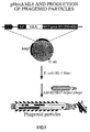
- the strategy consists in construction of a hybrid gene containing all the region coding for hIL-6 followed by the last 157 amino acids of protein pIII of the phage M13 and preceded by the sequence Pel B, which vectors the synthesized protein to the periplasmic space.
- This plasmid also contains a phage replication origin. If a bacterial cell containing this plasmid is infected by a bacteriophage known as a "helper", such as M13K07, single filament copies will be produced by the plasmid, and will be coated with the phage proteins, just like a true phage genome. These phage particles containing the plasmid are known as phasmids. Along with the normal pIII molecules, they also contain hIL6-pIII fusion molecules.
- a hIL-6 molecule and the gene coding its amino acid sequence are thus contained within the same unit.
- mutant molecules it will be possible to determine the amino acid sequence of the molecules exposed on the surfaces of the phages obtainied from selection processes, simply by sequencing the phasmid DNA.
- pHen hIL-6 A further characteristic of the construction pHen hIL-6 is the presence of a translation codon stop between the IL-6 gene and the pIII gene. Production of the hybrid protein is carried out in bacterial strains capable of suppressing this Stop codon. Vice versa the use of non-suppressor strains allows production of hIL-6 alone, directly in the periplasmic space.
- hIL-6 phasmids coated with hIL-6 phasmids or with M13K07 and made to react with shrIL-6R (soluble Human Recombinant IL-6R). After repeated washing the presence of the receptor was indicated using a specific monoclonal antibody conjugated with alkaline phosphatase. The signal obtained is greater in the case of hIL-6 phasmids and increases as the amount of receptor used increases.
- hIL-6 phasmid particles were mixed with particles of M13K07 at a ratio of 1:100. This mixture was incubated with polystyrene balls coated with shrIL-6R. After repeated washing at a neutral pH the balls underwent stringent washing at pH 4.2, followed by elution at pH 3.6. The proportion of hIL-6 phasmids:M13K07 in the resulting eluant was then determined. The proportion is found to be 1:10, with a consequent enrichment by ten times of the hIL-6 phasmids with respect to M13K07.
- Phasmid particles were produced having on their surface mutant molecules of hIL-6 with a higher (176 Arg) or lower (179 Ala) affinity with the receptor, compared with the natural version. These particles were mixed in a ratio of 1:1. The mixture was incubated with the receptor in a solid phase (as described in point b). In the eluate at pH 3.6, the ratio between the two types of particle was found to be 15:1 in favor of 176 Arg.
- Equal amounts of particles of the various types listed above were incubated separately with the balls coated with receptor. After the usual washing sequence the number of particles recovered in the eluant at 3.6 was determined for each type of phage. Considering the value of particles recovered for the phasmid hIL-6 as 1, a value of 3 is obtained for 176 Arg, 0.2 for 179 Ala and 0.18 for M13K07. These values reflect the relative affinities of the molecules of IL-6 non exposed on the phage.
- the plasmid Phen ⁇ hIL-6 was used as a template for all the mutagenesis reactions.
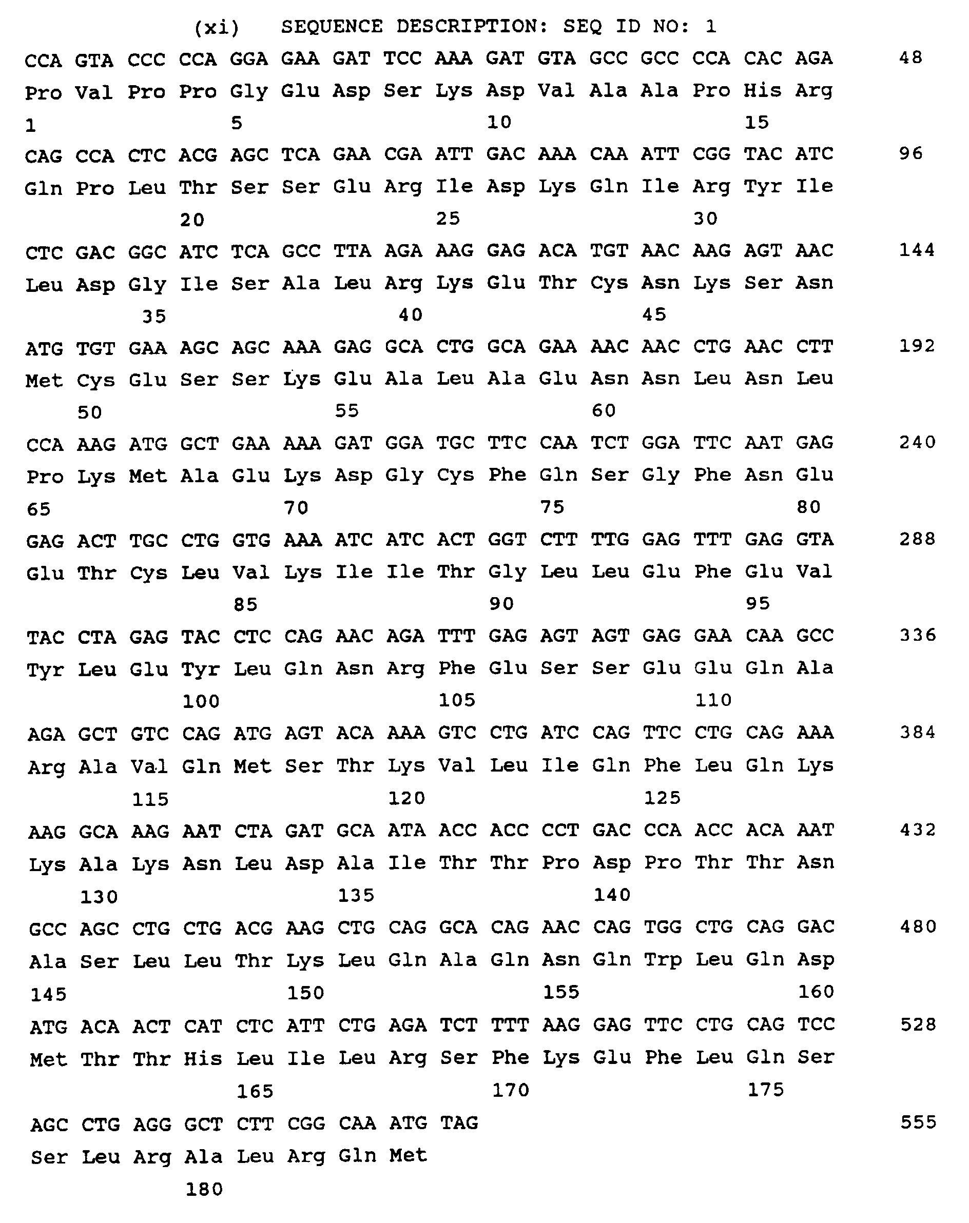
- This plasmid is a derivative of the plasmid pHEN1 and contains the area coding for human IL-6 (SEQ ID NO:1) upstream the area coding for the carboxy-terminal portion (from codon 250 to the COOH end) of the protein pIII of the bacteriophage M13; the two coding areas are in reading frame and are separated by an Amber UAG Stop codon, which can be suppressed in bacterial strains that have the gene of the suppressor SupE integrated in their genome.
- the area coding for human IL-6 is also in reading frame, below the peptide PelB, a signal sequence for secretion, and the whole gene is under the control of the promotor LacZ.
- a single site for the restriction enzyme SacI has been introduced in the nucleotide sequence coding for the amino acids 20-21-22 of hIL-6 without changing their identity and, equally, a single site for the restriction enzyme BfrI has been introduced in the area coding for the amino acids 38-39-40 of hIL-6, likewise without changing them.
- a PCR strategy (Polymerase Chain Reaction) was used to generate mutations within the codons selected in the area coding for human interleukin 6.
- the primer downstream is H9, a 32 nucleotide primer, corresponding to positions 426-457 (antisense filament) of hIL-6 cDNA (taking the first nucleotide of the first codon of the mature polypeptide to be 1).
- the primer hybridization site is downstream the recognition site for the enzyme XbaI, naturally present in the cDNA.
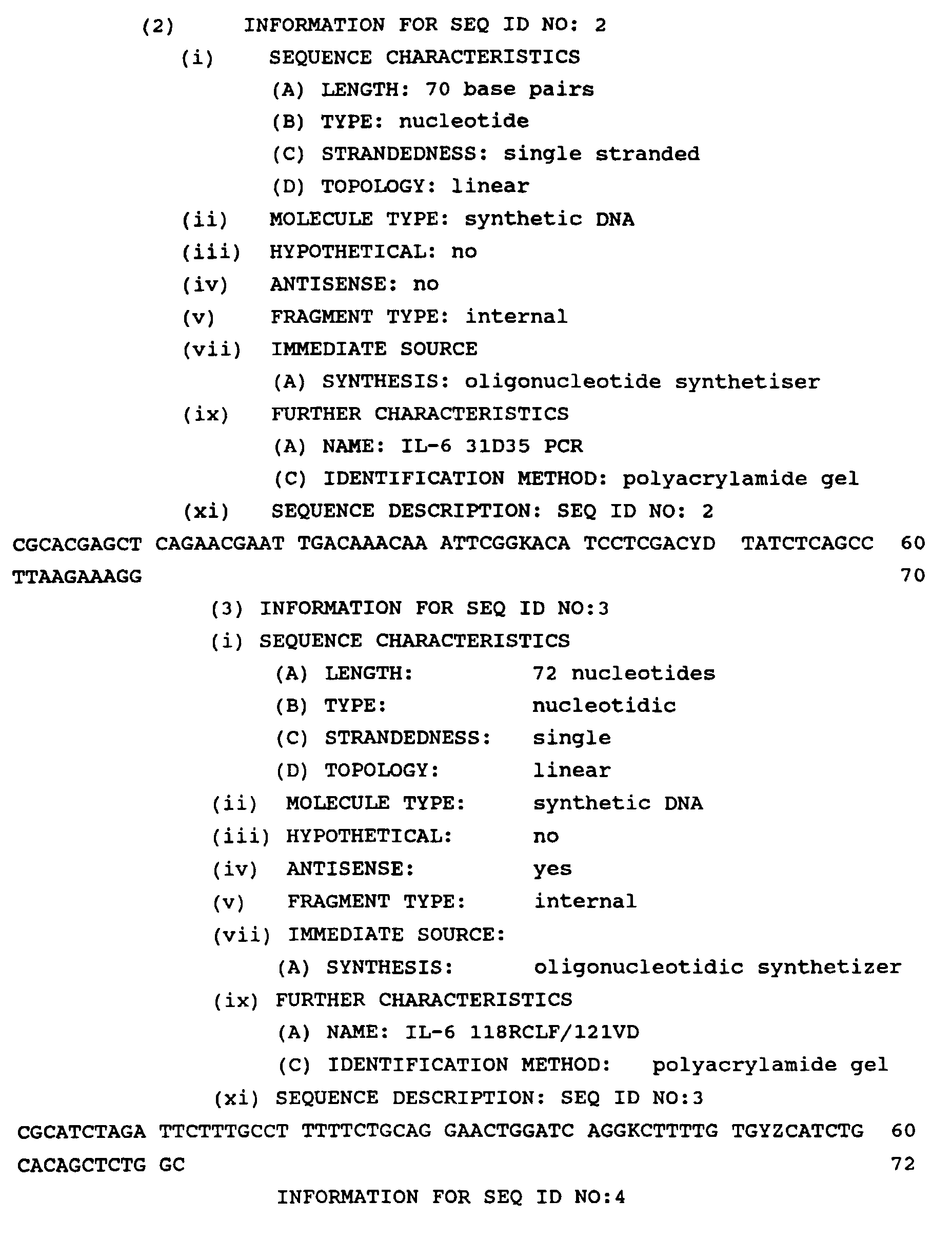
- the mutagenetic primer upstream is IL-6 31D/35 PCR, a 70 nucleotides primer, whose sequence is SEQ ID NO:2.
- the primer IL-6 31D/35 PCR extends from position 55 to position 124 (sense filament) of the hIL-6 cDNA and introduces degenerations into the codons coding for the amino acid 31 (wild type tyrosine) and 35 (wild type glycine).
- a DNA fragment of 403 pairs of bases is amplified using PCR according to standard PCR amplification protocols. Amplification is performed in 35 cycles. Each cycle consists of incubation for 2 minutes at 94°C for denaturation of the template, 2 minutes at 50°C for hybridization of the oligonucleotide and 3 minutes at 72°C for extension of the chain.
- the amplified fragment is digested with SacI and XbaI and purified using 2% agarose gel.
- the fragment generated by PCR and digested by the two enzymes is ligated into the vector pHen hIL-6 digested with the same two enzymes, purified on 0.8% agarose gel, to replace the wild type sequence.
- Table 1 shows the biological activity, in human hepatoma cells, and binding to the receptor gp 80 for wild type interleukin-6 and mutations thereof, the mutations being indicated with an asterix.
- the mutants (Tyr31Asp, Gly35Tyr), (Tyr31Asp, Gly35Phe) and (Tyr31Asp, Gly35Leu, Glu42Ala) show lower biological activity when compared with wild type interleukin 6. In all three cases the residual activity is 2-5% that of wild type interleukin-6.
- the three mutants maintain their full ability to bind to the interleukin 6 receptor gp 80.
- the three mutants have a decreasing signal transduction activity in hepatoma cells.
- they are antagonists of wild interleukin 6.
- the mutant (Tyr31Asp, Gly35Phe) is a particularly effective antagonist, as it is capable of reducing the activity of wild type interleukin 6 when used at 50 fold molar excess in tests on human hepatoma cells.
- Example 3 Generation and selection of further antagonists of interleukin 6 using the methodology according to the present invention
- a PCR strategy (Polymerase Chain Reaction) was used to generate mutations within the codons selected for the area coding for human interleukin 6.
- the primer above is HP/1, a primer with 29 nucleotides, corresponding to positions 1-19 (sense filament) of hIL-6 cDNA (taking the first nucleotide of the first codon of the mature polypeptide to be 1).
- the primer hybridization site is uspstream to the site recognizing the enzyme SacI, artificially introduced in the cDNA without changing the sequence coded by the latter, as described in example 2.
- the mutagenetic primer below is IL-6 118RCLF/121VD, a primer with 72 nucleotides, whose sequence is SEQ ID NO 3.
- the primer IL-6 118RCLF/121VD extends from position 334 to position 405 (antisense filament) of the cDNA of hIL- 6 and introduces degenerations into the codons coding for the amino acid 118 (wild type serine) and 121 (wild type valine).
- a DNA fragment of 415 pairs of bases is amplified using PCR according to standard PCR amplification protocols. Amplification is performed in 35 cycles. Each cycle consists of incubation for 2 minutes at 94°C for denaturation of the template, 2 minutes at 50°C for hybridization of the oligonucleotide and 3 minutes at 72°C for extension of the chain.
- the amplified fragment is digested with SacI and XbaI and purified using 2% agarose gel.
- the fragment generated by PCR and digested by the two enzymes is ligated into the vector pHen ⁇ hIL-6 digested with the same two enzymes, purified on 0.8% agarose gel, to replace the wild type sequence.
- Table 2 shows the biological activity, in human hepatoma cells, and binding to the receptor for wild interleukin-6 and versions thereof with mutations in the residues indicated.
- Receptor binding properties and biological activity of wild-type interleukin 6 and helix C mutations thereof 118 Ser 121 Val Biological activity 100% Receptor binding 100% Arg Val 66% 81% Leu Asp 36 ⁇ 4% 92% Arg Asp 3.5 ⁇ 0.5% 66 ⁇ 5% Ser Asp 58 ⁇ 23% 78 ⁇ 2%
- mutant IL-6 Ser118Arg/Val121Asp has characteristics very similar to those of the mutant IL-6 Tyr31Asp/Gly35Phe, described in Table 1, that is to say it binds normally to the type I receptor of interleukin 6, but cytokin biological activity is reduced approximately thirty-fold.
- the plasmid pHEN ⁇ hIL-6 Tyr31Asp/Gly35Phe obtained as described in Example 2, was used as a template for all the mutagenic reactions.
- a PCR strategy (Polymerase Chain Reaction) was used to generate mutations within the codons selected for the area coding for human interleukin 6.
- the primer above is HP/1, described in the previous example.
- the mutagenetic primer below is IL-6 118RCLF/121VD, also described in the previous example.
- a DNA fragment of 415 pairs of bases is amplified using PCR according to standard PCR amplification protocols. Amplification is performed in 35 cycles. Each cycle consists of incubation for 2 minutes at 94°C for denaturation of the template, 2 minutes at 50°C for hybridization of the oligonucleotide and 3 minutes at 72°C for extension of the chain.
- the amplified fragment is digested with SacI and XbaI and purified using 2% agarose gel.
- the fragment generated by PCR and digested by the two enzymes is ligated into the vector pHen hIL-6 digested with the same two enzymes, purified on 0.8% agarose gel, to replace the wild type sequence.
- Table 3 shows the biological activity, in human hepatoma cells, and binding to the receptor for wild interleukin-6 and versions thereof with mutations in the residues indicated.
- mutant IL-6 Tyr31Asp/Gly35Phe/Ser118Arg/Val121Asp was chosen for competition experiments with wild type interleukin 6 biological activity in human hepatoma cells.
- the cells were stimulated with wild interleukin 6 at 4 nanograms per milliliter (ng/ml) of culture medium, in the presence of increasing concentrations of mutant.
- increasing concentrations of the mutant manage to fully antagonize the effects of wild interleukin 6 on human hepatoma cells.
- Table 4 shows the levels of inhibition (in percentage) of the biological activity of wild interleukin 6 according to the concentration of antagonist (expressed in nanograms per milliliter, molar excess compared with wild type interleukin 6 and nanomoles per liter, respectively).
- the interleukin 6 antagonist generated and selected using the methodology described in the present invention, inhibit interleukin 6-dependent growth of human myeloma cells.
- all mutants are particularly effective antagonists of interleukin 6 biological activity on human myeloma cells.
- a phage library (containing mutations of residues Gln 175, Ser 177, Leu 181 and Leu 183 of wild type interleukin 6, present in the form of fusion product with filamentous phage protein) was constructed using the Primer Extension molecular biology technique
- the mutagenetic oligonucleotide is IL-6 QSLQ (AS), a 62 nucleotides oligo, whose sequence is SEQ ID NO:4.
- Primer IL-6 QSLQ extends from position 507 to the stop codon of the interleukin 6 cDNA (antisense strand), it introduces degenerations into codons coding for the amino acids 175 (wild type Gln), 177 (wild type Ser), 181 (wild type Leu) and 183 (wild type Gln) and it also introduces a NotI restriction site downstream of the interleukin 6 stop codon.
- Bam extends from position 503 to position 522 (sense strand) of the interleukin 6 cDNA and it contains a BamHI recognition site within the 5' nine nucleotides.
- the two oligonucleotides are complementary to each other on a region corresponding to position 507 through position 522 of the interleukin 6 cDNA.
- the two oligonucleotides were annealed in vitro, and the annealed oligonucleotides were used as substrate for a Primer Extension reaction, performed using the Klenow enzyme.
- the double-stranded DNA fragment thus obtained was then digested with BamHI (compatible with BglII) and with NotI and ligated into the plasmid phen ⁇ hIL-6, digested with BglII (compatible with BamHI) and with NotI, in order to replace the wild type sequence with the mutated ones.
- the ligation product was inserted in bacteria, yielding roughly one million independent transformants ("trasformant” is the definition given to a bacterium which has incorporated a recombinant plasmid).
- the transformed bacteria were infected by with the M13K07 helper bacteriophage to generate the phage library (a library of phasmids) as described in the example 1.
- the library underwent selection by incubation with polystirene balls coated with shrIL-6R, as described in the example 1.
- the phage population eluted at pH 3.6 was then amplified in bacteria. After five cycles of selection-amplification, randomly selected phages were seqenced over the mutagenized region, the corresponding mutant interleukin 6 proteins were produced in the periplasmic space of the appropiate bacterial strain (as described in the example 1) and tested for both receptor binding and biological activity on human hepatoma cells.
- Table 6 shows that, by using using the methodology to the present invention, it is possible to select superagonists of interleukin 6, mutant molecules which have increased both receptor binding and biological activity on human hepatoma cells.
- the mutations selected by the methodology according to the present invention can be used as starting point for the development of more potent interleukin 6 superagonists. This is shown in this example, in which the mutation identified in the phage 5-2 are combined with the mutation Ser176Arg, which by prior art is known to increase both receptor binding and biological activity (see International Publication WO 94/11402 of a PCT application of the present Applicant, filed 2.11.93 with Italian priority of 6.11.92). The three mutations were grouped on the same cDNA by mean of Oligonucleotide Directed Mutagenesis.
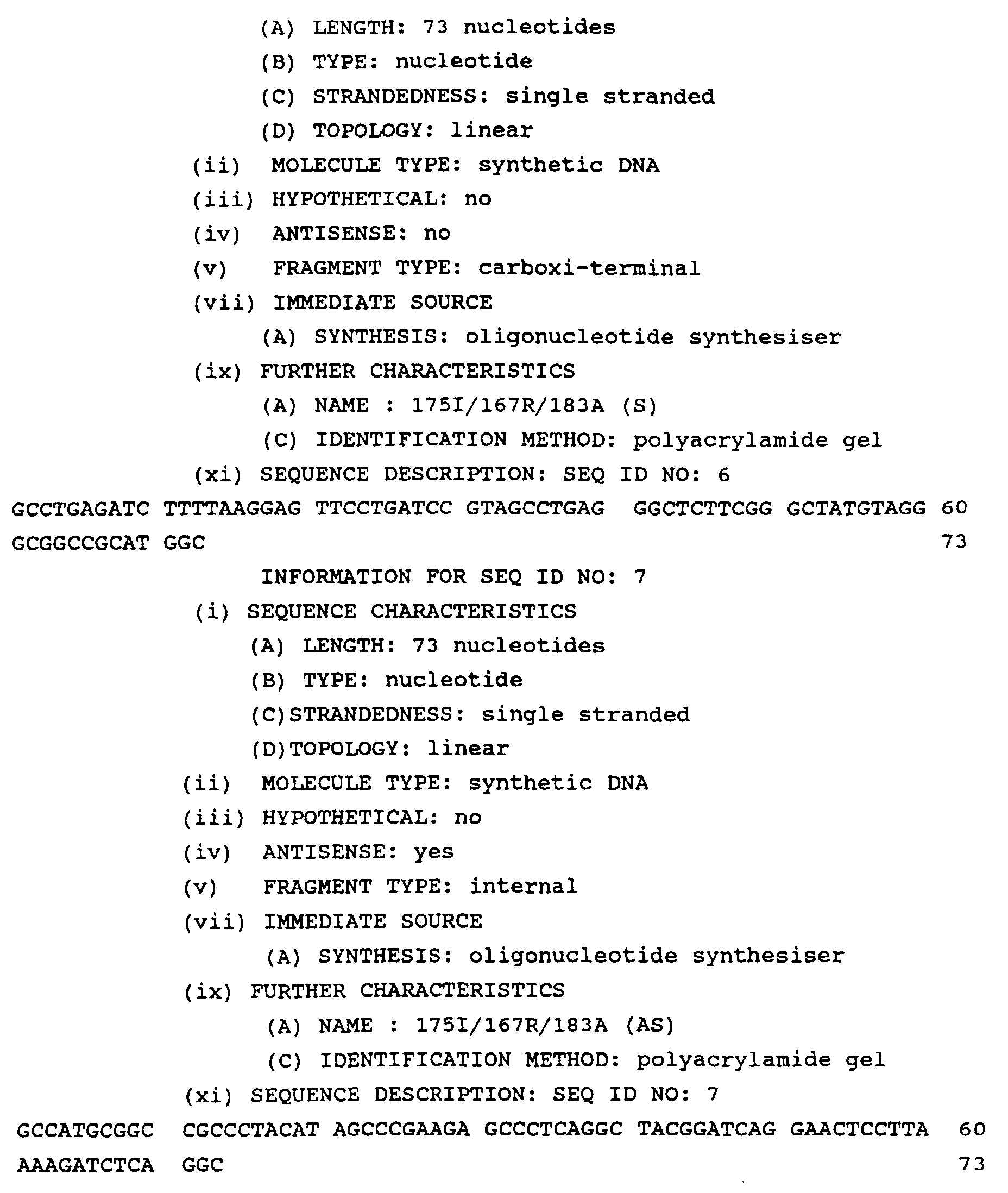
- Oligonucleotide 175I/176R/183A (S), whose sequence is SEQ ID NO:6 is 73 nucleotides long which extends from position 499 to the stop codon (sense strand) of the interleukin 6 cDNA present in phen hIL-6 and which has a recognition site for the enzyme NotI downstream of the stop codon.
- Oligonucleotide 175I/176R/183A (AS), whose sequence is SEQ ID NO:7, is 73 nucleotides long which extends from position 499 to the stop codon (antisense strand) of the interleukin 6 cDNA present in phen ⁇ hIL-6 and which has a recognition site for the enzyme NotI upstream of the stop codon.
- the two oligonucleotide are complementary to each other and they both encode the amino acid Isoleucine in position 175, the amino amino acid Serine in position 176 and the amino acid Alanine in position 183.
- the two oligonucleotides were annealed in vitro, the double-stranded DNA fragment thus obtained was digested with the restriction enzymes BglII and Not I and ligated into the vector phen ⁇ hlL-6 digested with the same two enzymes, in order to replace the wild type sequence with the mutated one.
- the corresponding interleukin 6 variant carrying the three desired mutations was produced in the periplasmic space of the appropiate bacterial strain (as described in the example 1) and tested for both receptor binding and biological activity on human hepatoma cells.
- the following Table 7 shows the receptor binding properties and the biological activity on human hepatoma cells of both the new triple mutant and of the parental double mutant.
- the mutant carrying the substitutions Gln175Ile/Ser176Arg/Gln183Ala is a much more effective interleukin 6 superagonist than the parental mutant carrying only the two substitutions Gln175Ile/Gln183Ala.
- the corresponding mutant protein was tested both for receptor binding and for antagonistic behaviour on human hepatoma and myeloma cells, the following table 8 shows the receptor binding properties of both SAnt 1 and DFRD (the mutant from which SAnt 1 was derived), together with the amounts (in nanograms of mutant per milliliter of colture medium) of mutant necessary to inhibit 50% of interleukin 6 biological activity (hepatoma cells were stimulated with 4 nanograms of wild type interleukin 6 per milliliter of colture medium, while myeloma cells were stimulated with 0.1 nanograms of interleukin 6 per milliliter of colture medium, due to the higher sensitivity of the latter cells to wild type interleukin 6).
- Antagonist Receptor binding 50% inhibition of interleukin 6 hepatoma cells activity on myeloma cells Hep3B HepG2 XG-1 DFRD 97% 164 ng/ml 132 ng/ml 190 ng/ml SAnt1 406% 19 ng/ml 32 ng/ml 22 ng/ml
Landscapes
- Life Sciences & Earth Sciences (AREA)
- Health & Medical Sciences (AREA)
- Chemical & Material Sciences (AREA)
- Molecular Biology (AREA)
- Engineering & Computer Science (AREA)
- Immunology (AREA)
- Biomedical Technology (AREA)
- Hematology (AREA)
- Urology & Nephrology (AREA)
- General Health & Medical Sciences (AREA)
- Cell Biology (AREA)
- Biochemistry (AREA)
- Medicinal Chemistry (AREA)
- Proteomics, Peptides & Aminoacids (AREA)
- Organic Chemistry (AREA)
- Analytical Chemistry (AREA)
- Physics & Mathematics (AREA)
- Food Science & Technology (AREA)
- Microbiology (AREA)
- General Physics & Mathematics (AREA)
- Pathology (AREA)
- Biotechnology (AREA)
- Gastroenterology & Hepatology (AREA)
- Zoology (AREA)
- Toxicology (AREA)
- Biophysics (AREA)
- Genetics & Genomics (AREA)
- Endocrinology (AREA)
- Peptides Or Proteins (AREA)
- Medicines That Contain Protein Lipid Enzymes And Other Medicines (AREA)
- Preparation Of Compounds By Using Micro-Organisms (AREA)
- Investigating Or Analysing Biological Materials (AREA)
- Pharmaceuticals Containing Other Organic And Inorganic Compounds (AREA)
- Medicines Containing Antibodies Or Antigens For Use As Internal Diagnostic Agents (AREA)
- Micro-Organisms Or Cultivation Processes Thereof (AREA)
Abstract
Description
- comparing the amino acid sequences of the growth
hormone with the sequences of said
interleukin 6; - comparing the amino acid sequences of the growth
hormone receptor with those of the two receptors of the
interleukin 6, that is with interleukin 6-specific receptor and gp 130; - on the basis of the above comparisons, formulating
a three-dimensional model of the receptor complex based
on the functional similarity between sequences of the
growth hormone receptor and the two
interleukin 6 receptors in question; and - identifying the residues of the
wild type interleukin 6 that are a part of the site of interaction with the specific receptor and of the site of interaction with gp 130, respectively; - introduction of mutations into the interaction
sites of
interleukin 6; and - evaluation of the biological activity of the mutants.
- production of a series of phage libraries
containing mutations of the following wild type residues
of
interleukin 6, present in the form of fusion product with filamentous phage proteins - Glu 42, Glu 51, Ser 52, Ser 53, Lys 54, Glu 55, Asn 63, Lys 66, Met 67, Ala 68, Glu 69, Lys 70, Asp 71, Phe 170, Gln 175, Ser 176, Ser 177, Leu 181, Gln 183;
- generation of a phage library, each phage having a
mutant interleukin 6 sequence; - selection, from the mixed population of
phages expressing interleukin 6 mutants, of that or those with an affinity for the specific receptor greater than that of wild type interleukin; and - identification of the best amino acid sequence or sequences binding the receptor by sequencing of the DNA extracted from the selected phage particles.
- mutation of the residues identified in
claim 1, to form part of the site of interaction with gp 130 (Arg 30, Tyr 31, Gly 33, Ser 37, Ala 38, Ser 118, Lys 120, Val 121, Gln 124, Phe 125, Gln 127, Lys 128 and Lys 129), using conventional molecular biology techniques; - evaluation of biological activity and affinity for
the
specific interleukin 6 receptor of the mutants produced as above, in order to identify variants ofinterleukin 6 whose affinity to the specific receptor is intact and that show reduction or loss of the biological activity; and - evaluation of the above variants of
interleukin 6 as antagonists for the biological activity ofwild interleukin 6.

| Receptor binding properties and biological activity of
wild- | ||||
| 118 Ser | 121 | Biological activity | 100% | Receptor binding 100% |
| Arg | Val | 66% | 81% | |
| Leu | Asp | 36▪4% | 92% | |
| Arg | Asp | 3.5▪0.5% | 66▪5% | |
| Ser | Asp | 58▪23% | 78▪2% |
| Receptor binding properties and biological activity of
| ||||||
| Helix A | Helix C | |||||
| 31 | 35 | 118 | 121 | Biological activity | Receptor binding | |
| Tyr | | Ser | Val | 100% | 100% | |
| Asp | Phe | Leu | Val | 1.4% | ||
| Asp | Phe | Arg | Val | 5.4▪1.1% | 66▪2% | |
| Asp | | Leu | Asp | 0% | 63▪4% | |
| Asp | | Arg | Asp | 0% | 97▪15% | |
| Asp | | Phe | Asp | 0% | 76▪26% |
| Inhibition of the biological activity of | |||
| IL-6 g/ml | Tyr31Asp/Gly35Phe/Ser118Arg/Val121Asp | Inhibition of wild IL-6 (%) | |
| molar excess | concentration (nM) | ||
| 16 | 4 X | 0.7 nM | 23% |
| 60 | 15 X | 2.7 nM | 28% |
| 164 | 41 X | 7.5 nM | 50% |
| 500 | 125 X | 22.7 nM | 75% |
| 1000 | 250 X | 45.5 nM | 82% |
| 2000 | 500 X | 90.0 nM | 90% |
| 4000 | 1000 X | 181.8 nM | 96% |
| Inhibition of | |||
| Antagonist | Inhibition of | ||
| DFRD | DFFD | DFLD | |
| 3.3 ng/ | 6% | 0% | 0% |
| 10 ng/ml | 12% | 7% | 3% |
| 30 ng/ml | 25% | 18% | 10% |
| 90 ng/ml | 42% | 35% | 31% |
| 270 ng/ml | 64% | 59% | 53% |
| 810 ng/ml | 84% | 84% | 71% |
| 2430 ng/ml | 90% | 90% | 76% |
| 7290 ng/ml | 93% | 95% | 81% |
| Receptor binding properties and biological activities
of | |||||||
| Position | 175 | 177 | 181 | 183 | Receptor binding (%) | Biological activity (%) | |
| wild type | Gln | | Leu | Gln | 100% | 100% | |
| phage 5-4 | Gln | Ser | Leu | Tyr | 240% | 130% | |
| phage 5-8 | Gln | Ser | Leu | Ala | 240% | 120% | |
| phage 5-2 | Ile | Ser | Leu | Ala | 260% | 150% | |
| phage 4-8 | Gln | | Ile | Asn | 100% | ||
| phage 5-3 | Ile | Ser | Val | His | 80% |
| Receptor binding properties and biological activities
of | ||||||
| Position | 175 | 176 | 183 | Receptor binding (%) | Biological activity (%) | |
| wild type | | Ser | Gln | 100% | 100% | |
| phage 5-2 | Ile | Ser | Ala | 260% | 150% | |
| triple mutant | Ile | Arg | Ala | 450% | 260% |
| Inhibition of | ||||
| Antagonist | Receptor binding (% of wt) | 50% inhibition of | activity on myeloma cells | |
| Hep3B | HepG2 | XG-1 | ||
| DFRD | 97% | 164 ng/ml | 132 ng/ml | 190 ng/ml |
| SAnt1 | 406% | 19 ng/ml | 32 ng/ml | 22 ng/ml |


Claims (6)
- A methodology for selecting superagonists, antagonists and superantagonists of interleukin 6 using the membrane molecule gp 130 to activate the mechanisms regulating cell physiology, comprising the following operations:comparing the amino acid sequences of the growth hormone with the sequences of interleukin 6;comparing the amino acid sequences of the growth hormone receptor with those of the two receptors of interleukin 6, that is with the interleukin 6-specific receptor and gp 130;on the basis of the above comparisons, formulating a three-dimensional model of the receptor complex based on the
functional similarity between sequences on the growth hormone receptor and the two interleukin 6 receptors in question;identifying the residues of the wild type interleukin 6 that form a part of the site of interaction with the specific receptor and the site of interaction with gp 130, respectively;
introduction of mutations into the interaction sites of interleukin 6; andevaluation of the biological activity of the mutants. - The methodology for selecting superagonists of interleukin 6 according to claim 1, further comprising the following additional operations:production of a series of phage libraries containing mutations of the following wild type residues of interleukin 6 present in the form of a fusion product with filamentous phage proteins
Glu 42, Glu 51, Ser 52, Ser 53, Lys 54, Glu 55, Asn 63, Lys 66, Met 67, Ala 68, Glu 69, Lys 70, Asp 71, Phe 170, Gln 175, Ser 176, Ser 177, Leu 181, Gln 183;generation of a phage library, each phage having a mutant interleukin 6 sequence;selection, from the mixed population of phages expressing interleukin 6 mutants, of that or those with an affinity for the specific receptor greater than that of wild type interleukin; andidentification of the best amino acid sequence or sequences binding the receptor by sequencing of the DNA extracted from the selected phage particles. - The methodology for selecting superagonists of interleukin 6 according to claim 2, in which a series of phage libraries are produced, containing mutants of said wild type residue of interleukin 6 present as a product of fusion with protein pIII of M13.
- The methodology for selecting antagonists of interleukin 6 according to claim 1 further comprising the following additional operations:mutation of the residues identified in claim 1, to form part of the site of interaction with gp 130 (Arg 30, Tyr 31, Gly 35, Ser 37, Ala 38, Ser 118, Lys 120, Val 121, Gln 124, Phe 125, Gln 127, Lys 128 and Lys 129), using conventional molecular biology techniques;evaluation of biological activity and affinity with the specific interleukin 6 receptor of the mutants produced as above, in order to identify variants of interleukin 6 whose affinity to the specific receptor is intact and that show reduction or loss of the biological activity; andevaluation of the above variants of interleukin 6 as antagonists for the biological activity of wild type interleukin 6.
- The methodology for selecting superantagonists of interleukin 6 according to claim 1 by combination of the variations of amino acid sequences responsible for antagonist activity, which are indicated in claim 4, with amino acid variations responsible for an increased affinity of the specific receptor for interleukin 6.
- A methodology for selecting superantagonists of interleukin 6 according to claim 5, in which the mutagenesis of the residues identified as above is performed using a molecular biology technique chosen from the group comprising Polymerase Chain Reaction, Primer Extension, Oligonucleotide Directed Mutagenesis, and combinations thereof.
Applications Claiming Priority (3)
| Application Number | Priority Date | Filing Date | Title |
|---|---|---|---|
| ITRM930409 | 1993-06-23 | ||
| ITRM930409A IT1261787B (en) | 1993-06-23 | 1993-06-23 | METHODOLOGY FOR THE SELECTION OF SUPERAGONISTS, ANTAGONISTS AND SUPER-ANTAGONISTS OF HORMONES OF WHICH RECEPTOR COMPLEX IS PART OF GP 130. |
| PCT/IT1994/000095 WO1995000852A2 (en) | 1993-06-23 | 1994-06-23 | A methodology for selecting superagonists, antagonists and superantagonists of hormones whose receptor complex includes gp 130 |
Publications (2)
| Publication Number | Publication Date |
|---|---|
| EP0656117A1 EP0656117A1 (en) | 1995-06-07 |
| EP0656117B1 true EP0656117B1 (en) | 2000-02-02 |
Family
ID=11401818
Family Applications (1)
| Application Number | Title | Priority Date | Filing Date |
|---|---|---|---|
| EP94920582A Expired - Lifetime EP0656117B1 (en) | 1993-06-23 | 1994-06-23 | A methodology for selecting superagonists, antagonists and superantagonists of interleukin 6 |
Country Status (13)
| Country | Link |
|---|---|
| EP (1) | EP0656117B1 (en) |
| JP (1) | JP2643604B2 (en) |
| CN (2) | CN1249439C (en) |
| AT (1) | ATE189528T1 (en) |
| AU (1) | AU687416B2 (en) |
| CA (1) | CA2142973C (en) |
| DE (1) | DE69422893T2 (en) |
| DK (1) | DK0656117T3 (en) |
| ES (1) | ES2144524T3 (en) |
| GR (1) | GR3033111T3 (en) |
| IT (1) | IT1261787B (en) |
| PT (1) | PT656117E (en) |
| WO (1) | WO1995000852A2 (en) |
Cited By (1)
| Publication number | Priority date | Publication date | Assignee | Title |
|---|---|---|---|---|
| WO2013147153A1 (en) | 2012-03-29 | 2013-10-03 | 株式会社未来創薬研究所 | Anti-lamp5 antibody and utilization thereof |
Families Citing this family (12)
| Publication number | Priority date | Publication date | Assignee | Title |
|---|---|---|---|---|
| IT1276555B1 (en) * | 1995-04-28 | 1997-11-03 | Angeletti P Ist Richerche Bio | HUMAN INTERLUCHINA-6 ANTAGONISTS COMPLETELY UNABLE TO FORM A BOND WITH GP 130, AND USE THEM FOR THE PREPARATION OF COMPOSITIONS |
| IT1277926B1 (en) * | 1995-09-01 | 1997-11-12 | Angeletti P Ist Richerche Bio | USE OF NATURAL CYTOKINE MUTEINS AS IMMUNOGENS FOR THE PREPARATION OF PHARMACEUTICAL COMPOSITIONS TO TREAT OR PREVENT |
| US5849293A (en) * | 1996-01-11 | 1998-12-15 | Cornell Research Foundation, Inc. | Use of human transferrin in controlling insulin levels |
| IT1285790B1 (en) * | 1996-09-24 | 1998-06-24 | Angeletti P Ist Richerche Bio | ADENOVIRUS RECOMBINANT DEFECTIVES CODING FOR MUTANTS OF HUMAN INTERLEUKINE 6 (HIL-6) WITH ANTAGONIST ACTIVITY OR |
| ES2312184T3 (en) | 1997-03-21 | 2009-02-16 | Chugai Seiyaku Kabushiki Kaisha | THERAPEUTIC PREVENTIVE AGENTS FOR THE TREATMENT OF MULTIPLE SCLEROSIS, CONTAINING ANTI-RECEIVING ANTIBODIES OF IL-6 ANTAGONISTS. |
| GB9806530D0 (en) | 1998-03-26 | 1998-05-27 | Glaxo Group Ltd | Inflammatory mediator |
| WO2003033015A1 (en) | 2001-10-11 | 2003-04-24 | Applied Research Systems Ars Holding N.V. | USE OF gp130 ACTIVATORS IN DIABETIC NEUROPATHY |
| FR2833011B1 (en) * | 2001-12-04 | 2004-10-29 | Univ Claude Bernard Lyon | NOVEL PROTEIN WITH IL-6 INHIBITING ACTIVITY |
| IL161673A0 (en) | 2004-04-29 | 2004-09-27 | Applied Research Systems | Compositions and methods for therapy of chemotherapy-induced neuropathy |
| CN101246160B (en) * | 2007-09-06 | 2012-11-21 | 三峡大学 | IL-6/sIL-6R combined molecular model established by ELISA method in vitro |
| DE102011104822A1 (en) * | 2011-06-18 | 2012-12-20 | Christian-Albrechts-Universität Zu Kiel | Ciliary Neutrophic factor variants |
| WO2018104564A2 (en) * | 2017-08-11 | 2018-06-14 | Solvay Specialty Polymers Italy S.P.A. | Method for stabilizing aqueous dispersions of fluorinated polymers |
Citations (2)
| Publication number | Priority date | Publication date | Assignee | Title |
|---|---|---|---|---|
| US5210075A (en) * | 1990-02-16 | 1993-05-11 | Tanabe Seiyaku Co., Ltd. | Interleukin 6 antagonist peptides |
| WO1994009138A1 (en) * | 1992-10-20 | 1994-04-28 | Cetus Oncology Corporation | Interleukin-6 receptor antagonists |
Family Cites Families (6)
| Publication number | Priority date | Publication date | Assignee | Title |
|---|---|---|---|---|
| EP0397834B1 (en) * | 1988-10-28 | 2000-02-02 | Genentech, Inc. | Method for identifying active domains and amino acid residues in polypeptides and hormone variants |
| JP2898064B2 (en) * | 1989-08-03 | 1999-05-31 | 忠三 岸本 | Human gp130 protein |
| JP2898040B2 (en) * | 1990-01-26 | 1999-05-31 | 忠三 岸本 | Antibodies to gp130 protein |
| DE69231467T2 (en) * | 1991-05-10 | 2001-01-25 | Genentech, Inc. | SELECTION OF AGONISTS AND ANTAGONISTS OF LIGANDS |
| US5470952A (en) * | 1993-10-20 | 1995-11-28 | Regeneron Pharmaceuticals, Inc. | CNTF and IL-6 antagonists |
| US5939534A (en) * | 1993-12-29 | 1999-08-17 | Sumitomo Pharmaceuticals Company, Limited | Factors mutated in the D1 cap region |
-
1993
- 1993-06-23 IT ITRM930409A patent/IT1261787B/en active IP Right Grant
-
1994
- 1994-06-23 WO PCT/IT1994/000095 patent/WO1995000852A2/en not_active Ceased
- 1994-06-23 AT AT94920582T patent/ATE189528T1/en not_active IP Right Cessation
- 1994-06-23 CN CN02107480.1A patent/CN1249439C/en not_active Expired - Fee Related
- 1994-06-23 EP EP94920582A patent/EP0656117B1/en not_active Expired - Lifetime
- 1994-06-23 JP JP7502633A patent/JP2643604B2/en not_active Expired - Lifetime
- 1994-06-23 DE DE69422893T patent/DE69422893T2/en not_active Expired - Fee Related
- 1994-06-23 DK DK94920582T patent/DK0656117T3/en active
- 1994-06-23 CN CN94190533.0A patent/CN1101937C/en not_active Expired - Fee Related
- 1994-06-23 AU AU71324/94A patent/AU687416B2/en not_active Ceased
- 1994-06-23 ES ES94920582T patent/ES2144524T3/en not_active Expired - Lifetime
- 1994-06-23 PT PT94920582T patent/PT656117E/en unknown
- 1994-06-23 CA CA002142973A patent/CA2142973C/en not_active Expired - Fee Related
-
2000
- 2000-03-29 GR GR20000400800T patent/GR3033111T3/en not_active IP Right Cessation
Patent Citations (2)
| Publication number | Priority date | Publication date | Assignee | Title |
|---|---|---|---|---|
| US5210075A (en) * | 1990-02-16 | 1993-05-11 | Tanabe Seiyaku Co., Ltd. | Interleukin 6 antagonist peptides |
| WO1994009138A1 (en) * | 1992-10-20 | 1994-04-28 | Cetus Oncology Corporation | Interleukin-6 receptor antagonists |
Cited By (1)
| Publication number | Priority date | Publication date | Assignee | Title |
|---|---|---|---|---|
| WO2013147153A1 (en) | 2012-03-29 | 2013-10-03 | 株式会社未来創薬研究所 | Anti-lamp5 antibody and utilization thereof |
Also Published As
| Publication number | Publication date |
|---|---|
| IT1261787B (en) | 1996-06-03 |
| WO1995000852A2 (en) | 1995-01-05 |
| CN1112795A (en) | 1995-11-29 |
| CN1249439C (en) | 2006-04-05 |
| AU7132494A (en) | 1995-01-17 |
| DE69422893T2 (en) | 2000-08-17 |
| HK1011081A1 (en) | 1999-07-02 |
| WO1995000852A3 (en) | 1995-03-30 |
| ITRM930409A0 (en) | 1993-06-23 |
| CA2142973C (en) | 2004-10-26 |
| DE69422893D1 (en) | 2000-03-09 |
| EP0656117A1 (en) | 1995-06-07 |
| DK0656117T3 (en) | 2000-07-24 |
| JP2643604B2 (en) | 1997-08-20 |
| PT656117E (en) | 2000-07-31 |
| ITRM930409A1 (en) | 1994-12-23 |
| AU687416B2 (en) | 1998-02-26 |
| CA2142973A1 (en) | 1995-01-05 |
| ES2144524T3 (en) | 2000-06-16 |
| ATE189528T1 (en) | 2000-02-15 |
| GR3033111T3 (en) | 2000-08-31 |
| JPH07509571A (en) | 1995-10-19 |
| CN1101937C (en) | 2003-02-19 |
| CN1538176A (en) | 2004-10-20 |
Similar Documents
| Publication | Publication Date | Title |
|---|---|---|
| US5849283A (en) | Human interleukin-6 receptor antagonists | |
| Valls et al. | Yeast carboxypeptidase Y vacuolar targeting signal is defined by four propeptide amino acids. | |
| Sparks et al. | Distinct ligand preferences of Src homology 3 domains from Src, Yes, Abl, Cortactin, p53bp2, PLCgamma, Crk, and Grb2. | |
| Gonzalez et al. | A cluster of phosphorylation sites on the cyclic AMP-regulated nuclear factor CREB predicted by its sequence | |
| Gearing et al. | Molecular cloning and expression of cDNA encoding a murine myeloid leukaemia inhibitory factor (LIF). | |
| Yanofsky et al. | High affinity type I interleukin 1 receptor antagonists discovered by screening recombinant peptide libraries. | |
| US5880096A (en) | Peptides and compounds that bind to the IL-1 receptor | |
| EP1243650B1 (en) | Process for constructing a cDNA library | |
| AU687416B2 (en) | A methodology for selecting superagonists, antagonists and superantagonists of hormones whose receptor complex includes GP 130 | |
| US5786331A (en) | Peptides and compounds that bind to the IL-1 receptor | |
| JP4493271B2 (en) | Combinatorial library of proteins having a skeleton structure of C-type lectin-like domain | |
| EP2022856A2 (en) | Improvements in or relating to binding proteins for recognition of DNA | |
| JP2001527417A (en) | Nucleic acid binding protein | |
| Jestin et al. | Improving the display of proteins on filamentous phage | |
| US5789552A (en) | Interleukin-6 receptor antagonists | |
| Cabibbo et al. | Monovalent phage display of human interleukin (hIL)-6: selection of superbinder variants from a complex molecular repertoire in the hIL-6 D-helix | |
| CA2218372C (en) | Antagonists of human interleukin-6 that are totally incapable of binding gp 130, and their use in the preparation of pharmaceutical compounds | |
| HK1011081B (en) | A methodology for selecting superagonists, antagonists and superantagonists of hormones whose receptor complex includes gp 130 | |
| JP4361684B2 (en) | Peptide ligands for erythropoietin receptors | |
| KR100220356B1 (en) | A method for selecting superagonists, antagonists and super antagonists of human interleukin-6 based receptor complex three dimensional modelling | |
| HK1011032B (en) | Superagonists and antagonists of human il-6, and method for their selection | |
| Boulurani | Molecular Cloning and Developmental Expression of SQ1, A cAMP Inducible Gene of Dictyostelium discoideum | |
| AU1003799A (en) | Improvements in or relating to binding proteins for recognition of DNA |
Legal Events
| Date | Code | Title | Description |
|---|---|---|---|
| PUAI | Public reference made under article 153(3) epc to a published international application that has entered the european phase |
Free format text: ORIGINAL CODE: 0009012 |
|
| AK | Designated contracting states |
Kind code of ref document: A1 Designated state(s): AT BE CH DE DK ES FR GB GR IE LI LU MC NL PT SE |
|
| D17D | Deferred search report published (deleted) | ||
| 17P | Request for examination filed |
Effective date: 19950428 |
|
| 17Q | First examination report despatched |
Effective date: 19980707 |
|
| GRAG | Despatch of communication of intention to grant |
Free format text: ORIGINAL CODE: EPIDOS AGRA |
|
| GRAG | Despatch of communication of intention to grant |
Free format text: ORIGINAL CODE: EPIDOS AGRA |
|
| GRAH | Despatch of communication of intention to grant a patent |
Free format text: ORIGINAL CODE: EPIDOS IGRA |
|
| RIC1 | Information provided on ipc code assigned before grant |
Free format text: 6G 01N 33/68 A, 6G 01N 33/74 B, 6C 12N 15/12 B, 6C 12N 15/18 B, 6C 07K 14/655 B |
|
| GRAH | Despatch of communication of intention to grant a patent |
Free format text: ORIGINAL CODE: EPIDOS IGRA |
|
| GRAA | (expected) grant |
Free format text: ORIGINAL CODE: 0009210 |
|
| AK | Designated contracting states |
Kind code of ref document: B1 Designated state(s): AT BE CH DE DK ES FR GB GR IE LI LU MC NL PT SE |
|
| REF | Corresponds to: |
Ref document number: 189528 Country of ref document: AT Date of ref document: 20000215 Kind code of ref document: T |
|
| RTI1 | Title (correction) |
Free format text: A METHODOLOGY FOR SELECTING SUPERAGONISTS, ANTAGONISTS AND SUPERANTAGONISTS OF INTERLEUKIN 6 |
|
| REG | Reference to a national code |
Ref country code: CH Ref legal event code: EP |
|
| REF | Corresponds to: |
Ref document number: 69422893 Country of ref document: DE Date of ref document: 20000309 |
|
| REG | Reference to a national code |
Ref country code: IE Ref legal event code: FG4D |
|
| ET | Fr: translation filed | ||
| REG | Reference to a national code |
Ref country code: CH Ref legal event code: NV Representative=s name: PATENTANWAELTE SCHAAD, BALASS, MENZL & PARTNER AG |
|
| EUG | Se: european patent has lapsed | ||
| REG | Reference to a national code |
Ref country code: ES Ref legal event code: FG2A Ref document number: 2144524 Country of ref document: ES Kind code of ref document: T3 |
|
| REG | Reference to a national code |
Ref country code: DK Ref legal event code: T3 |
|
| REG | Reference to a national code |
Ref country code: PT Ref legal event code: SC4A Free format text: AVAILABILITY OF NATIONAL TRANSLATION Effective date: 20000420 |
|
| PLBE | No opposition filed within time limit |
Free format text: ORIGINAL CODE: 0009261 |
|
| STAA | Information on the status of an ep patent application or granted ep patent |
Free format text: STATUS: NO OPPOSITION FILED WITHIN TIME LIMIT |
|
| 26N | No opposition filed | ||
| REG | Reference to a national code |
Ref country code: GB Ref legal event code: IF02 |
|
| PGFP | Annual fee paid to national office [announced via postgrant information from national office to epo] |
Ref country code: PT Payment date: 20060524 Year of fee payment: 13 |
|
| PGFP | Annual fee paid to national office [announced via postgrant information from national office to epo] |
Ref country code: MC Payment date: 20060613 Year of fee payment: 13 |
|
| PGFP | Annual fee paid to national office [announced via postgrant information from national office to epo] |
Ref country code: SE Payment date: 20060614 Year of fee payment: 13 Ref country code: NL Payment date: 20060614 Year of fee payment: 13 |
|
| PGFP | Annual fee paid to national office [announced via postgrant information from national office to epo] |
Ref country code: LU Payment date: 20060615 Year of fee payment: 13 Ref country code: GB Payment date: 20060615 Year of fee payment: 13 Ref country code: DK Payment date: 20060615 Year of fee payment: 13 Ref country code: CH Payment date: 20060615 Year of fee payment: 13 |
|
| PGFP | Annual fee paid to national office [announced via postgrant information from national office to epo] |
Ref country code: DE Payment date: 20060616 Year of fee payment: 13 Ref country code: AT Payment date: 20060616 Year of fee payment: 13 |
|
| PGFP | Annual fee paid to national office [announced via postgrant information from national office to epo] |
Ref country code: FR Payment date: 20060619 Year of fee payment: 13 |
|
| PGFP | Annual fee paid to national office [announced via postgrant information from national office to epo] |
Ref country code: GR Payment date: 20060621 Year of fee payment: 13 |
|
| PGFP | Annual fee paid to national office [announced via postgrant information from national office to epo] |
Ref country code: IE Payment date: 20060626 Year of fee payment: 13 |
|
| PGFP | Annual fee paid to national office [announced via postgrant information from national office to epo] |
Ref country code: ES Payment date: 20060629 Year of fee payment: 13 |
|
| PGFP | Annual fee paid to national office [announced via postgrant information from national office to epo] |
Ref country code: BE Payment date: 20060712 Year of fee payment: 13 |
|
| BERE | Be: lapsed |
Owner name: ISTITUTO DI RICERCHE DI BIOLOGIA MOLECOLARE P. *AN Effective date: 20070630 |
|
| REG | Reference to a national code |
Ref country code: PT Ref legal event code: MM4A Free format text: LAPSE DUE TO NON-PAYMENT OF FEES Effective date: 20071226 |
|
| PG25 | Lapsed in a contracting state [announced via postgrant information from national office to epo] |
Ref country code: PT Free format text: LAPSE BECAUSE OF NON-PAYMENT OF DUE FEES Effective date: 20071226 Ref country code: MC Free format text: LAPSE BECAUSE OF NON-PAYMENT OF DUE FEES Effective date: 20070630 |
|
| REG | Reference to a national code |
Ref country code: CH Ref legal event code: PL |
|
| REG | Reference to a national code |
Ref country code: DK Ref legal event code: EBP |
|
| EUG | Se: european patent has lapsed | ||
| GBPC | Gb: european patent ceased through non-payment of renewal fee |
Effective date: 20070623 |
|
| PG25 | Lapsed in a contracting state [announced via postgrant information from national office to epo] |
Ref country code: AT Free format text: LAPSE BECAUSE OF NON-PAYMENT OF DUE FEES Effective date: 20070623 |
|
| NLV4 | Nl: lapsed or anulled due to non-payment of the annual fee |
Effective date: 20080101 |
|
| REG | Reference to a national code |
Ref country code: IE Ref legal event code: MM4A |
|
| PG25 | Lapsed in a contracting state [announced via postgrant information from national office to epo] |
Ref country code: BE Free format text: LAPSE BECAUSE OF NON-PAYMENT OF DUE FEES Effective date: 20070630 |
|
| REG | Reference to a national code |
Ref country code: FR Ref legal event code: ST Effective date: 20080229 |
|
| PG25 | Lapsed in a contracting state [announced via postgrant information from national office to epo] |
Ref country code: NL Free format text: LAPSE BECAUSE OF NON-PAYMENT OF DUE FEES Effective date: 20080101 Ref country code: LI Free format text: LAPSE BECAUSE OF NON-PAYMENT OF DUE FEES Effective date: 20070630 Ref country code: DE Free format text: LAPSE BECAUSE OF NON-PAYMENT OF DUE FEES Effective date: 20080101 Ref country code: CH Free format text: LAPSE BECAUSE OF NON-PAYMENT OF DUE FEES Effective date: 20070630 |
|
| PG25 | Lapsed in a contracting state [announced via postgrant information from national office to epo] |
Ref country code: IE Free format text: LAPSE BECAUSE OF NON-PAYMENT OF DUE FEES Effective date: 20070625 Ref country code: GB Free format text: LAPSE BECAUSE OF NON-PAYMENT OF DUE FEES Effective date: 20070623 |
|
| PG25 | Lapsed in a contracting state [announced via postgrant information from national office to epo] |
Ref country code: SE Free format text: LAPSE BECAUSE OF NON-PAYMENT OF DUE FEES Effective date: 20070624 |
|
| PG25 | Lapsed in a contracting state [announced via postgrant information from national office to epo] |
Ref country code: DK Free format text: LAPSE BECAUSE OF NON-PAYMENT OF DUE FEES Effective date: 20070702 |
|
| REG | Reference to a national code |
Ref country code: ES Ref legal event code: FD2A Effective date: 20070625 |
|
| PG25 | Lapsed in a contracting state [announced via postgrant information from national office to epo] |
Ref country code: FR Free format text: LAPSE BECAUSE OF NON-PAYMENT OF DUE FEES Effective date: 20070702 |
|
| PG25 | Lapsed in a contracting state [announced via postgrant information from national office to epo] |
Ref country code: ES Free format text: LAPSE BECAUSE OF NON-PAYMENT OF DUE FEES Effective date: 20070625 |
|
| PG25 | Lapsed in a contracting state [announced via postgrant information from national office to epo] |
Ref country code: GR Free format text: LAPSE BECAUSE OF NON-PAYMENT OF DUE FEES Effective date: 20080104 |
|
| PG25 | Lapsed in a contracting state [announced via postgrant information from national office to epo] |
Ref country code: LU Free format text: LAPSE BECAUSE OF NON-PAYMENT OF DUE FEES Effective date: 20070623 |