EP0652746B1 - Lymphatic delivery composition - Google Patents
Lymphatic delivery composition Download PDFInfo
- Publication number
- EP0652746B1 EP0652746B1 EP93917936A EP93917936A EP0652746B1 EP 0652746 B1 EP0652746 B1 EP 0652746B1 EP 93917936 A EP93917936 A EP 93917936A EP 93917936 A EP93917936 A EP 93917936A EP 0652746 B1 EP0652746 B1 EP 0652746B1
- Authority
- EP
- European Patent Office
- Prior art keywords
- particle
- particles
- modifying agent
- active agent
- uptake
- Prior art date
- Legal status (The legal status is an assumption and is not a legal conclusion. Google has not performed a legal analysis and makes no representation as to the accuracy of the status listed.)
- Expired - Lifetime
Links
- 0 CC(C)(*)C(*)C(C(C(*)N=O)[O+])O Chemical compound CC(C)(*)C(*)C(C(C(*)N=O)[O+])O 0.000 description 1
Images


Classifications
-
- A—HUMAN NECESSITIES
- A61—MEDICAL OR VETERINARY SCIENCE; HYGIENE
- A61K—PREPARATIONS FOR MEDICAL, DENTAL OR TOILETRY PURPOSES
- A61K9/00—Medicinal preparations characterised by special physical form
- A61K9/48—Preparations in capsules, e.g. of gelatin, of chocolate
- A61K9/50—Microcapsules having a gas, liquid or semi-solid filling; Solid microparticles or pellets surrounded by a distinct coating layer, e.g. coated microspheres, coated drug crystals
- A61K9/51—Nanocapsules; Nanoparticles
- A61K9/5107—Excipients; Inactive ingredients
- A61K9/513—Organic macromolecular compounds; Dendrimers
- A61K9/5146—Organic macromolecular compounds; Dendrimers obtained otherwise than by reactions only involving carbon-to-carbon unsaturated bonds, e.g. polyethylene glycol, polyamines, polyanhydrides
-
- A—HUMAN NECESSITIES
- A61—MEDICAL OR VETERINARY SCIENCE; HYGIENE
- A61K—PREPARATIONS FOR MEDICAL, DENTAL OR TOILETRY PURPOSES
- A61K47/00—Medicinal preparations characterised by the non-active ingredients used, e.g. carriers or inert additives; Targeting or modifying agents chemically bound to the active ingredient
- A61K47/50—Medicinal preparations characterised by the non-active ingredients used, e.g. carriers or inert additives; Targeting or modifying agents chemically bound to the active ingredient the non-active ingredient being chemically bound to the active ingredient, e.g. polymer-drug conjugates
- A61K47/69—Medicinal preparations characterised by the non-active ingredients used, e.g. carriers or inert additives; Targeting or modifying agents chemically bound to the active ingredient the non-active ingredient being chemically bound to the active ingredient, e.g. polymer-drug conjugates the conjugate being characterised by physical or galenical forms, e.g. emulsion, particle, inclusion complex, stent or kit
- A61K47/6921—Medicinal preparations characterised by the non-active ingredients used, e.g. carriers or inert additives; Targeting or modifying agents chemically bound to the active ingredient the non-active ingredient being chemically bound to the active ingredient, e.g. polymer-drug conjugates the conjugate being characterised by physical or galenical forms, e.g. emulsion, particle, inclusion complex, stent or kit the form being a particulate, a powder, an adsorbate, a bead or a sphere
- A61K47/6927—Medicinal preparations characterised by the non-active ingredients used, e.g. carriers or inert additives; Targeting or modifying agents chemically bound to the active ingredient the non-active ingredient being chemically bound to the active ingredient, e.g. polymer-drug conjugates the conjugate being characterised by physical or galenical forms, e.g. emulsion, particle, inclusion complex, stent or kit the form being a particulate, a powder, an adsorbate, a bead or a sphere the form being a solid microparticle having no hollow or gas-filled cores
- A61K47/6929—Medicinal preparations characterised by the non-active ingredients used, e.g. carriers or inert additives; Targeting or modifying agents chemically bound to the active ingredient the non-active ingredient being chemically bound to the active ingredient, e.g. polymer-drug conjugates the conjugate being characterised by physical or galenical forms, e.g. emulsion, particle, inclusion complex, stent or kit the form being a particulate, a powder, an adsorbate, a bead or a sphere the form being a solid microparticle having no hollow or gas-filled cores the form being a nanoparticle, e.g. an immuno-nanoparticle
- A61K47/6931—Medicinal preparations characterised by the non-active ingredients used, e.g. carriers or inert additives; Targeting or modifying agents chemically bound to the active ingredient the non-active ingredient being chemically bound to the active ingredient, e.g. polymer-drug conjugates the conjugate being characterised by physical or galenical forms, e.g. emulsion, particle, inclusion complex, stent or kit the form being a particulate, a powder, an adsorbate, a bead or a sphere the form being a solid microparticle having no hollow or gas-filled cores the form being a nanoparticle, e.g. an immuno-nanoparticle the material constituting the nanoparticle being a polymer
- A61K47/6933—Medicinal preparations characterised by the non-active ingredients used, e.g. carriers or inert additives; Targeting or modifying agents chemically bound to the active ingredient the non-active ingredient being chemically bound to the active ingredient, e.g. polymer-drug conjugates the conjugate being characterised by physical or galenical forms, e.g. emulsion, particle, inclusion complex, stent or kit the form being a particulate, a powder, an adsorbate, a bead or a sphere the form being a solid microparticle having no hollow or gas-filled cores the form being a nanoparticle, e.g. an immuno-nanoparticle the material constituting the nanoparticle being a polymer the polymer being obtained by reactions only involving carbon to carbon, e.g. poly(meth)acrylate, polystyrene, polyvinylpyrrolidone or polyvinylalcohol
-
- A—HUMAN NECESSITIES
- A61—MEDICAL OR VETERINARY SCIENCE; HYGIENE
- A61K—PREPARATIONS FOR MEDICAL, DENTAL OR TOILETRY PURPOSES
- A61K9/00—Medicinal preparations characterised by special physical form
- A61K9/14—Particulate form, e.g. powders, Processes for size reducing of pure drugs or the resulting products, Pure drug nanoparticles
- A61K9/141—Intimate drug-carrier mixtures characterised by the carrier, e.g. ordered mixtures, adsorbates, solid solutions, eutectica, co-dried, co-solubilised, co-kneaded, co-milled, co-ground products, co-precipitates, co-evaporates, co-extrudates, co-melts; Drug nanoparticles with adsorbed surface modifiers
- A61K9/146—Intimate drug-carrier mixtures characterised by the carrier, e.g. ordered mixtures, adsorbates, solid solutions, eutectica, co-dried, co-solubilised, co-kneaded, co-milled, co-ground products, co-precipitates, co-evaporates, co-extrudates, co-melts; Drug nanoparticles with adsorbed surface modifiers with organic macromolecular compounds
Definitions
- the present invention relates to a lymphatic delivery composition, and more particularly to a composition for delivering an active agent to the lymphatic system.
- the lymphatic system has an important role in transporting body fluids and particulate materials to include proteins, fat particles, etc. Large proteins and certain cells (lymphocytes) pass from the blood plasma into the tissue fluid and it is the major function of the lymph to return these essential components to the blood circulation. The lymph also plays an important role in transporting the products of fat digestion in the gastrointestinal tract, the chylomicrons, into the blood circulation. The properties of the lymphatic system have been reviewed in detail by J.M. Yoffrey and F.C. Courtice (Lymphatics, lymph and the lymphomyeloid complex, Academic Press, London, 1970). The lymphatic system also plays an important role in the spread of tumours. Malignant cells can enter the lymphatic system and become captured by lymph nodes where secondary tumours can be produced.
- lymphatic drainage and lymph nodes in the diagnosis and treatment of malignant diseases.
- S.E. Strand and others L. Bergquist et al. in Microspheres and Drug Therapy, Immunological and Medical Aspects p.263 (Edited by Davis et al.) Elsevier 1984).
- colloidal particles can have an important role in characterising the properties of the lymphatic system as well as a possible role in delivering drugs to the lymphatic system.
- the use of colloidal particles as radiodiagnostic agents has been reviewed by Strand et al. (Bergquist et al. Seminars in Nuclear Med. 12 (1983) 9-19).
- a wide range of materials has been examined to include solid particles, emulsions and vesicles (liposomes).
- the distribution of colloidal agents depends strongly on their particle size and colloids suggested for lymphoscintigraphy were found to have a median size of about 40-60 nm. Uptake into regional lymph nodes after, for example, subcutaneous administration is quite small and values from 1-10% are typical after 2-5 hours (Strand, S.E. CRC Crit. Rev. Drug Carrier Systems 6 (1989) 211-237).
- a targeting system for the delivery of diagnostic and therapeutic agents to the lymphatic system should have the following characteristics:
- GB 2108967 describes the admixture of colloidal albumin particles with poloxamers and ethoxylated surfactants of the Cremophor series and measurements on lymphatic uptake are described. The quantities sequestered in lymph nodes are reported for times of 2 hours after administration. For the poloxamer materials only the hydrophilic materials containing 80% ethylene oxide content were examined. The uptake in the primary lymph nodes was increased with values as high as 17% reported for Poloxamer 238. For Poloxamer 188 about 13% of the colloid was captured in the primary nodes. However, only 1% was reported to be found in the secondary nodes.
- Illum and others have described the use of surfactants and in particular the poloxamers and poloxamines for drug delivery and drug targeting applications in the systemic circulation.
- US Patent No. 4904 479 Illum describes how the materials poloxamer 407 and poloxamine 908 can be used to target colloidal carriers to the bone marrow of the rabbit or retain such particles in the systemic circulation.
- These prior art materials have been studied in the work leading to the present invention; neither material has been found to have advantage in targeting colloidal particles to the regional lymph nodes.
- Such systems are well transported from the site of injection and are well retained in the lymph nodes (primary and secondary) by a process of capture by the resident macrophages. This process of uptake may involve a receptor mediated step where particles are coated with certain lymph proteins known as opsonins.
- one aspect of the present invention provides a composition for delivering an active agent to the lymphatic system comprising a plurality of colloidal particles having a size in the range of 1-250 nm, and an active agent associated with each particle, wherein the surface of each particle is modified to give a hydrophobicity ratio as herein defined of less than 10 and preferably less than 5.
- the hydrophobicity ratio is measured by the method of hydrophobic interaction chromatography using butyl agarose.
- the invention provides a composition for delivering an active agent to the lymphatic system comprising a plurality of colloidal particles and an active agent associated with each particle, a modifying agent being adsorbed onto or attached to the surface of each particle, wherein the modifying agent gives an advancing contact angle as herein defined of less than 60°.
- the advancing contact angle is that obtained when the modifying agent is adsorbed or attached to the surface of polystyrene at a concentration providing a plateau in the adsorption isotherm.
- the invention provides a composition for delivering an active agent to the lymphatic system comprising a plurality of colloidal particles and an active agent associated with each particle wherein a modifying agent is adsorbed or attached onto the surface of each particle such that the thickness of the layer of modifying agent is less than 10nm and preferably the layer thickness is 0.8 to 4.0 nm.
- the layer thickness is by photon correlation spectroscopy using uncoated particles as control, or by rheological measurements.
- a still further aspect of the invention provides a composition for delivering an active agent to the lymphatic system comprising a plurality of colloidal particles and an active agent associated with each particle wherein a modifying agent is adsorbed onto or attached to the surface of each particle such that the albumin uptake ratio (AUR) of the surface as herein defined is between 0.2 and 0.5.
- the albumin uptake ratio is defined as the ratio of the amount of human serum albumin adsorbed to test particles to the amount adsorbed to control polystyrene particles.
- the above requirements may be used as alternative measurements of the surface properties of particles of the composition according to the invention or may be used in combination such that the composition satisfies more than one of the requirements.
- active agent as used herein is defined as a pharmaceutically or therapeutically active agent or a diagnostic agent.
- the active agent may be associated with the particles by any well known technique, but should be in such a way that the active agent remains associated with the particles until the point of uptake of the particles by the lymph nodes.
- the surface of each particle is modified by adsorption or attachment of a modifying agent.
- modifying agent is used herein to describe any agent which modifies the surface hydrophobicity/hydrophilicity of the particle.
- Preferred materials are non-ionic surfactants, and particularly preferred materials are members of the poloxamer and poloxamine series that comprise block copolymers of polyoxyethylene and polyoxypropylene. It has been found that poloxamer 182, 183, 184, 185, 234, 282, 333, 401, 402, 403 and poloxamine 504, 904 and 1302 are particularly suitable.
- any other materials which provide the desired particle surface characteristics may be used as modifying agent, including polyethylene glycol (PEG), dextran, modified dextrans such as polyethylene glycol-dextran conjugates, gangliosides, polysaccharides, phospholipids, glycolipids, proteins and mixtures and modifications thereof.
- PEG polyethylene glycol
- dextran modified dextrans such as polyethylene glycol-dextran conjugates
- gangliosides such as polyethylene glycol-dextran conjugates
- polysaccharides such as polyethylene glycol-dextran conjugates
- phospholipids such as phospholipids, glycolipids, proteins and mixtures and modifications thereof.
- the modifying agent used desirably has a molecular weight of greater than 1000.
- the hydrophilic domain should preferably have less than 42 ethylene oxide groups.
- the polymer can be attached by adsorption, incorporation or grafting. If the materials are block copolymers of the poloxamine (Pluronic) or poloxamer (Tetronic) series, the polyoxyethylene content should prefereably be less than 80%.
- the colloidal particles are preferably in the size range 1-100 nm and most preferably up to 60nm.
- the particles can be non-biodegradable, for example polystyrene particles or carbon particles, or biodegradable.
- the biodegradable particles suitably include microspheres and nanoparticles, microcapsules or nanocapsules, emulsions, microemulsions, liposomes and mimics of lipoproteins and chylomicrons.
- Suitable materials for these include polylactic acid and polyglycolic acid and their mixtures, polylactidecoglycolide mixtures, polymalic acid, polyalkylcyanoacrylates, polyanhydrides, polycaprolactones, polyphosphazenes, natural materials such as hyaluronic acid, albumin, dextran, gelatin, starch, collagen, polysaccharides and derivatives thereof and vegetable oils such as soybean oil.
- derivatives we mean covalent esters and ethers of such natural materials.
- the modifying agent is preferably adsorbed to the surface of the particle, but may be attached by any other method which produces a particle with the desired surface hydrophobicity may be used.
- a suitable modifying agent that is preferentially located at the particle surface may be incorporated during particle production (see FEBS 268, vol 1, 1990) 235-237 and Polymer, 1992, vol 33, No 5, 1112 onwards), suitable chemical moieties may be grafted to reactive groupings on the particle surface (see Methods in Enzymology vol 112, 1985, p67 onwards), or more hydrophilic components may be phase separated during particle preparation.
- the term attached as used herein includes any method of incorporating the modifying agent into or grafting the modifying agent onto the particle.
- An attached modifying agent can be incorporated during particle preparation and the following is an example of a suitable method.
- Poly(D,L-lactide-co-glycolide) or PLGA (75:25, Mw GPC 63kD) was purchased in the form of Resomer® RG755 from Boehringer Ingelheim (Ingelheim, Germany).
- Poloxamer 407 and 188, and Poloxamine 908, 904 and 1504 were used as purchased.
- the copolymer (PLGA, PMLABe 100 , PMLABe 90 H 10 , or PMLABe 80 H 20 ) was dissolved in acetone (10 ml, 20.0 mg/ml for PLGA or 5.0 mg/ml for PMLABeH) and a mixture of deionized water and ethanol (1:1) was added dropwise (25G syringe needle) into the copolymer solution stirred by magnetic stirrer (Ika-Labortechink, Germany), until turbidity indicative of copolymer precipitation was visualy observed.
- the suspension of these preformed nanoparticles was then added to an aqueous surfactant solution (15ml) placed in a glass beaker (50 ml) and agitated by a magnetic stirrer at ambient temperature until complete evaporation of the organic solvent had taken place.
- the aqueous surfactant solution consisted of poloxamer and poloxamine stabilizers in concentrations of 1.0% w/v.
- the nanoparticles were separated from the preparation media by passing through a chromatography column (C 40/26 Pharmacia, Uppsala, Sweden) packed with Sepharose 4B gel (Pharmacia, Uppsala, Sweden) using deionized water as the eluent (flow rate 1.5 ml/minute).
- a suitable particle with a grafted modifying agent would be for example human serum albumin microspheres in the size region 80-140 nm with a grafted surface PEG chain. These can be prepared by precipitating a 2% solution of human serum albumin-polyethylene glycol co-polymer in a water/accetone mixture with ethyl acetate during stirring. The microspheres are crosslinked with glutaraldehyde. Alternatively human serum albumin microspheres with surface grafted PEG chains can be produced by mixing a solution of DextranolxPEG with acetone and PEG 400, adding a 20% solution of human serum albumin dropwise to the mixture and stirring overnight. The dextranox-PEG (a copolymer of modified dextran and PEG) crosslinks the albumin by means of the active aldehyde functions on the modified dextran molecules.
- human serum albumin microspheres in the size region 80-140 nm with a grafted surface PEG chain.
- Microparticles with covalently bonded PEO chains, grafted particles can be produced by a copolymerisation process.
- An example is the use of a polyethylene glycol macromonomer with styrene.
- the composition of the copolymerisation reaction mixture can be varied to prepare microparticles with varying amounts of macromonomer in the surface.
- the surfactant-free copolymerization of styrene with PEG2000 is based on the method used by Ottewill et al (Br. Polym. J 19 , 435, 1987) using potassium persulphate as the initiator.
- the polymerization was performed in a three-necked round-bottomed flask under constant nitrogen flow with a water cooled reflux condenser and a thermometer placed in the other two outlets.
- the reaction flask was maintained at a constant temperature (70°C) using a thermostatted oil bath and the flask contents were stirred by a magnetic stirrer at constant speed.
- Ultrapure water was added to the flask and allowed to equilibrate to 70°C at which time styrene was added to the flask beneath the surface of the water (to minimise evaporation) through one outlet.
- the PEG2000 (dissolved in ultrapure water) and the initiator potassium persulphate (dissolved in ultrapure water) were added separately, immediately after the styrene and the reaction was allowed to proceed for 24 hours at 70°C.
- the resulting latices were filtered through Watman No 1 filter paper (UK) and then a portion of each latex cleaned by dialysis as follows. Approximately 20ml latex was dialysed against 900ml ultrapure water for two weeks replacing the dialysate daily.
- UK Watman No 1 filter paper
- Suitable liposome systems can be prepared with a mixture of phospholipids and cholesterol. The molar ratio can be varied for the desired composition.
- Polyethyleneglycol-phosphatidylethanolamine is an example of a modified phospholipid that can be used to provide liposomes with appropriate surface characteristics.
- liposomes containing polyethyleneglycol phospholipids are well described in the literature (see for example, Litzinger and Huang. Biochim. Biophys. Acta. 1127, 249 1992).
- the polyethyleneglycol phospholipid is added to a mixture of phospholipid and cholesterol in chloroform.
- the lipid mixture is placed in a round bottom glass tube and the organic solvent is evaporated under vacuum.
- the lipid film so obtained is suspended by the addition of buffer to form multilamellar liposomes. Sonication or passage of these liposomes through nucleopore filters results in the formation of small unilameller vesicles.
- composition should preferably achieve the following distribution when measured in the rat-foot pad model at 24 hours:
- lymphatic delivery composition will be advantageous for many applications to include:
- compositions may be administered by standard methods and particularly by subcutaneous injection.
- the compositions should satisfy the usual requirements for injection and are therefore administered in sterile, non-pyrogenic and preferably non-inflammatory solutions such as saline.
- Single or multiple injections can be used for administration of the compounds.
- the dose administered depends on the anatomical area to be treated or investigated and typically the volume of solution injected is less than about 0.5 ml.
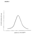
- uptake of colloidal particles by lymph nodes is surprisingly increased by modifying the surface of the particles to have an intermediate hydrophobicity/hydrophilicity. In this way we have achieved an uptake of at least 15% of the administered dose in the primary and secondary lymph nodes with at least 5% in each of the primary and secondary nodes and less than 25% of the dose remaining at the injection site.
- intermediate hydrophobicity/ hydrophilicity required by the particles is based on concepts of surface free energy (contact angles or hydrophobic interaction chromatography or albumin uptake ratio) or thickness of the layer of the modifying agent. These concepts and the measurement of objective parameters have been discussed previously by the present inventors and others, see Muller, R.H., Davis, S.S. and Mak, E. in Advances in Separations using Aqueous Phase Systems, Applications in Cell Biology and Biotechnology, Eds. Fisher et al. Plenum Press, New York, 1989, pp 149-155. Muller, R.H., Davis, S.S., Illum, L. and Mak, E. in Targeting of Drugs with Synthetic Systems. Eds. Gregoriadis et al. Plenum, New York, 1986, pp 239-263.
- the contact angle between a surface and a solution of the modifying agent can be used as a measure of surface hydrophobicity.
- the method used is that described by Troster, S.D. and Kreuter, J. Contact angles of surfactants with a potential to alter body distribution of colloidal drug carriers on poly(methyl methacrylate) surfaces. Int. J. Pharm. 45 (1988) 91-100.
- the contact angle measured is the advancing contact angle between an aqueous 0.2% solution of the block copolymer and a plane surface of a reference polymeric material, in our case polystyrene.
- the greater the value of the contact angle the more hydrophilic is the nature of the modifying agent.
- a low value of the advancing contact angle indicates lower hydrophilicity.
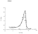
- the hydrophobicity-hydrophilicity of a particle can be determined by HIC as discussed by Muller et al. (1989) cited above. This approach has been adapted from techniques described in the field of microbiology.
- HIC hydrophobicity-hydrophilicity of a particle
- a butyl agarose column (bed volume 2.5ml; Sigma, UK) was used. 150 ⁇ l of a 0.1% w/v 60 mm polystyrene particles suspension in 0.1% w/v of polymer solution (incubated overnight) was loaded onto the column and eluted with 5ml of phosphate buffer (pH 6.8, 0.02M) containing 0.2 M NaCl (elution peak). The column was then washed with buffer containing 0.1% v/v Triton X-100 until all the particles had been recovered (washed peak).
- HIC ratio Area of elution peak Area of wash peak
- the thickness of the layer of a modifying agent which is adsorbed to the particle surface can be measured by various techniques.
- PCS photon correlation spectroscopy
- standard particles of, for example, polystyrene of a size similar to that to be used in animal experiments is equibrated with a concentration of polymer in aqueous solution such that at equilibrium the concentration remaining in the bulk phase will provide conditions such that adsorption is occurring in the plateau region of the adsorption isotherm. This plateau region is described in more detail in the publication of Kayes and Rawlins, Colloid Polymer Sci. 257 , 622-629 (1979).
- the size of these particles is determined by the PCS method and is compared to that of control particles of identical properties which have not been incubated with the polymer. Half of the difference in size is the adsorbed layer thickness. Particle solutions were incubated overnight at the concentrations of 0.1% w/v particles suspensions in 0.1% w/v of polymer solution prior to measurement of the adsorbed layer thickness by Photon Correlation Spectroscopy.
- the thickness of the layer of modifying agent can be determined by rheological measurements, as described by Tadros in (Polymer colloids as models for studying inferfacial forces in concentrated dispersions in F. Candau and RH Ottewill (eds), Scientific Methods for the Study of Polymer Colloids and Their Applications, Kluwer Academic Publishers, Netherlands, 1990, pp393-411) and by Prestidge and Tadros (Viscoelastic Properties of aqueous concentrated polystyrene latex dispersions containing grafted poly (ethylene oxide) chains, J. Colloid Interf. Sci, 124, 1988, 660-665), or Costello et al , J. Colloid Interface Sci, 152 , 237, 1992.
- the adsorbed layer or the layer of a grafted hydrophilic polymer such as PEG can be measured by studying the relation between relative viscosity and effective volume fraction of a high concentrated dispersion of particles.
- Rheological measurements can be conducted using a Haake-Rotovisco (Model RV100) fitted with a M500/M150 head, concentric cylinder geometry. The experiments are conducted at 25°C. Flow curves showing the relation between steady shear stress and shear rate are recorded on an X-Y recorder. The values of the relative viscosity are obtained from the gradients of such curves. For low volume fractions the curves are linear throughout, demonstrating Newtonian behaviour. For systems with higher volume fraction the dispersions are non-Newtonian and the linear portion of the flow curve is used.
- ⁇ is obtained by an iterative process wherein values of ⁇ are used to fit the experimental points to the theoretical curve given by the Dougherty-Krieger equation.
- This method can also be used to determine an adsorbed layer thickness as follows.
- Polystyrene dispersion at 8% volume fraction of average size, 250nm, are coated with an adsorbed layer of block copolymer by equilibration for 24 hours in a 1% w/v solution of the copolymer at 25°C.
- Dispersions of such coated systems or PEG-grafted with various volume fractions are prepared and measured as above in terms of their relative viscosities.
- Systems of high volume fraction are prepared by centrifugation of more dilute systems. Volume fractions in the range 0.3 to 0.6 are selected.
- the thickness of a grafted layer or modifying agent can also be measured by small angle neutron scattering (SANS) according to the method described in Harris et al , 'Determination of the adsorption of surface active agents by small angle scattering' in 'Adsorption from solution', Eds, Ottewill et al p139, Academic Press 1983 or p287 Cogrove et al , An experimental study of polymer conformations at the solid/solution interface.
- SANS small angle neutron scattering
- the relative hydrophobicity of a particle surface can also be determined by the uptake of a probe material such as a protein. It is well known that hydrophobic particles strongly adsorb proteins such as human serum albumin, while hydrophilic surfaces adsorb much less. Thus a scale of relative hydrophobicity can be created by studying the adsorption of a single purified protein species to a plane surface or to the surface of particles. The amount adsorbed in terms of milligrams per unit surface area, can be used as a suitable parameter. Preferably the albumin uptake ratio (AUR) is used and this is defined as the ratio of the amount taken up on a test surface in comparison to a control surface.
- AUR albumin uptake ratio
- the AUR can be determined by the following method. To 20.0ml of 0.5mg/ml human serum albumin (HSA) solution, 246 ⁇ l of HSA-labelled with iodine-125 was added. The specific activity of the HSA solution was 8840 cpm/ ⁇ g. The particles under study and any reference particles at a known concentration and particle size were incubated with the radio labelled albumin. Thus 1ml of 2% w/v poloxamer or poloxamine solution were incubated overnight with 1ml of 2% (w/v) polystyrene particles of 250nm diameter. To 0.3ml of the particle suspension 0.2ml of buffer was added followed by 1.0ml of labelled HSA added dropwise.
- HSA human serum albumin
- Controls were prepared containing HSA and poloxamer or poloxamine. Incubation lasted 12 hours (the incubation tubes were precoated with 4ml of 1mg/ml HSA solution. After 2 hours the mixture was discarded and the tube rinsed three times with water.) After incubation, the particles were centrifuged at 13,500rpm until pelleted. The particles were washed and sonicated to provide an even resuspension. The amount of labelled HSA attached to the particles was determined using a gamma counter.
- Polystyrene particles (Polysciences Ltd UK) of a particle size of 60 nm as determined by PCS were surface labelled with Iodine-125 using the method of Huh Y., Donaldson, G.W., and Johnston, F.J., A Radiation-induced bonding of iodine at the surface of uniform polystyrene particles. Radiation Research 60 , 42-53 (1974). The labelled particles were cleaned by dialysis and then incubated with a series of block co-polymers of the poloxamer and poloxamine series. These block copolymers contain polyoxyethylene and polyoxypropylene groups. The concentration of block copolymer was 0.1%.
- male Wistar rats weighing 160-200g received 100 ⁇ l of injection volume (0.1mg polystyrene particles iodine-125 labelled; 0.1% w/v polymer) by subcutaneous injection into the hind foot pads, under halothane anaesthetic.
- the regional lymph nodes namely the popliteal, iliac and renal nodes, and major body organs were removed and counted in a gamma counter for associated Iodine-125 activity.
- the results were calculated as a % of administered dose and expressed as the mean ⁇ sd of four injections. The results are summarised in Table 1.
- Certain materials increases the sequestration of polystyrene microspheres in the lymph nodes by a surprising amount in comparison to uncoated particles and those coating agents previsouly described in the prior art.
- the uptake is more pronouced in local (primary) than distal modes (secondary and tertiary). These results demonstrate that the highest lymph node uptake an be achieved by the appropriate selection of a particle with a modified surface.
- the uptake is far greater than has been obtained previously for any reported colloidal system. It is useful to calculate a ratio in terms of the amount in the lymph nodes relative to that at the injection site. This value demonstrates the degree to which the particles move from their administration site to the target site.
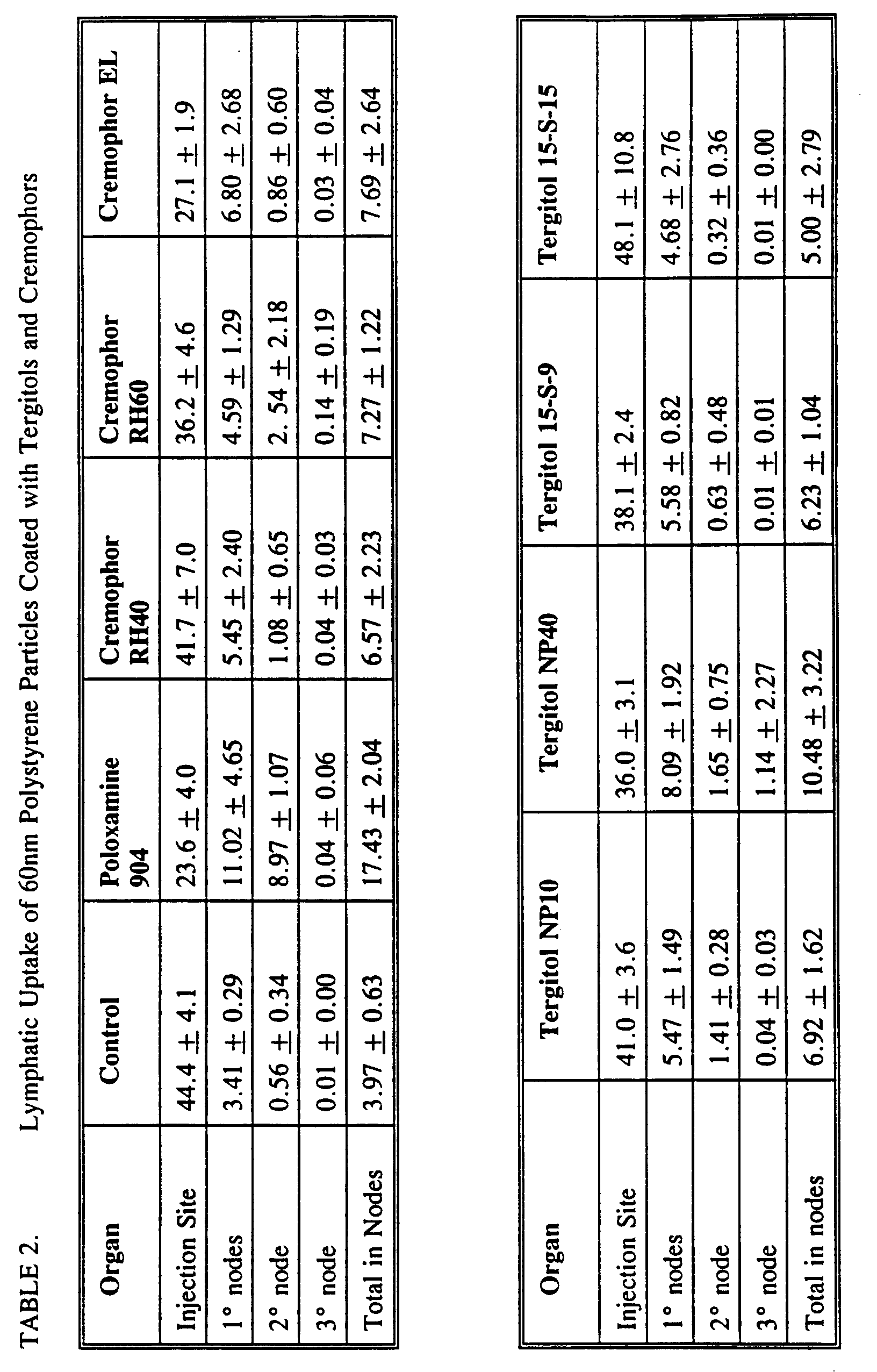
- polystyrene particles were also coated with the Cremophor materials, EL RH40 and RH60 (polyethylated castor oil, polyoxyethylene 40 and 60, hydrogenated castor oil) and Tergitol materials, NP10, NP40, 15-S-9, 15-S-15 (nonoxynol 10, 40, Pareth 9 and 15) described in the patent GB 2108967 (Table 2). Concentrations of 0.1% were employed for the coating step. Uncoated particles and particles coated with poloxamine-904 were used as controls. For all these materials except poloxamine 904 the performance was poor. The uptake of the coated particles in the lymph node was low and high quantities were retained at the site of administration.
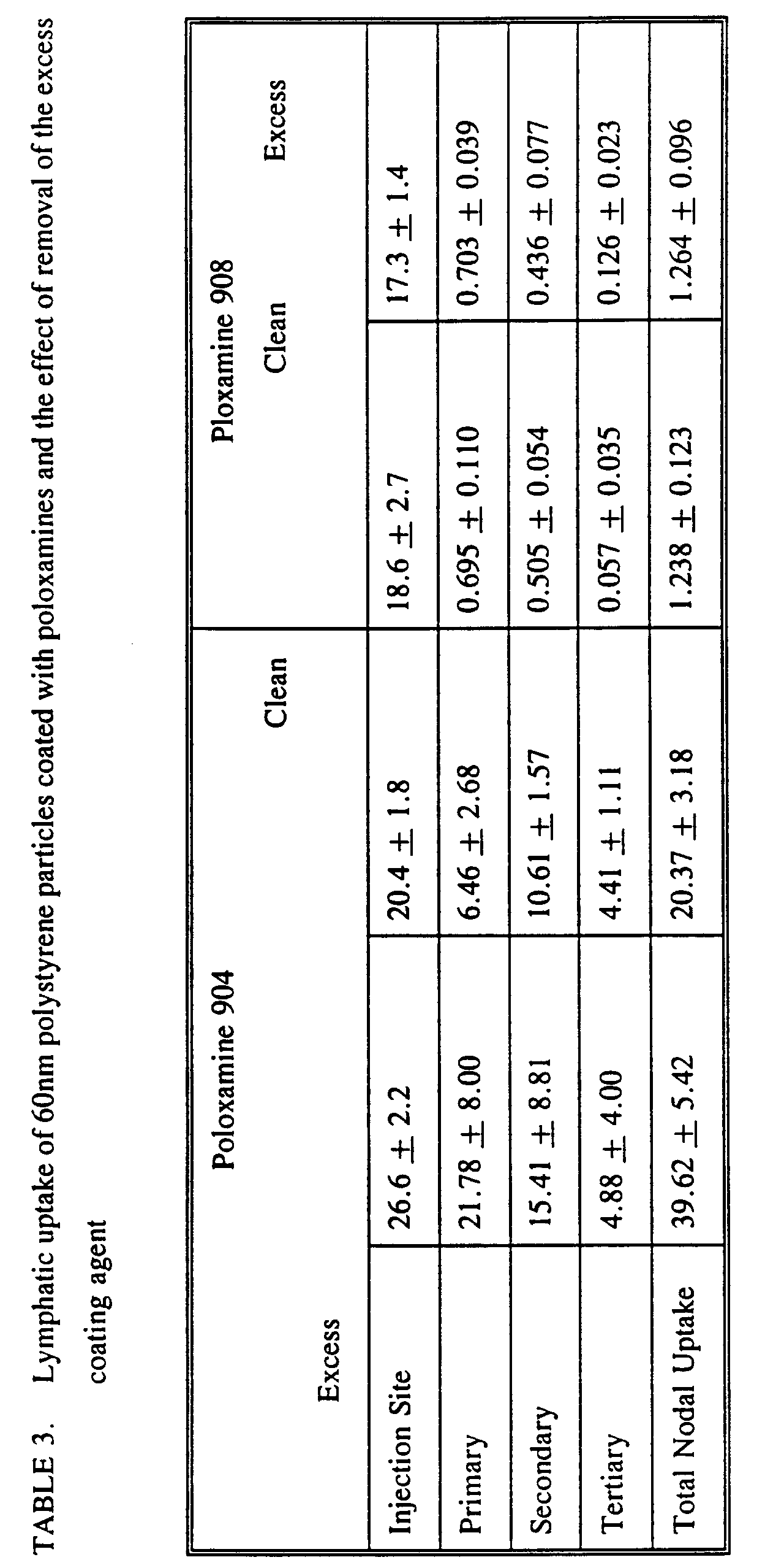
- the results are shown in Table 3. The results demonstrate that the targeting effects found with certain of the poloxamers and poloxamines is due to the presence of the modified surface layer rather than to the polymer acting as some form of penetration enhancer. Indeed removal of the excess coating polymer can increase further the lymphatic uptake of the coated particles for the case of Poloxamine 904.
- HSA-I 125 I 125 labelled HSA (HSA-I 125 ), specific activity 46 kBg/ ⁇ g. Prepared from HSA (Albutein) and I 125 from Amersham, Buckinghamshire).
- the specific activity of the labelled HSA determined by measuring the radioactivity of five 50 ⁇ l samples, was found to be 130.10 Bq/ ⁇ g.
- Triplicate samples were prepared. A known volume of particles (0.168ml - 0.423ml), sufficient to yield approximately 3 mg of particles was added to a plastic 5 ml test tube. Buffer was added to 0.5ml. 1 ml of labelled HSA was added dropwise whilst shaking at a speed of 200 on an IKA-Vibrax-VX horizontal shaker from IKA-Labortechnik (Staufen, Germany). After addition the speed of shaking was increased to 400. Incubation lasted 2 hours at room temperature.
Landscapes
- Health & Medical Sciences (AREA)
- Chemical & Material Sciences (AREA)
- Engineering & Computer Science (AREA)
- Bioinformatics & Cheminformatics (AREA)
- Medicinal Chemistry (AREA)
- Public Health (AREA)
- Nanotechnology (AREA)
- Veterinary Medicine (AREA)
- General Health & Medical Sciences (AREA)
- Pharmacology & Pharmacy (AREA)
- Epidemiology (AREA)
- Life Sciences & Earth Sciences (AREA)
- Animal Behavior & Ethology (AREA)
- Physics & Mathematics (AREA)
- Biomedical Technology (AREA)
- Optics & Photonics (AREA)
- Immunology (AREA)
- Medicinal Preparation (AREA)
- Medicines That Contain Protein Lipid Enzymes And Other Medicines (AREA)
Description
| Uptake of coated colloidal particles by regional lymph notes in the rat foot pad model Data at 24 hours post injection. | |||||
| Coating Polymer | Injection site | Primary | Regional Lymph Nodes Secondary | Total | T/I ratio |
| Poloxamers | |||||
| 101 | 30.8 ± 6.2 | 6.97 ± 2.53 | 2.00 ± 0.81 | 8.99 ± 3.09 | 0.292 |
| 105 | 32.8 ± 1.8 | 10.68 ± 1.23 | 2.18 ± 2.04 | 12.94 ± 3.09 | 0.394 |
| 108 | 29.4 ± 1.2 | 7.44 ± 3.39 | 4.55 ± 2.25 | 12.12 ± 2.97 | 0.412 |
| 124 | 15.6 ± 6.3 | 8.27 ± 0.06 | 2.79 ± 2.18 | 11.56 ± 1.56 | 0.741 |
| 181 | 34.3 ± 6.3 | 8.79 ± 2.73 | 1.87 ± 1.43 | 10.75 ± 3.79 | 0.313 |
| 182 | 19.6 ± 1.9 | 10.55 ± 0.82 | 10.86 ± 0.52 | 21.93 ± 31.3 | 1.119 |
| 183 | 21.4 ± 0.3 | 11.97 ± 3.99 | 11.68 ± 3.6 | 24.24 ± 4.34 | 1.133 |
| 184 | 22.0 ± 2.8 | 18.45 ± 6.25 | 9.26 ± 5.74 | 28.53 ± 1.04 | 1.298 |
| 185 | 19.2 ± 1.2 | 14.49 ± 3.92 | 14.91 ± 3.34 | 29.49 ± 6.24 | 1.536 |
| 188 | 17.9 ± 6.0 | 5.02 ± 2.20 | 9.08 ± 2.25 | 14.82 ± 5.15 | 0.828 |
| 234 | 11.8 ± 1.6 | 19.46 ± 1.22 | 5.57 ± 4.33 | 25.17 ± 5.61 | 2.133 |
| 238 | 15.6 ± 1.8 | 0.94 ± 0.24 | 0.36 ± 0.35 | 1.46 ± 0.32 | 0.094 |
| 282 | 12.8 ± 0.3 | 7.23 ± 5.62 | 13.55 ± 2.30 | 21.79 ± 4.51 | 1.702 |
| 288 | 11.8 ± 1.0 | 0.44 ± 0.35 | 0.22 ± 0.18 | 0.79 ± 0.41 | 0.067 |
| 333 | 11.8 ± 2.3 | 12.44 ± 4.78 | 6.39 ± 1.43 | 21.48 ± 7.32 | 1.820 |
| 338 | 9.6 ± 1.8 | 0.59 ± 0.24 | 0.26 ± 0.01 | 0.99 ± 0.13 | 0.199 |
| 401 | 15.5 ± 1.1 | 7.10 ± 1.18 | 16.73 ± 1.89 | 28.62 ± 3.64 | 1.843 |
| 402 | 11.6 ± 3.3 | 6.08 ± 1.06 | 10.70 ± 1.74 | 20.96 ± 2.44 | 1.807 |
| 403 | 12.3 ± 0.8 | 2.72 ± 1.17 | 5.90 ± 2.55 | 13.59 ± 4.09 | 1.105 |
| 407 | 11.1 ± 1.4 | 0.52 ± 0.21 | 0.51 ± 0.24 | 1.07 ± 0.39 | 0.096 |
| Poloxamines | |||||
| 304 | 29.4 ± 0.6 | 5.25 ± 4.89 | 1.82 ± 1.35 | 7.09 ± 4.97 | 0.241 |
| 504 | 18.5 ± 3.5 | 13.95 ± 2.33 | 11.05 ± 1.12 | 25.00 ± 2.00 | 1.351 |
| 704 | 14.9 ± 4.8 | 5.93 ± 1.18 | 15.70 ± 1.85 | 27.47 ± 2.47 | 1.846 |
| 901 | 45.0 ± 9.4 | 1.94 ± 0.20 | 0.41 ± 0.33 | 2.36 ± 0.19 | 0.052 |
| 904 | 13.0 ± 3.1 | 2.97 ± 0.82 | 10.73 ± 2.08 | 18.04 ± 2.70 | 1.388 |
| 908 | 22.1 ± 3.2 | 0.68 ± 0.16 | 0.53 ± 0.15 | 1.23 ± 0.27 | 0.056 |
| 1104 | 14.8 ± 4.1 | 2.75 ± 1.58 | 8.02 ± 4.49 | 11.63 ± 4.96 | 0.786 |
| 1302 | 19.1 ± 6.2 | 14.60 ± 0.39 | 4.94 ± 1.07 | 19.55 ± 0.68 | 1.024 |
| 1304 | 10.6 ± 1.1 | 1.34 ± 0.17 | 1.83 ± 0.26 | 3.28 ± 0.51 | 0.309 |
| 1504 | 12.9 ± 0.5 | 0.79 ± 0.31 | 0.93 ± 0.19 | 1.79 ± 0.28 | 0.139 |
| 1508 | 15.9 ± 2.3 | 0.54 ± 0.17 | 0.37 ± 0.12 | 1.00 ± 0.34 | 0.063 |
| Physicochemical data for coated particles | |||
| Coating Polymer | Adsorbed layer thickness (nm) | HIC Ratio | Contact angle (°) |
| 101 | 0.15 | 0.06 ± 0.03 | - |
| 105 | 0.19 | 0.56 ± 0.09 | - |
| 108 | 0.99 | 0.78 ± 0.11 | - |
| 124 | 0.04 | 1.23 ± 0.18 | - |
| 181 | 0.19 | 0.13 ± 0.05 | 43.7 ± 0.9 |
| 182 | 0.79 | 1.71 ± 0.32 | 47.1 ± 0.9 |
| 183 | 0.76 | 1.95 ± 0.16 | 47.8 ± 1.1 |
| 184 | 1.01 | 2.13 ± 0.06 | 49.0 ± 0.7 |
| 185 | 1.64 | 3.25 ± 0.13 | 53.5 ± 1.0 |
| 188 | 4.19 | 5.39 ± 0.43 | 63.2 ± 1.2 |
| 234 | 1.55 | 3.65 ± 0.29 | - |
| 238 | 8.45 | 17.39 ± 0.77 | - |
| 282 | 1.26 | 2.09 ± 0.11 | - |
| 288 | 7.34 | 19.76 ± 0.24 | - |
| 333 | 2.76 | 4.14 ± 0.52 | - |
| 338 | 7.47 | 20.31 ± 0.76 | - |
| 401 | 1.24 | 4.02 ± 0.25 | 32.1 ± 1.9 |
| 402 | 2.36 | 3.87 ± 0.06 | 24.1 ± 1.7 |
| 403 | 3.09 | 4.54 ± 0.11 | 27.3 ± 1.6 |
| 407 | 6.05 | 18.37 ± 4.57 | 56.2 ± 1.0 |
| Poloxamines | |||
| 304 | - | 0.21 ± 0.04 | - |
| 504 | 0.84 | 1.66 ± 0.25 | - |
| 704 | 1.34 | 4.21 ± 0.61 | - |
| 901 | 0.43 | 0.62 ± 0.19 | 27.6 ± 1.2 |
| 904 | 1.67 | 3.35 ± 0.68 | 44.3 ± 1.2 |
| 908 | 8.14 | 18.65 ± 1.58 | 69.0 ± 1.1 |
| 1104 | 2.57 | 5.17 ± 0.446 | - |
| 1302 | 3.00 | 3.67 ± 0.67 | - |
| 1304 | 3.79 | 5.33 ± 0.25 | - |
| 1504 | 4.89 | 5.68 ± 0.61 | - |
| 1508 | 9.39 | 15.50 ± 2.31 | - |
| Uptake of Solconanocoll into the Lymphatics | |||
| Organ | 2 hours | 6 hours | 24 hours |
| Injection Site | 39.6 ± 5.1 | 36.8 ± 3.7 | 23.2 ± 1.2 |
| Primary nodes | 4.59 ± 1.05 | 4.57 ± 1.67 | 5.12 ± 1.12 |
| Secondary node | 3.59 ± 1.73 | 4.62 ± 0.76 | 3.54 ± 0.37 |
| Tertiary node | 0.66 ± 0.51 | 1.02 ± 0.52 | 1.22 ± 0.28 |
| Total in nodes | 8.85 ± 2.70 | 9.95 ± 1.97 | 9.55 ± 0.54 |
| Liver | 7.40 ± 1.65 | 7.89 ± 0.59 | 4.47 ± 0.46 |
| Spleen | 0.40 ± 0.02 | 0.29 ± 0.05 | 0.22 ± 0.04 |
| Kidneys | 6.00 ± 0.83 | 8.53 ± 0.27 | 8.98 ± 0.30 |
| Blood | 0.66 ± 0.18 | 0.32 ± 0.02 | 0.20 ± 0.04 |
| Time Course of Particle Uptake by the Lymphatics | |||
| 2 hours | 6 hours | 24 hours | |
| CONTROL | |||
| Injection Site | 52.2 ± 5.7 | 45.8 ± 2.11 | 38.1 ± 3.1 |
| Primary | 4.05 ± 0.51 | 4.99 ± 1.10 | 8.17 ± 2.98 |
| Secondary | 0.654 ± 0.058 | 1.14 ± 0.463 | |
| Tertiary | 0.071 ± 0.033 | 1.10 ± 1.85 | 0.078 ± 0.083 |
| Total in nodes | 4.77 ± 0.47 | 6.91 ± 2.04 | 9.39 ± 3.36 |
| Poloxamine 904 jection | |||
| site | 27.1 ± 5.4 | 24.6 ± 2.7 | 19.0 ± 2.0 |
| Primary | 7.21 ± 2.56 | 5.20 ± 0.814 | 7.08 ± 1.59 |
| Secondary | 9.11 ± 4.38 | 9.90 ± 2.71 | 12.16 ± 3.82 |
| Tertiary | 5.95 ± 2.62 | 7.48 ± 3.86 | 4.99 ± 2.80 |
| Total in nodes | 20.78 ± 4.19 | 22.83 ± 1.80 | 24.22 ± 3.01 |
| Poloxamine 908 | |||
| Injection Site | 31.8 ± 5.7 | 26.6 ± 4.1 | 17.5 ± 1.3 |
| Primary | 0.684 ± 0.293 | 1.262 ± 0.251 | 0.969 ± 0.105 |
| Secondary | 0.394 ± 0.053 | 0.372 ± 0.315 | 0.271 ± 0.028 |
| Tertiary | 0.188 ± 0.050 | 0.095 ± 0.078 | 0.032 ± 0.002 |
| Total in nodes | 1.167 ± 0.131 | 1.73 ± 0.514 | 1.27 ± 0.123 |
| Relation between lymph node uptake (T/I) and adsorption of albumin to particle surface (AUR). | ||
| Coating | Albumin uptake ratio | Lymph node uptake (T/I) |
| Control | 1.0 | 0.07 |
| 904 | 0.21 | 1.39 |
| 908 | 0.05 | 0.06 |
| 182 | 0.336 | 1.12 |
| 183 | 0.343 | 1.13 |
| 184 | 0.338 | 1.30 |
| 185 | 0.340 | 1.54 |
| 188 | 0.184 | 0.83 |
| 333 | 0.249 | 1.82 |
| 234 | 0.277 | 2.13 |
| 238 | 0.113 | 0.09 |
| 282 | 0.277 | 1.70 |
| 407 | 0.09 | 0.10 |
Claims (14)
- A composition for delivering an active agent to the lymphatic system comprising a plurality of colloidal particles having a size in the range of 1-250nm, and an active agent associated with each particle wherein the surface of each particle has a hydrophobicity ratio as herein defined of less than 10.
- A composition according to claim 1 wherein the hydrophobicity ratio is less than 5.
- A composition for delivering an active agent to the lymphatic system comprising a plurality of colloidal particles having a size in the range of 1-250 nm, and an active agent associated with each particle, wherein a modifying agent is adsorbed onto or attached to the surface of each particle, such that the modifying agent gives an advancing contact angle as herein defined of less than 60°.
- A composition for delivering an active agent to the lymphatic system comprising a plurality of colloidal particles having a size in the range of 1-250 nm, and an active agent associated with each particle wherein a modifying agent is adsorbed onto or attached to the surface of each particle such that the thickness of the layer of modifying agent is 0.8 to 4.0 nm.
- A composition for delivering an active agent to the lymphatic system comprising a plurality of colloidal particles having a size in the range of 1-250 nm, and an active agent associated with each particle wherein a modifying agent is adsorbed onto or attached to the surface of each particle such that the albumin uptake ratio (AUR) of the surface as herein defined is between 0.2 and 0.5.
- A composition for delivering an active agent to the lymphatic system comprising a plurality of colloidal particles having a size in the range of 1-250 nm, and an active agent associated with each particle wherein a modifying agent is adsorbed onto or attached to the surface of each particle such that the thickness of the layer of modifying agent is less than 10nm and the surface of each particle has a hydrophobicity ratio as herein defined of less than 5.
- A composition for delivering an active agent to the lymphatic system comprising a plurality of colloidal particles and an active agent associated with each particle wherein a modifying agent is adsorbed onto or attached to the surface of each particle such that the thickness of the layer of modifying agent is less than 10nm and the modifying agent gives an advancing contact angle as herein defined of less than 60°.
- A composition for delivering an active agent to the lymphatic system comprising a plurality of colloidal particles and an active agent associated with each particle wherein a modifying agent is adsorbed onto or attached to the surface of each particle such that the thickness of the layer of modifying agent is less than 10nm and the albumin uptake ratio (AUR) of the surface as herein defined is between 0.2 and 0.5.
- A composition for delivering an active agent to the lymphatic system comprising a plurality of colloidal particles and an active agent associated with each particle wherein a modifying agent is adsorbed onto or attached to the surface of each particle such that the thickness of the layer of modifying agent is less than 10nm and the surface of each particle has a hydrophobicity ratio as herein defined of less than 10 and the modifying agent gives an advancing contact angle as herein defined of less than 60° and the albumin uptake ratio (AUR) of the surface as herein defined is between 0.2 and 0.5.
- A composition according to any one of claims 6 to 9 wherein the thickness of the layer of modifying agent is 0.8 to 4.0 nm.
- A composition according to claim 1 or 2 wherein the surface of each particle is modified by adsorption or attachment of a modifying agent.
- A composition according to any one of the preceding claims wherein the modifying agent is a non-ionic surfactant.
- A composition according to claim 12 wherein the modifying agent is a block copolymer.
- A composition according to claim 13 wherein the block copolymer contains polyethyleneglycol.
Applications Claiming Priority (3)
| Application Number | Priority Date | Filing Date | Title |
|---|---|---|---|
| GB929216082A GB9216082D0 (en) | 1992-07-28 | 1992-07-28 | Lymphatic delivery composition |
| GB9216082 | 1992-07-28 | ||
| PCT/GB1993/001596 WO1994002122A1 (en) | 1992-07-28 | 1993-07-28 | Lymphatic delivery composition |
Publications (2)
| Publication Number | Publication Date |
|---|---|
| EP0652746A1 EP0652746A1 (en) | 1995-05-17 |
| EP0652746B1 true EP0652746B1 (en) | 1998-10-28 |
Family
ID=10719471
Family Applications (1)
| Application Number | Title | Priority Date | Filing Date |
|---|---|---|---|
| EP93917936A Expired - Lifetime EP0652746B1 (en) | 1992-07-28 | 1993-07-28 | Lymphatic delivery composition |
Country Status (10)
| Country | Link |
|---|---|
| US (2) | US5792475A (en) |
| EP (1) | EP0652746B1 (en) |
| JP (1) | JPH08501534A (en) |
| AU (1) | AU4717593A (en) |
| DE (1) | DE69321839T2 (en) |
| DK (1) | DK0652746T3 (en) |
| ES (1) | ES2124791T3 (en) |
| GB (2) | GB9216082D0 (en) |
| NO (1) | NO307169B1 (en) |
| WO (1) | WO1994002122A1 (en) |
Families Citing this family (33)
| Publication number | Priority date | Publication date | Assignee | Title |
|---|---|---|---|---|
| US5346702A (en) * | 1992-12-04 | 1994-09-13 | Sterling Winthrop Inc. | Use of non-ionic cloud point modifiers to minimize nanoparticle aggregation during sterilization |
| US5439686A (en) * | 1993-02-22 | 1995-08-08 | Vivorx Pharmaceuticals, Inc. | Methods for in vivo delivery of substantially water insoluble pharmacologically active agents and compositions useful therefor |
| US20070116761A1 (en) * | 1993-02-22 | 2007-05-24 | Desai Neil P | Novel formulations of pharmacological agents, methods for the preparation thereof and methods for the use thereof |
| US5981719A (en) | 1993-03-09 | 1999-11-09 | Epic Therapeutics, Inc. | Macromolecular microparticles and methods of production and use |
| US6090925A (en) | 1993-03-09 | 2000-07-18 | Epic Therapeutics, Inc. | Macromolecular microparticles and methods of production and use |
| US5543158A (en) * | 1993-07-23 | 1996-08-06 | Massachusetts Institute Of Technology | Biodegradable injectable nanoparticles |
| US5565215A (en) * | 1993-07-23 | 1996-10-15 | Massachusettes Institute Of Technology | Biodegradable injectable particles for imaging |
| HUT75469A (en) * | 1994-02-28 | 1997-05-28 | Medinova Med Consulting Gmbh | Drug targeting system, method for preparing same and its use |
| US6007845A (en) * | 1994-07-22 | 1999-12-28 | Massachusetts Institute Of Technology | Nanoparticles and microparticles of non-linear hydrophilic-hydrophobic multiblock copolymers |
| WO1996025918A1 (en) | 1995-02-24 | 1996-08-29 | Nanosystems L.L.C. | Aerosols containing nanoparticle dispersions |
| US5834025A (en) * | 1995-09-29 | 1998-11-10 | Nanosystems L.L.C. | Reduction of intravenously administered nanoparticulate-formulation-induced adverse physiological reactions |
| US5611344A (en) * | 1996-03-05 | 1997-03-18 | Acusphere, Inc. | Microencapsulated fluorinated gases for use as imaging agents |
| US6511477B2 (en) * | 1997-03-13 | 2003-01-28 | Biocardia, Inc. | Method of drug delivery to interstitial regions of the myocardium |
| US6443949B2 (en) * | 1997-03-13 | 2002-09-03 | Biocardia, Inc. | Method of drug delivery to interstitial regions of the myocardium |
| US8853260B2 (en) * | 1997-06-27 | 2014-10-07 | Abraxis Bioscience, Llc | Formulations of pharmacological agents, methods for the preparation thereof and methods for the use thereof |
| US20030199425A1 (en) * | 1997-06-27 | 2003-10-23 | Desai Neil P. | Compositions and methods for treatment of hyperplasia |
| US6369132B2 (en) * | 1997-10-17 | 2002-04-09 | Hercules Incorporated | Fine particle size low bulk density thermoplastic polymers |
| US6444192B1 (en) * | 1999-02-05 | 2002-09-03 | The Regents Of The University Of California | Diagnostic imaging of lymph structures |
| US6815170B1 (en) | 1999-06-30 | 2004-11-09 | John Wayne Cancer Institute | Methods for lymph node identification |
| AU5905400A (en) | 1999-07-14 | 2001-02-05 | Board Of Regents, The University Of Texas System | Methods and compositions for delivery and retention of active agents to lymph nodes |
| FR2835749B1 (en) * | 2002-02-08 | 2006-04-14 | Inst Nat Sante Rech Med | PHARMACEUTICAL COMPOSITION IMPROVING IN VIVO GENE TRANSFER |
| US20040197264A1 (en) * | 2003-04-04 | 2004-10-07 | Alexander Schwarz | Microspheres comprising therapeutic and diagnostic radioactive isotopes |
| CN101287507B (en) * | 2005-08-12 | 2012-12-05 | 刘江 | Methods and devices for lymphatic targeting |
| AU2007285472B2 (en) * | 2006-03-30 | 2013-10-24 | Engene, Inc. | Non-viral compositions and methods for transfecting gut cells in vivo |
| WO2008022263A2 (en) * | 2006-08-17 | 2008-02-21 | Epix Pharmaceuticals, Inc. | Methods for lymph system imaging |
| WO2010102066A1 (en) | 2009-03-05 | 2010-09-10 | Bend Research, Inc. | Dextran polymer powder for inhalation administration of pharmaceuticals |
| WO2010138192A2 (en) | 2009-05-27 | 2010-12-02 | Selecta Biosciences, Inc. | Nanocarriers possessing components with different rates of release |
| CN102905728B (en) | 2010-05-26 | 2015-11-25 | 西莱克塔生物科技公司 | Nanocarrier compositions with uncoupled adjuvants |
| US8815294B2 (en) | 2010-09-03 | 2014-08-26 | Bend Research, Inc. | Pharmaceutical compositions of dextran polymer derivatives and a carrier material |
| US9060938B2 (en) | 2011-05-10 | 2015-06-23 | Bend Research, Inc. | Pharmaceutical compositions of active agents and cationic dextran polymer derivatives |
| US20130028857A1 (en) | 2011-07-29 | 2013-01-31 | Selecta Biosciences, Inc. | Synthetic nanocarriers comprising polymers comprising multiple immunomodulatory agents |
| US9687573B2 (en) | 2013-03-13 | 2017-06-27 | Biosphere Medical, Inc. | Compositions and associated methods for radioisotope-binding microparticles |
| CN111514317B (en) * | 2020-04-09 | 2022-08-19 | 温州医科大学附属第一医院 | Lymphatic system bimodal imaging contrast agent |
Family Cites Families (11)
| Publication number | Priority date | Publication date | Assignee | Title |
|---|---|---|---|---|
| CH652599A5 (en) * | 1981-08-28 | 1985-11-29 | Solco Basel Ag | Method for producing colloidal physiological degradable, human serum albumin containing products and their use. |
| GB8601100D0 (en) * | 1986-01-17 | 1986-02-19 | Cosmas Damian Ltd | Drug delivery system |
| US5219577A (en) * | 1990-06-22 | 1993-06-15 | The Regents Of The University Of California | Biologically active composition having a nanocrystalline core |
| EP0654973A4 (en) * | 1992-07-21 | 1995-08-09 | Gen Hospital Corp | LYPHATIC TISSUE DRUG ADMINISTRATION SYSTEM. |
| DE69327659T2 (en) * | 1992-10-14 | 2000-09-21 | Nycomed Imaging As, Oslo | THERAPEUTIC AND DIAGNOSTIC IMAGE GENERATION COMPOSITION AND METHOD FOR PRODUCING THE SAME |
| US5336507A (en) * | 1992-12-11 | 1994-08-09 | Sterling Winthrop Inc. | Use of charged phospholipids to reduce nanoparticle aggregation |
| ATE156706T1 (en) * | 1993-03-17 | 1997-08-15 | Silica Gel Gmbh | SUPERPARAMAGNETIC PARTICLES, METHOD FOR THE PRODUCTION AND USE OF THE SAME |
| US5565215A (en) * | 1993-07-23 | 1996-10-15 | Massachusettes Institute Of Technology | Biodegradable injectable particles for imaging |
| US5543158A (en) * | 1993-07-23 | 1996-08-06 | Massachusetts Institute Of Technology | Biodegradable injectable nanoparticles |
| US5571536A (en) * | 1995-02-06 | 1996-11-05 | Nano Systems L.L.C. | Formulations of compounds as nanoparticulate dispersions in digestible oils or fatty acids |
| US5560931A (en) * | 1995-02-14 | 1996-10-01 | Nawosystems L.L.C. | Formulations of compounds as nanoparticulate dispersions in digestible oils or fatty acids |
-
1992
- 1992-07-28 GB GB929216082A patent/GB9216082D0/en active Pending
-
1993
- 1993-07-28 US US08/374,671 patent/US5792475A/en not_active Expired - Fee Related
- 1993-07-28 EP EP93917936A patent/EP0652746B1/en not_active Expired - Lifetime
- 1993-07-28 JP JP6504329A patent/JPH08501534A/en active Pending
- 1993-07-28 AU AU47175/93A patent/AU4717593A/en not_active Abandoned
- 1993-07-28 ES ES93917936T patent/ES2124791T3/en not_active Expired - Lifetime
- 1993-07-28 DK DK93917936T patent/DK0652746T3/en active
- 1993-07-28 DE DE69321839T patent/DE69321839T2/en not_active Expired - Fee Related
- 1993-07-28 WO PCT/GB1993/001596 patent/WO1994002122A1/en not_active Ceased
- 1993-07-28 GB GB9500063A patent/GB2283422B/en not_active Expired - Fee Related
-
1995
- 1995-01-23 NO NO950238A patent/NO307169B1/en unknown
-
1998
- 1998-06-15 US US09/094,959 patent/US5928669A/en not_active Expired - Fee Related
Also Published As
| Publication number | Publication date |
|---|---|
| GB9216082D0 (en) | 1992-09-09 |
| NO307169B1 (en) | 2000-02-21 |
| DK0652746T3 (en) | 1999-07-12 |
| JPH08501534A (en) | 1996-02-20 |
| DE69321839D1 (en) | 1998-12-03 |
| DE69321839T2 (en) | 1999-04-15 |
| NO950238D0 (en) | 1995-01-23 |
| AU4717593A (en) | 1994-02-14 |
| NO950238L (en) | 1995-01-23 |
| US5928669A (en) | 1999-07-27 |
| EP0652746A1 (en) | 1995-05-17 |
| GB2283422A (en) | 1995-05-10 |
| WO1994002122A1 (en) | 1994-02-03 |
| GB9500063D0 (en) | 1995-03-08 |
| ES2124791T3 (en) | 1999-02-16 |
| GB2283422B (en) | 1996-05-01 |
| US5792475A (en) | 1998-08-11 |
Similar Documents
| Publication | Publication Date | Title |
|---|---|---|
| US5792475A (en) | Lymphatic delivery composition | |
| Wang et al. | Strategies for liposome drug delivery systems to improve tumor treatment efficacy | |
| US6207195B1 (en) | Therapeutic nanospheres | |
| Jain et al. | RGD-anchored magnetic liposomes for monocytes/neutrophils-mediated brain targeting | |
| Howard et al. | PEGylation of nanocarrier drug delivery systems: state of the art | |
| Bhadra et al. | PEGylated Peptide Dendrimeric Carriers for the Delivery of Antimalarial Drug Chloroquine Phosphate: Bhadra, Bhadra, and Jain | |
| JP2886171B2 (en) | Bioadhesive drug carriers for endothelial and epithelial uptake of therapeutic and diagnostic agents and localization to lesions | |
| Müller | Colloidal carriers for controlled drug delivery and targeting: Modification, characterization and in vivo distribution | |
| US7465716B2 (en) | Targeted drug delivery with a hyaluronan ligand | |
| Tiefenauer et al. | In vivo evaluation of magnetite nanoparticles for use as a tumor contrast agent in MRI | |
| EP2106806A1 (en) | Nanoparticles for targeted delivery of active agents to the lung | |
| Moghimi | Mechanisms regulating body distribution of nanospheres conditioned with pluronic and tetronic block co-polymers | |
| Lin et al. | Preparation of surface-modified albumin nanospheres | |
| Song et al. | Surface-modified PLGA nanoparticles with PEG/LA-chitosan for targeted delivery of arsenic trioxide for liver cancer treatment: Inhibition effects enhanced and side effects reduced | |
| AU1947395A (en) | Drug targeting system, method for preparing same and its use | |
| Nakamura et al. | Identification of polyethylene glycol-resistant macrophages on stealth imaging in vitro using fluorescent organosilica nanoparticles | |
| TW200815047A (en) | Nanoparticle compositions | |
| Wohlfart et al. | Treatment of glioblastoma with poly (isohexyl cyanoacrylate) nanoparticles | |
| US20060270030A1 (en) | Multimodally altered cells as a form for administering active substances and as diagnostic particles | |
| CN112274654B (en) | Targeted drug-loaded nano-micelle, and preparation method and application thereof | |
| Borchardt et al. | Body distribution of 75Se-radiolabeled silica nanoparticles covalently coated with co-functionalized surfactants after intravenous injection in rats | |
| Khalil et al. | Colloidal polymeric nanoparticles and brain drug delivery | |
| Nishikawa | Pharmacokinetic properties of nanomaterials | |
| JPH08511553A (en) | Surface-modified albumin microspheres and pharmaceutical compositions containing them | |
| CN120305205A (en) | A kidney-targeted small molecule polypeptide nanoliposome and its preparation method and application |
Legal Events
| Date | Code | Title | Description |
|---|---|---|---|
| PUAI | Public reference made under article 153(3) epc to a published international application that has entered the european phase |
Free format text: ORIGINAL CODE: 0009012 |
|
| 17P | Request for examination filed |
Effective date: 19950109 |
|
| AK | Designated contracting states |
Kind code of ref document: A1 Designated state(s): BE CH DE DK ES FR IE IT LI PT SE |
|
| 17Q | First examination report despatched |
Effective date: 19960704 |
|
| GRAG | Despatch of communication of intention to grant |
Free format text: ORIGINAL CODE: EPIDOS AGRA |
|
| GRAG | Despatch of communication of intention to grant |
Free format text: ORIGINAL CODE: EPIDOS AGRA |
|
| GRAH | Despatch of communication of intention to grant a patent |
Free format text: ORIGINAL CODE: EPIDOS IGRA |
|
| GRAH | Despatch of communication of intention to grant a patent |
Free format text: ORIGINAL CODE: EPIDOS IGRA |
|
| GRAA | (expected) grant |
Free format text: ORIGINAL CODE: 0009210 |
|
| AK | Designated contracting states |
Kind code of ref document: B1 Designated state(s): BE CH DE DK ES FR IE IT LI PT SE |
|
| REG | Reference to a national code |
Ref country code: CH Ref legal event code: NV Representative=s name: R. A. EGLI & CO. PATENTANWAELTE Ref country code: CH Ref legal event code: EP |
|
| REF | Corresponds to: |
Ref document number: 69321839 Country of ref document: DE Date of ref document: 19981203 |
|
| REG | Reference to a national code |
Ref country code: IE Ref legal event code: FG4D |
|
| ET | Fr: translation filed | ||
| REG | Reference to a national code |
Ref country code: ES Ref legal event code: FG2A Ref document number: 2124791 Country of ref document: ES Kind code of ref document: T3 |
|
| REG | Reference to a national code |
Ref country code: PT Ref legal event code: SC4A Free format text: AVAILABILITY OF NATIONAL TRANSLATION Effective date: 19990128 |
|
| REG | Reference to a national code |
Ref country code: DK Ref legal event code: T3 |
|
| PLBE | No opposition filed within time limit |
Free format text: ORIGINAL CODE: 0009261 |
|
| STAA | Information on the status of an ep patent application or granted ep patent |
Free format text: STATUS: NO OPPOSITION FILED WITHIN TIME LIMIT |
|
| 26N | No opposition filed | ||
| REG | Reference to a national code |
Ref country code: CH Ref legal event code: PFA Free format text: DANBIOSYST UK LIMITED TRANSFER- WEST PHARMACEUTICAL SERVICES DRUG DELIVERY & CLINICAL RESEARCH CENTRE LIMITED ALBERT EINSTEIN CENTRE |
|
| REG | Reference to a national code |
Ref country code: FR Ref legal event code: CD |
|
| REG | Reference to a national code |
Ref country code: PT Ref legal event code: PD4A Free format text: WEST P. S. DRUG DELIVERY & CLINICAL RES. CEN. LTD.GB Effective date: 19991012 |
|
| PGFP | Annual fee paid to national office [announced via postgrant information from national office to epo] |
Ref country code: PT Payment date: 20050630 Year of fee payment: 13 |
|
| PGFP | Annual fee paid to national office [announced via postgrant information from national office to epo] |
Ref country code: SE Payment date: 20050706 Year of fee payment: 13 |
|
| PGFP | Annual fee paid to national office [announced via postgrant information from national office to epo] |
Ref country code: FR Payment date: 20050708 Year of fee payment: 13 |
|
| PGFP | Annual fee paid to national office [announced via postgrant information from national office to epo] |
Ref country code: IE Payment date: 20050713 Year of fee payment: 13 |
|
| PGFP | Annual fee paid to national office [announced via postgrant information from national office to epo] |
Ref country code: DK Payment date: 20050715 Year of fee payment: 13 |
|
| PGFP | Annual fee paid to national office [announced via postgrant information from national office to epo] |
Ref country code: DE Payment date: 20050721 Year of fee payment: 13 |
|
| PGFP | Annual fee paid to national office [announced via postgrant information from national office to epo] |
Ref country code: CH Payment date: 20050727 Year of fee payment: 13 |
|
| PGFP | Annual fee paid to national office [announced via postgrant information from national office to epo] |
Ref country code: ES Payment date: 20050818 Year of fee payment: 13 |
|
| PGFP | Annual fee paid to national office [announced via postgrant information from national office to epo] |
Ref country code: BE Payment date: 20050916 Year of fee payment: 13 |
|
| PG25 | Lapsed in a contracting state [announced via postgrant information from national office to epo] |
Ref country code: IE Free format text: LAPSE BECAUSE OF NON-PAYMENT OF DUE FEES Effective date: 20060728 |
|
| PG25 | Lapsed in a contracting state [announced via postgrant information from national office to epo] |
Ref country code: SE Free format text: LAPSE BECAUSE OF NON-PAYMENT OF DUE FEES Effective date: 20060729 |
|
| PG25 | Lapsed in a contracting state [announced via postgrant information from national office to epo] |
Ref country code: LI Free format text: LAPSE BECAUSE OF NON-PAYMENT OF DUE FEES Effective date: 20060731 Ref country code: DK Free format text: LAPSE BECAUSE OF NON-PAYMENT OF DUE FEES Effective date: 20060731 Ref country code: CH Free format text: LAPSE BECAUSE OF NON-PAYMENT OF DUE FEES Effective date: 20060731 Ref country code: BE Free format text: LAPSE BECAUSE OF NON-PAYMENT OF DUE FEES Effective date: 20060731 |
|
| PGFP | Annual fee paid to national office [announced via postgrant information from national office to epo] |
Ref country code: IT Payment date: 20060731 Year of fee payment: 14 |
|
| PG25 | Lapsed in a contracting state [announced via postgrant information from national office to epo] |
Ref country code: PT Free format text: LAPSE BECAUSE OF NON-PAYMENT OF DUE FEES Effective date: 20070129 |
|
| PG25 | Lapsed in a contracting state [announced via postgrant information from national office to epo] |
Ref country code: DE Free format text: LAPSE BECAUSE OF NON-PAYMENT OF DUE FEES Effective date: 20070201 |
|
| REG | Reference to a national code |
Ref country code: PT Ref legal event code: MM4A Free format text: LAPSE DUE TO NON-PAYMENT OF FEES Effective date: 20070129 |
|
| REG | Reference to a national code |
Ref country code: DK Ref legal event code: EBP |
|
| REG | Reference to a national code |
Ref country code: CH Ref legal event code: PL |
|
| EUG | Se: european patent has lapsed | ||
| REG | Reference to a national code |
Ref country code: IE Ref legal event code: MM4A |
|
| REG | Reference to a national code |
Ref country code: FR Ref legal event code: ST Effective date: 20070330 |
|
| REG | Reference to a national code |
Ref country code: ES Ref legal event code: FD2A Effective date: 20060729 |
|
| BERE | Be: lapsed |
Owner name: *WEST PHARMACEUTICAL SERVICES DRUG DELIVERY & CLIN Effective date: 20060731 |
|
| PG25 | Lapsed in a contracting state [announced via postgrant information from national office to epo] |
Ref country code: ES Free format text: LAPSE BECAUSE OF NON-PAYMENT OF DUE FEES Effective date: 20060729 |
|
| PG25 | Lapsed in a contracting state [announced via postgrant information from national office to epo] |
Ref country code: FR Free format text: LAPSE BECAUSE OF NON-PAYMENT OF DUE FEES Effective date: 20060731 |
|
| PG25 | Lapsed in a contracting state [announced via postgrant information from national office to epo] |
Ref country code: IT Free format text: LAPSE BECAUSE OF NON-PAYMENT OF DUE FEES Effective date: 20070728 |