EP0649468B1 - Soluble cr1 derivatives - Google Patents
Soluble cr1 derivatives Download PDFInfo
- Publication number
- EP0649468B1 EP0649468B1 EP93913383A EP93913383A EP0649468B1 EP 0649468 B1 EP0649468 B1 EP 0649468B1 EP 93913383 A EP93913383 A EP 93913383A EP 93913383 A EP93913383 A EP 93913383A EP 0649468 B1 EP0649468 B1 EP 0649468B1
- Authority
- EP
- European Patent Office
- Prior art keywords
- scr
- mature
- dna
- polypeptide according
- residues
- Prior art date
- Legal status (The legal status is an assumption and is not a legal conclusion. Google has not performed a legal analysis and makes no representation as to the accuracy of the status listed.)
- Expired - Lifetime
Links
- 108020004414 DNA Proteins 0.000 claims description 104
- 108090000765 processed proteins & peptides Proteins 0.000 claims description 61
- 229920001184 polypeptide Polymers 0.000 claims description 60
- 102000004196 processed proteins & peptides Human genes 0.000 claims description 60
- 238000000034 method Methods 0.000 claims description 44
- 150000001413 amino acids Chemical class 0.000 claims description 39
- 101000668170 Homo sapiens RNA-binding motif, single-stranded-interacting protein 2 Proteins 0.000 claims description 30
- 102100039690 RNA-binding motif, single-stranded-interacting protein 2 Human genes 0.000 claims description 30
- 102000053602 DNA Human genes 0.000 claims description 18
- 208000037265 diseases, disorders, signs and symptoms Diseases 0.000 claims description 14
- 101100365087 Arabidopsis thaliana SCRA gene Proteins 0.000 claims description 11
- 101150105073 SCR1 gene Proteins 0.000 claims description 11
- 101100134054 Saccharomyces cerevisiae (strain ATCC 204508 / S288c) NTG1 gene Proteins 0.000 claims description 11
- FFEARJCKVFRZRR-BYPYZUCNSA-N L-methionine Chemical compound CSCC[C@H](N)C(O)=O FFEARJCKVFRZRR-BYPYZUCNSA-N 0.000 claims description 10
- 229930182817 methionine Natural products 0.000 claims description 10
- 239000002773 nucleotide Substances 0.000 claims description 10
- 125000003729 nucleotide group Chemical group 0.000 claims description 10
- 208000035475 disorder Diseases 0.000 claims description 9
- 239000013604 expression vector Substances 0.000 claims description 9
- 238000004519 manufacturing process Methods 0.000 claims description 9
- 230000024203 complement activation Effects 0.000 claims description 8
- 239000004475 Arginine Substances 0.000 claims description 6
- ODKSFYDXXFIFQN-UHFFFAOYSA-N arginine Natural products OC(=O)C(N)CCCNC(N)=N ODKSFYDXXFIFQN-UHFFFAOYSA-N 0.000 claims description 6
- 230000008569 process Effects 0.000 claims description 6
- 201000010099 disease Diseases 0.000 claims description 5
- 239000003814 drug Substances 0.000 claims description 5
- 101000668165 Homo sapiens RNA-binding motif, single-stranded-interacting protein 1 Proteins 0.000 claims description 4
- 206010061218 Inflammation Diseases 0.000 claims description 4
- 102100039692 RNA-binding motif, single-stranded-interacting protein 1 Human genes 0.000 claims description 4
- 230000004054 inflammatory process Effects 0.000 claims description 4
- 239000000126 substance Substances 0.000 claims description 4
- 239000003937 drug carrier Substances 0.000 claims description 3
- HNDVDQJCIGZPNO-UHFFFAOYSA-N histidine Natural products OC(=O)C(N)CC1=CN=CN1 HNDVDQJCIGZPNO-UHFFFAOYSA-N 0.000 claims description 3
- 239000000546 pharmaceutical excipient Substances 0.000 claims description 3
- 230000001225 therapeutic effect Effects 0.000 claims description 3
- 239000008194 pharmaceutical composition Substances 0.000 claims description 2
- 108090000623 proteins and genes Proteins 0.000 description 102
- 102000004169 proteins and genes Human genes 0.000 description 73
- FAPWRFPIFSIZLT-UHFFFAOYSA-M Sodium chloride Chemical compound [Na+].[Cl-] FAPWRFPIFSIZLT-UHFFFAOYSA-M 0.000 description 62
- 239000013612 plasmid Substances 0.000 description 46
- 108091034117 Oligonucleotide Proteins 0.000 description 44
- 102000006834 complement receptors Human genes 0.000 description 43
- 108010047295 complement receptors Proteins 0.000 description 43
- 210000004027 cell Anatomy 0.000 description 42
- 230000000295 complement effect Effects 0.000 description 38
- XSQUKJJJFZCRTK-UHFFFAOYSA-N Urea Chemical compound NC(N)=O XSQUKJJJFZCRTK-UHFFFAOYSA-N 0.000 description 36
- 239000000243 solution Substances 0.000 description 36
- DGVVWUTYPXICAM-UHFFFAOYSA-N β‐Mercaptoethanol Chemical compound OCCS DGVVWUTYPXICAM-UHFFFAOYSA-N 0.000 description 35
- 239000011780 sodium chloride Substances 0.000 description 31
- 238000003556 assay Methods 0.000 description 29
- 239000000047 product Substances 0.000 description 29
- JLCPHMBAVCMARE-UHFFFAOYSA-N [3-[[3-[[3-[[3-[[3-[[3-[[3-[[3-[[3-[[3-[[3-[[5-(2-amino-6-oxo-1H-purin-9-yl)-3-[[3-[[3-[[3-[[3-[[3-[[5-(2-amino-6-oxo-1H-purin-9-yl)-3-[[5-(2-amino-6-oxo-1H-purin-9-yl)-3-hydroxyoxolan-2-yl]methoxy-hydroxyphosphoryl]oxyoxolan-2-yl]methoxy-hydroxyphosphoryl]oxy-5-(5-methyl-2,4-dioxopyrimidin-1-yl)oxolan-2-yl]methoxy-hydroxyphosphoryl]oxy-5-(6-aminopurin-9-yl)oxolan-2-yl]methoxy-hydroxyphosphoryl]oxy-5-(6-aminopurin-9-yl)oxolan-2-yl]methoxy-hydroxyphosphoryl]oxy-5-(6-aminopurin-9-yl)oxolan-2-yl]methoxy-hydroxyphosphoryl]oxy-5-(6-aminopurin-9-yl)oxolan-2-yl]methoxy-hydroxyphosphoryl]oxyoxolan-2-yl]methoxy-hydroxyphosphoryl]oxy-5-(5-methyl-2,4-dioxopyrimidin-1-yl)oxolan-2-yl]methoxy-hydroxyphosphoryl]oxy-5-(4-amino-2-oxopyrimidin-1-yl)oxolan-2-yl]methoxy-hydroxyphosphoryl]oxy-5-(5-methyl-2,4-dioxopyrimidin-1-yl)oxolan-2-yl]methoxy-hydroxyphosphoryl]oxy-5-(5-methyl-2,4-dioxopyrimidin-1-yl)oxolan-2-yl]methoxy-hydroxyphosphoryl]oxy-5-(6-aminopurin-9-yl)oxolan-2-yl]methoxy-hydroxyphosphoryl]oxy-5-(6-aminopurin-9-yl)oxolan-2-yl]methoxy-hydroxyphosphoryl]oxy-5-(4-amino-2-oxopyrimidin-1-yl)oxolan-2-yl]methoxy-hydroxyphosphoryl]oxy-5-(4-amino-2-oxopyrimidin-1-yl)oxolan-2-yl]methoxy-hydroxyphosphoryl]oxy-5-(4-amino-2-oxopyrimidin-1-yl)oxolan-2-yl]methoxy-hydroxyphosphoryl]oxy-5-(6-aminopurin-9-yl)oxolan-2-yl]methoxy-hydroxyphosphoryl]oxy-5-(4-amino-2-oxopyrimidin-1-yl)oxolan-2-yl]methyl [5-(6-aminopurin-9-yl)-2-(hydroxymethyl)oxolan-3-yl] hydrogen phosphate Polymers Cc1cn(C2CC(OP(O)(=O)OCC3OC(CC3OP(O)(=O)OCC3OC(CC3O)n3cnc4c3nc(N)[nH]c4=O)n3cnc4c3nc(N)[nH]c4=O)C(COP(O)(=O)OC3CC(OC3COP(O)(=O)OC3CC(OC3COP(O)(=O)OC3CC(OC3COP(O)(=O)OC3CC(OC3COP(O)(=O)OC3CC(OC3COP(O)(=O)OC3CC(OC3COP(O)(=O)OC3CC(OC3COP(O)(=O)OC3CC(OC3COP(O)(=O)OC3CC(OC3COP(O)(=O)OC3CC(OC3COP(O)(=O)OC3CC(OC3COP(O)(=O)OC3CC(OC3COP(O)(=O)OC3CC(OC3COP(O)(=O)OC3CC(OC3COP(O)(=O)OC3CC(OC3COP(O)(=O)OC3CC(OC3COP(O)(=O)OC3CC(OC3CO)n3cnc4c(N)ncnc34)n3ccc(N)nc3=O)n3cnc4c(N)ncnc34)n3ccc(N)nc3=O)n3ccc(N)nc3=O)n3ccc(N)nc3=O)n3cnc4c(N)ncnc34)n3cnc4c(N)ncnc34)n3cc(C)c(=O)[nH]c3=O)n3cc(C)c(=O)[nH]c3=O)n3ccc(N)nc3=O)n3cc(C)c(=O)[nH]c3=O)n3cnc4c3nc(N)[nH]c4=O)n3cnc4c(N)ncnc34)n3cnc4c(N)ncnc34)n3cnc4c(N)ncnc34)n3cnc4c(N)ncnc34)O2)c(=O)[nH]c1=O JLCPHMBAVCMARE-UHFFFAOYSA-N 0.000 description 28
- 241000588724 Escherichia coli Species 0.000 description 27
- 210000003000 inclusion body Anatomy 0.000 description 26
- 239000000203 mixture Substances 0.000 description 25
- 108020004705 Codon Proteins 0.000 description 24
- 230000004913 activation Effects 0.000 description 24
- 239000000872 buffer Substances 0.000 description 24
- BDAGIHXWWSANSR-UHFFFAOYSA-N methanoic acid Natural products OC=O BDAGIHXWWSANSR-UHFFFAOYSA-N 0.000 description 24
- 239000006228 supernatant Substances 0.000 description 24
- 239000007983 Tris buffer Substances 0.000 description 23
- LENZDBCJOHFCAS-UHFFFAOYSA-N tris Chemical compound OCC(N)(CO)CO LENZDBCJOHFCAS-UHFFFAOYSA-N 0.000 description 23
- 239000012634 fragment Substances 0.000 description 22
- 210000002966 serum Anatomy 0.000 description 22
- 230000014509 gene expression Effects 0.000 description 21
- 239000012528 membrane Substances 0.000 description 21
- YBYRMVIVWMBXKQ-UHFFFAOYSA-N phenylmethanesulfonyl fluoride Chemical compound FS(=O)(=O)CC1=CC=CC=C1 YBYRMVIVWMBXKQ-UHFFFAOYSA-N 0.000 description 20
- 239000008188 pellet Substances 0.000 description 19
- KCXVZYZYPLLWCC-UHFFFAOYSA-N EDTA Chemical compound OC(=O)CN(CC(O)=O)CCN(CC(O)=O)CC(O)=O KCXVZYZYPLLWCC-UHFFFAOYSA-N 0.000 description 18
- 239000004202 carbamide Substances 0.000 description 18
- 238000000746 purification Methods 0.000 description 18
- 238000002415 sodium dodecyl sulfate polyacrylamide gel electrophoresis Methods 0.000 description 18
- 238000004458 analytical method Methods 0.000 description 17
- 230000003068 static effect Effects 0.000 description 17
- HZAXFHJVJLSVMW-UHFFFAOYSA-N 2-Aminoethan-1-ol Chemical compound NCCO HZAXFHJVJLSVMW-UHFFFAOYSA-N 0.000 description 16
- 229920000392 Zymosan Polymers 0.000 description 16
- 239000013598 vector Substances 0.000 description 16
- 238000004587 chromatography analysis Methods 0.000 description 15
- 239000003112 inhibitor Substances 0.000 description 15
- 238000002360 preparation method Methods 0.000 description 15
- 238000007792 addition Methods 0.000 description 14
- 238000010276 construction Methods 0.000 description 14
- 239000000499 gel Substances 0.000 description 14
- 230000037361 pathway Effects 0.000 description 14
- 108091008146 restriction endonucleases Proteins 0.000 description 14
- AVKUERGKIZMTKX-NJBDSQKTSA-N ampicillin Chemical compound C1([C@@H](N)C(=O)N[C@H]2[C@H]3SC([C@@H](N3C2=O)C(O)=O)(C)C)=CC=CC=C1 AVKUERGKIZMTKX-NJBDSQKTSA-N 0.000 description 13
- 229960000723 ampicillin Drugs 0.000 description 13
- 238000009472 formulation Methods 0.000 description 13
- 230000005764 inhibitory process Effects 0.000 description 13
- 239000002609 medium Substances 0.000 description 13
- 239000000523 sample Substances 0.000 description 13
- OSWFIVFLDKOXQC-UHFFFAOYSA-N 4-(3-methoxyphenyl)aniline Chemical compound COC1=CC=CC(C=2C=CC(N)=CC=2)=C1 OSWFIVFLDKOXQC-UHFFFAOYSA-N 0.000 description 12
- 235000019253 formic acid Nutrition 0.000 description 12
- 239000003085 diluting agent Substances 0.000 description 11
- 230000000694 effects Effects 0.000 description 11
- RWSXRVCMGQZWBV-WDSKDSINSA-N glutathione Chemical compound OC(=O)[C@@H](N)CCC(=O)N[C@@H](CS)C(=O)NCC(O)=O RWSXRVCMGQZWBV-WDSKDSINSA-N 0.000 description 11
- 238000002955 isolation Methods 0.000 description 11
- UXVMQQNJUSDDNG-UHFFFAOYSA-L Calcium chloride Chemical compound [Cl-].[Cl-].[Ca+2] UXVMQQNJUSDDNG-UHFFFAOYSA-L 0.000 description 10
- 229920002684 Sepharose Polymers 0.000 description 10
- 239000001110 calcium chloride Substances 0.000 description 10
- 229910001628 calcium chloride Inorganic materials 0.000 description 10
- 230000029087 digestion Effects 0.000 description 10
- 239000003527 fibrinolytic agent Substances 0.000 description 10
- 230000002949 hemolytic effect Effects 0.000 description 10
- 238000010186 staining Methods 0.000 description 10
- 229960000103 thrombolytic agent Drugs 0.000 description 10
- PEDCQBHIVMGVHV-UHFFFAOYSA-N Glycerine Chemical compound OCC(O)CO PEDCQBHIVMGVHV-UHFFFAOYSA-N 0.000 description 9
- OKKJLVBELUTLKV-UHFFFAOYSA-N Methanol Chemical compound OC OKKJLVBELUTLKV-UHFFFAOYSA-N 0.000 description 9
- 229920005654 Sephadex Polymers 0.000 description 9
- 239000012507 Sephadex™ Substances 0.000 description 9
- 230000009089 cytolysis Effects 0.000 description 9
- 239000006167 equilibration buffer Substances 0.000 description 9
- 210000003743 erythrocyte Anatomy 0.000 description 9
- 230000001404 mediated effect Effects 0.000 description 9
- 230000002829 reductive effect Effects 0.000 description 9
- JKMHFZQWWAIEOD-UHFFFAOYSA-N 2-[4-(2-hydroxyethyl)piperazin-1-yl]ethanesulfonic acid Chemical compound OCC[NH+]1CCN(CCS([O-])(=O)=O)CC1 JKMHFZQWWAIEOD-UHFFFAOYSA-N 0.000 description 8
- 239000011543 agarose gel Substances 0.000 description 8
- YPZRWBKMTBYPTK-BJDJZHNGSA-N glutathione disulfide Chemical compound OC(=O)[C@@H](N)CCC(=O)N[C@H](C(=O)NCC(O)=O)CSSC[C@@H](C(=O)NCC(O)=O)NC(=O)CC[C@H](N)C(O)=O YPZRWBKMTBYPTK-BJDJZHNGSA-N 0.000 description 8
- 238000003752 polymerase chain reaction Methods 0.000 description 8
- 238000003127 radioimmunoassay Methods 0.000 description 8
- 102000004190 Enzymes Human genes 0.000 description 7
- 108090000790 Enzymes Proteins 0.000 description 7
- 238000002835 absorbance Methods 0.000 description 7
- 238000010790 dilution Methods 0.000 description 7
- 239000012895 dilution Substances 0.000 description 7
- 239000002158 endotoxin Substances 0.000 description 7
- 229940088598 enzyme Drugs 0.000 description 7
- 238000000855 fermentation Methods 0.000 description 7
- 230000004151 fermentation Effects 0.000 description 7
- 239000012465 retentate Substances 0.000 description 7
- 239000000725 suspension Substances 0.000 description 7
- 108700010070 Codon Usage Proteins 0.000 description 6
- LFQSCWFLJHTTHZ-UHFFFAOYSA-N Ethanol Chemical compound CCO LFQSCWFLJHTTHZ-UHFFFAOYSA-N 0.000 description 6
- 108091028043 Nucleic acid sequence Proteins 0.000 description 6
- 238000000246 agarose gel electrophoresis Methods 0.000 description 6
- 230000027455 binding Effects 0.000 description 6
- 125000000484 butyl group Chemical group [H]C([*])([H])C([H])([H])C([H])([H])C([H])([H])[H] 0.000 description 6
- 238000006243 chemical reaction Methods 0.000 description 6
- 239000011521 glass Substances 0.000 description 6
- 108010017826 DNA Polymerase I Proteins 0.000 description 5
- 102000004594 DNA Polymerase I Human genes 0.000 description 5
- 238000001712 DNA sequencing Methods 0.000 description 5
- 241001198387 Escherichia coli BL21(DE3) Species 0.000 description 5
- 108010024636 Glutathione Proteins 0.000 description 5
- 238000010367 cloning Methods 0.000 description 5
- 238000001962 electrophoresis Methods 0.000 description 5
- 230000002255 enzymatic effect Effects 0.000 description 5
- 239000000463 material Substances 0.000 description 5
- 238000013492 plasmid preparation Methods 0.000 description 5
- 238000012163 sequencing technique Methods 0.000 description 5
- 230000001131 transforming effect Effects 0.000 description 5
- BFSVOASYOCHEOV-UHFFFAOYSA-N 2-diethylaminoethanol Chemical compound CCN(CC)CCO BFSVOASYOCHEOV-UHFFFAOYSA-N 0.000 description 4
- 241001131785 Escherichia coli HB101 Species 0.000 description 4
- TWRXJAOTZQYOKJ-UHFFFAOYSA-L Magnesium chloride Chemical compound [Mg+2].[Cl-].[Cl-] TWRXJAOTZQYOKJ-UHFFFAOYSA-L 0.000 description 4
- 241001494479 Pecora Species 0.000 description 4
- BFNBIHQBYMNNAN-UHFFFAOYSA-N ammonium sulfate Chemical compound N.N.OS(O)(=O)=O BFNBIHQBYMNNAN-UHFFFAOYSA-N 0.000 description 4
- 229910052921 ammonium sulfate Inorganic materials 0.000 description 4
- 230000001580 bacterial effect Effects 0.000 description 4
- 230000008033 biological extinction Effects 0.000 description 4
- 230000004154 complement system Effects 0.000 description 4
- 238000004520 electroporation Methods 0.000 description 4
- 238000002474 experimental method Methods 0.000 description 4
- 238000000605 extraction Methods 0.000 description 4
- 238000000338 in vitro Methods 0.000 description 4
- 230000000977 initiatory effect Effects 0.000 description 4
- 108020004999 messenger RNA Proteins 0.000 description 4
- 229920002401 polyacrylamide Polymers 0.000 description 4
- FGDZQCVHDSGLHJ-UHFFFAOYSA-M rubidium chloride Chemical compound [Cl-].[Rb+] FGDZQCVHDSGLHJ-UHFFFAOYSA-M 0.000 description 4
- 229910000162 sodium phosphate Inorganic materials 0.000 description 4
- 238000012360 testing method Methods 0.000 description 4
- 238000012546 transfer Methods 0.000 description 4
- QTBSBXVTEAMEQO-UHFFFAOYSA-N Acetic acid Chemical compound CC(O)=O QTBSBXVTEAMEQO-UHFFFAOYSA-N 0.000 description 3
- CSCPPACGZOOCGX-UHFFFAOYSA-N Acetone Chemical compound CC(C)=O CSCPPACGZOOCGX-UHFFFAOYSA-N 0.000 description 3
- 206010001052 Acute respiratory distress syndrome Diseases 0.000 description 3
- 229920000936 Agarose Polymers 0.000 description 3
- 108091026890 Coding region Proteins 0.000 description 3
- 102000012410 DNA Ligases Human genes 0.000 description 3
- 108010061982 DNA Ligases Proteins 0.000 description 3
- 108010053070 Glutathione Disulfide Proteins 0.000 description 3
- 101000727061 Homo sapiens Complement receptor type 1 Proteins 0.000 description 3
- 241001465754 Metazoa Species 0.000 description 3
- 208000013616 Respiratory Distress Syndrome Diseases 0.000 description 3
- DBMJMQXJHONAFJ-UHFFFAOYSA-M Sodium laurylsulphate Chemical compound [Na+].CCCCCCCCCCCCOS([O-])(=O)=O DBMJMQXJHONAFJ-UHFFFAOYSA-M 0.000 description 3
- 239000002253 acid Substances 0.000 description 3
- 208000011341 adult acute respiratory distress syndrome Diseases 0.000 description 3
- 201000000028 adult respiratory distress syndrome Diseases 0.000 description 3
- 238000000137 annealing Methods 0.000 description 3
- 230000015572 biosynthetic process Effects 0.000 description 3
- 238000005119 centrifugation Methods 0.000 description 3
- 239000004074 complement inhibitor Substances 0.000 description 3
- 229960003180 glutathione Drugs 0.000 description 3
- 238000011534 incubation Methods 0.000 description 3
- 230000037230 mobility Effects 0.000 description 3
- 230000000717 retained effect Effects 0.000 description 3
- 238000004007 reversed phase HPLC Methods 0.000 description 3
- 150000003839 salts Chemical class 0.000 description 3
- 239000002002 slurry Substances 0.000 description 3
- AJPJDKMHJJGVTQ-UHFFFAOYSA-M sodium dihydrogen phosphate Chemical compound [Na+].OP(O)([O-])=O AJPJDKMHJJGVTQ-UHFFFAOYSA-M 0.000 description 3
- 235000019333 sodium laurylsulphate Nutrition 0.000 description 3
- 238000003786 synthesis reaction Methods 0.000 description 3
- 238000002560 therapeutic procedure Methods 0.000 description 3
- 230000014616 translation Effects 0.000 description 3
- XLYOFNOQVPJJNP-UHFFFAOYSA-N water Substances O XLYOFNOQVPJJNP-UHFFFAOYSA-N 0.000 description 3
- HRPVXLWXLXDGHG-UHFFFAOYSA-N Acrylamide Chemical compound NC(=O)C=C HRPVXLWXLXDGHG-UHFFFAOYSA-N 0.000 description 2
- 108010089414 Anaphylatoxins Proteins 0.000 description 2
- 108010058207 Anistreplase Proteins 0.000 description 2
- IJGRMHOSHXDMSA-UHFFFAOYSA-N Atomic nitrogen Chemical compound N#N IJGRMHOSHXDMSA-UHFFFAOYSA-N 0.000 description 2
- 208000023275 Autoimmune disease Diseases 0.000 description 2
- HEDRZPFGACZZDS-UHFFFAOYSA-N Chloroform Chemical compound ClC(Cl)Cl HEDRZPFGACZZDS-UHFFFAOYSA-N 0.000 description 2
- 229940124073 Complement inhibitor Drugs 0.000 description 2
- 102100030886 Complement receptor type 1 Human genes 0.000 description 2
- 101710196208 Fibrinolytic enzyme Proteins 0.000 description 2
- 241000233866 Fungi Species 0.000 description 2
- 102000000588 Interleukin-2 Human genes 0.000 description 2
- 108010002350 Interleukin-2 Proteins 0.000 description 2
- ONIBWKKTOPOVIA-BYPYZUCNSA-N L-Proline Chemical compound OC(=O)[C@@H]1CCCN1 ONIBWKKTOPOVIA-BYPYZUCNSA-N 0.000 description 2
- ISWSIDIOOBJBQZ-UHFFFAOYSA-N Phenol Chemical compound OC1=CC=CC=C1 ISWSIDIOOBJBQZ-UHFFFAOYSA-N 0.000 description 2
- ONIBWKKTOPOVIA-UHFFFAOYSA-N Proline Natural products OC(=O)C1CCCN1 ONIBWKKTOPOVIA-UHFFFAOYSA-N 0.000 description 2
- 240000004808 Saccharomyces cerevisiae Species 0.000 description 2
- 238000012300 Sequence Analysis Methods 0.000 description 2
- 102000003990 Urokinase-type plasminogen activator Human genes 0.000 description 2
- 108090000435 Urokinase-type plasminogen activator Proteins 0.000 description 2
- 208000027418 Wounds and injury Diseases 0.000 description 2
- 150000007513 acids Chemical class 0.000 description 2
- 239000012190 activator Substances 0.000 description 2
- 230000002776 aggregation Effects 0.000 description 2
- 238000004220 aggregation Methods 0.000 description 2
- 239000001166 ammonium sulphate Substances 0.000 description 2
- 235000011130 ammonium sulphate Nutrition 0.000 description 2
- AIYUHDOJVYHVIT-UHFFFAOYSA-M caesium chloride Chemical compound [Cl-].[Cs+] AIYUHDOJVYHVIT-UHFFFAOYSA-M 0.000 description 2
- 230000015556 catabolic process Effects 0.000 description 2
- 239000003153 chemical reaction reagent Substances 0.000 description 2
- 239000003795 chemical substances by application Substances 0.000 description 2
- 210000004978 chinese hamster ovary cell Anatomy 0.000 description 2
- HVYWMOMLDIMFJA-DPAQBDIFSA-N cholesterol Chemical compound C1C=C2C[C@@H](O)CC[C@]2(C)[C@@H]2[C@@H]1[C@@H]1CC[C@H]([C@H](C)CCCC(C)C)[C@@]1(C)CC2 HVYWMOMLDIMFJA-DPAQBDIFSA-N 0.000 description 2
- 230000002860 competitive effect Effects 0.000 description 2
- 239000002299 complementary DNA Substances 0.000 description 2
- 238000007796 conventional method Methods 0.000 description 2
- 238000012258 culturing Methods 0.000 description 2
- 238000012217 deletion Methods 0.000 description 2
- 230000037430 deletion Effects 0.000 description 2
- 238000003745 diagnosis Methods 0.000 description 2
- 238000009585 enzyme analysis Methods 0.000 description 2
- 238000007429 general method Methods 0.000 description 2
- ZDXPYRJPNDTMRX-UHFFFAOYSA-N glutamine Natural products OC(=O)C(N)CCC(N)=O ZDXPYRJPNDTMRX-UHFFFAOYSA-N 0.000 description 2
- 125000000404 glutamine group Chemical group N[C@@H](CCC(N)=O)C(=O)* 0.000 description 2
- 238000001727 in vivo Methods 0.000 description 2
- 208000027866 inflammatory disease Diseases 0.000 description 2
- 230000002401 inhibitory effect Effects 0.000 description 2
- 238000001990 intravenous administration Methods 0.000 description 2
- 239000007788 liquid Substances 0.000 description 2
- 229910001629 magnesium chloride Inorganic materials 0.000 description 2
- 210000004962 mammalian cell Anatomy 0.000 description 2
- -1 mannose and mannitol Chemical class 0.000 description 2
- 238000013507 mapping Methods 0.000 description 2
- 238000002156 mixing Methods 0.000 description 2
- 208000010125 myocardial infarction Diseases 0.000 description 2
- 102000039446 nucleic acids Human genes 0.000 description 2
- 108020004707 nucleic acids Proteins 0.000 description 2
- 150000007523 nucleic acids Chemical class 0.000 description 2
- 239000002777 nucleoside Substances 0.000 description 2
- 235000015097 nutrients Nutrition 0.000 description 2
- 239000006880 nzcym-medium Substances 0.000 description 2
- 239000002953 phosphate buffered saline Substances 0.000 description 2
- 230000008488 polyadenylation Effects 0.000 description 2
- SCVFZCLFOSHCOH-UHFFFAOYSA-M potassium acetate Chemical compound [K+].CC([O-])=O SCVFZCLFOSHCOH-UHFFFAOYSA-M 0.000 description 2
- 238000001556 precipitation Methods 0.000 description 2
- 230000002797 proteolythic effect Effects 0.000 description 2
- 238000011084 recovery Methods 0.000 description 2
- 238000011160 research Methods 0.000 description 2
- 238000011218 seed culture Methods 0.000 description 2
- 238000003756 stirring Methods 0.000 description 2
- 229960005202 streptokinase Drugs 0.000 description 2
- 238000006467 substitution reaction Methods 0.000 description 2
- 230000001732 thrombotic effect Effects 0.000 description 2
- 230000002103 transcriptional effect Effects 0.000 description 2
- 238000013519 translation Methods 0.000 description 2
- 238000002054 transplantation Methods 0.000 description 2
- 108020005087 unfolded proteins Proteins 0.000 description 2
- 229960005356 urokinase Drugs 0.000 description 2
- AZUYLZMQTIKGSC-UHFFFAOYSA-N 1-[6-[4-(5-chloro-6-methyl-1H-indazol-4-yl)-5-methyl-3-(1-methylindazol-5-yl)pyrazol-1-yl]-2-azaspiro[3.3]heptan-2-yl]prop-2-en-1-one Chemical compound ClC=1C(=C2C=NNC2=CC=1C)C=1C(=NN(C=1C)C1CC2(CN(C2)C(C=C)=O)C1)C=1C=C2C=NN(C2=CC=1)C AZUYLZMQTIKGSC-UHFFFAOYSA-N 0.000 description 1
- QKNYBSVHEMOAJP-UHFFFAOYSA-N 2-amino-2-(hydroxymethyl)propane-1,3-diol;hydron;chloride Chemical compound Cl.OCC(N)(CO)CO QKNYBSVHEMOAJP-UHFFFAOYSA-N 0.000 description 1
- DVLFYONBTKHTER-UHFFFAOYSA-N 3-(N-morpholino)propanesulfonic acid Chemical compound OS(=O)(=O)CCCN1CCOCC1 DVLFYONBTKHTER-UHFFFAOYSA-N 0.000 description 1
- QFVHZQCOUORWEI-UHFFFAOYSA-N 4-[(4-anilino-5-sulfonaphthalen-1-yl)diazenyl]-5-hydroxynaphthalene-2,7-disulfonic acid Chemical compound C=12C(O)=CC(S(O)(=O)=O)=CC2=CC(S(O)(=O)=O)=CC=1N=NC(C1=CC=CC(=C11)S(O)(=O)=O)=CC=C1NC1=CC=CC=C1 QFVHZQCOUORWEI-UHFFFAOYSA-N 0.000 description 1
- FWMNVWWHGCHHJJ-SKKKGAJSSA-N 4-amino-1-[(2r)-6-amino-2-[[(2r)-2-[[(2r)-2-[[(2r)-2-amino-3-phenylpropanoyl]amino]-3-phenylpropanoyl]amino]-4-methylpentanoyl]amino]hexanoyl]piperidine-4-carboxylic acid Chemical compound C([C@H](C(=O)N[C@H](CC(C)C)C(=O)N[C@H](CCCCN)C(=O)N1CCC(N)(CC1)C(O)=O)NC(=O)[C@H](N)CC=1C=CC=CC=1)C1=CC=CC=C1 FWMNVWWHGCHHJJ-SKKKGAJSSA-N 0.000 description 1
- PZASAAIJIFDWSB-CKPDSHCKSA-N 8-[(1S)-1-[8-(trifluoromethyl)-7-[4-(trifluoromethyl)cyclohexyl]oxynaphthalen-2-yl]ethyl]-8-azabicyclo[3.2.1]octane-3-carboxylic acid Chemical compound FC(F)(F)C=1C2=CC([C@@H](N3C4CCC3CC(C4)C(O)=O)C)=CC=C2C=CC=1OC1CCC(C(F)(F)F)CC1 PZASAAIJIFDWSB-CKPDSHCKSA-N 0.000 description 1
- 208000024827 Alzheimer disease Diseases 0.000 description 1
- 208000000104 Arthus reaction Diseases 0.000 description 1
- 201000001320 Atherosclerosis Diseases 0.000 description 1
- 108091003079 Bovine Serum Albumin Proteins 0.000 description 1
- 241000701822 Bovine papillomavirus Species 0.000 description 1
- 238000009010 Bradford assay Methods 0.000 description 1
- OYPRJOBELJOOCE-UHFFFAOYSA-N Calcium Chemical compound [Ca] OYPRJOBELJOOCE-UHFFFAOYSA-N 0.000 description 1
- CURLTUGMZLYLDI-UHFFFAOYSA-N Carbon dioxide Chemical compound O=C=O CURLTUGMZLYLDI-UHFFFAOYSA-N 0.000 description 1
- 206010053567 Coagulopathies Diseases 0.000 description 1
- 208000035473 Communicable disease Diseases 0.000 description 1
- 108010077840 Complement C3a Proteins 0.000 description 1
- 108010078546 Complement C5a Proteins 0.000 description 1
- 108010069112 Complement System Proteins Proteins 0.000 description 1
- 102000000989 Complement System Proteins Human genes 0.000 description 1
- 206010011017 Corneal graft rejection Diseases 0.000 description 1
- 241000699802 Cricetulus griseus Species 0.000 description 1
- 208000011231 Crohn disease Diseases 0.000 description 1
- PJWWRFATQTVXHA-UHFFFAOYSA-N Cyclohexylaminopropanesulfonic acid Chemical compound OS(=O)(=O)CCCNC1CCCCC1 PJWWRFATQTVXHA-UHFFFAOYSA-N 0.000 description 1
- FBPFZTCFMRRESA-KVTDHHQDSA-N D-Mannitol Chemical compound OC[C@@H](O)[C@@H](O)[C@H](O)[C@H](O)CO FBPFZTCFMRRESA-KVTDHHQDSA-N 0.000 description 1
- WQZGKKKJIJFFOK-QTVWNMPRSA-N D-mannopyranose Chemical compound OC[C@H]1OC(O)[C@@H](O)[C@@H](O)[C@@H]1O WQZGKKKJIJFFOK-QTVWNMPRSA-N 0.000 description 1
- 108050009160 DNA polymerase 1 Proteins 0.000 description 1
- 230000007018 DNA scission Effects 0.000 description 1
- 108010014303 DNA-directed DNA polymerase Proteins 0.000 description 1
- 102000016928 DNA-directed DNA polymerase Human genes 0.000 description 1
- 241000255581 Drosophila <fruit fly, genus> Species 0.000 description 1
- 108010042407 Endonucleases Proteins 0.000 description 1
- 102000004533 Endonucleases Human genes 0.000 description 1
- 241000672609 Escherichia coli BL21 Species 0.000 description 1
- 241001452028 Escherichia coli DH1 Species 0.000 description 1
- 208000001034 Frostbite Diseases 0.000 description 1
- 108010010803 Gelatin Proteins 0.000 description 1
- 102000006395 Globulins Human genes 0.000 description 1
- 108010044091 Globulins Proteins 0.000 description 1
- 206010018364 Glomerulonephritis Diseases 0.000 description 1
- WQZGKKKJIJFFOK-GASJEMHNSA-N Glucose Natural products OC[C@H]1OC(O)[C@H](O)[C@@H](O)[C@@H]1O WQZGKKKJIJFFOK-GASJEMHNSA-N 0.000 description 1
- 206010070476 Haemodialysis complication Diseases 0.000 description 1
- 206010018910 Haemolysis Diseases 0.000 description 1
- 241000238631 Hexapoda Species 0.000 description 1
- 241000282412 Homo Species 0.000 description 1
- 101000801481 Homo sapiens Tissue-type plasminogen activator Proteins 0.000 description 1
- 206010020772 Hypertension Diseases 0.000 description 1
- 206010022680 Intestinal ischaemia Diseases 0.000 description 1
- NNJVILVZKWQKPM-UHFFFAOYSA-N Lidocaine Chemical compound CCN(CC)CC(=O)NC1=C(C)C=CC=C1C NNJVILVZKWQKPM-UHFFFAOYSA-N 0.000 description 1
- 241000239218 Limulus Species 0.000 description 1
- 208000005777 Lupus Nephritis Diseases 0.000 description 1
- KDXKERNSBIXSRK-UHFFFAOYSA-N Lysine Natural products NCCCCC(N)C(O)=O KDXKERNSBIXSRK-UHFFFAOYSA-N 0.000 description 1
- 239000004472 Lysine Substances 0.000 description 1
- 229910021380 Manganese Chloride Inorganic materials 0.000 description 1
- GLFNIEUTAYBVOC-UHFFFAOYSA-L Manganese chloride Chemical compound Cl[Mn]Cl GLFNIEUTAYBVOC-UHFFFAOYSA-L 0.000 description 1
- 229930195725 Mannitol Natural products 0.000 description 1
- 208000034486 Multi-organ failure Diseases 0.000 description 1
- 208000010718 Multiple Organ Failure Diseases 0.000 description 1
- 208000009525 Myocarditis Diseases 0.000 description 1
- 208000012902 Nervous system disease Diseases 0.000 description 1
- 208000025966 Neurological disease Diseases 0.000 description 1
- 241000283973 Oryctolagus cuniculus Species 0.000 description 1
- 238000012408 PCR amplification Methods 0.000 description 1
- 206010033645 Pancreatitis Diseases 0.000 description 1
- 206010033647 Pancreatitis acute Diseases 0.000 description 1
- 208000018737 Parkinson disease Diseases 0.000 description 1
- 108010001014 Plasminogen Activators Proteins 0.000 description 1
- 102000001938 Plasminogen Activators Human genes 0.000 description 1
- 108010021757 Polynucleotide 5'-Hydroxyl-Kinase Proteins 0.000 description 1
- 102000008422 Polynucleotide 5'-hydroxyl-kinase Human genes 0.000 description 1
- 239000004793 Polystyrene Substances 0.000 description 1
- 239000012564 Q sepharose fast flow resin Substances 0.000 description 1
- 108020004511 Recombinant DNA Proteins 0.000 description 1
- 206010063897 Renal ischaemia Diseases 0.000 description 1
- 206010040047 Sepsis Diseases 0.000 description 1
- 206010040070 Septic Shock Diseases 0.000 description 1
- 108091081024 Start codon Proteins 0.000 description 1
- 108010023197 Streptokinase Proteins 0.000 description 1
- 210000001744 T-lymphocyte Anatomy 0.000 description 1
- 101710137500 T7 RNA polymerase Proteins 0.000 description 1
- 108010006785 Taq Polymerase Proteins 0.000 description 1
- 208000007536 Thrombosis Diseases 0.000 description 1
- 208000030886 Traumatic Brain injury Diseases 0.000 description 1
- 206010053614 Type III immune complex mediated reaction Diseases 0.000 description 1
- 206010046851 Uveitis Diseases 0.000 description 1
- 206010046865 Vaccinia virus infection Diseases 0.000 description 1
- 241000700605 Viruses Species 0.000 description 1
- 206010052428 Wound Diseases 0.000 description 1
- SRXKIZXIRHMPFW-UHFFFAOYSA-N [4-[6-[amino(azaniumylidene)methyl]naphthalen-2-yl]oxycarbonylphenyl]-(diaminomethylidene)azanium;methanesulfonate Chemical compound CS([O-])(=O)=O.CS([O-])(=O)=O.C1=CC(N=C([NH3+])N)=CC=C1C(=O)OC1=CC=C(C=C(C=C2)C([NH3+])=N)C2=C1 SRXKIZXIRHMPFW-UHFFFAOYSA-N 0.000 description 1
- 238000010521 absorption reaction Methods 0.000 description 1
- 206010000891 acute myocardial infarction Diseases 0.000 description 1
- 201000003229 acute pancreatitis Diseases 0.000 description 1
- 230000000172 allergic effect Effects 0.000 description 1
- 230000004075 alteration Effects 0.000 description 1
- 230000003321 amplification Effects 0.000 description 1
- 208000007502 anemia Diseases 0.000 description 1
- 238000002399 angioplasty Methods 0.000 description 1
- 229960000983 anistreplase Drugs 0.000 description 1
- 239000003146 anticoagulant agent Substances 0.000 description 1
- 239000002518 antifoaming agent Substances 0.000 description 1
- 239000000427 antigen Substances 0.000 description 1
- 108091007433 antigens Proteins 0.000 description 1
- 102000036639 antigens Human genes 0.000 description 1
- 238000003149 assay kit Methods 0.000 description 1
- 208000006673 asthma Diseases 0.000 description 1
- 208000010668 atopic eczema Diseases 0.000 description 1
- 210000003719 b-lymphocyte Anatomy 0.000 description 1
- 244000052616 bacterial pathogen Species 0.000 description 1
- 238000010923 batch production Methods 0.000 description 1
- 239000011324 bead Substances 0.000 description 1
- WQZGKKKJIJFFOK-VFUOTHLCSA-N beta-D-glucose Chemical compound OC[C@H]1O[C@@H](O)[C@H](O)[C@@H](O)[C@@H]1O WQZGKKKJIJFFOK-VFUOTHLCSA-N 0.000 description 1
- 230000004071 biological effect Effects 0.000 description 1
- 210000004369 blood Anatomy 0.000 description 1
- 239000008280 blood Substances 0.000 description 1
- 229940098773 bovine serum albumin Drugs 0.000 description 1
- UDSAIICHUKSCKT-UHFFFAOYSA-N bromophenol blue Chemical compound C1=C(Br)C(O)=C(Br)C=C1C1(C=2C=C(Br)C(O)=C(Br)C=2)C2=CC=CC=C2S(=O)(=O)O1 UDSAIICHUKSCKT-UHFFFAOYSA-N 0.000 description 1
- 239000007975 buffered saline Substances 0.000 description 1
- 210000004899 c-terminal region Anatomy 0.000 description 1
- 239000011575 calcium Substances 0.000 description 1
- 229910052791 calcium Inorganic materials 0.000 description 1
- 150000001720 carbohydrates Chemical class 0.000 description 1
- 235000014633 carbohydrates Nutrition 0.000 description 1
- 230000002612 cardiopulmonary effect Effects 0.000 description 1
- 125000002091 cationic group Chemical group 0.000 description 1
- 230000006037 cell lysis Effects 0.000 description 1
- 210000002421 cell wall Anatomy 0.000 description 1
- 230000001413 cellular effect Effects 0.000 description 1
- 230000035605 chemotaxis Effects 0.000 description 1
- 235000012000 cholesterol Nutrition 0.000 description 1
- 238000003776 cleavage reaction Methods 0.000 description 1
- 230000035602 clotting Effects 0.000 description 1
- 238000000975 co-precipitation Methods 0.000 description 1
- 238000004440 column chromatography Methods 0.000 description 1
- 239000012141 concentrate Substances 0.000 description 1
- 238000009833 condensation Methods 0.000 description 1
- 230000005494 condensation Effects 0.000 description 1
- 239000000356 contaminant Substances 0.000 description 1
- 238000001816 cooling Methods 0.000 description 1
- NKLPQNGYXWVELD-UHFFFAOYSA-M coomassie brilliant blue Chemical compound [Na+].C1=CC(OCC)=CC=C1NC1=CC=C(C(=C2C=CC(C=C2)=[N+](CC)CC=2C=C(C=CC=2)S([O-])(=O)=O)C=2C=CC(=CC=2)N(CC)CC=2C=C(C=CC=2)S([O-])(=O)=O)C=C1 NKLPQNGYXWVELD-UHFFFAOYSA-M 0.000 description 1
- 238000012937 correction Methods 0.000 description 1
- 230000001351 cycling effect Effects 0.000 description 1
- 210000005220 cytoplasmic tail Anatomy 0.000 description 1
- SUYVUBYJARFZHO-RRKCRQDMSA-N dATP Chemical compound C1=NC=2C(N)=NC=NC=2N1[C@H]1C[C@H](O)[C@@H](COP(O)(=O)OP(O)(=O)OP(O)(O)=O)O1 SUYVUBYJARFZHO-RRKCRQDMSA-N 0.000 description 1
- SUYVUBYJARFZHO-UHFFFAOYSA-N dATP Natural products C1=NC=2C(N)=NC=NC=2N1C1CC(O)C(COP(O)(=O)OP(O)(=O)OP(O)(O)=O)O1 SUYVUBYJARFZHO-UHFFFAOYSA-N 0.000 description 1
- RGWHQCVHVJXOKC-SHYZEUOFSA-J dCTP(4-) Chemical compound O=C1N=C(N)C=CN1[C@@H]1O[C@H](COP([O-])(=O)OP([O-])(=O)OP([O-])([O-])=O)[C@@H](O)C1 RGWHQCVHVJXOKC-SHYZEUOFSA-J 0.000 description 1
- HAAZLUGHYHWQIW-KVQBGUIXSA-N dGTP Chemical compound C1=NC=2C(=O)NC(N)=NC=2N1[C@H]1C[C@H](O)[C@@H](COP(O)(=O)OP(O)(=O)OP(O)(O)=O)O1 HAAZLUGHYHWQIW-KVQBGUIXSA-N 0.000 description 1
- NHVNXKFIZYSCEB-XLPZGREQSA-N dTTP Chemical compound O=C1NC(=O)C(C)=CN1[C@@H]1O[C@H](COP(O)(=O)OP(O)(=O)OP(O)(O)=O)[C@@H](O)C1 NHVNXKFIZYSCEB-XLPZGREQSA-N 0.000 description 1
- 230000006378 damage Effects 0.000 description 1
- 239000008367 deionised water Substances 0.000 description 1
- 239000003398 denaturant Substances 0.000 description 1
- 230000001419 dependent effect Effects 0.000 description 1
- 108010032939 des-Arginine Complement C5a Proteins 0.000 description 1
- 238000013461 design Methods 0.000 description 1
- 239000008121 dextrose Substances 0.000 description 1
- LOKCTEFSRHRXRJ-UHFFFAOYSA-I dipotassium trisodium dihydrogen phosphate hydrogen phosphate dichloride Chemical compound P(=O)(O)(O)[O-].[K+].P(=O)(O)([O-])[O-].[Na+].[Na+].[Cl-].[K+].[Cl-].[Na+] LOKCTEFSRHRXRJ-UHFFFAOYSA-I 0.000 description 1
- 208000037765 diseases and disorders Diseases 0.000 description 1
- VHJLVAABSRFDPM-QWWZWVQMSA-N dithiothreitol Chemical compound SC[C@@H](O)[C@H](O)CS VHJLVAABSRFDPM-QWWZWVQMSA-N 0.000 description 1
- 229940079593 drug Drugs 0.000 description 1
- 206010014599 encephalitis Diseases 0.000 description 1
- 108010072542 endotoxin binding proteins Proteins 0.000 description 1
- 238000012869 ethanol precipitation Methods 0.000 description 1
- DNJIEGIFACGWOD-UHFFFAOYSA-N ethyl mercaptane Natural products CCS DNJIEGIFACGWOD-UHFFFAOYSA-N 0.000 description 1
- 238000001704 evaporation Methods 0.000 description 1
- 230000008020 evaporation Effects 0.000 description 1
- 239000012467 final product Substances 0.000 description 1
- 210000000285 follicular dendritic cell Anatomy 0.000 description 1
- 238000004108 freeze drying Methods 0.000 description 1
- 230000005714 functional activity Effects 0.000 description 1
- 108010074605 gamma-Globulins Proteins 0.000 description 1
- BTCSSZJGUNDROE-UHFFFAOYSA-N gamma-aminobutyric acid Chemical compound NCCCC(O)=O BTCSSZJGUNDROE-UHFFFAOYSA-N 0.000 description 1
- 239000008273 gelatin Substances 0.000 description 1
- 229920000159 gelatin Polymers 0.000 description 1
- 235000019322 gelatine Nutrition 0.000 description 1
- 235000011852 gelatine desserts Nutrition 0.000 description 1
- 210000001282 glomerular podocyte Anatomy 0.000 description 1
- 239000011544 gradient gel Substances 0.000 description 1
- 210000003714 granulocyte Anatomy 0.000 description 1
- 229960000789 guanidine hydrochloride Drugs 0.000 description 1
- PJJJBBJSCAKJQF-UHFFFAOYSA-N guanidinium chloride Chemical compound [Cl-].NC(N)=[NH2+] PJJJBBJSCAKJQF-UHFFFAOYSA-N 0.000 description 1
- 238000001631 haemodialysis Methods 0.000 description 1
- 238000010438 heat treatment Methods 0.000 description 1
- 230000008588 hemolysis Effects 0.000 description 1
- 230000023597 hemostasis Effects 0.000 description 1
- 102000046508 human CR1 Human genes 0.000 description 1
- 210000005260 human cell Anatomy 0.000 description 1
- 208000026278 immune system disease Diseases 0.000 description 1
- 230000000899 immune system response Effects 0.000 description 1
- 238000002513 implantation Methods 0.000 description 1
- 208000000509 infertility Diseases 0.000 description 1
- 230000036512 infertility Effects 0.000 description 1
- 231100000535 infertility Toxicity 0.000 description 1
- 210000004969 inflammatory cell Anatomy 0.000 description 1
- 230000002757 inflammatory effect Effects 0.000 description 1
- 238000001802 infusion Methods 0.000 description 1
- 239000007972 injectable composition Substances 0.000 description 1
- 238000002347 injection Methods 0.000 description 1
- 239000007924 injection Substances 0.000 description 1
- 208000014674 injury Diseases 0.000 description 1
- 230000003834 intracellular effect Effects 0.000 description 1
- 230000002147 killing effect Effects 0.000 description 1
- 238000013493 large scale plasmid preparation Methods 0.000 description 1
- 229960004194 lidocaine Drugs 0.000 description 1
- 238000012417 linear regression Methods 0.000 description 1
- 229920006008 lipopolysaccharide Polymers 0.000 description 1
- 239000002502 liposome Substances 0.000 description 1
- 229960005015 local anesthetics Drugs 0.000 description 1
- 239000006166 lysate Substances 0.000 description 1
- 210000002540 macrophage Anatomy 0.000 description 1
- WRUGWIBCXHJTDG-UHFFFAOYSA-L magnesium sulfate heptahydrate Chemical compound O.O.O.O.O.O.O.[Mg+2].[O-]S([O-])(=O)=O WRUGWIBCXHJTDG-UHFFFAOYSA-L 0.000 description 1
- 230000014759 maintenance of location Effects 0.000 description 1
- 239000011565 manganese chloride Substances 0.000 description 1
- 239000000594 mannitol Substances 0.000 description 1
- 235000010355 mannitol Nutrition 0.000 description 1
- 238000001471 micro-filtration Methods 0.000 description 1
- 239000002480 mineral oil Substances 0.000 description 1
- 235000010446 mineral oil Nutrition 0.000 description 1
- 238000010369 molecular cloning Methods 0.000 description 1
- 210000001616 monocyte Anatomy 0.000 description 1
- 208000029744 multiple organ dysfunction syndrome Diseases 0.000 description 1
- 201000006417 multiple sclerosis Diseases 0.000 description 1
- 230000035772 mutation Effects 0.000 description 1
- 206010028417 myasthenia gravis Diseases 0.000 description 1
- 230000017074 necrotic cell death Effects 0.000 description 1
- 201000008383 nephritis Diseases 0.000 description 1
- 210000000440 neutrophil Anatomy 0.000 description 1
- 229910052757 nitrogen Inorganic materials 0.000 description 1
- 238000003199 nucleic acid amplification method Methods 0.000 description 1
- 150000003833 nucleoside derivatives Chemical class 0.000 description 1
- 125000003835 nucleoside group Chemical group 0.000 description 1
- 230000014207 opsonization Effects 0.000 description 1
- 230000003287 optical effect Effects 0.000 description 1
- 210000001672 ovary Anatomy 0.000 description 1
- 201000003045 paroxysmal nocturnal hemoglobinuria Diseases 0.000 description 1
- 239000002245 particle Substances 0.000 description 1
- 210000001539 phagocyte Anatomy 0.000 description 1
- 239000012071 phase Substances 0.000 description 1
- 150000008300 phosphoramidites Chemical class 0.000 description 1
- OJMIONKXNSYLSR-UHFFFAOYSA-N phosphorous acid Chemical compound OP(O)O OJMIONKXNSYLSR-UHFFFAOYSA-N 0.000 description 1
- 229940012957 plasmin Drugs 0.000 description 1
- 229940127126 plasminogen activator Drugs 0.000 description 1
- 229920002223 polystyrene Polymers 0.000 description 1
- 235000011056 potassium acetate Nutrition 0.000 description 1
- 238000011533 pre-incubation Methods 0.000 description 1
- 238000012808 pre-inoculation Methods 0.000 description 1
- 239000002244 precipitate Substances 0.000 description 1
- 230000001376 precipitating effect Effects 0.000 description 1
- 125000002924 primary amino group Chemical group [H]N([H])* 0.000 description 1
- 238000012545 processing Methods 0.000 description 1
- 230000002062 proliferating effect Effects 0.000 description 1
- 239000012460 protein solution Substances 0.000 description 1
- 238000010188 recombinant method Methods 0.000 description 1
- 239000006176 redox buffer Substances 0.000 description 1
- 230000010410 reperfusion Effects 0.000 description 1
- 230000001850 reproductive effect Effects 0.000 description 1
- 230000019254 respiratory burst Effects 0.000 description 1
- 230000004044 response Effects 0.000 description 1
- 208000037803 restenosis Diseases 0.000 description 1
- 238000004366 reverse phase liquid chromatography Methods 0.000 description 1
- 230000002441 reversible effect Effects 0.000 description 1
- 206010039073 rheumatoid arthritis Diseases 0.000 description 1
- 239000012723 sample buffer Substances 0.000 description 1
- 230000007017 scission Effects 0.000 description 1
- 230000035945 sensitivity Effects 0.000 description 1
- 230000036303 septic shock Effects 0.000 description 1
- 230000037432 silent mutation Effects 0.000 description 1
- 239000001488 sodium phosphate Substances 0.000 description 1
- 239000007790 solid phase Substances 0.000 description 1
- 238000001179 sorption measurement Methods 0.000 description 1
- 241000894007 species Species 0.000 description 1
- 230000003393 splenic effect Effects 0.000 description 1
- 239000003381 stabilizer Substances 0.000 description 1
- 238000003860 storage Methods 0.000 description 1
- 235000000346 sugar Nutrition 0.000 description 1
- 150000008163 sugars Chemical class 0.000 description 1
- 230000004083 survival effect Effects 0.000 description 1
- 208000011580 syndromic disease Diseases 0.000 description 1
- 201000000596 systemic lupus erythematosus Diseases 0.000 description 1
- 239000012085 test solution Substances 0.000 description 1
- 238000010257 thawing Methods 0.000 description 1
- 238000005382 thermal cycling Methods 0.000 description 1
- 230000002537 thrombolytic effect Effects 0.000 description 1
- 230000000451 tissue damage Effects 0.000 description 1
- 231100000827 tissue damage Toxicity 0.000 description 1
- 238000004448 titration Methods 0.000 description 1
- 230000000699 topical effect Effects 0.000 description 1
- 230000001988 toxicity Effects 0.000 description 1
- 231100000419 toxicity Toxicity 0.000 description 1
- 238000013518 transcription Methods 0.000 description 1
- 230000035897 transcription Effects 0.000 description 1
- 230000009261 transgenic effect Effects 0.000 description 1
- 230000009529 traumatic brain injury Effects 0.000 description 1
- 230000001960 triggered effect Effects 0.000 description 1
- 239000013638 trimer Substances 0.000 description 1
- 239000001226 triphosphate Substances 0.000 description 1
- 235000011178 triphosphate Nutrition 0.000 description 1
- 125000002264 triphosphate group Chemical class [H]OP(=O)(O[H])OP(=O)(O[H])OP(=O)(O[H])O* 0.000 description 1
- RYFMWSXOAZQYPI-UHFFFAOYSA-K trisodium phosphate Chemical compound [Na+].[Na+].[Na+].[O-]P([O-])([O-])=O RYFMWSXOAZQYPI-UHFFFAOYSA-K 0.000 description 1
- 238000000108 ultra-filtration Methods 0.000 description 1
- 241000701447 unidentified baculovirus Species 0.000 description 1
- 241001515965 unidentified phage Species 0.000 description 1
- 238000011144 upstream manufacturing Methods 0.000 description 1
- 208000007089 vaccinia Diseases 0.000 description 1
- 230000008728 vascular permeability Effects 0.000 description 1
- 239000003643 water by type Substances 0.000 description 1
- 229920003169 water-soluble polymer Polymers 0.000 description 1
- 230000029663 wound healing Effects 0.000 description 1
Images

Classifications
-
- C—CHEMISTRY; METALLURGY
- C07—ORGANIC CHEMISTRY
- C07K—PEPTIDES
- C07K14/00—Peptides having more than 20 amino acids; Gastrins; Somatostatins; Melanotropins; Derivatives thereof
- C07K14/435—Peptides having more than 20 amino acids; Gastrins; Somatostatins; Melanotropins; Derivatives thereof from animals; from humans
- C07K14/705—Receptors; Cell surface antigens; Cell surface determinants
- C07K14/70596—Molecules with a "CD"-designation not provided for elsewhere
-
- A—HUMAN NECESSITIES
- A61—MEDICAL OR VETERINARY SCIENCE; HYGIENE
- A61P—SPECIFIC THERAPEUTIC ACTIVITY OF CHEMICAL COMPOUNDS OR MEDICINAL PREPARATIONS
- A61P29/00—Non-central analgesic, antipyretic or antiinflammatory agents, e.g. antirheumatic agents; Non-steroidal antiinflammatory drugs [NSAID]
-
- A—HUMAN NECESSITIES
- A61—MEDICAL OR VETERINARY SCIENCE; HYGIENE
- A61P—SPECIFIC THERAPEUTIC ACTIVITY OF CHEMICAL COMPOUNDS OR MEDICINAL PREPARATIONS
- A61P37/00—Drugs for immunological or allergic disorders
-
- A—HUMAN NECESSITIES
- A61—MEDICAL OR VETERINARY SCIENCE; HYGIENE
- A61K—PREPARATIONS FOR MEDICAL, DENTAL OR TOILETRY PURPOSES
- A61K38/00—Medicinal preparations containing peptides
Definitions
- the present invention relates to polypeptides and their use in the diagnosis and therapy of disorders involving complement activity and various inflammatory and immune disorders.
- the complement system is composed of many different proteins that are important in the immune system's response to foreign antigens.
- the complement system becomes activated when its primary components are cleaved and the products alone or with other proteins, activate additional complement proteins resulting in a proteolytic cascade.
- Activation of the complement system leads to a variety of responses including increased vascular permeability, chemotaxis of phagocytic cells, activation of inflammatory cells, opsonization of foreign particles, direct killing of cells and tissue damage.
- Activation of the complement system may be triggered by antigen-antibody complexes (the classical pathway) or, for example, by lipopolysaccharides present in cell walls of pathogenic bacteria (the alternative pathway).
- Complement receptor type 1 has been shown to be present on the membranes of erythrocytes, monocytes/macrophages, granulocytes, B cells, some T cells, splenic follicular dendritic cells, and glomerular podocytes.
- CR1 binds to the complement components C3b and C4b and has also been referred to as the C3b/C4b receptor.
- the structural organisation and primary sequence of one allotype of CR1 is known (Klickstein et al ., 1987, J. Exp. Med. 165:1095-1112, Vogelstein et al ., 1988, J. Exp. Med. 168:1699-1717; Hourcade et al .,1988, J.
- LHRs long homologous repeats
- the CR1 molecule consists of the N-terminal LHR-A, the next two repeats, LHR-B and LHR-C, and the most C-terminal LHR-D followed by 2 additional SCRs, a 25 residue putative transmembrane region and a 43 residue cytoplasmic tail.
- residue 1 Based on the mature CR1 molecule having a predicted N-terminal glutamine residue, hereinafter designated as residue 1, the first four SCR domains of LHR-A are defined herein as consisting of residues 2-58, 63-120, 125-191 and 197-252, respectively, of mature CR1.
- soluble fragments of CR1 have also been generated via recombinant DNA procedures by eliminating the transmembrane region from the DNAs being expressed (WO 89/09220, WO 91/05047).
- the soluble CR1 fragments were functionally active, bound C3b and/or C4b and demonstrated Factor I cofactor activity depending upon the regions they contained.
- Such constructs inhibited in vitro complement-related functions such as neutrophil oxidative burst, complement mediated hemolysis, and C3a and C5a production.
- a particular soluble construct, sCR1/pBSCR1c also demonstrated in vivo activity in a reversed passive Arthus reaction (WO 89/09220, WO 91/05047; Yeh et al ., 1991, J. Immunol. 146:250), suppressed post-ischemic myocardial inflammation and necrosis (WO 89/09220, WO 91/05047; Weisman et al ., Science, 1990, 249:146-1511; Dupe, R. et al . Thrombosis & Haemostasis (1991) 65(6) 695.) and extended survival rates following transplantation (Pruitt & Bollinger, 1991, J. Surg.
- Soluble polypeptides corresponding to part of CR1 have now been found to possess functional complement inhibitory, including anti-haemolytic, activity.
- a soluble polypeptide comprising, in sequence, one to four short consensus repeats (SCR) selected from SCR 1, 2, 3 and 4 of long homologous repeat A (LHR-A) as the only structurally and functionally intact SCR domains of CR1 and including at least SCR3, wherein SCR 1, 2, 3 and 4 consist, respectively, of residues 2-58, 63-120, 125-191 and 197-252 of mature CR1.
- SCR short consensus repeats
- LHR-A long homologous repeat A
- the above defined polypeptide comprises, in sequence, SCR 1, 2, 3 and 4 of LHR-A or SCR 1, 2 and 3 of LHR-A as the only structurally and functionally intact SCR domains of CR1.
- amino acid sequence of the polypeptide of the invention by way of addition, deletion or conservative substitution of residues, including allelic variations, in which the biological activity of the polypeptide is retained, are encompassed by the invention.
- Conservative substitution is understood to mean the retention of the charge and size characteristics of the amino acid side chain, for example arginine replaced by histidine.
- the polypeptide of the invention may be represented symbolically as follows: NH 2 -V 1 -SCR1-W 1 -SCR2-X 1 -SCR3-Y 1 -OH in which SCR1 represents residues 2-58 of mature CR1, SCR2 represents residues 63-120 of mature CR1, SCR3 represents residues 125-191 of mature CR1, and V 1 , W 1 , X 1 and Y 1 represent bonds or short linking sequences of amino acids, preferably 1 to 5 residues in length and which are preferably derived from native interdomain sequences in CR1.
- W 1 , X 1 and Y 1 represent residues 59-62, 121-124 and 192-196, respectively, of mature CR1 and V 1 represents residue 1 of mature CR1 optionally linked via its N-terminus to methionine.
- polypeptide of the invention may be represented symbolically as follows: NH 2 -V 2 -SCR1-W 2 -SCR2-X 2 -SCR3-Y 2 -SCR4-Z 2 OH in which SCR1, SCR2 and SCR3 are as hereinbefore defined, SCR4 represents residues 197-252 of mature CR1 and V 2 , W 2 , X 2 , Y 2 and Z 2 represents bonds or short linking sequences of amino adds, preferably 1 to 5 residues in length and which are preferably derived from native interdomain sequences in CR1.
- W 2 , X 2 , Y 2 and Z 2 represent residues 59-62, 121-124, 192-196, and residues 253 respectively, of mature CR1 and V 2 represents residue 1 of mature CR1 optionally linked via its N-terminus to methionine.
- arginine 235 is replaced by histidine.
- residue 235 is arginine.
- polypeptide of the invention may be represented symbolically as follows: NH 2 -X 3 -SCR3-Y 3 -OH in which SCR3 is as hereinbefore defined and X 3 and Y 3 represent bonds or short linking sequences of amino acids, preferably 1 to 5 residues in length and which are preferably derived from native interdomain sequences in CR1.
- X 3 represents amino acids 122-124 of mature CR1 optionally linked to methionine at its N-terminus and Y 4 represents amino acids 192-196 of mature CR1.
- polypeptide of the invention may be represented symbolically as follows: NH 2 -X 4 -SCR3-Y 4 -SCR4-Z 4 -OH in which SCR3 and SCR4 are as hereinbefore defined and X 4 , Y 4 and Z 4 represent bonds or short linking sequences of amino acids, preferably 1 to 5 residues in length and which are preferably derived from native interdomain sequences in CR1.
- X 4 represents amino acids 122-124 of mature CR1 optionally linked to methionine at its N-terminus and Y 4 and Z 4 represent amino acids 192-196 and 253 respectively of mature CR1.
- the invention provides a process for preparing a CR1 polypeptide according to the invention which process comprises expressing DNA encoding said polypeptide in a recombinant host cell and recovering the product.
- the process may comprise the steps of:
- the DNA polymer encoding the polypeptide also forms part of the invention.
- the process of the invention may be performed by conventional recombinant techniques such as described in Sambrook et al ., Molecular Cloning : A laboratory manual 2nd Edition. Cold Spring Harbor Laboratory Press (1989) and DNA Cloning vols I, II and III (D. M. Glover ed., IRL Press Ltd).
- the invention also provides a process for preparing the DNA polymer by the condensation of appropriate mono-, di- or oligomeric nucleotide units.
- the preparation may be carried out chemically, enzymatically, or by a combination of the two methods, in vitro or in vivo as appropriate.
- the DNA polymer may be prepared by the enzymatic ligation of appropriate DNA fragments, by conventional methods such as those described by D. M. Roberts et al ., in Biochemistry 1985, 24 , 5090-5098.
- the DNA fragments may be obtained by digestion of DNA containing the required sequences of nucleotides with appropriate restriction enzymes, by chemical synthesis, by enzymatic polymerisation, or by a combination of these methods.
- Digestion with restriction enzymes may be performed in an appropriate buffer at a temperature of 20°-70°C, generally in a volume of 50 ⁇ l or less with 0.1-10 ⁇ g DNA.
- Enzymatic polymerisation of DNA may be carried our in vitro using a DNA polymerase such as DNA polymerase 1 (Klenow fragment) in an appropriate buffer containing the nucleoside triphosphates dATP, dCTP, dGTP and dTTP as required at a temperature of 10°-37°C, generally in a volume of 50 ⁇ l or less.
- a DNA polymerase such as DNA polymerase 1 (Klenow fragment) in an appropriate buffer containing the nucleoside triphosphates dATP, dCTP, dGTP and dTTP as required at a temperature of 10°-37°C, generally in a volume of 50 ⁇ l or less.
- Enzymatic ligation of DNA fragments may be carried out using a DNA ligase such as T4 DNA ligase in an appropriate buffer at a temperature of 4°C to 37°C, generally in a volume of 50 ⁇ l or less.
- a DNA ligase such as T4 DNA ligase in an appropriate buffer at a temperature of 4°C to 37°C, generally in a volume of 50 ⁇ l or less.
- the chemical synthesis of the DNA polymer or fragments may be carried our by conventional phosphotriester, phosphite or phosphoramidite chemistry, using solid phase techniques such as those described in 'Chemical and Enzymatic Synthesis of Gene Fragments - A Laboratory Manual' (ed. H.G. Gassen and A. Lang), Verlag Chemie, Weinheim (1982), or in other scientific publications, for example M.J.Gait, H.W.D. Matthes M. Singh, B.S. Sproat and R.C. Titmas, Nucleic Acids Research, 1982, 10 , 6243; B.S. Sproar and W. Bannwarth, Tetrahedron Letters, 1983, 24 , 5771; M.D.
- the DNA polymer is preferably prepared by ligating two or more DNA molecules which together comprise a DNA sequence encoding the polypeptide.
- the DNA molecules may be obtained by the digestion with suitable restriction enzymes of vectors carrying the required coding sequences.
- DNA molecules and the way in which they are obtained depends upon the structure of the desired product.
- design of a suitable strategy for the construction of the DNA molecule coding for the polypeptide is a routine matter for the skilled worker in the art.
- codon usage may be optimised for high level expression in E. coli using the principles set out in Devereux et al ., (1984) Nucl. Acid Res., 12 , 387.
- the expression of the DNA polymer encoding the polypeptide in a recombinant host cell may be carried out by means of a replicable expression vector capable, in the host cell, of expressing the DNA polymer.
- the expression vector is novel and also forms part of the invention.
- the replicable expression vector may be prepared in accordance with the invention, by cleaving a vector compatible with the host cell to provide a linear DNA segment having an intact replicon, and combining said linear segment with one or more DNA molecules which, together with said linear segment, encode the polypeptide, under ligating conditions.
- the ligation of the linear segment and more than one DNA molecule may be carried out simultaneously or sequentially as desired.
- the DNA polymer may be preformed or formed during the construction of the vector, as desired.
- the choice of vector will be determined in part by the host cell, which may be prokaryotic, such as E. coli , or eukaryotic, such as mouse C127, mouse mycloma, chinese hamster ovary, fungi e.g. filamentous fungi or unicellular 'yeast' or an insect cell such as Drosophila.
- the host cell may also be in a transgenic animal.
- Suitable vectors include plasmids, bacteriophages, cosmids and recombinant viruses derived from, for example, baculoviruses or vaccinia.
- the DNA polymer may be assembled into vectors designed for isolation of stable transformed mammalian cell lines expressing the fragment e.g. bovine papillomavirus vectors in mouse C127 cells, or amplified vectors in chinese hamster ovary cells (DNA Cloning Vol. II D.M. Glover ed. IRL Press 1985; Kaufman, R.J. et al. . Molecular and Cellular Biology 5, 1750-1759, 1985; Pavlakis G.N. and Hamer, D.H. Proceedings of the National Academy of Sciences (USA) 80, 397-401, 1983; Goeddel, D.V. et al .,European Patent Application No. 0093619, 1983).
- bovine papillomavirus vectors in mouse C127 cells
- amplified vectors in chinese hamster ovary cells DNA Cloning Vol. II D.M. Glover ed. IRL Press 1985; Kaufman, R.J. et al.
- the preparation of the replicable expression vector may be carried out conventionally with appropriate enzymes for restriction, polymerisation and ligation of the DNA, by procedures described in, for example, Sambrook et al .,cited above. Polymerisation and ligation may be performed as described above for the preparation of the DNA polymer. Digestion with restriction enzymes may be performed in an appropriate buffer at a temperature of 20°-70°C, generally in a volume of 50 ⁇ l or less with 0.1-10 ⁇ g DNA.
- the recombinant host cell is prepared, in accordance with the invention, by transforming a host cell with a replicable expression vector of the invention under transforming conditions.
- Suitable transforming conditions are conventional and are described in, for example, Sambrook et al. , cited above, or "DNA Cloning" Vol. II, D.M. Glover ed., IRL Press Ltd, 1985.
- a bacterial host such as E.coli
- a solution of CaCl 2 (Cohen et al .,Proc. Nat. Acad. Sci., 1973, 69 , 2110) or with a solution comprising a mixture of RbCl, MnCl 2 , potassium acetate and glycerol, and then with 3-[N-morpholino]-propane-sulphonic acid, RbCl and glycerol or by electroporation as for example described by Bio-Rad Laboratories, Richmond, California, USA, manufacturers of an electroporator.
- Mammalian cells in culture may be transformed by calcium co-precipitation of the vector DNA onto the cells or by using cationic liposomes.
- the invention also extends to a host cell transformed with a replicable expression vector of the invention.
- Culturing the transformed host cell under conditions permitting expression of the DNA polymer is carried out conventionally, as described in, for example, Sambrook et al ., and "DNA Cloning" cited above.
- the cell is supplied with nutrient and cultured at a temperature below 45°C.
- the protein product is recovered by conventional methods according to the host cell.
- the host cell is bacterial such as E. coli and the protein is expressed intracellularly, it may be lysed physically, chemically or enzymatically and the protein product isolated from the resulting lysate.
- the product is usually isolated from the nutrient medium.
- the product obtained from the culture may require folding for optimum functional activity. This is most likely if the protein is expressed as inclusion bodies.
- the polypeptide is preferably partially purified before folding, in order to minimise formation of aggregates with contaminating proteins and minimise misfolding of the polypeptide.
- the removal of contaminating E. coli proteins by specifically isolating the inclusion bodies and the subsequent additional purification prior to folding are important aspects of the procedure.
- the folding process is carried out in such a way as to minimise aggregation of intermediate-folded states of the polypeptide.
- careful consideration needs to be given to, among others, the salt type and concentration, temperature, protein concentration, redox buffer concentrations and duration of folding.
- the exact condition for any given polypeptide generally cannot be predicted and must be determined by experiment.
- the methods generally involve breaking all the disulphide bonds in the inclusion body, for example with 50mM 2-mercaptoethanol, in the presence of a high concentration of denaturant such as 8M urea or 6M guanidine hydrochloride. The next step is to remove these agents to allow folding of the proteins to occur. Formation of the disulphide bridges requires an oxidising environment and this may be provided in a number of ways, for example by air, or by incorporating a suitable redox system, for example a mixture of reduced and oxidised gluzathione.
- the inclusion body is solubilised using 8M urea, in the presence of mercaptoethanol, and protein is folded, after initial removal of contaminating proteins, by addition of cold buffer.
- a preferred buffer is 20mM ethanolamine containing 1mM reduced glutathione and 0.5mM oxidised glutathione. The folding is preferably carried out at a temperature in the range 1 to 5°C over a period of 1 to 4 days.
- the aggregated protein can be removed in a number of ways, for example by centrifugation or by treatment with precipicants such as ammonium sulphate. Where either of these procedures are adopted, monomeric polypeptide is the major soluble product.
- polypeptide of this invention is useful in the treatment or diagnosis of many complement-mediated or complement-related diseases and disorders including, but not limited to, those listed below.
- the present invention is also directed to a pharmaceutical composition
- a pharmaceutical composition comprising a therapeutically effective amount of a polypeptide, as above, and a pharmaceutically acceptable carrier or excipient.
- the present invention also provides a method of treating a disease or disorder associated with inflammation or inappropriate complement activation comprising administering to a subject in need of such treatment a therapeutically effective amount of a polypeptide of this invention.
- the subject is preferably a human.
- An effective amount of the polypeptide for the treatment of a disease or disorder is in the dose range of 0.01-100mg/kg; preferably 0.1mg-10mg/kg.
- the polypeptide should be formulated into an appropriate pharmaceutical or therapeutic composition.
- a composition typically contains a therapeutically active amount of the polypeptide and a pharmaceutically acceptable excipient or carrier such as saline, buffered saline, dextrose, or water.
- compositions may also comprise specific stabilising agents such as sugars, including mannose and mannitol, and local anaesthetics for injectable compositions, including, for example, lidocaine.
- polypeptide of this invention in the manufacture of a medicament for the treatment of a disease or disorder associated with inflammation or inappropriate complement activation.
- compositions which further comprise a therapeutically active amount of a thrombolytic agent.
- An effective amount of a thrombolytic agent is in the dose range of 0.01-10mg/kg; preferably 0.1-5mg/kg.
- Preferred thrombolytic agents include, but are not limited to, streptokinase, human tissue type plasminogen activator and urokinase molecules and derivatives, fragments or conjugates thereof.
- the thrombolytic agents may comprise one or more chains that may be fused or reversibly linked to other agents to form hybrid molecules (EP-A-0297882 and EP 155387), such as, for example, urokinase linked to plasmin (EP-A-0152736), a fibrinolytic enzyme linked to a water-soluble polymer (EP-A-0183503).
- the thrombolytic agents may also comprise muteins of plasminogen activators (EP-A-0207589).
- the thrombolytic agent may comprise a reversibly blocked in vitro fibrinolytic enzyme as described in U.S. Patent No. 4,285,932.
- a most preferred enzyme is a p-anisoyl plasminogen-streptokinase activator complex as described in U.S. Patent No. 4,808,405, and marketed by SmithKline Beecham Pharmaceuticals under the Trademark EMINASE (generic name anistreplase, also referred to as APSAC; Monk et al .,1987, Drugs 34:25-49).
- Routes of administration for the individual or combined therapeutic compositions of the present invention include standard mutes, such as, for example, intravenous infusion or bolus injection. Active complement inhibitors and thrombolytic agents may be administered together or sequentially, in any order.
- the present invention also provides a method for treating a thrombotic condition, in particular acute myocardial infarction, in a human or non-human animal.
- This method comprises administering to a human or animal in need of this treatment an effective amount of a polypeptide according to this invention and an effective amount of a thrombolytic agent.
- a polypeptide of this invention and a thrombolytic agent in the manufacture of a medicament for the treatment of a thrombotic condition in a human or animal.
- Such methods and uses may be carried out as described in WO 91/05047.
- This invention further provides a method for treating adult respiratory distress syndrome (ARDS) in a human or non-human animal.
- This method comprises administering to the patient an effective amount of a polypeptide according to this invention.
- the invention also provides a method of delaying hyperacute allograft or hyperacute xenograft rejection in a human or non-human animal which receives a transplant by administering an effective amount of a polypeptide according to this invention. Such administration may be to the patient or by application to the transplant prior to implantation.
- the invention yet further provides a method of treating wounds in a human or non-human animal by administering by either topical or parenteral e.g. intravenous mutes, an effective amount of a polypeptide according to this invention.
- references in the Examples to amino acid numbering relate to the corresponding residues of mature CR1 protein.
- a DNA sequence for SCR 1 + 2 corresponding to amino acid 1 and ending at amino acid 124 of mature human complement receptor 1 was designed such that the 5' end of the gene contained an Nde I site.
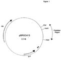
- This site comprises an ATG codon to give the initiating methionine required for the start of mRNA translation and places the gene an optimum distance from the Shine-Dalgarno ribosome binding sequence of pBROC413.
- the 3' end of the gene finished on two stop codons followed by a Hin dIII site.
- Restriction endonucleases that do not cut pBROC413 and that were commercially available were identified. The sequences of the restriction sites recognised by the endonucleases were translated into all three reading frames. The sites that contained rarely used codons for E. coli expression were discarded. The remaining sites were matched with the DNA coding for SCR 1 + 2. If the restriction site could be fitted into the DNA sequence so as to preserve the coding sequence and not add a rarely used codon, the DNA sequence was altered to include this restriction site. 10 unique restriction sites were so identified and incorporated. To enable intracellular expression of protein in E. coli , an ATG codon was added to the 5' end of the gene immediately preceding the codon for the first amino acid of mature CR-1.
- the codon ATG is part of the Nde I restriction site which can be used for cloning into vectors such as pBROC413.
- the codon corresponding to proline 124 of mature CR-1 has been changed to one encoding glutamine, which also encompasses an Eco RI site.
- the DNA coding for SCR 1+2+3 was constructed by ligating DNA coding for SCR 1+2 (Example 1a) to DNA encoding SCR 3.
- the SCR 3 coding unit corresponding to amino acid 122 and ending at amino acid 196 of mature CR1 was designed such that 5' end of the unit contained the Eco RI site at the junction of SCR's 2 & 3 as well as an Nde I site 5' to the Eco RI site. The 3' end of the unit finished on two stop codons followed by a Hin dIII site.
- the plasmids containing the SCR 3 coding unit and the SCR 1+2 coding unit were digested with Eco RI and Hin dIII.
- the SCR 3 coding unit was isolated and inserted downstream of the SCR 1+2 coding unit in the Eco RI/ Hin dIII-cut SCR 1+2 - containing plasmid, to give a plasmid containing the SCR 1+2+3 coding unit, which corresponds to amino acids 1 to 196 of mature CR1.
- zymosan A activated serum was carried out as described in Method (xvii). Different concentrations of SCR 1+2+3 (typically in the range 1 - 1000 ⁇ g/ml) were incubated with serum in the presence of zymosan A and the % inhibition of activation at each concentration was determined. From several different experiments the IC50 was determined as 20 -40 ⁇ g/ml indicating that SCR 1+2+3 can inhibit the alternative pathway of complement.
- a batch of final product SCR 1 + 2 + 3 was prepared essentially as described in Example 4 above and was measured for endotoxin content using a method based on the gel-clot reaction of limulus amoebocyte lysate (LAL) (Atlas Bioscan Ltd.). The sensitivity of the assay was 0.125 EU/ml and this was checked by titration against a doubling dilution series prepared from standard E. coli endotoxin supplied with the LAL kit.
- LAL limulus amoebocyte lysate
- the sequence for SCR 3 corresponding to amino acid 122 and ending at amino acid 196 of mature human complement receptor 1 was designed such that the 5' end of the gene contained an Nde I restriction endonuclease site.
- This site comprises an ATG start codon to give the initiating methionine required for the start of mRNA translation and allows the placement of the gene an optimum distance from the Shine-Dalgarno ribosome binding site of pBROC413.
- This codon is followed immediately by the gene coding for SCR 3 starting with arginine 122 of mature human complement receptor 1.
- the 3' end of the gene finishes with a codon for lysine 196 followed by two stop codons followed by a Hin dIII site.
- the DNA coding for SCR3 was modified for optimum codon usage in E. coli as described in the methods.
- the gene was also altered to incorporate unique restriction endonuclease sites. This was carried out in the following way. Restriction endonucleases that do not cut pBROC413 and were commercially available were identified. The DNA sequence of these restriction endonuclease sites was then translated into all three reading frames and the codon usage examined. Sites that contained codons that are rarely used by E. coli were discarded. The remaining sites were examined for their translated sequence and if that sequence matched with SCR 3, the restriction site was incorporated into the sequence.
- Two constructs were prepared by making a plasmid encoding SCR 1+2, incorporating SCR3 and finally adding SCR4.
- the two constructs encoded consensus SCR1 to 4 and the R235H mutation of SCR1 to 4 (Example 12).
- a plasmid containing the SCR 1 + 2 + 3 + 4 coding unit was constructed by adding the DNA encoding SCR 4 onto the construct coding for SCR 1 + 2 + 3 (Example 2).
- the SCR 4 DNA coding unit was made by synthesising the DNA encoding the last 17 amino acids of SCR 3 followed by the DNA coding for the linker region followed by SCR 4. This DNA started at the Spe I site of the SCR 1 + 2 + 3 coding construct which corresponds to T175 of mature CR-1 followed by the DNA coding for the linker region followed by SCR 4 ending on the codon corresponding to S253 followed by two stop codons and a Hind III site.
- the DNA encoding SCR 4 was altered for optimised codon usage and restriction sites as previously described in Example 1. This unit of DNA was ligated to the plasmid coding for SCR 1 + 2 + 3 which had been cut with Spe I and Hind III to give a construct coding for SCR 1 + 2 + 3 + 4.
Landscapes
- Health & Medical Sciences (AREA)
- Chemical & Material Sciences (AREA)
- Life Sciences & Earth Sciences (AREA)
- Organic Chemistry (AREA)
- Medicinal Chemistry (AREA)
- General Health & Medical Sciences (AREA)
- Immunology (AREA)
- Genetics & Genomics (AREA)
- Zoology (AREA)
- General Chemical & Material Sciences (AREA)
- Pharmacology & Pharmacy (AREA)
- Chemical Kinetics & Catalysis (AREA)
- Animal Behavior & Ethology (AREA)
- Proteomics, Peptides & Aminoacids (AREA)
- Public Health (AREA)
- Veterinary Medicine (AREA)
- Molecular Biology (AREA)
- Biophysics (AREA)
- Cell Biology (AREA)
- Toxicology (AREA)
- Nuclear Medicine, Radiotherapy & Molecular Imaging (AREA)
- Gastroenterology & Hepatology (AREA)
- Biochemistry (AREA)
- Rheumatology (AREA)
- Engineering & Computer Science (AREA)
- Pain & Pain Management (AREA)
- Bioinformatics & Cheminformatics (AREA)
- Medicines That Contain Protein Lipid Enzymes And Other Medicines (AREA)
- Preparation Of Compounds By Using Micro-Organisms (AREA)
- Peptides Or Proteins (AREA)
- Micro-Organisms Or Cultivation Processes Thereof (AREA)
- Saccharide Compounds (AREA)
Description
- The present invention relates to polypeptides and their use in the diagnosis and therapy of disorders involving complement activity and various inflammatory and immune disorders.
- Constituting about 10% of the globulins in normal serum, the complement system is composed of many different proteins that are important in the immune system's response to foreign antigens. The complement system becomes activated when its primary components are cleaved and the products alone or with other proteins, activate additional complement proteins resulting in a proteolytic cascade. Activation of the complement system leads to a variety of responses including increased vascular permeability, chemotaxis of phagocytic cells, activation of inflammatory cells, opsonization of foreign particles, direct killing of cells and tissue damage. Activation of the complement system may be triggered by antigen-antibody complexes (the classical pathway) or, for example, by lipopolysaccharides present in cell walls of pathogenic bacteria (the alternative pathway).
- Complement receptor type 1 (CR1) has been shown to be present on the membranes of erythrocytes, monocytes/macrophages, granulocytes, B cells, some T cells, splenic follicular dendritic cells, and glomerular podocytes. CR1 binds to the complement components C3b and C4b and has also been referred to as the C3b/C4b receptor. The structural organisation and primary sequence of one allotype of CR1 is known (Klickstein et al., 1987, J. Exp. Med. 165:1095-1112, Klickstein et al., 1988, J. Exp. Med. 168:1699-1717; Hourcade et al.,1988, J. Exp. Med. 168:1255-1270, WO 89/09220, WO 91/05047). It is composed of 30 short consensus repeats (SCRs) that each contain around 60-70 amino acids. In each SCR, around 29 of the average 65 amino acids are conserved. Each SCR has been proposed to form a three dimensional triple loop structure trough disulphide linkages with the third and first and the fourth and second half-cystines in disulphide bonds. CR1 is further arranged as 4 long homologous repeats (LHRs) of 7 SCRs each. Following a leader sequence, the CR1 molecule consists of the N-terminal LHR-A, the next two repeats, LHR-B and LHR-C, and the most C-terminal LHR-D followed by 2 additional SCRs, a 25 residue putative transmembrane region and a 43 residue cytoplasmic tail.
- Based on the mature CR1 molecule having a predicted N-terminal glutamine residue, hereinafter designated as residue 1, the first four SCR domains of LHR-A are defined herein as consisting of residues 2-58, 63-120, 125-191 and 197-252, respectively, of mature CR1.
- Hourcade et al.,1988, J. Exp. Med. 168:1255-1270 observed an alternative polyadenylation site in the human CR1 transcriptional unit that was predicted to produce a secreted form of CR1. The mRNA encoded by this truncated sequence comprises the first 8.5 SCRs of CR1, and encodes a protein of about 80 kDa which was proposed to include the C4b binding domain. When a cDNA corresponding to this truncated sequence was transfected into COS cells and expressed, it demonstrated the expected C4b binding activity but did not bind to C3b (Krych et al.,1989, FASEB J. 3:A368; Krych et al. Proc. Nat. Acad. Sci. 1991, 88, 4353-7). Krych et al., also observed a mRNA similar to the predicted one in several human cell lines and postulated that such a truncated soluble form of CR1 with C4b binding activity may be synthesised in humans.
- In addition, Makrides et al. (1992, J. Biol. Chem. 267 (34) 24754-61) have expressed SCR 1 + 2 and 1 + 2 + 3 + 4 of LHR-A as membrane-attached proteins in CHO cells.
- Several soluble fragments of CR1 have also been generated via recombinant DNA procedures by eliminating the transmembrane region from the DNAs being expressed (WO 89/09220, WO 91/05047). The soluble CR1 fragments were functionally active, bound C3b and/or C4b and demonstrated Factor I cofactor activity depending upon the regions they contained. Such constructs inhibited in vitro complement-related functions such as neutrophil oxidative burst, complement mediated hemolysis, and C3a and C5a production. A particular soluble construct, sCR1/pBSCR1c, also demonstrated in vivo activity in a reversed passive Arthus reaction (WO 89/09220, WO 91/05047; Yeh et al., 1991, J. Immunol. 146:250), suppressed post-ischemic myocardial inflammation and necrosis (WO 89/09220, WO 91/05047; Weisman et al., Science, 1990, 249:146-1511; Dupe, R. et al. Thrombosis & Haemostasis (1991) 65(6) 695.) and extended survival rates following transplantation (Pruitt & Bollinger, 1991, J. Surg. Res 50:350; Pruitt et al., 1991 Transplantation 52; 868). Furthermore, co-formulation of sCR1/pBSCR1c with p-anisoylated human plasminogen-streptokinase-activator complex (APSAC) resulted in similar anti-haemolytic activity as sCR1 alone, indicating that the combination of the complement inhibitor sCR1 with a thrombolytic agent was feasible (WO 91/05047).
- Soluble polypeptides corresponding to part of CR1 have now been found to possess functional complement inhibitory, including anti-haemolytic, activity.
- According to the present invention there is provided a soluble polypeptide comprising, in sequence, one to four short consensus repeats (SCR) selected from
SCR SCR - In preferred aspects, the above defined polypeptide comprises, in sequence,
SCR SCR - It is to be understood that variations in the amino acid sequence of the polypeptide of the invention by way of addition, deletion or conservative substitution of residues, including allelic variations, in which the biological activity of the polypeptide is retained, are encompassed by the invention. Conservative substitution is understood to mean the retention of the charge and size characteristics of the amino acid side chain, for example arginine replaced by histidine.
- In one aspect, the polypeptide of the invention may be represented symbolically as follows:
- In a preferred embodiment of formula (I), W1, X1 and Y1 represent residues 59-62, 121-124 and 192-196, respectively, of mature CR1 and V1 represents residue 1 of mature CR1 optionally linked via its N-terminus to methionine.
- In another aspect the polypeptide of the invention may be represented symbolically as follows:
- In preferred embodiments of formula (II), W2, X2, Y2 and Z2 represent residues 59-62, 121-124, 192-196, and residues 253 respectively, of mature CR1 and V2 represents residue 1 of mature CR1 optionally linked via its N-terminus to methionine.
- In one particular embodiment of formula (II) arginine 235 is replaced by histidine.
- In the preferred embodiment of formula (II), residue 235 is arginine.
- In one further aspect, the polypeptide of the invention may be represented symbolically as follows:
- In a preferred embodiment of formula (III) X3 represents amino acids 122-124 of mature CR1 optionally linked to methionine at its N-terminus and Y4 represents amino acids 192-196 of mature CR1.
- In another further aspect, the polypeptide of the invention may be represented symbolically as follows:
- In a preferred embodiment of formula (IV) X4 represents amino acids 122-124 of mature CR1 optionally linked to methionine at its N-terminus and Y4 and Z4 represent amino acids 192-196 and 253 respectively of mature CR1.
- In a further aspect, the invention provides a process for preparing a CR1 polypeptide according to the invention which process comprises expressing DNA encoding said polypeptide in a recombinant host cell and recovering the product.
- In particular, the process may comprise the steps of:
- i) preparing a replicable expression vector capable, in a host cell, of expressing a DNA polymer comprising a nucleotide sequence that encodes said polypeptide;
- ii) transforming a host cell with said vector,
- iii) culturing said transformed host cell under conditions permitting expression of said DNA polymer to produce said polypeptide; and
- iv) recovering said polypeptide.
-
- The DNA polymer encoding the polypeptide also forms part of the invention.
- The process of the invention may be performed by conventional recombinant techniques such as described in Sambrook et al., Molecular Cloning : A laboratory manual 2nd Edition. Cold Spring Harbor Laboratory Press (1989) and DNA Cloning vols I, II and III (D. M. Glover ed., IRL Press Ltd).
- The invention also provides a process for preparing the DNA polymer by the condensation of appropriate mono-, di- or oligomeric nucleotide units.
- The preparation may be carried out chemically, enzymatically, or by a combination of the two methods, in vitro or in vivo as appropriate. Thus, the DNA polymer may be prepared by the enzymatic ligation of appropriate DNA fragments, by conventional methods such as those described by D. M. Roberts et al., in Biochemistry 1985, 24, 5090-5098.
- The DNA fragments may be obtained by digestion of DNA containing the required sequences of nucleotides with appropriate restriction enzymes, by chemical synthesis, by enzymatic polymerisation, or by a combination of these methods.
- Digestion with restriction enzymes may be performed in an appropriate buffer at a temperature of 20°-70°C, generally in a volume of 50µl or less with 0.1-10µg DNA.
- Enzymatic polymerisation of DNA may be carried our in vitro using a DNA polymerase such as DNA polymerase 1 (Klenow fragment) in an appropriate buffer containing the nucleoside triphosphates dATP, dCTP, dGTP and dTTP as required at a temperature of 10°-37°C, generally in a volume of 50µl or less.
- Enzymatic ligation of DNA fragments may be carried out using a DNA ligase such as T4 DNA ligase in an appropriate buffer at a temperature of 4°C to 37°C, generally in a volume of 50µl or less.
- The chemical synthesis of the DNA polymer or fragments may be carried our by conventional phosphotriester, phosphite or phosphoramidite chemistry, using solid phase techniques such as those described in 'Chemical and Enzymatic Synthesis of Gene Fragments - A Laboratory Manual' (ed. H.G. Gassen and A. Lang), Verlag Chemie, Weinheim (1982), or in other scientific publications, for example M.J.Gait, H.W.D. Matthes M. Singh, B.S. Sproat and R.C. Titmas, Nucleic Acids Research, 1982, 10, 6243; B.S. Sproar and W. Bannwarth, Tetrahedron Letters, 1983, 24, 5771; M.D. Matteucci and M.H. Caruthers, Tetrahedron Letters, 1980, 21, 719; M.D. Matteucci and M.H. Caruthers, Journal of the American Chemical Society, 1981, 103, 3185; S.P. Adams et al., Journal of the American Chemical Society, 1983, 105, 661; N.D. Sinha, J. Biernat, J. McMannus and H. Koester, Nucleic Acids Research, 1984, 12, 4539; and H.W.D. Matthes et al., EMBO Journal, 1984, 3, 801. Preferably an automated DNA synthesiser (for example, Applied Biosysrems 381A Synthesiser) is employed.
- The DNA polymer is preferably prepared by ligating two or more DNA molecules which together comprise a DNA sequence encoding the polypeptide.
- The DNA molecules may be obtained by the digestion with suitable restriction enzymes of vectors carrying the required coding sequences.
- The precise structure of the DNA molecules and the way in which they are obtained depends upon the structure of the desired product. The design of a suitable strategy for the construction of the DNA molecule coding for the polypeptide is a routine matter for the skilled worker in the art.
- In particular, consideration may be given to the codon usage of the particular host cell. The codons may be optimised for high level expression in E. coli using the principles set out in Devereux et al., (1984) Nucl. Acid Res., 12, 387.
- The expression of the DNA polymer encoding the polypeptide in a recombinant host cell may be carried out by means of a replicable expression vector capable, in the host cell, of expressing the DNA polymer. The expression vector is novel and also forms part of the invention.
- The replicable expression vector may be prepared in accordance with the invention, by cleaving a vector compatible with the host cell to provide a linear DNA segment having an intact replicon, and combining said linear segment with one or more DNA molecules which, together with said linear segment, encode the polypeptide, under ligating conditions.
- The ligation of the linear segment and more than one DNA molecule may be carried out simultaneously or sequentially as desired.
- Thus, the DNA polymer may be preformed or formed during the construction of the vector, as desired. The choice of vector will be determined in part by the host cell, which may be prokaryotic, such as E. coli, or eukaryotic, such as mouse C127, mouse mycloma, chinese hamster ovary, fungi e.g. filamentous fungi or unicellular 'yeast' or an insect cell such as Drosophila. The host cell may also be in a transgenic animal. Suitable vectors include plasmids, bacteriophages, cosmids and recombinant viruses derived from, for example, baculoviruses or vaccinia.
- The DNA polymer may be assembled into vectors designed for isolation of stable transformed mammalian cell lines expressing the fragment e.g. bovine papillomavirus vectors in mouse C127 cells, or amplified vectors in chinese hamster ovary cells (DNA Cloning Vol. II D.M. Glover ed. IRL Press 1985; Kaufman, R.J. et al.. Molecular and Cellular Biology 5, 1750-1759, 1985; Pavlakis G.N. and Hamer, D.H. Proceedings of the National Academy of Sciences (USA) 80, 397-401, 1983; Goeddel, D.V. et al.,European Patent Application No. 0093619, 1983).
- The preparation of the replicable expression vector may be carried out conventionally with appropriate enzymes for restriction, polymerisation and ligation of the DNA, by procedures described in, for example, Sambrook et al.,cited above. Polymerisation and ligation may be performed as described above for the preparation of the DNA polymer. Digestion with restriction enzymes may be performed in an appropriate buffer at a temperature of 20°-70°C, generally in a volume of 50µl or less with 0.1-10µg DNA.
- The recombinant host cell is prepared, in accordance with the invention, by transforming a host cell with a replicable expression vector of the invention under transforming conditions. Suitable transforming conditions are conventional and are described in, for example, Sambrook et al., cited above, or "DNA Cloning" Vol. II, D.M. Glover ed., IRL Press Ltd, 1985.
- The choice of transforming conditions is determined by the host cell. Thus, a bacterial host such as E.coli, may be treated with a solution of CaCl2 (Cohen et al.,Proc. Nat. Acad. Sci., 1973, 69, 2110) or with a solution comprising a mixture of RbCl, MnCl2, potassium acetate and glycerol, and then with 3-[N-morpholino]-propane-sulphonic acid, RbCl and glycerol or by electroporation as for example described by Bio-Rad Laboratories, Richmond, California, USA, manufacturers of an electroporator. Mammalian cells in culture may be transformed by calcium co-precipitation of the vector DNA onto the cells or by using cationic liposomes.
- The invention also extends to a host cell transformed with a replicable expression vector of the invention.
- Culturing the transformed host cell under conditions permitting expression of the DNA polymer is carried out conventionally, as described in, for example, Sambrook et al., and "DNA Cloning" cited above. Thus, preferably the cell is supplied with nutrient and cultured at a temperature below 45°C.
- The protein product is recovered by conventional methods according to the host cell. Thus, where the host cell is bacterial such as E. coli and the protein is expressed intracellularly, it may be lysed physically, chemically or enzymatically and the protein product isolated from the resulting lysate. Where the host cell is mammalian, the product is usually isolated from the nutrient medium.
- Where the host cell is bacterial, such as E. coli, the product obtained from the culture may require folding for optimum functional activity. This is most likely if the protein is expressed as inclusion bodies. There are a number of aspects of the isolation and folding process that are regarded as important. In particular, the polypeptide is preferably partially purified before folding, in order to minimise formation of aggregates with contaminating proteins and minimise misfolding of the polypeptide. Thus, the removal of contaminating E. coli proteins by specifically isolating the inclusion bodies and the subsequent additional purification prior to folding are important aspects of the procedure.
- The folding process is carried out in such a way as to minimise aggregation of intermediate-folded states of the polypeptide. Thus, careful consideration needs to be given to, among others, the salt type and concentration, temperature, protein concentration, redox buffer concentrations and duration of folding. The exact condition for any given polypeptide generally cannot be predicted and must be determined by experiment.
- There axe numerous methods available for the folding of proteins from inclusion bodies and these are known to the skilled worker in this field. The methods generally involve breaking all the disulphide bonds in the inclusion body, for example with 50mM 2-mercaptoethanol, in the presence of a high concentration of denaturant such as 8M urea or 6M guanidine hydrochloride. The next step is to remove these agents to allow folding of the proteins to occur. Formation of the disulphide bridges requires an oxidising environment and this may be provided in a number of ways, for example by air, or by incorporating a suitable redox system, for example a mixture of reduced and oxidised gluzathione.
- Preferably, the inclusion body is solubilised using 8M urea, in the presence of mercaptoethanol, and protein is folded, after initial removal of contaminating proteins, by addition of cold buffer. A preferred buffer is 20mM ethanolamine containing 1mM reduced glutathione and 0.5mM oxidised glutathione. The folding is preferably carried out at a temperature in the range 1 to 5°C over a period of 1 to 4 days.
- If any precipitation or aggregation is observed, the aggregated protein can be removed in a number of ways, for example by centrifugation or by treatment with precipicants such as ammonium sulphate. Where either of these procedures are adopted, monomeric polypeptide is the major soluble product.
- If the bacterial cell secretes the protein, folding is not usually necessary.
- The polypeptide of this invention is useful in the treatment or diagnosis of many complement-mediated or complement-related diseases and disorders including, but not limited to, those listed below.
-
- multiple sclerosis
- stroke
- Guillain Barré Syndrome
- traumatic brain injury
- Parkinson's disease
- allergic encephalitis
- Alzheimer's disease
-
-
- haemodialysis complications
- hyperacute allograft rejection
- xenograft rejection
- corneal graft rejection
- interleukin-2 induced toxicity during IL-2 therapy
- paroxysmal nocturnal haemoglobinuria
-
-
- inflammation of autoimmune diseases
- Crohn's Disease
- adult respiratory distress syndrome
- thermal injury including burns or frostbite
- uveitis
- psorasis
- asthma
- acute pancreatitis
-
-
- myocardial infarction
- balloon angioplasty
- atherosclerosis (cholesterol-induced) & restenosis
- hypertension
- post-pump syndrome in cardiopulmonary bypass or renal haemodialysis
- renal ischemia
- intestinal ischaemia
-
-
- multiple organ failure
- septic shock
-
-
- rheumatoid arthritis
- systemic lupus erythematosus (SLE)
- SLE nephritis
- proliferative nephritis
- glomerulonephritis
- haemolytic anemia
- myasthenia gravis
-
-
- antibody- or complement-mediated infertility
-
- The present invention is also directed to a pharmaceutical composition comprising a therapeutically effective amount of a polypeptide, as above, and a pharmaceutically acceptable carrier or excipient.
- The present invention also provides a method of treating a disease or disorder associated with inflammation or inappropriate complement activation comprising administering to a subject in need of such treatment a therapeutically effective amount of a polypeptide of this invention.
- In the above methods, the subject is preferably a human.
- An effective amount of the polypeptide for the treatment of a disease or disorder is in the dose range of 0.01-100mg/kg; preferably 0.1mg-10mg/kg.
- For administration, the polypeptide should be formulated into an appropriate pharmaceutical or therapeutic composition. Such a composition typically contains a therapeutically active amount of the polypeptide and a pharmaceutically acceptable excipient or carrier such as saline, buffered saline, dextrose, or water. Compositions may also comprise specific stabilising agents such as sugars, including mannose and mannitol, and local anaesthetics for injectable compositions, including, for example, lidocaine.
- Further provided is the use of a polypeptide of this invention in the manufacture of a medicament for the treatment of a disease or disorder associated with inflammation or inappropriate complement activation.
- In order to inhibit complement activation and, at the same time, provide thrombolytic therapy, the present invention provides compositions which further comprise a therapeutically active amount of a thrombolytic agent. An effective amount of a thrombolytic agent is in the dose range of 0.01-10mg/kg; preferably 0.1-5mg/kg. Preferred thrombolytic agents include, but are not limited to, streptokinase, human tissue type plasminogen activator and urokinase molecules and derivatives, fragments or conjugates thereof. The thrombolytic agents may comprise one or more chains that may be fused or reversibly linked to other agents to form hybrid molecules (EP-A-0297882 and EP 155387), such as, for example, urokinase linked to plasmin (EP-A-0152736), a fibrinolytic enzyme linked to a water-soluble polymer (EP-A-0183503). The thrombolytic agents may also comprise muteins of plasminogen activators (EP-A-0207589). In a preferred embodiment, the thrombolytic agent may comprise a reversibly blocked in vitro fibrinolytic enzyme as described in U.S. Patent No. 4,285,932. A most preferred enzyme is a p-anisoyl plasminogen-streptokinase activator complex as described in U.S. Patent No. 4,808,405, and marketed by SmithKline Beecham Pharmaceuticals under the Trademark EMINASE (generic name anistreplase, also referred to as APSAC; Monk et al.,1987, Drugs 34:25-49).
- Routes of administration for the individual or combined therapeutic compositions of the present invention include standard mutes, such as, for example, intravenous infusion or bolus injection. Active complement inhibitors and thrombolytic agents may be administered together or sequentially, in any order.
- The present invention also provides a method for treating a thrombotic condition, in particular acute myocardial infarction, in a human or non-human animal. This method comprises administering to a human or animal in need of this treatment an effective amount of a polypeptide according to this invention and an effective amount of a thrombolytic agent.
- Also provided is the use of a polypeptide of this invention and a thrombolytic agent in the manufacture of a medicament for the treatment of a thrombotic condition in a human or animal. Such methods and uses may be carried out as described in WO 91/05047.
- This invention further provides a method for treating adult respiratory distress syndrome (ARDS) in a human or non-human animal. This method comprises administering to the patient an effective amount of a polypeptide according to this invention.
- The invention also provides a method of delaying hyperacute allograft or hyperacute xenograft rejection in a human or non-human animal which receives a transplant by administering an effective amount of a polypeptide according to this invention. Such administration may be to the patient or by application to the transplant prior to implantation.
- The invention yet further provides a method of treating wounds in a human or non-human animal by administering by either topical or parenteral e.g. intravenous mutes, an effective amount of a polypeptide according to this invention.
-
- (i) DNA cleavage Cleavage of DNA by restriction endonucleases was carried out according to the manufacturer's instructions using supplied buffers. Double digests were carried out simultaneously if the buffer conditions were suitable for both enzymes. Otherwise double digests were carried out sequentially where enzyme requiring the lowest salt concentration was added first to the digest. Once that digest was complete the salt concentration was altered and the second enzyme added.
- (ii) Production of blunt ended DNA fragments The recessed 3' termini of DNA fragments were filled in using the Klenow fragment of DNA polymerase I as described in Sambrook et al (1989).
- (iii) DNA purification/ concentration and analysis Removal of protein contaminants, nucleosides was with phenol/CHCl3 followed by precipitation with ethanol. DNA was analysed on horizontal agarose gel electrophoresis: both methods are described in Sambrook et al (1989).
- (iv) DNA fragment isolation
- 1. DNA purification on DEAE NA45 membranes DNA fragments were purified from agarose gels by making an incision in the agarose above and just below the required DNA fragment. NA45 membranes from Schleicher & Schuell (Anderman, Great Britain) that had been soaked in TE (10 mM Tris pH 8.0, 1 mM EDTA) were inserted into the incisions and current reapplied to the gel until the DNA fragment was trapped on the lower membrane; higher molecular weight DNA was trapped on the upper membrane. The lower membrane was removed from the gel and the DNA eluted into 0.05 M arginine/1 M NaCl at 70°C for 2 hours. The DNA was then concentrated by ethanol precipitation as described in Sambrook et al (1989).
- 2. Electroelution DNA fragments were excised from agarose gels and DNA extracted by electroelution using the Unidirectional Electroeluter (IBI Ltd.,Cambridge, England) according to the manufacturer's instructions.
- 3. Gel purification DNA fragments were excised from agarose gels and DNA extracted using the QIAEX gel extraction kit according to the manufacturers instructions (QIAGEN Inc., USA).
- (v) Plasmid preparation Large scale plasmid preparation of plasmid DNA was carried out using CsCl as described in Sambrook et al (1989) or using Magic Maxipreps (Promega Corporation, Madison, USA) according to the manufacturers instructions. Mini-plasmid preparations were carried out using either the alkaline lysis method described in Sambrook et al (1989) or Magic Minipreps (Promega Corporation, Madison, USA) according to the manufacturer's instructions.
- (vi) Introduction of plasmid DNA into E. coli
- 1. Plasmids were transformed into E. coli HB101 or E. coli BL21 (DE3) (Studier and Moffat, 1986) that had been made competent using calcium chloride as described in Sambrook et al (1989).
- 2. Alternatively plasmids were introduced into E.coli DH1 (Low,1968) or E.coli BL21 (DE3) by electroporation using the Gene Pulsar and Pulse Controller of Bio-Rad (Bio-Rad Laboratories, Richmond, California,USA) according to the manufacturer's instructions.
- (vii) Kinasing of oligonucleotides Oligonucleotides or annealed oligonucleotides possessing 5' overhangs were kinased using T4 polynucleotide kinase as described in Sambrook et al (1989).
- (viii) Annealing and ligation of oligonucleotides Oligonucleotides were annealed together by mixing generally equimolar concentrations of the complementary oligonucleotides in 10 mM Tris pH 8.5, 5 mM MgCl2 and placing at 100°C for 5 minutes and then cooling very slowly to room temperature. Annealed oligonucleotides with sticky ends were ligated to vector or other oligonucleotides containing complementary sticky ends using T4 DNA ligase as described in Sambrook et al (1989).
- (ix) PCR (Polymerase Chain Reaction) amplification of DNA
DNA fragments from ligation reactions or DNA fragments excised and
purified from agarose gels were amplified by PCR from two primers complementary
to the 5' ends of the DNA fragment. Approximately 0.1 - 1µg of ligation reaction or
the purified DNA from the agarose gel was mixed in 10 mM Tris pH 8.3 (at 25°C),
50 mM KCl, 0.1% gelatin; MgCl2 concentrations were varied from 1.5 mM to 6 mM
to find a suitable concentration for each reaction. Both primers were added to a
final concentration of 2 µM; each dNTP was added to a final concentration of 0.2
mM. The final reaction volume was either 75 µl or 100 µl, which was overlayed with
mineral oil to prevent evaporation. Thermal cycling was then started on a thermal
cycler eg. Hybaid Thermal reactor, and a typical example of conditions used was
94°C 7 mins, 45°
C 2 mins, hold at 45°C for less than 5 mm., and then add 5 units of Taq DNA polymerase (purchased from a commercial source, e.g. Gibco). The DNA fragment was amplified by cycling the temperature at 72°C 2 mins, 94°C 1 mm and 45°C 2 min a total of 35 times. - (x) DNA sequencing using the double stranded method Sequencing was carried out using "Sequenase™" (United States Biochemical Corporation) essentially as described in the manufacturer's instructions.
- (xi) DNA sequence analysis and manipulation Analysis of sequences were carried out on a digital VAX computer using the GCG package of programmes as described in Devereux et al (1984).
- (xii) Production of oligonucleotides
- 1. Oligonucleotides were synthesised using a Gene Assembler Plus (Pharmacia LKB Biotechnology, Milton Keynes, England) or a 381A Synthesiser (Applied BioSystems) according to the manufacturer's instructions.
- 2. Oligonucleotide purification was carried out either using MonoQ as recommended by Pharmacia or by UV shadowing where recovery of synthetic oligonucleotides was by electrophoresis through a denaturing polyacrylamide gel. The oligonucleotides were loaded onto a 12% acrylamide/7M urea gel and run at 1500V until the oligonucleotide had migrated approximately two thirds of the length of the gel. The DNA was visualised using a hand-held, long-wavelength ultraviolet lamp; and the DNA bands excised. The oligonucleotide was recovered using Sep-Pak C18 reverse phase columns (Waters) as described in Sambrook et al (1989).
- (xiii) Sodium dodecyl sulphate polyacrylamide gel electrophoresis (SDS PAGE) SDS PAGE was carried out generally using the Novex system (British Biotechnology) according to the manufacturer's instructions. Prepacked gels of acrylamide concentrations 14%, 16%, 4 - 20% or 10 - 27% were the ones most frequently used. Samples for electrophoresis, including protein molecular weight standards (LMW Kit, Pharmacia) welt usually diluted in 1%(w/v)SDS - containing buffer (with or without 5%(v/v) 2-mercaptoethanol), and left at room temperature for about 0.5 to 1 h before application to the gel.
- (xiv) Alteration of codon usage The non random use of synonymous codons has been demonstrated in E. coli and there is some evidence to support the belief that protein production from genes containing non-optimal or minor codons (particularly at the 5' end of the gene) is less efficient than that from genes with no such codons (e.g. Chen an Inouye, 1990). A codon usage table compiled from genes highly expressed in E. coli (supplied as part of the GCG sequence analysis software package, Devereux et al, (1984)) was used to determine the optimal codons for expression in E. coli. All of the first 30 codons of all constructs (where compatible with restriction enzyme sites) were optimised for high level expression. The codons for the seven amino acids: arg, gly, ile, leu, pro, ser, ala were optimised (where compatible with restriction enzyme sites) throughout the coding sequence.
- (xv) Construction of vector pBROC413 The plasmid pT7-7 (Tabor, 1990) contains DNA corresponding to nucleotides 2065-4362 of pBR322 and like pBR322 can be mobilized by a conjugative plasmid in the presence of a third plasmid ColK. A mobility protein encoded by ColK acts on the nic site at nucleotide 2254 of pBR322 initiating mobilization from this point. pT7-7 was digested with LspI and BglII and the protruding 5' ends filled in with the Klenow fragment of DNA Polymerase I. The plasmid DNA fragment was purified by agarose gel electrophoresis, the blunt ends ligated together and transformed into E. coli DH1 by electroporation. The resultant plasmid pBROC413 (Fig.1) was identified by restriction enzyme analysis of plasmid DNA.The deletion in pBROC413 from the LspI site immediately upstream of the 10 promoter to the BglII site at nucleotide 434 of pT7-7 deletes the DNA corresponding to nucleotides 2065-2297 of pBR322. The nic site and adjacent sequences are therefore deleted making pBROC413 non mobilizable.
- (xvi) Haemolytic assay The anti-haemolytic activity of polypeptides was assessed by measuring the inhibition of complement mediated lysis of sheep erythrocytes sensitised with rabbit antibodies (obtained from Diamedix Corporation, Miami, USA). Human serum diluted 1:125 in 0.1 M Hepes/ 0.15 M NaCl pH 7.4 buffer was the source of complement and was prepared from a pool of volunteers essentially as described in (Dacie & Lewis, 1975). Briefly, blood was warmed to 37°C for 5 minutes, the clot removed and the remaining serum clarified by centrifugation. The serum fraction was split into small aliquots and stored at -196°C. Aliquots were thawed as required and diluted in the Hepes buffer immediately before use.Inhibition of complement-mediated lysis of sensitised sheep erythrocytes was measured using a standard haemolytic assay using a v-bottom microtitre plate format as follows.50 µl of a range of concentrations (0.01-100µg/ml but typically 0.05 - 25 µg/ml) of test protein diluted in Hepes buffer were incubated with 50 µl of the diluted serum for 15 minutes at 37°C. 100 µl of prewarmed sensitised sheep erythrocytes were added for 1 hour at 37°C in a final reaction volume of 200 µl. Samples were spun at 300g at 4°C for 15 minutes before transferring 150 µl of supernatant to flat bottomed microtitre plates and determining the absorption at 410 nm, which reflects the amount of lysis in each test solution. Maximum lysis was determined by incubating serum with erythrocytes in the absence of any inhibitor from which the proportion of background lysis had been subtracted (determined by incubating erythrocytes with buffer). The background lysis by inhibitor was assessed by incubating inhibitor with erythrocytes and then subtracting that from test samples. Inhibition was expressed as a fraction of the total cell lysis such that IH50 represents the concentration of inhibitor required to give 50% inhibition of lysis.
- (xvii) C3a RIA Assay Activation of complement pathways can be followed by measuring the release of the anaphylatoxin, C3a and its breakdown product C3a des Arg. Both products can be measured using a competitive radio-immuno assay purchased from Amersham International plc, U. K., (human complement C3a des Arg [125I]assay, code RPA 518).
- (a) Alternative pathway activation by Zymosan A
The alternative pathway of complement was activated with zymosan A, a
complex carbohydrate from yeast (Sigma, catalogue number Z-4250). Zymosan A
was made 50 mg/ml in Hepes buffer (0.1M Hepes/0.15M NaCl pH 7.4) or in PBS (50
mM sodium phosphate/0.1 M NaCl pH 7.4) and vortexed until a fine suspension had
formed. Serum (prepared as described for the haemolytic assay; Method xvi) was
preincubated with different concentrations of complement inhibitor diluted in Hepes
buffer for 15 mins at 37°C using the volumes given below. Zymosan A was then
vortexed for a few seconds each time before addition to the samples after which
samples were incubated for a further 30 mins at 37°C. The zymosan A was then
spun down at approximately 11,000g for 30 seconds at ambient temperature.
Typically 100 µl of supernatant were added to an equal volume of precipitating
solution provided in the kit and the subsequent supernatant assayed as described in the
technical bulletin supplied by Amersham with the C3a des Arg assay RIA kit. Each
sample was assayed in duplicate and useful dilutions of the supernatant, to ensure that
sample counts were on the standard curve, were found to be 1/50 - 1/100. EDTA or
Futhan were not used in any solutions or tubes as suggested in the technical bulletin.Each sample was counted for 1 minute on an LKB-Wallac 1272 Clinigamma.
Data was processed using the RiaCalc program for RIA assays as supplied with the
Clinigamma. The data was computed essentially as described in the Amersham
technical bulletin with the standrd curve constructed by non-linear regression fit to
the data.The miniaturised assay was carried out essentially as described above but
using smaller total volumes for the activation of serum.Volumes of samples added
serum inhibitor Zymosan A Normal Assay 79 µl 20 µl 21 µl Miniaturised Assay 26.3 µl 6.7 µl 7 µl - (b) Classical pathway activation by heat aggregated IgG Activation of the classical pathway by IgG was performed as follows. Human γ-globulin (Sigma, catalogue number G-4386) was made 14 mg/ml in 0.1 M Hepes/0.15 M NaCl pH 7.4 and heated at 60°C for 1 hour. Samples of heat aggregated IgG were then stored as small aliquots at -80°C until required. Serum was activated using heat aggregated IgG using the same volumes as described for the zymosan A normal or miniaturised assay. Preincubation of inhibitor with serum was for 15 mins at 37°C followed by addition of the heat aggregated IgG. Incubation was continued for a further 45 mins at 37°C. The samples were then assayed directly for C3a levels using either the normal or miniaturised assay. (xviii) C5a RIA AssayActivation of complement pathways can be followed by measuring the release of the anaphylatoxin C5a and its breakdown product C5a des Arg. Both products can be measured using a competitive radio-immuno assay purchased from Amersham International plc, U. K., (human complement C5a des Arg [125I]assay, code RPA 520).The alternative pathway of complement was activated with zymosan A, as described for the C3a RIA assay (Method (xvii)). The assay was carried out in the miniaturised form as described for the C3a assay using the reagents provided in the C5a des-Arg RIA kit.
-
- A DNA sequence for SCR 1 + 2 corresponding to amino acid 1 and ending at amino acid 124 of mature human complement receptor 1 was designed such that the 5' end of the gene contained an NdeI site. This site comprises an ATG codon to give the initiating methionine required for the start of mRNA translation and places the gene an optimum distance from the Shine-Dalgarno ribosome binding sequence of pBROC413. The 3' end of the gene finished on two stop codons followed by a HindIII site.
- Restriction endonucleases that do not cut pBROC413 and that were commercially available were identified. The sequences of the restriction sites recognised by the endonucleases were translated into all three reading frames. The sites that contained rarely used codons for E. coli expression were discarded. The remaining sites were matched with the DNA coding for SCR 1 + 2. If the restriction site could be fitted into the DNA sequence so as to preserve the coding sequence and not add a rarely used codon, the DNA sequence was altered to include this restriction site. 10 unique restriction sites were so identified and incorporated. To enable intracellular expression of protein in E. coli, an ATG codon was added to the 5' end of the gene immediately preceding the codon for the first amino acid of mature CR-1. The codon ATG is part of the NdeI restriction site which can be used for cloning into vectors such as pBROC413. The codon corresponding to proline 124 of mature CR-1 has been changed to one encoding glutamine, which also encompasses an EcoRI site.
- (a) Construction of plasmid
Oligonucleotides coding for SCR 1 + 2 (Table 1; 1 - 8) were synthesised as 4
complementary pairs of 87 - 101 mers that could be ligated in a unique fashion via
complementary 8 bp overhangs between the pairs of oligonucleotides. The four
complementary pairs of oligonucleotides were designated Pair A (oligos 1+2) , Pair B
(
oligos 3+4), Pair C (oligos 5+6) and Pair D (oligos 7+8). Pair A which corresponded to the 5' end of the gene contained an NdeI restriction site overhang and Pair D contained a HindIII restriction site overhang at the 3' end. All oligonucleotides apart from 1 and 2 were purified on Pharmacia Mono Q columns prior to use.Oligonucleotide 2 of pair A and oligonucleotide 7 of pair D were kinased before annealing with their unkinased complementary oligonucleotides 1 and 8 respectively. Oligonucleotides pairs B and C were annealed first and then kinased. The kinased oligonuleotide pairs were ligated Pair A (approx. 0.1µg) to Pair B (approx. 0.2µg) and Pair C (approx. 2µg) to Pair D (approx. 4µg). The ligated oligonucleotides (A+B) were in turn ligated to (C+D) to form the gene coding for SCR 1 + 2.The DNA coding for SCR 1 + 2 was amplified by PCR using two oligonucleotides (Table 1; 15 and 16) complementary to the two strands of DNA. Both oligonucleotides contained 5' unmatched ends that contained 6 bp of random sequence followed by the sequence of either NdeI or HindIII restriction sites followed by 18 bp complementary to the gene. Following PCR, a band of approximately 400 bp was visualised on horizontal agarose gel electrophoresis, which was excised and purified on DEAE NA45 membranes. The DNA was then cut with NdeI and HindIII before ligating into pBROC413 that had been cut with the same enzymes. The vector was transformed into E. coli HB101 made competent with calcium chloride. Mini-plasmid preparations were made and the plasmid DNA was analysed by digestion with NdeI and HindIII. Plasmids containing the correct sized insert, were further subjected to restriction mapping with EcoRI, HpaI, KpnI and SmaI. The plasmids that displayed the correct restriction maps were analysed by DNA sequencing of both strands across the gene coding for SCR 1 + 2. Plasmid pDB1010-D11 was identified as having the correct sequence across the gene coding for SCR 1 + 2. -
- The DNA coding for SCR 1+2+3 was constructed by ligating DNA coding for SCR 1+2 (Example 1a) to
DNA encoding SCR 3. - General points relating to
SCR 3 are presented in Example 9. - The
SCR 3 coding unit corresponding to amino acid 122 and ending at amino acid 196 of mature CR1, was designed such that 5' end of the unit contained the EcoRI site at the junction of SCR's 2 & 3 as well as an NdeI site 5' to the EcoRI site. The 3' end of the unit finished on two stop codons followed by a HindIII site. The plasmids containing theSCR 3 coding unit and the SCR 1+2 coding unit were digested with EcoRI and HindIII. TheSCR 3 coding unit was isolated and inserted downstream of the SCR 1+2 coding unit in the EcoRI/HindIII-cut SCR 1+2 - containing plasmid, to give a plasmid containing the SCR 1+2+3 coding unit, which corresponds to amino acids 1 to 196 of mature CR1. The addition of theSCR 3 coding unit through the EcoRI site, converts the codon corresponding to a glutamine at position 124 back to the authentic amino acid (proline) that is found in CR1. - (a) Construction of plasmid pDB1013-5-4 encoding SCR 1 + 2 + 3
Three pairs of oligonucleotides (Table 1; 9 - 14) encompassing the SCR3
coding sequence were synthesised. The oligonucleotides were first annealed as pairs
(9, 10; 11,12; 13,14) and the middle pair kinased thus allowing the three pairs to be
ligated together via 8 base pair overlapping sequences. The 5' end of this trimeric
molecule was designed to be complementary to NdeI digested DNA and the 3' end to
HindIII digested DNA. This enabled the trimer to be cloned into NdeI/HindIII
digested pBROC413 generating pBROC435 (Fig.2). The identity of pBROC435 was
checked by restriction enzyme analysis and confirmed by DNA sequencing.Plasmid DNA from pBROC435 and pDB1010-D11 (Example 1) were both
cut with EcoRI and HindIII; the EcoRI/HindIII band of pBROC435 coding for
SCR 3 was purified on an DEAE NA45 membrane as was the cut vector pDB1010-D11. TheSCR 3 coding unit was then ligated into pDB1010-D11 to generate pDB1013-5 which was then transformed into calcium chloride competent E. coli HB101. The resulting colonies were analysed by mini-plasmid preparation of DNA followed by restriction mapping. One of the colonies, termed pDB1013-5-4 (Fig.2), contained the SCR 1+2+3 coding unit. This plasmid was then analysed for expression of the gene product. - (b) Expression of SCR 1 + 2 + 3 pDB1013-5-4 was transformed into calcium chloride competent E.coli BL21(DE3) and resulting colonies analysed by restriction digestion of mini-plasmid DNA preparations. Single colonies were inoculated into universals containing 10 ml of L broth or NCYZM medium and 50 µg/ml ampicillin and allowed to grow overnight at 37°C, 220 r.p.m. The overnight cultures (typically 5ml) were used to inoculate each of 2 L conical flasks containing 500 mls of NCYZM medium, 150 µg/ml ampicillin; cultures were grown at 37°C, 220 r.p.m. until A600 was 0.5 absorbance units. Cultures were induced with 1 mM isopropylthioβ-D-galactoside (IPTG) and allowed to grow a further 3 hours under the same conditions. The cultures were centrifuged (approx. 8000g/10 min) and the supernatants discarded. The cell pellets were stored at -40°C. L broth was 1% (w/v) Bactotryptone, 0.5% (w/v) Bactoyeastextract, 0.5% (w/v) NaCl. NCYZM media was L-broth containing 0.1% (w/v) casamino acids and 0.2%(w/v) MgSO4.7H2O, pH 7.0.
- (c) Isolation of solubilised inclusion bodies Frozen cell pellet of E.coli BL21 DE3 (pDB1013-5-4) (1 litre culture) prepared in a similar way to that described in Example 2b. was allowed to thaw at 4°C for 2 h and was then resuspended in 50 mM Tris/50 mM NaCl/1 mM EDTA/0.1 mM PMSF pH 8.0 (33 ml). The suspension was transferred to a 100 ml glass beaker and sonicated (Heat Systems - Ultrasonics W380; 70 Watts, 50 x 50% pulse, pulse time =5 sec.). The sonicate was immediately centrifuged (6000g/4°C/10 min) and the supernatant was discarded. The pellet, containing the inclusion bodies, was resuspended in 20 mM Tris/8 M urea/50 mM 2-mercaptoethanol/l mM EDTA/0.1 mM PMSF pH 8.5 (100 ml) and left static at room temperature (approx. 23°C) for 1 h. The resulting solution was centrifuged (approx. 2000 g at 4°C for 10 min) to remove any material that had failed to solubilise. The supernatant of this spin was retained at -40°C as the solubilised inclusion body product.
- (d) Purification of SCR 1 + 2 + 3 from the solubilised inclusion body A column (i.d., 16 mm; h, 10 mm) of S-Sepharose Fast Flow was prepared and connected into an FPLC (Pharmacia) system. The column was equilibrated with 20 mM Tris/8M urea/1 mM EDTA/50 mM 2-mercaptoethanol pH 8.5. 10 ml of thawed, solubilised inclusion body, prepared as described in Example 2c, was applied to the column and washed through with equilibration buffer. The column was then developed with a liner gradient to 1M NaCl (in equilibration buffer) followed by rinses with 1M NaCl and 2M NaCl (also in equilibration buffer). All the chromatography was at 1.0 ml min -1 and at room temperature.Analysis by SDS PAGE/protein staining of the fractions collected during the chromatography indicated that virtually all the SCR 1 + 2 + 3 polypeptide had absorbed to the column and had been dissociated by the 1M NaCl - containing buffer. The appropriate fractions were stored at -40°C.Subsequent assay for protein content of the peak fraction using the Bradford protein assay and a bovine serum albumin standard showed it contained 2.8 mg protein.
- (e) Folding S-Sepharose - purified SCR 1 + 2 + 3 that had been purified in a similar way to that described in Example 2d and stored at -40°C was thawed and 0.4 ml buffer-exchanged into 0.05 M formic acid using Sephaex G25 (P10). The absorbance at 280 nm of the buffer-exchanged solution was determined as 0.52, and, using ∈ = 34000 and appropriate correction factors for dilution, the protein concentration of the original preparation (prior to buffer-exchange) was calculated to be 0.6 mg/ml.Based on this figure, 1.7 ml S-Sepharose - purified protein was diluted with 0.85 ml 20 mM Tris/8M urea/50 mM 2-mercaptoethanol/1M NaCl pH 8.5 to give a 0.4 mg/ml solution, on which the folding was carried outFolding was effected by a series of dilutions, using cold diluent at all times.At t=0 h, 0.8 ml SCR1 + 2 + 3 (0.4 mg/ml) was added to 0.8 ml 20 mM Tris/1M urea/5 mM EDTA/3 mM 2-mercaptoethanol pH 8.0 ('diluent') in a 30 ml polystyrene universal container. The solution was mixed thoroughly by gentle swirling and left static, capped, in a cold room (approx. 2 to 3°C).At 1 h, 1.6 ml diluent was added and mixed.At 2 h, 3.2 ml diluent was added and mixed.At 4 h, 6.4 ml diluent was added and mixed.The solution was left a further 20 h in the cold room, then ultrafiltered (YM5, Amicon Ltd) to approx. 1.4 ml. This was buffer-exchanged into 0.1M NH4HCO3 (2.5 ml) using Sephadex G25 (PD 10) in the cold room. The eluate was aliquoted and was stored at -40°C or lyophilised.The product containing SCR 1 + 2 + 3 was analysed by SDS PAGE, followed by protein staining. In both non-reduced and reduced (with 2-mercaptoethanol 5% (v/v)) samples there was a single major band. The molecular weight of the reduced band, compared to reduced protein standards of known Mr, was approx. 24,000. The non-reduced protein (band) had a slightly faster mobility than the reduced protein (band).The product was analysed in a functional haemolytic assay utilising antibody-sensitized sheep erythrocytes (Method (xvi)). The product showed concentration-dependent inhibition of the complement - mediated lysis of the erythrocytes with an IH50 around 0.4 µg/ml.
- (f) Folding
Preparation, folding, processing and analysis were carried out exactly as
described in Example 2e except
- (1) the diluent for the folding was 20 mM ethanolamine (pH 10.0)
- (2) the folded solution was ultrafiltered to a final volume of 1.55 ml, and
- (3) the IH50 figure was determined as about 0.6 µg/ml.
- (4) the recovery of product was approx. 100 per cent.
- (g) Determination of N-terminal sequence of SCR 1 + 2 + 3
1ml samples of growing E.coli BL21 (DE3) containing plasmid pDB1013-5-4
were removed 3 hours post-induction with 1 mM IPTG as described in Example 2b.
These samples were spun in an eppendorf centrifuge and the resultant pellets each
resuspended in 200µl of reducing buffer (100 mM Tris pH6.8/200 mM
dithiothreitol/4% (w/v) SDS/2% (w/v) bromophenol blue and 20% (v/v) glycerol and
boiled for 5 minutes. 25µl samples were applied to a 14% polyacrylamide gel.
When the electrophoresis was complete the proteins were transferred to a ProBlott
membrane (Applied Biosystems) using a Sartoblot electroblotting apparatus
(Sartorius) at 0.8 mA/cm2 for 1 hour 40 mins using CAPS (3-[cyclohexylamino]-1-propanesulphonic
acid) transfer buffer. After transfer the ProBlott membrane was
stained (0.1% (w/v) Coomassie Blue R-250/40% (v/v) methanol/1% v/v acetic acid)
for 20 seconds and destained using 50% (v/v) methanol. A band corresponding to a
Mr approx 23,000 protein was excised and the N-terminal sequence determined using
a Blott cartridge in an Applied Biosystems 477A Protein Sequencer.The sequence of the first 20 amino acids was found to agree with the predicted
sequence except that
residue 3 could not be identified by the sequencing protocol used. -
-
- (a) Fermentation of E. coli harbouring the plasmid pDB1013-5-4 E. coli BL21 (DE3) : pDB1013-5-4 was recovered from storage in liquid nitrogen by thawing a vial containing 1 ml of the culture and this was used to inoculate 100 ml of seed medium (NCYZM) containing ampicillin at 75 µg/ml. The primary and secondary seed stage fermentations were carried out in plain 500 ml shake flasks batched with 100 ml aliquots of NCYZM medium. The primary and secondary seed fermentation conditions were as follows : 37°C, 230 rpm on an orbital shaker with a 50 mm throw. The primary seed incubation time was 2 hours. The primary seed culture was used to inoculate secondary seed fermentation medium at 0.1% (v/v). The secondary seed was incubated for 14.5 hours.Two 15 litre Biolafitte fermenters were each batched with 10 litres of NCYZM medium and 0.01% (v/v) Dow Coming DC1510 antifoam. The vessels plus media were sterilised using steam to 121°C for 45 minutes. Ampicillin sterilised by microfiltration (0.2 µm) was added aseptically to the vessel media to a final concentration of 150 µg/ml. The fermenters were inoculated at a level of 3% (v/v) from pooled secondary seed culture. The final stage incubation conditions were 37°C, agitator 400 rpm, airflow 5 l/min (0.5 vvm). The final stage fermentations were sampled aseptically pre-inoculation, at 0 hours and thence every half hour. The samples were monitored for increases in optical density (600 nm). When the OD600 was ≥ 0.5, IPTG was added to give a final concentration of 1 mM. The fermentations were incubated for a further 3 hours.The cells were recovered by centrifuganon using 7000 g for 25 minutes. The total cell yield (wet weight) was 49.8 grammes.
- (b) Inclusion body isolation. Inclusion bodies from 23 g (wet weight) cell pellet were isolated and solubilised essentially as described in Example 2.
- (c) Purification of denatured SCR 1 + 2 + 3 The volume of solubilised inclusion body from Example 3b was approx. 800 ml. To this viscous solution was added SP-Sepharose FF (100 ml gel bed, water washed and suction dried). The mixture was swirled vigorously and left static for 1h at room temperature. The supernatant was decanted and stored at -40°C. The remaining slurry was resuspended to a uniform suspension and poured into a glass jacket (i.d., 41.5 mm) and allowed to settle into a packed bed. This packed bed was connected into a low pressure chromatography system at 4°C and equilibrated with 0.02M Tris/8M urea/0.05M 2-mercaptoethanol/0.001 M EDTA pH 8.5. When the A280 of the eluate had minimised, the buffer was changed (step-wise) to the equilibration buffer additionally containing 1 M NaCl. A single A280 peak was eluted, in a volume of 90 ml (equivalent to approx. 1 Vt). The solution was clear and colourless and was estimated, by A280 determination of a buffer-exchanged sample (using an ε = 25,000), to contain about 300 mg target protein. By SDS PAGE followed by protein stain the target protein was the major band present. The 90 ml product was stored at -40°C.
- (d) Folding and further purification. 18 ml of the above product (nominal 60 mg) was diluted with 12 ml 0.02 M Tris/8M urea/1 M NaCl/0.05M 2-mercaptoechanol pH 8.5. The product (30 ml) was added as 5 ml aliquots at 1 min intervals to 930 ml freshly prepared, cold 0.02 M ethanolamine/0.001 M EDTA, with swirling, and left static for 1 h/4°C. Then reduced glutathione was added to 1 mM (by addition of 9.6 ml 0.1M GSH) and oxidised glutathione was added to 0.5 mM (by addition of 9.6 ml 0.05M GSSG). The solution was clear and was left static in the cold for approx. 70 h. The solution was then ultrafiltered using a YM10 membrane to a final retentare volume of about 10 ml; this retentate was cloudy. It was mixed with 90 ml 0.1 M NaH2PO4/1 M (NH4)2SO4 pH 7.0 (Buffer A) at room temperature and then centrifuged at 4000 rpm for 20 min. The supernatant was decanted and SCR 1 + 2 + 3 protein isolated by chromatography of the supernatant on Butyl Toyopearl 650 S.The column of Butyl Toyopearl (Vt ∼ 12 ml) was equilibrated with Buffer A. The 100 ml supernatant was applied to the column and the column washed with Buffer A. It was then developed with a linear gradient of 100% Buffer A to 100% 0.1 M NaH2PO4 pH 7.0. All the chromatography was at room temperature at approx. 30 cmh-1.A major A280 peak was eluted during the gradient. Fractions spanning the peak were analysed by SDS PAGE followed by protein stain. The most concentrated fractions of the peak contained essentially pure SCR 1 + 2 + 3 and were active in the haemolytic assay (Method (xvi))-(IH50 ∼ 0.3 µg/ml). They were stored at -40°C.
-
- Batches of SCR 1 + 2 + 3 that had been expressed, folded and purified in similar ways to batches described in Examples 2 and 3 and further purified by ammonium sulphate treatment and Butyl Toyopearl chromatography essentially as described in Example 3d were formulated into a useable product as follows.
- Three such Butyl Toyopearl products were pooled to give a volume of about 31 ml. All 31 ml were buffer-exchanged into 0.05 M formic acid (prepared using 0.2 µm-filtered 'MilliQ' water) using a column of Sephadex G25. All the chromatography was at 50 cmh-1 at 40°C. The eluate from the column was monitored at 280 nm and the Vo fraction was collected as a single fraction. The bulk of this fraction was lyophilised in aliquots.
- Analysis of the Vo pool prior to lyophilisation by both SDS PAGE/stain and C8 reverse phase HPLC showed it to be essentially pure target protein. The pool demonstrated anti-haemolytic activity (IH50 approx. 0.3 µg/ml) and the endotoxin content was low (< 1ng/mg).
- One of the lyophilised aliquots was shown to be soluble at 10mg ml-1 in phosphate-buffered saline and showed complement inhibitory activity in the haemolytic assay (Method xvi); the IH50 was 0.3µg/ml.
- Another of the lyophilised aliquots was examined to determine the disulphide bridge pattern. All six correct (as predicted on the basis of a consensus SCR motif) disulphides were detected.
- Inhibition of heat aggregated IgG activated serum was carried out as described in Method (xvii). Heat aggregated IgG activates the classical pathway of complement. Different concentrations (typically 4 - 1000 µg/ml) of inhibitor were incubated with serum in the presence of heat aggregated IgG and the % inhibition of activation at each concentration was determined. The IC50 of SCR 1+2+3 was determined as approximately 22 µg/ml indicating that SCR 1+2+3 can inhibit the classical pathway of complement.
- Inhibition of zymosan A activated serum was carried out as described in Method (xvii). Different concentrations of SCR 1+2+3 (typically in the range 1 - 1000 µg/ml) were incubated with serum in the presence of zymosan A and the % inhibition of activation at each concentration was determined. From several different experiments the IC50 was determined as 20 -40 µg/ml indicating that SCR 1+2+3 can inhibit the alternative pathway of complement.
- Inhibition of zymosan A activated serum was carried out as described in Method (xvii) and assayed as described in Method (xviii). Different concentrations of SCR 1+2+3 (typically in the range 4 - 700 µg/ml) were incubated with serum in the presence of zymosan A and the % inhibition of activation at each concentration was determined. From several different experiments the IC50 was determined as approximately 20 - 30 µg/ml, indicating that SCR 1 + 2 + 3 can inhibit the alternative pathway of complement.
- A batch of final product SCR 1 + 2 + 3 was prepared essentially as described in Example 4 above and was measured for endotoxin content using a method based on the gel-clot reaction of limulus amoebocyte lysate (LAL) (Atlas Bioscan Ltd.). The sensitivity of the assay was 0.125 EU/ml and this was checked by titration against a doubling dilution series prepared from standard E. coli endotoxin supplied with the LAL kit.
- 10-fold dilutions of ∼ 1.3 mg/ml SCR 1 + 2 + 3 protein stock were tested in quadruplicate for their effect on LAL by adding 10µl of sample to 10µl LAL. After 1h at 37°C the mixtures were tested for either clotting or remaining liquid. (Solutions that contain at least 0.125 EU endotoxin will clot this LAL preparation.) After taking into account the results of simultaneous tests designed to test for interference, it was concluded that the endotoxin content of the SCR 1 + 2 + 3 protein preparation was < 12.5 EU/ml, equivalent to approx. < 1 ng/mg protein.
- The sequence for
SCR 3 corresponding to amino acid 122 and ending at amino acid 196 of mature human complement receptor 1 was designed such that the 5' end of the gene contained an NdeI restriction endonuclease site. This site comprises an ATG start codon to give the initiating methionine required for the start of mRNA translation and allows the placement of the gene an optimum distance from the Shine-Dalgarno ribosome binding site of pBROC413. This codon is followed immediately by the gene coding forSCR 3 starting with arginine 122 of mature human complement receptor 1. The 3' end of the gene finishes with a codon for lysine 196 followed by two stop codons followed by a HindIII site. - The DNA coding for SCR3 was modified for optimum codon usage in E. coli as described in the methods. The gene was also altered to incorporate unique restriction endonuclease sites. This was carried out in the following way. Restriction endonucleases that do not cut pBROC413 and were commercially available were identified. The DNA sequence of these restriction endonuclease sites was then translated into all three reading frames and the codon usage examined. Sites that contained codons that are rarely used by E. coli were discarded. The remaining sites were examined for their translated sequence and if that sequence matched with
SCR 3, the restriction site was incorporated into the sequence. - (a) Construction of plasmid
pBROC435 encoding SCR 3 The construction of pBROC435 is described in Example 2a - (b) Expression of
SCR 3 from pBROC435 pBROC435 was transformed by electroporation into E.coli BL21(DE3) and resulting colonies analysed by restriction digestion of mini-plasmid DNA preparations. Single colonies were inoculated into universals containing 10 ml of L broth or NCYZM medium and 50-75 µg/ml ampicillin and allowed to grow overnight at 37°C, 220 r.p.m. Typically 4ml of overnight cultures were used to inoculate each of 2 L conical flasks containing 500 ml of NCYZM medium, 150 µg/ml ampicillin; cultures were grown at 37°C, 230r.p.m. until A600 was 0.5 absorbance units. Cultures were induced with 1 mM IPTG and allowed to grow a further 3 hours under the same conditions. The cultures were centrifuged (approx, 8000g/10 min) and the supernatants discarded. The cell pellets were stored at -40°C. - (c) Isolation of solubilised inclusion bodies The frozen cell pellet of E. coli (from 3 1 growth culture in NCYZM) described in Example 9b was allowed to thaw at room temperature for 2 h and was then resuspended in 50 mM Tris/50mM NaCl/1mM EDTA/0.1 mM PMSF pH 8.0 (90ml). The suspension was transferred to a 200 ml glass beaker and sonicated (Heat Systems - Ultrasonics W380 70 Watts, 50 x 50% pulse, pulse time =5 sec.). The sonicate was immediately centrifuged (6000g/4°C/10 min) and the supernatant was discarded. The pellet, containing the inclusion bodies, was resuspended in 20 mM Tris/8 M urea/50 mM 2-mercaptoethanol/1 mM EDTA/0.1mM PMSF pH 8.5 (300ml) with gentle pipetting to mix. After mixing, the solution was left static at room temperature (approx. 23° C) for 1h. The resulting solution was centrifuged (2000 g at 4° C for 10 min) to remove material that failed to solubilise. The supernatant of this spin was retained at -40° C as the solubilised inclusion body product.
- (d) Purification of SCR3 from the solubilised inclusion body A column (i.d., 32 mm; h, 32mm) of Q-Sepharose Fast flow (Pharmacia) was prepared and equilibrated with 20 mM Tris/8M urea/50 mM 2-mercaptoethanol pH 9.0. 200 ml of thawed, solubilised inclusion body, prepared as in Example 9c, was applied to the column and washed through with equilibration buffer. The column was connected to an FPLC system and developed via a stepwise gradient of 0.1, 1.0, 2.0M NaCl (also in equilibriation buffer). All chromatography was at 2.0 ml min-1 and at room temperature.Analysis by SDS PAGE/protein staining of the fractions collected during the chromatography indicated that virtually all the SCR3 did not bind to the column. Many other proteins had absorbed to the column however and had been dissociated by the 0.1M and 1M NaCl - containing buffers. The purity of SCR3 in the unadsorbed fraction was estimated to be about 80%.
- (e) Folding of SCR3 Q-Sepharose-purified SCR3 that had been purified as described in Example 9d and stored at -40°C was thawed and was folded by a series of dilutions, using cold diluent. At t=0, 100 ml 20 mM Tris/1M urea/5mM EDTA/3mM 2-mercaptoethanol pH 8.0 (diluent) were added to 100ml SCR3. At this stage the solution was turbid in appearance. The solution was mixed thoroughly by gentle swirling and left static, capped, in a cold room (2-3°C). At 1 h, 200ml diluent was added and mixed, final volume = 400 ml. At 2 h, 400ml diluent was added and mixed, final volume = 800ml. At 4 h 800ml of diluent was added and mixed, final volume = 1.6L. The solution was left for a further 20 h in the cold room. The solution now appeared clear, and it was stored at -40°C in aliquors.
- (f) Formulation of SCR3 50 ml of SCR3 prepared as in Example 9e were thawed and ultrafiltered to 3.5 ml using a 2000 Da cut-off membrane (Amicon). 2.5 ml of the concentrate was buffer-exchanged into 0.1M NH4HCO3 (3.0 ml) using Sephadex G25 (PD 10). Subsequent analysis for protein content using the molar extinction coefficient of 11000 showed this sample contained approx 0.24mg/ml.Analysis of this material by SDS PAGE/protein staining indicated that the protein was about 80% pure. Samples reduced with 2-mercaptoethanol had a lower electrophoretic mobility suggesting the presence of disulphide bonds in SCR3.Analysis of this sample in the haemolytic assay (Method (xvi)) showed it had an IH50 of approx. 10- 20 µg/ml.
- (g) Determination of N-terminal sequence of expressed SCR3 200µl SCR3 prepared and formulated in 0.1M NH4HCO3 as in Example 9f was precipitated with 800µl cold acetone in a cardice/ethanol bath for 60 mins. The sample was then spun in an Eppendorf centrifuge (approx 10,000g/20 mins) and the resultant pellet resuspended with heating in sample buffer containing 5% (v/v) 2-mercaptoethanol. 30 µl samples were electrophoresed on a 4 to 20% SDS-containing polyacrylamide gradient gel. When the electrophoresis was complete the proteins were transferred to a ProBlott membrane (Applied Biosystems) using an electroblotting apparatus at 200mA for 2 h using CAPS in 10% methanol/90%H2O (v/v)) transfer buffer. After transfer the ProBlott membrane was stained (0.1%(w/v) Coomassie Blue), destained, rinsed and air dried according to the manufacturers instructions. Sections of the membrane were excised and used for N-terminal sequencing.The sequence of the first 20 amino acids of the major band was as expected for SCR3 with the exception of residue 5, which could not be identified.
- (h) Preparation, folding and formulation of SCR3
Preparation and folding were carried out exactly as described in Example 9a-9e.
400 ml of folded SCR3 was ultrafiltered through a 30 KDa cut-off filter
(Amicon) at 4° C. Samples of the ultrafiltrate were processed in two ways.
- 1. 50 ml were ultrafiltered using a 2 KDa cut-off membrane to a final volume of 3.5ml and buffer-exchanged into 0.05 M formic acid (6.7 ml) using Sephadex G25 (PD10) columns. The total amount of SCR3 estimated by the absorbance at 280 nm was 0.6mg. Analysis by SDS PAGE/protein staining indicated that the protein had a purity of about 95%. The sample was freeze-dried and stored at -40°C.
- 2. 100ml of the ultrafiltrate were adjusted to pH 5.5 with HCl. The sample was applied to a Mono S column (1ml) at 1.5 ml min-1 and washed through with equilibration buffer (20mM Tris.HCl pH 5.5). The column was then developed with a step gradient of 0.1, 1.0 and 2.0M NaCl (also in equilibration buffer). All remaining chromatography was at 1.0 ml min-1 and at room temperature. Analysis by SDS PAGE/protein staining of the fractions collected during the chromatography demonstrated that the major band dissociated at 1M NaCl contained SCR3 at about 95% purity.
-
-
- (a) Construction of plasmid
pDB1019 encoding SCR 3 + 4 The DNA coding forSCR 3 + 4 was constructed from the plasmids pBROC435 (Example 2) and pDB1018-1 (Example 11) which carry the genes coding forSCR 3 and SCR 1 + 2 + 3 +4 respectively. TheSCR 4 coding unit was excised from pDB1018-1 and ligated onto the end of theSCR 3 coding unit in pBROC435.pDB1018-1 was digested with SpeI and HindIII and separated on a 1 % agarose gel. The band which codes for SCR 4 (∼ 245 bp) was excised from the gel and purified using the QIAEX extraction kit. Plasmid pBROC435 was also cut with SpeI and HindIII, separated on 1 % agarose, excised from the agarose and purified with the QIAEX kit. TheSCR 4 coding DNA was then ligated into the cut pBROC435 plasmid to give pDB1019. This DNA was used to transform E. coli HB101 made competent with CaCl2. Transformants were analysed by restriction analysis using EcoRI and HindIII. Clones carrying the correct sized insert were used for expression studies. - (b) Expression of
SCR 3 + 4 from pDB1019-1C pDB1019 was transformed into E. coli BL21(DE3) made competent with CaCl2 and the resulting colonies were analysed by restriction digestion of mini-plasmid DNA preparations. Plasmid pDB1019-1C was identified as carrying the correct sized insert. Single colonies of E. coli BL21(DE3) carrying pDB1019-1C were inoculated into ten universals containing 10 mls of NCYZM medium and 75 µg/ml ampicillin and allowed to grow overnight at 37°C, 240 r.p.m. The overnight cultures were then used to inoculate eight 2 L conical flasks (5 ml/flask) containing 500 ml of NCYZM medium, 150 µg/ml ampicillin. Cultures were grown at 37°C, 240 r.p.m. until A600 was 0.5 absorbance units. At this point cultures were induced with 1 mM IPTG and allowed to grow a further 3 hours under the saint conditions. The cultures were centrifuged (approx. 8000g/ 10 mins) and the supernatants were discarded. The cell pellets were stored at -40°C. - (c) Isolation, purification, folding and formulation of
SCR 3 + 4 The methods used generally follow those described earlier for the preparation of SCR 1 + 2 + 3.Isolation of solubilised inclusion bodies from cell pellet derived from 21 culture was carried out as described in Example 2c. The volume of solubilisate was 200 ml.Some of the contaminating (host) E. coli proteins were removed from the preparation by adsorption onto S-Sepharose, either in a batch process or by column chromatography, using systems similar to those described in Example 2d. The protein present in the unadsorbed fractions was shown by SDS PAGE/stain to contain significant amounts ofSCR 3 + 4 protein. About half of these fractions were uhrafiltered using a YM1 (Amicon) membrane to approx. 35 to 40 ml. This retentate was estimated to contain about 0.3 mg protein/ml (based on A280 determination of a buffer-exchanged sample. using ε= 21,000). 10.5 ml of the retentate was mixed with 325 ml cold 20 mM ethanolainine and left static at 4°C for 1 hour. Then reduced glutathione was added to 1 mM (by addition of 3.4 ml 100mM GSH) and oxidised glutathione was added to 0.5mM (by addition of 3.4ml 50mM GSSG). The solution was mixed and left static at 4°C for ∼ 72 h. The solution was clear. The solution was then ultrafiltered using a YM1 membrane to a retentate of 5 ml. The retentate was divided in two and buffer-exchanged into either 20 mM ethanolamine or 50 mM formic acid using Sephadex G25 (PD10 columns).Analysis of theformic acid SCR 3 +4 product by reverse phase HPLC and by SDS PAGE followed by protein staining showed only one major protein species (> 90% pure). The protein concentration was estimated to be 0.3 mg/ml using A280 determinations. The product was active in the haemolytic assay (Method (xvi)); the IH50 value was approx. 30 µg/ml -
- Two constructs were prepared by making a plasmid encoding SCR 1+2, incorporating SCR3 and finally adding SCR4. The two constructs encoded consensus SCR1 to 4 and the R235H mutation of SCR1 to 4 (Example 12).
- A plasmid containing the SCR 1 + 2 + 3 + 4 coding unit was constructed by adding the
DNA encoding SCR 4 onto the construct coding for SCR 1 + 2 + 3 (Example 2). For convenience of DNA manipulation, theSCR 4 DNA coding unit was made by synthesising the DNA encoding the last 17 amino acids ofSCR 3 followed by the DNA coding for the linker region followed bySCR 4. This DNA started at the SpeI site of the SCR 1 + 2 + 3 coding construct which corresponds to T175 of mature CR-1 followed by the DNA coding for the linker region followed bySCR 4 ending on the codon corresponding to S253 followed by two stop codons and a HindIII site. As for the previous constructs theDNA encoding SCR 4 was altered for optimised codon usage and restriction sites as previously described in Example 1. This unit of DNA was ligated to the plasmid coding for SCR 1 + 2 + 3 which had been cut with SpeI and HindIII to give a construct coding for SCR 1 + 2 + 3 + 4. - (a) Construction of plasmid pDB1018 encoding SCR 1 + 2 + 3 + 4
Oligonucleotides (Table 1; oligos 21 - 26 coding for SCR4) were synthesised
as 3 complementary pairs of 68-90 mers that could be ligated in a unique fashion via
complementary 8 bp overhangs between the pairs of oligonucleotides. The 3
complementary pairs of oligonucleotides were designated Pair E (oligos 21, 22), Pair
F (oligos 23, 24) and Pair G (oligos 25, 26). Pair E which corresponds to the 5' end
of the gene contained a SpeI restriction site overhang and Pair G contained a Hind III
restriction site overhang at the 3' end. All oligonucleotides apart from 22 and 24
were purified by electrophoresis through a denaturing polyacrylamide gel followed by
reverse phase chromatography (C18). Oligonucleotides 22, 23, 24 and 25 were
kinased before annealing to their complementary oligonucleotides. The
oligonucleotides were ligated pair E to pair F to pair G to form the gene coding for
part of SCR3 and the whole of SCR4 which for convenience will be called the SCR4
gene in the subsequent text.The DNA coding for SCR4 was initially amplified by PCR using two
oligonucleotides (Table 1; oligos 17 and 18) complementary to the two strands of
DNA. Both oligonucleotides contained 5' unmatched ends that contained 6bp of
random sequence followed by the sequence of either SpeI (oligo 17) or Hind III
(oligo 18) restriction sites followed by 18 bp complementary to the gene. Following
PCR a band of approximately 250 bp was visualised on horizontal agarose gel
electrophoresis, which was excised and purified on DEAE NA45 membranes. This
DNA was used for a second PCR amplification using nested primers that had been
moved inwards by four nucleotides at their 5' ends (Table 1; oligo 19, oligo 20).
These oligo's incorporated the SpeI and HindIII restriction sites but now only had 2
nucleotides beyond the end of each restriction site. Following PCR a band of
approx. 250 bp was visualised on horizontal agarose gel electrophoresis. This band
was excised and purified using the QIAEX agarose gel extraction kit.The DNA for
SCR 4 was blunt-end ligated to itself following kinasing. The multimers formed were visualized by horizontal agarose gel electrophoresis and the bands excised and purified using the QIAEX agarose gel extraction kit. The DNA was then cut with Spe I and Hind III and ligated into pDB1013-5-4 that had been cut with the same enzymes to produce pDB1018 (Fig.3). The vector was transformed into E.coli HB101 made competent with calcium chloride. Mini-plasmid preparations were made and plasmid DNA analysed by digestion with Nde I, Hind III, Stu I, Spe I and Kpn I. The plasmids with the correct restriction maps were analysed by DNA sequencing of both strands across the gene encoding SCR4. Two plasmids were selected for further study. pDB1018-1, which encoded MQI-S253 (consensus SCR1 to 4) and pDB1018-6, which encoded the R235H mutant of MQ1-S253. The amino acid sequences of the two polypeptides encoded by pDB1018-1 and pDB1018-6 are shown in Table 2.Taking the first residue as being the A of the ATG initiating codon, DNA sequencing revealed that residue 600 of pDB1018-6 had been altered from 0 -> A. This is a silent mutation and does not alter the amino acid at this position. - (b) Expression of MQ1-S253 from pDB1018-1 pDB1018-1, constructed as described in Example 11a, was transformed into calcium chloride competent E.coli BL21(DE3). Single colonies were inoculated into universals containing 10ml of NZCYM medium and 75 µg/ml ampicillin and allowed to grow overnight at 37°C, 230 r.p.m. 3ml of overnight culture were used to inoculate each of 8 x 2 litre conical flasks containing 500ml of NZCYM medium, 150 µg/ml ampicillin; cultures were grown at 37°C, 230 r.p.m. until A600 reached 0.5 absorbance units. The cultures were induced with 1mM IPTG and allowed to grow for a further 3 hours under the same conditions. The cultures were centrifuged (approx. 7000g/10 mins/4°C) and the supernatants discarded. The cell pellets were stored at -40°C.
- (c) Isolation of solubilised inclusion bodies The frozen cell pellets of E.coli BL21(DE3) (pDB1018-1) each equivalent to 1 litre of culture prepared as described in Example 11b were allowed to thaw at 0-4°C over 2 hours. The pellets were resuspended in 50mM Tris/50mM NaCl/1mM EDTA/0.1mM PMSF pH 8.0; 30ml for each litre pellet. Each suspension was transferred to a 100ml glass beaker and sonicated (Heat systems - Ultrasonics W380; 70 Watts, 50 x 50% pulse, pulse time = 5 seconds). The sonicates were pooled and immediately centrifuged (6,000 g/ 4°C/10 mins) and the supernatant discarded. The pellet containing the inclusion bodies was resuspended in 20 mM Tris/8 M urea/50mM 2-mercaptoethanol/1mM EDTA/0.1mM PMSF pH 8.5 (400 ml), thoroughly mixed and left static at room temperature (approx. 23°C) for 1 hour.
- (d) Purification of MQI-S253 from the solubilised inclusion body. 30ml of S-Sepharose FF that had been washed with deionised water and suction dried was added to the inclusion body solution described in Example 11c, and vigorously shaken for 30 seconds. The S-Sepharose mixture was left static at room temperature (23°C) for 1.5 hours and then the supernatant was discarded. The remaining slurry was packed into a column (id, 4.1cm). The column was equilibrated using 20mM Tris/8M urea/50mM 2-mercaptoethanol/1mM EDTA/0.1mM PMSF pH 8.5 at 60 cmh-1, 4°C. MQ1-S253 protein was eluted using the equilibration buffer containing 1M NaCl. Analysis by SDS PAGE/protein staining of the fractions collected during the chromatography indicated that virtually all the target protein had adsorbed to the column and had been dissociated by the 1M NaCl wash. The appropriate fraction was stored at -40°C.
- (e) Folding and formulation Based on a molar extinction coefficient of 25,000 and A280 values determined in 50mM formic acid, 60 mg of the S-Sepharose purified unfolded protein described in Example 11d was folded and formulated as follows :-8.0ml of solution (equivalent to 60 mg protein) was diluted with 22ml cold 20mM Tris/8M urea/50mM 2-mereaptoethanol/1M NaCl/1mM EDTA/0.1mM PMSF pH8.5, to give 30ml of a 2.0 mg/ml solution. The 30ml was diluted rapidly with constant stirring into 930ml cold (0-4°C) freshly prepared 20mM ethanolamine. The solution was left static at 0-4°C for 1 hour. Reduced glutathione was added to 1mM (by addition of 9.6ml of 100mM stock) and then oxidised glutathione was added to 0.5mM (by addition of 9.6ml of 50mM stock). The solution was left static at 0-4°C for a further 48 hours and then ultrafiltered using a stirred cell (Amicon) and a YM10 membrane (Amicon, nominal 10,000 Da molecular weight cut-off) to approx. 29ml. The ultrafiltered retentate was buffer exchanged into 50 mM formic acid using Sephadex G25 (i.d., 26mm; h, 245mm Vt, 123ml) and a flow rate of 50 cmh-1 to a final volume of 40ml. Using a molar extinction coefficient of 25,000 for the protein 51mg of protein was recovered. The purified protein gave an IH50 value (see Method xvi) of approximately 2 µg/ml.
- (f) Further purification and formulation of SCR1 + 2 + 3 + 4. Folded SCR1 + 2 + 3 + 4 (nominal 25mg) in 50mM formic acid prepared essentially as described in Example 11e was lyophilised. The lyophilisate was resolubilised in 20mM ethanolamine (10ml) to give a cloudy solution. The 10ml were then added to 90ml 0.1M NaH2PO4/1M (NH4)2SO4 pH 7.0, thoroughly mixed, and then clarified by centrifugation (4000 rpm/20 min). The supernatant (100ml) was decanted and was chromatographed on Butyl Toyopearl (exactly as described for SCR1 + 2 + 3 in Example 3d). The peak A280 fractions, eluting at about 100% of the 1M NaCl-containing buffer, were pooled and buffer-exchanged using Sephadex G25 into 50mM formic acid. The Vo pool (29.5ml) was lyophilised in aliquots.The purity of the protein was assessed by SDS PAGE followed by protein staining and by C8 reverse-phase HPLC; the protein was estimated to be >95% pure. One of the lyophilised aliquots was resolubilised to 4mg protein/ml in 0.1M Hepes/0.15M NaCl pH7.4. The product showed activity in the haemolytic assay (Method (xvi)); the IH50 was calculated to be 0.3 µg/ml.Another of the lyophilised aliquots was examined to determine the disulphide bridge pattern using proteolytic digestion and peptide identification by amino acid sequencing. All eight correct (as predicted on the basis of a consensus SCR motif) disulphides were detected.
-
-
- (a) Expression of MQ1-S253 (R235H) pDB1018-6 (prepared as described in Example 11a) was transformed into calcium chloride competent E. coli BL21(DE3). Single colonies were inoculated into universals containing 10 mls of NCYZM medium and 50 µg/ml ampicillin and allowed to grow overnight at 37°C, 220 r.p.m. The overnight cultures (approx. 3ml) were used to inoculate each of 21 conical flasks containing 500 ml of NCYZM medium, 150 µg/ml ampicillin; cultures were grown at 37°C, 220 r.p.m. until A600 was 0.5 absorbance units. Cultures were induced with 1 mM IPTG and allowed to grow a further 3 hours under the same conditions. The cultures were centrifuged (approx. 8000g/10 min/4°C) and the supernatants discarded. The cell pellets were stored at -40°C.
- (b) Isolation of solubilised inclusion bodies and purification of unfolded MQ1-S253 (R235H) Frozen cell pellet of E. coli BL21 DE3 (pDB1018-6) (2 litre culture) described in Example 12a was allowed to thaw at 4°C for 2 h and was then resuspended in 50 mM Tris/50 mM NaCl/1 mM EDTA/0.1 mM PMSF pH 8.0 (66 ml). The suspension was transferred to a 250 ml glass beaker and sonicated (Heat Systems - Ultrasonics W380; 70 Watts, 30 x 50% pulse time = 5 seconds). The sonicate was immediately centrifuged (6000g/4°C/10 mm) and the supernatant was discarded. The pellet, containing the inclusion bodies, was resuspended by vigorous swirling in 20 mM Tris/8 M urea/50 mM 2-mercaptoethanol/1 mM EDTA/0.1 mM PMSF pH 8.5 (200 ml) and left static at room temperature (approx. 23°C) for 1.5h. Water-washed, suction-dried 5-Sepharose (equivalent to approx. 25 ml packed bed volume) was added to the 200 ml solubilised inclusion body and the mixture swirled vigorously to wet the Sepharose beads thoroughly. The mixture was left static at room temperature for 1h. The supernatant (approximately 150 ml) was decanted and discarded. The slurry remaining was resuspended to a uniform suspension by swirling and then poured into a 32 mm (i.d.) glass jacket and allowed to settle. The gel bed was connected into a low pressure chromatography system and was equilibrated with 20 mM Tris/8 M urea/1mM EDTA/50mM 2-mercaptoethanol pH 8.5 at 4°C until the A280 baseline stabilised. The column was then developed with equilibration buffer containing 1M NaCl. All the chromatography was at approx. 1 ml min-1. Analysis by SDS PAGE/protein staining of the fractions collected during the chromatography indicated that most of the MQ1-S253 (R235H) polypeptide had adsorbed to the column and had been dissociated by the 1M NaCl - containing buffer wash and that the purity of the material was about 90%.A sample of the pool was buffer-exchanged into 50mM formic acid using Sephadex G25 column to allow some assays to be carried out.Amino acid analysis of the pool of the MQ1-S253 (R235H) - containing fractions gave a total protein content of about 120 mg.
- (c) Folding and formulation of SCR 1 + 2 + 3 (R235H) Based on A280 values and a molar extinction coefficient of 25,000 for the protein in 50mM formic acid, 20 mg of the S-Sepharose-purified unfolded protein described in Example 12b was folded and formulated as follows.5.2 ml protein solution (equivalent to 20 mg) was diluted with 4.8 ml cold 20 mM Tris/8M urea/50 mM 2-mercaptoethamol/1M NaCl pH8.5, to yield 10 ml of a 2.0 mg/ml solution.The 10 ml was diluted rapidly with constant stirring into 310 ml freshly prepared, cold (approx. 0-4°C) 20 mM ethanolamine. The solution was left static at 0-4°C for 1 h. Then reduced glutathione was added to 1 mM (by addition of 2.56 ml 125 mM GSH). Then oxidised glutathione was added to 0.5 mM (by addition of 3.2 ml 50 mM GSSG). The solution was left static, in the cold room (∼ 2-3°C), for a further 48 h. The solution was then ultrafiltered using a stirred cell and a YM10 (nominal 10,000 molecular weight out-off) membrane to approximately 2 ml. The solution was clear. The ultrafiltration cell was washed with approximately 2 ml 20 mM ethanolamine and the wash and the ultrafiltered retentate were pooled to give a final volume of 3.7 ml.2.2 ml of this solution was buffer-exchanged into 3.2 ml 50 mM formic acid using Sephadex G25 (PD10). The buffer-exchanged material was regarded as the product, and it was stored at -40°C. Analysis of an aliquot of the product showed it contained 1.6 mg protein/ml, that by SDS PAGE under non-reducing conditions a single major band of Mr∼28,000 was present and that N-terminal sequencing of the band (MQXNAPE) was consistent with the expected sequence. In addition the preparation gave an IH50 value (see Method (xvi)) of approximately 1 µg/ml.
-
-
- Fig. 1 Plasmid pBROC413. bla indicates the ampicillin resistance gene, 10 the T7 RNA polymerase promoter and rbs the ribosome binding site. Arrows for 10 and bla give the direction of transcription. The polylinker site has been indicated. The plasmid is not drawn to scale and the size is approximate.
- Figure 2 illustrates the construction from pDB1010-D11 and pBROC435 of plasmid pDB1013-5-4 coding for SCR 1 + 2 + 3. Plasmid sizes are approximate and are not drawn to scale.
- Figure 3 illustrates the construction from pDB1013-5-4 of pDB1018 coding for SCR 1+2+3+4. Plasmid sizes are approximate and are not drawn to scale.
-
-
- 1. Chen G-F. and Inouye M. (1990). Suppression of the negative effect of minor arginine codons on gene expression; preferential usage of minor codons within the first 25 codons of the E.coli genes. Nuc.Acids.Res. 18 (6): 1465-1473.
- 2. Dacie, J.V. & Lewis S.M., (1975) Practical haematology Fifth Edition, Ed. Churchill Livingstone, Edinburgh & New York pp 3 -4
- 3. Devereux, Haeberli and Smithies (1984). A comprehensive set of sequence analysis programmes for the vax. Nuc.Acids.Res. 12(1): 387 - 395
- 4. Hourcade D., Miesner D. R., Atkinson J. P. & Holers V. M. (1988). Identification of an alternative polyadenylation site in the human C3b/C4b receptor (complement receptor type 1) transcriptional unit and prediction of a secreted form of complement receptor type 1. J. Exp. Med. 168 1255 - 1270
- 5. Low B. (1968). Proc.Natl.Acad.Sci.USA 60: 160
- 6. Sambrook J., Fritsch E.F. and Maniatis J. (1989). Molecular Cloning: A Laboratory Manual 2nd Edition. Cold Spring Harbour Laboratory Press
- 7. Studier F.W. and Moffat B.A. (1986). J.Mol.Biol 189: 113
- 8. Tabor S. (1990). Expression using the T7 RNA polymerase/promoter system. In Current Protocols in Molecular Biology (F.A. Ausubel, R. Brent, R.E. Kingston, .D.D. Moore, J.G. Seidman, J.A. Smith and K. Struhl, eds) pp. 16.2.1 - 16.2.11 Greene Publishing and Wiley-Interscience, New York
-
| OLIGO 1 = SEQ ID NO: 1 |
| |
| |
| |
| OLIGO 5 = SEQ ID NO: 5 |
| OLIGO 6 = SEQ ID NO: 6 |
| OLIGO 7 = SEQ ID NO: 7 |
| OLIGO 8 = SEQ ID NO: 8 |
| OLIGO 9 = SEQ ID NO: 9 |
| OLIGO 10 = SEQ ID NO: 10 |
| OLIGO 11 = SEQ ID NO: 11 |
| OLIGO 12 = SEQ ID NO: 12 |
| OLIGO 13 = SEQ ID NO: 13 |
| OLIGO 14 = SEQ ID NO: 14 |
| OLIGO 15 = SEQ ID NO: 15 |
| OLIGO 16 = SEQ ID NO: 16 |
| OLIGO 17 = SEQ ID NO: 17 |
| OLIGO 18 = SEQ ID NO: 18 |
| OLIGO 19 = SEQ ID NO: 19 |
| OLIGO 20 = SEQ ID NO: 20 |
| OLIGO 21 = SEQ ID NO: 21 |
| OLIGO 22 = SEQ ID NO: 22 |
| OLIGO 23 = SEQ ID NO: 23 |
| OLIGO 24 = SEQ ID NO: 24 |
| OLIGO 25 = SEQ ID NO: 25 |
| OLIGO 26 = SEQ ID NO: 26 |
| Amino acid sequences of proteins, deduced from the cDNA constructs. |
| The full deduced sequence of the proteins of the Examples are given as follows: |
| MQ1->K196 of CR-1 is given in SEQ ID NO: 27 |
| MR122->K196 of CR-1 is given in SEQ ID NO: 28 |
| MQ1-S253 of CR-1 is given in SEQ ID NO: 29 |
| The R235H mutant of MQ1-S253 of CR-1 is given in SEQ ID NO: 30 |
| MR122-S253 of CR-1 is given in SEQ ID NO: 31. |


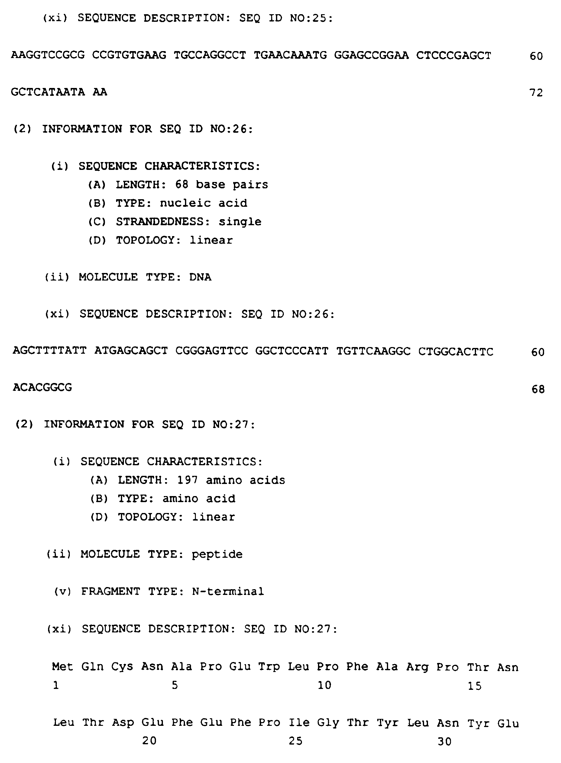

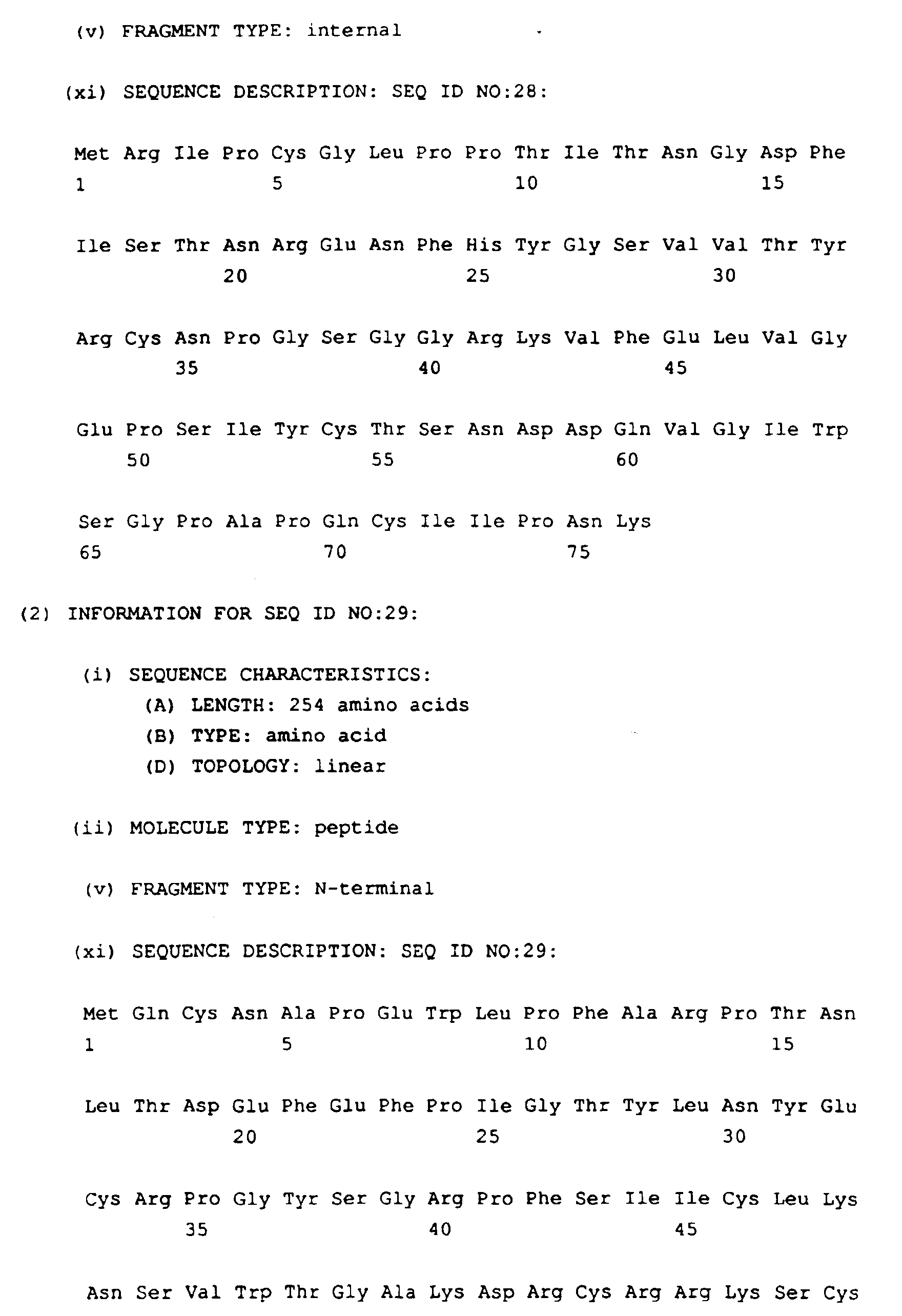
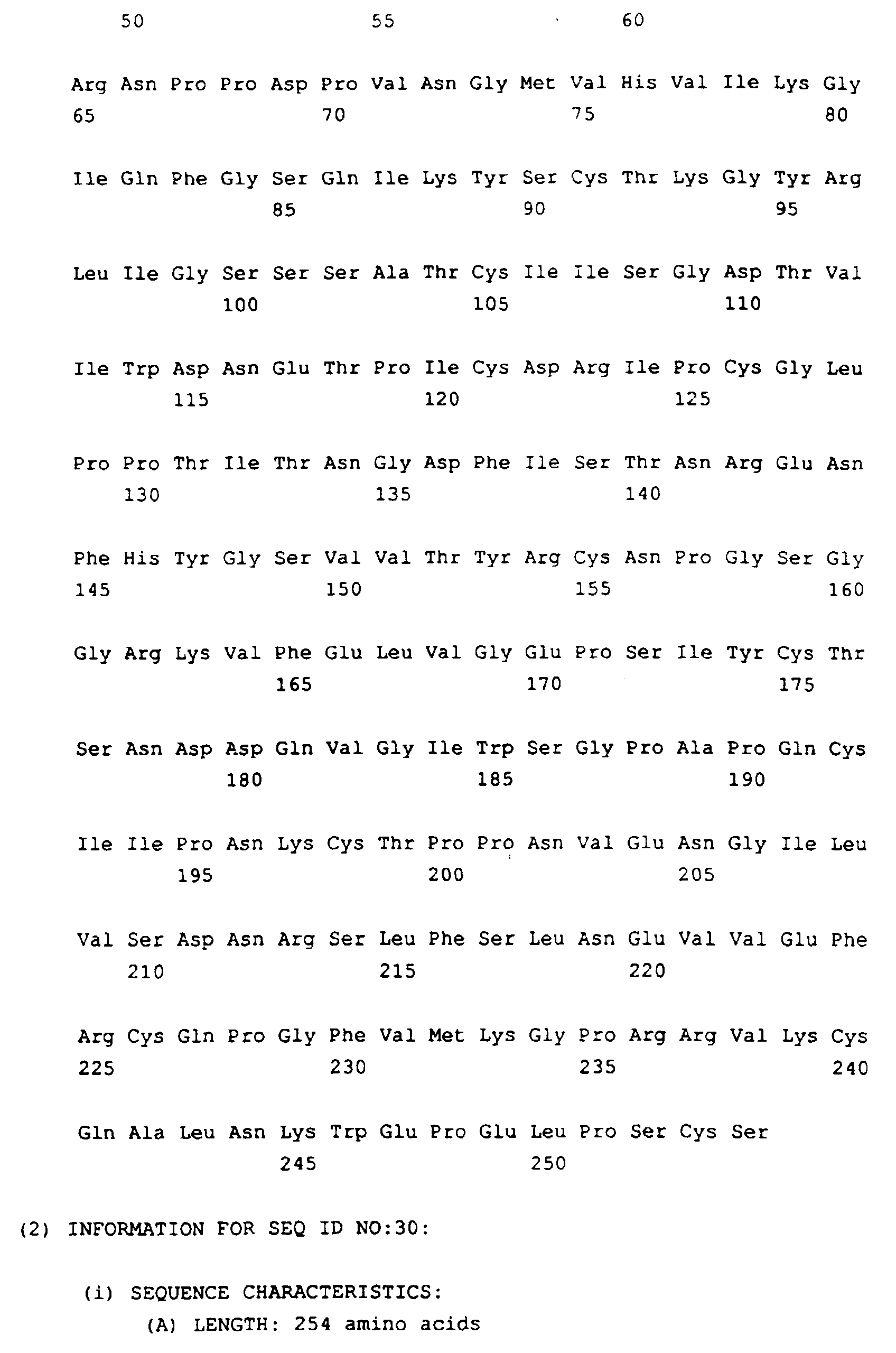
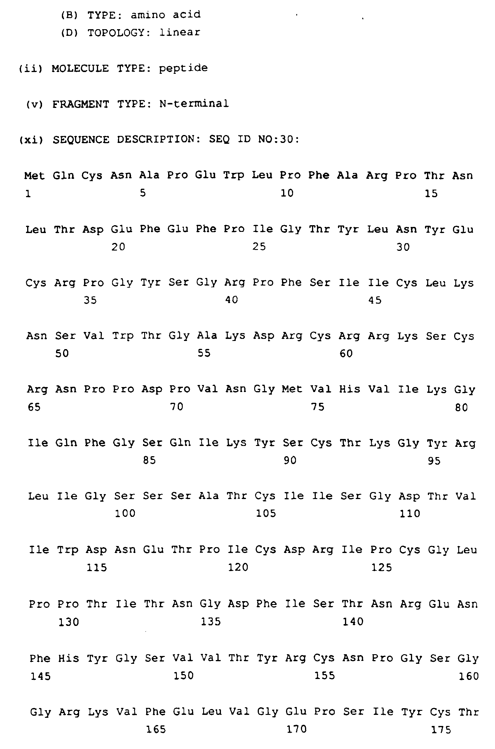

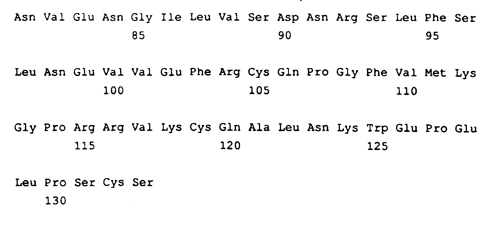
Claims (21)
- A soluble polypeptide comprising, in sequence, one to four short consensus repeats (SCR) selected from SCR 1, 2, 3 and 4 of long homologous repeat A (LHR-A) as the only structurally and functionally intact SCR domains of CR1 and including at least SCR3, wherein SCR 1, 2, 3 and 4 consist respectively, of residues 2-58, 63-120, 125-191 and 197-252 of mature CR1.
- A polypeptide according to claim 1 which comprises, in sequence, SCR 1, 2 and 3 of LHR-A as the only structurally and functionally intact SCR domains of CR1.
- A polypeptide according to claim 2 of formula (I):
- A polypeptide according to claim 3 in which W1, X1 and Y1 represent residues 59-62, 121-124 and 192-196, respectively, of mature CR1 and V1 represents residue 1 of mature CR1 optionally linked via its N-terminus to methionine.
- A polypeptide according to claim 1 which comprises, in sequence, SCR 1, 2, 3 and 4 of LHR-A as the only structurally and functionally intact SCR domains of CR1.
- A polypeptide according to claim 5 of formula (II):
- A polypeptide according to claim 6 in which W2, X2, Y2 and Z2 represent residues 59-62, 121-124, 192-196, and residues 253 respectively, of mature CR1 and V2 represents residue 1 of mature CR1 optionally linked via its N-terminus to methionine.
- A polypeptide according to claim 7 in which arginine 235 is replaced by histidine.
- A polypeptide according to claim 1 of formula (III):
- A polypeptide according to claim 9 in which X3 represents amino acids 122-124 of mature CR1 optionally linked to methionine at its N-terminus and Y4 represents amino acids 192- 196 of mature CR1.
- A polypeptide according to claim 1 of formula (IV):
- A polypeptide according to claim 11 in which X4 represents amino acids 122-124 of mature CR1 optionally linked to methionine at its N-terminus and Y4 and Z4 represent amino acids 192-196 and 253 respectively of mature CR1.
- A polypeptide according to claim 1 having the amino acid sequence given in SEQ ID NO: 27, SEQ ID NO:28, SEQ ID NO:29, SEQ ID NO:30 or SEQ ID NO:31.
- A DNA molecule encoding a soluble polypeptide according to claim 1.
- A DNA polymer according to claim 14 having the nucleotide sequence given in SEQ ID No: 32 or SEQ ID No. 33, as follows:


- A replicable expression vector capable, in a host cell, of expressing the DNA polymer of claim 14 or 15.
- A host cell transformed with the replicable expression vector of claim 16.
- A process for preparing a CR1 polypeptide according to claim 1 which process comprises expressing DNA encoding said polypeptide in a recombinant host cell and recovering the product.
- A pharmaceutical composition comprising a therapeutically effective amount of a polypeptide according to claim 1 and a pharmaceutically acceptable carrier or excipient.
- The use of a polypeptide of claim 1 in the manufacture of a medicament for the treatment of a disease or disorder associated with inflammation or inappropriate complement activation.
- A polypeptide according to claim 1 for use as an active therapeutic substance.
Applications Claiming Priority (5)
| Application Number | Priority Date | Filing Date | Title |
|---|---|---|---|
| GB929213376A GB9213376D0 (en) | 1992-06-24 | 1992-06-24 | Novel compounds |
| GB9213376 | 1992-06-24 | ||
| GB939304057A GB9304057D0 (en) | 1993-03-01 | 1993-03-01 | Novel compounds |
| GB9304057 | 1993-03-01 | ||
| PCT/GB1993/001282 WO1994000571A1 (en) | 1992-06-24 | 1993-06-16 | Soluble cr1 derivatives |
Publications (2)
| Publication Number | Publication Date |
|---|---|
| EP0649468A1 EP0649468A1 (en) | 1995-04-26 |
| EP0649468B1 true EP0649468B1 (en) | 2000-03-15 |
Family
ID=26301119
Family Applications (1)
| Application Number | Title | Priority Date | Filing Date |
|---|---|---|---|
| EP93913383A Expired - Lifetime EP0649468B1 (en) | 1992-06-24 | 1993-06-16 | Soluble cr1 derivatives |
Country Status (5)
| Country | Link |
|---|---|
| US (2) | US5833989A (en) |
| EP (1) | EP0649468B1 (en) |
| JP (1) | JP3939747B2 (en) |
| DE (1) | DE69328098T2 (en) |
| WO (1) | WO1994000571A1 (en) |
Cited By (2)
| Publication number | Priority date | Publication date | Assignee | Title |
|---|---|---|---|---|
| WO2004022096A1 (en) * | 2002-09-06 | 2004-03-18 | Alexion Pharmaceuticals, Inc. | Method of treatment of asthma using antibodies to complement component c5 |
| US9352035B2 (en) | 2002-09-06 | 2016-05-31 | Alexion Pharmaceuticals, Inc. | High concentration antibody formulations |
Families Citing this family (18)
| Publication number | Priority date | Publication date | Assignee | Title |
|---|---|---|---|---|
| WO1994017822A1 (en) * | 1993-02-12 | 1994-08-18 | T Cell Sciences, Inc. | PULMONARY ADMINISTRATION OF sCR1 AND OTHER COMPLEMENT INHIBITORY PROTEINS |
| US6074642A (en) * | 1994-05-02 | 2000-06-13 | Alexion Pharmaceuticals, Inc. | Use of antibodies specific to human complement component C5 for the treatment of glomerulonephritis |
| GB9604518D0 (en) * | 1996-03-02 | 1996-05-01 | Smithkline Beecham Plc | Novel compounds |
| GB9614871D0 (en) * | 1996-07-15 | 1996-09-04 | Smithkline Beecham Plc | Compounds |
| GB9624731D0 (en) * | 1996-11-28 | 1997-01-15 | Univ Leicester | Complement inhibitor |
| GB9704519D0 (en) | 1997-03-05 | 1997-04-23 | Smithkline Beecham Plc | Compounds |
| AU6549398A (en) * | 1997-03-10 | 1998-09-29 | Beth Israel Deaconess Medical Center | Immune complexes and methods of detection and treatment thereof |
| GB9706950D0 (en) * | 1997-04-05 | 1997-05-21 | Chernajovsky Yuti | Immune modulation by polypeptides related to CR1 |
| GB9905503D0 (en) | 1999-03-10 | 1999-05-05 | Adprotech Plc | Novel compound formulations and methods of delivery |
| EP1186299A1 (en) * | 2000-09-12 | 2002-03-13 | Universitair Medisch Centrum Utrecht | The diagnosis, prevention, and/or succesful treatment of atherosclerosis, infectious diseases, and disturbances in the immune system |
| US20050271660A1 (en) | 2002-09-06 | 2005-12-08 | Alexion Pharmaceuticals, Inc. | Nebulization of monoclonal antibodies for treating pulmonary diseases |
| US8124097B2 (en) * | 2004-01-21 | 2012-02-28 | Case Western Reserve University | Hybrid and chimeric polypeptides that regulate activation of complement |
| WO2006034032A2 (en) * | 2004-09-17 | 2006-03-30 | The Johns Hopkins University | Biomarkers for breast cancer |
| CA2654440C (en) | 2005-06-06 | 2013-09-03 | Girish J. Kotwal | Methods for treatment or prophylaxis of atherosclerosis and reperfusion injury |
| DE102007010843B3 (en) * | 2007-03-04 | 2008-09-11 | Cytocentrics Ag | Method and device |
| GB201800620D0 (en) | 2018-01-15 | 2018-02-28 | Univ Manchester | C3b Binding Polypeptide |
| AU2019268410B2 (en) | 2018-05-16 | 2025-01-02 | Csl Limited | Soluble complement receptor type 1 variants and uses thereof |
| IL299048A (en) | 2020-06-14 | 2023-02-01 | Vertex Pharma | Complement factor i variants, fusion constructs and compositions comprising same and uses thereof |
Family Cites Families (2)
| Publication number | Priority date | Publication date | Assignee | Title |
|---|---|---|---|---|
| US5212071A (en) * | 1988-04-01 | 1993-05-18 | The Johns Hopkins University | Nucleic acids encoding a human C3b/C4b receptor (CR1) |
| IL89790A (en) * | 1988-04-01 | 2002-05-23 | Johns Hopking University | Nucleic acid sequences that encode and cells that produce cr1 protein and methods of production and purification |
-
1993
- 1993-06-16 EP EP93913383A patent/EP0649468B1/en not_active Expired - Lifetime
- 1993-06-16 DE DE69328098T patent/DE69328098T2/en not_active Expired - Lifetime
- 1993-06-16 JP JP50213294A patent/JP3939747B2/en not_active Expired - Fee Related
- 1993-06-16 US US08/356,361 patent/US5833989A/en not_active Expired - Lifetime
- 1993-06-16 WO PCT/GB1993/001282 patent/WO1994000571A1/en not_active Ceased
-
1996
- 1996-12-19 US US08/769,967 patent/US5859223A/en not_active Expired - Lifetime
Cited By (4)
| Publication number | Priority date | Publication date | Assignee | Title |
|---|---|---|---|---|
| WO2004022096A1 (en) * | 2002-09-06 | 2004-03-18 | Alexion Pharmaceuticals, Inc. | Method of treatment of asthma using antibodies to complement component c5 |
| AU2003270330B2 (en) * | 2002-09-06 | 2009-07-30 | Alexion Pharmaceuticals, Inc. | Method of treatment of asthma using antibodies to complement component C5 |
| US9352035B2 (en) | 2002-09-06 | 2016-05-31 | Alexion Pharmaceuticals, Inc. | High concentration antibody formulations |
| US9415102B2 (en) | 2002-09-06 | 2016-08-16 | Alexion Pharmaceuticals, Inc. | High concentration formulations of anti-C5 antibodies |
Also Published As
| Publication number | Publication date |
|---|---|
| JP3939747B2 (en) | 2007-07-04 |
| HK1014552A1 (en) | 1999-09-30 |
| DE69328098T2 (en) | 2000-08-24 |
| US5859223A (en) | 1999-01-12 |
| WO1994000571A1 (en) | 1994-01-06 |
| US5833989A (en) | 1998-11-10 |
| EP0649468A1 (en) | 1995-04-26 |
| JPH07508516A (en) | 1995-09-21 |
| DE69328098D1 (en) | 2000-04-20 |
Similar Documents
| Publication | Publication Date | Title |
|---|---|---|
| US5833989A (en) | Soluble CR1 derivatives | |
| EP0571602B1 (en) | Dimer of molecular variant of apolipoprotein and processes for the production thereof | |
| US7655617B2 (en) | Conjugates of soluble peptidic compounds with membrane-binding agents | |
| Schreiber et al. | Assembly of the cytolytic alternative pathway of complement from 11 isolated plasma proteins. | |
| JPH03191791A (en) | Production of biologically active protein | |
| EP1719780A2 (en) | Mammalian cytokine-like polypeptide-10 | |
| AU6942994A (en) | Recombinant bpi-based and lbp-based proteins, nucleic acid molecules encoding same, methods of producing same, and uses thereof | |
| CN111499762B (en) | Fusion protein containing PDGFR beta specific affinity body and TNF alpha and application thereof | |
| US6833437B2 (en) | Complement receptor type 1 (CR1)-like sequences | |
| HK1014552B (en) | Soluble cr1 derivatives | |
| JP2002541073A (en) | Method for molecular refolding of Ig domain-containing polypeptide | |
| US6797806B2 (en) | Fragments of CR1 and their use | |
| Barsoum et al. | Production, safety and antitumor efficacy of recombinant Oncofetal Antigen/immature laminin receptor protein | |
| Veeraragavan | Studies on two major contaminating proteins of the cytoplasmic inclusion bodies in Escherichia coli | |
| CN101883784B (en) | N-terminal modified interferon-alpha | |
| JPH07188292A (en) | Human-derived glycoprotein, physiologically active factor comprising the glycoprotein, and preparation containing the same as an active ingredient | |
| CN1986817A (en) | Plant oil-body expression vector and process of expressing human acid fibroblast growth factor with plant oil-body | |
| KR20020067108A (en) | Transducing domain, transducing domain-cargo molecule complex and uses thereof | |
| US5827731A (en) | Thrombin-inhibitory protein from ticks | |
| Schwaeble et al. | Human complement factor B: functional properties of a recombinant zymogen of the alternative activation pathway convertase | |
| EP2365822A1 (en) | Snake factor v and methods of use as a procoagulant | |
| JPH0925297A (en) | Human-derived glycoprotein, physiologically active factor comprising the glycoprotein, and preparation containing the same as an active ingredient |
Legal Events
| Date | Code | Title | Description |
|---|---|---|---|
| PUAI | Public reference made under article 153(3) epc to a published international application that has entered the european phase |
Free format text: ORIGINAL CODE: 0009012 |
|
| 17P | Request for examination filed |
Effective date: 19941201 |
|
| AK | Designated contracting states |
Kind code of ref document: A1 Designated state(s): BE CH DE FR GB IT LI NL |
|
| 17Q | First examination report despatched |
Effective date: 19960507 |
|
| RAP1 | Party data changed (applicant data changed or rights of an application transferred) |
Owner name: ADPROTECH PLC |
|
| GRAG | Despatch of communication of intention to grant |
Free format text: ORIGINAL CODE: EPIDOS AGRA |
|
| GRAG | Despatch of communication of intention to grant |
Free format text: ORIGINAL CODE: EPIDOS AGRA |
|
| GRAH | Despatch of communication of intention to grant a patent |
Free format text: ORIGINAL CODE: EPIDOS IGRA |
|
| GRAH | Despatch of communication of intention to grant a patent |
Free format text: ORIGINAL CODE: EPIDOS IGRA |
|
| GRAA | (expected) grant |
Free format text: ORIGINAL CODE: 0009210 |
|
| AK | Designated contracting states |
Kind code of ref document: B1 Designated state(s): BE CH DE FR GB IT LI NL |
|
| REG | Reference to a national code |
Ref country code: CH Ref legal event code: NV Representative=s name: KIRKER & CIE SA Ref country code: CH Ref legal event code: EP |
|
| REF | Corresponds to: |
Ref document number: 69328098 Country of ref document: DE Date of ref document: 20000420 |
|
| ET | Fr: translation filed | ||
| ITF | It: translation for a ep patent filed | ||
| PLBE | No opposition filed within time limit |
Free format text: ORIGINAL CODE: 0009261 |
|
| STAA | Information on the status of an ep patent application or granted ep patent |
Free format text: STATUS: NO OPPOSITION FILED WITHIN TIME LIMIT |
|
| 26N | No opposition filed | ||
| REG | Reference to a national code |
Ref country code: CH Ref legal event code: PFA Free format text: ADPROTECH PLC TRANSFER- ADPROTECH LIMITED |
|
| REG | Reference to a national code |
Ref country code: CH Ref legal event code: PFA Free format text: ADPROTECH LIMITED,UNIT 3, NUMBER 2 ORCHARD ROAD,ROYSTON/HERTS SG8 5HD (GB) TRANSFER- ADPROTECH LIMITED,SECOND FLOOR, UNITS 7 & 8 THE MALTINGS, GREEN DRIFT,ROYSTON/HERTS SG8 5DY (GB) |
|
| REG | Reference to a national code |
Ref country code: GB Ref legal event code: IF02 |
|
| PGFP | Annual fee paid to national office [announced via postgrant information from national office to epo] |
Ref country code: CH Payment date: 20090721 Year of fee payment: 17 |
|
| PGFP | Annual fee paid to national office [announced via postgrant information from national office to epo] |
Ref country code: BE Payment date: 20090805 Year of fee payment: 17 |
|
| PGFP | Annual fee paid to national office [announced via postgrant information from national office to epo] |
Ref country code: IT Payment date: 20090718 Year of fee payment: 17 |
|
| PGFP | Annual fee paid to national office [announced via postgrant information from national office to epo] |
Ref country code: NL Payment date: 20100611 Year of fee payment: 18 |
|
| BERE | Be: lapsed |
Owner name: *ADPROTECH P.L.C. Effective date: 20100630 |
|
| REG | Reference to a national code |
Ref country code: CH Ref legal event code: PL |
|
| PG25 | Lapsed in a contracting state [announced via postgrant information from national office to epo] |
Ref country code: IT Free format text: LAPSE BECAUSE OF NON-PAYMENT OF DUE FEES Effective date: 20100616 |
|
| PG25 | Lapsed in a contracting state [announced via postgrant information from national office to epo] |
Ref country code: LI Free format text: LAPSE BECAUSE OF NON-PAYMENT OF DUE FEES Effective date: 20100630 Ref country code: CH Free format text: LAPSE BECAUSE OF NON-PAYMENT OF DUE FEES Effective date: 20100630 |
|
| PG25 | Lapsed in a contracting state [announced via postgrant information from national office to epo] |
Ref country code: BE Free format text: LAPSE BECAUSE OF NON-PAYMENT OF DUE FEES Effective date: 20100630 |
|
| REG | Reference to a national code |
Ref country code: NL Ref legal event code: V1 Effective date: 20120101 |
|
| PG25 | Lapsed in a contracting state [announced via postgrant information from national office to epo] |
Ref country code: NL Free format text: LAPSE BECAUSE OF NON-PAYMENT OF DUE FEES Effective date: 20120101 |
|
| PGFP | Annual fee paid to national office [announced via postgrant information from national office to epo] |
Ref country code: DE Payment date: 20120613 Year of fee payment: 20 |
|
| PGFP | Annual fee paid to national office [announced via postgrant information from national office to epo] |
Ref country code: GB Payment date: 20120613 Year of fee payment: 20 Ref country code: FR Payment date: 20120619 Year of fee payment: 20 |
|
| REG | Reference to a national code |
Ref country code: DE Ref legal event code: R071 Ref document number: 69328098 Country of ref document: DE |
|
| REG | Reference to a national code |
Ref country code: DE Ref legal event code: R071 Ref document number: 69328098 Country of ref document: DE |
|
| REG | Reference to a national code |
Ref country code: GB Ref legal event code: PE20 Expiry date: 20130615 |
|
| PG25 | Lapsed in a contracting state [announced via postgrant information from national office to epo] |
Ref country code: GB Free format text: LAPSE BECAUSE OF EXPIRATION OF PROTECTION Effective date: 20130615 Ref country code: DE Free format text: LAPSE BECAUSE OF EXPIRATION OF PROTECTION Effective date: 20130618 |