EP0648123B1 - INHIBITORS OF PROTEIN TYROSINE-PHOSPHATASE $g(a) FOR TREATMENT OF TUMORS - Google Patents
INHIBITORS OF PROTEIN TYROSINE-PHOSPHATASE $g(a) FOR TREATMENT OF TUMORS Download PDFInfo
- Publication number
- EP0648123B1 EP0648123B1 EP93915598A EP93915598A EP0648123B1 EP 0648123 B1 EP0648123 B1 EP 0648123B1 EP 93915598 A EP93915598 A EP 93915598A EP 93915598 A EP93915598 A EP 93915598A EP 0648123 B1 EP0648123 B1 EP 0648123B1
- Authority
- EP
- European Patent Office
- Prior art keywords
- cells
- ptpα
- src
- cell
- tumour
- Prior art date
- Legal status (The legal status is an assumption and is not a legal conclusion. Google has not performed a legal analysis and makes no representation as to the accuracy of the status listed.)
- Expired - Lifetime
Links
Images


Classifications
-
- C—CHEMISTRY; METALLURGY
- C07—ORGANIC CHEMISTRY
- C07K—PEPTIDES
- C07K16/00—Immunoglobulins [IG], e.g. monoclonal or polyclonal antibodies
- C07K16/40—Immunoglobulins [IG], e.g. monoclonal or polyclonal antibodies against enzymes
-
- C—CHEMISTRY; METALLURGY
- C12—BIOCHEMISTRY; BEER; SPIRITS; WINE; VINEGAR; MICROBIOLOGY; ENZYMOLOGY; MUTATION OR GENETIC ENGINEERING
- C12N—MICROORGANISMS OR ENZYMES; COMPOSITIONS THEREOF; PROPAGATING, PRESERVING, OR MAINTAINING MICROORGANISMS; MUTATION OR GENETIC ENGINEERING; CULTURE MEDIA
- C12N9/00—Enzymes; Proenzymes; Compositions thereof; Processes for preparing, activating, inhibiting, separating or purifying enzymes
- C12N9/14—Hydrolases (3)
- C12N9/16—Hydrolases (3) acting on ester bonds (3.1)
-
- A—HUMAN NECESSITIES
- A61—MEDICAL OR VETERINARY SCIENCE; HYGIENE
- A61K—PREPARATIONS FOR MEDICAL, DENTAL OR TOILETRY PURPOSES
- A61K38/00—Medicinal preparations containing peptides
-
- A—HUMAN NECESSITIES
- A61—MEDICAL OR VETERINARY SCIENCE; HYGIENE
- A61K—PREPARATIONS FOR MEDICAL, DENTAL OR TOILETRY PURPOSES
- A61K48/00—Medicinal preparations containing genetic material which is inserted into cells of the living body to treat genetic diseases; Gene therapy
Definitions
- This invention relates to the treatment of tumours, in particular tumours exhibiting an elevated level of pp60 c-src kinase activity such as human colon carcinoma.
- pp60 c-src The kinase activity of pp60 c-src is specifically and transiently increased during mitosis and repressed during interphase 1 .
- Loss of cell cycle control of pp60 c-src occurs upon mutation of Tyr 527 to Phe or when pp60 c-src is associated with polyoma middle-T-antigen, and these conditions result in cell transformation or tumourigenesis 2,3 .
- pp60 c-src has elevated kinase activity which is maintained throughout the cell cycle and accompanied by dephosphorylation of the carboxy-terminal negative regulatory 4-7 Tyr 527 site, or mimicry of Tyr 527 dephosphorylation in the case of the mutant.
- PTPases Protein tyrosine phosphatases
- HPTP ⁇ receptor-like human PTP ⁇
- an inhibitor of PTP ⁇ for use in the treatment of a tumour exhibiting an elevated level of pp60 c-src kinase activity.
- PTPases may be more fully termed as protein tyrosine phosphate phosphohydrolases (EC 3.1.3.48). They may be divided into three classes based on their structural organisation. Class I contains the low molecular mass non-receptor molecules possessing a single catalytic domain (PTP IB, TCPTP, rat brain PTP). Class II and II PTPases are receptor-like transmembrane proteins. The sole member of Class II is human PTP ⁇ (HPTP ⁇ ) which has a single cytoplasmic catalytic domain. Class III members are LCA, LAR, HPTP ⁇ , HPTP ⁇ , HPTP ⁇ , HPTP ⁇ , DPTP, DLAR and possess two repeated putative catalytic domains in the cytoplasmic region of the molecule.
- Inhibitors of PTP ⁇ include chemical compounds, for example zinc ions, vanadates such as sodium orthovanadate and arsenites such as phenylarsine oxide. These compounds are, however, fairly toxic. Non-toxic inhibitors are preferred.
- the inhibitor may therefore be an antibody, polyclonal or monoclonal.
- the antibody is typically an inhibitory antibody to the extracellular domain of PTP ⁇ or to the active site in the catalytic domain of PTP ⁇ .
- Inhibitors such as zinc, vanadate, arsenite and antibody act directly on PTP ⁇ . However, inhibition may be achieved in other ways. Inhibition of endogenous PTP ⁇ may be effected by gene transfer of a mutant PTP ⁇ cDNA where the mutant protein product interacts with and blocks endogenous PTP ⁇ activity or competes with endogenous PTP ⁇ for activating proteins, ligands, etc.
- a suitable cDNA encodes a mutant PTP ⁇ in which the Cys residues of the catalytic domain(s) have been mutated to Ser.
- the inhibitors are used to treat a tumour exhibiting an elevated level of pp60 c-src kinase activity. Any tumour which has abnormally active or overactive pp60 c-src , which may be a result of PTP ⁇ overexpression or overactivation in the tumour, may be treated.
- the tumour may be a tumour with increased pp60 c-src activity which cannot be accounted for by a proportional increase in pp60 c-src amount.
- Such tumours include human colon carcinoma, rhabdomyosarcoma, osteogenic sarcoma and Ewing's sarcoma 10-14 .
- the inhibitors may be particularly used in the treatment of human colon carcinoma which is the third most common human malignancy. The inhibitors may therefore be used in the treatment of colorectal cancer.
- An agent for use in the treatment of a tumour exhibiting an elevated level of pp60 c-src kinase activity can therefore be provided.
- the agent comprises a HPTP ⁇ inhibitor.
- a pharmaceutical composition may be formulated, comprising the inhibitor and a pharmaceutically acceptable carrier or diluent.
- a chemical or antibody inhibitor may be given by injection.
- a pH controlling agent, a buffer, a stabilizing agent and/or an excipient may be added.
- lyophilized injectable preparations may be produced. There can be produced preparations for subcutaneous, intramuscular or intravenous injection.
- a chemical inhibitor may alternatively be administered by the oral or rectal route.
- Orally administrable solid preparations may be produced by adding an excipient and, if desired, a binding agent, disintegrator, lubricating agent, coloring agent, and/or flavouring agent, and the like to the inhibitor, and forming tablets, coated tablets, granules, powders or capsules according to conventional techniques.
- Oral liquid compositions may be prepared by adding a flavouring agent, buffer, and/or stabilizer to the main drug and forming a syrup or dry syrup.
- a rectal suppository may be prepared by adding an excipient and, if desired, a surfactant to the inhibitor to form the rectal suppository according to conventional techniques.
- a DNA sequence encoding a mutant PTP ⁇ may be incorporated in a retroviral or adenoviral vector or in liposomes for the purpose of gene transfer 15 .
- the vectors may transfer the DNA into cultured human tumour cells which are administered to a patient by injection, for example intratumoural injection. Cells from the tumour it is wished to treat may therefore be removed from a patient, grown in culture, infected with the vector, selected for positive infection and then reintroduced into the patient.
- Liposomes containing DNA for example plasmid DNA, are injected directly into tumour cells to accomplish gene transfer.
- physical DNA transfer may be achieved using biolistic technology i.e. a gene gun.
- a therapeutically effective amount of the inhibitor is administered to a host having a tumour exhibiting an elevated level of pp60 c-src kinase activity.
- the host is typically a human but may be another mammal. The condition of a patient may thus be improved. The tumour from which a patient suffers may be ameliorated. This treatment may be given in conjunction with another method of treatment, for example surgery, to eradicate the tumour.
- the dosage levels for the inhibitor differ depending on a variety of factors including condition of the patient, the age and body weight of the patient, the disease state being treated and the administration form. Dosage levels of the order of about 0.05-500 mg per day are useful for injection of a chemical or antibody inhibitor though the dosage levels may differ from portion to portion. When a chemical inhibitor is administered orally or rectally, the dosage levels are about 0.05-1000 mg per day. The dosage levels may be changed according to the requirements of a doctor.
- DNA is used to introduce at least one gene per target cell such as 1 to 10000 DNA molecules per cell.
- the amount of DNA used depends on the tumour mass and estimated number of cells per tumour. It also depends on the amount of endogenous PTP ⁇ per tumour cell. Generally an amount of inhibitory mutant PTP ⁇ DNA is introduced that gives expression of an excess amount of mutant PTP ⁇ protein.
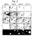
- the PTP ⁇ -overexpressing cells had a fibroblastic phenotype (Fig. 2a).
- the ⁇ -21 cells were beginning to exhibit a transformed phenotype with some of the cells appearing rounded and highly refractile.
- none of the PTP ⁇ -overexpressing cell lines were growth arrested through contact inhibition (Table 1). They grew in multilayers and formed foci containing refractile and disordered cells (Fig. 2b).
- the ⁇ -7, ⁇ -21 and ⁇ -29 cells also had a reduced requirement for serum growth factors as evidenced by their continued proliferation in medium with 0.1% FCS, whereas control cells became quiescent (Table 1).
- pp60 c-src A few enzymes are activated by tyrosine dephosphorylation, such as the src family tyrosine kinases 4, 22-24 and the cell cycle serine/threonine kinase p34 cdc2 25-27 .
- certain mutant forms of pp60 c-src including its oncogenic counterpart pp60 v-src , have transforming ability 5-7, 28 .
- the activity of pp60 c-src in the control REF-neo and PTP ⁇ overexpressing cells was tested by immunoprecipitating pp60 c-src and measuring its ability to phosphorylate the exogenous substrate enolase.
- activation of pp60 c-src involves dephosphorylation of Tyr 527 4-7, 29 .
- the activation state of pp60 c-src reflects the balance of opposing activities of a Tyr 527 kinase and phosphatase, where phosphorylation of Tyr 527 negatively regulates pp60 c-src activity.
- a specific Tyr 527 kinase has recently been identified and cloned 31, 32 , although as yet nothing is known of the regulation of its activity.
- Cell cycle-regulated inhibition of such a kinase or activation of an appropriate tyrosine phosphatase, or both, could result in Tyr 527 dephosphorylation and activation of mitotic pp60 c-src .
- the unregulated occurrence of these events may lead to uncontrolled cell proliferation.
- we have shown that the constitutive overexpression of active PTP ⁇ subverts the normal control of cell growth and have identified pp60 c-src as a target for PTP ⁇ action.
- pp60 c-src may be a direct substrate of PTP ⁇ . Since PTP ⁇ is widely expressed and is particularly abundant in brain 21, 34 while pp60 c-src is expressed in most cells and at high levels in neural tissue 35 , PTP ⁇ may function as an in vivo regulator of src kinase activity in normal cells. TABLE 1 Growth characteristics of parental, control and PTP ⁇ -overexpressing cells.
- Vanadate conc ( ⁇ M) REF-neo Number of cells/5 cm dish ⁇ -7 ⁇ -21 ⁇ -29 0 1.20 x 10 6 1.80 x 10 6 1.60 x 10 6 1.72 x 10 6 5 1.10 x 10 6 1.72 x 10 6 1.42 x 10 6 1.62 x 10 6 30 1.04 x 10 6 1.41 x 10 6 7.00 x 10 5 7.20 x 10 5 60 9.90 x 10 5 1.18 x 10 6 3.70 x 10 5 4.70 x 10 5 120 7.60 x 10 5 5.80 x 10 5 1.60 x 10 5 2.10 x 10 5 2.
- DME medium 10% FCS for 48 h.
- the medium was discarded, and fresh medium containing 10% FCS and the indicated concentration (below) of sodium orthovanadate was added to the cells.
- After 72 h of culture in this medium the cells from two replicate dishes were harvested and counted.
- Table 3 The results are shown in Table 3 below and are essentially the same as in 1 above.
- Vanadate conc Number of cells/5cm dish REF-neo ⁇ -21 0 1.84 x 10 6 1.38 x 10 6 5 1.90 x 10 6 1.20 x 10 6 30 1.80 x 10 6 4.80 x 10 5 60 1.60 x 10 6 2.60 x 10 5 120 6.70 x 10 5 1.76 x 10 5 3.
- PTP ⁇ -overexpressing cells ⁇ -21 were grown for 48 h (to about 60% confluency) in DME medium containing 10% FCS. The medium was removed and replaced with medium containing the indicated concentration of sodium orthovanadate (below) and either 10% (sets #1 and #3) or 0.1% FCS (set #2).
- the cells were cultured for 48 h and counted (sets #1 and 2), or the medium was removed from set #3 and replaced with medium containing 10% FCS without sodium orthovanadate and cultured for another 48 h before counting.
- the results are shown in Table 4 below and demonstrate that the proliferation of ⁇ -21 cells grown in either 10% or 0.1% FCS is similarly affected by vanadate in a dose-dependent manner and that vanadate is not irreversibly toxic to these cells since they will resume proliferation once vandate is removed from the medium as may be appreciated by comparison of the cell numbers in sets #1 and 3.
- Example 4 Determination whether vanadate can block or inhibit tumourigenesis caused by REF ⁇ -21 PTP ⁇ -overexpressing cells.
- the ⁇ -21 cells were grown in DME medium containing 10% FCS and then in DME medium with 0.1% FCS in the presence of 50 ⁇ M sodium orthovanadate for 48 h. The cells were then harvested by trypsinisation, centrifuged to pellet the cells, and the cell pellet resuspended in DME containing 50 ⁇ M sodium orthovanadate. Other ⁇ -21 cells were grown and prepared in a similar manner, except in the absence of sodium orthovanadate throughout. The resuspended cells (plus or minus vanadate) were subcutaneously injected into nude mice (2 x 10 6 cells/mouse, 4 mice for each treatment), and nine days later the tumour size was measured.
Landscapes
- Chemical & Material Sciences (AREA)
- Health & Medical Sciences (AREA)
- Life Sciences & Earth Sciences (AREA)
- Organic Chemistry (AREA)
- Genetics & Genomics (AREA)
- Zoology (AREA)
- Medicinal Chemistry (AREA)
- Bioinformatics & Cheminformatics (AREA)
- General Health & Medical Sciences (AREA)
- Engineering & Computer Science (AREA)
- Biochemistry (AREA)
- Wood Science & Technology (AREA)
- Molecular Biology (AREA)
- Proteomics, Peptides & Aminoacids (AREA)
- General Engineering & Computer Science (AREA)
- Biotechnology (AREA)
- Biomedical Technology (AREA)
- Immunology (AREA)
- Biophysics (AREA)
- Microbiology (AREA)
- Medicines That Contain Protein Lipid Enzymes And Other Medicines (AREA)
- Preparation Of Compounds By Using Micro-Organisms (AREA)
- Medicines Containing Plant Substances (AREA)
- Pharmaceuticals Containing Other Organic And Inorganic Compounds (AREA)
- Acyclic And Carbocyclic Compounds In Medicinal Compositions (AREA)
- Medicines Containing Antibodies Or Antigens For Use As Internal Diagnostic Agents (AREA)
Abstract
Description
- This invention relates to the treatment of tumours, in particular tumours exhibiting an elevated level of pp60c-src kinase activity such as human colon carcinoma.
- The kinase activity of pp60c-src is specifically and transiently increased during mitosis and repressed during interphase1. Loss of cell cycle control of pp60c-src occurs upon mutation of Tyr 527 to Phe or when pp60c-src is associated with polyoma middle-T-antigen, and these conditions result in cell transformation or tumourigenesis 2,3. In both cases pp60c-src has elevated kinase activity which is maintained throughout the cell cycle and accompanied by dephosphorylation of the carboxy-terminal negative regulatory 4-7 Tyr 527 site, or mimicry of Tyr 527 dephosphorylation in the case of the mutant.
- Protein tyrosine phosphatases (PTPases) have been considered as potential anti-oncogenes 8,9, where mutations giving rise to a non-functional gene product or gene deletion could result in tumourigenesis. We have now found that over-expression of the receptor-like human PTPα (HPTPα) results in persistent activation of pp60c-src with concomitant cell transformation and tumourigenesis. This indicates instead that PTPα may function as an oncogene and not an anti-oncogene.
- According to the present invention, there is provided an inhibitor of PTPα for use in the treatment of a tumour exhibiting an elevated level of pp60c-src kinase activity.
- PTPases may be more fully termed as protein tyrosine phosphate phosphohydrolases (EC 3.1.3.48). They may be divided into three classes based on their structural organisation. Class I contains the low molecular mass non-receptor molecules possessing a single catalytic domain (PTP IB, TCPTP, rat brain PTP). Class II and II PTPases are receptor-like transmembrane proteins. The sole member of Class II is human PTPβ (HPTPβ) which has a single cytoplasmic catalytic domain. Class III members are LCA, LAR, HPTPα, HPTPγ, HPTPδ, HPTPε, DPTP, DLAR and possess two repeated putative catalytic domains in the cytoplasmic region of the molecule.
- Inhibitors of PTPα include chemical compounds, for example zinc ions, vanadates such as sodium orthovanadate and arsenites such as phenylarsine oxide. These compounds are, however, fairly toxic. Non-toxic inhibitors are preferred. The inhibitor may therefore be an antibody, polyclonal or monoclonal. The antibody is typically an inhibitory antibody to the extracellular domain of PTPα or to the active site in the catalytic domain of PTPα.
- Inhibitors such as zinc, vanadate, arsenite and antibody act directly on PTPα. However, inhibition may be achieved in other ways. Inhibition of endogenous PTPα may be effected by gene transfer of a mutant PTPα cDNA where the mutant protein product interacts with and blocks endogenous PTPα activity or competes with endogenous PTPα for activating proteins, ligands, etc. A suitable cDNA encodes a mutant PTPα in which the Cys residues of the catalytic domain(s) have been mutated to Ser.
- The inhibitors are used to treat a tumour exhibiting an elevated level of pp60c-src kinase activity. Any tumour which has abnormally active or overactive pp60c-src, which may be a result of PTPα overexpression or overactivation in the tumour, may be treated. The tumour may be a tumour with increased pp60c-src activity which cannot be accounted for by a proportional increase in pp60c-src amount. Such tumours include human colon carcinoma, rhabdomyosarcoma, osteogenic sarcoma and Ewing's sarcoma 10-14. The inhibitors may be particularly used in the treatment of human colon carcinoma which is the third most common human malignancy. The inhibitors may therefore be used in the treatment of colorectal cancer.
- An agent for use in the treatment of a tumour exhibiting an elevated level of pp60c-src kinase activity can therefore be provided. The agent comprises a HPTPα inhibitor. A pharmaceutical composition may be formulated, comprising the inhibitor and a pharmaceutically acceptable carrier or diluent.
- A chemical or antibody inhibitor may be given by injection. When an injectable preparation is produced, a pH controlling agent, a buffer, a stabilizing agent and/or an excipient may be added. Further, according to conventional techniques, lyophilized injectable preparations may be produced. There can be produced preparations for subcutaneous, intramuscular or intravenous injection.
- A chemical inhibitor may alternatively be administered by the oral or rectal route. Orally administrable solid preparations may be produced by adding an excipient and, if desired, a binding agent, disintegrator, lubricating agent, coloring agent, and/or flavouring agent, and the like to the inhibitor, and forming tablets, coated tablets, granules, powders or capsules according to conventional techniques. Oral liquid compositions may be prepared by adding a flavouring agent, buffer, and/or stabilizer to the main drug and forming a syrup or dry syrup. A rectal suppository may be prepared by adding an excipient and, if desired, a surfactant to the inhibitor to form the rectal suppository according to conventional techniques.
- A DNA sequence encoding a mutant PTPα may be incorporated in a retroviral or adenoviral vector or in liposomes for the purpose of gene transfer15. The vectors may transfer the DNA into cultured human tumour cells which are administered to a patient by injection, for example intratumoural injection. Cells from the tumour it is wished to treat may therefore be removed from a patient, grown in culture, infected with the vector, selected for positive infection and then reintroduced into the patient. Liposomes containing DNA, for example plasmid DNA, are injected directly into tumour cells to accomplish gene transfer. In an alternative embodiment, physical DNA transfer may be achieved using biolistic technology i.e. a gene gun.
- In use, a therapeutically effective amount of the inhibitor is administered to a host having a tumour exhibiting an elevated level of pp60c-src kinase activity. The host is typically a human but may be another mammal. The condition of a patient may thus be improved. The tumour from which a patient suffers may be ameliorated. This treatment may be given in conjunction with another method of treatment, for example surgery, to eradicate the tumour.
- The dosage levels for the inhibitor differ depending on a variety of factors including condition of the patient, the age and body weight of the patient, the disease state being treated and the administration form. Dosage levels of the order of about 0.05-500 mg per day are useful for injection of a chemical or antibody inhibitor though the dosage levels may differ from portion to portion. When a chemical inhibitor is administered orally or rectally, the dosage levels are about 0.05-1000 mg per day. The dosage levels may be changed according to the requirements of a doctor.
- For the purpose of gene transfer, enough DNA is used to introduce at least one gene per target cell such as 1 to 10000 DNA molecules per cell. The amount of DNA used depends on the tumour mass and estimated number of cells per tumour. It also depends on the amount of endogenous PTPα per tumour cell. Generally an amount of inhibitory mutant PTPα DNA is introduced that gives expression of an excess amount of mutant PTPα protein.
- The following Examples illustrate the invention. In the accompanying drawings:
- Figure 1 shows overexpression of active PTPα. a, Western blotting with anti-PTPα antiserum of solubilised membrane (M) and cytosolic (C) fractions from parental cells (REF) and cells transfected with PTPα cDNA (α-7, α-21, α-29). b, PTPase specific activities of cytosolic (solid bars) and solubilised membrane (hatched bars) fractions measured towards the phosphotyrosyl-peptide RR-src. Fractions from control transfected cells (REF-neo) are designated as neo. c, PTPase activity of immunoprecipitated PTPα from parental REF (□), control transfected REF-neo (◆) and PTPα-transfected (●, α-7; ○, α-21; △ , α-29) cells.
- Figure 2 shows the morphologies of control REF-neo and PTPα-overexpressing cells. a, Subconfluent and b, four day post-confluent cells in DME medium with 10% fetal calf serum (FCS) (320X). c, 24 h after changing to Dulbecco's minimum essential (DME) medium with 0.1% FCS (320X). d, 72 h after changing to DME medium with 0.1% FCS (320X). e, Colonies on soft agar with serum-free DME medium (32X).
- Figure 3 shows the protein kinase activity, immunoblotting and cyanogen bromide phosphopeptide analysis of pp60c-src. Cells maintained in medium with 0.1% FCS for 48 h were harvested (0 h) or treated with 10% FCS for 8 h prior to harvesting (8 h). Lysates of control REF-neo cells (neo) and PTPα-overexpressing (α-7, α-21, α-29) cells were equalised for protein and immunoprecipitated with anti-src monoclonal antibody 327. a, Top; A portion of each immunoprecipitate was incubated with the exogenous substrate enolase and [γ-32P]ATP and the mixture resolved on 10% sodium dodecyl sulphate-polyacrylamide gel electrophoresis (SDS-PAGE) followed by autoradiography. Bottom; Another portion of each immunoprecipitate was run on 10% SDS-PAGE, immunoblotted and probed with antibody 327 to measure pp60c-src levels. b, Cyanogen bromide phosphopeptides of immunoprecipitated pp60c-src from lysates of cells maintained in medium with 0.1% FCS for 48 h and then metabolically labelled with 32P-orthophosphate for 16 h. c, as in b except that pp60c-src immunoprecipitated from quiescent control REF-neo cells was incubated without (minus) or with (plus) the bacterially expressed and purified intracellular region of PTPα prior to digestion with cyanogen bromide. In b and c, the position of the 32 kDa phosphopeptide is designated by the open arrow and the position of the 4 kDa phosphopeptide containing Tyr 527 is designated by the closed arrow.
-
- 1. To facilitate cloning of the full-length PTPα cDNA16 into the BamH1-HindIII site of the expression vector pXJ4117, a 570 bp fragment of the 5' portion of the gene was first amplified by PCR. A BamH1 site (GGATCC) and an optimal Kozak consensus sequence (CCACC) were added to the 5' oligonucleotide (5'-AAGGATCCAACCACCATGGATTCCTGG-3') and the 3' oligonucleotide contained in the unique Ec1XI restriction site in the PTPα cDNA. The fidelity of amplification was confirmed by sequencing.
The PCR fragment was cleaved with BamH1 and Ec1XI and ligated with a Ec1XI-HindIII fragment containing the rest of the PTPα coding sequence, and this was inserted into a BamH1-HindIII site in the expression vector pXJ41 to give pXJ41-PTPα. The neo gene in a pMAM-neo vector (Pharmacia) was excised with BamH1 and blunt-ended by filling with Klenow. It was inserted into the pXJ41-PTPα and pXJ41 vectors which had been cut at a Sa1I site (downstream of the PTPα insert in the former) and likewise blunt-ended. The REF cells (about 106 per 15 cm dish) were transfected with 20 µg of either pXJ41-PTPα-neo or pXJ41-neo expression vector using the calcium phosphate method 18.
After 16 h the cells were washed with DME medium (containing 4.5 g/l glucose and penicillin/streptomycin) and incubated with DME medium containing G418 (500 µg/ml) and 10% FCS. The medium was changed every four days, and colonies were isolated after 21 days. To prepare cell membrane and cytosolic fractions, confluent cells were rinsed twice with cold phosphate-buffered saline, scraped into harvesting buffer (50mM Tris-Cl pH 7.6, 150mM NaCl, 2mM PMSF, 10 µg/ml aprotinin) and pelleted by low speed centrifugation. After resuspension of the pellets in harvesting buffer and brief sonication to lyse the cells, the lysates were centrifuged at 100,000xg for 30 min at 4°C to obtain the sytosolic supernatants.
The pellets were rinsed once with harvesting buffer, resuspended in harvesting buffer with 1% Triton X-100 and solubilised on ice for 60 min. After centrifugation at 100,000xg as above the supernatants comprised the solubilised membrane fractions. For Western blotting, 150 µg protein from each fraction was electrophoresed on 7.5% SDS-PAGE and transferred to nitrocellulose membranes in 50mM Tris base/0.56% glycine/10% methanol (80V, 75 min, 25°C). The membranes were probed with a 1:500 dilution of anti-PTPα antiserum (raised in rabbits against a peptide comprising the carboxy-terminal amino acid sequence IDAFSDYANFK of PTPα) followed by a 1:3000 dilution of goat anti-rabbit IgG conjugated to peroxidase (Sigma), and developed using the ECL system (Amersham).
PTPase specific activity was measured at 30°C in reactions containing 50mM Mes pH 6.0, 0.5 mg/ml bovine serum albumin (BSA), 0.5mM dithiothreitol (DTT), (5µM phosphotyrosyl-RR-src peptide (RRLIEDAEY(P)AARG, corresponding to the sequence encompassing Tyr 416 of pp60c-src and phosphorylated as described16) and 3.3 µg/ml solubilised membrane protein or 6.6 µg/ml cytosolic protein. Reactions were linear over the times assayed. For immunoprecipitation of PTPα, confluent cells were lysed in harvesting buffer with 1% Triton X-100 and centrifuged at 100,000xg for 30 min.
Anti-PTPα antiserum (150 µl) was added to 6 ml of 2 mg/ml supernatant and mixed at 4°C for 16 h. Protein A-Sepharose (Pharmacia) was added and mixed at 4°C for 2 h. The immunoprecipitated pellets obtained by centrifugation were washed three times with 50mM Tris-C1 pH 7.6, 0.5M NaCl, 1mM PMSF, 10 µg/ml aprotinin and packed into small columns in the same buffer. Protein was eluted from the beads with 0.6 ml 0.1M glycine, pH 2.5 and immediately neutralised with 20 µl 1M Tris-C1, pH 8.0. The results are shown in Figures 1 and 2 and Table 1. - 2. Cells were lysed in RIPA buffer19 (2 ml/15 cm dish) and the lysates centrifuged at 100,000xg for 30 min. Anti-src monoclonal antibody (Mab 327, Oncogene Science) was added to the lysates (1 µg/2ml) and mixed at 4°C for 1 h. Goat anti-mouse IgG (Sigma) was added (4 µg/2 ml) and mixed as above, followed by a one-tenth volume of Protein A cell suspension (Sigma) and mixing as above. After low speed centrifugation the immunoprecipitates were washed as described19.
Immunoblot analysis of immunoprecipitated pp60c-src levels was as described in Example 1 except immunoblots were probed with a 1:1000 dilution of 0.1 mg/ml Mab 327 followed by goat anti-mouse IgG conjugated to peroxidase (1:1000) (Sigma). The kinase activity of immunoprecipitated pp60c-src was determined in 20 µl reactions containing 10mM Pipes, pH 7.0, 5mM MnC12, 0.5mM DTT, 2.5 µg acid-treated enolase and 10 µCi [γ-32P]ATP at 37°C for 5 min. For cyanogen bromide phosphopeptide analysis, cells were labelled for 16 h with 1.5 mCi/ml 32P-orthophosphate (Amersham) in phosphate-free DME containing normal DME (95:5, vol:vol) and 0.1% FCS.
Immunoprecipitates of pp60c-src, prepared from the various cells as just described, were electrophoresed on 10% SDS-PAGE and the position of 32P-labelled pp60c-src determined by autoradiography. Corresponding pieces of the gel were cut out and electroeluted in 25mM Tris, 192mM glycine, 0.1% SDS, 2% 2-mercaptoethanol at 90V for 2 h. Electroeluted protein was precipitated with 10% trichloroacetic acid with 100 µg bovine serum albumin as carrier. The precipitate was washed once with cold ethanol, dried, digested with cyanogen bromide and analysed on 20% SDS-PAGE as described20. For in vitro treatment of pp60c-src with PTPα prior to cyanogen bromide digestion, 32P-labelled pp60c-src immunoprecipitates were washed four times with 50mM Mes, pH 6.0, 0.5mM DTT and incubated at 30°C for 60 min in the same buffer with 0.5 µg/ml bovine serum albumin in the presence or absence of 15 µg of PTPα (intracellular domain)16. The results are shown in Figure 3. - 3. Fischer rat embryo fibroblasts (REF) were transfected with the full length human receptor-like protein tyrosine phosphatase α (PTPα) cDNA in an expression vector driven by the human cytomegalovirus promoter and G418-resistant cell lines were isolated. Western blotting with anti-PTPα antibodies detected elevated levels of a protein of an expected21 Mr of about 130 kDa in total lysates of several cell lines (not shown), and three lines (α-7, α-21, α-29) were selected for further analysis.
- In accord with the transmembrane nature of PTPα solubilized membrane fractions from these cells contained much higher amounts of PTPα than those from parental cells (REF), and the overexpressed PTPα was predominantly localized to the membrane although some was detectable in the cytosolic fraction (Fig. 1a). The membrane PTPase specific activities were elevated 3-fold over those in parental and control cells transfected with expression plasmid lacking PTPα cDNA (REF-neo), while cytosolic PTPase specific activities were increased only 1.5-fold (Fig. 1b). Immunoprecipitates of PTPα from α-7, α-21 and α-29 cell lysates also had higher phosphatase activity than those prepared from control or parental cell lysates (Fig. 1c), demonstrating that the observed increases in PTPase specific activities are due to PTPα overexpression.
- At subconfluency, the PTPα-overexpressing cells had a fibroblastic phenotype (Fig. 2a). At this stage the α-21 cells were beginning to exhibit a transformed phenotype with some of the cells appearing rounded and highly refractile. In contrast to the control cells, none of the PTPα-overexpressing cell lines were growth arrested through contact inhibition (Table 1). They grew in multilayers and formed foci containing refractile and disordered cells (Fig. 2b). The α-7, α-21 and α-29 cells also had a reduced requirement for serum growth factors as evidenced by their continued proliferation in medium with 0.1% FCS, whereas control cells became quiescent (Table 1).
- In particular, a high proportion of α-21 cells appeared transformed within 24 h of serum deprivation (Fig. 2c). The α-7 and α-29 cells likewise appeared transformed after the concentration of FCS in the medium was reduced from 10% to 0.1%, although this phenotypic change was more apparent after 48-72 h (Fig. 2d). All three PTPα-overexpressing cells exhibited anchorage-independent growth in soft agar (with α-7 and α-21 cells showing enhanced colony formation in the absence of serum) (Fig. 2e and Table 1) and the ability to form tumours in nude mice with a short latency period (Table 1). The above features of these cells in culture in conjunction with their demonstrated in vitro tumorigenicity indicates that PTPα has transforming capability and may be oncogenic when overexpressed.
- A few enzymes are activated by tyrosine dephosphorylation, such as the src family tyrosine kinases4, 22-24 and the cell cycle serine/threonine kinase p34cdc2 25-27. Of these, certain mutant forms of pp60c-src, including its oncogenic counterpart pp60v-src, have transforming ability5-7, 28. The activity of pp60c-src in the control REF-neo and PTPα overexpressing cells was tested by immunoprecipitating pp60c-src and measuring its ability to phosphorylate the exogenous substrate enolase. In cells maintained in medium with 0.1% FCS for 48 h, the kinase activity of pp60c-src from α-7, α-21 and α-29 cells was increased 3- to 6-fold over that of pp60c-src from control cells, and the autophosphorylation activity of pp60c-src was also higher (Fig. 3a, top).
- A similar activation of pp60c-src was observed in cells maintained as above but treated with 10% FCS for 8 h (Fig. 3a, top) or 24 h (not shown). The amounts of pp60c-src protein immunoprecipitated from all cells were comparable, as determined by probing immunoblots with an anti-src monoclonal antibody (Fig. 3a, bottom). Thus the higher activity of pp60c-src in the PTPα-overexpressing cells is not due to a corresponding increase in the level of pp60c-src protein.
- In many instances, activation of pp60c-src involves dephosphorylation of Tyr 5274-7, 29. To determine whether the activation of pp60c-src in PTPα-overexpressing cells could be accounted for by dephosphorylation of this residue, we immunoprecipitated pp60c-src from 32P-labelled cells and analysed its cyanogen bromide phosphopeptides. A 32 kDa phosphopeptide containing amino-terminal sites for serine and threonine phosphorylation20, 30 was recovered from cleaved pp60c-src from both control (REF-neo) and PTPα-overexpressing cells (Fig. 3b, open arrow).
- However, the pp60c-src from control REF-neo cells was phosphorylated at Tyr 527 (represented by a 4 kDa phosphopeptide20), while no phosphorylation of Tyr 527 was detected in pp60c-src from α-7, α-21 and α-29 cells (Fig. 3b, closed arrow). A direct interaction between PTPα and pp60c-src could be demonstrated in vitro, where bacterially expressed and purified PTPα was able to dephosphorylate Tyr 527 of pp60c-src immunoprecipitated from quiescent control cells (Fig. 3c).
- The activation state of pp60c-src reflects the balance of opposing activities of a Tyr 527 kinase and phosphatase, where phosphorylation of Tyr 527 negatively regulates pp60c-src activity. A specific Tyr 527 kinase has recently been identified and cloned31, 32, although as yet nothing is known of the regulation of its activity. Cell cycle-regulated inhibition of such a kinase or activation of an appropriate tyrosine phosphatase, or both, could result in Tyr 527 dephosphorylation and activation of mitotic pp60c-src. The unregulated occurrence of these events may lead to uncontrolled cell proliferation. In accord with this, we have shown that the constitutive overexpression of active PTPα subverts the normal control of cell growth and have identified pp60c-src as a target for PTPα action.
- The in vivo dephosphorylation of Tyr 527 of pp60c-src in PTPα-overexpressing cells and the cellular association of both proteins with membranes33, when coupled with the ability of purified PTPα to dephosphorylate the same site of pp60c-src in vitro, suggests that pp60c-src may be a direct substrate of PTPα. Since PTPα is widely expressed and is particularly abundant in brain21, 34 while pp60c-src is expressed in most cells and at high levels in neural tissue35, PTPα may function as an in vivo regulator of src kinase activity in normal cells.
TABLE 1 Growth characteristics of parental, control and PTPα-overexpressing cells. cells Saturation density (cells/cm2) cell proliferation at 0.1% FCS colonies in soft agarc tumours in nude mice (latency)d 10 % FCS 0% FCS REF NDa No 0 0 0/5 REF-neo 1.2x10⁵ No 0 0 0/4 REFα-7 >1.5x10⁶ b Yes 217 ± 20 416 ± 11 4/4 (5-6d) REFα-21 >1.5x10⁶ Yes 420 ± 18 789 ± 42 4/4 (5-6d) REFα-29 >1.5x10⁴ Yes 781 ± 1 456 ± 28 4/4 (5-6d) a ND, not determined b No apparent saturation density was observed with α-7, α-21 or α-29 cells. Proliferation continued to the point where fresh medium was required every 3-5 hours and eventually these cells lifted off the dishes in sheets. c 400 cells were added to each 6 cm petri dish and colonies were counted after three weeks. Counts are the average of triplicate experiments ± standard error. d Each mouse was injected subcutaneously at one site on the back with 2x10⁵ cells. -
1. Cells were grown on 5 cm petri dishes in DME medium with 10% FCS for 48 h. After 48 h of growth, each dish of α-7, α-21, and α-29 cells contained about 0.4 x 10⁵ to 0.6 x 10⁵ cells, and each dish of REF-neo cells contained 1.8 x 10⁵ cells. The medium was discarded, and fresh medium containing 10% FCS and the indicated concentration (below) of sodium orthovanadate was added to the cells. After 48 h of culture in this medium the cells from two replicate dishes were harvested and counted. The results are shown in Table 2 below. They demonstrate that vanadate inhibits the proliferation of the PTPα-overexpressing cells (α-7, α-21, α-29) in a dose-dependent manner while having relatively little effect on that of the control cells (REF-neo).TABLE 2 Vanadate conc (µM) REF-neo Number of cells/5 cm dish α-7 α-21 α-29 0 1.20 x 10⁶ 1.80 x 10⁶ 1.60 x 10⁶ 1.72 x 10⁶ 5 1.10 x 10⁶ 1.72 x 10⁶ 1.42 x 10⁶ 1.62 x 10⁶ 30 1.04 x 10⁶ 1.41 x 10⁶ 7.00 x 10⁵ 7.20 x 10⁵ 60 9.90 x 10⁵ 1.18 x 10⁶ 3.70 x 10⁵ 4.70 x 10⁵ 120 7.60 x 10⁵ 5.80 x 10⁵ 1.60 x 10⁵ 2.10 x 10⁵
2. Cells (control cells = REF-neo, PTPα-overexpressing cells = α-21) were grown on 5 cm petri dishes in DME medium with 10% FCS for 48 h. The medium was discarded, and fresh medium containing 10% FCS and the indicated concentration (below) of sodium orthovanadate was added to the cells. After 72 h of culture in this medium the cells from two replicate dishes were harvested and counted. The results are shown in Table 3 below and are essentially the same as in 1 above.TABLE 3 Vanadate conc (µM) Number of cells/5cm dish REF-neo α-21 0 1.84 x 10⁶ 1.38 x 10⁶ 5 1.90 x 10⁶ 1.20 x 10⁶ 30 1.80 x 10⁶ 4.80 x 10⁵ 60 1.60 x 10⁶ 2.60 x 10⁵ 120 6.70 x 10⁵ 1.76 x 10⁵
3. PTPα-overexpressing cells (α-21) were grown for 48 h (to about 60% confluency) in DME medium containing 10% FCS. The medium was removed and replaced with medium containing the indicated concentration of sodium orthovanadate (below) and either 10% (sets #1 and #3) or 0.1% FCS (set #2). The cells were cultured for 48 h and counted (sets #1 and 2), or the medium was removed fromset # 3 and replaced with medium containing 10% FCS without sodium orthovanadate and cultured for another 48 h before counting. The results are shown in Table 4 below and demonstrate that the proliferation of α-21 cells grown in either 10% or 0.1% FCS is similarly affected by vanadate in a dose-dependent manner and that vanadate is not irreversibly toxic to these cells since they will resume proliferation once vandate is removed from the medium as may be appreciated by comparison of the cell numbers insets # 1 and 3.TABLE 4 Vanadate conc (µM) set #1 (+10% FCS) set #2 (+0.1% FCS) set #3 0 1.62 x 10⁶ 1.52 x 10⁶ 5.12 x 10⁶ 5 1.30 x 10⁶ 1.18 x 10⁶ 2.90 x 10⁶ 30 5.00 x 10⁵ 5.40 x 10⁵ 1.46 x 10⁶ 60 3.20 x 10⁵ 3.80 x 10⁵ 9.60 x 10⁵ 120 1.80 x 10⁵ 2.00 x 10⁵ 5.20 x 10⁵ - The α-21 cells were grown in soft agar in the presence of the indicated concentration of sodium orthovanadate (below) on duplicate dishes. After 9 days the colonies were counted on each dish and the approximate size of the colonies noted. The results are shown in Table 5 below and demonstrate the reduced ability of the PTPα-overexpressing cells to form colonies in soft agar in the presence of vanadate.
TABLE 5 Vanadate conc (µM) Number of colonies Exp 1 Exp 2Average Colony size 0 1650 1461 1556 +++ 5 1127 1392 1260 +++ 10 1458 934 1196 +++ 20 692 641 667 ++ 40 312 271 292 + 80 90 73 82 +/- - The α-21 cells were grown in DME medium containing 10% FCS and then in DME medium with 0.1% FCS in the presence of 50 µM sodium orthovanadate for 48 h. The cells were then harvested by trypsinisation, centrifuged to pellet the cells, and the cell pellet resuspended in DME containing 50 µM sodium orthovanadate. Other α-21 cells were grown and prepared in a similar manner, except in the absence of sodium orthovanadate throughout. The resuspended cells (plus or minus vanadate) were subcutaneously injected into nude mice (2 x 10⁶ cells/mouse, 4 mice for each treatment), and nine days later the tumour size was measured. The results below show that tumours arising from cells grown and injected in the presence of vanadate are on average 2.6 times smaller than those arising from cells grown and injected in the absence of vanadate.
cells + vanadate tumour size average tumour size mouse 1: 0.30 x 0.8 cm = 0.24 cm2 0.618 cm2 mouse 2: 0.70 x 1.5 cm = 1.05 cm2 mouse 3: 0.40 x 0.7 cm = 0.28 cm2 mouse 4 0.75 x 1.2 cm = 0.90 cm2 cells - vanadate tumour size average tumour size mouse 5: 0.80 x 1.4 cm = 1.12 cm2 1.603 cm2 mouse 6: 0.75 x 2.2 cm = 1.65 cm2 mouse 7: 0.90 x 1.8 cm = 1.62 cm2 mouse 8: 0.88 x 2.3 cm = 2.02 cm2 -
- 1. Chackalaparampil, I. & Shalloway, D. Cell 52, 801-810 (1988).
- 2. Bagrodia, S., Chackalaparampil, I., Kmiecik, T.E. & Shalloway, D. Nature 349, 172-175 (1991).
- 3. Kaech, S. Covik, L., Wyss, A. & Ballmer-Hofer, K. Nature 350, 431-433 (1991).
- 4. Cooper, J.A., Gould, K.L., Cartwright, C.A. & Hunter, T. Science 231, 1431-1434 (1986).
- 5. Kmiecik, T.E. & Shalloway, D. Cell 49, 65-73 (1987).
- 6. Piwnica-Worms, H., Saunders, K.B. Roberts, T.M., Smith, A.E. & Cheng S.H. Cell 49, 75-82 (1987).
- 7. Cartwright, C.A., Eckhart, W., Simon, S. & Kaplan, P.L. Cell 49, 83-91 (1987).
- 8. Fischer, E.H., Charbonneau, H . & Tonks, N.K. Science 253, 401-406 (1991).
- 9. La Forgia, S., Morse, B., Levy, J., Barnea, G., Cannizaro, L.A., Li, F., Nowell, P.C., Boghosian-Sell, L., Glick, J., Weston, A., Harris, C.C., Drabkin, H., Patterson, D., Croce, C.M., Schlessinger, J. & Huebner, K. Proc. Natl. Acad. Sci. USA 88, 5036-5040 (1991).
- 10. Rosen, N., Bolen, J.B. Schwartz, A.M. Cohen, P., DeSeau, V. & Israel, M.A. J.Biol. Chem. 261, 13754-13756 (1986).
- 11. Bolen, J.B., Veillette, A., Schwartz, A.M., DeSeau, V. and Rosen, N. Proc. Natl.
Acad. Sci. USA 84, 2251-2255 (1987). - 12. Cartwright, C.A., Kamps M.P., Meister, A.I., Pipos, J.M. and Eckhart, W. J. Clin. Invest. 83,2025-2033 (1989).
- 13. Cartwright, C.A., Meister, A.I. and Eckhart, W. Proc. Natl. Acad. Sci. USA 87, 558-562, 1990.
- 14. Fanning, P., Bulovas, K., Saini, K.S. Libertino, J.A. Joyce, A.D. and Summerhayes, I.C. Cancer Research, 52, 1457-1462, 1992.
- 15. Miller, A.D. Nature 357, 455-460, 11 June 1992 and references cited therein.
- 16. Wang, Y. & Pallen, C.J. EMBO J. 10, 3231-3237 (1991).
- 17. Xiao, J.H., Davidson, I., Matthes, H., Garnier, J.M. & Chambon, P. Cell 65, 551-568 (1991).
- 18. Graham, F.L. & Van Der Eb, J. Virology 52, 456-467 (1973).
- 19. Gould, K.L. & Hunter, T. Mol. Cell. Biol. 8, 3345-3356 (1988).
- 20. Bolen, J.B., Veillette, A., Schwartz, A.M. DeSeau, V. & Rosen, N. Oncogene Res. 1, 149-168 (1987).
- 21. Sap, J., D'Eustachio, P., Givol, D. & Schlessinger, J. Proc. Natl. Acad. Sci. USA 87, 6112-6116 (1990).
- 22. Marth, J.D., Cooper, J.A., King, C.S., Ziegler, S.F., Tinker, D.A., Overell, R.W., Krebs, E.G. & Perlmutter, R.M. Mol. Cell. Biol. 8, 540-540 (1988).
- 23. Amrein, K.E. & Sefton, B.M. Proc. Natl. Acad. Sci. USA 85, 4247-4251 (1988).
- 24. Kawakami,T., Kawakami, Y., Aaronson, S.A. & Robbins, K.C. Proc. Natl. Acad. Sci. USA 85, 3870-3874 (1988).
- 25. Dunphy, W.G. & Newport, J.W. Cell 58, 181-191 (1989).
- 26. Morla, A.O., Draetta, G., Beach, D. & Wang, J Y.J. Cell 58, 193-204 (1990).
- 27. Gould, K.L. & Nurse, P. Nature 342, 39-45 (1989).
- 28. Parker, R.C., Varmus, H.E. & Bishop, J.M. Cell 37, 131-139 (1984).
- 29. Courtneige, S.A. EMBO J. 4, 1471-1477 (1985).
- 30. Shenoy, S., Choi, J.-K., Bagrodia, S., Copeland, T.D., Maller, J.L. & Shalloway, D. Cell 57, 763-774 (1989).
- 31. Nada, S., Okada, M., MacAuley, A., Cooper J.A. & Nakagawa, H. Nature 351, 69-72 (1991).
- 32. Okada, M. & Nakagawa, H. J. Biol. Chem. 264, 20886-20893 (1989).
- 33. Courtneige, S.A., Levinson, A.D. & Bishop, J.M. Proc. Natl. Acad. Sci. USA 77, 3783-3787 (1980).
- 34. Matthews, R.J., Cahir, E.D. & Thomas, M.L. Proc. Natl. Acad. Sci. USA 87, 4444-4448 (1990).
- 35. Cotton, P.C. & Brugge, J.S. Mol. Cell. Biol. 3, 1157-1162 (1983).
Claims (6)
- An inhibitor of protein tyrosine phosphatase α for use in the treatment of a tumour exhibiting an elevated level of pp60c-src kinase activity.
- An inhibitor according to claim 1, in which the tumour is human colon carcinoma.
- An inhibitor according to claim 1 or 2, which is a chemical compound.
- An inhibitor according to claim 1 or 2, which is an antibody.
- An inhibitor according to claim 1 to 2, which is a cDNA encoding a mutant protein tyrosine phosphatase α.
- Use of an inhibitor of protein tyrosine phosphatase α in the manufacture of a medicament for use in the treatment of a tumour exhibiting an elevated level of pp60c-src kinase activity.
Applications Claiming Priority (3)
| Application Number | Priority Date | Filing Date | Title |
|---|---|---|---|
| GB929214754A GB9214754D0 (en) | 1992-07-10 | 1992-07-10 | Tumour treatment |
| GB9214754 | 1992-07-10 | ||
| PCT/CA1993/000285 WO1994001119A1 (en) | 1992-07-10 | 1993-07-09 | INHIBITORS OF PROTEIN TYROSINE-PHOSPHATASE α FOR TREATMENT OF TUMORS |
Publications (2)
| Publication Number | Publication Date |
|---|---|
| EP0648123A1 EP0648123A1 (en) | 1995-04-19 |
| EP0648123B1 true EP0648123B1 (en) | 1996-10-09 |
Family
ID=10718558
Family Applications (1)
| Application Number | Title | Priority Date | Filing Date |
|---|---|---|---|
| EP93915598A Expired - Lifetime EP0648123B1 (en) | 1992-07-10 | 1993-07-09 | INHIBITORS OF PROTEIN TYROSINE-PHOSPHATASE $g(a) FOR TREATMENT OF TUMORS |
Country Status (5)
| Country | Link |
|---|---|
| EP (1) | EP0648123B1 (en) |
| AT (1) | ATE143808T1 (en) |
| DE (1) | DE69305348T2 (en) |
| GB (1) | GB9214754D0 (en) |
| WO (1) | WO1994001119A1 (en) |
Families Citing this family (9)
| Publication number | Priority date | Publication date | Assignee | Title |
|---|---|---|---|---|
| US20050032087A1 (en) | 1990-07-11 | 2005-02-10 | New York University | Novel receptor-type phosphotyrosine phosphatase-alpha |
| US6682905B1 (en) | 1990-07-11 | 2004-01-27 | New York University | Receptor-type phosphotyrosine phosphatase-alpha |
| US5861266A (en) * | 1994-02-28 | 1999-01-19 | New York University | Treatment of diabetes mellitus and insulin receptor signal transduction |
| GB9411671D0 (en) * | 1994-06-10 | 1994-08-03 | Univ Singapore | Tumor diagnosis and prognosis |
| GB9516059D0 (en) * | 1995-08-04 | 1995-10-04 | Medical Res Council | Novel genes and proteins |
| US6818440B2 (en) | 1997-04-28 | 2004-11-16 | Sugen, Inc. | Diagnosis and treatment of alk-7 related disorders |
| US6342593B1 (en) | 1997-06-11 | 2002-01-29 | Sugen, Inc. | Diagnosis and treatment of ALP related disorders |
| US6039569A (en) * | 1997-08-12 | 2000-03-21 | Jeneric/Pentron Incorporated | Fiber-reinforced dental structures and method of manufacture thereof |
| CN102108365A (en) * | 2009-12-29 | 2011-06-29 | 上海新号源生物科技有限公司 | PTP alpha (protein tyrosine phosphatase alpha) mutant genes in malignant tumors and application thereof |
Family Cites Families (3)
| Publication number | Priority date | Publication date | Assignee | Title |
|---|---|---|---|---|
| US4710469A (en) * | 1986-05-02 | 1987-12-01 | Merck & Co., Inc. | Novel phosphotyrosyl protein phosphatase |
| EP0627489B1 (en) * | 1990-03-14 | 1998-09-02 | Washington Research Foundation | Methods and compositions for the treatment of malignancies in which a protein kinase is associated |
| CA2087008A1 (en) * | 1990-07-11 | 1992-01-12 | Joseph Schlessinger | Novel receptor-type phosphotyrosine phosphatase |
-
1992
- 1992-07-10 GB GB929214754A patent/GB9214754D0/en active Pending
-
1993
- 1993-07-09 EP EP93915598A patent/EP0648123B1/en not_active Expired - Lifetime
- 1993-07-09 AT AT93915598T patent/ATE143808T1/en not_active IP Right Cessation
- 1993-07-09 WO PCT/CA1993/000285 patent/WO1994001119A1/en not_active Ceased
- 1993-07-09 DE DE69305348T patent/DE69305348T2/en not_active Expired - Fee Related
Also Published As
| Publication number | Publication date |
|---|---|
| ATE143808T1 (en) | 1996-10-15 |
| DE69305348D1 (en) | 1996-11-14 |
| DE69305348T2 (en) | 1997-02-20 |
| EP0648123A1 (en) | 1995-04-19 |
| GB9214754D0 (en) | 1992-08-19 |
| WO1994001119A1 (en) | 1994-01-20 |
Similar Documents
| Publication | Publication Date | Title |
|---|---|---|
| Zheng et al. | Cell transformation and activation of pp60 c-src by overexpression of a protein tyrosine phosphatase | |
| Kmiecik et al. | Activation and suppression of pp60c-src transforming ability by mutation of its primary sites of tyrosine phosphorylation | |
| Bomalaski et al. | Human extracellular recombinant phospholipase A2 induces an inflammatory response in rabbit joints | |
| Chen et al. | Intratumoral activation and enhanced chemotherapeutic effect of oxazaphosphorines following cytochrome P-450 gene transfer: development of a combined chemotherapy/cancer gene therapy strategy | |
| Stábeli et al. | Bothrops moojeni myotoxin-II, a Lys49-phospholipase A2 homologue: an example of function versatility of snake venom proteins | |
| US6407058B1 (en) | Modifying the permeability of physiological barriers | |
| EP0690918B1 (en) | Protein tyrosine phosphatase ptp-s31 | |
| US20010026796A1 (en) | TCL1 enhances Akt kinase activity and mediates its nuclear translocation | |
| KR20150128731A (en) | Inhibition of Pulmonary Fibrosis with Nutlin-3a and Peptides | |
| WO1996012820A1 (en) | INTERACTIONS BETWEEN Raf PROTO-ONCOGENES AND CDC25 PHOSPHATASES, AND USES RELATED THERETO | |
| Acs et al. | Both the catalytic and regulatory domains of protein kinase C chimeras modulate the proliferative properties of NIH 3T3 cells | |
| Cho-Chung et al. | Suppression of malignancy targeting the intracellular signal transducing proteins of cAMP: the use of site-selective cAMP analogs, antisense strategy, and gene transfer | |
| EP0648123A1 (en) | TYROSINE PHOSPHATASE -g (a) PROTEIN INHIBITORS FOR THE TREATMENT OF TUMORS. | |
| JPH10504441A (en) | Compositions for inhibiting TNF formation and uses thereof | |
| Cheng et al. | Structural elements that regulate pp59c-fyn catalytic activity, transforming potential, and ability to associate with polyomavirus middle-T antigen | |
| Sun et al. | Expression, purification, and initial characterization of human Yes protein tyrosine kinase from a bacterial expression system | |
| EP2322561B1 (en) | Anti-NIK antibodies and uses thereof | |
| EP0981610B1 (en) | Compositions for treatment of disorders involving programmed cell death | |
| Ohta et al. | Alterations in CD45 glycosylation pattern accompanying different cell proliferation states | |
| Rachie et al. | Identification of an inducible 85-kDa nuclear protein kinase. | |
| US20060067919A1 (en) | Colon cancer metastasis inhibitor | |
| William et al. | Lack of phosphorylation of lipocortin I in A431 epidermoid carcinoma cells treated with phorbol esters | |
| JPH0687761A (en) | Remedy for tumor | |
| Steeg et al. | Nm23, breast differentiation, and cancer metastasis | |
| AU742176B2 (en) | Suppression of malignancy utilizing ribonucleotide reductase R1 |
Legal Events
| Date | Code | Title | Description |
|---|---|---|---|
| PUAI | Public reference made under article 153(3) epc to a published international application that has entered the european phase |
Free format text: ORIGINAL CODE: 0009012 |
|
| 17P | Request for examination filed |
Effective date: 19950109 |
|
| AK | Designated contracting states |
Kind code of ref document: A1 Designated state(s): AT BE CH DE DK ES FR GB GR IE IT LI LU MC NL PT SE |
|
| 17Q | First examination report despatched |
Effective date: 19950707 |
|
| GRAH | Despatch of communication of intention to grant a patent |
Free format text: ORIGINAL CODE: EPIDOS IGRA |
|
| GRAH | Despatch of communication of intention to grant a patent |
Free format text: ORIGINAL CODE: EPIDOS IGRA |
|
| RIN1 | Information on inventor provided before grant (corrected) |
Inventor name: WANG, YUE, INSTITUTE OF MOLECULAR AND CELL BIOLOG Inventor name: ZHENG, XIN MIN, INSTITUTE OF MOLECULAR AND Inventor name: PALLEN, CATHERINE JANE, INSTITUTE OF MOLECULAR AND |
|
| GRAA | (expected) grant |
Free format text: ORIGINAL CODE: 0009210 |
|
| AK | Designated contracting states |
Kind code of ref document: B1 Designated state(s): AT BE CH DE DK ES FR GB GR IE IT LI LU MC NL PT SE |
|
| PG25 | Lapsed in a contracting state [announced via postgrant information from national office to epo] |
Ref country code: NL Free format text: LAPSE BECAUSE OF FAILURE TO SUBMIT A TRANSLATION OF THE DESCRIPTION OR TO PAY THE FEE WITHIN THE PRESCRIBED TIME-LIMIT Effective date: 19961009 Ref country code: GR Free format text: LAPSE BECAUSE OF FAILURE TO SUBMIT A TRANSLATION OF THE DESCRIPTION OR TO PAY THE FEE WITHIN THE PRESCRIBED TIME-LIMIT Effective date: 19961009 Ref country code: ES Free format text: THE PATENT HAS BEEN ANNULLED BY A DECISION OF A NATIONAL AUTHORITY Effective date: 19961009 Ref country code: DK Effective date: 19961009 Ref country code: BE Effective date: 19961009 |
|
| REF | Corresponds to: |
Ref document number: 143808 Country of ref document: AT Date of ref document: 19961015 Kind code of ref document: T |
|
| REG | Reference to a national code |
Ref country code: CH Ref legal event code: NV Representative=s name: PATENTANWAELTE SCHAAD, BALASS, MENZL & PARTNER AG |
|
| ITF | It: translation for a ep patent filed | ||
| REF | Corresponds to: |
Ref document number: 69305348 Country of ref document: DE Date of ref document: 19961114 |
|
| REG | Reference to a national code |
Ref country code: IE Ref legal event code: FG4D Free format text: 70194 |
|
| ET | Fr: translation filed | ||
| PG25 | Lapsed in a contracting state [announced via postgrant information from national office to epo] |
Ref country code: SE Effective date: 19970109 Ref country code: PT Effective date: 19970109 |
|
| NLV1 | Nl: lapsed or annulled due to failure to fulfill the requirements of art. 29p and 29m of the patents act | ||
| PG25 | Lapsed in a contracting state [announced via postgrant information from national office to epo] |
Ref country code: IE Free format text: LAPSE BECAUSE OF NON-PAYMENT OF DUE FEES Effective date: 19970709 |
|
| PG25 | Lapsed in a contracting state [announced via postgrant information from national office to epo] |
Ref country code: LU Free format text: LAPSE BECAUSE OF NON-PAYMENT OF DUE FEES Effective date: 19970731 |
|
| PLBE | No opposition filed within time limit |
Free format text: ORIGINAL CODE: 0009261 |
|
| STAA | Information on the status of an ep patent application or granted ep patent |
Free format text: STATUS: NO OPPOSITION FILED WITHIN TIME LIMIT |
|
| 26N | No opposition filed | ||
| PG25 | Lapsed in a contracting state [announced via postgrant information from national office to epo] |
Ref country code: MC Free format text: LAPSE BECAUSE OF NON-PAYMENT OF DUE FEES Effective date: 19980131 |
|
| PGFP | Annual fee paid to national office [announced via postgrant information from national office to epo] |
Ref country code: AT Payment date: 19980713 Year of fee payment: 6 |
|
| REG | Reference to a national code |
Ref country code: GB Ref legal event code: 732E |
|
| REG | Reference to a national code |
Ref country code: CH Ref legal event code: PUE Owner name: NATIONAL UNIVERSITY OF SINGAPORE TRANSFER- INSTITU |
|
| REG | Reference to a national code |
Ref country code: FR Ref legal event code: TP |
|
| PG25 | Lapsed in a contracting state [announced via postgrant information from national office to epo] |
Ref country code: AT Free format text: LAPSE BECAUSE OF NON-PAYMENT OF DUE FEES Effective date: 19990709 |
|
| REG | Reference to a national code |
Ref country code: GB Ref legal event code: IF02 |
|
| PGFP | Annual fee paid to national office [announced via postgrant information from national office to epo] |
Ref country code: DE Payment date: 20070705 Year of fee payment: 15 |
|
| PGFP | Annual fee paid to national office [announced via postgrant information from national office to epo] |
Ref country code: CH Payment date: 20070713 Year of fee payment: 15 |
|
| PGFP | Annual fee paid to national office [announced via postgrant information from national office to epo] |
Ref country code: GB Payment date: 20070704 Year of fee payment: 15 |
|
| PGFP | Annual fee paid to national office [announced via postgrant information from national office to epo] |
Ref country code: IT Payment date: 20070730 Year of fee payment: 15 |
|
| PGFP | Annual fee paid to national office [announced via postgrant information from national office to epo] |
Ref country code: FR Payment date: 20070710 Year of fee payment: 15 |
|
| REG | Reference to a national code |
Ref country code: CH Ref legal event code: PL |
|
| GBPC | Gb: european patent ceased through non-payment of renewal fee |
Effective date: 20080709 |
|
| PG25 | Lapsed in a contracting state [announced via postgrant information from national office to epo] |
Ref country code: DE Free format text: LAPSE BECAUSE OF NON-PAYMENT OF DUE FEES Effective date: 20090203 |
|
| REG | Reference to a national code |
Ref country code: FR Ref legal event code: ST Effective date: 20090331 |
|
| PG25 | Lapsed in a contracting state [announced via postgrant information from national office to epo] |
Ref country code: LI Free format text: LAPSE BECAUSE OF NON-PAYMENT OF DUE FEES Effective date: 20080731 Ref country code: GB Free format text: LAPSE BECAUSE OF NON-PAYMENT OF DUE FEES Effective date: 20080709 Ref country code: CH Free format text: LAPSE BECAUSE OF NON-PAYMENT OF DUE FEES Effective date: 20080731 |
|
| PG25 | Lapsed in a contracting state [announced via postgrant information from national office to epo] |
Ref country code: IT Free format text: LAPSE BECAUSE OF NON-PAYMENT OF DUE FEES Effective date: 20080709 Ref country code: FR Free format text: LAPSE BECAUSE OF NON-PAYMENT OF DUE FEES Effective date: 20080731 |