EP0640835B1 - Method for determining sugar chain structure - Google Patents
Method for determining sugar chain structure Download PDFInfo
- Publication number
- EP0640835B1 EP0640835B1 EP94305980A EP94305980A EP0640835B1 EP 0640835 B1 EP0640835 B1 EP 0640835B1 EP 94305980 A EP94305980 A EP 94305980A EP 94305980 A EP94305980 A EP 94305980A EP 0640835 B1 EP0640835 B1 EP 0640835B1
- Authority
- EP
- European Patent Office
- Prior art keywords
- linkage
- sugar
- sugar chain
- residue
- specific
- Prior art date
- Legal status (The legal status is an assumption and is not a legal conclusion. Google has not performed a legal analysis and makes no representation as to the accuracy of the status listed.)
- Expired - Lifetime
Links
- 0 CC(C(*)C([C@@](*)I)O)OCCO[C@]1(C)OC(*(OI(*(C2)=C2C(C2O)O)OC2*=C)I)OC(COCC(C(C)OC(COCC(C(*)O)C(C(*)*=*)O)C2*)*2O)C1O Chemical compound CC(C(*)C([C@@](*)I)O)OCCO[C@]1(C)OC(*(OI(*(C2)=C2C(C2O)O)OC2*=C)I)OC(COCC(C(C)OC(COCC(C(*)O)C(C(*)*=*)O)C2*)*2O)C1O 0.000 description 5
Images


Classifications
-
- C—CHEMISTRY; METALLURGY
- C07—ORGANIC CHEMISTRY
- C07H—SUGARS; DERIVATIVES THEREOF; NUCLEOSIDES; NUCLEOTIDES; NUCLEIC ACIDS
- C07H13/00—Compounds containing saccharide radicals esterified by carbonic acid or derivatives thereof, or by organic acids, e.g. phosphonic acids
- C07H13/02—Compounds containing saccharide radicals esterified by carbonic acid or derivatives thereof, or by organic acids, e.g. phosphonic acids by carboxylic acids
- C07H13/04—Compounds containing saccharide radicals esterified by carbonic acid or derivatives thereof, or by organic acids, e.g. phosphonic acids by carboxylic acids having the esterifying carboxyl radicals attached to acyclic carbon atoms
-
- C—CHEMISTRY; METALLURGY
- C07—ORGANIC CHEMISTRY
- C07H—SUGARS; DERIVATIVES THEREOF; NUCLEOSIDES; NUCLEOTIDES; NUCLEIC ACIDS
- C07H15/00—Compounds containing hydrocarbon or substituted hydrocarbon radicals directly attached to hetero atoms of saccharide radicals
- C07H15/26—Acyclic or carbocyclic radicals, substituted by hetero rings
-
- C—CHEMISTRY; METALLURGY
- C07—ORGANIC CHEMISTRY
- C07H—SUGARS; DERIVATIVES THEREOF; NUCLEOSIDES; NUCLEOTIDES; NUCLEIC ACIDS
- C07H3/00—Compounds containing only hydrogen atoms and saccharide radicals having only carbon, hydrogen, and oxygen atoms
- C07H3/06—Oligosaccharides, i.e. having three to five saccharide radicals attached to each other by glycosidic linkages
-
- G—PHYSICS
- G01—MEASURING; TESTING
- G01N—INVESTIGATING OR ANALYSING MATERIALS BY DETERMINING THEIR CHEMICAL OR PHYSICAL PROPERTIES
- G01N33/00—Investigating or analysing materials by specific methods not covered by groups G01N1/00 - G01N31/00
- G01N33/48—Biological material, e.g. blood, urine; Haemocytometers
- G01N33/50—Chemical analysis of biological material, e.g. blood, urine; Testing involving biospecific ligand binding methods; Immunological testing
- G01N33/5005—Chemical analysis of biological material, e.g. blood, urine; Testing involving biospecific ligand binding methods; Immunological testing involving human or animal cells
-
- G—PHYSICS
- G01—MEASURING; TESTING
- G01N—INVESTIGATING OR ANALYSING MATERIALS BY DETERMINING THEIR CHEMICAL OR PHYSICAL PROPERTIES
- G01N33/00—Investigating or analysing materials by specific methods not covered by groups G01N1/00 - G01N31/00
- G01N33/48—Biological material, e.g. blood, urine; Haemocytometers
- G01N33/50—Chemical analysis of biological material, e.g. blood, urine; Testing involving biospecific ligand binding methods; Immunological testing
- G01N33/53—Immunoassay; Biospecific binding assay; Materials therefor
- G01N33/575—Immunoassay; Biospecific binding assay; Materials therefor for cancer
- G01N33/5756—Immunoassay; Biospecific binding assay; Materials therefor for cancer involving tumour-associated glycolinkage [TAG]
-
- Y—GENERAL TAGGING OF NEW TECHNOLOGICAL DEVELOPMENTS; GENERAL TAGGING OF CROSS-SECTIONAL TECHNOLOGIES SPANNING OVER SEVERAL SECTIONS OF THE IPC; TECHNICAL SUBJECTS COVERED BY FORMER USPC CROSS-REFERENCE ART COLLECTIONS [XRACs] AND DIGESTS
- Y10—TECHNICAL SUBJECTS COVERED BY FORMER USPC
- Y10S—TECHNICAL SUBJECTS COVERED BY FORMER USPC CROSS-REFERENCE ART COLLECTIONS [XRACs] AND DIGESTS
- Y10S435/00—Chemistry: molecular biology and microbiology
- Y10S435/81—Packaged device or kit
Definitions
- the present invention relates to a method for determining sugar chain structures, a kit to be used for said method and novel oligosaccharides.
- the present invention in particular, relates to a method for determining the structure of an N -acetyllactosamine type of sugar chain, a kit to be used for said method, and novel N -acetyllactosamine type of sugar chains.
- Exoglycosidases can recognize a kind of sugar residue at the non-reducing terminus of a sugar chain and the anomeric isomers of the sugar residue ( ⁇ and ⁇ ), and the exoglycosidases with limited substrate specificities can recognize the position of the linkages also.
- the combination of the anomeric isomers and the position of the linkage e.g.: ⁇ -2,3-linkage or ⁇ -1,4-linkage will be referred to as the "mode of linkage.”
- mode of linkage For example, ⁇ -galactosidase from Diplococcus pneumoniae recognizes only Gal ⁇ -1,4-linkage at a non-reducing terminus and hydrolyzes it, and ⁇ -galactosidase from jack beans recognizes ⁇ anomeric isomer of Gal at a non-reducing terminus and releases it.
- the kind of a sugar residue and its mode of linkage at the non-reducing terminus of a sugar chain can be estimated by consideration of the substrate specificities of exoglycosidases. For example, when a sugar chain sample can be digested with ⁇ -galactosidase from D. pneumoniae , it can be estimated that the sugar chain has a Gal ⁇ -1,4-linkage at the non-reducing terminus.
- the enzymatic digestion is generally conducted stepwise. For example, when a sugar chain sample 1 is digested with an enzyme A, a sugar chain 2 is obtained as the digestion product. The sugar chain 2 is recovered and then digested with an enzyme B to give a sugar chain 3, which is further digested with an enzyme C. Such a process is repeated. Therefore, the technique of structural analysis by the enzymatic digestion has serious defect that it requires the time consuming step for the recovery of the digestion product even though the techique can distinguish a difference in a fine structure.
- This technique requires only one chromatographic analysis and it does not require the recovery of sugar chains, so the structural analysis of the sugar chain by the enzymatic digestion has been simplified and expedited by this technique.
- WO 92/19768 discloses oligosaccharide sequencing including an oligosaccharide containing the M3 core and a acetylglucosamine residue linked to a mannose residue.
- Various sequencing agents are used, including exoglycosidase residues.
- the cause of the above-described defects of this technique resides in lack of identification of each of the reaction product.
- identification means that a substance having an unknown structure is determined to be identical with a substance having a known structure by chromatographic analysis or the like. It is indispensable for the identification to collect all of the possible sugar chains as the standard for the analysis and they should be separated from each other by any analytical method. It is impossible, however, to have a complete collection of all of the possible sugar chains as the standard and to separate and distinguish them from each other because the number of such sugar chains is enormous. For these reasons, the approximation of the experimental spectrum to the theoretical spectrum is employed in place of the identification in this technique.
- the present invention may provide a more exact, reliable, and simple method for determining the sugar chain structure, a kit to be used for said method, and novel oligosaccharides useful for said method as standards.
- the present invention relates to a method for determining the structure of an N -acetyllactosamine type of sugar chain characterized by determining the site of linkage and the mode of linkage of sugar residues at the non-reducing terminal side of a ⁇ - N -acetylglucosamine residue linked to an ⁇ -mannose residue in M3 core by means of detecting the presence of the ⁇ - N -acetylglucosamine residue after enzymatic or chemical treatment of said sugar chain.
- one of the modes of the method comprises the following steps:
- the present invention also relates to a kit for determining the structure of an N -acetyllactosamine type of sugar chain comprising the components set out in claims 16 to 17.
- the present invention also relates to oligosaccharides set out in claims 18 to 21.
- the inventors of this invention achieved the purpose of this invention during research into a method of the structural analysis of sugar chains by development of a method of treating a sugar chain in order that the number of the possible products from the treatment should be limited for the sake of the employment of a process of identification, a kit to be used for this method, and novel oligosaccharides useful for a process of identification as standards.
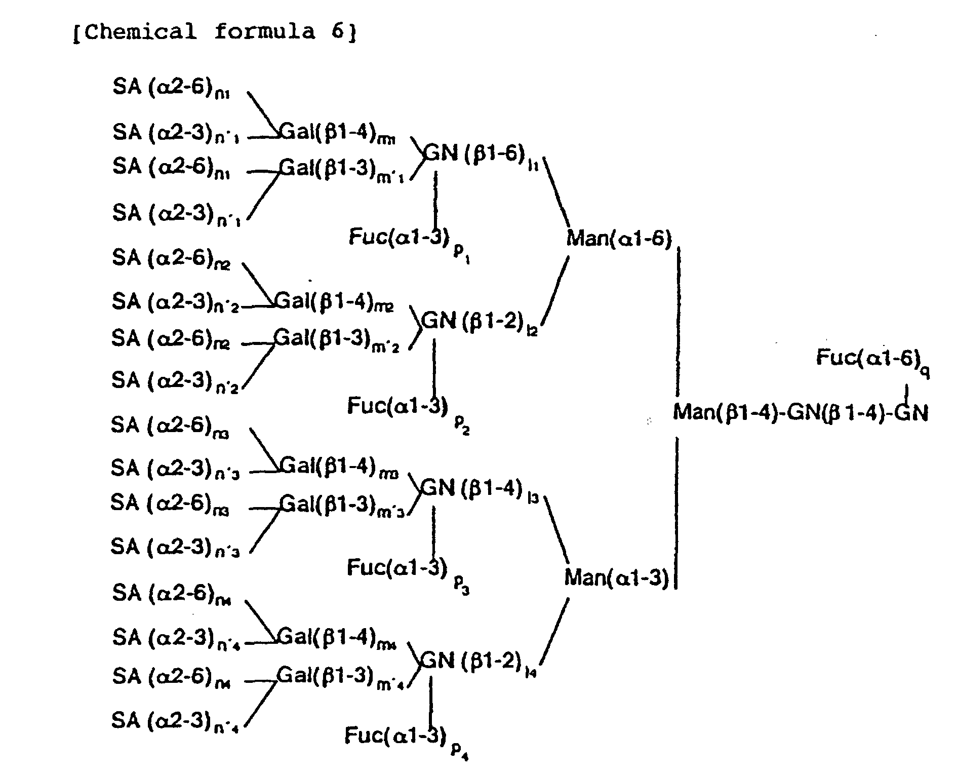
- Sugar chains of the N -acetyllactosamine type include, for example, those represented by the following general formula (6).
- SA represents a sialic acid residue
- Gal represents a galactose residue
- GN represents an N -acetylglucosamine residue
- Fuc represents a fucose residue
- Man represents a mannose residue
- M3 core a common structural unit
- the N -acetyllactosamine type of sugar chain has a dendritic structure comprising the M3 core having branches through four GN's.
- the branches (hereinafter the group comprising a branched GN and sugar residues at the non-reducing terminal side of the GN will be referred to as "branch") comprise GN, Gal, SA and Fuc sugar residues.
- Gal and SA each have two modes of linkage, i.e. Gal ⁇ -1,3 or Gal ⁇ -1,4, and SA ⁇ -2,3 or SA ⁇ -2,6, respectively. These modes of linkage must be distinguished from each other.
- the number of each sugar residue constituting a branch is at most 1 and the sequence thereof is fixed.
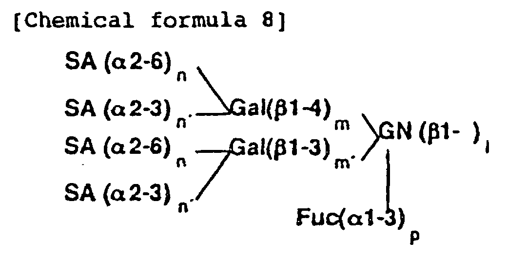
- the contents of each branch are represented by the following formula 8.
- the structure of the N -acetyllactosamine type of sugar chain can be determined merely by determining the presence or absence of each sugar residue in each branch, and the sequence of the sugar residues is determined as a matter of course.
- the determination of the presence or absence of each sugar residue in each branch is synonymous with the determination of the branch having a certain sugar residue. Therefore, the structure of the N -acetyllactosamine type of sugar chain can be determined by determining the branch having a certain sugar residue in each case.
- the branches having a certain sugar residue of which the presence should be determined can be distinguished from the branches not having the residue by removing the branches not having the residue from a sugar chain sample by the enzymatic or chemical treatment.
- branches not having GN When the branches do not have GN, a sugar chain sample has no branch and thus the removal of the branches not having GN is unnecessary.
- the branches not having Gal, SA, or Fuc can be removed from a sugar chain sample, for example, in the following way.
- the branches do not have Gal, they may have GN and/or Fuc and thus the branches can be removed, for example, by the treatment of a sugar chain sample with the mixture of two enzymes, ⁇ - N -acetylglucosaminidase and ⁇ -fucosidase.
- GN is protected from the treatment by Gal and thus the branches having Gal are not removed.
- the branches do not have SA they may have GN, Gal, and/or Fuc and thus the branches can be removed, for example, by the treatment of a sugar chain sample with the mixture of three enzymes, ⁇ - N -acetylglucosaminidase, ⁇ -galactosidase, and ⁇ -fucosidase.
- GN and Gal are protected from the treatment by SA and thus the branches having SA are not removed.
- the branches do not have Fuc, they may have GN, Gal, and/or SA and thus the branches can be removed, for example, by the treatment of a sugar chain sample with the mixture of three enzymes, ⁇ - N -acetylglucosaminidase, ⁇ -galactosidase, and sialidase.
- GN is protected from the treatment by Fuc and thus the branches having Fuc are not removed.
- the remaining branches should be identified.
- the remaining branches are converted into those with the simplest structure, that are to say the branches having only GN by the enzymatic or chemical treatment. After this treatment, the number of final products is so limited that the final products can be easily identified.
- the presence or absence of each branch in the treatment product can be determined by identifying the treatment product of the sugar chain with any of the 16 sugar chains, with the proviso that the 16 sugar chains can be separated by any method.
- the branch information for all the sugar residues constituting the branch can be obtained when the treatment can be conducted so as to leave only GN of the branch having a certain sugar residue among the branches and the 16 sugar chains given in Table 1 can be separated and identified by chromatographic analysis or the like. From these data, the structure of the N -acetyllactosamine type of sugar chain represented by the chemical formula 6 can be identified.
- the structure of a typical N -acetyllactosamine type of sugar chain can be principally determined by determining (i) the site of linkage of GN, namely the site of GN linkage (hereinafter the term "the site of linkage of a certain sugar residue” means the branch which has a certain sugar residue and its linkage) (ii) the site and mode of Gal linkage, (iii) the site and mode of SA linkage, and (iv) the site of Fuc linkage.
- n x n' x 0, namely, m x , m' x ⁇ n x , n' x , because SA is linked to only Gal.
- Fuc can be linked to GN at the reducing terminus of an N -acetyllactosamine type of sugar chain in ⁇ -1,6-linkage mode, and also to four GN's in branches in ⁇ -1,3-linkage mode. Therefore, the determination of the site and mode of Fuc linkage means the determination of the five variables, q, p 1 , p 2 , p 3 and p 4 in the above formula (6).
- Gal is linked to GN in ⁇ -1,3-linkage mode
- Fuc is linked to the GN in ⁇ -1,3-linkage mode.
- p x m' x 0 (x being 1, 2, 3 or 4).
- the following informations, (i) to (iv), of a certain sugar chain can be determined by digesting the sugar chain with at least one kind of glycosidase and analyzing the reaction products: (i) the site of GN linkage, (ii) the site and mode of Gal linkage, (iii) the site and mode of SA linkage, and (iv) the site of Fuc linkage.
- the whole structure of a sugar chain sample can be determined by the enzymatic digestions, which are designed so that only GN linked to M3 core remains; that only GN's at the branches having Gal remain; that only GN's at the branches having SA remain; and that only GN's to which Fuc is linked remain, and by identification of each reaction product by using standard sugar chains.
- the standard sugar chains used in this invention are 16 sugar chains in which only GN's are linked to the M3 core represented by the following formula (9), which include M3 core also. wherein Y 1 , Y 2 , Y 3 and Y 4 each represent hydrogen or ⁇ -GN.
- the site of GN linkage to the M3 core in the final reaction product should be determined.
- the relationship between 16 sugar chains, namely, oligosaccharides I to XVI, with the site of GN linkage is given in Table 1.
- the oligosaccharides I to XVI can be prepared by digestion with several glycosidases from N -acetyllactosamine type of sugar chain.
- the oligosaccharide XVI can be prepared by complete digestion of a sugar chain, which can be obtained from human ⁇ 1 -acid glycoprotein (Sigma) by hydrazinolysis and N-acetylation, with sialidase, ⁇ -galactosidase, and fucosidase.
- the oligosaccharide XVI can be partially digested with ⁇ - N -acetylglucosaminidase from bovine kidney to form a mixture of the oligosaccharides I to XVI, which can be purified by HPLC or the like.
- the oligosaccharide VIII can be prepared by complete digestion of a sugar chain, which can be obtained from bovine fetuin (Sigma) by hydrazinolysis and N-acetylation, with sialidase and ⁇ -galactosidase.
- the oligosaccharide VIII can be partially digested with ⁇ - N -acetylglucosaminidase from bovine kidney to form a mixture of the oligosaccharides I to VIII, which can be purified by HPLC or the like. These oligosaccharides can be detectd with the pulsed amperometric detector (PAD) with high sensitivity.
- PID pulsed amperometric detector
- oligosaccharides may be used as intact form or may be used as the form represented by the following formula (10). wherein Y 1 , Y 2 , Y 1 and Y 4 each represent hydrogen or ⁇ -GN, and R 2 represents an aldehyde group, a labeled methylene group or a labeled methine group.
- the methylene group can be labeled with, for example, 2-aminopyridine [ Agricultural and Biological Chemistry , 54 , 2169-2170 (1990)], p-aminobenzoic acid ethyl ester (ABEE) [ Analytical Biochemistry , 141 , 366-381 (1984)], or 8-aminonaphthalene-1,3,6-trisulfonic acid (ANTS) [ Biochemical Journal , 270 , 705-713 (1990)].
- 2-aminopyridine Agricultural and Biological Chemistry , 54 , 2169-2170 (1990)
- ABEE p-aminobenzoic acid ethyl ester
- ANTS 8-aminonaphthalene-1,3,6-trisulfonic acid
- the methine group can be labeled with, for example, radioactive material (tritium) [ Methods in Enzymology , 50 , 50 (1978)] or 1-phenyl-3-methyl-5-pyrazolone (PMP) [ Analytical Biochemistry , 180 , 351-357 (1989)].
- Oligosaccharides I to XVI listed in Table 1 may be labeled with 2-aminopyridine. PA-oligosaccharide I to XVI will be referred to as I-PA to XVI-PA, respectively.
- I-PA to VII-PA can be prepared by, for example, partial digestion of VIII-PA, which can be obtained from oligosaccharide VIII by PA-method, with ⁇ - N -acetylglucosaminidase from bovine kidney, and by purification of the digestion products by HPLC.
- IX-PA to XV-PA can be prepared by, for example, partial digestion of XVI-PA, which can be obtained from oligosaccharide XVI by PA-method, with ⁇ - N -acetylglucosaminidase from bovine kidney, and by purification of the digestion products by HPLC.
- PA-oligosaccharides can be used as the standard to identify the structure of the final reaction product of this invention.
- the mixture of the eight oligosaccharides I-PA to VIII-PA can be separated by, for example, HPLC [eluent: 100 mM acetic acid/triethylamine (pH 4.0) containing 0.035% 1-butanol] with PALPAK® TYPE R (Takara Shuzo).
- Figure 1 shows an example of the separation of an equimolar mixture of I-PA to VIII-PA.
- the ordinate represents the relative fluorescence intensity
- the abscissa represents the elution time (min) (the same shall apply hereinafter).
- the mixture of 16 oligosaccharides I-PA to XVI-PA can be separated by HPLC.
- the mixture is analyzed by HPLC [eluent: 50 mM acetic acid/triethylamine (pH 7.3) containing acetonitrile; linear gradient of acetonitrile 70% (0 min) to 50% (300 min)] with PALPAK® TYPE N (Takara Shuzo).
- I-PA to XVI-PA can be separated from each other on the basis of a difference in the number of the component sugar residues.
- An example thereof is given in Fig. 2.
- Fig. 2 an oligosaccharide in which no GN is linked to the M3 core at all, i.e.
- IV-PA is eluted during the period of 13 to 17 min; those in which one GN is linked to the M3 core (I-PA, V-PA, VI-PA and XII-PA) are eluted during the period of 18 to 23 min (fraction 2); those in which two GN's are linked to the M3 core (II-PA, III-PA, VII-PA, IX-PA, XIII-PA and XIV-PA) are eluted during the period of 24 to 29 min (fraction 3); those in which three GN's are linked to the M3 core (VIII-PA, X-PA, XI-PA and XV-PA) are eluted during the period of 32 to 38 min (fraction 4); and that in which four GN's are linked to the M3 core (XVI-PA) is eluted during the period of 42 to 46 min.
- fraction 3 is further purified by HPLC [eluent: 100 mM acetic acid/triethylamine containing 0.035% 1-butanol (pH 4.0)] with PALPAK® TYPE R, six oligosaccharides, i.e. II-PA, III-PA, VII-PA, IX-PA, XIII-PA and XIV-PA, can be separated.
- Figure 5 shows an example thereof.
- 16 oligosaccharides, I-PA to XVI-PA can be separated.
- the 16 PA-oligosaccharides can be isolated by pooling the fractions containing each peak in Figs. 2 to 5.
- XIV-PA can be isolated by pooling the fraction containing the peak in Fig. 5, it can be more easily obtained by the following method.
- a portion of the fraction 3 can be completely digested with ⁇ - N -acetylglucosaminidase from D. pneumoniae specific for ⁇ -1,2-linkage and then the products can be analyzed by HPLC under the same conditions as those of Fig. 5, and then the peaks of III-PA and VII-PA disappear (because they are converted into I-PA and IV-PA, respectively), leaving only XIV-PA.
- XIV-PA can be obtained by pooling the fraction containing the remaining peak.
- IX-PA to XV-PA can also be efficiently prepared from XVI-PA, for example, PA-sugar chain 004 (Takara Shuzo) by use of sialyl transferase and galactose oxidase.
- PALPAK® TYPE N column [eluent A, a 25:75 mixture of 0.2 M acetic acid/triethylamine buffer at pH 7.3 and acetonitrile; eluent B, a 50:50 mixture of 0.1 M acetic acid/triethylamine buffer at pH 7.3 and acetonitrile; after injection of a sample into the column equilibrated with a 80:20 mixture of eluent A and B, the ratio of eluent B is increased on a linear gradient to 40% over a 75-min period.], and PALPAK® TYPE R column [eluent: 100 mM acetic acid/triethylamine (pH 4.0) containing 0.02% 1-butanol].
- FIG. 7 A typical elution profiles of the mixture of the sixteen PA-oligosaccharides on the two columns are shown in Fig. 7.
- the elution position of each PA-oligosaccharide on the two columns can be plotted and thus the two-dimensional sugar map can be made.
- Figure 7 also shows an example of two-dimensional sugar map. Final reaction product of this invention can be easily identified by use of this two-dimensional sugar map.
- I-PA The proton nuclear magnetic resonance ( 1 H-NMR) spectrum of I-PA is given in Figs. 8 and 9, that of II-PA in Figs. 10 and 11, that of III-PA in Figs. 12 and 13, that of IX-PA in Fig. 14, that of X-PA in Figs. 15 and 16, that of XI-PA in Fig. 17, that of XII-PA in Fig. 18, that of XIII-PA in Fig. 19 and that of XIV-PA in Fig. 20.
- a sugar chain sample is digested with glycosidases in one or more steps including a reaction-termination operation in the course of the reaction, and the final product is subjected to chromatographic analysis comparing with the above-described standard sugar chains.
- the sugar chain to be analyzed may be any of that represented by above chemical formula (6). Even sugar chain which is not represented by the formula (6) can be analyzed by the method of the present invention so far as it can be converted into that represented by the formula (6) by enzymatic or chemical treatments.
- a sugar chain with a polylactosamine structure which has the repeating N -acetyllactosamine [(-3)Gal( ⁇ 1-4)GlcNAc( ⁇ 1-)] being linked to the Gal residues of the formula (6) is not represented by the formula (6).
- the sugar chain with polylactosamine can be converted into a sugar chain represented by the formula (6) by sequential digestion with ⁇ -galactosidase and ⁇ - N -acetylglucosaminidase [T. Ohkura et al. , J. Biol. Chem. (1981) 256 , 8485-8490] or by digestion with endo- ⁇ -galactosidase from Escherichia freundii .
- the sugar chain to be analyzed may be labeled or not.
- the sugar chain sample may be a mixture of several sugar chains, the purity thereof is preferably at least 80%.
- the glycosidase used in the respective steps may be a single one or a mixture of two or more glycosidases.
- the glycosidase to be used is preferably exoglycosidases, endoglycosidases can also be used.
- the reaction temperature which may not be particularly limited is usually 20 to 50°C, preferably 37°C.
- the reaction time which may not be particularly limited is usually 10 min to 20 h, preferably 3 to 5 h.
- the reaction is terminated by inactivation of the glycosidases by incubating the reaction mixture at 80 to 120°C for 3 to 10 min. Such an inactivation step is not always necessary.
- the inactivation step is not always necessary when the glycosidases used in the previous step show no activities because the optimum temperature, pressure, pH, concentration of the organic solvent, etc., of the subsequent step are extremely different from those of the previous step, or when the glycosidase used in the previous step can be removed with antibody beads or an ion-exchange resin.
- the analytical method of the reaction product may include thin layer chromatography, electrophoresis, mass spectrometry, gas chromatography, etc., and the analysis by HPLC is preferable.
- the reaction sequence include usually 1 to 6 reactions per sample. The products of each reaction are analyzed, and the sugar chain structure is determined by combining various pieces of the information thus obtained. Below, each reaction is described in detail.
- the reaction product should be one of sugar chains repesented by chemical formula (9) or (10).
- the reaction product can be identified by using the above-described standard sugar chains, and then the site of GN linkage in the starting sugar chain sample, i.e. the variables l 1 , l 2 , l 3 and l 4 in the formula (6), can be determined.
- glycosidases used herein can be preferably those having a low substrate specificity, such as ⁇ -galactosidase from jack beans (Seikagaku-Kogyo), ⁇ -galactosidase from Streptococcus (Seikagaku-Kogyo), sialidase from Arthrobacter ureafaciens (Nacalai Tesque), ⁇ -fucosidase from bovine kidney (Boehringer Mannheim), etc.
- glycosidases used herein can be preferably those having a low substrate specificity, such as ⁇ - N -acetylglucosaminidase from bovine kidney (Boehringer Mannheim), ⁇ -galactosidase from jack beans, ⁇ -galactosidase from Streptococcus , sialidase from A. ureafaciens , and ⁇ -fucosidase from bovine kidney.
- a sugar chain sample is digested with the same mixture of glycosidases as described above except that a linkage-specific ⁇ -galactosidase is used.
- the sugar chain is digested with a mixture of ⁇ - N -acetylglucosaminidase, sialidase, ⁇ -fucosidase and ⁇ -1,4-linkage specific or ⁇ -1,3-linkage-specific ⁇ -galactosidase.
- Gal which can be digested with the linkage-specific ⁇ -galactosidase, and GN at the same branch, and Fuc and GN, which are not substituted by Gal, can be removed from the chain, leaving Gal, which cannot be digested with the linkage-specific ⁇ -galactosidase, and GN at the same branch.
- the sugar chain is further digested with non-specific ⁇ -galactosidase to remove Gal.
- the reaction product must be one of sugar chains repesented by chemical formula (9) or (10).
- the reaction product can be identified by using the above-described standard sugar chains, and then the site of Gal linkage, which cannot be hydrolyzed with the linkage specific ⁇ -galactosidase, in the starting sugar chain sample can be determined.
- the site of Gal ⁇ -1,3-linkage i.e. the variables m' 1 , m' 2 , m' 3 and m' 4 in the formula (6), can be determined.
- the site of Gal ⁇ -1,4-linkage namely, the variables m 1 , m 2 , m 3 and m 4 in the formula (6), can be determined by combining these data.
- n x or n' x (x being 1, 2, 3 or 4) is 1 or, in other words, it is determined that (n x + n' x ) is either 0 or 1.
- the glycosidases used herein can be preferably those having a low substrate specificity, such as ⁇ - N -acetylglucosaminidase from bovine kidney, ⁇ -galactosidase from jack beans, ⁇ -galactosidase from Streptococcus , sialidase from A. ureafaciens , ⁇ -fucosidase from bovine kidney, etc.
- a sugar chain sample is digested with the same mixture of glycosidases as described above except that a linkage-specific sialidase is used.
- the sugar chain is digested with a mixture of ⁇ - N -acetylglucosaminidase, ⁇ -galactosidase, ⁇ -fucosidase and ⁇ -2,3-linkage- or ⁇ -2,6-linkage-specific sialidase.
- SA which cannot be hydrolyzed with the specific sialidase used in the reaction, and Gal and GN at the same branch are left after the reaction.
- the sugar chain is further digested with non-specific sialidase and ⁇ -galactosidase to remove SA and Gal.
- the reaction product must be one of sugar chains repesented by chemical formula (9) or (10).
- the reaction product can be identified by using the above-described standard sugar chains, and then the site of SA linkage, which cannot be hydrolyzed with the linkage specific sialidase, in the starting sugar chain sample can be determined. For example, when ⁇ -2,3-linkage-specific sialidase from Salmonella typhimurium (Takara Shuzo) is used, the site of SA ⁇ -2,6-linkage by this reaction, i.e.
- the variables n 1 n 2 , n 3 and n 4 in the formula (6) can be determined. Since n 1 +n' 1 , n 2 +n' 2 , n 3 +n' 3 and n 4 +n' 4 have been determined by the above-described reaction, the site of SA ⁇ -2,3-linkage, namely, the variables n' 1 , n' 2 , n' 3 and n' 4 in the formula (6), can be determined by combining these data.
- the standard oligosaccharides represented by the formula (9) or (10) are not necessarily used.
- the sugar chain sample is digested with ⁇ -galactosidase first, and then the enzyme is removed or inactivated. Then the reaction mixture is digested with sialidase.
- the site of SA linkage can be determined, and also, the mode of SA linkage can be determined by use of a sialidase that is specific for ⁇ -2,3-linkage.
- the reaction product can be identified by using the above-described standard sugar chains, and then the site of Fuc ⁇ -1,3-linkage in the starting sugar chain sample, i.e., the variables p 1 , p 2 , p 3 and p 4 in the formula (6), can be determined.
- the glycosidase used herein can be preferably one having a low substrate specificity, such as ⁇ - N -acetylglucosaminidase from bovine kidney, ⁇ -galactosidase from jack beans, ⁇ -galactosidase from Streptococcus , sialidase from A. ureafaciens , ⁇ -fucosidase from bovine kidney, etc.
- the presence or absence of Fuc ⁇ -1,6-linkage to GN at the reducing terminus of the sugar chain sample, namely, the variable q, in the formula (6) can be determined by using ⁇ -1,3/4-linkage-specific ⁇ -fucosidase.
- ⁇ -1,3/4-linkage-specific ⁇ -fucosidase When the sugar chain is digested with a mixture of ⁇ - N -acetylglucosaminidase, ⁇ -galactosidase, sialidase and, for example, ⁇ -1,3/4-fucosidase from Streptomyces (Takara Shuzo), GN, Gal, SA and ⁇ -1,3-linked Fuc are removed, leaving ⁇ -1,6-linked Fuc.
- the product when the sugar chain sample is free from ⁇ -1,6-linked Fuc or, in other words, when q is 0, the product must be the M3 core, which can be distinguished from M3 core with ⁇ -1,6-linked Fuc. From these results, the presence or absence of Fuc ⁇ -1,6-linkage can be determined.
- the N -acetyllactosamine type of sugar chain occasionally contains Fuc( ⁇ 1-2)Gal sequence, in addition to the modes of linkage represented by the formula (6).
- the site of Fuc ⁇ -1,2-linkage can be determined by the same reaction as described above except that an ⁇ -1,2-linkage-specific fucosidase, such as ⁇ -1,2-fucosidase from Arthrobacter (Takara Shuzo), is used.
- the definite structure can be determined from the above-described results, (i) to (iv).
- the site of GN linkage namely, the variables, l 1 , l 2 , l 3 and l 4 in the formula (6), can be determined from the results of (i);
- the site and mode of Gal linkage namely, the variables, m 1 , m' 1 , m 2 , m' 2 , m 3 , m' 3 , m 4 and m' 4 in the formula (6), can be determined from the results of (ii);
- the site and mode of SA linkage namely, the variables n 1 , n' 1 , n 2 , n' 2 , n 3 , n' 3 , n 4 and n' 4 in the formula (6), can be determined from the results of (iii);
- the site of Fuc linkage namely, the variables q, p
- the branches having a certain sugar residue of which the presence should be determined can be distinguished from the branches not having the residue by removing the branches having the residue from a sugar chain sample by the enzymatic or chemical treatment.
- GN residues in the branches having Gal ⁇ -1,3-linkage can be specifically removed by digestion with lacto- N -biosidase, which releases disaccharide, Gal( ⁇ 1-3)GN, from the non-reducing terminus of a sugar chain [M. Sano et al. , Proc. Natl. Acad. Sci. U.S.A. 89, 8512-8516 (1992)].
- the branches having Gal( ⁇ 1-3)GN can be specifically removed by the treatment with the mixture of sialidase, ⁇ -fucosidase, and lacto- N -biosidase. Then the reaction products can be digested with ⁇ -galactosidase and the final reaction product must be one of sugar chains represented by chemical formula (9) or (10).
- the branches being missing from the branches having GN by this treatment can be determined as the branches having Gal( ⁇ 1-3)GN, i.e. the branches having Gal ⁇ -1,3-linkage.
- oligosaccharides represented by the following formula (12), as the standard sugar chains.
- Z 1 , Z 2 , Z 3 , Z 4 , Z 5 and Z 6 each represent hydrogen or ⁇ -GN.
- sugar chain has the core structure represented by the above formula (12), which can be modified with Gal, SA and Fuc residues.
- the standard sugar chains represented by formula (12) might be useful for the analysis of these sugar chains. Five oligosaccharides which are particularly useful for the analysis are given in Table 2.
- oligosaccarides can be used without labeling or after labeling like the oligosaccharides I to XVI.
- the typical mode of a method of the present invention includes a process of identifying the resulting product from the treatment by comparing with the standard sugar chains, however, if the structure of the resulting product can be determined by other analytical techniques including NMR spectroscopy and mass spectrometry, these can be employed for the present invention.
- the structure of the sugar chain can be determined more easily by combining reagents used in the process of the present invention to form a kit.
- the kit comprises at least one of the oligosaccharides of the formula (4) and, if necessary, another oligosaccharide of the formula (9) or (10).
- a kit containing I-PA to XVI-PA is widely usable for the analysis of the N -acetyllactosamine type of sugar chains.
- the kit may contain glycosidases, a column for the analysis, buffer solution, etc., in addition to the oligosaccharide.
- the reagents to be contained in the kit may be in the form of either a solution or freeze-dried product.
- BPLC conditions were as follows: apparatus LC 6A (Shimadzu), column PALPAK® TYPE R (4.6 mm ⁇ x 250 mm) (Takara Shuzo) eluent 100 mM acetic acid/triethylamine containing 0.035% of 1-butanol (pH 4.0) detection conducted with fluorescence detector RF-535 (Shimadzu) at excitation wavelength of 320 nm and fluorescence wavelength of 400 nm flow rate 1 ml/min column temperature 40°C (hereinafter referred to as "Condition 1")
- composition of each of three purified oligosaccharides, I-PA, II-PA and III-PA were analyzed by mass spectrometry with a API-III mass spectrograph (Perkin-Elmer Sciex), NMR spectroscopy (in D 2 O) with a 400 MHz proton nuclear magnetic resonance spectrometer (Bruker), and monosaccharide analysis after acid hydrolysis.
- a solution comprising each 1 pmol of I-PA, II-PA and III-PA obtained in Example 1 and commercially available IV-PA (PA Sugar Chain 016; Takara Shuzo), V-PA (PA standard sugar chain 100.1; Nakano Vinegar), VI-PA (PA standard sugar chain 100.2; Nakano Vinegar), VII-PA (PA Sugar Chain 012; Takara Shuzo) and VIII-PA (PA Sugar Chain 013; Takara Shuzo) was treated by HPLC under the Condition 1 (Fig. 1). Eight kinds of the PA-oligosaccharides were separated.
- the mixture was separated by HPLC under the Condition 2.
- the obtained chart is given in Fig. 2.
- the peaks were divided into five fractions, i.e. fraction 1 eluted during the period of 13 to 17 min, fraction 2 during the period of 18 to 23 min, fraction 3 during the period of 24 to 29 min, fraction 4 during the period of 32 to 38 min, and fraction 5 during the period of 42 to 46 min.
- the sugar chain could be separated on the basis of the number of sugar residues constituting the chain.
- the fraction 3 was one in which two GN's were linked to the M3 core, namely, it was identified with a mixture of six oligosaccharides, i.e. II-PA, III-PA, VII-PA, IX-PA, XIII-PA and XIV-PA;
- the fraction 4 was one in which three GN's were linked to the M3 core, namely, it was identified with a mixture of four oligosaccharides, i.e. VIII-PA, X-PA, XI-PA and XV-PA; and the peak of the fraction 5 obtained at 44.0 min was one in which four GN's were linked to the M3 core, namely, it was identified with XVI-PA.
- the oligosaccharides of the respective peaks thus fractionated were partially digested with ⁇ - N -acetylglucosaminidase from bovine kidney and then treated by HPLC under the Condition 2 to separate a fraction in which one GN was linked to the M3 core.
- the fraction thus recovered was then analyzed by HPLC under the Condition 3. Since V-PA, VI-PA and XII-PA were detected in the digest of the peak of 10.5 min in Fig. 4, the peak was identified with XV-PA. Also, since I-PA, V-PA and XII-PA were detected in the digest of the peak of 13.3 min in Fig. 4, the peak was identified with X-PA.
- the reaction mixture was desalted through a SephadexTM G-15 column, and the sugar chain fraction was injected to a monoQTM column (Pharmacia) and eluted, the gradient being 10 mM to 100 mM ammonium acetate (pH 8.5). A fraction containing 3 mol of sialic acid per mole of the sugar chain was recovered from the eluted fractions. The amount of the recovered sialylated sugar chain was 646 nmol.
- reaction mixture was treated on a boiling water bath for 10 min to terminate the reaction. Then 1 U of sialidase from A. ureafaciens (Kyokuto Pharmaceutical) was added to the reaction mixture to conduct digestion at 37°C for 2 h. Then 250 U of ⁇ -galactosidase from Aspergillus (Toyobo) was added to the reaction mixture and the reaction was continued at 37°C for 5.5 h.
- the pH of the reaction mixture was adjusted to about 11 with 5 N sodium hydroxide.
- 20 mg of sodium borohydride (NaBH 4 ) was added to the reaction mixture, and the resultant mixture was left to stand at room temperature for 30 min. Additional 30 mg of sodium borohydride was added to the reaction mixture, and the resultant mixture was left to stand at room temperature for 30 min to complete the reduction reaction.
- the reduction reaction mixture was desalted through a SephadexTM G-15 column and then injected to a PALPAK® TYPE R column.
- the elution was conducted with 0.1% 1-butanol and 0.05% trifluoroacetic acid, and the main peak eluting at 30 min was recovered.
- the resultant sugar chain was XVI-PA in which ⁇ -1,6-linked GN was protected with Gal.
- the amount of the recovered sugar chain was 300 nmol.
- a portion (250 nmol) of the protected sugar chain thus obtained was digested with 2.5 U of N -acetylglucosaminidase from bovine kidney in 50 mM citrate buffer (pH 5.0) in presence of 50 mM D-(+)-galactono-1,4-lactone (Nacalai Tesque). After the reaction for 3.5 h, the reaction mixture was treated on a boiling water bath for 10 min to terminate the reaction.
- the reaction mixture was desalted through a SephadexTM G-15 column and three fractions were recovered by HPLC, the conditions are described below, depending on the number of GN units linked to the M3 core (fraction A with one GN unit linked; fraction B with two GN units linked; and fraction C with three GN units linked).
- the HPLC pattern is shown in Fig. 21.
- each of six purified oligosaccharides IX-PA, X-PA, XI-PA, XII-PA, XIII-PA and XIV-PA, were analyzed by mass spectrometry with a API-III mass spectrograph, NMR spectroscopy (in D 2 O) with a 500 MHz proton nuclear magnetic resonance spectrometer (JEOL) or that of Bruker above-described (only X-PA), and monosaccharide analysis after acid hydrolysis.
- API-III mass spectrograph NMR spectroscopy (in D 2 O) with a 500 MHz proton nuclear magnetic resonance spectrometer (JEOL) or that of Bruker above-described (only X-PA)
- the 1 H-NMR spectrum of IX-PA is given in Fig. 14, that of X-PA in Figs. 15 and 16, that of XI-PA in Fig. 17, that of XII-PA in Fig. 18, that of XIII-PA in Fig. 19, and that of XIV-PA in Fig. 20.
- the chemical shift values in these 1 H-NMR spectra are represented by taking the chemical shift value of the methyl proton of acetone in D 2 O at 37°C as 2.218 ppm when DSS is used as the standard.
- the signal at 2.218 ppm was that of the methyl proton of acetone used as the internal standard.
- Each of the sixteen PA-oligosaccharides, I-PA to XVI-PA was analysed by HPLC with two kinds of columns, PALPAK® TYPE N column [condition 2], and PALPAK® TYPE R column [eluent: 100 mM acetic acid/triethylamine (pH 4.0) containing 0.02% 1-butanol; other conditions being the same as those of the condition 1 (hereinafter referred to as condition 4)].
- the elution profiles of the mixture of the sixteen PA-oligosaccharides on the two columns were shown in Fig. 7. The elution position of each PA-oligosaccharide on the two columns was plotted and thus the two-dimensional sugar map was made.
- Figure 7 also shows the two-dimensional sugar map.
- Final reaction product of this invention can be easily identified by use of this two-dimensional sugar map.
- Each of the elution time obtained by reversed-phase PPLC (PALPAK® TYPE R column) was expressed on the ordinate, and that obtained by size-fractionation HPLC (PALPAK® TYPE N column) was expressed on the abscissa.
- Kits 1 and 2 for determining the structure of N -acetyllactosamine type of sugar chains were prepared (Tables 3 and 4).
- oligosaccharides IV-PA, VII-PA, VIII-PA and XVI-PA to be contained in the kits were the above-mentioned ones produced by Takara Shuzo.
- the oligosaccharides V-PA and VI-PA were those produced by Nakano Vinegar.
- the oligosaccharides IX-PA to XV-PA were obtained by the method described in Example 3 or partial hydrolysis of the oligosaccharide XVI-PA with N -acetylglucosaminidase from bovine kideny followed by purification by HPLC by the method described in Example 2-(2).
- Contents of Kit 1 (for 100 runs) solution Reagent Amount (ml) 1 solution of mixture of oligosaccharides I-PA to VIII-PA (each 1 pmol/ ⁇ l) 0.1 2 250 mM citrate buffer (pH 5.0) 0.4 3 50 U/ml sialidase from A.
- ureafaciens 0.5 4 15 U/ml ⁇ -2,3-sialidase from S.
- ureafaciens 0.5 4 15 U/ml ⁇ -2,3-sialidase from S. typhimurium 0.5 5 5 U/ml ⁇ -galactosidase from D. pneumonia 0.5 6 5 U/ml ⁇ -galactosidase from Streptococcus 0.5 7 100 U/ml ⁇ - N -acetylglucosaminidase from bovine kidney 0.5 8 10 U/ml ⁇ -fucosidase from bovine kidney 0.5 9 1 U/ml ⁇ -1,3/4-fucosidase derived from Streptomyces 0.5 10 100 mM acetic acid/triethylamine (pH 4.0) containing 0.035% 1-butanol 1000 11 100 mM acetic acid/triethylamine (pH 4.0) containing 0.07% 1-butanol 1000 12-A 50 mM acetic acid/triethylamine (pH 7.3)
- Bovine fetuin is a glycoprotein having a molecular weight of about 48,000 and about 22% by weight thereof comprises sugar chains.
- the structure of the sugar chains of the fetuin has been analyzed in detail, so that it is suitable for use in the investigation of the sugar chains of glycoproteins.
- the main asparagine-binding sugar chains of the fetuin have such a structure that 0 to 5 SA's are linked to the structure of XXII or XXIII in Table 5.
- the structure of the fetuin varies depending on the number, site and mode of linkage of SA. Since most of the various structures of the fetuin are represented by the formula (6), they could be analyzed with the kit of the present invention.
- the PA sugar chain was divided into five fractions wherein 0 to 4 SA's were linked to each molecule. From the five fractions, a fraction having one SA and another fraction having two SA's were used. A part (1/100) of each fraction was separated by HPLC [eluent: 50 mM acetic acid/triethylamine (pH 5.0) containing 0.15% of 1-butanol] with a reversed-phase column CosmosilTM 5C 18 AR (4.6 mm ⁇ x 250 mm; Nacalai Tesque).
- a peak of 40 min was prepared from the fraction having one SA linked, and peaks of 38, 48 and 72 min were prepared from that having two SA's linked, and they were further purified by HPLC with ASAHIPAK® NH 2 P50.
- the sugar chain in the fraction of 48 min having 2 SA's, that of 38 min having 2 SA's, that of 40 min having 1 SA and that of 72 min having 2 SA's will be referred to as A, B, C and D, respectively.
- Each sugar chain was dissolved in 100 ⁇ l of distilled water to prepare a sample solution.
- the reaction mixture obtained by the reaction 2 was analyzed to find out that the product was III-PA as judged from the elution time thereof.
- the sugar chain A had SA in two Gal's which were linked to the two GN's on the ⁇ -1,3-linked Man of the PA oligosaccharide XXII (XXII-PA) in Table 5.
- the reaction mixture obtained by the reaction 3 was analyzed to find out that the product comprised about 80% of VI-PA and about 20% of III-PA as judged from the elution time thereof.
- the sugar chain A was determined to be a mixture of XXIV-PA and XXV-PA in a ratio of about 4:1, each having a structure given in Table 5.
- the reaction mixture obtained by the reaction 3 was analyzed to find out that the product was IV-PA as judged from the elution time thereof.
- the purified sugar chain C was determined to be a nearly 1:1 mixture of XXVII-PA and XXVIII-PA in Table 5.
- the oligosaccharides XXII to XXIX satisfy the conditions relating to variables l x , m x , m' x , n x , n' x , q and p x (x being 1, 2, 3 or 4) in the formula (6) given in the following Table 5.
- XXII-PA to XXIX-PA are obtained by labeling the oligosaccharides XXII to XXIX respectively with 2-aminopyridine.
- the symbol (1) represents that one of the two variables is 1 and the other is 0.
- the sugar chain fraction was obtained from 1 mg of ⁇ 1 -acid glycoprotein (Sigma) by hydrazinolysis and N-acetylation.
- sugar chain F 50 pmol of sugar chain E were digested with 5 mU of endo- ⁇ -galactosidase from Escherichia freundii at 37°C for 2 h, and a portion was analyzed by HPLC with an amide-silica column [ Anal. Biochem. 171 , 73-90, (1988)]. The product peak was eluted about 2.5 glucose units earlier than sugar chain E. This product was named sugar chain F.
- Endo- ⁇ -galactosidase might release Gal( ⁇ 1-4)GN( ⁇ 1-3)Gal from sugar chain E producing sugar chain F.
- the branch having N -acetyllactosamine unit was determined with Kit 2 of Example 4.
- the present invention provides a more exact, reliable, and simple method for determining the sugar chain structure, a kit to be used for said method, and novel oligosaccharides useful for said method as standards.
Landscapes
- Chemical & Material Sciences (AREA)
- Health & Medical Sciences (AREA)
- Life Sciences & Earth Sciences (AREA)
- Engineering & Computer Science (AREA)
- Molecular Biology (AREA)
- Immunology (AREA)
- Organic Chemistry (AREA)
- Biochemistry (AREA)
- Biotechnology (AREA)
- General Health & Medical Sciences (AREA)
- Biomedical Technology (AREA)
- Hematology (AREA)
- Urology & Nephrology (AREA)
- Genetics & Genomics (AREA)
- Food Science & Technology (AREA)
- Microbiology (AREA)
- Cell Biology (AREA)
- Medicinal Chemistry (AREA)
- Physics & Mathematics (AREA)
- Analytical Chemistry (AREA)
- General Physics & Mathematics (AREA)
- Pathology (AREA)
- Tropical Medicine & Parasitology (AREA)
- Measuring Or Testing Involving Enzymes Or Micro-Organisms (AREA)
- Saccharide Compounds (AREA)
Description
- The present invention relates to a method for determining sugar chain structures, a kit to be used for said method and novel oligosaccharides. The present invention, in particular, relates to a method for determining the structure of an N-acetyllactosamine type of sugar chain, a kit to be used for said method, and novel N-acetyllactosamine type of sugar chains.
- Recently, many evidences have been presented for proving the fact that the sugar chains per se function in the recognition of cells, adhesion of cells or metastasis of cancer and, therefore, the structural analysis of the sugar chain in glycoconjugates including glycoproteins and glycolipids is becoming more and more important. In the prior art, the chain structure was analyzed by various analytical methods including methylation analysis, periodate oxidation, enzymatic digestion, nuclear magnetic resonance spectroscopy, and mass spectrometry. In addition, it has become possible to analyze sugar chains by high-performance liquid chromatography (HPLC), which has been rapidly developed in recent years. A method for the fluorescence labeling of sugars with 2-aminopyridine has been developed by Hase et al. [Journal of Biochemistry, 95, 197-203 (1984); Hereinafter, this method and pyridylamino sugar will be referred to as PA-method and PA-sugar, respectively), and the combination of pyridylamination with HPLC using two kinds of columns, i.e. so-called two-dimensional mapping method makes it possible to analyze many oligosaccharides sensitively [Analytical Biochemistry, 171, 73-90 (1988)].
- However, it is impossible to distinguish sugar chains having the same elution position or elution positions which are very close to each other in the two kinds of columns from each other by this method. Further, this method is ineffective for sugar chains not mentioned in the map, particularly, for those having sialic acid residues, and it is thus unsuitable for the determination of the structure of a sugar chain having an unknown structure.
- When the structure of a sugar chain is to be determined by a method, which combines any labeling methods with any separation method, such as the combination of pyridylamination with HPLC, all of the possible sugar chains should be available for the standard sugar chains with the structures determined by any method, and moreover, they should be separated and identified by the separation methods, but this must be impossible because of the diversity of sugar chain structures. Thus, digestion with exoglycosidases with limited substrate specificities is usually used for determination of sugar chain structures. In this method, the products from exoglycosidase digestion are separated and identified by chromatography [see, for example, E. Tsuda et al., Biochemistry, 27, 5646-5654 (1988)]. The digestion with the exoglycosidase with limited substrate specificity will be hereinafter referred to as "enzymatic digestion".
- Exoglycosidases can recognize a kind of sugar residue at the non-reducing terminus of a sugar chain and the anomeric isomers of the sugar residue (α and β), and the exoglycosidases with limited substrate specificities can recognize the position of the linkages also. Here the combination of the anomeric isomers and the position of the linkage, e.g.: α-2,3-linkage or β-1,4-linkage will be referred to as the "mode of linkage." For example, β-galactosidase from Diplococcus pneumoniae recognizes only Galβ-1,4-linkage at a non-reducing terminus and hydrolyzes it, and β-galactosidase from jack beans recognizes β anomeric isomer of Gal at a non-reducing terminus and releases it. The kind of a sugar residue and its mode of linkage at the non-reducing terminus of a sugar chain can be estimated by consideration of the substrate specificities of exoglycosidases. For example, when a sugar chain sample can be digested with β-galactosidase from D. pneumoniae, it can be estimated that the sugar chain has a Galβ-1,4-linkage at the non-reducing terminus.
- The enzymatic digestion is generally conducted stepwise. For example, when a
sugar chain sample 1 is digested with an enzyme A, asugar chain 2 is obtained as the digestion product. Thesugar chain 2 is recovered and then digested with an enzyme B to give asugar chain 3, which is further digested with an enzyme C. Such a process is repeated. Therefore, the technique of structural analysis by the enzymatic digestion has serious defect that it requires the time consuming step for the recovery of the digestion product even though the techique can distinguish a difference in a fine structure. - To overcome this defect, a technique of systematically conducting the enzymatic digestion of the sugar chain to estimate the structure thereof has been developed [US Patent No. 5,100,778 and C. J. Edge et al., Proc. Natl. Acad. Sci. U.S.A., 89, 6338-6342 (1992)].
- This technique is characterized by a series of the operation comprising:
- 1. division in equal parts of a sugar chain sample the structure of which is to be determined,
- 2. complete digestion of the respective parts with a series of different enzyme mixtures,
- 3. chromatographic analysis of a mixture comprising each of the reaction mixture,
- 4. comparison of the analytical spectrum, namely, the elusion position and intensity of each peak obtained by the chromatographic analysis with the spectrum that is theoretically calculated from the sugar chain structure in data bases, and
- 5. estimation of the structure of a sugar chain sample by making a choice of structure giving an analytical spectrum that is statistically most close to that of the sugar chain sample among the database.
-
- This technique requires only one chromatographic analysis and it does not require the recovery of sugar chains, so the structural analysis of the sugar chain by the enzymatic digestion has been simplified and expedited by this technique.
- However, since this technique is based on the approximation of the experimental spectrum to the theoretical spectrum, there is a possibility that the estimated structure might be erroneous or two or more structures might be estimated, when the experimental spectrum is fundamentally distant from the theoretical one or when two or more sugar chains having identical or remarkably similar theoretical spectra are found in the data base. For example, according to C. J. Edge et al. [Proc. Natl. Acad. Sci. U.S.A., 89, 6338-6342 (1992)], even when a sugar chain having a simple structure of the following
formula 5is analyzed by this technique, two structures are theoretically estimated for it in equal probability from the analytical results and it is impossible to determine the true structure thereof. Edge and his co-workers say that this problem might be solved to some extent by adding an enzyme having a more exact linkage specificity to the enzyme mixture. However, when the number of enzymes to be used increases, the number of required enzyme mixtures also increases, therefore this technique is not practical, and moreover, the number of such specific enzymes is limited, and it is practically impossible to establish an enzyme mixture capable of distinguishing all of the possible structures of sugar chains.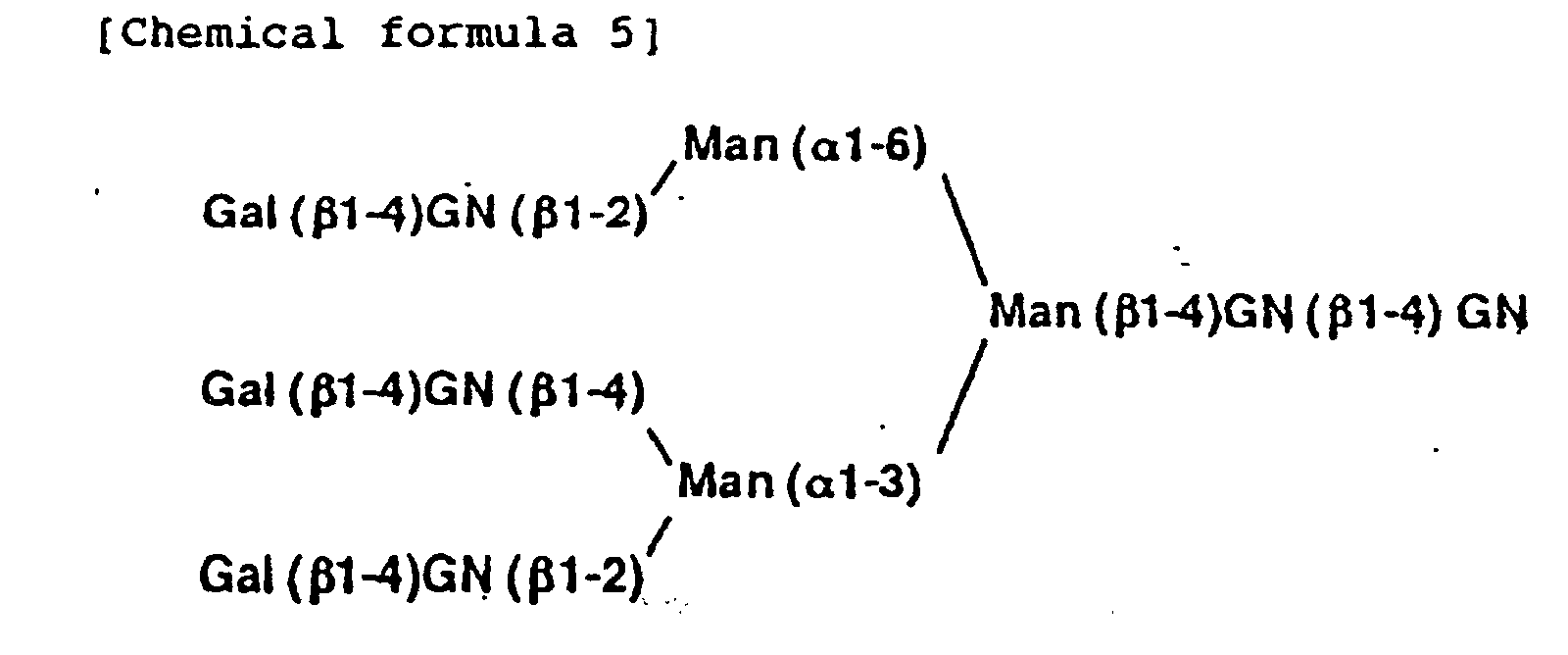
- This technique has another serious defect. Namely, the structures of many sugar chains, particularly asparagine-linked sugar chains, are not linear but complicatedly branched. Therefore, one sugar chain has two or more non-reducing termini, i.e. branches. Even when the kinds and number of the sugar residues and the mode of linkage of them are determined by the technique, it is impossible to determine which branches have the sugar residues if all of the branches do not have the residue. Thus the structure cannot be identified in some cases. In addition, this technique is very sensitive to factors which disturb the experimental spectrum, such as reduction in the activity of the enzyme used, contamination with a very small amount of an unexpected enzyme activity and contamination with other sugar chains. However, there are no means to confirm whether the obtained spectrum is correct or not. Although the analysis method is easy and rapid, it lacks accuracy of obtained spectrum and therefore it will not be enough for a means of the structural analysis of sugar chain.
- Biomedical Chromatography, vol. 6, pages 77-83 (1992) describes the analysis of oligosaccharides labelled with p-Aminobenzoic ethyl ester (ABEE-derivates). HPLC is used to separate the labelled derivatives and the resalting two-dimensional maps are used for structural analysis.
- Carbohydrate Research, vol. 130, 1984, pages 85-101 concerns the synthesis of the pentasaccharide core of N-acetyl glucosamine-type sugar chains, referred to herein as the M3 core.
- WO 92/19768 discloses oligosaccharide sequencing including an oligosaccharide containing the M3 core and a acetylglucosamine residue linked to a mannose residue. Various sequencing agents are used, including exoglycosidase residues.
- The cause of the above-described defects of this technique resides in lack of identification of each of the reaction product. The term "identification" as used herein means that a substance having an unknown structure is determined to be identical with a substance having a known structure by chromatographic analysis or the like. It is indispensable for the identification to collect all of the possible sugar chains as the standard for the analysis and they should be separated from each other by any analytical method. It is impossible, however, to have a complete collection of all of the possible sugar chains as the standard and to separate and distinguish them from each other because the number of such sugar chains is enormous. For these reasons, the approximation of the experimental spectrum to the theoretical spectrum is employed in place of the identification in this technique.
- Fundamentally, however, an indefinite approximation technique should not be employed for the determination of the structure of a substance. So long as the approximation technique is employed, there should be danger that the structure cannot be determined or an incorrect structure is given. Therefore, it has been desired to develop a more exact, reliable, and simple method for determining sugar chain structure.
- The present invention may provide a more exact, reliable, and simple method for determining the sugar chain structure, a kit to be used for said method, and novel oligosaccharides useful for said method as standards.
- To sum up, the present invention relates to a method for determining the structure of an N-acetyllactosamine type of sugar chain characterized by determining the site of linkage and the mode of linkage of sugar residues at the non-reducing terminal side of a β-N-acetylglucosamine residue linked to an α-mannose residue in M3 core by means of detecting the presence of the β-N-acetylglucosamine residue after enzymatic or chemical treatment of said sugar chain. Further, one of the modes of the method comprises the following steps:
- (1) selectively removing particular β-N-acetylglucosamine residues linked to an α-mannose residue in pentasaccharide core of N-acetyllactosamine-type sugar chain (formula 7 below) (M3 core);
- (2) in the event that the product of step (1) has a remaining sugar residue which is linked to the non-reducing terminal of β-N-acetylglucosamine residues linked to an α-mannose residue in the M3 core, removing the said remaining sugar residue; and
- (3) identifying the resulting products of step (1) or (2) by comparing them with standard sugar chains.
-
- The present invention also relates to a kit for determining the structure of an N-acetyllactosamine type of sugar chain comprising the components set out in claims 16 to 17.
- The present invention also relates to oligosaccharides set out in claims 18 to 21.
- The inventors of this invention achieved the purpose of this invention during research into a method of the structural analysis of sugar chains by development of a method of treating a sugar chain in order that the number of the possible products from the treatment should be limited for the sake of the employment of a process of identification, a kit to be used for this method, and novel oligosaccharides useful for a process of identification as standards.
- A detailed description will be made on the present invention.
- Sugar chains of the N-acetyllactosamine type include, for example, those represented by the following general formula (6).wherein SA represents a sialic acid residue, Gal represents a galactose residue, GN represents an N-acetylglucosamine residue, Fuc represents a fucose residue, Man represents a mannose residue, the symbol in the parentheses following each residue indicates the mode of linkage, and symbols lx, mx, m'x, nx, n'x, and px (x being 1, 2, 3 or 4) and q each represent a variable, with the proviso that mxm'x = nxn'x = m'xpx =0 and lx ≥ mx,m'x ≥ nx,n'x and lx ≥ px.
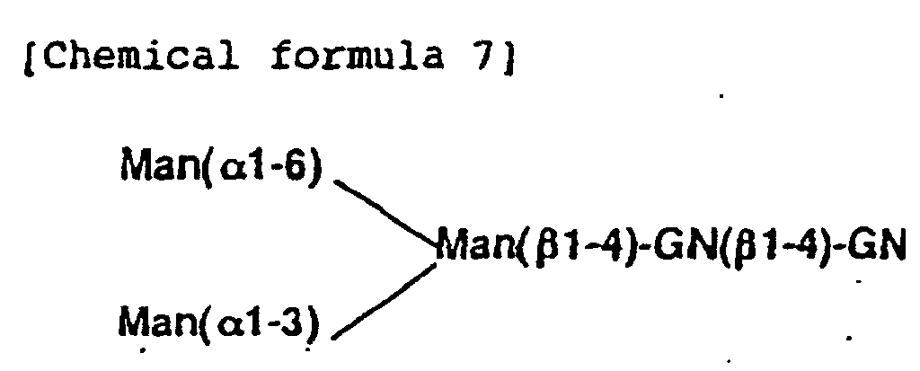
- Although the fine structures of the sugar chains of N-acetyllactosamine type are full of variety, the basic structure thereof has a common structural unit (hereinafter referred to as "M3 core") represented by the following formula (7).
- All the ordinary structures are represented by the above formula (6). The N-acetyllactosamine type of sugar chain has a dendritic structure comprising the M3 core having branches through four GN's. The branches (hereinafter the group comprising a branched GN and sugar residues at the non-reducing terminal side of the GN will be referred to as "branch") comprise GN, Gal, SA and Fuc sugar residues. Gal and SA each have two modes of linkage, i.e. Galβ-1,3 or Galβ-1,4, and SAα-2,3 or SAα-2,6, respectively. These modes of linkage must be distinguished from each other. The number of each sugar residue constituting a branch is at most 1 and the sequence thereof is fixed. The contents of each branch are represented by the following formula 8.
- Since the number of each sugar residue in a branch is limited to 0 or 1 and the sequence thereof has been specified as the chemical formula 8, the structure of the N-acetyllactosamine type of sugar chain can be determined merely by determining the presence or absence of each sugar residue in each branch, and the sequence of the sugar residues is determined as a matter of course.
- The determination of the presence or absence of each sugar residue in each branch is synonymous with the determination of the branch having a certain sugar residue. Therefore, the structure of the N-acetyllactosamine type of sugar chain can be determined by determining the branch having a certain sugar residue in each case.
- A description will be made on the technique of determining which branch has a certain sugar residue (branch information).
- In a typical mode of the method provided by this invention, the branches having a certain sugar residue of which the presence should be determined, can be distinguished from the branches not having the residue by removing the branches not having the residue from a sugar chain sample by the enzymatic or chemical treatment.
- When the branches do not have GN, a sugar chain sample has no branch and thus the removal of the branches not having GN is unnecessary. The branches not having Gal, SA, or Fuc can be removed from a sugar chain sample, for example, in the following way.
- When the branches do not have Gal, they may have GN and/or Fuc and thus the branches can be removed, for example, by the treatment of a sugar chain sample with the mixture of two enzymes, β-N-acetylglucosaminidase and α-fucosidase. In the branches having Gal, GN is protected from the treatment by Gal and thus the branches having Gal are not removed. When the branches do not have SA, they may have GN, Gal, and/or Fuc and thus the branches can be removed, for example, by the treatment of a sugar chain sample with the mixture of three enzymes, β-N-acetylglucosaminidase, β-galactosidase, and α-fucosidase. In the branches having SA, GN and Gal are protected from the treatment by SA and thus the branches having SA are not removed. When the branches do not have Fuc, they may have GN, Gal, and/or SA and thus the branches can be removed, for example, by the treatment of a sugar chain sample with the mixture of three enzymes, β-N-acetylglucosaminidase, β-galactosidase, and sialidase. In the branches having Fuc, GN is protected from the treatment by Fuc and thus the branches having Fuc are not removed. After the removal of the branches not having each sugar residue, Gal, SA, and Fuc, according to the method described above, the remaining branches should be identified. For the purpose of the identification, the remaining branches are converted into those with the simplest structure, that are to say the branches having only GN by the enzymatic or chemical treatment. After this treatment, the number of final products is so limited that the final products can be easily identified.
- The number of the structures with the M3 core having GN's is calculated as follows: 2x2x2x2=16, because each of the four GN's has two possibilities, i.e. presence and absence. These structures are given in Table 1. The presence or absence of each branch in the treatment product can be determined by identifying the treatment product of the sugar chain with any of the 16 sugar chains, with the proviso that the 16 sugar chains can be separated by any method.
- As described above, the branch information for all the sugar residues constituting the branch can be obtained when the treatment can be conducted so as to leave only GN of the branch having a certain sugar residue among the branches and the 16 sugar chains given in Table 1 can be separated and identified by chromatographic analysis or the like. From these data, the structure of the N-acetyllactosamine type of sugar chain represented by the
chemical formula 6 can be identified. - Below, a method of the present invention which employs digestion with glycosidases as the treatment of the sugar chain will be explained.
- The structure of a typical N-acetyllactosamine type of sugar chain can be principally determined by determining (i) the site of linkage of GN, namely the site of GN linkage (hereinafter the term "the site of linkage of a certain sugar residue" means the branch which has a certain sugar residue and its linkage) (ii) the site and mode of Gal linkage, (iii) the site and mode of SA linkage, and (iv) the site of Fuc linkage.
- The following four sites of GN linkage are usually possible in typical sugar chains: GN(β1-6)Man(α1-6), GN(β1-2)Man(α1-6), GN(β1-4)Man(α1-3) and GN(β1-2)Man(α1-3). Determination of the site of GN linkage among these four sites means the determination of the four variables, i.e. l1, l2, l3 and l4, in the formula (6).
- The sugar residues to which Gal can be linked are the above-mentioned four GN's, and two modes of Gal linkage, i.e. β-1,3 and β-1,4, are possible. Therefore, the determination of the site and mode of Gal linkage means the determination of the eight variables, m1, m'1, m2, m'2, m3, m'3, m4 and m'4. Because two or more Gal's are not linked to one GN, mxm'x = 0 (x being 1, 2, 3 or 4). Further, when lx = 0, then mx, m'x = 0, namely, lx ≥ mx, m'x, because Gal is linked to only GN.
- SA can be linked to Gal in two modes of linkage, i.e. α-2,3 and α-2,6. Therefore, the determination of the site and mode of SA linkage means the determination of the eight variables, n1, n'1, n2, n'2, n3, n'3, n4 and n'4 in the above formula (6). Because two or more SA's are not linked to one Gal, nxn'x = 0 (x being 1, 2, 3 or 4). Further, when mx, m'x = 0, then nx n'x = 0, namely, mx, m'x ≥ nx, n'x, because SA is linked to only Gal.
- Fuc can be linked to GN at the reducing terminus of an N-acetyllactosamine type of sugar chain in α-1,6-linkage mode, and also to four GN's in branches in α-1,3-linkage mode. Therefore, the determination of the site and mode of Fuc linkage means the determination of the five variables, q, p1, p2, p3 and p4 in the above formula (6). However, when Gal is linked to GN in β-1,3-linkage mode, it is impossible that Fuc is linked to the GN in α-1,3-linkage mode. Namely, pxm'x = 0 (x being 1, 2, 3 or 4). Further, when lx = 0, then px = 0, namely, px ≤ lx, since Fuc can be linked to only GN.
- The following informations, (i) to (iv), of a certain sugar chain can be determined by digesting the sugar chain with at least one kind of glycosidase and analyzing the reaction products: (i) the site of GN linkage, (ii) the site and mode of Gal linkage, (iii) the site and mode of SA linkage, and (iv) the site of Fuc linkage. In other words, the whole structure of a sugar chain sample can be determined by the enzymatic digestions, which are designed so that only GN linked to M3 core remains; that only GN's at the branches having Gal remain; that only GN's at the branches having SA remain; and that only GN's to which Fuc is linked remain, and by identification of each reaction product by using standard sugar chains.
- A description will be made on the standard sugar chains used in the present invention.
- The standard sugar chains used in this invention are 16 sugar chains in which only GN's are linked to the M3 core represented by the following formula (9), which include M3 core also.wherein Y1, Y2, Y3 and Y4 each represent hydrogen or β-GN.
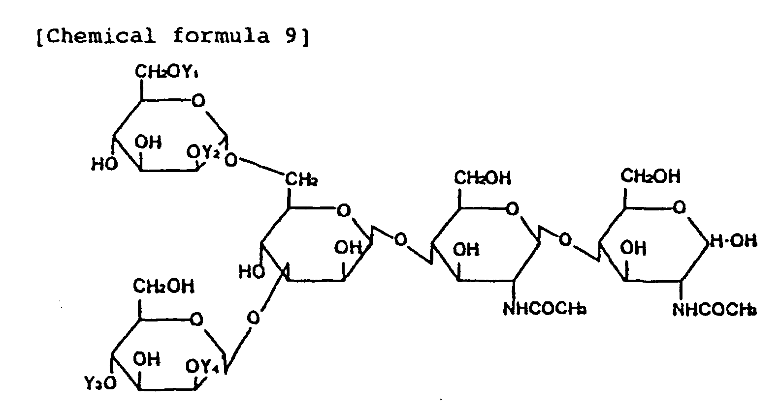
- By comparing the 16 standard sugar chains with the final reaction products, the site of GN linkage to the M3 core in the final reaction product should be determined. The relationship between 16 sugar chains, namely, oligosaccharides I to XVI, with the site of GN linkage is given in Table 1.
oligosaccharides Y1 Y2 Y3 Y4 I H H β-GN H II H β-GN β-GN H III H H β-GN β-GN IV H H H H V H β-GN H H VI H H H β-GN VII H β-GN H β-GN VIII H β-GN β-GN β-GN IX β-GN H H β-GN X β-GN β-GN β-GN H XI β-GN H β-GN β-GN XII β-GN H H H XIII β-GN β-GN H H XIV β-GN H β-GN H XV β-GN β-GN H β-GN XVI β-GN β-GN β-GN β-GN - The oligosaccharides I to XVI can be prepared by digestion with several glycosidases from N-acetyllactosamine type of sugar chain. For example, the oligosaccharide XVI can be prepared by complete digestion of a sugar chain, which can be obtained from human α1-acid glycoprotein (Sigma) by hydrazinolysis and N-acetylation, with sialidase, β-galactosidase, and fucosidase. The oligosaccharide XVI can be partially digested with β-N-acetylglucosaminidase from bovine kidney to form a mixture of the oligosaccharides I to XVI, which can be purified by HPLC or the like. Another example is that the oligosaccharide VIII can be prepared by complete digestion of a sugar chain, which can be obtained from bovine fetuin (Sigma) by hydrazinolysis and N-acetylation, with sialidase and β-galactosidase. The oligosaccharide VIII can be partially digested with β-N-acetylglucosaminidase from bovine kidney to form a mixture of the oligosaccharides I to VIII, which can be purified by HPLC or the like. These oligosaccharides can be detectd with the pulsed amperometric detector (PAD) with high sensitivity.
- These oligosaccharides may be used as intact form or may be used as the form represented by the following formula (10).wherein Y1, Y2, Y1 and Y4 each represent hydrogen or β-GN, and R2 represents an aldehyde group, a labeled methylene group or a labeled methine group.
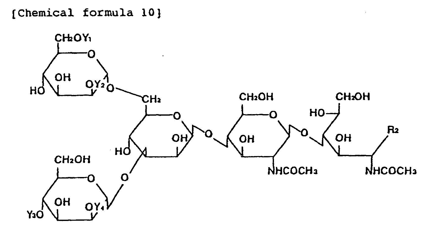
- The methylene group can be labeled with, for example, 2-aminopyridine [Agricultural and Biological Chemistry, 54, 2169-2170 (1990)], p-aminobenzoic acid ethyl ester (ABEE) [Analytical Biochemistry, 141, 366-381 (1984)], or 8-aminonaphthalene-1,3,6-trisulfonic acid (ANTS) [Biochemical Journal, 270, 705-713 (1990)].
- The methine group can be labeled with, for example, radioactive material (tritium) [Methods in Enzymology, 50, 50 (1978)] or 1-phenyl-3-methyl-5-pyrazolone (PMP) [Analytical Biochemistry, 180, 351-357 (1989)].
Oligosaccharides I to XVI listed in Table 1 may be labeled with 2-aminopyridine. PA-oligosaccharide I to XVI will be referred to as I-PA to XVI-PA, respectively. I-PA to VII-PA can be prepared by, for example, partial digestion of VIII-PA, which can be obtained from oligosaccharide VIII by PA-method, with β-N-acetylglucosaminidase from bovine kidney, and by purification of the digestion products by HPLC. IX-PA to XV-PA can be prepared by, for example, partial digestion of XVI-PA, which can be obtained from oligosaccharide XVI by PA-method, with β-N-acetylglucosaminidase from bovine kidney, and by purification of the digestion products by HPLC. These PA-oligosaccharides can be used as the standard to identify the structure of the final reaction product of this invention. - The mixture of the eight oligosaccharides I-PA to VIII-PA can be separated by, for example, HPLC [eluent: 100 mM acetic acid/triethylamine (pH 4.0) containing 0.035% 1-butanol] with PALPAK® TYPE R (Takara Shuzo). Figure 1 shows an example of the separation of an equimolar mixture of I-PA to VIII-PA. In Fig. 1, the ordinate represents the relative fluorescence intensity and the abscissa represents the elution time (min) (the same shall apply hereinafter).
- The mixture of 16 oligosaccharides I-PA to XVI-PA can be separated by HPLC. For example, the mixture is analyzed by HPLC [eluent: 50 mM acetic acid/triethylamine (pH 7.3) containing acetonitrile; linear gradient of
acetonitrile 70% (0 min) to 50% (300 min)] with PALPAK® TYPE N (Takara Shuzo). I-PA to XVI-PA can be separated from each other on the basis of a difference in the number of the component sugar residues. An example thereof is given in Fig. 2. In Fig. 2, an oligosaccharide in which no GN is linked to the M3 core at all, i.e. IV-PA, is eluted during the period of 13 to 17 min; those in which one GN is linked to the M3 core (I-PA, V-PA, VI-PA and XII-PA) are eluted during the period of 18 to 23 min (fraction 2); those in which two GN's are linked to the M3 core (II-PA, III-PA, VII-PA, IX-PA, XIII-PA and XIV-PA) are eluted during the period of 24 to 29 min (fraction 3); those in which three GN's are linked to the M3 core (VIII-PA, X-PA, XI-PA and XV-PA) are eluted during the period of 32 to 38 min (fraction 4); and that in which four GN's are linked to the M3 core (XVI-PA) is eluted during the period of 42 to 46 min. - When
fraction 2 is further purified by HPLC [eluent: 100 mM acetic acid/triethylamine (pH 4.0) containing 0.07% 1-butanol] with PALPAK® TYPE R, four oligosaccharides in which one GN is linked (i.e. I-PA, V-PA, VI-PA and XII-PA) can be separated. Figure 3 shows an example thereof. In the same manner, four oligosaccharides, i.e. VIII-PA, X-PA, XI-PA and XV-PA, can be purified fromfraction 4. Figure 4 shows an example thereof. Whenfraction 3 is further purified by HPLC [eluent: 100 mM acetic acid/triethylamine containing 0.035% 1-butanol (pH 4.0)] with PALPAK® TYPE R, six oligosaccharides, i.e. II-PA, III-PA, VII-PA, IX-PA, XIII-PA and XIV-PA, can be separated. Figure 5 shows an example thereof. Thus, 16 oligosaccharides, I-PA to XVI-PA, can be separated. - The 16 PA-oligosaccharides can be isolated by pooling the fractions containing each peak in Figs. 2 to 5. Although XIV-PA can be isolated by pooling the fraction containing the peak in Fig. 5, it can be more easily obtained by the following method. A portion of the
fraction 3 can be completely digested with β-N-acetylglucosaminidase from D. pneumoniae specific for β-1,2-linkage and then the products can be analyzed by HPLC under the same conditions as those of Fig. 5, and then the peaks of III-PA and VII-PA disappear (because they are converted into I-PA and IV-PA, respectively), leaving only XIV-PA. Thus XIV-PA can be obtained by pooling the fraction containing the remaining peak. An example thereof is given in Fig. 6.
IX-PA to XV-PA can also be efficiently prepared from XVI-PA, for example, PA-sugar chain 004 (Takara Shuzo) by use of sialyl transferase and galactose oxidase. - Each of the sixteen PA-oligosaccharides, I-PA to XVI-PA, can be analyzed by HPLC with two kinds of columns, PALPAK® TYPE N column [eluent A, a 25:75 mixture of 0.2 M acetic acid/triethylamine buffer at pH 7.3 and acetonitrile; eluent B, a 50:50 mixture of 0.1 M acetic acid/triethylamine buffer at pH 7.3 and acetonitrile; after injection of a sample into the column equilibrated with a 80:20 mixture of eluent A and B, the ratio of eluent B is increased on a linear gradient to 40% over a 75-min period.], and PALPAK® TYPE R column [eluent: 100 mM acetic acid/triethylamine (pH 4.0) containing 0.02% 1-butanol]. A typical elution profiles of the mixture of the sixteen PA-oligosaccharides on the two columns are shown in Fig. 7. The elution position of each PA-oligosaccharide on the two columns can be plotted and thus the two-dimensional sugar map can be made. Figure 7 also shows an example of two-dimensional sugar map. Final reaction product of this invention can be easily identified by use of this two-dimensional sugar map.
- A description will be made on the properties of I-PA, II-PA, III-PA, IX-PA, X-PA, XI-PA, XII-PA, XIII-PA, and XIV-PA. The proton nuclear magnetic resonance (1H-NMR) spectrum of I-PA is given in Figs. 8 and 9, that of II-PA in Figs. 10 and 11, that of III-PA in Figs. 12 and 13, that of IX-PA in Fig. 14, that of X-PA in Figs. 15 and 16, that of XI-PA in Fig. 17, that of XII-PA in Fig. 18, that of XIII-PA in Fig. 19 and that of XIV-PA in Fig. 20. Chemical shifts are expressed in parts per million from the
internal sodium 4,4-dimethyl-4-silapentane-1-sulfonate (DSS) but were actually measured with an internal standard of acetone (2.218 ppm in D2O at 37°C). In Figs. 9, 11, 13, 14, 15, 17, 18, 19 and 20, the signal at 2.218 ppm was that of the methyl proton of acetone used as the internal standard. -
- Mol. wt.
- 1192.7 (by mass spectrometry)
- 1H-NMR
- 4.556 (H-1, GN-7), 2.067 (NAc, GN-7)
- Sugar compn.
- Man: GN = 3.0 : 2.5, free from Gal and Fuc.
-
- Mol. wt.
- 1395.4 (by mass spectrometry)
- 1H-NMR
- 4.541 (H-1, GN-5'), 4.556 (H-1, GN-7), 2.040 (NAc, GN-5'), 2.066 (NAc, GN-7)
- Sugar compn.
- Man: GN = 3.0 : 3.5, free from Gal and Fuc.
-
- Mol. wt.
- 1395.5 (by mass spectrometry)
- 1H-NMR
- 4.527 (H-1, GN-5), 4.515 (H-1, GN-7), 2.045 (NAc, GN-5), 2.067 (NAc, GN-7)
- Sugar compn.
- Man: GN = 3.0 : 3.5, free from Gal and Fuc.
-
- Mol. wt.
- 1395.0 (by mass spectrometry)
- 1H-NMR
- 4.552 (H-1, GN-7'), 4.535 (H-1, GN-5), 2.046 (NAc, GN-7'), 2.046 (NAc, GN-5)
- Sugar compn.
- Man: GN = 3.0 : 3.5, free from Gal and Fuc.
-
- Mol. wt.
- 1599.0 (by mass spectrometry)
- 1H-NMR
- 4.528 (H-1, GN-7'), 4.549 (H-1, GN-5'),
4.557 (H-1, GN-7), 2.028 (NAc, GN-7'),
2.038 (NAc, GN-5'), 2.067 (NAc, GN-7) - Sugar compn.
- Man: GN = 3.0 : 4.5, free from Gal and Fuc.
-
- Mol. wt.
- 1598.0 (by mass spectrometry)
- 1H-NMR
- 4.546 (H-1, GN-7'), 4.515 (H-1, GN-7),
4.529 (H-1, GN-5), 2.045 (NAc, GN-7'),
2.065 (NAc, GN-7), 2.045 (NAc, GN-5) - Sugar compn.
- Man: GN = 3.0 4.5, free from Gal and Fuc.
-
- Mol. wt.
- 1191.5 (by mass spectrometry)
- 1H-NMR
- 4.542 (H-1, GN-7'), 2.043 (NAc, GN-7')
- Sugar compn.
- Man: GN = 3.0 : 2.5, free from Gal and Fuc.
-
- Mol. wt.
- 1395.0 (by mass spectrometry)
- 1H-NMR
- 4.530 (H-1, GN-7'), 4.548 (H-1, GN-5'), 2.027 (NAc, GN-7'), 2.038 (NAc, GN-5')
- Sugar compn.
- Man: GN = 3.0 : 3.5, free from Gal and Fuc.
-
- Mol. wt.
- 1395.0 (by mass spectrometry)
- 1H-NMR
- 4.530 (H-1, GN-7'), 4.556 (H-1, GN-7),
2.043 (NAc, GN-7'), 2.038 (NAc, GN-7) - Sugar compn.
- Man: GN 3.0 : 3.5, free from Gal and Fuc.
- The reference numerals of the sugar residues in the 1H-NMR spectra are as shown in the following chemical formula (11):
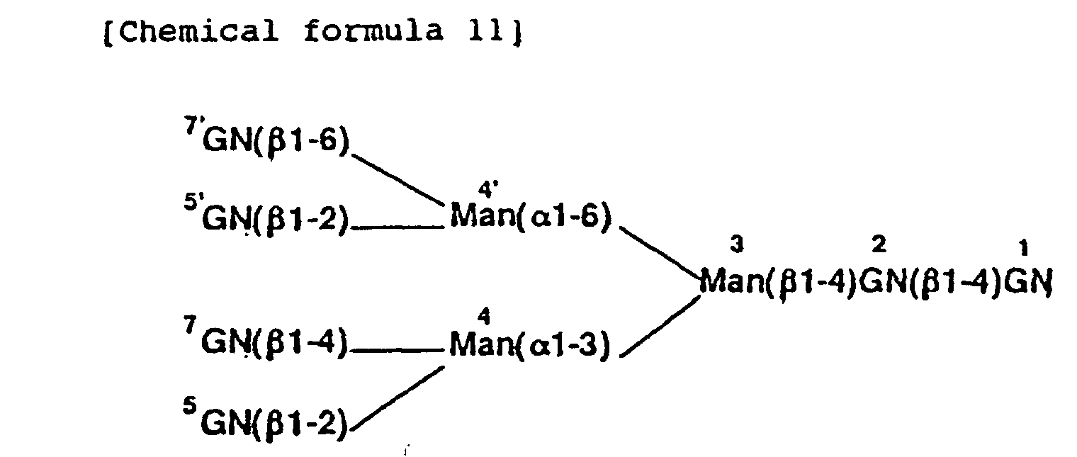
- When the enzymatic digestion is employed as the treatment in the method of the present invention, a sugar chain sample is digested with glycosidases in one or more steps including a reaction-termination operation in the course of the reaction, and the final product is subjected to chromatographic analysis comparing with the above-described standard sugar chains.
- The sugar chain to be analyzed may be any of that represented by above chemical formula (6). Even sugar chain which is not represented by the formula (6) can be analyzed by the method of the present invention so far as it can be converted into that represented by the formula (6) by enzymatic or chemical treatments.
- For example, a sugar chain with a polylactosamine structure, which has the repeating N-acetyllactosamine [(-3)Gal(β1-4)GlcNAc(β1-)] being linked to the Gal residues of the formula (6) is not represented by the formula (6). The sugar chain with polylactosamine can be converted into a sugar chain represented by the formula (6) by sequential digestion with β-galactosidase and β-N-acetylglucosaminidase [T. Ohkura et al., J. Biol. Chem. (1981) 256, 8485-8490] or by digestion with endo-β-galactosidase from Escherichia freundii.
- The sugar chain to be analyzed may be labeled or not. Although the sugar chain sample may be a mixture of several sugar chains, the purity thereof is preferably at least 80%.
- The glycosidase used in the respective steps may be a single one or a mixture of two or more glycosidases. Although the glycosidase to be used is preferably exoglycosidases, endoglycosidases can also be used. The reaction temperature which may not be particularly limited is usually 20 to 50°C, preferably 37°C. The reaction time which may not be particularly limited is usually 10 min to 20 h, preferably 3 to 5 h. After each step, the reaction is terminated by inactivation of the glycosidases by incubating the reaction mixture at 80 to 120°C for 3 to 10 min. Such an inactivation step is not always necessary. In other words, the inactivation step is not always necessary when the glycosidases used in the previous step show no activities because the optimum temperature, pressure, pH, concentration of the organic solvent, etc., of the subsequent step are extremely different from those of the previous step, or when the glycosidase used in the previous step can be removed with antibody beads or an ion-exchange resin.
- The analytical method of the reaction product, which may not be particularly limited, may include thin layer chromatography, electrophoresis, mass spectrometry, gas chromatography, etc., and the analysis by HPLC is preferable. The reaction sequence include usually 1 to 6 reactions per sample. The products of each reaction are analyzed, and the sugar chain structure is determined by combining various pieces of the information thus obtained. Below, each reaction is described in detail.
- When a sugar chain sample is digested with a mixture of β-galactosidase, sialidase, and α-fucosidase, Gal, SA, and Fuc can be removed from the chain, leaving GN. Therefore, the reaction product should be one of sugar chains repesented by chemical formula (9) or (10). The reaction product can be identified by using the above-described standard sugar chains, and then the site of GN linkage in the starting sugar chain sample, i.e. the variables l1, l2, l3 and l4 in the formula (6), can be determined. The glycosidases used herein can be preferably those having a low substrate specificity, such as β-galactosidase from jack beans (Seikagaku-Kogyo), β-galactosidase from Streptococcus (Seikagaku-Kogyo), sialidase from Arthrobacter ureafaciens (Nacalai Tesque), α-fucosidase from bovine kidney (Boehringer Mannheim), etc.
- When a sugar chain sample is digested with a mixture of β-N-acetylglucosaminidase, sialidase, and α-fucosidase, Fuc and GN, which is not substituted by Gal, can be removed from the chain, leaving Gal and GN at the same branch. After removal or inactivation the enzymes, the sugar chain is further digested with β-galactosidase to remove Gal. The reaction product must be one of sugar chains repesented by chemical formula (9) or (10). The reaction product can be identified by using the above-described standard sugar chains, and then the site of Gal linkage in the starting sugar chain sample can be determined. By this reaction, the mode of Gal linkage cannot be determined. Namely, it can be determined only that either mx or m'x (x being 1, 2, 3 or 4) is 1 or, in other words, it can be determined that (mx + m'x) is either 0 or 1. The glycosidases used herein can be preferably those having a low substrate specificity, such as β-N-acetylglucosaminidase from bovine kidney (Boehringer Mannheim), β-galactosidase from jack beans, β-galactosidase from Streptococcus, sialidase from A. ureafaciens, and α-fucosidase from bovine kidney.
- To determine the mode of Gal linkage, a sugar chain sample is digested with the same mixture of glycosidases as described above except that a linkage-specific β-galactosidase is used. The sugar chain is digested with a mixture of β-N-acetylglucosaminidase, sialidase, α-fucosidase and β-1,4-linkage specific or β-1,3-linkage-specific β-galactosidase. Gal, which can be digested with the linkage-specific β-galactosidase, and GN at the same branch, and Fuc and GN, which are not substituted by Gal, can be removed from the chain, leaving Gal, which cannot be digested with the linkage-specific β-galactosidase, and GN at the same branch. After removal or inactivation of the enzymes, the sugar chain is further digested with non-specific β-galactosidase to remove Gal. The reaction product must be one of sugar chains repesented by chemical formula (9) or (10). The reaction product can be identified by using the above-described standard sugar chains, and then the site of Gal linkage, which cannot be hydrolyzed with the linkage specific β-galactosidase, in the starting sugar chain sample can be determined. For example, when β-1,4-linkage specific β-galactosidase from D. pneumoniae (Boehringer Mannheim) is used, the site of Galβ-1,3-linkage, i.e. the variables m'1, m'2, m'3 and m'4 in the formula (6), can be determined. Because m1+m'1, m2+m'2, m3+m'3 and m4+m'4 have been determined by the above-described reaction, the site of Galβ-1,4-linkage, namely, the variables m1, m2, m3 and m4 in the formula (6), can be determined by combining these data.
- When a sugar chain sample is digested with a mixture of β-N-acetylglucosaminidase, β-galactosidase, and α-fucosidase, Gal and GN, which are not substituted by SA, can be removed from the chain, leaving SA, Gal, and GN at the same branch. After removal or inactivation of the enzymes, the sugar chain is further digested with the mixture of sialidase and β-galactosidase. The reaction product must be one of sugar chains repesented by chemical formula (9) or (10). The reaction product can be identified by using the above-described standard sugar chains, and then the site of SA linkage in the starting sugar chain sample can be determined. By this reaction, the mode of SA linkage cannot be determined. Namely, it is determined only that either nx or n'x (x being 1, 2, 3 or 4) is 1 or, in other words, it is determined that (nx + n'x) is either 0 or 1. The glycosidases used herein can be preferably those having a low substrate specificity, such as β-N-acetylglucosaminidase from bovine kidney, β-galactosidase from jack beans, β-galactosidase from Streptococcus, sialidase from A. ureafaciens, α-fucosidase from bovine kidney, etc.
- To determine the mode of SA linkage, a sugar chain sample is digested with the same mixture of glycosidases as described above except that a linkage-specific sialidase is used. The sugar chain is digested with a mixture of β-N-acetylglucosaminidase, β-galactosidase, α-fucosidase and α-2,3-linkage- or α-2,6-linkage-specific sialidase. SA, which cannot be hydrolyzed with the specific sialidase used in the reaction, and Gal and GN at the same branch are left after the reaction. After removal or inactivation of the enzymes, the sugar chain is further digested with non-specific sialidase and β-galactosidase to remove SA and Gal. The reaction product must be one of sugar chains repesented by chemical formula (9) or (10). The reaction product can be identified by using the above-described standard sugar chains, and then the site of SA linkage, which cannot be hydrolyzed with the linkage specific sialidase, in the starting sugar chain sample can be determined. For example, when α-2,3-linkage-specific sialidase from Salmonella typhimurium (Takara Shuzo) is used, the site of SAα-2,6-linkage by this reaction, i.e. the variables n1 n2, n3 and n4 in the formula (6) can be determined. Since n1+n'1, n2+n'2, n3+n'3 and n4+n'4 have been determined by the above-described reaction, the site of SAα-2,3-linkage, namely, the variables n'1, n'2, n'3 and n'4 in the formula (6), can be determined by combining these data.
- When only the site and the mode of SA linkage in a sugar chain sample should be determined, the standard oligosaccharides represented by the formula (9) or (10) are not necessarily used. For example, when the site and the mode of SA linkage in the so-called biantennary sugar chain, i.e. the variables, n2, n'2, n4, and n'4 in the formula (6) should be determined, the sugar chain sample is digested with β-galactosidase first, and then the enzyme is removed or inactivated. Then the reaction mixture is digested with sialidase. After the enzymatic digestion, Gal that is linked by SA must remain and thus the reaction product must be one of the four kinds of product that is to say the oligosaccharides represented by the formula (6) with the variables, (l2=l4=1, m2=m4=0), (l2=l4=1, m2=0, m4=1), (l2=l4=1, m2=1, m4=0), and (l2=l4=1, m2=m4=1). Thesefore if the reaction product is identified with these four standard oligosacchrides, the site of SA linkage can be determined, and also, the mode of SA linkage can be determined by use of a sialidase that is specific for α-2,3-linkage.
- When a sugar chain sample is digested with a mixture of β-N-acetylglucosaminidase, β-galactosidase, and sialidase, Fuc and GN substituted by Fuc are left, while other GN, Gal and SA are removed from the chain. After removal or inactivation of the enzymes, the sugar chain is further digested with α-fucosidase to remove Fuc. If necessary, β-galactosidase is also added in the mixture, because Gal which is linked to the same GN as Fuc is linked may remain after the above-described reaction. The reaction product must be one of sugar chains repesented by chemical formula (9) or (10). The reaction product can be identified by using the above-described standard sugar chains, and then the site of Fucα-1,3-linkage in the starting sugar chain sample, i.e., the variables p1, p2, p3 and p4 in the formula (6), can be determined. The glycosidase used herein can be preferably one having a low substrate specificity, such as β-N-acetylglucosaminidase from bovine kidney, β-galactosidase from jack beans, β-galactosidase from Streptococcus, sialidase from A. ureafaciens, α-fucosidase from bovine kidney, etc.
- The presence or absence of Fucα-1,6-linkage to GN at the reducing terminus of the sugar chain sample, namely, the variable q, in the formula (6) can be determined by using α-1,3/4-linkage-specific α-fucosidase. When the sugar chain is digested with a mixture of β-N-acetylglucosaminidase, β-galactosidase, sialidase and, for example, α-1,3/4-fucosidase from Streptomyces (Takara Shuzo), GN, Gal, SA and α-1,3-linked Fuc are removed, leaving α-1,6-linked Fuc. Thus, when the sugar chain sample is free from α-1,6-linked Fuc or, in other words, when q is 0, the product must be the M3 core, which can be distinguished from M3 core with α-1,6-linked Fuc. From these results, the presence or absence of Fucα-1,6-linkage can be determined.
- The N-acetyllactosamine type of sugar chain occasionally contains Fuc(α1-2)Gal sequence, in addition to the modes of linkage represented by the formula (6). The site of Fucα-1,2-linkage can be determined by the same reaction as described above except that an α-1,2-linkage-specific fucosidase, such as α-1,2-fucosidase from Arthrobacter (Takara Shuzo), is used.
- When the structure of the sugar chain sample to be analyzed is represented by the formula (6), the definite structure can be determined from the above-described results, (i) to (iv). The site of GN linkage, namely, the variables, l1, l2, l3 and l4 in the formula (6), can be determined from the results of (i); the site and mode of Gal linkage, namely, the variables, m1, m'1, m2, m'2, m3, m'3, m4 and m'4 in the formula (6), can be determined from the results of (ii); the site and mode of SA linkage, namely, the variables n1, n'1, n2, n'2, n3, n'3, n4 and n'4 in the formula (6), can be determined from the results of (iii); and the site of Fuc linkage, namely, the variables q, p1, p2, p3 and p4 in the formula (6), can be determined from the results of (iv).
- In another mode of the method provided by this invention, the branches having a certain sugar residue of which the presence should be determined, can be distinguished from the branches not having the residue by removing the branches having the residue from a sugar chain sample by the enzymatic or chemical treatment. For example, GN residues in the branches having Galβ-1,3-linkage can be specifically removed by digestion with lacto-N-biosidase, which releases disaccharide, Gal(β1-3)GN, from the non-reducing terminus of a sugar chain [M. Sano et al., Proc. Natl. Acad. Sci. U.S.A. 89, 8512-8516 (1992)]. The branches having Gal(β1-3)GN can be specifically removed by the treatment with the mixture of sialidase, α-fucosidase, and lacto-N-biosidase. Then the reaction products can be digested with β-galactosidase and the final reaction product must be one of sugar chains represented by chemical formula (9) or (10). The branches being missing from the branches having GN by this treatment can be determined as the branches having Gal(β1-3)GN, i.e. the branches having Galβ-1,3-linkage.
- The usefulness of the method of the present invention for determining the structure of the sugar chain can be further improved by using oligosaccharides represented by the following formula (12), as the standard sugar chains.wherein Z1, Z2, Z3, Z4, Z5 and Z6 each represent hydrogen or β-GN.
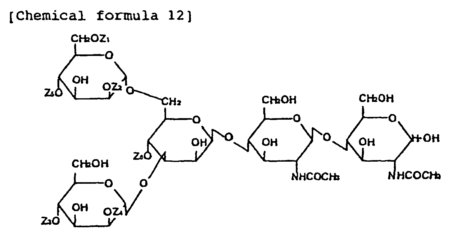
- Though most of the N-acetyllactosamine type of sugar chains are represented by the formula (6), some sugar chain has the core structure represented by the above formula (12), which can be modified with Gal, SA and Fuc residues. The standard sugar chains represented by formula (12) might be useful for the analysis of these sugar chains. Five oligosaccharides which are particularly useful for the analysis are given in Table 2.
oligosaccharides Z1 Z2 Z3 Z4 Z5 Z6 XVII H β-GN H β-GN H β-GN XVIII H β-GN β-GN β-GN H β-GN XIX β-GN β-GN H β-GN H β-GN XX β-GN β-GN β-GN β-GN H β-GN XXI β-GN β-GN β-GN β-GN β-GN β-GN - These oligosaccarides can be used without labeling or after labeling like the oligosaccharides I to XVI.
- The typical mode of a method of the present invention includes a process of identifying the resulting product from the treatment by comparing with the standard sugar chains, however, if the structure of the resulting product can be determined by other analytical techniques including NMR spectroscopy and mass spectrometry, these can be employed for the present invention.
- The structure of the sugar chain can be determined more easily by combining reagents used in the process of the present invention to form a kit. The kit comprises at least one of the oligosaccharides of the formula (4) and, if necessary, another oligosaccharide of the formula (9) or (10). For example, a kit containing I-PA to VIII-PA is effective particularly in analyzing a biantennary sugar of the formula (6) wherein l1=l3=0 and l2=l4=1, or a triantennary sugar of the same formula (6) wherein l1=0 and l2=l3=l4=1. For example, a kit containing I-PA to XVI-PA is widely usable for the analysis of the N-acetyllactosamine type of sugar chains.
- The kit may contain glycosidases, a column for the analysis, buffer solution, etc., in addition to the oligosaccharide. The reagents to be contained in the kit may be in the form of either a solution or freeze-dried product.
-
- Figure 1 shows a chart obtained by HPLC of a mixture of
8 oligosaccharides I-PA to VIII-PA under the
Condition 1. - Figure 2 shows a chart obtained by HPLC of a mixture of
16 ologosaccharides I-PA to XVI-PA under the
Condition 2. - Figure 3 shows a chart obtained by HPLC of
fraction 2 in Fig. 2 under theCondition 3. - Figure 4 shows a chart obtained by HPLC of
fraction 4 in Fig. 2 under theCondition 3. - Figure 5 shows a chart obtained by HPLC of
fraction 3 in Fig. 2 under theCondition 1. - Figure 6 shows a chart obtained by HPLC of fraction in
Fig. 2 under the
Condition 1 after the complete digestion with β-N-acetylglucosaminidase from D. pneumoniae. - Figure 7 shows a elution profile of a mixture of 16
oligosaccharides I-PA to XVI-PA under the
conditions - Figure 8 shows the 1H-NMR spectrum, in the range of chemical shift of 3.00 to 5.50 ppm, of I-PA.
- Figure 9 shows the 1H-NMR spectrum, in the range of chemical shift of 1.85 to 2.50-ppm, of I-PA.
- Figure 10 shows the 1H-NMR spectrum, in the range of chemical shift of 3.00 to 5.50 ppm, of II-PA.
- Figure 11 shows the 1H-NMR spectrum, in the range of chemical shift of 1.85 to 2.50 ppm, of II-PA.
- Figure 12 shows the 1H-NMR spectrum, in the range of chemical shift of 3.00 to 5.50 ppm, of III-PA.
- Figure 13 shows the 1H-MNR spectrum, in the range of chemical shift of 1.85 to 2.50 ppm, of III-PA.
- Figure 14 shows the 1H-MNR spectrum, in the range of chemical shift of 1.60 to 2.50 ppm, of IX-PA.
- Figure 15 shows the 1H-MNR spectrum, in the range of chemical shift of 1.85 to 2.50 ppm, of X-PA.
- Figure 16 shows the 1H-MNR spectrum, in the range of chemical shift of 3.00 to 5.50 ppm, of X-PA.
- Figure 17 shows the 1H-MNR spectrum, in the range of chemical shift of 1.60 to 5.50 ppm, of XI-PA.
- Figure 18 shows the 1H-MNR spectrum, in the range of chemical shift of 1.60 to 5.50 ppm, of XII-PA.
- Figure 19 shows the 1H-MNR spectrum, in the range of chemical shift of 1.60 to 5.50 ppm, of XIII-PA.
- Figure 20 shows the 1H-MNR spectrum, in the range of chemical shift of 1.60 to 5.50 ppm, of XIV-PA.
- Figure 21 shows a elution profile of the products obtained by partial digestion of the sugar chain with β-N-acetylglucosaminidase from bovine kidney. The GN residue in the sequence, GN(β1-6)Man(α), in the sugar chain is protected.
-
- The following Examples will further illustrate the present invention, which by no means limit the invention.
- 200 nmol of commercially available oligosaccharide VIII-PA (PA Sugar Chain 013; Takara Shuzo) was digested with 20 mU of β-N-acetylglucosaminidase from D. pneumoniae (Boehringer Mannheim) in 50 mM phosphate/citrate buffer (pH 5.0) at 37°C. A part of the reaction mixture was analyzed by HPLC, and when the nondegraded substance had amounted to about 25% of the whole, the reaction mixture was treated at 100°C for 5 min to terminate the reaction. Then the products were separated treated by HPLC to obtain I-PA (22.4 min), II-PA (42.5 min) and III-PA (25.2 min), which were then purified. The BPLC conditions were as follows:
apparatus LC 6A (Shimadzu), column PALPAK® TYPE R (4.6 mm x 250 mm) (Takara Shuzo) eluent 100 mM acetic acid/triethylamine containing 0.035% of 1-butanol (pH 4.0) detection conducted with fluorescence detector RF-535 (Shimadzu) at excitation wavelength of 320 nm and fluorescence wavelength of 400 nm flow rate 1 ml/ min column temperature 40°C (hereinafter referred to as " Condition 1") - Then the composition of each of three purified oligosaccharides, I-PA, II-PA and III-PA, were analyzed by mass spectrometry with a API-III mass spectrograph (Perkin-Elmer Sciex), NMR spectroscopy (in D2O) with a 400 MHz proton nuclear magnetic resonance spectrometer (Bruker), and monosaccharide analysis after acid hydrolysis.
- The properties of the respective oligosaccharides are given below. Further, the 1H-NMR spectrum of I-PA is given in Figs. 8 and 9, that of II-PA in Figs. 10 and 11 and that of III-PA in Figs. 12 and 13. The chemical shift values in these 1H-NMR spectra are represented by taking the chemical shift value of the methyl proton of acetone in D2O at 37°C as 2.218 ppm when DSS is used as the standard. In Figs. 9, 11 and 13, the signal at 2.218 ppm was that of the methyl proton of acetone used as the internal standard.
-
- Mol. wt.
- 1192.7
- 1H-NMR
- 4.556 (H-1, GN-7), 2.067 (NAc, GN-7)
- Sugar compn.
- Man: GN = 3.0 : 2.5, free from Gal and Fuc.
-
- Mol. wt.
- 1395.4
- 1H-NMR
- 4.541 (H-1, GN-5'), 4.556 (H-1, GN-7),
2.040 (NAc, GN-5'), 2.066 (NAc, GN-7) - Sugar compn.
- Man: GN = 3.0 : 3.5, free from Gal and Fuc.
-
- Mol. wt.
- 1395.5
- 1H-NMR
- 4.527 (H-1, GN-5), 4.515 (H-1, GN-7),
2,045-(NAc, GN-5), 2.067 (NAc, GN-7) - Sugar compn.
- Man:GN = 3.0 : 3.5, free from Gal and Fuc.
- The reference numerals of the sugar residues in the 1H-NMR spectra are as shown in the following chemical formula (11):
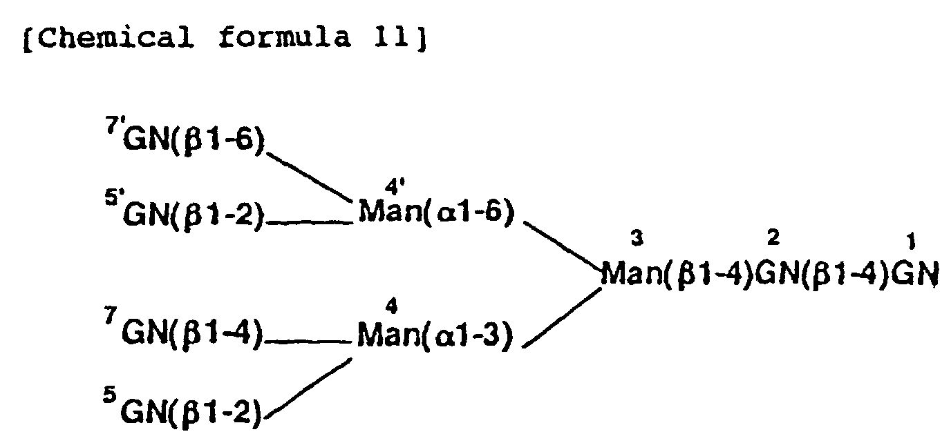
- A solution comprising each 1 pmol of I-PA, II-PA and III-PA obtained in Example 1 and commercially available IV-PA (PA Sugar Chain 016; Takara Shuzo), V-PA (PA standard sugar chain 100.1; Nakano Vinegar), VI-PA (PA standard sugar chain 100.2; Nakano Vinegar), VII-PA (PA Sugar Chain 012; Takara Shuzo) and VIII-PA (PA Sugar Chain 013; Takara Shuzo) was treated by HPLC under the Condition 1 (Fig. 1). Eight kinds of the PA-oligosaccharides were separated.
- 1 nmol of XVI-PA (PA Sugar Chain 014; Takara Shuzo) was digested with 1 mU of β-N-acetylglucosaminidase from bovine kidney, which has a comparatively low substrate specificity, in 50 mM phosphate/citrate buffer (pH 5.0) at 37°C. A part of the reaction mixture was analyzed by HPLC with PALPACK® TYPE N [eluent: 50 mM acetic acid/triethylamine (pH 7.3); linear gradient from
acetonitrile 70% (0 min) to 50% (300 min); other conditions being the same as those of the Condition 1 (hereinafter referred to as "Condition 2")].
When the nondegrated substance had amounted to about 20% of the whole, the reaction mixture was treated at 100°C for 5 min to terminate the reaction to thereby obtain a mixture of I-PA to XVI-PA. - The mixture was separated by HPLC under the
Condition 2. The obtained chart is given in Fig. 2. The peaks were divided into five fractions, i.e.fraction 1 eluted during the period of 13 to 17 min,fraction 2 during the period of 18 to 23 min,fraction 3 during the period of 24 to 29 min,fraction 4 during the period of 32 to 38 min, andfraction 5 during the period of 42 to 46 min. Under theCondition 2, the sugar chain could be separated on the basis of the number of sugar residues constituting the chain. By comparison with the elution position of the commercially available IV-PA, V-PA, VII-PA, VIII-PA and XVI-PA in HPLC under theCondition 2, it was found that the peak of thefraction 1 obtained at 15.4 min was one in which GN was not linked to the M3 core, namely, it was identified with IV-PA. In the same manner, thefraction 2 was one in which one GN was linked to the M3 core, namely, it was identified with a mixture of four oligosaccharides, i.e. I-PA, V-PA, VI-PA and XII-PA; thefraction 3 was one in which two GN's were linked to the M3 core, namely, it was identified with a mixture of six oligosaccharides, i.e. II-PA, III-PA, VII-PA, IX-PA, XIII-PA and XIV-PA; thefraction 4 was one in which three GN's were linked to the M3 core, namely, it was identified with a mixture of four oligosaccharides, i.e. VIII-PA, X-PA, XI-PA and XV-PA; and the peak of thefraction 5 obtained at 44.0 min was one in which four GN's were linked to the M3 core, namely, it was identified with XVI-PA. - The peak of the
fraction 1 obtained at 15.4 min (IV-PA) and that of thefraction 5 obtained at 44.0 min (XVI-PA) appeared as the single peaks of 12.0 min and 15.7 min, respectively, also in HPLC with PALPACK® TYPE R [eluent: acetic acid/triethylamine (pH 4.0) containing 0.07% of 1-butanol; other conditions being the same as those of the Condition 1 (hereinafter referred to as "Condition 3" ) ]. - When the
fraction 2 was separated by HPLC under theCondition 3, VI-PA was eluted at 11.2 min, XII-PA at 12.8 min, I-PA at 15.2 min, and V-PA at 20.7 min. The pattern is given in Fig. 3. When thefraction 3 was analyzed by HPLC under theCondition 1, XIII-PA was eluted at 15.0 min, IX-PA at 16.8 min, III-PA at 25.2 min, XIV-PA at 25.9 min, VII-PA at 26.7 min, and II-PA at 42.5 min. The pattern is given in Fig. 5. When thefraction 4 was analyzed by HPLC under theCondition 3, XV-PA was eluted at 10.4 min, X-PA at 13.3 min, XI-PA at 17.6 min, and VIII-PA at 25.9 min. The pattern is given in Fig. 4. Thus the 16 kinds of the PA-oligosaccharides, I-PA to XVI-PA, could be separated. - The respective peaks were identified as follows. I-PA, II-PA and III-PA obtained in Example 1 and commercially available IV-PA, V-PA, VI-PA, VII-PA, VIII-PA and XVI-PA were used as the standard sugar chains. By comparing the elution positions of the peaks, the peak of 15.4 min in Fig. 2 was identified with IV-PA, that of 44.0 min in Fig. 2 with XVI-PA, that of 11.2 min in Fig. 3 with VI-PA, that of 15.2 min in Fig. 3 with I-PA, that of 20.7 min in Fig. 3 with V-PA, that of 25.9 mim in Fig. 4 with VIII-PA, that of 25.2 min in Fig. 5 with III-PA, that of 26.7 min in Fig. 5 with VII-PA, and that of 42.5 min in Fig. 5 with II-PA.
- The peak at 12.8 min in Fig. 3 was identified with XII-PA based on the finding that when it was recovered and then treated with β-N-acetylglucosaminidase from bovine kidney, the M3 core was formed, that the starting material was XVI-PA, and that three other oligosaccharides were eluted at different times.
- As for the peaks of the remaining six oligosaccharides, each of them was fractionated and identified by a method which will be described below. The peak of 25.9 min in Fig. 5 was treated as follows: when a part of the
fraction 3 was completely digested with β-1,2-linkage-specific β-N-acetylglucosaminidase from D. pneumoniae and then treated by HPLC under theCondition 1, the peaks of 25.2 min (III-PA) and 26.7 min (VII-PA) disappeared (i.e. they were hydrolyzed to form I-PA and IV-PA, respectively), leaving only the peak of 25.9 min (Fig. 6), which was recovered. - The oligosaccharides of the respective peaks thus fractionated were partially digested with β-N-acetylglucosaminidase from bovine kidney and then treated by HPLC under the
Condition 2 to separate a fraction in which one GN was linked to the M3 core. The fraction thus recovered was then analyzed by HPLC under theCondition 3. Since V-PA, VI-PA and XII-PA were detected in the digest of the peak of 10.5 min in Fig. 4, the peak was identified with XV-PA. Also, since I-PA, V-PA and XII-PA were detected in the digest of the peak of 13.3 min in Fig. 4, the peak was identified with X-PA. Further, since I-PA, VI-PA and XII-PA were detected in the digest of the peak of 17.6 min in Fig. 4, the peak was identified with XI-PA. Since V-PA and XII-PA were detected in the digest of the peak of 15.0 min in Fig. 5, the peak was identified with XIII-PA. Since VI-PA and XII-PA were detected in the digest of the peak of 16.8 min in Fig. 5, the peak was identified with IX-PA. Since I-PA and XII-PA were detected in the digest of the peak of 25.9 min in Fig. 6, the peak was identified with XIV-PA. -
- (1) A mixture of 2.5 µmol of CMP-sialic acid (cytidine 5'-monophospho-N-acetylneuraminic acid, CMP-NANA; Sigma) with 850 nmol of a tetraantennary asialosugar chain (PA-Sugar Chain 004; Takara Shuzo) was reacted with 0.1 U of α2,6-sialyltransferase from rat liver (Boehringer Mannheim) in 12.5 mM cacodylate buffer (pH 6.8) containing 0.5% Triton CF-54 at 37°C for 36 h. In the course of the reaction, 2.5 µmol of CMP-NANA was added to the reaction mixture 16 h and 24 h after the initiation of the reaction.
-
- The reaction mixture was desalted through a Sephadex™ G-15 column, and the sugar chain fraction was injected to a monoQ™ column (Pharmacia) and eluted, the gradient being 10 mM to 100 mM ammonium acetate (pH 8.5). A fraction containing 3 mol of sialic acid per mole of the sugar chain was recovered from the eluted fractions. The amount of the recovered sialylated sugar chain was 646 nmol.
- 500 nmol of the recovered sialylated sugar chain was reacted with 100 U of galactose oxidase from Dactylium dendoroides (Takara Shuzo) in the presence of 4.7 mU of catalase (Sigma) in 50 mM phosphate buffer (pH 7.0) at 37°C for 2 h.
- The reaction mixture was treated on a boiling water bath for 10 min to terminate the reaction. Then 1 U of sialidase from A. ureafaciens (Kyokuto Pharmaceutical) was added to the reaction mixture to conduct digestion at 37°C for 2 h. Then 250 U of β-galactosidase from Aspergillus (Toyobo) was added to the reaction mixture and the reaction was continued at 37°C for 5.5 h.
- The pH of the reaction mixture was adjusted to about 11 with 5 N sodium hydroxide. 20 mg of sodium borohydride (NaBH4) was added to the reaction mixture, and the resultant mixture was left to stand at room temperature for 30 min. Additional 30 mg of sodium borohydride was added to the reaction mixture, and the resultant mixture was left to stand at room temperature for 30 min to complete the reduction reaction.
- The reduction reaction mixture was desalted through a Sephadex™ G-15 column and then injected to a PALPAK® TYPE R column. The elution was conducted with 0.1% 1-butanol and 0.05% trifluoroacetic acid, and the main peak eluting at 30 min was recovered. The resultant sugar chain was XVI-PA in which β-1,6-linked GN was protected with Gal. The amount of the recovered sugar chain was 300 nmol.
- A portion (250 nmol) of the protected sugar chain thus obtained was digested with 2.5 U of N-acetylglucosaminidase from bovine kidney in 50 mM citrate buffer (pH 5.0) in presence of 50 mM D-(+)-galactono-1,4-lactone (Nacalai Tesque). After the reaction for 3.5 h, the reaction mixture was treated on a boiling water bath for 10 min to terminate the reaction.
- The reaction mixture was desalted through a Sephadex™ G-15 column and three fractions were recovered by HPLC, the conditions are described below, depending on the number of GN units linked to the M3 core (fraction A with one GN unit linked; fraction B with two GN units linked; and fraction C with three GN units linked). The HPLC pattern is shown in Fig. 21.
-
- chromatograph: LC 6A (Shimadzu),
- column: Asahi PAK® NH2 P-50 (6.0 mm x 150 mm) (Asahi Chemical Industry),
- eluent A: 50 mM acetic acid/triethylamine containing 75% of acetonitrile (pH 7.3),
- eluent B: 50 mM acetic acid/triethylamine containing 50% of acetonitrile (pH 7.3),
- elution: gradient elution wherein the content of eluent B was increased from 10% to 25% during 100 min,
- detection: fluorescence detector RF-535 (Shimadzu),
- excitation wavelength: 315 nm,
- fluorescent wavelength: 380 nm,
- flow rate: 1 ml/min, and
- column temp. : 40°C.
-
- Each fraction was digested with 7.5 U of β-galactosidase from Aspergillus at 37°C for 15 h. Then the digestion mixture was treated on a boiling water bath for 10 min to terminate the reaction and reaction products were separated by HPLC, the conditions of which are described below. XII-PA (23.6 min) was recovered from the reaction product of fraction A; XIII-PA (19.4 min), IX-PA (21.4 min) and XIV-PA (31.3 min) from that of the fraction B; and XV-PA (18.9 min), X-PA (25.4 min) and XI-PA (33.4 min) from that of the fraction C.
-
- chromatograph: LC 6A (Shimadzu),
- column: YMC Pack AM (10 mm x 150 mm) (YMC),
- eluent: 0.05% trifluoroacetic acid containing 0.1% 1-butanol,
- detection: fluorescence detector RF-535 (Shimadzu),
- excitation wavelength: 320 nm,
- fluorescent wavelength: 400 nm,
- flow rate: 2 ml/min, and
- column temp. : 40°C.
-
- Then each of six purified oligosaccharides, IX-PA, X-PA, XI-PA, XII-PA, XIII-PA and XIV-PA, were analyzed by mass spectrometry with a API-III mass spectrograph, NMR spectroscopy (in D2O) with a 500 MHz proton nuclear magnetic resonance spectrometer (JEOL) or that of Bruker above-described (only X-PA), and monosaccharide analysis after acid hydrolysis.
- The properties of the respective oligosaccharides are given below. Further, the 1H-NMR spectrum of IX-PA is given in Fig. 14, that of X-PA in Figs. 15 and 16, that of XI-PA in Fig. 17, that of XII-PA in Fig. 18, that of XIII-PA in Fig. 19, and that of XIV-PA in Fig. 20. The chemical shift values in these 1H-NMR spectra are represented by taking the chemical shift value of the methyl proton of acetone in D2O at 37°C as 2.218 ppm when DSS is used as the standard. In Figs. 14, 15, 17, 18, 19 and 20, the signal at 2.218 ppm was that of the methyl proton of acetone used as the internal standard.
- The reference numerals of the sugar residues in the 1H-NMR spectra are as shown in the
above chemical formula 11. -
- Mol. wt.
- 1395.0 (by mass spectrometry)
- 1H-NMR
- 4.552 (H-1, GN-7'), 4.535 (H-1, GN-5),
2.046 (NAc, GN-7'), 2.046 (NAc, GN-5) - Sugar compn.
- Man: GN = 3.0 : 3.5, free from Gal and Fuc.
-
- Mol. wt.
- 1599.0 (by mass spectrometry)
- 1H-NMR
- 4.528 (H-1, GN-7'), 4.549 (H-1, GN-5'),
4.557 (H-1, GN-7), 2.028 (NAc, GN-7'),
2.038 (NAc, GN-5'), 2.067 (NAc, GN-7) - Sugar compn.
- Man: GN = 3.0 : 4.5, free from Gal and Fuc.
-
- Mol. wt.
- 1598.0 (by mass spectrometry)
- 1H-NMR
- 4.546 (H-1, GN-7'), 4.515 (H-1, GN-7),
4.529 (H-1, GN-5), 2.045 (NAc, GN-7'),
2.065 (NAc, GN-7), 2.045 (NAc, GN-5) - Sugar compn.
- Man: GN = 3.0 : 4.5, free from Gal and Fuc.
-
- Mol. wt.
- 1191.5 (by mass spectrometry)
- 1H-NMR
- 4.542 (H-1, GN-7'), 2.043 (NAc, GN-7'),
- Sugar compn.
- Man: GN = 3.0 : 2.5, free from Gal and Fuc.
-
- Mol. wt.
- 1395.0 (by mass spectrometry)
- 1H-NMR
- 4.530 (H-1, GN-7'), 4.548 (H-1, GN-5)
2.027 (NAc, GN-7'), 2.038 (NAc, GN-5') - Sugar compn.
- Man: GN = 3.0 : 3.5, free from Gal and Fuc.
-
- Mol. wt.
- 1395.0 (by mass spectrometry)
- 1H-NMR
- 4.545 (H-1, GN-7'), 4.556 (H-1, GN-7),
2.043 (NAc, GN-7'), 2.065 (NAc, GN-7) - Sugar compn.
- Man: GN = 3.0 : 3.5, free from Gal and Fuc.
- Each of the sixteen PA-oligosaccharides, I-PA to XVI-PA (500 fmol each), was analysed by HPLC with two kinds of columns, PALPAK® TYPE N column [condition 2], and PALPAK® TYPE R column [eluent: 100 mM acetic acid/triethylamine (pH 4.0) containing 0.02% 1-butanol; other conditions being the same as those of the condition 1 (hereinafter referred to as condition 4)]. The elution profiles of the mixture of the sixteen PA-oligosaccharides on the two columns were shown in Fig. 7. The elution position of each PA-oligosaccharide on the two columns was plotted and thus the two-dimensional sugar map was made. Figure 7 also shows the two-dimensional sugar map. Final reaction product of this invention can be easily identified by use of this two-dimensional sugar map. Each of the elution time obtained by reversed-phase PPLC (PALPAK® TYPE R column) was expressed on the ordinate, and that obtained by size-fractionation HPLC (PALPAK® TYPE N column) was expressed on the abscissa.
-
Kits - The oligosaccharides IV-PA, VII-PA, VIII-PA and XVI-PA to be contained in the kits were the above-mentioned ones produced by Takara Shuzo. Similarly, the oligosaccharides V-PA and VI-PA were those produced by Nakano Vinegar.
- The oligosaccharides IX-PA to XV-PA were obtained by the method described in Example 3 or partial hydrolysis of the oligosaccharide XVI-PA with N-acetylglucosaminidase from bovine kideny followed by purification by HPLC by the method described in Example 2-(2).
Contents of Kit 1 (for 100 runs) solution Reagent Amount (ml) 1 solution of mixture of oligosaccharides I-PA to VIII-PA (each 1 pmol/µl) 0.1 2 250 mM citrate buffer (pH 5.0) 0.4 3 50 U/ml sialidase from A. ureafaciens 0.5 4 15 U/ml α-2,3-sialidase from S. typhimurium 0.5 5 5 U/ml β-galactosidase from D. pneumoniae 0.5 6 5 U/ml β-galactosidase from Streptococcus 0.5 7 100 U/ml β-N-acetylglucosaminidase from bovine kidney 0.5 8 10 U/ml α-fucosidase from bovine kidney 0.5 9 1 U/ml α-1,3/4-fucosidase derived from Streptomyces 0.5 10 100 mM acetic acid/triethylamine (pH 4.0) containing 0.035% 1-butanol 1000 Attachment Number Comumn 1: PALPAK TYPE R (4.6 mm x 250 mm) 1 Contents of Kit 2 (for 100 runs) solution Reagent Amount (ml) 1 solution of mixture of oligosaccharides I-PA to VXI-PA (each 1 pmol/µl) 0.1 2 250 mM citrate buffer (pH 5.0) 0.4 3 50 U/ml sialidase from A. ureafaciens 0.5 4 15 U/ml α-2,3-sialidase from S. typhimurium 0.5 5 5 U/ml β-galactosidase from D. pneumonia 0.5 6 5 U/ml β-galactosidase from Streptococcus 0.5 7 100 U/ml β-N-acetylglucosaminidase from bovine kidney 0.5 8 10 U/ml α-fucosidase from bovine kidney 0.5 9 1 U/ml α-1,3/4-fucosidase derived from Streptomyces 0.5 10 100 mM acetic acid/triethylamine (pH 4.0) containing 0.035% 1-butanol 1000 11 100 mM acetic acid/triethylamine (pH 4.0) containing 0.07% 1-butanol 1000 12-A 50 mM acetic acid/triethylamine (pH 7.3) containing 75% acetonitrile 1000 12-B 50 mM acetic acid/triethylamine (pH 7.3) containing 50% acetonitrile 1000 Attachment Number Column 1: PALPAK@ TYPE R (4.6 mm x 250 mm) 1 Colum 2: PALPAK@ TYPE RN(4.6 mm x 250 mm) 1 - Bovine fetuin is a glycoprotein having a molecular weight of about 48,000 and about 22% by weight thereof comprises sugar chains.. The structure of the sugar chains of the fetuin has been analyzed in detail, so that it is suitable for use in the investigation of the sugar chains of glycoproteins. The main asparagine-binding sugar chains of the fetuin have such a structure that 0 to 5 SA's are linked to the structure of XXII or XXIII in Table 5. The structure of the fetuin varies depending on the number, site and mode of linkage of SA. Since most of the various structures of the fetuin are represented by the formula (6), they could be analyzed with the kit of the present invention.
- 1 g of bovine fetuin (Takara Shuzo) was hydrazinolyzed and then the product was N-acetylated to obtain a free sugar chain fraction. This fraction was labeled with 2-aminopyridine. Excess reagents were removed with a Sephadex™ G15 column (Pharmacia) to obtain the PA sugar chain, which was subjected to the chromatography with DEAE-Toyopearl™ 650 M column (18 mm x 250 mm; Tosoh) equilibrated with 10 mM tris hydrochloride buffer (pH 9.0) to conduct elution with the NaCl concentration gradient from 0 to 100 mM.
- Thus, the PA sugar chain was divided into five fractions wherein 0 to 4 SA's were linked to each molecule. From the five fractions, a fraction having one SA and another fraction having two SA's were used. A part (1/100) of each fraction was separated by HPLC [eluent: 50 mM acetic acid/triethylamine (pH 5.0) containing 0.15% of 1-butanol] with a reversed-phase column Cosmosil™ 5C18AR (4.6 mm x 250 mm; Nacalai Tesque). A peak of 40 min was prepared from the fraction having one SA linked, and peaks of 38, 48 and 72 min were prepared from that having two SA's linked, and they were further purified by HPLC with ASAHIPAK® NH2 P50. The sugar chain in the fraction of 48 min having 2 SA's, that of 38 min having 2 SA's, that of 40 min having 1 SA and that of 72 min having 2 SA's will be referred to as A, B, C and D, respectively. Each sugar chain was dissolved in 100 µl of distilled water to prepare a sample solution.
- The following operation was conducted with
Kit 1 of Example 4: - (Reaction 1) 4 µl of
reagent reagent reagent Condition 1. - (Reaction 2) 4 µl of
reagent reagent reagent 7 and 3 µl of distilled water were added to 5 µl of the sample to effect a reaction at 37°C for 5 h. The reaction mixture was treated at 100°C for 10 min, and 2 µl ofreagent reagent 5 were added to the mixture to effect a reaction for additional 5 h. The reaction mixture was treated at 100°C for 10 min, and then 2 µl of the product was analyzed by HPLC in the same manner as that described above. - (Reaction 3) 4 µl of
reagent reagent reagent reagent reagent 5 were added to the mixture to effect a reaction for additional 5 h. The reaction mixture was treated at 100°C for 10 min, and then 2 µl of the product was analyzed by HPLC in the same manner as that described above. -
- Results: The reaction mixture obtained by the
reaction 1 was analyzed to find out that the product was VIII-PA as judged from the elution time thereof. This suggests that Gal of the sugar chain A was completely removed with β-1,4-linkage-specific β-galactosidase, i.e. thereagent 5 used in thereaction 1. Namely, all the Gal linkages of the sugar chain A were β-1,4-linkage. Therefore, the sugar chain (A) was determined to be one wherein l1=m1=m'1=m'2=m'3=m'4=q=p1=p2=p3=p4=0, and l2=l3=l4=m2=m3=m4=1 in the formula (6). Then, the reaction mixture obtained by thereaction 2 was analyzed to find out that the product was III-PA as judged from the elution time thereof. Thus, the sugar chain A had SA in two Gal's which were linked to the two GN's on the α-1,3-linked Man of the PA oligosaccharide XXII (XXII-PA) in Table 5. Namely, n1=n2=n'1=n'2=0 and n3+n'3=n4+n'4=1 in the formula (6). Thereafter, the reaction mixture obtained by thereaction 3 was analyzed to find out that the product comprised about 80% of VI-PA and about 20% of III-PA as judged from the elution time thereof. Thus, the sugar chain A was determined to be composed of about 20% of chains which have SA linked through α-2,6 to both of two Gal's to which SA had been proved to be linked by the result above, in other words, wherein n3=1 and n4=1, and about 80% of chains which have SA linked through α-2,6 to the only Gal linked to the GN(β1-2)Man(α1-3), in other words, wherein n3=0 and n4=1. As a result, the sugar chain A was determined to be a mixture of XXIV-PA and XXV-PA in a ratio of about 4:1, each having a structure given in Table 5. - The following operation was conducted with
Kit 1 of Example 4: - (Reaction 1) 4 µl of
reagent reagent reagent - (Reaction 2) 4 µl of
reagent reagent reagent 7 and 3 µl of distilled water were added to 5 µl of the sample to effect a reaction at 37°C for 5 h. The reaction mixture was treated at 100°C for 10 min and 2 µl ofreagent reagent 5 were added to the mixture to effect a reaction for additional 5 h. The reaction mixture was treated at 100°C for 10 min, and then 2 µl of the product was analyzed by HPLC in the same manner as that described above. - (Reaction 3) 4 µl of
reagent reagent reagent reagent reagent 5 were added to the mixture to effect a reaction for additional 5 h. The reaction mixture was treated at 100°C for 10 min, and then 2 µl of the product was analyzed by HPLC in the same manner as that described above. -
- Results: The reaction mixture obtained by the
reaction 1 was analyzed to find out that the product was VII-PA as judged from the elution time thereof. Therefore, the sugar chain B was determined to be one wherein l1=l3=m1=m3=m'2=m'3=q=p1=p2=p3=p4=0, and l2=l4=m2=m4=1 in the formula (6). Then, the reaction mixture obtained by thereaction 2 was analyzed to find out that the product was VII-PA as judged from the elution time thereof. Thus, the sugar chain B was determined to be one which had SA in both Gal's in the PA oligosaccharide XXIII (XXIII-PA) in Table 5, in other words, wherein n2+n'2=n4+n'4=1 in the formula (6). Thereafter, the reaction mixture obtained by thereaction 3 was analyzed to find out that the product was VI-PA as judged from the elution time thereof. Thus, the sugar chain B was determined to be one which had α-2,6-linked SA on the branch of Man which was linked through α-1,3-linkage in the XXIII-PA, in other words, wherein n2=0 and n4=1. As a result, the sugar chain B was determined to be XXVI-PA in Table 5. - The following operation was conducted with
Kit 1 of Example 4: - (Reaction 1) 4 µl of
reagent reagent reagent - (Reaction 2) 4 µl of
reagent reagent reagent 7 and 3 µl of distilled water were added to 5 µl of the sample to effect a reaction at 37°C for 5 h. The reaction mixture was treated at 100°C for 10 min and 2 µl ofreagent reagent 5 were added to the mixture to effect a reaction for additional 5 h. The reaction mixture was treated at 100°C for 10 min, and then 2 µl of the product was analyzed by HPLC in the same manner as that described above. - (Reaction 3) 4 µl of
reagent reagent reagent reagent reagent 5 were added to the mixture to effect a reaction for additional 5 h. The reaction mixture was treated at 100°C for 10 min, and then 2 µl of the product was analyzed by HPLC in the same manner as that described above. -
- Results: The reaction mixture obtained by the
reaction 1 was analyzed to find out that the product was VIII-PA as judged from the elution time thereof. Therefore, the sugar chain C was determined to be one wherein l1=m1=m'1=m'2=m'3=m'4=q=p1=p2=p3=p4=0, and l2=l3=l4=m2=m3=m4=1 in the formula (6). Then, the reaction mixture obtained by thereaction 2 was analyzed to find out that the product comprised about 50% of I-PA and about 50% of V-PA as judged from the elution time thereof. Thus, the sugar chain C was determined to be a nearly 1:1 mixture of an oligosaccharide which had SA linked to the Gal linked to the GN(β1-4)Man(α1-3) of XXII-PA, in other words, wherein n2=n4=n'2=n'4=0 and n3+n'3=1 in the formula (6) and an oligosaccharide having SA linked to the Gal linked to the GN(β1-2)Man(α1-6), in other words, wherein n3=n4=n'3=n'4=0 and n2+n'2=1 in the formula (6). Thereafter, the reaction mixture obtained by thereaction 3 was analyzed to find out that the product was IV-PA as judged from the elution time thereof. Thus, the sugar chain C was determined to be one which did not have SA linked through α-2,6 but SA's all linked through α-2,3, in other words, wherein n'2=1 or n'3=1 in the formula (6). As a result, the purified sugar chain C was determined to be a nearly 1:1 mixture of XXVII-PA and XXVIII-PA in Table 5. - The following operation was conducted with
Kit 1 of Example 4: - (Reaction 1) 4 µl of
reagent reagent reagent - (Reaction 2) 4 µl of
reagent reagent reagent 7 and 3 µl of distilled water were added to 5 µl of the sample to effect a reaction at 37°C for 5 h. The reaction mixture was treated at 100°C for 10 min and 2 µl ofreagent reagent 5 were added to the mixture to effect a reaction for additional 5 h. The reaction mixture was treated at 100°C for 10 min, and then 2 µl of the product was analyzed by HPLC in the same manner as that described above. -
- Results: The reaction mixture obtained by the
reaction 1 was analyzed to find out that the product was VIII-PA as judged from the elution time thereof. Therefore, the sugar chain D was determined to be one wherein l1=m1=m'1=m'2=m'3=m'4=q=p1=p2=p3=p4=0, and l2=l3=l4=m2=m3=m4=1 in the formula (6). Then, the reaction mixture obtained by thereaction 2 was analyzed to find out that the product was II-PA as judged from the elution time thereof. Thus, the sugar chain D was determined to be one which had SA linked to the Gal linked to the GN(β1-4)Man(α1-3) and also SA linked to the Gal linked to the GN(β1-2)Man(α1-6) of XXII-PA, in other words, wherein n2+n'2=n3+n'3=1 in the formula (6). Therefore, the sugar chain D was determined to be XXIX-PA in Table 5. - The oligosaccharides XXII to XXIX satisfy the conditions relating to variables lx, mx, m'x, nx, n'x, q and px (x being 1, 2, 3 or 4) in the formula (6) given in the following Table 5. And XXII-PA to XXIX-PA are obtained by labeling the oligosaccharides XXII to XXIX respectively with 2-aminopyridine.In the above Table 5, the symbol (1) represents that one of the two variables is 1 and the other is 0.

- The sugar chain fraction was obtained from 1 mg of α1-acid glycoprotein (Sigma) by hydrazinolysis and N-acetylation.
- The fraction was treated with 0.1 N HCl for 1 h to remove SA residues, and then the mixture was chromatographed on Bio-gel™ P-4 (Bio-Rad) column (25 mm x 900 mm x 2). Sugar chains were eluted with H2O, and the fractions with the elution position corresponding to that of isomaltooligosaccharide (DP=21), of which the degree of polimerization was 21, were pooled and lyophilized. The pooled fraction was labeled with 2-aminopyridine. PA-oligosaccharides were purified by HPLC with PALPAK® TYPE R column and then sugar chain E was obtained.
- 50 pmol of sugar chain E were digested with 5 mU of endo-β-galactosidase from Escherichia freundii at 37°C for 2 h, and a portion was analyzed by HPLC with an amide-silica column [Anal. Biochem. 171, 73-90, (1988)]. The product peak was eluted about 2.5 glucose units earlier than sugar chain E. This product was named sugar chain F.
- From these results, sugar chain E might have a N-acetyllactosamine unit in a certain branch of a sugar chain represented by the formula (6) with the variables, (l1=l2=l3=l4=m1=m2=m3=m4=1, and the rest are 0), i.e. so-called tetraantennary sugar chain. Endo-β-galactosidase might release Gal(β1-4)GN(β1-3)Gal from sugar chain E producing sugar chain F.
- The branch having N-acetyllactosamine unit was determined with
Kit 2 of Example 4. - (Reaction 1) Sugar chain F, 10 pmol/5 µl, was mixed with
4 µl of
reagent reagent 5, and 9 µl of distilled water, and the mixture was incubated at 37°C for 2 h. After the mixture was boiled at 100°C for 10 min to stop the reaction, 2 µl of the mixture was analyzed by HPLC under the conditions of 2 and 4. - (Reaction 2) Sugar chain F, 10 pmol/5 µl, was mixed with
4 µl of
reagent reagent 5 was added to the mixture and the mixture was incubated for 2 h. After the mixture was boiled at 100°C for 10 min to stop the reaction, 2 µl of the mixture was analyzed by HPLC under the conditions of 2 and 4. -
- Results: The product from the
reaction 1 was identified with XVI-PA judging from the elution position of the product and standard sugar chain in HPLC under the conditions of 2 and 4. Thus sugar chain F should be represented by the formula (6) with the variables, (l1=l2=l3=l4=1). The product from thereaction 2 was identified with XI-PA in the same way, and thus sugar chain F was identified with a sugar chain represented by the formula (6) with the variables, (m1=m2=m4=1, m2=0). From these results, sugar chain F was represented by the formula (6) with the variables, (l1=l2=l3=l4=m1=m3=m4=1, and the rest were 0). The results also show that N-acetyllactosamine unit was linked to Gal linked to the GN(β1-2)Man(α1-6). - The present invention provides a more exact, reliable, and simple method for determining the sugar chain structure, a kit to be used for said method, and novel oligosaccharides useful for said method as standards.
Claims (21)
- A method for determining the structure of an N-acetyllactosamine type of sugar chain characterized by determining the site of linkage and the mode of linkage of sugar residues at the non-reducing terminal side of a β-N-acetylglucosamine residue linked to an α-mannose residue in pentasaccharide core of N-acetyllactosamine-type sugar chain (formula 7) (M3 core)by means of detecting the presence of a β-N-acetylglucosamine residue after enzymatic or chemical treatment of said sugar chain.

- A method of claim 1 comprising the following steps:(1) selectively removing particular β-N-acetylglucosamine residues linked to an α-mannose residue in the M3 core;(2) in the event that the product of step (1) has a remaining sugar residue which is linked to the non-reducing terminal of β-N-acetylglucosamine residues linked to an α-mannose residue in the M3 core, removing the said remaining sugar residue; and(3) identifying the resulting products of step (1) or (2) by comparing them with standard sugar chains.
- A method of claim 2, wherein the β-N-acetylglucosamine residues of step (1) do not have a specified sugar residue at the non-reducing terminal side, wherein the site of linkage and the mode of linkage of the specified sugar residue should be determined.
- A method of claim 3, wherein the sugar residues are removed by enzymatic treatment.
- A method of claim 4, wherein the enzymatic treatment is digestion with glycosidases.
- A method of claim 5, wherein the glycosidases are selected from the group consisting of β-N-acetylglucosaminidase, β-galactosidase, sialidase and α-fucosidase.
- A method of claim 6, wherein at least one of the glycosidases is a linkage-specific glycosidase.
- A method of claim 7, wherein the linkage-specific glycosidase is selected from the group consisting of β-1,3-linkage-specific β-galactosidase, β-1,4-linkage-specific β-galactosidase, α-2,3-linkage-specific sialidase, α-2,6-linkage-specific sialidase, α-1,3/4-linkage-specific α-fucosidase and α-1,2-linkage-specific α-fucosidase.
- A method of claim 2, wherein the β-N-acetylglucosamine residues of step (1) have a specified sugar residue at the non-reducing terminal side, wherein the site of linkage and the mode of linkage of the specified sugar residue should be determined.
- A method of claim 9, wherein the sugar residues are removed by enzymatic treatment.
- A method of claim 10, wherein the enzymatic treatment is digestion With glycosidases.
- A method of claim 11, wherein the glycosidases are selected from the group consisting of β-N-acetylglucosaminidase, β-galactosidase, sialidase, α-fucosidase, lacto-N-biosidase and endo-β-galactosidase.
- A method of claim 12, wherein at least one of the glycosidases is a linkage-specific glycosidase.
- A method of claim 13, wherein the linkage-specific glycosidase is selected from the group consisting of β-1,3-linkage-specific β-galactosidase, β-1,4-linkage-specific β-galactosidase, α-2,3-linkage-specific sialidase, α-2,6-linkage-specific sialidase, α-1,3/4-linkage-specific a-fucosidase and α-1,2-linkage-specific α-fucosidase.
- A method of according to any of claims 2 to 14 comprising the step of identifying the resulting products of step (1) or (2) by comparing them with standard sugar chains of the following chemical formula:wherein X1, X2, X3, X4, X5, and X6 represent hydrogen or a β-N-acetyl glucosamine residue and R1 is represented by the following formula 2 or 3:
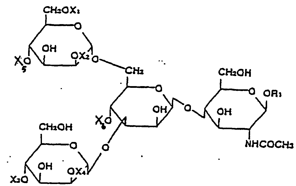
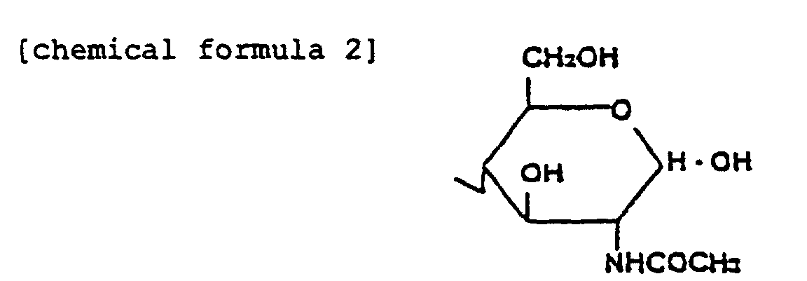 wherein R2 represents an aldehyde group, a labeled methylene group or a labeled methine group.
- A kit for use in determining sugar chain structure according to any of claims 1-15 comprising:wherein X1, X2, X3, X4, X5 and X6 represent hydrogen or a β-N-acetyl glucosamine residue and R1 is represented by the following chemical formula 2 or 3:(i) a reagent to enzymatically or chemically remove branches not having one of the sugar residues Gal, SA or Fuc from the sugar chain and(ii) at least one standard sugar chain according to the formula:
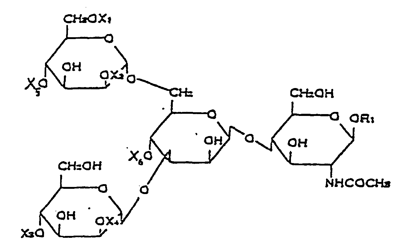
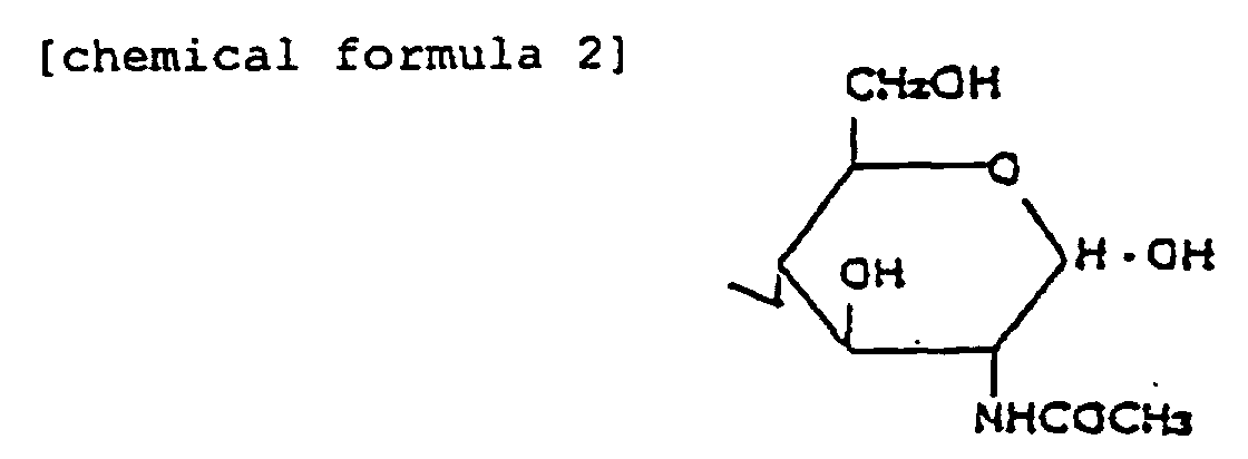 wherein R2 is an aldehyde group, a labeled methylene group or a labeled methine group.
- A kit according to claim 16 further comprising a second oligosaccharide represented by the following chemical formula:wherein X1, X2, X3 and X4 represent hydrogen or a β-N-acetyl glucosamine residue and R1 is represented by the following chemical formula 2 or 3:

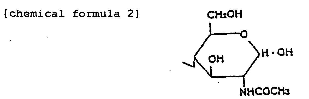 wherein R2 represents an aldehyde group, a labeled methylene group or a labeled methine group.
- An oligosaccharide represented by the following chemical formula:wherein X1, X2, X3 and X4 represent hydrogen or a β-N-acetyl glucosamine residue and R1 is represented by the following chemical formula 2 or 3:
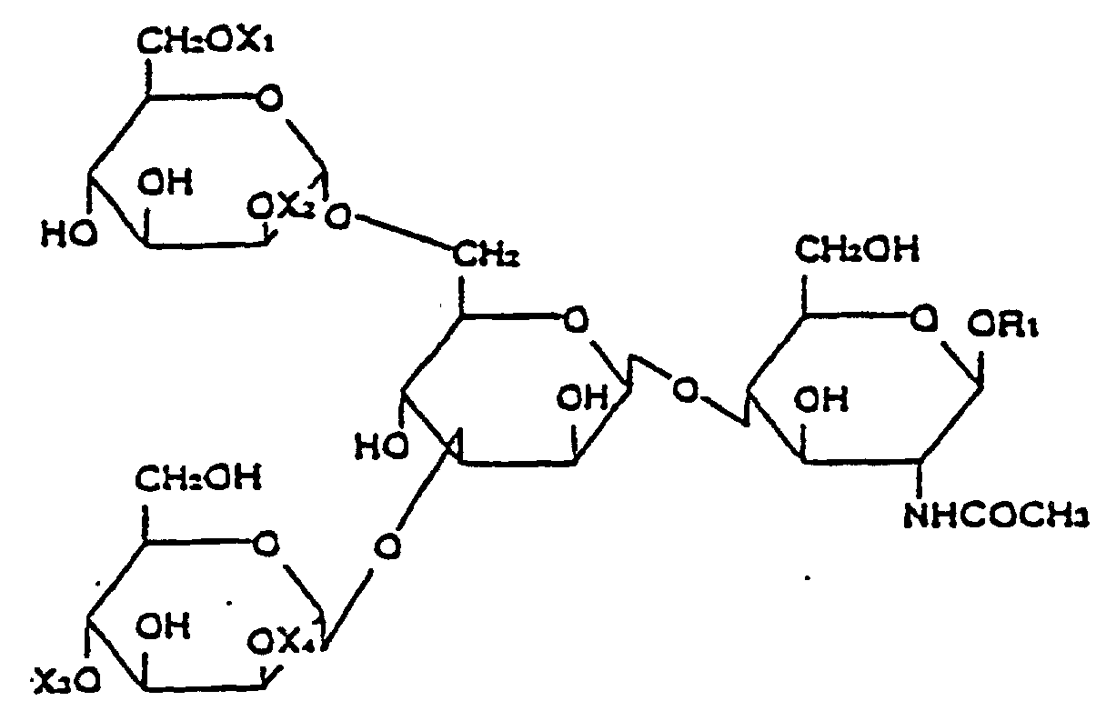
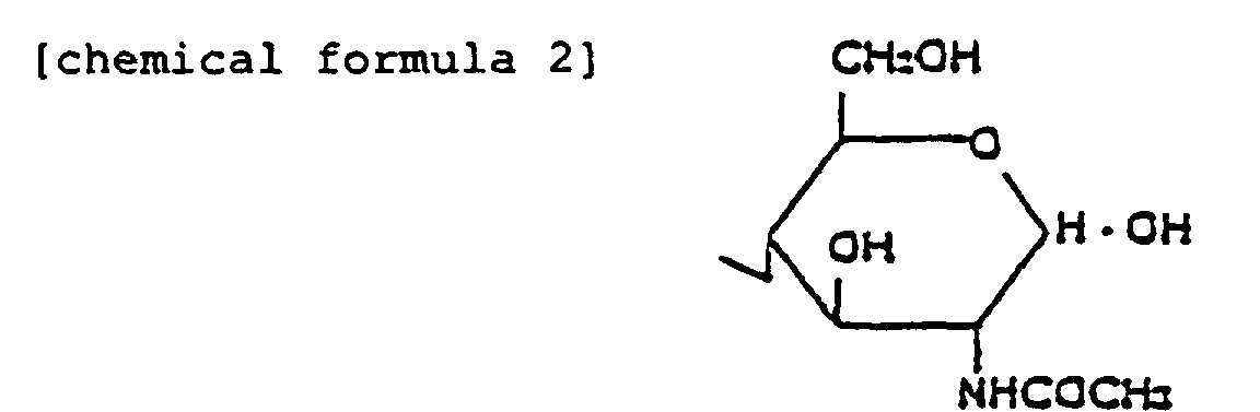 wherein R2 represents an aldehyde group, a labeled methylene group or a labeled methine group;
with the two provisos that (i) the cases where both X1 and X3 represent hydrogen simultaneously are excluded and (ii) the cases where both X2 and X4 represent β-N-acetyl glucosamine residues simultaneously are excluded. - An oligosaccharide of claim 18, wherein R1 is represented by the chemical formula 3.
- An oligosaccharide of claim 19, wherein R2 represents a labeled methylene group.
- An oligosaccharide of claim 20, wherein the labeled methylene group is a methylene group labeled with 2-aminopyridine.
Applications Claiming Priority (6)
| Application Number | Priority Date | Filing Date | Title |
|---|---|---|---|
| JP22787793 | 1993-08-23 | ||
| JP22787793 | 1993-08-23 | ||
| JP227877/93 | 1993-08-23 | ||
| JP6179748A JP3020811B2 (en) | 1993-08-23 | 1994-07-08 | Sugar chain structure determination method |
| JP17974894 | 1994-07-08 | ||
| JP179748/94 | 1994-07-08 |
Publications (3)
| Publication Number | Publication Date |
|---|---|
| EP0640835A2 EP0640835A2 (en) | 1995-03-01 |
| EP0640835A3 EP0640835A3 (en) | 1996-04-03 |
| EP0640835B1 true EP0640835B1 (en) | 2004-04-07 |
Family
ID=26499503
Family Applications (1)
| Application Number | Title | Priority Date | Filing Date |
|---|---|---|---|
| EP94305980A Expired - Lifetime EP0640835B1 (en) | 1993-08-23 | 1994-08-12 | Method for determining sugar chain structure |
Country Status (5)
| Country | Link |
|---|---|
| US (1) | US5500342A (en) |
| EP (1) | EP0640835B1 (en) |
| JP (1) | JP3020811B2 (en) |
| CA (1) | CA2130028C (en) |
| DE (1) | DE69433675T2 (en) |
Families Citing this family (14)
| Publication number | Priority date | Publication date | Assignee | Title |
|---|---|---|---|---|
| WO2003025133A2 (en) * | 2001-09-14 | 2003-03-27 | Mimeon, Inc. | Methods of making glycolmolecules with enhanced activities and uses thereof |
| JP4374447B2 (en) * | 2003-12-25 | 2009-12-02 | 独立行政法人産業技術総合研究所 | Method for analyzing the interaction between protein and sugar chain |
| WO2005064327A1 (en) * | 2003-12-25 | 2005-07-14 | National Institute Of Advanced Industrial Science And Technology | Sugar chain structure profiling technique |
| US20090216705A1 (en) * | 2005-04-13 | 2009-08-27 | National Institute Of Advanced Industrial Science And Technology | Method for predicting sugar chain structure |
| CA2794697A1 (en) | 2010-04-07 | 2011-10-13 | Momenta Pharmaceuticals, Inc. | High mannose glycans |
| CN102812290B (en) | 2010-04-09 | 2016-08-03 | 皇家飞利浦电子股份有限公司 | There is the luminaire of smooth cut-off |
| US9170249B2 (en) | 2011-03-12 | 2015-10-27 | Momenta Pharmaceuticals, Inc. | N-acetylhexosamine-containing N-glycans in glycoprotein products |
| EP3009441B1 (en) | 2011-09-20 | 2016-12-21 | Wakayama University | Process for producing novel sialo-sugar chain |
| EP2856159A4 (en) | 2012-06-01 | 2016-04-13 | Momenta Pharmaceuticals Inc | Methods related to denosumab |
| EP2877589B1 (en) | 2012-07-26 | 2024-03-06 | Momenta Pharmaceuticals, Inc. | Glycoproteins with anti-inflammatory properties |
| EP2855533A4 (en) | 2013-03-15 | 2015-11-25 | Momenta Pharmaceuticals Inc | Methods related to ctla4-fc fusion proteins |
| US10464996B2 (en) | 2013-05-13 | 2019-11-05 | Momenta Pharmaceuticals, Inc. | Methods for the treatment of neurodegeneration |
| WO2015057622A1 (en) | 2013-10-16 | 2015-04-23 | Momenta Pharmaceuticals, Inc. | Sialylated glycoproteins |
| CN113185558B (en) * | 2021-05-08 | 2023-01-24 | 河南大学 | Galactose modified detection probe capable of being identified by OGT (one glass solution) |
Family Cites Families (10)
| Publication number | Priority date | Publication date | Assignee | Title |
|---|---|---|---|---|
| US5260433A (en) * | 1982-06-23 | 1993-11-09 | Enzo Diagnostics, Inc. | Saccharide specific binding system labeled nucleotides |
| SE451849B (en) * | 1985-12-11 | 1987-11-02 | Svenska Sockerfabriks Ab | VIEW TO SYNTHETIZE GYCLOSIDIC BINDINGS AND USE OF THIS RECEIVED PRODUCTS |
| JPS6410177A (en) * | 1987-07-02 | 1989-01-13 | Takara Shuzo Co | Fluorescent labeling method for saccharides |
| JP2579497B2 (en) * | 1987-09-07 | 1997-02-05 | オリエンタル酵母工業株式会社 | Liver disease diagnostic agent |
| DK626488D0 (en) * | 1988-11-10 | 1988-11-10 | Torben Falck Oerntoft | PROCEDURE FOR SELECTING A SECRET SAMPLE THROUGH A BODY OPENING AND PREPARATION OF THE SAMPLE FOR THE QUANTITATIVE DETERMINATION OF MOLECULAR STRUCTURES AND ENZYME ACTIVITY OF THE SAMPLE SAMPLING SAMPLING SAMPLE SUMMARY |
| JPH05500563A (en) * | 1989-09-27 | 1993-02-04 | アストロスキャン・リミテッド | Carbohydrate analysis |
| US5100778A (en) * | 1989-10-03 | 1992-03-31 | Monsanto Company | Oligosaccharide sequencing |
| US5284558A (en) * | 1990-07-27 | 1994-02-08 | University Of Iowa Research Foundation | Electrophoresis-based sequencing of oligosaccharides |
| JPH04221393A (en) * | 1990-12-20 | 1992-08-11 | Kikkoman Corp | N-acetyl-beta-d-glucosamine derivative, reagent for determination of n-acetyl-beta-d-glucosaminidase activity containing the derivative as active component and determination of n-acetyl-beta-d-glucosaminidase activity using the same |
| US5667984A (en) * | 1991-05-07 | 1997-09-16 | Oxford Glycosystems Ltd. | Sequencing of oligosaccharides |
-
1994
- 1994-07-08 JP JP6179748A patent/JP3020811B2/en not_active Expired - Fee Related
- 1994-08-11 US US08/288,768 patent/US5500342A/en not_active Expired - Fee Related
- 1994-08-12 DE DE69433675T patent/DE69433675T2/en not_active Expired - Fee Related
- 1994-08-12 EP EP94305980A patent/EP0640835B1/en not_active Expired - Lifetime
- 1994-08-12 CA CA002130028A patent/CA2130028C/en not_active Expired - Fee Related
Also Published As
| Publication number | Publication date |
|---|---|
| DE69433675D1 (en) | 2004-05-13 |
| US5500342A (en) | 1996-03-19 |
| CA2130028A1 (en) | 1995-02-24 |
| JPH07112996A (en) | 1995-05-02 |
| EP0640835A3 (en) | 1996-04-03 |
| CA2130028C (en) | 2001-06-05 |
| DE69433675T2 (en) | 2005-03-03 |
| JP3020811B2 (en) | 2000-03-15 |
| EP0640835A2 (en) | 1995-03-01 |
Similar Documents
| Publication | Publication Date | Title |
|---|---|---|
| Schauer | [10] Analysis of sialic acids | |
| US5500342A (en) | Method for determining sugar chain structure | |
| Ashford et al. | The β1→ 2‐d‐xylose and α1→ 3‐l‐fucose substituted N‐linked oligosaccharides from Erythrina cristagalli lectin: Isolation, characterisation and comparison with other legume lectins | |
| Van Tilbeurgh et al. | Fluorogenic and chromogenic glycosides as substrates and ligands of carbohydrases | |
| US5100778A (en) | Oligosaccharide sequencing | |
| JP6602747B2 (en) | Synthesis and use of isotopically labeled glycans | |
| Piller et al. | Biosynthesis of blood group I antigens. Identification of a UDP-GlcNAc: GlcNAc beta 1-3Gal (-R) beta 1-6 (GlcNAc to Gal) N-acetylglucosaminyltransferase in hog gastric mucosa. | |
| Murata et al. | Facile enzymatic conversion of lactose into lacto-N-tetraose and lacto-N-neotetraose | |
| Kimura et al. | The N‐glycans of jack bean α‐mannosidase: Structure, topology and function | |
| Lee et al. | Carbohydrate analysis of glycoproteins A review | |
| Prime et al. | Oligosaccharide sequencing based on exo-and endoglycosidase digestion and liquid chromatographic analysis of the products | |
| US7094563B2 (en) | Isolation and composition of novel glycosidases | |
| US5753454A (en) | Sequencing of oligosaccharides: the reagent array-electrochemical detection method | |
| Ishii et al. | A fluorogenic probe for core-fucosylated glycan-preferred ENGase | |
| US5667984A (en) | Sequencing of oligosaccharides | |
| Ohta et al. | Further characterization of allergenically active oligosaccharitols isolated from a sea squirt H-antigen | |
| Takegawa et al. | Enzymatic Synthesis of a Neoglycoconjugate by Transglycosylation withArthrobacterEndo-β-N-acetylglucosaminidase: A Substrate for Colorimetric Detection of Endo-β-N-acetylglucosaminidase Activity | |
| US6458573B1 (en) | Isolation and composition of a novel glycosidase from chryseobacterium | |
| EP0675204B1 (en) | Transglycosylating process for producing carbohydrates or glycoconjugates | |
| Hokke et al. | Action of rat liver Galβ1-4GlcNAc α (2-6)-sialyltransferase on Manβ1-4GlcNAcβ-OMe, GalNAcβ1-4GlcNAcβ-OMe, Glcβ1-4GlcNAcβ-OMe and GlcNAcβ1-4GlcNAcβ-OMe as synthetic substrates | |
| US6423525B1 (en) | Isolation and composition of a novel glycosidase from chryseobacterium | |
| Yoshima et al. | The Asparagine-Linked Sugar Chains of the Glycoproteins in Calf Thymocyte Plasma Membrane Structural Studies of Neutral Oligosaccharides | |
| EP0586419A1 (en) | Sequencing of oligosaccharides | |
| Lee et al. | Conformational studies of glycopeptides by energy transfer: introduction of fluorophore at specific branches of biantennary glycopeptides | |
| US6358724B1 (en) | Isolation and composition of novel glycosidases |
Legal Events
| Date | Code | Title | Description |
|---|---|---|---|
| PUAI | Public reference made under article 153(3) epc to a published international application that has entered the european phase |
Free format text: ORIGINAL CODE: 0009012 |
|
| AK | Designated contracting states |
Kind code of ref document: A2 Designated state(s): CH DE ES FR GB IT LI SE |
|
| PUAL | Search report despatched |
Free format text: ORIGINAL CODE: 0009013 |
|
| AK | Designated contracting states |
Kind code of ref document: A3 Designated state(s): CH DE ES FR GB IT LI SE |
|
| 17P | Request for examination filed |
Effective date: 19960417 |
|
| 17Q | First examination report despatched |
Effective date: 19981218 |
|
| RAP1 | Party data changed (applicant data changed or rights of an application transferred) |
Owner name: TAKARA BIO INC. |
|
| GRAP | Despatch of communication of intention to grant a patent |
Free format text: ORIGINAL CODE: EPIDOSNIGR1 |
|
| GRAS | Grant fee paid |
Free format text: ORIGINAL CODE: EPIDOSNIGR3 |
|
| GRAA | (expected) grant |
Free format text: ORIGINAL CODE: 0009210 |
|
| AK | Designated contracting states |
Kind code of ref document: B1 Designated state(s): CH DE ES FR GB IT LI SE |
|
| PG25 | Lapsed in a contracting state [announced via postgrant information from national office to epo] |
Ref country code: LI Free format text: LAPSE BECAUSE OF FAILURE TO SUBMIT A TRANSLATION OF THE DESCRIPTION OR TO PAY THE FEE WITHIN THE PRESCRIBED TIME-LIMIT Effective date: 20040407 Ref country code: IT Free format text: LAPSE BECAUSE OF FAILURE TO SUBMIT A TRANSLATION OF THE DESCRIPTION OR TO PAY THE FEE WITHIN THE PRESCRIBED TIME-LIMIT;WARNING: LAPSES OF ITALIAN PATENTS WITH EFFECTIVE DATE BEFORE 2007 MAY HAVE OCCURRED AT ANY TIME BEFORE 2007. THE CORRECT EFFECTIVE DATE MAY BE DIFFERENT FROM THE ONE RECORDED. Effective date: 20040407 Ref country code: CH Free format text: LAPSE BECAUSE OF FAILURE TO SUBMIT A TRANSLATION OF THE DESCRIPTION OR TO PAY THE FEE WITHIN THE PRESCRIBED TIME-LIMIT Effective date: 20040407 |
|
| REG | Reference to a national code |
Ref country code: GB Ref legal event code: FG4D |
|
| REG | Reference to a national code |
Ref country code: CH Ref legal event code: EP |
|
| REF | Corresponds to: |
Ref document number: 69433675 Country of ref document: DE Date of ref document: 20040513 Kind code of ref document: P |
|
| PG25 | Lapsed in a contracting state [announced via postgrant information from national office to epo] |
Ref country code: SE Free format text: LAPSE BECAUSE OF FAILURE TO SUBMIT A TRANSLATION OF THE DESCRIPTION OR TO PAY THE FEE WITHIN THE PRESCRIBED TIME-LIMIT Effective date: 20040707 |
|
| PG25 | Lapsed in a contracting state [announced via postgrant information from national office to epo] |
Ref country code: ES Free format text: LAPSE BECAUSE OF FAILURE TO SUBMIT A TRANSLATION OF THE DESCRIPTION OR TO PAY THE FEE WITHIN THE PRESCRIBED TIME-LIMIT Effective date: 20040718 |
|
| REG | Reference to a national code |
Ref country code: CH Ref legal event code: PL |
|
| ET | Fr: translation filed | ||
| PLBE | No opposition filed within time limit |
Free format text: ORIGINAL CODE: 0009261 |
|
| STAA | Information on the status of an ep patent application or granted ep patent |
Free format text: STATUS: NO OPPOSITION FILED WITHIN TIME LIMIT |
|
| 26N | No opposition filed |
Effective date: 20050110 |
|
| PGFP | Annual fee paid to national office [announced via postgrant information from national office to epo] |
Ref country code: FR Payment date: 20060808 Year of fee payment: 13 |
|
| PGFP | Annual fee paid to national office [announced via postgrant information from national office to epo] |
Ref country code: GB Payment date: 20060809 Year of fee payment: 13 |
|
| PGFP | Annual fee paid to national office [announced via postgrant information from national office to epo] |
Ref country code: DE Payment date: 20060810 Year of fee payment: 13 |
|
| GBPC | Gb: european patent ceased through non-payment of renewal fee |
Effective date: 20070812 |
|
| REG | Reference to a national code |
Ref country code: FR Ref legal event code: ST Effective date: 20080430 |
|
| PG25 | Lapsed in a contracting state [announced via postgrant information from national office to epo] |
Ref country code: DE Free format text: LAPSE BECAUSE OF NON-PAYMENT OF DUE FEES Effective date: 20080301 |
|
| PG25 | Lapsed in a contracting state [announced via postgrant information from national office to epo] |
Ref country code: FR Free format text: LAPSE BECAUSE OF NON-PAYMENT OF DUE FEES Effective date: 20070831 |
|
| PG25 | Lapsed in a contracting state [announced via postgrant information from national office to epo] |
Ref country code: GB Free format text: LAPSE BECAUSE OF NON-PAYMENT OF DUE FEES Effective date: 20070812 |