EP0626861B1 - Treatment for asthma - Google Patents
Treatment for asthma Download PDFInfo
- Publication number
- EP0626861B1 EP0626861B1 EP93902914A EP93902914A EP0626861B1 EP 0626861 B1 EP0626861 B1 EP 0626861B1 EP 93902914 A EP93902914 A EP 93902914A EP 93902914 A EP93902914 A EP 93902914A EP 0626861 B1 EP0626861 B1 EP 0626861B1
- Authority
- EP
- European Patent Office
- Prior art keywords
- antibody
- vla
- asthma
- use according
- composition
- Prior art date
- Legal status (The legal status is an assumption and is not a legal conclusion. Google has not performed a legal analysis and makes no representation as to the accuracy of the status listed.)
- Expired - Lifetime
Links
Images


Classifications
-
- C—CHEMISTRY; METALLURGY
- C07—ORGANIC CHEMISTRY
- C07K—PEPTIDES
- C07K14/00—Peptides having more than 20 amino acids; Gastrins; Somatostatins; Melanotropins; Derivatives thereof
- C07K14/435—Peptides having more than 20 amino acids; Gastrins; Somatostatins; Melanotropins; Derivatives thereof from animals; from humans
- C07K14/705—Receptors; Cell surface antigens; Cell surface determinants
- C07K14/70503—Immunoglobulin superfamily
- C07K14/70542—CD106
-
- A—HUMAN NECESSITIES
- A61—MEDICAL OR VETERINARY SCIENCE; HYGIENE
- A61P—SPECIFIC THERAPEUTIC ACTIVITY OF CHEMICAL COMPOUNDS OR MEDICINAL PREPARATIONS
- A61P11/00—Drugs for disorders of the respiratory system
-
- A—HUMAN NECESSITIES
- A61—MEDICAL OR VETERINARY SCIENCE; HYGIENE
- A61P—SPECIFIC THERAPEUTIC ACTIVITY OF CHEMICAL COMPOUNDS OR MEDICINAL PREPARATIONS
- A61P11/00—Drugs for disorders of the respiratory system
- A61P11/06—Antiasthmatics
-
- A—HUMAN NECESSITIES
- A61—MEDICAL OR VETERINARY SCIENCE; HYGIENE
- A61P—SPECIFIC THERAPEUTIC ACTIVITY OF CHEMICAL COMPOUNDS OR MEDICINAL PREPARATIONS
- A61P37/00—Drugs for immunological or allergic disorders
- A61P37/08—Antiallergic agents
-
- C—CHEMISTRY; METALLURGY
- C07—ORGANIC CHEMISTRY
- C07K—PEPTIDES
- C07K16/00—Immunoglobulins [IG], e.g. monoclonal or polyclonal antibodies
- C07K16/18—Immunoglobulins [IG], e.g. monoclonal or polyclonal antibodies against material from animals or humans
- C07K16/28—Immunoglobulins [IG], e.g. monoclonal or polyclonal antibodies against material from animals or humans against receptors, cell surface antigens or cell surface determinants
- C07K16/2839—Immunoglobulins [IG], e.g. monoclonal or polyclonal antibodies against material from animals or humans against receptors, cell surface antigens or cell surface determinants against the integrin superfamily
- C07K16/2842—Immunoglobulins [IG], e.g. monoclonal or polyclonal antibodies against material from animals or humans against receptors, cell surface antigens or cell surface determinants against the integrin superfamily against integrin beta1-subunit-containing molecules, e.g. CD29, CD49
-
- A—HUMAN NECESSITIES
- A61—MEDICAL OR VETERINARY SCIENCE; HYGIENE
- A61K—PREPARATIONS FOR MEDICAL, DENTAL OR TOILETRY PURPOSES
- A61K38/00—Medicinal preparations containing peptides
Definitions
- the present invention relates to a treatment for asthma. More particularly, this invention relates to the use of antibodies recognizing Very Late Antigen-4 (VLA-4), a ligand on certain leukocytes for the endothelial cell receptor Vascular Cell Adhesion Molecule-1 (VCAM-1), in the treatment of asthma.
- VLA-4 Very Late Antigen-4
- VCAM-1 Vascular Cell Adhesion Molecule-1
- Asthma is a condition of the respiratory tract characterized by widespread, reversible narrowing of the airways (bronchoconstriction) and increased sensitivity (hyperresponsiveness) of the airways to a variety of stimuli.
- the familiar symptomology of asthma i.e., coughing, wheezing, chest tightness, dyspnea, is caused by airway smooth muscle contraction, increased bronchial mucus secretion, and inflammation.
- asthma has been estimated to affect 10-20% of school-aged children around the world, and hospital admissions for asthma in children have increased dramatically in recent years, one survey for the United States indicating that hospital admissions for children under 15 with asthma increased by at least 145% between 1970 and 1984. (See, M.R. Sears, 1990 [1].)
- it is estimated that 10 million Americans (4% of the population) have asthma, and some $4 billion is spent in treatment per year. L.K. Altman, 1991 [2]; C. Starr, 1991 [3].
- Triggers exacerbate asthma by causing transient enhancement of airway hyperresponsiveness. Triggers that have been found to induce airway hyperresponsiveness include inhaled allergens, inhaled low molecular weight agents to which the subject has become sensitized (e.g., by occupational exposure), viral or mycoplasma respiratory infections, and oxidizing gases such as ozone and nitrogen dioxide.
- inducing triggers can be distinguished from “inciting" triggers of bronchospastic episodes which include exercise, cold air, emotional stress, pharmacological triggers, inhaled irritants.
- the common feature of inducing triggers is that they are associated with airways inflammation; inciting triggers produce smooth muscle contractions (bronchospasms) which depend on the underlying degree of hyperresponsiveness, rather than increasing airways responsiveness themselves. (See, D.W. Cockcroft, 1990 [4].)
- DSCG disodium cromoglycate
- nedocromil sodium nedocromil sodium
- antihistamines such as ketotifen
- corticosteroids such as prednisolone
- the inflammatory response in asthma is typical for tissues covered by a mucosa and is characterized by vasodilation, plasma exudation, recruitment of inflammatory cells such as neutrophils, monocytes, macrophages, lymphocytes and eosinophils to the sites of inflammation, and release of inflammatory mediators by resident tissue cells (e.g., mast cells) or by migrating inflammatory cells.
- inflammatory cells such as neutrophils, monocytes, macrophages, lymphocytes and eosinophils to the sites of inflammation
- resident tissue cells e.g., mast cells
- migrating inflammatory cells e.g., mast cells
- Late phase response in allergen-induced asthma and persistent hyperresponsiveness have been associated with the recruitment of leukocytes, and particularly eosinophils, to inflamed lung tissue.
- leukocytes and particularly eosinophils
- Eosinophils are known to release several inflammatory mediators, e.g., 15-HETE, leukotriene C 4 , PAF, cationic proteins, eosinophil peroxidase.
- beta 2 -adrenoceptor agonists and DSCG are potent stabilizers of mast cells, which are capable of releasing many mediators, including histamine, prostaglandins, leukotrienes, platelet activating factor (PAF), and chemotactic factors for neutrophils and eosinophils; corticosteroids, as another example, complex with steroid hormone receptors, which leads to the synthesis of proteins, such as lipocortins, that produce anti-inflammatory effects.
- PAF platelet activating factor
- corticosteroids as another example, complex with steroid hormone receptors, which leads to the synthesis of proteins, such as lipocortins, that produce anti-inflammatory effects.
- Inflammatory leukocytes are recruited to sites of inflammation by cell adhesion molecules that are expressed on the surface of endothelial cells and which act as receptors for leukocyte surface proteins or protein complexes.
- Eosinophils have recently been found to participate in three distinct cell adhesion pathways to vascular endothelium, binding to cells expressing intercellular adhesion molecule-1 (ICAM-1), endothelial ceil adhesion molecule-1 (ELAM-1), and vascular cell adhesion molecule-1 (VCAM-1).
- IAM-1 intercellular adhesion molecule-1
- ELAM-1 endothelial ceil adhesion molecule-1
- VCAM-1 vascular cell adhesion molecule-1
- VCAM1 binds to the ⁇ 4 ⁇ 1 integrin, VLA-4, which is expressed on various lymphoid cells, including eosinophils (Weller et al., 1991 [10]; Elices et al. 1990 [14]). That eosinophils express VLA-4 differentiates them from other inflammatory cells such as neutrophils, which bind to ELAM-1 and ICAM-1 but not VCAM-1.
- the pharmaceutical composition comprises a VCAM, or an anti-VCAM antibody, and a pharmaceutically acceptable excipient or carrier.
- the document falls for consideration under Article 54 (3) EPC.
- the VLA-4 mediated adhesion pathway was investigated in an asthma model to examine the possible role of VLA-4 in leukocyte recruitment to inflammed lung tissue. It has now been discovered that administering anti-VLA-4 antibody inhibits both the late phase response and airway hyperresponsiveness in allergic sheep. Surprisingly, administration of anti-VLA-4 led to a reduction in the number of both neutrophils and eosinophils in the lung at 4 hours after allergen challenge, even though both cells have alternate adhesion pathways by which they can be recruited to lung tissues. Also surprisingly, inhibition of hyperresponsiveness in the treated sheep was observed which continued to 1 week, even though infiltration of leukocytes, including neutrophils and eosinophils, was not significantly reduced over time.
- the present invention is based on novel methods for the treatment of asthma and provides new pharmaceutical compositions useful in the treatment of asthma.
- the present invention provides the manufacture of a medicament for use in a method comprising the step of administering to an asthma sufferer an effective amount of an anti-VLA-4 antibody, such as monoclonal antibody HP1/2.
- the anti-VLA-4 antibody is advantageously administered in vivo to a patient with chronic allergen-induced asthma, and serves to inhibit late phase response to allergens and to attenuate airway hyperresponsiveness.
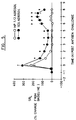
- Figure 1 is a graph depicting the effect of monoclonal antibody HP1/2 (intravenous) on the response to allergen ( Ascaris suum antigen) in dual responder allergic sheep. Percentage change in specific lung resistance (SR L ) is measured over time post allergen challenge. Asterisks indicate statistically significant results.
- Figure 2 is a graph depicting plasma concentration of monoclonal antibody HP1/2 (intravenous) in sheep, measured over time after initial administration.
- Figure 3 is a graph depicting the effect of monoclonal antibody HP1/2 (intravenous) on airway hyperresponsiveness in dual responder sheep.
- Airway responsiveness measured in breath units (BU) of cumulative breaths of a 1% weight/volume carbachol solution (a known bronchoconstrictor) that increases specific lung resistance 400% over the value obtained using diluent alone.
- BU breath units
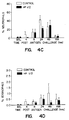
- Figure 4 is a series of four graphs showing the total cells (Fig. 4A) and the levels of different leukocytes (lymphocytes (Fig. 4B), neutrophils (Fig. 4C), and eosinophils (Fig. 4D)) detected by bronchoalveolar lavage in allergic sheep challenged with Ascaris suum antigen alone and after pretreatment with monoclonal antibody HP1/2 (intravenous).
- Figure 5 is a graph depicting the effect of monoclonal antibody HP1/2 (16 mg, aerosol) and 1E6 (16 mg, aerosol) on the response to allergen ( Ascaris suum antigen) in dual responder allergic sheep. Percentage change in specific lung resistance (SR L ) is measured over time post allergen challenge. Asterisks indicate statistically significant results.
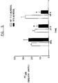
- Figure 6 is a graph depicting the effect of monoclonal antibody HP1/2 (16 mg, aerosol) and 1E6 (16 mg, aerosol) on airway hyperresponsiveness in dual responder sheep.
- Airway responsiveness measured in breath units (BU) of cumulative breaths of a 1% weight/volume carbachol solution (a known bronchoconstrictor) that increases specific lung resistance 400% over the value obtained using diluent alone.
- BU breath units
- an immortal cell line typically myeloma cells
- lymphocytes typically splenocytes
- a given antigen e.g., VLA-4
- Immunization may be accomplished using standard procedures. The unit dose and immunization regimen depend on the species of mammal immunized, its immune status, the body weight of the mammal, etc. Typically, the immunized mammals are bled and the serum from each blood sample is assayed for particular antibodies using appropriate screening assays. For example, anti-VLA-4 antibodies may be identified by immunoprecipitation of 125 I-labeled cell lysates from VLA-4-expressing cells. (See, Sanchez-Madrid et al. 1986 [16] and Hemler et al.
- Anti-VLA-4 antibodies may also be identified by flow cytometry, e.g., by measuring flourescent staining of Ramos cells incubated with an antibody believed to recognize VLA-4 (see, Elices et al., (1990) [14]).
- the lymphocytes used in the production of hybridoma cells typically are isolated from immunized mammals whose sera have already tested positive for the presence of anti-VLA-4 antibodies using such screening assays.
- the immortal cell line (e.g., a myeloma cell line) is derived from the same mammalian species as the lymphocytes.
- Preferred immortal cell lines are mouse myeloma cell lines that are sensitive to culture medium containing hypoxanthine, aminopterin and thymidine ("HAT medium").
- HAT-sensitive mouse myeloma cells are fused to mouse splenocytes using 1500 molecular weight polyethylene glycol ("PEG 1500").
- Hybridoma cells resulting from the fusion are then selected using HAT medium, which kills unfused and unproductively fused myeloma cells (unfused splenocytes die after several days because they are not transformed).
- Hybridomas producing a desired antibody are detected by screening the hybridoma culture supernatants.
- hybridomas prepared to produce anti-VLA-4 antibodies may be screened by testing the hybridoma culture supernatant for secreted antibodies having the ability to bind to a recombinant ⁇ 4 -subunit-expressing cell line, such as transfected K-562 cells (see, Elices et al. [14]).
- hybridoma cells that tested positive in such screening assays were cultured in a nutrient medium under conditions and for a time sufficient to allow the hybridoma cells to secrete the monoclonal antibodies into the culture medium.
- Tissue culture techniques and culture media suitable for hybridoma cells are well known.
- the conditioned hybridoma culture supernatant may be collected and the anti-VLA-4 antibodies optionally further purified by well-known methods.
- the desired antibody may be produced by injecting the hybridoma cells into the peritoneal cavity of an unimmunized mouse.
- the hybridoma cells proliferate in the peritoneal cavity, secreting the antibody which accumulates as ascites fluid.
- the antibody may be harvested by withdrawing the ascites fluid from the peritoneal cavity with a syringe.
- anti-VLA-4 monoclonal antibodies have been previously described (see, e.g., Sanchez-Madrid et al., 1986 [16]; Hemler et al. (1987) [17]; Pulido et al. (1991) [19]).
- an anti-VLA-4 monoclonal antibody designated HP1/2 obtained from Biogen, Inc., Cambridge, MA
- HP1/2 obtained from Biogen, Inc., Cambridge, MA
- Such a chimeric HP1/2 antibody is similar in specificity and potency to the murine HP1/2 antibody, and may be useful in methods of treatment according to the present invention.
- humanized recombinant anti-VLA-4 antibodies may be useful in these methods.
- the HP1/2 V H DNA sequence and its translated amino acid sequences are set forth in SEQ ID NO: 1 and SEQ ID NO: 2, respectively.
- the HP1/2 V K DNA sequence and its translated amino acid sequence are set forth in SEQ ID NO: 3 and SEQ ID NO: 4, respectively.
- Monoclonal antibodies such as HP1/2 and other anti-VLA-4 antibodies e.g., Mab HP2/1, HP2/4, L25, P4C2
- the antibodies will recognize the B1 or B2 epitopes of the VLA- ⁇ 4 chain (see, Pulido et al. (1991) [19]).
- anti-VLA-4 antibodies used according to the method of the present invention may specifically inhibit, at least for an initial period following allergen challenge, the migration of VLA-4-expressing leukocytes to inflamed sections of the lung.
- VLA-4 leukocyte migration could, in turn, prevent secondary pathological effects of leukocyte infiltration, e.g., release of toxic substances, inducement of soluble inflammatory cell mediators, release or inducement of leukocyte chemotactic agents (such as neutrophil chemotactic factors), etc.
- leukocyte chemotactic agents such as neutrophil chemotactic factors
- late phase response to the allergen and continuing hypersensitivity of the airways may be attenuated.
- the anti-VLA-4 antibodies may attenuate signal transduction necessary for the release of inflammatory mediators and/or cell chemotactic agents.
- the present invention provides the manufacture of a medicament for use in a method for the treatment of asthma which comprises administering to a mammal suffering from allergic asthma a composition comprising an anti-VLA-4 antibody.
- a composition comprising an anti-VLA-4 antibody.
- the examples below set forth the results observed in asthmatic sheep. However, the similarity between physiological responses and pharmacological effects in sheep and in humans has been documented (see, e.g., W.M. Abraham, 1989 [20]); and similarities between sheep and other animal asthma models (rabbits, squirrel monkeys, guinea pigs, and sensitized dogs) have been noted (see, e.g., W.M. Abraham et al., 1988 [8]). Accordingly, the results reported herein will be relevant and applicable to, and the method claimed will be useful in, any mammal, including humans, suffering from allergic asthma.
- the anti-VLA-4 antibody administered in accordance with the present invention may be administered prophylactically, before exposure to an asthma-inducing allergen. Beneficial effects will also be obtained if the antibody is administered at the time of or immediately after allergen exposure, between early phase and late phase response to attenuate the severity of late phase response, or at any time following allergen exposure to reduce or eliminate airway hyperresponsiveness.
- the anti-VLA-4 antibody can be administered in the form of a composition comprising an anti-VLA-4 antibody and a pharmaceutically acceptable carrier.
- the composition will be in a form suitable for intravenous injection.
- antibody compositions in the form of a sterile aqueous or phosphate-buffered saline solution which can be nebulized (atomized) and breathed directly into the lungs by the asthma sufferer, e.g., using an inhaler.
- Dosages will vary depending on the sensitivity of the asthma sufferer to particular allergens, the concentration of allergen on exposure and frequency/duration of exposure(s), the proposed mode of administration (e.g., injection or inhalation), the desired plasma level of antibody, the effectiveness of a particular antibody or combination of antibodies in suppressing airway responsiveness, the clearance rate or half-life of the antibody composition, and other such factors familiar to physicians experienced in the treatment of allergic asthma.
- dosages will be calculated and adjusted to maintain a plasma level of antibody in the range of from 1-1000 ⁇ g/ml, although higher or lower dosages may be indicated with consideration to the age, sensitivity, tolerance, and other characteristics of the patient, the acuteness of the flareup, the history and course of the disease, and other similar factors routinely considered by an attending physician.
- Suitable pharmaceutical carriers include, e.g., sterile saline and physiological buffer solutions. Phosphate buffered saline (PBS) is preferred for inhalant administration.
- PBS Phosphate buffered saline
- the pharmaceutical compositions may additionally be formulated to control the release of the active ingredients or to prolong their presence in the patient's system.
- suitable drug delivery systems include, e.g., hydrogels, hydroxmethylcellulose, microcapsules, liposomes, microemulsions, microspheres, and the like.
- antibodies capable of binding to the ⁇ 4 subunit of VLA-4 must be employed. It is preferred that monoclonal antibodies be used.
- suitable recombinant antibodies capable of binding to VLA-4 may alternatively be used.
- Such recombinant antibodies include antibodies produced via recombinant DNA techniques, e.g., by transforming a host cell with a suitable expression vector containing DNA encoding the light and heavy immunoglobulin chains of the desired antibody, and recombinant chimeric antibodies, wherein some or all of the hinge and constant regions of the heavy and/or the light chain of the anti-VLA-4 antibody have been substituted with corresponding regions of an immunoglobulin light or heavy chain of a different species (i.e., preferably the same species as the asthma sufferer being treated, to minimize immune response to the administered antibody).
- a different species i.e., preferably the same species as the asthma sufferer being treated, to minimize immune response to the administered antibody.
- VLA-4-binding fragments of anti-VLA-4 antibodies such as Fab, Fab', F(ab') 2 , and F(v) fragments; heavy chain monomers or dimers; light chain monomers or dimers; and dimers consisting of one heavy chain and one light chain are also contemplated herein.
- antibody fragments may be produced by chemical methods, e.g., by cleaving an intact antibody with a protease, such as pepsin or papain, or via recombinant DNA techniques, e.g., by using host cells transformed with truncated heavy and/or light chain genes.
- Heavy and light chain monomers may similarly be produced by treating an intact antibody with a reducing agent such as dithiothreitol or ⁇ -mercaptoethanol or by using host cells transformed with DNA encoding either the desired heavy chain or light chain or both.
- VCAM-1 an endothelial cell receptor for VLA-4
- Small molecules such as oligosaccharides that mimic the binding domain of an VLA-4 ligand and fit the receptor domain of VLA-4 may also be employed.
- VLA-4-binding polypeptides or molecules that effectively inhibit late phase response or airway hyperresponsiveness in allergic subjects is contemplated herein as an alternative method for treatment of asthma.
- anti-VLA-4 antibodies may be used in combination with other antibodies having a therapeutic effect on airway responsiveness.
- beneficial effects reported herein are due to the inhibition of leukocyte recruitment to VCAM-1-expressing endothelium
- combinations of anti-VLA-4 antibodies with other antibodies that interfere with the adhesion between leukocyte antigens and endothelial cell receptor molecules may be advantageous.
- anti-ELAM-1 and/or anti-ICAM-1 antibodies may be advantageous. [See, Gundel et al. (1991) [27]; Wegner et al. (1990) [28].)
- compositions contemplated herein may be administered by any suitable means such as orally, intraesophageally or intranasally, intrabronchially (local treatment, e.g., via bronchoscope), as well as subcutaneously, intramuscularly, intravenously, intra-arterially, or parenterally. Ordinarily administration via inhalation is preferred.
- a balloon catheter was advanced through one nostril under topical anesthesia with 2% lidocaine solution to the lower esophagus, and a cuffed endotracheal tube was advanced through the other nostril (using a flexible fiberoptic bronchoscope as a guide) for the measurement of airway mechanics and during aerosol challenges.
- Pleural pressure was estimated with the esophageal balloon catheter (filled with 1 ml of air) positioned 5-10 cm from the gastroesophageal junction. In this position, end expiratory pleural pressure ranged between -2 and -5 cm H 2 O. Once the balloon was placed, it was secured so that it remained in position for the duration of the experiment.
- Airway responsiveness was determined by performing dose response curves to inhaled carbachol.
- the dose response curves were plotted using measurements of SR L taken immediately after inhalation of buffer (PBS) alone and after each consecutive administration of 10 breaths of increasing concentrations of carbachol in PBS.
- the concentrations of carbachol were 0.25%, 0.5%, 1.0%, 2.0% and 4.0% wt/vol in PBS.
- the provocation test was discontinued when SR L increased over 400% from the post-PBS value or after the highest carbachol concentration had been administered.
- Airway responsiveness was determined by calculating from the dose response curves the cumulative carbachol dose in breath units (BU) that increased specific lung resistance 400% over the post buffer value (PD 400% ).
- BU cumulative carbachol dose in breath units
- One breath unit was defined as one breath of a 1% wt/vol carbachol solution.
- Each sheep was subjected to a trial as a control in which a placebo (PBS without additive) was used as a pretreatment 30 minutes before allergen challenge with Ascaris suum antigen (Greer Diagnostics, Lenoir, NC). Subsequently, the sheep were subjected to an identical trial, except that 1 mg/kg of monoclonal antibody HP1/2 was administered to each sheep 30 minutes prior to antigen challenge.
- the placebo buffer control or isotope-matched antibody (1E6, anti-LFA3) control
- HP1/2 compositions were administered by intravenous injection.
- the HP1/2 composition (and the 1E6 control) was prepared by diluting pure antibody obtained from a hybridoma (Biogen, Inc., Cambridge MA) in sterile, endotoxin-free PBS and adjusting to deliver 1 mg/kg antibody based on the weight of each sheep.
- the antigen solution was delivered as an aerosol using a disposable medical nebulizer (RAINDROP®, Puritan Bennett, Lenexa, KS) that provided an aerosol with a mass median aerodynamic diameter of 3.2 ⁇ M (geometric SD 1.9) as determined by an Andersen cascade impactor.
- the Ascaris suum extract was diluted in PBS to a concentration of 82,000 Protein Nitrogen Units(PNU)/ml.
- the output of the nebulizer was directed into a plastic T-tube, one end of which was connected to the inspiratory port of a Harvard respirator.
- a dosimeter connected to the nebulizer consisting of a solenoid valve and a 20 psi compressed air source and the solenoid valve was activated at the beginning of the inspiratory cycle of the Harvard respirator for one second.
- the aerosol delivered at a tidal volume of 500 ml and a rate of 20 breaths per min. for 20 min.
- Each sheep was challenged with an equivalent dose of antigen (400 breaths) in the control and HP1/2 trials.
- Carbachol aerosols for the dose response curves were also generated by nebulizer as described above.
- bronchoalveolar lavage BAL
- the distal tip of the specially designed 80 cm fiberoptic bronchoscope was gently wedged into a randomly selected subsegmental bronchus.
- Lung lavage was performed by slow infusion and gentle aspiration of 3 x 30 ml of PBS (pH 7.4) at 39° C, using 30 ml syringes attached to the working channel of the bronchoscope.
- the lavage return was collected, strained through gauze to remove mucus and then centrifuged at 420 g for 15 min. Supernatant was decanted, and the cells were resuspended in PBS.
- Plasma level of antibody and white blood cell counts were determined from venous blood samples (approx. 5 ml) from peripheral leg vein or jugular vein.
- BSL Baseline
- PD 400% baseline airway responsiveness
- BAL baseline bronchoalveolar lavage
- SR L specific lung resistance
- SR L was measured immediately after challenge, hourly from 1-6 hours following challenge, every half-hour from 6.5 hours to 8 hours, and also at 24 hours, 48 hours and 1 week (i.e., 168 hours) after antigen challenge.
- BALs were performed following SR L measurements at 4, 8, 24 and 48 hours and at 1 week.
- peripheral blood was drawn and total white blood cell counts and assessment of cell populations were taken before treatment (baseline), immediately after challenge, and at 1, 2, 3, 4, 6, 8, 24 and 48 hours, and 1 week after challenge. The results of this trial are shown in the figures:
- HP1/2 This effect of HP1/2 is specific because the same dose of 1E6 had no protective effect (e.g., 1E6 treated animals showed a significant fall in PC 400 , whereas HP1/2 blocked the effect).
- the differences in the physiological responses between HP1/2 and 1E6 are not the result of deficiencies in total WBC or differential counts between the groups. Total WBC and differential in both the HP1/2 and 1E6 groups showed a pattern of responses similar to those seen in the intravenous trials.
Landscapes
- Health & Medical Sciences (AREA)
- Chemical & Material Sciences (AREA)
- Life Sciences & Earth Sciences (AREA)
- Organic Chemistry (AREA)
- Medicinal Chemistry (AREA)
- General Health & Medical Sciences (AREA)
- Immunology (AREA)
- Pulmonology (AREA)
- Veterinary Medicine (AREA)
- Nuclear Medicine, Radiotherapy & Molecular Imaging (AREA)
- General Chemical & Material Sciences (AREA)
- Pharmacology & Pharmacy (AREA)
- Chemical Kinetics & Catalysis (AREA)
- Animal Behavior & Ethology (AREA)
- Bioinformatics & Cheminformatics (AREA)
- Public Health (AREA)
- Engineering & Computer Science (AREA)
- Biochemistry (AREA)
- Biophysics (AREA)
- Genetics & Genomics (AREA)
- Molecular Biology (AREA)
- Proteomics, Peptides & Aminoacids (AREA)
- Toxicology (AREA)
- Zoology (AREA)
- Gastroenterology & Hepatology (AREA)
- Cell Biology (AREA)
- Medicines Containing Antibodies Or Antigens For Use As Internal Diagnostic Agents (AREA)
- Peptides Or Proteins (AREA)
- Medicines That Contain Protein Lipid Enzymes And Other Medicines (AREA)
- Pharmaceuticals Containing Other Organic And Inorganic Compounds (AREA)
- Medicines Containing Material From Animals Or Micro-Organisms (AREA)
- Saccharide Compounds (AREA)
- Medicinal Preparation (AREA)
- Medicines Containing Plant Substances (AREA)
Abstract
Description
- The present invention relates to a treatment for asthma. More particularly, this invention relates to the use of antibodies recognizing Very Late Antigen-4 (VLA-4), a ligand on certain leukocytes for the endothelial cell receptor Vascular Cell Adhesion Molecule-1 (VCAM-1), in the treatment of asthma.
- Asthma is a condition of the respiratory tract characterized by widespread, reversible narrowing of the airways (bronchoconstriction) and increased sensitivity (hyperresponsiveness) of the airways to a variety of stimuli. The familiar symptomology of asthma, i.e., coughing, wheezing, chest tightness, dyspnea, is caused by airway smooth muscle contraction, increased bronchial mucus secretion, and inflammation. Though seldom fatal, asthma has been estimated to affect 10-20% of school-aged children around the world, and hospital admissions for asthma in children have increased dramatically in recent years, one survey for the United States indicating that hospital admissions for children under 15 with asthma increased by at least 145% between 1970 and 1984. (See, M.R. Sears, 1990 [1].) Overall, it is estimated that 10 million Americans (4% of the population) have asthma, and some $4 billion is spent in treatment per year. (L.K. Altman, 1991 [2]; C. Starr, 1991 [3].)
- The causes of asthma are not completely understood, however the study of agents that trigger acute asthmatic episodes supports the theory that asthma is an immunological reaction by a subject in response to specific allergens of the subject's environment. These "triggers" exacerbate asthma by causing transient enhancement of airway hyperresponsiveness. Triggers that have been found to induce airway hyperresponsiveness include inhaled allergens, inhaled low molecular weight agents to which the subject has become sensitized (e.g., by occupational exposure), viral or mycoplasma respiratory infections, and oxidizing gases such as ozone and nitrogen dioxide. These "inducing" triggers can be distinguished from "inciting" triggers of bronchospastic episodes which include exercise, cold air, emotional stress, pharmacological triggers, inhaled irritants. The common feature of inducing triggers is that they are associated with airways inflammation; inciting triggers produce smooth muscle contractions (bronchospasms) which depend on the underlying degree of hyperresponsiveness, rather than increasing airways responsiveness themselves. (See, D.W. Cockcroft, 1990 [4].)
- The recognition that airways inflammation is a cause of transient (acute) and also persistent airway hyperresponsiveness has had an impact on the treatment of asthma sufferers. Early treatments for asthma focused on bronchoconstriction and led to the development of many effective bronchodilator drugs. The most commonly prescribed were beta2-adrenoceptor agonists (epinephrine, isoproterenol, albuterol, salmeterol, etc.), xanthines (caffeine, theophylline, etc.) and cholinoceptor antagonists (atropine, acetylcholine, etc.). More recently, however, anti-inflammatory drugs have begun to replace bronchodilators as first-line treatments for asthma. Commonly prescribed anti-inflammatory agents for asthma include disodium cromoglycate (DSCG), nedocromil sodium, antihistamines such as ketotifen, and corticosteroids such as prednisolone. (See, F.M.C. Cuss, 1990 [5] and P.M. O'Byrne, 1990 [6].)
- The inflammatory response in asthma is typical for tissues covered by a mucosa and is characterized by vasodilation, plasma exudation, recruitment of inflammatory cells such as neutrophils, monocytes, macrophages, lymphocytes and eosinophils to the sites of inflammation, and release of inflammatory mediators by resident tissue cells (e.g., mast cells) or by migrating inflammatory cells. (J.C. Hogg, 1990 [7].) In allergen-induced asthma, sufferers often exhibit a dual response to exposure to an allergen --an "early phase" response beginning immediately after exposure and lasting until 1-2 hours after exposure, followed by a "late phase" response beginning about 3 hours after exposure and lasting sometimes until 8-10 hours or longer after exposure. (D.W. Cockroft, 1990 [4].) Late phase response in allergen-induced asthma and persistent hyperresponsiveness have been associated with the recruitment of leukocytes, and particularly eosinophils, to inflamed lung tissue. (W.M. Abraham et al., 1988 [8].) Eosinophils are known to release several inflammatory mediators, e.g., 15-HETE, leukotriene C4, PAF, cationic proteins, eosinophil peroxidase. (K.F. Chung, 1990 [9].)
- Many of the drugs used to treat asthma have been found to block or neutralize the effects of the release of inflammatory mediators which regulate the inflammatory response. For example, beta2-adrenoceptor agonists and DSCG are potent stabilizers of mast cells, which are capable of releasing many mediators, including histamine, prostaglandins, leukotrienes, platelet activating factor (PAF), and chemotactic factors for neutrophils and eosinophils; corticosteroids, as another example, complex with steroid hormone receptors, which leads to the synthesis of proteins, such as lipocortins, that produce anti-inflammatory effects. (F.M.C. Cuss, 1990 [5].)
- Although known asthma medications have some effect on leukocyte recruitment into the lung (W.M. Abraham et al., 1990 [8]), none of these drugs is effective to directly block migration of leukocytes into inflamed tissues.
- Inflammatory leukocytes are recruited to sites of inflammation by cell adhesion molecules that are expressed on the surface of endothelial cells and which act as receptors for leukocyte surface proteins or protein complexes. Eosinophils have recently been found to participate in three distinct cell adhesion pathways to vascular endothelium, binding to cells expressing intercellular adhesion molecule-1 (ICAM-1), endothelial ceil adhesion molecule-1 (ELAM-1), and vascular cell adhesion molecule-1 (VCAM-1). (P.F. Weller et al., 1991 [10]; G.M. Walsh et al., 1991 [11]; B.S. Bochner et al., 1991 [12]; and A. Dobrina et al., 1991 [13].) VCAM1 binds to the α4β1 integrin, VLA-4, which is expressed on various lymphoid cells, including eosinophils (Weller et al., 1991 [10]; Elices et al. 1990 [14]). That eosinophils express VLA-4 differentiates them from other inflammatory cells such as neutrophils, which bind to ELAM-1 and ICAM-1 but not VCAM-1.
- PCT/WO92/00751 published on 23 January 1992 describes a pharmaceutical composition for the prophylaxis or treatment of diseases involving the binding of eosinophils to cells expressing surface vascular cell adhesion molecules. The pharmaceutical composition comprises a VCAM, or an anti-VCAM antibody, and a pharmaceutically acceptable excipient or carrier. The document falls for consideration under Article 54 (3) EPC.
- The VLA-4 mediated adhesion pathway was investigated in an asthma model to examine the possible role of VLA-4 in leukocyte recruitment to inflammed lung tissue. It has now been discovered that administering anti-VLA-4 antibody inhibits both the late phase response and airway hyperresponsiveness in allergic sheep. Surprisingly, administration of anti-VLA-4 led to a reduction in the number of both neutrophils and eosinophils in the lung at 4 hours after allergen challenge, even though both cells have alternate adhesion pathways by which they can be recruited to lung tissues. Also surprisingly, inhibition of hyperresponsiveness in the treated sheep was observed which continued to 1 week, even though infiltration of leukocytes, including neutrophils and eosinophils, was not significantly reduced over time.
- The present invention is based on novel methods for the treatment of asthma and provides new pharmaceutical compositions useful in the treatment of asthma. In particular, the present invention provides the manufacture of a medicament for use in a method comprising the step of administering to an asthma sufferer an effective amount of an anti-VLA-4 antibody, such as monoclonal antibody HP1/2. The anti-VLA-4 antibody is advantageously administered in vivo to a patient with chronic allergen-induced asthma, and serves to inhibit late phase response to allergens and to attenuate airway hyperresponsiveness.
- Figure 1 is a graph depicting the effect of monoclonal antibody HP1/2 (intravenous) on the response to allergen (Ascaris suum antigen) in dual responder allergic sheep. Percentage change in specific lung resistance (SRL) is measured over time post allergen challenge. Asterisks indicate statistically significant results.
- Figure 2 is a graph depicting plasma concentration of monoclonal antibody HP1/2 (intravenous) in sheep, measured over time after initial administration.
- Figure 3 is a graph depicting the effect of monoclonal antibody HP1/2 (intravenous) on airway hyperresponsiveness in dual responder sheep. Airway responsiveness, measured in breath units (BU) of cumulative breaths of a 1% weight/volume carbachol solution (a known bronchoconstrictor) that increases
specific lung resistance 400% over the value obtained using diluent alone. Asterisks indicate statistically significant results. - Figure 4 (comprised of Figures 4A, 4B, 4C and 4D) is a series of four graphs showing the total cells (Fig. 4A) and the levels of different leukocytes (lymphocytes (Fig. 4B), neutrophils (Fig. 4C), and eosinophils (Fig. 4D)) detected by bronchoalveolar lavage in allergic sheep challenged with Ascaris suum antigen alone and after pretreatment with monoclonal antibody HP1/2 (intravenous). Total cells, and the percentage of total cells that were lymphocytes or neutrophils or eosinophils, were measured at 4-hour, 8-hour, 24-hour, 48-hour and 1-week time points post allergen challenge.
- Figure 5 is a graph depicting the effect of monoclonal antibody HP1/2 (16 mg, aerosol) and 1E6 (16 mg, aerosol) on the response to allergen (Ascaris suum antigen) in dual responder allergic sheep. Percentage change in specific lung resistance (SRL) is measured over time post allergen challenge. Asterisks indicate statistically significant results.
- Figure 6 is a graph depicting the effect of monoclonal antibody HP1/2 (16 mg, aerosol) and 1E6 (16 mg, aerosol) on airway hyperresponsiveness in dual responder sheep. Airway responsiveness, measured in breath units (BU) of cumulative breaths of a 1% weight/volume carbachol solution (a known bronchoconstrictor) that increases
specific lung resistance 400% over the value obtained using diluent alone. Asterisks indicate statistically significant results. - The technology for producing monoclonal antibodies is well known. Briefly, an immortal cell line (typically myeloma cells) is fused to lymphocytes (typically splenocytes) from a mammal immunized with whole cells expressing a given antigen, e.g., VLA-4, and the culture supernatants of the resulting hybridoma cells are screened for antibodies against the antigen. (See, generally, Kohler et al., 1975 [15].)
- Immunization may be accomplished using standard procedures. The unit dose and immunization regimen depend on the species of mammal immunized, its immune status, the body weight of the mammal, etc. Typically, the immunized mammals are bled and the serum from each blood sample is assayed for particular antibodies using appropriate screening assays. For example, anti-VLA-4 antibodies may be identified by immunoprecipitation of 125I-labeled cell lysates from VLA-4-expressing cells. (See, Sanchez-Madrid et al. 1986 [16] and Hemler et al. 1987 [17].) Anti-VLA-4 antibodies may also be identified by flow cytometry, e.g., by measuring flourescent staining of Ramos cells incubated with an antibody believed to recognize VLA-4 (see, Elices et al., (1990) [14]). The lymphocytes used in the production of hybridoma cells typically are isolated from immunized mammals whose sera have already tested positive for the presence of anti-VLA-4 antibodies using such screening assays.
- Typically, the immortal cell line (e.g., a myeloma cell line) is derived from the same mammalian species as the lymphocytes. Preferred immortal cell lines are mouse myeloma cell lines that are sensitive to culture medium containing hypoxanthine, aminopterin and thymidine ("HAT medium").
- Typically, HAT-sensitive mouse myeloma cells are fused to mouse splenocytes using 1500 molecular weight polyethylene glycol ("PEG 1500"). Hybridoma cells resulting from the fusion are then selected using HAT medium, which kills unfused and unproductively fused myeloma cells (unfused splenocytes die after several days because they are not transformed). Hybridomas producing a desired antibody are detected by screening the hybridoma culture supernatants. For example, hybridomas prepared to produce anti-VLA-4 antibodies may be screened by testing the hybridoma culture supernatant for secreted antibodies having the ability to bind to a recombinant α4-subunit-expressing cell line, such as transfected K-562 cells (see, Elices et al. [14]).
- To produce anti VLA-4-antibodies, hybridoma cells that tested positive in such screening assays were cultured in a nutrient medium under conditions and for a time sufficient to allow the hybridoma cells to secrete the monoclonal antibodies into the culture medium. Tissue culture techniques and culture media suitable for hybridoma cells are well known. The conditioned hybridoma culture supernatant may be collected and the anti-VLA-4 antibodies optionally further purified by well-known methods.
- Alternatively, the desired antibody may be produced by injecting the hybridoma cells into the peritoneal cavity of an unimmunized mouse. The hybridoma cells proliferate in the peritoneal cavity, secreting the antibody which accumulates as ascites fluid. The antibody may be harvested by withdrawing the ascites fluid from the peritoneal cavity with a syringe.
- Several anti-VLA-4 monoclonal antibodies have been previously described (see, e.g., Sanchez-Madrid et al., 1986 [16]; Hemler et al. (1987) [17]; Pulido et al. (1991) [19]). For the experiments herein, an anti-VLA-4 monoclonal antibody designated HP1/2 (obtained from Biogen, Inc., Cambridge, MA) was used. The variable regions of the heavy and light chains of the anti-VLA-4 antibody HP1/2 have been cloned, sequenced and expressed in combination with constant regions of human immunoglobulin heavy and light chains. Such a chimeric HP1/2 antibody is similar in specificity and potency to the murine HP1/2 antibody, and may be useful in methods of treatment according to the present invention. Similarly, humanized recombinant anti-VLA-4 antibodies may be useful in these methods. The HP1/2 VH DNA sequence and its translated amino acid sequences are set forth in SEQ ID NO: 1 and SEQ ID NO: 2, respectively. The HP1/2 VK DNA sequence and its translated amino acid sequence are set forth in SEQ ID NO: 3 and SEQ ID NO: 4, respectively.
- Monoclonal antibodies such as HP1/2 and other anti-VLA-4 antibodies (e.g., Mab HP2/1, HP2/4, L25, P4C2) capable of recognizing the α chain of VLA-4 will be useful in the present invention. It is most preferred that the antibodies will recognize the B1 or B2 epitopes of the VLA-α4 chain (see, Pulido et al. (1991) [19]). While not wishing to be bound by one scientific theory, anti-VLA-4 antibodies used according to the method of the present invention may specifically inhibit, at least for an initial period following allergen challenge, the migration of VLA-4-expressing leukocytes to inflamed sections of the lung. This inhibition of VLA-4 leukocyte migration could, in turn, prevent secondary pathological effects of leukocyte infiltration, e.g., release of toxic substances, inducement of soluble inflammatory cell mediators, release or inducement of leukocyte chemotactic agents (such as neutrophil chemotactic factors), etc. As a result, late phase response to the allergen and continuing hypersensitivity of the airways may be attenuated. Alternatively, the anti-VLA-4 antibodies may attenuate signal transduction necessary for the release of inflammatory mediators and/or cell chemotactic agents.
- The present invention provides the manufacture of a medicament for use in a method for the treatment of asthma which comprises administering to a mammal suffering from allergic asthma a composition comprising an anti-VLA-4 antibody. The examples below set forth the results observed in asthmatic sheep. However, the similarity between physiological responses and pharmacological effects in sheep and in humans has been documented (see, e.g., W.M. Abraham, 1989 [20]); and similarities between sheep and other animal asthma models (rabbits, squirrel monkeys, guinea pigs, and sensitized dogs) have been noted (see, e.g., W.M. Abraham et al., 1988 [8]). Accordingly, the results reported herein will be relevant and applicable to, and the method claimed will be useful in, any mammal, including humans, suffering from allergic asthma.
- The anti-VLA-4 antibody administered in accordance with the present invention may be administered prophylactically, before exposure to an asthma-inducing allergen. Beneficial effects will also be obtained if the antibody is administered at the time of or immediately after allergen exposure, between early phase and late phase response to attenuate the severity of late phase response, or at any time following allergen exposure to reduce or eliminate airway hyperresponsiveness.
- The anti-VLA-4 antibody can be administered in the form of a composition comprising an anti-VLA-4 antibody and a pharmaceutically acceptable carrier. Preferably, the composition will be in a form suitable for intravenous injection. Also contemplated are antibody compositions in the form of a sterile aqueous or phosphate-buffered saline solution which can be nebulized (atomized) and breathed directly into the lungs by the asthma sufferer, e.g., using an inhaler. Dosages will vary depending on the sensitivity of the asthma sufferer to particular allergens, the concentration of allergen on exposure and frequency/duration of exposure(s), the proposed mode of administration (e.g., injection or inhalation), the desired plasma level of antibody, the effectiveness of a particular antibody or combination of antibodies in suppressing airway responsiveness, the clearance rate or half-life of the antibody composition, and other such factors familiar to physicians experienced in the treatment of allergic asthma. In general, dosages will be calculated and adjusted to maintain a plasma level of antibody in the range of from 1-1000 µg/ml, although higher or lower dosages may be indicated with consideration to the age, sensitivity, tolerance, and other characteristics of the patient, the acuteness of the flareup, the history and course of the disease, and other similar factors routinely considered by an attending physician. Depending on the potency and half-life of the antibody employed, it is preferred to use from about 0.05 mg/kg to 5.0 mg/kg of antibody, most preferably from 0.5 to 2.0 mg/kg of antibody, based on the weight of the patient receiving treatment.
- Suitable pharmaceutical carriers include, e.g., sterile saline and physiological buffer solutions. Phosphate buffered saline (PBS) is preferred for inhalant administration. The pharmaceutical compositions may additionally be formulated to control the release of the active ingredients or to prolong their presence in the patient's system. Numerous suitable drug delivery systems are known for this purpose and include, e.g., hydrogels, hydroxmethylcellulose, microcapsules, liposomes, microemulsions, microspheres, and the like.
- It will also be recognized that for the purposes of the present invention, antibodies capable of binding to the α4 subunit of VLA-4 must be employed. It is preferred that monoclonal antibodies be used.
- In addition to naturally produced antibodies, suitable recombinant antibodies capable of binding to VLA-4 may alternatively be used. Such recombinant antibodies include antibodies produced via recombinant DNA techniques, e.g., by transforming a host cell with a suitable expression vector containing DNA encoding the light and heavy immunoglobulin chains of the desired antibody, and recombinant chimeric antibodies, wherein some or all of the hinge and constant regions of the heavy and/or the light chain of the anti-VLA-4 antibody have been substituted with corresponding regions of an immunoglobulin light or heavy chain of a different species (i.e., preferably the same species as the asthma sufferer being treated, to minimize immune response to the administered antibody). (See, e.g., P.T. Jones et al., 1986 [21], E.S. Ward et al., 1989 [22], and U.S. Patent 4,816,397 (Boss et al.) [23].)
- Furthermore, VLA-4-binding fragments of anti-VLA-4 antibodies, such as Fab, Fab', F(ab')2, and F(v) fragments; heavy chain monomers or dimers; light chain monomers or dimers; and dimers consisting of one heavy chain and one light chain are also contemplated herein. Such antibody fragments may be produced by chemical methods, e.g., by cleaving an intact antibody with a protease, such as pepsin or papain, or via recombinant DNA techniques, e.g., by using host cells transformed with truncated heavy and/or light chain genes. Heavy and light chain monomers may similarly be produced by treating an intact antibody with a reducing agent such as dithiothreitol or β-mercaptoethanol or by using host cells transformed with DNA encoding either the desired heavy chain or light chain or both.
- Also, from the foregoing discussion it will be apparent that other polypeptides and molecules which inhibit or block VLA-4-mediated binding will be effective in the treatment of asthma in the same manner as anti-VLA-4 antibodies. For example, a soluble form of VCAM-1 (an endothelial cell receptor for VLA-4) or a fragment thereof may be administered to compete for the VLA-4 binding site, thereby leading to effects similar to the administration of anti-VLA-4 antibodies. Small molecules such as oligosaccharides that mimic the binding domain of an VLA-4 ligand and fit the receptor domain of VLA-4 may also be employed. (See, J.J. Devlin et al., 1990 [24], J.K. Scott and G.P. Smith, 1990 [25], and U.S. Patent 4,833,092 (Geysen) [26].) The use of such VLA-4-binding polypeptides or molecules that effectively inhibit late phase response or airway hyperresponsiveness in allergic subjects is contemplated herein as an alternative method for treatment of asthma.
- It is also contemplated that anti-VLA-4 antibodies may be used in combination with other antibodies having a therapeutic effect on airway responsiveness. For instance, to the extent that the beneficial effects reported herein are due to the inhibition of leukocyte recruitment to VCAM-1-expressing endothelium, combinations of anti-VLA-4 antibodies with other antibodies that interfere with the adhesion between leukocyte antigens and endothelial cell receptor molecules may be advantageous. For example, in addition to the use of anti-VLA-4 antibodies in accordance with this invention, the use of anti-ELAM-1 and/or anti-ICAM-1 antibodies may be advantageous. [See, Gundel et al. (1991) [27]; Wegner et al. (1990) [28].)
- When formulated in the appropriate vehicle, the pharmaceutical compositions contemplated herein may be administered by any suitable means such as orally, intraesophageally or intranasally, intrabronchially (local treatment, e.g., via bronchoscope), as well as subcutaneously, intramuscularly, intravenously, intra-arterially, or parenterally. Ordinarily administration via inhalation is preferred.
- Experiments were performed essentially as described by Abraham et al. [8]. Briefly, allergic sheep having natural allergic cutaneous reaction to 1:1000 or 1:10,000 dilutions of Ascaris suum extract (Greer Diagnostics, Lenoir NC) were tested, and sheep demonstrating both early and late phase airway response ("dual responders") to inhalation challenge with Ascaris suum antigen were selected. To measure breathing mechanics and physical changes in the airways, the sheep were restrained in a prone position with heads immobilized. A balloon catheter was advanced through one nostril under topical anesthesia with 2% lidocaine solution to the lower esophagus, and a cuffed endotracheal tube was advanced through the other nostril (using a flexible fiberoptic bronchoscope as a guide) for the measurement of airway mechanics and during aerosol challenges. Pleural pressure was estimated with the esophageal balloon catheter (filled with 1 ml of air) positioned 5-10 cm from the gastroesophageal junction. In this position, end expiratory pleural pressure ranged between -2 and -5 cm H2O. Once the balloon was placed, it was secured so that it remained in position for the duration of the experiment. Lateral pressure in the trachea was measured with a sidehole catheter, (inner diam. 2.5 mm) advanced through and positioned distal to the tip of thee endotracheal tube. Transpulmonary pressure (the difference between tracheal and pleural pressure) was measured with a differential pressure transducer catheter system (MP45, Validyne, Northridge, CA). The pressure transducer catheter system showed no phase shift between pressure and flow to a frequency of 9 HZ. Pulmonary resistance (RL) was measured by connecting the proximal end of the endotracheal tube to a Fleich pneumotachograph (Dyna Sciences, Blue Bell PA). Signals indicating flow and transpulmonary pressure were recorded on an oscilloscope recorder (Model DR-12; Electronics for Medicine, White Plains, NY) linked to a computer for automatic calculation of pulmonary resistance (RL) from transpulmonary pressure, respiratory volume (obtained by digital integration) and flow by the mid-volume technique, analyzed from 5-10 breaths. Thoracic gas volume (Vtg) was measured immediately after determination of RL in a constant volume body plethysmograph. Specific lung resistance (SRL) was calculated from these values (

- Airway responsiveness was determined by performing dose response curves to inhaled carbachol. The dose response curves were plotted using measurements of SRL taken immediately after inhalation of buffer (PBS) alone and after each consecutive administration of 10 breaths of increasing concentrations of carbachol in PBS. The concentrations of carbachol were 0.25%, 0.5%, 1.0%, 2.0% and 4.0% wt/vol in PBS. The provocation test was discontinued when SRL increased over 400% from the post-PBS value or after the highest carbachol concentration had been administered. Airway responsiveness was determined by calculating from the dose response curves the cumulative carbachol dose in breath units (BU) that increased
specific lung resistance 400% over the post buffer value (PD400%). One breath unit was defined as one breath of a 1% wt/vol carbachol solution. Thus, the greater the suppression of airway hyper-responsiveness, the greater the number of breath units would be required before observing the same bronchoconstriction as seen in the controls. - Each sheep was subjected to a trial as a control in which a placebo (PBS without additive) was used as a
pretreatment 30 minutes before allergen challenge with Ascaris suum antigen (Greer Diagnostics, Lenoir, NC). Subsequently, the sheep were subjected to an identical trial, except that 1 mg/kg of monoclonal antibody HP1/2 was administered to eachsheep 30 minutes prior to antigen challenge. The placebo (buffer control or isotope-matched antibody (1E6, anti-LFA3) control) and HP1/2 compositions were administered by intravenous injection. The HP1/2 composition (and the 1E6 control) was prepared by diluting pure antibody obtained from a hybridoma (Biogen, Inc., Cambridge MA) in sterile, endotoxin-free PBS and adjusting to deliver 1 mg/kg antibody based on the weight of each sheep. The antigen solution was delivered as an aerosol using a disposable medical nebulizer (RAINDROP®, Puritan Bennett, Lenexa, KS) that provided an aerosol with a mass median aerodynamic diameter of 3.2 µM (geometric SD 1.9) as determined by an Andersen cascade impactor. The Ascaris suum extract was diluted in PBS to a concentration of 82,000 Protein Nitrogen Units(PNU)/ml. The output of the nebulizer was directed into a plastic T-tube, one end of which was connected to the inspiratory port of a Harvard respirator. A dosimeter connected to the nebulizer consisting of a solenoid valve and a 20 psi compressed air source and the solenoid valve was activated at the beginning of the inspiratory cycle of the Harvard respirator for one second. The aerosol delivered at a tidal volume of 500 ml and a rate of 20 breaths per min. for 20 min. Each sheep was challenged with an equivalent dose of antigen (400 breaths) in the control and HP1/2 trials. Carbachol aerosols for the dose response curves were also generated by nebulizer as described above. - For cellular analysis, bronchoalveolar lavage (BAL) was performed on each sheep. The distal tip of the specially designed 80 cm fiberoptic bronchoscope was gently wedged into a randomly selected subsegmental bronchus. Lung lavage was performed by slow infusion and gentle aspiration of 3 x 30 ml of PBS (pH 7.4) at 39° C, using 30 ml syringes attached to the working channel of the bronchoscope. The lavage return was collected, strained through gauze to remove mucus and then centrifuged at 420 g for 15 min. Supernatant was decanted, and the cells were resuspended in PBS. An aliquot of the suspension was transferred to a hemocytometer chamber to estimate total cells. Total viable cells were estimated by trypan blue exclusion. A second aliquot of the cell suspension was spun in a cytospin (600 rpm for 10 minutes) and stained by Wright-Giemsa and observed at 100X to identify cell populations. 500 cells per slide were characterized to establish the differential cell counts. Cells characterized included epithelial cells, macrophages, basophils, monocytes and unidentifiable cells (grouped into a category termed "others"), in addition to lymphocytes, neutrophils and eosinophils.
- Plasma level of antibody and white blood cell counts were determined from venous blood samples (approx. 5 ml) from peripheral leg vein or jugular vein.
- An airway challenge trial using eight dual responder allergic sheep was conducted according to the foregoing protocols. Baseline (BSL) airway responsiveness (PD400%) was established 2-3 days prior to antigen challenge and a baseline bronchoalveolar lavage (BAL) was performed one day prior to challenge. On challenge day, baseline values for specific lung resistance (SRL) was measured, then the sheep were administered buffer (control) or HP1/2 by injection. After this first administration ("treatment"), SRL was measured, and 30 min. after treatment, the sheep were challenged with Ascaris suum antigen. SRL was measured immediately after challenge, hourly from 1-6 hours following challenge, every half-hour from 6.5 hours to 8 hours, and also at 24 hours, 48 hours and 1 week (i.e., 168 hours) after antigen challenge. BALs were performed following SRL measurements at 4, 8, 24 and 48 hours and at 1 week. For these studies, peripheral blood was drawn and total white blood cell counts and assessment of cell populations were taken before treatment (baseline), immediately after challenge, and at 1, 2, 3, 4, 6, 8, 24 and 48 hours, and 1 week after challenge. The results of this trial are shown in the figures:
- Figure 1 shows the effect of HP1/2 treatment on antigen-induced airway responses in the subject sheep. HP1/2 treatment resulted in significant (indeed, virtually complete) inhibition of the late phase response experienced by the controls.
- Figure 2 is a graph of plasma concentration of HP1/2 in µg/ml in the treated subjects, measured immediately following antigen challenge and then at 1, 2, 3, 4, 6, 8, 24 and 48 hours after challenge. After equilibration, the antibody concentration settled to a concentration of approximately 20 µg/ml, which concentration was maintained out to the 48-hour point.
- Figure 3 is a graph showing the effect of HP1/2 treatment on airway responsiveness. At 24, 48, and 1 week after antigen challenge, treated subjects showed significant decrease in airway responsiveness. Even at 2 weeks after antigen challenge, treated subjects continued to show decreases in airway responsiveness. The fact that the virtually complete inhibitory effect of the antibody lasted out to 1 week is especially surprising and encouraging in terms of the therapeutic value of the treatment.
- Figure 4 is a series of graphs illustrating the results of BALs performed at 4, 8, 24 and 48 hours after antigen challenge, and at 1 week after antigen challenge. The results show no significant changes over controls in total cells recovered from treated subjects. However, treated subjects showed reduced levels of both neutrophils and eosinophils at the 4-hour time point after challenge. This is somewhat surprising, given that the administration of anti-VLA-4 would not be expected to influence neutrophil recruitment, since neutrophils do not express VLA-4. Also, both neutrophils and eosinophils express alternative ligands involved in adhesion to endothelium; both types of cells have been shown to bind to endothelial cells via the LFA-1/ICAM-1 pathway and the CDX/ELAM-1 pathway.
- Similar therapeutic effects with the anti-VLA-4 antibody HP1/2 were observed when the subjects were treated with HP1/2
antibody 2 hours after antigen challenge as opposed to 30 minutes prior to challenge as described above. The effect of HP1/2 was dose-dependent. For example, reducing the dose to 0.2 mg/kg was not sufficient to protect against the late response. For the antigen challenge studies in which 1E6 (anti-LFA3) was used as an isotope-matched control antibody for the HP1/2 treatment, no effect on the early or late response was observed using 1E6 in a control trial. The 1E6-2C12 hybridoma cell line producing the 1E6 antibody has been deposited as ATCC HB 10693. - A subsequent experiment was performed to investigate the efficacy of aerosol delivery of the antibody. The trials were performed essentially as described above, except that two sheep were used, and the HP1/2 was delivered via nebulizer in the form of an aerosol (dose = 8 mg HP1/2 per animal, administered 1/2 hour prior to antigen challenge).
- In control sheep (receiving placebo), the late phase response was characterized by an average increase in SRL of 126% of the baseline value, whereas when the sheep were treated with the anti-VLA-4 antibody, average rise in SRL was 26% of baseline. These results amount to approximately 80% inhibition of late phase response. The results also indicated about 70% inhibition of airway responsiveness at 24 hours. From this trial, it is apparent that inhalant delivery of the antibody may be used to obtain the benefits of this invention.
- These data were confirmed and extended to 5 sheep with controls (isotype-matched 1E6 (anti-LFA3) antibody control) using a 16 mg/kg aerosol dose of HP1/2 (n=5) or 1E6 (n=4). Figures 5 and 6 show that treatment with HP1/2 aerosol at this
dose 30 minutes before antigen challenge is also effective in blocking the late response and airway hyperresponsiveness. HP1/2 aerosol treatment resulted in significant (indeed, virtually complete) inhibition of the late phase response experienced by the 1E6 controls. 1E6 aerosol treatment was without effect. Although comparable protection was achieved in both the intravenous and aerosol trials, the protection afforded by HP1/2 in the aerosol trials was achieved without detectable blood levels of the drug. This effect of HP1/2 is specific because the same dose of 1E6 had no protective effect (e.g., 1E6 treated animals showed a significant fall in PC400, whereas HP1/2 blocked the effect). The differences in the physiological responses between HP1/2 and 1E6 are not the result of deficiencies in total WBC or differential counts between the groups. Total WBC and differential in both the HP1/2 and 1E6 groups showed a pattern of responses similar to those seen in the intravenous trials. - From the foregoing disclosure, numerous modifications and additional embodiments of the invention will be apparent to those experienced in this art. For example, actual dosage used, the type of antibody or antibody fragment used, mode of administration, exact composition, time and manner of administration of the treatment, and many other features all may be varied without departing from the description above.
-
- [1] M. R. Sears, "Epidemiology of Asthma," in Asthma as an Inflammatory Disease, P. O'Byrne, ed. (Marcel Dekker, Inc.; New York 1990), pp. 15-48
- [2] L. K. Altman, "Despite Gains in Treatment, Asthma Worsens," New York Times, The Doctor's World, March 26, 1991
- [3] C. Starr, "Treating Asthma: A Killer Gathers Strength," Drug Topics, cover story, issue of April 8, 1991
- [4] D. W. Cockcroft, "Atopy and Asthma," in Asthma as an Inflammatory Disease, P. O'Byrne, ed. (Marcel Dekker, Inc.; New York 1990), pp. 103-125
- [5] F. M. C. Cuss, "The Pharmacology of Antiasthma Medications," in Asthma as an Inflammatory Disease, P. O'Byrne, ed. (Marcel Dekker, Inc.; New York 1990), pp. 199-250
- [6] P. M. O'Byrne, "Airway Inflammation and Asthma," in Asthma as an Inflammatory Disease, P. O'Byrne, ed. (Marcel Dekker, Inc.; New York 1990), pp. 143-157
- [7] J. C. Hogg, "Pathology of Asthma," in Asthma as an Inflammatory Disease, P. O'Byrne, ed. (Marcel Dekker, Inc.; New York 1990), pp. 1-13
- [8] W. M. Abraham et al., "Cellular Markers of Inflammation in the Airways of Allergic Sheep with and without Allergen-induced Late Responses," Am. Rev. Respir. Dis., 138, 1565-1571 (1988)
- [9] K. F. Chung, "Inflammatory Mediators in Asthma," in Asthma as an Inflammatory Disease, P. O'Byrne, ed. (Marcel Dekker, Inc.; New York 1990), pp. 159-183
- [10] P. F. Weller et al., "Human eosinophil adherence to vascular endothelium mediated by binding to vascular
cell adhesion molecule 1 and endothelialleukocyte adhesion molecule 1," Proc. Natl. Acad. Sci. USA, 88, 7430-7433 (1991) - [11] G. M. Walsh et al., "Human Eosinophil, But Not Neutrophil, Adherence to IL-1-Stimulated Human Umbilical Vascular Endothelial Cells Is α4β1 (Very Late Antigen-4) Dependent," J. Immunol., 146, 3419-3423 (1991)
- [12] B. S. Bochner et al., "Adhesion of Human Basophils, Eosinophils, and Neutrophils to Interleukin 1-activated Human Vascular Endothelial Cells: Contributions of Endothelial Cell Adhesion Molecules," J. Exp. Med., 173, 1553-1556 (1991)
- [13] A. Dobrina et al., "Mechanisms of Eosinophil Adherence to Cultured Vascular Endothelial Cells," J. Clin. Invest., 88, 20-26 (1991)
- [14] M. J. Elices et al., "VCAM-1 on Activated Endothelium Interacts with the Leukocyte Integrin VLA-4 at a Site Distinct from the VLA-4/Fibronectin Binding Site," Cell, 60, 577-584 (1990)
- [15] Kohler and Milstein, "Continuous Cultures of Fused Cells Secreting Antibody of Predefined Specificity," Nature, 256, pp. 495-7 (1975)
- [16] F. Sanchez-Madrid et al., "VLA-3: A novel polypeptide association within the VLA molecular complex: cell distribution and biochemical characterization," Eur. J. Immunol., 16, 1343-1349 (1986)
- [17] M. E. Hemler et al., "Characterization of the Cell Surface Heterodimer VLA-4 and Related Peptides," J. Biol. Chem., 262(24), 11478-11485 (1987)
- [18] L. Osborn et al., "Direct cloning of vascular
cell adhesion molecule 1, a cytokine-induced endothelial protein that binds to lymphocytes," Cell, 59, 1203-1211 (1989) - [19] R. Pulido et al., "Functional Evidence for Three Distinct and Independently inhibitable Adhesion Activities Mediated by the Human Integrin VLA-4," J. Biol. Chem., 266(16), 10241-10245 (1991)
- [20] W. M. Abraham, "Pharmacology of Allergen-Induced Early and Late Airway Responses and Antigen-Induced Airway Hyperresponsiveness in Allergic Sheep," Pulmonary Pharmacology, 2, pp. 33-40 (1989)
- [21] P. T. Jones et al., "Replacing the Complementarity-Determining Regions in a Human Antibody with Those From a Mouse", Nature, 321, pp. 522-525 (1986)
- [22] E. S. Ward et al., "Binding Activities of a Repertoire of Single Immunoglobulin Variable Domains Secreted From Escherichia coli", Nature, 341, pp. 544-546 (1989).
- [23] U.S. Patent No. 4,816,397, Boss et al., "Multichain Polypeptides Or Proteins And Processes For Their Production", March 28, 1989.
- [24] J. J. Devlin et al., "Random Peptide Libraries: A Source of Specific Protein Binding Molecules", Science, 249, pp. 40-406 (1990)
- [25] J. K. Scott and G. P. Smith, "Searching for Peptide Ligands with an Epitope Library", Science, 249, pp. 386-390 (1990)
- [26] U.S. Patent No. 4,833,092, Geysen, "Method For Determining Mimotopes", May 23, 1989.
- [27] R. H. Gundel et al., "Endothelial Leukocyte Adhesion Molecule-1 Mediates Antigen-induced Acute Airway Inflammation and Late-phase Airway Obstruction in Monkeys," J. Clin. Invest., 88, 1407-1411 (1991)
- [28] C. D. Wegner et al., "Intercellular Adhesion Molecule-1 (ICAM-1) in the Pathogenesis of Asthma," Science, 247, 456-459 (1990)


Claims (22)
- Use of an anti-VLA-4 antibody capable of binding to the α4 subunit of VLA-4 for the manufacture of a medicament for use in the treatment of asthma, said treatment comprising administering to a mammal suffering from asthma a composition comprising an anti-VLA-4 antibody capable of binding to the α4 subunit of VLA-4.
- Use according to claim 1, wherein the anti-VLA-4 antibody composition is administered intravenously.
- Use according to claim 1, wherein the anti-VLA-4 antibody composition is administered in the form of an aerosol by inhalation.
- Use according to claim 1, 2 or 3, wherein the anti-VLA-4 antibody is selected from HP1/2, HP2/1, HP2/4, L25 and P4C2.
- Use according to claim 1, 2 or 3 wherein the anti-VLA-4 antibody is HP1/2, or a fragment thereof capable of binding to VLA-4.
- Use according to any preceding claim, wherein the composition is administered at a dosage so as to provide from 0.05 to 5.0 mg/kg of antibody, based on the weight of the asthma sufferer.
- Use according to claim 6, wherein the composition is administered at a dosage so as to provide from 0.5 to 2.0 mg/kg of antibody, based on the weight of the asthma sufferer.
- Use according to any preceding claim, wherein the composition is administered in an amount effective to provide a plasma level of antibody in the mammal of at least 10 µg/ml.
- Use according to any preceding claim, wherein the composition is administered prior to exposure to an allergen to which the asthma sufferer is hypersensitive.
- Use according to any of claims 1 to 8, wherein the composition is administered after exposure to an allergen to which said mammal is hypersensitive.
- Use according to any preceding claim, wherein the mammal is human.
- Use of a molecule capable of binding to the α4 subunit of VLA-4, the molecule being an antibody, a recombinant antibody, a chimeric antibody, a fragment of such antibodies, a polypeptide, or a small molecule, or combinations of any of the foregoing molecules capable of binding to the α4 subunit of VLA-4, for the manufacture of a medicament for use in the treatment of asthma, said treatment comprising administering to a mammal suffering from allergic asthma a molecule capable of binding to the α4 subunit of VLA-4, the molecule being an antibody, a recombinant antibody, a chimeric antibody, a fragment of such antibodies, a polypeptide or a small molecule, or combinations of any of the foregoing molecules capable of binding to the α4 subunit of VLA-4, in an amount effective to provide inhibition of late phase response to an allergen to which the sufferer is hypersensitive or to provide decreased airway hypersensitivity in said mammal following allergen challenge.
- Use according to claim 12, wherein the antibody, polypeptide or molecule is selected from monoclonal antibody HP1/2; Fab, Fab', F(ab')2 or F(v) fragments of such antibody; soluble VCAM-1 polypeptides; or small molecules that bind to the VCAM-1-binding domain of VLA-4.
- Use according to claim 12, wherein the composition comprises a plurality of anti-VLA-4 monoclonal antibodies or VLA-4-binding fragments thereof.
- Use according to claim 12, wherein the composition includes, in addition to anti-VLA-4, an anti-ELAM-1 antibody, or an anti-ICAM-1 antibody, or both anti-ELAM-1 and anti-ICAM antibodies.
- Use according to claim 12, wherein the anti-VLA-4 is HP1/2, or a fragment thereof capable of binding to VLA-4.
- Use according to any of claims 12 to 16, wherein the composition is administered at a dosage so as to provide from 0.05 to 5.0 mg/kg of antibody, antibody fragment, polypeptide or small molecule, based on the weight of the asthma sufferer.
- Use according to claim 17, wherein the composition is administered at a dosage so as to provide 1.0 - 2.0 mg/kg of antibody, antibody fragment, polypeptide or small molecule, based on the weight of the asthma sufferer.
- Use according to any of claims 12 to 18, wherein the composition is administered in an amount effective to provide a plasma level of antibody in the mammal of at least 10 µg/ml over a period of seven days.
- Use of a composition comprising an anti-VLA-4 antibody HP1/2 or a fragment thereof capable of binding to the α4 subunit of VLA-4 for the manufacture of a medicament for use in the treatment of asthma, said treatment comprising administering to a mammal suffering from asthma a composition comprising anti-VLA-4 antibody HP1/2 or a fragment thereof capable of binding to the α4 subunit of VLA-4.
- A pharmaceutical composition effective to attenuate late phase response or significantly reduce airway hypersensitivity in an asthmatic mammal consisting essentially of a monoclonal antibody recognizing the α4 subunit of VLA-4 in a pharmaceutically-acceptable carrier.
- Use of a polypeptide capable of binding to the α4 subunit of VLA-4 in the manufacture of an aerosol medicament for use in the treatment of asthma, said treatment comprising administering to a mammal suffering from allergic asthma a polypeptide capable of binding to the α4 subunit of VLA-4 in an aerosol preparation, in an amount effective to provide decreased airway hypersensitivity in said mammal following allergen challenge.
Applications Claiming Priority (3)
| Application Number | Priority Date | Filing Date | Title |
|---|---|---|---|
| US82176892A | 1992-01-13 | 1992-01-13 | |
| US821768 | 1992-01-13 | ||
| PCT/US1993/000030 WO1993013798A1 (en) | 1992-01-13 | 1993-01-12 | Treatment for asthma |
Publications (3)
| Publication Number | Publication Date |
|---|---|
| EP0626861A1 EP0626861A1 (en) | 1994-12-07 |
| EP0626861B1 true EP0626861B1 (en) | 1997-03-19 |
| EP0626861B2 EP0626861B2 (en) | 2004-04-28 |
Family
ID=25234272
Family Applications (1)
| Application Number | Title | Priority Date | Filing Date |
|---|---|---|---|
| EP93902914A Expired - Lifetime EP0626861B2 (en) | 1992-01-13 | 1993-01-12 | Treatment for asthma |
Country Status (10)
| Country | Link |
|---|---|
| EP (1) | EP0626861B2 (en) |
| JP (4) | JPH07506091A (en) |
| AT (1) | ATE150319T1 (en) |
| CA (1) | CA2127462C (en) |
| DE (1) | DE69309044T3 (en) |
| DK (1) | DK0626861T4 (en) |
| ES (1) | ES2102009T5 (en) |
| GR (1) | GR3023203T3 (en) |
| HK (1) | HK1007682A1 (en) |
| WO (1) | WO1993013798A1 (en) |
Families Citing this family (30)
| Publication number | Priority date | Publication date | Assignee | Title |
|---|---|---|---|---|
| US5932214A (en) * | 1994-08-11 | 1999-08-03 | Biogen, Inc. | Treatment for inflammatory bowel disease with VLA-4 blockers |
| EP0670735B1 (en) | 1992-11-13 | 1997-04-02 | Board Of Regents Of The University Of Washington | Peripheralization of hematopoietic stem cells |
| NZ261259A (en) * | 1993-01-12 | 1996-12-20 | Biogen Inc | Humanised recombinant anti-vla4 antibody and diagnostic compositions and medicaments |
| AU727187B2 (en) * | 1993-02-09 | 2000-12-07 | Biogen Idec Ma Inc. | Treatment for insulin dependent diabetes |
| JP3593343B2 (en) * | 1993-02-09 | 2004-11-24 | バイオジェン インコーポレイテッド | Treatment of insulin-dependent diabetes |
| ZA947006B (en) * | 1993-09-15 | 1995-05-02 | Univ Emory | Method of inhibiting binding of reticulocytes to endothelium by interfering with vla-4/vcam-1 interactions |
| DE19647381A1 (en) | 1996-11-15 | 1998-05-20 | Hoechst Ag | New heterocycles as leukocyte adhesion inhibitors and VLA-4 antagonists |
| DE19647382A1 (en) | 1996-11-15 | 1998-05-20 | Hoechst Ag | Heterocycles as inhibitors of leukocyte adhesion and VLA-4 antagonists |
| DE19741235A1 (en) | 1997-09-18 | 1999-03-25 | Hoechst Marion Roussel De Gmbh | Novel imidazolidine derivatives, their preparation, their use and pharmaceutical compositions containing them |
| DE19741873A1 (en) * | 1997-09-23 | 1999-03-25 | Hoechst Marion Roussel De Gmbh | New 5-ring heterocycles, their preparation, their use and pharmaceutical preparations containing them |
| DE19751251A1 (en) | 1997-11-19 | 1999-05-20 | Hoechst Marion Roussel De Gmbh | Substituted imidazolidine derivatives, their manufacture, their use and pharmaceutical preparations containing them |
| DE19821483A1 (en) | 1998-05-14 | 1999-11-18 | Hoechst Marion Roussel De Gmbh | New imidazolidine derivatives useful as leukocyte adhesion and migration inhibitors and/or VLA-4 receptor antagonists for treating E.G. inflammatory and allergic disorders |
| US7618630B2 (en) | 1998-09-14 | 2009-11-17 | Board Of Regents, The University Of Texas System | Methods of treating multiple myeloma and myeloma-induced bone resorption using integrin antagonists |
| AU757873B2 (en) | 1998-09-14 | 2003-03-06 | Board Of Regents, The University Of Texas System | Methods of treating multiple myeloma and myeloma-induced bone resorption using integrin antagonists |
| PL198975B1 (en) | 1999-04-22 | 2008-08-29 | Biogen Idec Inc | Method for the treatment of fibrosis using an antagonist of the integrin alpha−4 subunit |
| DE19922462A1 (en) | 1999-05-17 | 2000-11-23 | Aventis Pharma Gmbh | New phenylureidobenzyl-substituted spiro-imidazolidinedione derivatives, are inhibitors of leukocyte adhesion or migration or VLA-4 receptors, useful e.g. for treating inflammatory or allergic disease |
| HK1043320A1 (en) | 1999-06-01 | 2002-09-13 | Biogen, Inc. | A blocking monoclonal antibody to vla-1 and its use for the treatment of inflammatory disorders |
| DE10111877A1 (en) | 2001-03-10 | 2002-09-12 | Aventis Pharma Gmbh | Novel imidazolidine derivatives, their preparation, their use and pharmaceutical compositions containing them |
| UA83791C2 (en) | 2001-04-13 | 2008-08-26 | Байоджен Айдек Ма Инк. | Antibody to vla-1, pharmaceutical composition, containing thereof, and use of them for treatment individual having immunological disorder, mediated by vla-1 |
| DE10137595A1 (en) | 2001-08-01 | 2003-02-13 | Aventis Pharma Gmbh | New 3-alkylaminoalkyl-imdazolidin-4-one derivatives, are VLA-4 receptor and leukocyte adhesion and/or migration inhibitors, useful e.g. for treating inflammatory, allergic, autoimmune or cardiovascular disease |
| EP1477482B1 (en) | 2002-02-20 | 2010-04-14 | Ajinomoto Co., Inc. | Novel phenylalanine derivative |
| EP1700850B1 (en) | 2003-12-22 | 2015-07-15 | Ajinomoto Co., Inc. | Phenylalanine derivative |
| GB0521621D0 (en) * | 2005-10-24 | 2005-11-30 | Domantis Ltd | Tumor necrosis factor receptor 1 antagonists for treating respiratory diseases |
| BRPI0712607A8 (en) | 2006-05-25 | 2019-06-04 | Biogen Idec Inc | stroke treatment methods |
| EP2510941A3 (en) | 2007-02-20 | 2013-01-23 | Merrimack Pharmaceuticals, Inc. | Methods of treating multiple sclerosis by administration of alpha-fetoprotein in combination with an integrin antagonist |
| BRPI0911652A2 (en) | 2008-04-11 | 2015-08-04 | Merrimack Pharmaceuticals Inc | Human serum albumin binders and their conjugates |
| EA201291065A1 (en) | 2010-04-16 | 2013-03-29 | Байоджен Айдек Ма Инк. | ANTIBODIES AGAINST VLA-4 |
| ES2732243T3 (en) | 2012-02-16 | 2019-11-21 | Santarus Inc | Pharmaceutical compositions of ANTI-VLA1 antibodies (CD49A) |
| JP7457661B2 (en) | 2018-06-04 | 2024-03-28 | バイオジェン・エムエイ・インコーポレイテッド | Anti-VLA-4 antibodies with reduced effector function |
| EP4093508A1 (en) | 2020-01-24 | 2022-11-30 | Pfizer Inc. | Anti-e-selectin antibodies, compositions and methods of use |
Family Cites Families (4)
| Publication number | Priority date | Publication date | Assignee | Title |
|---|---|---|---|---|
| US4816567A (en) † | 1983-04-08 | 1989-03-28 | Genentech, Inc. | Recombinant immunoglobin preparations |
| GB8607679D0 (en) † | 1986-03-27 | 1986-04-30 | Winter G P | Recombinant dna product |
| IL95501A (en) | 1989-09-01 | 1997-04-15 | Hutchinson Fred Cancer Res | Inhibition by antibodies of lymphocyte adherence to vascular endothelium utilizing an extracellular matrix receptor-ligand interaction, and pharmaceutical compositions containing said antibodies |
| DK162890D0 (en) | 1990-07-06 | 1990-07-06 | Novo Nordisk As | POLYPEPTIDE |
-
1993
- 1993-01-12 WO PCT/US1993/000030 patent/WO1993013798A1/en not_active Ceased
- 1993-01-12 ES ES93902914T patent/ES2102009T5/en not_active Expired - Lifetime
- 1993-01-12 DK DK93902914T patent/DK0626861T4/en active
- 1993-01-12 EP EP93902914A patent/EP0626861B2/en not_active Expired - Lifetime
- 1993-01-12 DE DE69309044T patent/DE69309044T3/en not_active Expired - Lifetime
- 1993-01-12 HK HK98106927A patent/HK1007682A1/en not_active IP Right Cessation
- 1993-01-12 CA CA002127462A patent/CA2127462C/en not_active Expired - Lifetime
- 1993-01-12 JP JP5512512A patent/JPH07506091A/en not_active Withdrawn
- 1993-01-12 AT AT93902914T patent/ATE150319T1/en active
-
1997
- 1997-04-21 GR GR970400864T patent/GR3023203T3/en unknown
-
2002
- 2002-09-30 JP JP2002286910A patent/JP4590152B2/en not_active Expired - Lifetime
-
2006
- 2006-08-10 JP JP2006218925A patent/JP2006316070A/en not_active Withdrawn
-
2010
- 2010-07-27 JP JP2010168709A patent/JP2010285444A/en not_active Withdrawn
Also Published As
| Publication number | Publication date |
|---|---|
| JP2003176239A (en) | 2003-06-24 |
| JP2010285444A (en) | 2010-12-24 |
| DK0626861T4 (en) | 2004-08-16 |
| JP2006316070A (en) | 2006-11-24 |
| EP0626861B2 (en) | 2004-04-28 |
| ES2102009T5 (en) | 2004-12-16 |
| DE69309044T3 (en) | 2004-12-23 |
| AU665232B2 (en) | 1995-12-21 |
| CA2127462C (en) | 1999-03-23 |
| AU3431793A (en) | 1993-08-03 |
| JP4590152B2 (en) | 2010-12-01 |
| JPH07506091A (en) | 1995-07-06 |
| GR3023203T3 (en) | 1997-07-30 |
| HK1007682A1 (en) | 1999-04-23 |
| EP0626861A1 (en) | 1994-12-07 |
| ATE150319T1 (en) | 1997-04-15 |
| DE69309044T2 (en) | 1997-09-25 |
| DK0626861T3 (en) | 1997-09-22 |
| DE69309044D1 (en) | 1997-04-24 |
| CA2127462A1 (en) | 1993-07-22 |
| WO1993013798A1 (en) | 1993-07-22 |
| ES2102009T3 (en) | 1997-07-16 |
Similar Documents
| Publication | Publication Date | Title |
|---|---|---|
| EP0626861B2 (en) | Treatment for asthma | |
| HK1007682B (en) | Treatment for asthma | |
| US5871734A (en) | Treatment for asthma with VLA-4 blocking agents | |
| Abraham et al. | Alpha 4-integrins mediate antigen-induced late bronchial responses and prolonged airway hyperresponsiveness in sheep. | |
| ABRAHAM et al. | A small-molecule, tight-binding inhibitor of the integrin α4β1 blocks antigen-induced airway responses and inflammation in experimental asthma in sheep | |
| EP0528931B1 (en) | Humanized chimeric anti-icam-1 antibodies, methods of preparation and use | |
| Pretolani et al. | Antibody to very late activation antigen 4 prevents antigen-induced bronchial hyperreactivity and cellular infiltration in the guinea pig airways. | |
| EP0841946B1 (en) | Methods for treatment of allergic asthma | |
| MXPA98000760A (en) | Methods for the treatment of asthma allergy | |
| Håkansson et al. | Priming of eosinophil adhesion in patients with birch pollen allergy during pollen season: effect of immunotherapy | |
| KR0177519B1 (en) | Use of intercellular adhesion molecules, and their binding ligands in the treatment of asthma | |
| AU783054B2 (en) | Method for the treatment of fibrosis using an antagonist of the integrin alpha-4 subunit | |
| WO2008003319A1 (en) | Cd20 binding molecules for the treatment of copd | |
| AU2004275860A1 (en) | Treatment of respiratory diseases with anti-IL-2 receptor antibodies | |
| US20040197326A1 (en) | Method for treatment of allergic asthma | |
| JP2001505181A (en) | How to treat allergic diseases | |
| AU665232C (en) | Treatment for asthma | |
| EP0757697A1 (en) | Use of agents which block intercellular adhesion molecule/receptor interaction in the treatment of respiratory viral infection | |
| US7691591B2 (en) | Methods of identifying and isolating cells expressing DC-sign | |
| WO1997010004A1 (en) | Methods for treatment of interstitial cystitis | |
| CN120000781A (en) | Application of Lcn2 antibody in fighting against novel coronavirus | |
| MXPA06003129A (en) | Treatment of respiratory diseases with anti-il-2 receptor antibodies | |
| MXPA99010259A (en) | Methods for eosinophil depletion and compositions therefor |
Legal Events
| Date | Code | Title | Description |
|---|---|---|---|
| PUAI | Public reference made under article 153(3) epc to a published international application that has entered the european phase |
Free format text: ORIGINAL CODE: 0009012 |
|
| 17P | Request for examination filed |
Effective date: 19940811 |
|
| AK | Designated contracting states |
Kind code of ref document: A1 Designated state(s): AT BE CH DE DK ES FR GB GR IE IT LI LU MC NL PT SE |
|
| 17Q | First examination report despatched |
Effective date: 19950712 |
|
| GRAG | Despatch of communication of intention to grant |
Free format text: ORIGINAL CODE: EPIDOS AGRA |
|
| GRAH | Despatch of communication of intention to grant a patent |
Free format text: ORIGINAL CODE: EPIDOS IGRA |
|
| GRAH | Despatch of communication of intention to grant a patent |
Free format text: ORIGINAL CODE: EPIDOS IGRA |
|
| GRAA | (expected) grant |
Free format text: ORIGINAL CODE: 0009210 |
|
| AK | Designated contracting states |
Kind code of ref document: B1 Designated state(s): AT BE CH DE DK ES FR GB GR IE IT LI LU MC NL PT SE |
|
| REF | Corresponds to: |
Ref document number: 150319 Country of ref document: AT Date of ref document: 19970415 Kind code of ref document: T |
|
| REG | Reference to a national code |
Ref country code: CH Ref legal event code: EP |
|
| REF | Corresponds to: |
Ref document number: 69309044 Country of ref document: DE Date of ref document: 19970424 |
|
| ITF | It: translation for a ep patent filed | ||
| REG | Reference to a national code |
Ref country code: IE Ref legal event code: FG4D Free format text: 72590 |
|
| REG | Reference to a national code |
Ref country code: CH Ref legal event code: NV Representative=s name: RITSCHER & SEIFERT PATENTANWAELTE VSP |
|
| ET | Fr: translation filed | ||
| REG | Reference to a national code |
Ref country code: GR Ref legal event code: FG4A Free format text: 3023203 |
|
| REG | Reference to a national code |
Ref country code: ES Ref legal event code: FG2A Ref document number: 2102009 Country of ref document: ES Kind code of ref document: T3 |
|
| REG | Reference to a national code |
Ref country code: PT Ref legal event code: SC4A Free format text: AVAILABILITY OF NATIONAL TRANSLATION Effective date: 19970428 |
|
| REG | Reference to a national code |
Ref country code: DK Ref legal event code: T3 |
|
| PLBQ | Unpublished change to opponent data |
Free format text: ORIGINAL CODE: EPIDOS OPPO |
|
| PLBI | Opposition filed |
Free format text: ORIGINAL CODE: 0009260 |
|
| PLBQ | Unpublished change to opponent data |
Free format text: ORIGINAL CODE: EPIDOS OPPO |
|
| PLBI | Opposition filed |
Free format text: ORIGINAL CODE: 0009260 |
|
| 26 | Opposition filed |
Opponent name: NEUMANN LYDIA ELLEN Effective date: 19971218 |
|
| PLBF | Reply of patent proprietor to notice(s) of opposition |
Free format text: ORIGINAL CODE: EPIDOS OBSO |
|
| 26 | Opposition filed |
Opponent name: CYTEL CORPORATION Effective date: 19971219 Opponent name: CELLTECH THERAPEUTICS LTD Effective date: 19971219 Opponent name: NEUMANN LYDIA ELLEN Effective date: 19971218 |
|
| NLR1 | Nl: opposition has been filed with the epo |
Opponent name: NEUMANN LYDIA ELLEN |
|
| NLR1 | Nl: opposition has been filed with the epo |
Opponent name: CYTEL CORPORATION Opponent name: CELLTECH THERAPEUTICS LTD Opponent name: NEUMANN LYDIA ELLEN |
|
| PLBF | Reply of patent proprietor to notice(s) of opposition |
Free format text: ORIGINAL CODE: EPIDOS OBSO |
|
| PLBF | Reply of patent proprietor to notice(s) of opposition |
Free format text: ORIGINAL CODE: EPIDOS OBSO |
|
| PLBF | Reply of patent proprietor to notice(s) of opposition |
Free format text: ORIGINAL CODE: EPIDOS OBSO |
|
| PLBF | Reply of patent proprietor to notice(s) of opposition |
Free format text: ORIGINAL CODE: EPIDOS OBSO |
|
| PLBF | Reply of patent proprietor to notice(s) of opposition |
Free format text: ORIGINAL CODE: EPIDOS OBSO |
|
| TPAD | Observations filed by third parties |
Free format text: ORIGINAL CODE: EPIDOS TIPA |
|
| REG | Reference to a national code |
Ref country code: GB Ref legal event code: IF02 |
|
| PLAW | Interlocutory decision in opposition |
Free format text: ORIGINAL CODE: EPIDOS IDOP |
|
| APAC | Appeal dossier modified |
Free format text: ORIGINAL CODE: EPIDOS NOAPO |
|
| APBU | Appeal procedure closed |
Free format text: ORIGINAL CODE: EPIDOSNNOA9O |
|
| PUAH | Patent maintained in amended form |
Free format text: ORIGINAL CODE: 0009272 |
|
| STAA | Information on the status of an ep patent application or granted ep patent |
Free format text: STATUS: PATENT MAINTAINED AS AMENDED |
|
| 27A | Patent maintained in amended form |
Effective date: 20040428 |
|
| AK | Designated contracting states |
Kind code of ref document: B2 Designated state(s): AT BE CH DE DK ES FR GB GR IE IT LI LU MC NL PT SE |
|
| REG | Reference to a national code |
Ref country code: CH Ref legal event code: AEN Free format text: MAINTIEN DU BREVET DONT L'ETENDUE A ETE MODIFIEE |
|
| NLR2 | Nl: decision of opposition |
Effective date: 20040428 |
|
| REG | Reference to a national code |
Ref country code: SE Ref legal event code: RPEO |
|
| REG | Reference to a national code |
Ref country code: DK Ref legal event code: T4 |
|
| NLR3 | Nl: receipt of modified translations in the netherlands language after an opposition procedure | ||
| REG | Reference to a national code |
Ref country code: GR Ref legal event code: EP Ref document number: 20040402571 Country of ref document: GR |
|
| REG | Reference to a national code |
Ref country code: CH Ref legal event code: NV Representative=s name: RITSCHER & PARTNER AG |
|
| REG | Reference to a national code |
Ref country code: ES Ref legal event code: DC2A Date of ref document: 20040726 Kind code of ref document: T5 |
|
| APAA | Appeal reference recorded |
Free format text: ORIGINAL CODE: EPIDOS REFN |
|
| ET3 | Fr: translation filed ** decision concerning opposition | ||
| APAH | Appeal reference modified |
Free format text: ORIGINAL CODE: EPIDOSCREFNO |
|
| REG | Reference to a national code |
Ref country code: CH Ref legal event code: PFA Owner name: BIOGEN IDEC MA INC. Free format text: BIOGEN, INC.#14 CAMBRIDGE CENTER#CAMBRIDGE MASSACHUSETTS 02142 (US) -TRANSFER TO- BIOGEN IDEC MA INC.#14 CAMBRIDGE CENTER#CAMBRIDGE, MA 02142 (US) |
|
| REG | Reference to a national code |
Ref country code: CH Ref legal event code: PCAR Free format text: RITSCHER & PARTNER AG;RESIRAIN 1;8125 ZOLLIKERBERG (CH) |
|
| PLAB | Opposition data, opponent's data or that of the opponent's representative modified |
Free format text: ORIGINAL CODE: 0009299OPPO |
|
| PGFP | Annual fee paid to national office [announced via postgrant information from national office to epo] |
Ref country code: LU Payment date: 20110201 Year of fee payment: 19 |
|
| PGFP | Annual fee paid to national office [announced via postgrant information from national office to epo] |
Ref country code: PT Payment date: 20111222 Year of fee payment: 20 Ref country code: MC Payment date: 20111220 Year of fee payment: 20 |
|
| PGFP | Annual fee paid to national office [announced via postgrant information from national office to epo] |
Ref country code: IE Payment date: 20120126 Year of fee payment: 20 Ref country code: CH Payment date: 20120125 Year of fee payment: 20 Ref country code: FR Payment date: 20120130 Year of fee payment: 20 |
|
| PGFP | Annual fee paid to national office [announced via postgrant information from national office to epo] |
Ref country code: DE Payment date: 20120127 Year of fee payment: 20 |
|
| PGFP | Annual fee paid to national office [announced via postgrant information from national office to epo] |
Ref country code: IT Payment date: 20120124 Year of fee payment: 20 Ref country code: GR Payment date: 20120130 Year of fee payment: 20 Ref country code: SE Payment date: 20120127 Year of fee payment: 20 Ref country code: DK Payment date: 20120125 Year of fee payment: 20 Ref country code: GB Payment date: 20120126 Year of fee payment: 20 Ref country code: BE Payment date: 20120125 Year of fee payment: 20 |
|
| PGFP | Annual fee paid to national office [announced via postgrant information from national office to epo] |
Ref country code: NL Payment date: 20120130 Year of fee payment: 20 |
|
| REG | Reference to a national code |
Ref country code: DE Ref legal event code: R071 Ref document number: 69309044 Country of ref document: DE Ref country code: CH Ref legal event code: PL |
|
| REG | Reference to a national code |
Ref country code: DK Ref legal event code: EUP |
|
| REG | Reference to a national code |
Ref country code: PT Ref legal event code: MM4A Free format text: MAXIMUM VALIDITY LIMIT REACHED Effective date: 20130112 |
|
| REG | Reference to a national code |
Ref country code: NL Ref legal event code: V4 Effective date: 20130112 |
|
| BE20 | Be: patent expired |
Owner name: *BIOGEN INC. Effective date: 20130112 |
|
| REG | Reference to a national code |
Ref country code: GB Ref legal event code: PE20 Expiry date: 20130111 |
|
| REG | Reference to a national code |
Ref country code: SE Ref legal event code: EUG |
|
| REG | Reference to a national code |
Ref country code: AT Ref legal event code: MK07 Ref document number: 150319 Country of ref document: AT Kind code of ref document: T Effective date: 20130112 |
|
| PGFP | Annual fee paid to national office [announced via postgrant information from national office to epo] |
Ref country code: AT Payment date: 20111221 Year of fee payment: 20 |
|
| REG | Reference to a national code |
Ref country code: GR Ref legal event code: MA Ref document number: 20040402571 Country of ref document: GR Effective date: 20130113 |
|
| PG25 | Lapsed in a contracting state [announced via postgrant information from national office to epo] |
Ref country code: GB Free format text: LAPSE BECAUSE OF EXPIRATION OF PROTECTION Effective date: 20130111 Ref country code: DE Free format text: LAPSE BECAUSE OF EXPIRATION OF PROTECTION Effective date: 20130115 |
|
| PG25 | Lapsed in a contracting state [announced via postgrant information from national office to epo] |
Ref country code: PT Free format text: LAPSE BECAUSE OF EXPIRATION OF PROTECTION Effective date: 20130122 |
|
| PGFP | Annual fee paid to national office [announced via postgrant information from national office to epo] |
Ref country code: ES Payment date: 20120126 Year of fee payment: 20 |
|
| REG | Reference to a national code |
Ref country code: ES Ref legal event code: FD2A Effective date: 20130729 |
|
| PG25 | Lapsed in a contracting state [announced via postgrant information from national office to epo] |
Ref country code: ES Free format text: LAPSE BECAUSE OF EXPIRATION OF PROTECTION Effective date: 20130113 |
|
| REG | Reference to a national code |
Ref country code: IE Ref legal event code: MK9A |
|
| PG25 | Lapsed in a contracting state [announced via postgrant information from national office to epo] |
Ref country code: IE Free format text: LAPSE BECAUSE OF EXPIRATION OF PROTECTION Effective date: 20130112 |