EP0602048B1 - Removable implanted device - Google Patents
Removable implanted device Download PDFInfo
- Publication number
- EP0602048B1 EP0602048B1 EP92913861A EP92913861A EP0602048B1 EP 0602048 B1 EP0602048 B1 EP 0602048B1 EP 92913861 A EP92913861 A EP 92913861A EP 92913861 A EP92913861 A EP 92913861A EP 0602048 B1 EP0602048 B1 EP 0602048B1
- Authority
- EP
- European Patent Office
- Prior art keywords
- probe body
- probe
- wire
- suture attachment
- suture
- Prior art date
- Legal status (The legal status is an assumption and is not a legal conclusion. Google has not performed a legal analysis and makes no representation as to the accuracy of the status listed.)
- Expired - Lifetime
Links
- 239000000523 sample Substances 0.000 claims description 42
- WABPQHHGFIMREM-UHFFFAOYSA-N lead(0) Chemical compound [Pb] WABPQHHGFIMREM-UHFFFAOYSA-N 0.000 claims description 6
- 238000012544 monitoring process Methods 0.000 claims description 5
- 210000000056 organ Anatomy 0.000 claims description 4
- 230000006835 compression Effects 0.000 claims description 3
- 238000007906 compression Methods 0.000 claims description 3
- 238000000605 extraction Methods 0.000 claims description 2
- 238000002604 ultrasonography Methods 0.000 claims description 2
- 230000017531 blood circulation Effects 0.000 description 7
- 238000000034 method Methods 0.000 description 5
- 230000000747 cardiac effect Effects 0.000 description 4
- 210000001367 artery Anatomy 0.000 description 3
- 238000003780 insertion Methods 0.000 description 3
- 230000037431 insertion Effects 0.000 description 3
- 238000001356 surgical procedure Methods 0.000 description 3
- 239000013078 crystal Substances 0.000 description 2
- 239000004033 plastic Substances 0.000 description 2
- 210000003462 vein Anatomy 0.000 description 2
- 229920004934 Dacron® Polymers 0.000 description 1
- 230000000454 anti-cipatory effect Effects 0.000 description 1
- 239000000560 biocompatible material Substances 0.000 description 1
- 239000008280 blood Substances 0.000 description 1
- 210000004369 blood Anatomy 0.000 description 1
- 238000010586 diagram Methods 0.000 description 1
- 239000007943 implant Substances 0.000 description 1
- 238000004519 manufacturing process Methods 0.000 description 1
- 239000002184 metal Substances 0.000 description 1
- 238000012806 monitoring device Methods 0.000 description 1
- 210000000214 mouth Anatomy 0.000 description 1
- 210000003205 muscle Anatomy 0.000 description 1
- 230000002107 myocardial effect Effects 0.000 description 1
- 210000003928 nasal cavity Anatomy 0.000 description 1
- 239000005020 polyethylene terephthalate Substances 0.000 description 1
- 230000000638 stimulation Effects 0.000 description 1
- 230000008719 thickening Effects 0.000 description 1
- 210000001519 tissue Anatomy 0.000 description 1
- 230000002792 vascular Effects 0.000 description 1
Images
Classifications
-
- A—HUMAN NECESSITIES
- A61—MEDICAL OR VETERINARY SCIENCE; HYGIENE
- A61B—DIAGNOSIS; SURGERY; IDENTIFICATION
- A61B8/00—Diagnosis using ultrasonic, sonic or infrasonic waves
- A61B8/06—Measuring blood flow
-
- A—HUMAN NECESSITIES
- A61—MEDICAL OR VETERINARY SCIENCE; HYGIENE
- A61B—DIAGNOSIS; SURGERY; IDENTIFICATION
- A61B8/00—Diagnosis using ultrasonic, sonic or infrasonic waves
- A61B8/12—Diagnosis using ultrasonic, sonic or infrasonic waves in body cavities or body tracts, e.g. by using catheters
Definitions
- This invention relates to a removable implanted device, and more particularly, to a ultrasound sensor for measuring blood flow which can be implanted, then removed from the patient without reopening the implant wound.
- U.S. Patent No. 3,661,146 entitled “Transducer Arrangement For Measuring Blood Flow", issued May 9, 1972 to Peronneau, is directed to a transducer for measuring the speed and flow of blood.
- the transducer is fixed to the vessel with a band of dacron which is sutured shut. This device does not allow for removal without reopening the wound.
- U.S. Patent No. 4,313,448, entitled “Myocardial Sutureless Lead”, issued February 2, 1982 to Stokes, is directed to a device which anchors itself in tissue using a barbed end. This device does not utilize sutures in conjunction with a wire to allow for removal without reopening the wound.
- US Patent No. 4,926,875 entitled “Implantable And Extractable Biological Sensor Probe”, issued May 22, 1990 to Rabinovitz, is directed to a probe body 10 which includes a doppler transducer to sense blood flow, and which is wrapped around the vessel and secured using suture 34 and release wire 28 (see Fig. 1, 1b). To release the device wire 28 is pulled back, releasing suture 34 and allowing the probe 10 to unwrap. The device is then gently pulled out of the patient without surgery.
- an implantable, extractable probe for attachment to a vessel or organ comprising: (a) an elongate probe body comprising a flexible tube having front and rear ends; (b) at least one biological sensor attached to the probe body; (c) at least one lead wire connected to the biological sensor said lead wire extending through the flexible tube; and (d) a suture attachment wire for attaching at least one suture to secure the probe body to a predetermined sensor site, the suture attachment wire being releasably connected to the front end of the elongate probe body and extending through the flexible tube, at least a portion of the suture attachment wire being exposed to provide a suture attachment point to secure the probe in place at a monitoring site; whereby the elongate probe body may be secured to a predetermined sensor site by attaching at least one suture to the suture attachment wire, such that when the suture attachment wire is released, the probe body is released for extraction from the body; characterised in that suture attachment wire is bent or looped to form
- the inventive implantable and removable device or probe is shown generally at 10.
- the device is comprised of a transducer support 12 carried at the distal end 14 of the device, and which is connected to the proximal end 16 of the device by means of a flexible tube 18, as is well known in the art.
- the device is inserted into the body though insertion wound 20, then implanted on a vessel, such as an artery or vein.
- the device is primarily intended for use in monitoring blood flow through arteries or veins, but could also be used on other organs.
- the device is approximately 1.9 cm (3 ⁇ 4 inch) long and 0.95 cm (3/8 inch) in cross-section.
- the device is intended to be secured onto the vessel such that it would lie generally axially along the vessel.
- transducer support 12 is shown secured to artery 22.
- a pair of release wires 24 and 26 extend through the flexible tubing 18 and are releasably attached to the front of transducer support 12 by insertion into wire channels 27 and 28.
- wires 24 and 26 are held in wire channels 27 and 28 by means of a compression spring.
- wires 24 and 26 or wire channels 27 and 28 could be sized so that the wires 24 and 26 were friction or compression loaded into wire channels 27 and 28.
- wires 24 and 26 are exposed at cut-outs 30, 32, 34 and 36, to allow sutures 38 to be attached to transducer support 12 to secure the transducer support 12 to the monitor site.
- the transducer is comprised of a generally flat piezoelectric crystal 40 which is mounted at an angle of between 5° and 85°, as is well known in the art. The preferred angle is approximately 50°, which provides a good compromise between measuring blood flow and vessel diameter, as is well known in the art. It should also be understood that the angle is measured between a line perpendicular to the vessel and the face of crystal 40.
- Lead wires 42 extend through flexible tubing 18 and are attached to electrical connector 44 (shown in Figure 2) for ease of connection to a monitoring device, as is well known in the art.
- Release wires 24 and 26 can be extracted from channels 27 and 28 via detach access port 46 (shown in Figure 2). Cap 48 is removed, wires 24 and 26 are pulled out of channels 27 and 28, thereby releasing transducer support 12 from sutures 38, as shown in Figure 4. The transducer support 12 can then be withdrawn from the body by pulling on flexible tube 18. The transducer can be extracted through insertion wound 20 without the need for surgery.
- the implanted device could carry any biological sensor, not just a doppler transducer for measuring blood flow. It should also be understood that all parts of the invention placed inside the body are made of a biocompatible material such as plastic or rubber, as is well known in the art. It should also be understood that the wire could be made of a flexible metal, plastic, or monofilament.
Landscapes
- Health & Medical Sciences (AREA)
- Life Sciences & Earth Sciences (AREA)
- Heart & Thoracic Surgery (AREA)
- Medical Informatics (AREA)
- Biophysics (AREA)
- Nuclear Medicine, Radiotherapy & Molecular Imaging (AREA)
- Pathology (AREA)
- Radiology & Medical Imaging (AREA)
- Engineering & Computer Science (AREA)
- Biomedical Technology (AREA)
- Veterinary Medicine (AREA)
- Physics & Mathematics (AREA)
- Molecular Biology (AREA)
- Surgery (AREA)
- Animal Behavior & Ethology (AREA)
- General Health & Medical Sciences (AREA)
- Public Health (AREA)
- Hematology (AREA)
- Ultra Sonic Daignosis Equipment (AREA)
- Measuring Pulse, Heart Rate, Blood Pressure Or Blood Flow (AREA)
Description
- This invention relates to a removable implanted device, and more particularly, to a ultrasound sensor for measuring blood flow which can be implanted, then removed from the patient without reopening the implant wound.
- The art described in this section is not intended to constitute an admission that any patent, publication or other information referred to herein is "prior art" with respect to this invention, unless specifically designated as such. In addition, this section should not be construed to mean that a search has been made or that no other pertinent information as defined in 37 C.F.R. §1.56(a) exists.
- U.S. Patent No. 3,661,146, entitled "Transducer Arrangement For Measuring Blood Flow", issued May 9, 1972 to Peronneau, is directed to a transducer for measuring the speed and flow of blood. The transducer is fixed to the vessel with a band of dacron which is sutured shut. This device does not allow for removal without reopening the wound.
- U.S. Patent No. 4,313,448, entitled "Myocardial Sutureless Lead", issued February 2, 1982 to Stokes, is directed to a device which anchors itself in tissue using a barbed end. This device does not utilize sutures in conjunction with a wire to allow for removal without reopening the wound.
- U.S. Patent No. 4,355,643, entitled "Vacuum Cup Doppler Flow Transducer And Method For Using Same", issued October 26, 1982 to Laughlin, is directed to a device which uses a suction cup to create a vacuum, and thereby attach the device to the vessel.
- U.S. Patent No. 4,541,433, entitled "Cardiac Output Monitor", issued September 17, 1985 to Baudino, is directed to a device which uses a pair of fixation wires, which puncture the vessel and secure the instrument to the vessel.
- U.S. Patent No. 4,671,295, entitled "Method For Measuring Cardiac Output", issued June 9, 1987 to Abrams, is directed to a device which does not require surgery. This device is inserted through the nasal or oral cavity.
- U.S. Patent No. 4,722,347, entitled "Apparatus For Measuring Cardiac Output", issued February 2, 1988 to Abrams, is similar to U.S. Patent No. 4,671,295.
- US Patent No 4,823,800 entitled "Implantable Ultrasonic Probe And Method Of Manufacturing The Same", issued April 25, 1989 to Compos, is directed to a device which may be removed without a complex procedure.
- US Patent No. 4,915,113 entitled "Method And Apparatus For Monitoring The Patency Of Vascular Grafts", issued April 10, 1990 to Holman, is directed to a implantable device which is anchored to the vessel using a collar (see Fig. 2). This reference is not considered anticipatory or suggestive of the proposed invention.
- US Patent No. 4,917,115 entitled "Pacing System and Method For Physiological Stimulation Of The Heart Utilizing Doppler Means", issued April 17, 1990 to Flammang, is directed to a cardiac pacing device.
- US Patent No. 4,926,875, entitled "Implantable And Extractable Biological Sensor Probe", issued May 22, 1990 to Rabinovitz, is directed to a probe body 10 which includes a doppler transducer to sense blood flow, and which is wrapped around the vessel and secured using
suture 34 and release wire 28 (see Fig. 1, 1b). To release thedevice wire 28 is pulled back, releasingsuture 34 and allowing the probe 10 to unwrap. The device is then gently pulled out of the patient without surgery. - US Patent No. 4,947,854, entitled "Epicardil Multifunctional Probe", issued August 14, 1990 to Rabinovitz, is directed to an implanted device which measures blood flow velocity and muscle thickening with two sensors.
- It is an object of the invention to provide an implantable, extractable probe which allows easy placement on the monitoring site, most commonly a vessel, and allows for ease of removal without unduly stressing the vessel or organ.
- According to the present invention there is provided an implantable, extractable probe for attachment to a vessel or organ, comprising: (a) an elongate probe body comprising a flexible tube having front and rear ends; (b) at least one biological sensor attached to the probe body; (c) at least one lead wire connected to the biological sensor said lead wire extending through the flexible tube; and (d) a suture attachment wire for attaching at least one suture to secure the probe body to a predetermined sensor site, the suture attachment wire being releasably connected to the front end of the elongate probe body and extending through the flexible tube, at least a portion of the suture attachment wire being exposed to provide a suture attachment point to secure the probe in place at a monitoring site; whereby the elongate probe body may be secured to a predetermined sensor site by attaching at least one suture to the suture attachment wire, such that when the suture attachment wire is released, the probe body is released for extraction from the body; characterised in that suture attachment wire is bent or looped to form two substantially equal lengths of wire, and in that the probe body has sides and is provided with two cut-outs on each side to provide said exposed portions and where one length of suture attachment wire extends though both cut-outs on one side of the probe body, while the other length of suture attachment wire extends though both cut-outs on the other side of the probe body, whereby the exposed lengths of suture attachment wire are spaced a predetermined distance from the probe body.
- An implantable extractable probe embodying the present invention, will now be described, by way of example, with reference to the accompanying diagrammatic drawings, in which:
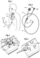
- Fig. 1 is a simplified pictorial diagram of the preferred embodiment of the probe;
- Fig. 2 is a perspective view of the probe of Figure 1;
- Fig. 3 is a fragmentary perspective view of the probe of Figure 1 implanted;
- Fig. 4 is a fragmentary perspective view of the probe of Figure 1 during withdrawal.
-
- Referring now to Figures 1 and 2, the inventive implantable and removable device or probe is shown generally at 10. The device is comprised of a
transducer support 12 carried at thedistal end 14 of the device, and which is connected to theproximal end 16 of the device by means of aflexible tube 18, as is well known in the art. The device is inserted into the body though insertion wound 20, then implanted on a vessel, such as an artery or vein. The device is primarily intended for use in monitoring blood flow through arteries or veins, but could also be used on other organs. The device is approximately 1.9 cm (¾ inch) long and 0.95 cm (3/8 inch) in cross-section. The device is intended to be secured onto the vessel such that it would lie generally axially along the vessel. - Referring now to Figures 3 and 4,
transducer support 12 is shown secured toartery 22. In the preferred embodiment, a pair ofrelease wires flexible tubing 18 and are releasably attached to the front oftransducer support 12 by insertion intowire channels wires wire channels wires wire channels wires wire channels - As can be seen best in Figures 3 and 4,
wires outs sutures 38 to be attached totransducer support 12 to secure thetransducer support 12 to the monitor site. The transducer is comprised of a generally flatpiezoelectric crystal 40 which is mounted at an angle of between 5° and 85°, as is well known in the art. The preferred angle is approximately 50°, which provides a good compromise between measuring blood flow and vessel diameter, as is well known in the art. It should also be understood that the angle is measured between a line perpendicular to the vessel and the face ofcrystal 40. Lead wires 42 extend throughflexible tubing 18 and are attached to electrical connector 44 (shown in Figure 2) for ease of connection to a monitoring device, as is well known in the art. -
Release wires channels Cap 48 is removed,wires channels transducer support 12 fromsutures 38, as shown in Figure 4. Thetransducer support 12 can then be withdrawn from the body by pulling onflexible tube 18. The transducer can be extracted throughinsertion wound 20 without the need for surgery. - It should be understood that the implanted device could carry any biological sensor, not just a doppler transducer for measuring blood flow. It should also be understood that all parts of the invention placed inside the body are made of a biocompatible material such as plastic or rubber, as is well known in the art. It should also be understood that the wire could be made of a flexible metal, plastic, or monofilament.
- This completes the description of the preferred embodiment of the invention. Those skilled in the art may recognize other equivalents to the specific embodiment described herein which equivalents are intended to be encompassed by the claims attached hereto.
Claims (4)
- An implantable, extractable probe (10) for attachment to a vessel or organ, comprising:(a) an elongate probe body (12) comprising a flexible tube (18) having front and rear ends;(b) at least one biological sensor (40) attached to the probe body (12);(c) at least one lead wire (42) connected to the biological sensor (40) said lead wire (42) extending through the flexible tube (18); and(d) a suture attachment wire (24,26) for attaching at least one suture (38) to secure the probe body (12) to a predetermined sensor site, the suture attachment wire (24,26) being releasably connected to the front end of the elongate probe body (12) and extending through the flexible tube (18), at least a portion of the suture attachment wire (24,26) being exposed to provide a suture attachment point to secure the probe (10) in place at a monitoring site;
whereby the elongate probe body (12) may be secured to a predetermined sensor site by attaching at least one suture to the suture attachment wire (24,26), such that when the suture attachment wire (24,26) is released, the probe body (12) is released for extraction from the patient body;
characterised in that the suture attachment wire (24,26) is bent or looped to form two substantially equal lengths of wire, and in that the probe body (12) has sides and is provided with two cut-outs (30 to 36) on each side to provide said exposed portions and where one length (24) of suture attachment wire extends through both cut-outs (30,32) on one side of the probe body (12), while the other length of suture attachment wire (26) extends through both cut-outs (30,32) on the other side of the probe body (12), whereby the exposed lengths of suture attachment wire (24,26) are spaced a predetermined distance from the probe body (12). - A probe according to Claim 1, characterised in that the biological sensor (4) is an ultrasound transducer.
- A probe according to Claim 1, characterised in that the probe body has a sensor site contacting face and in that the biological sensor is attached to the probe body such that said sensor is inclined at a 5-85° angle to said sensor site contacting face.
- A probe according to any one of Claims 1 to 3, characterised in that the suture attachment wire (24,26) is friction or compression loaded into a channel located in the front end of the probe body (12).
Applications Claiming Priority (3)
| Application Number | Priority Date | Filing Date | Title |
|---|---|---|---|
| US709343 | 1991-06-03 | ||
| US07/709,343 US5205292A (en) | 1991-06-03 | 1991-06-03 | Removable implanted device |
| PCT/US1992/004542 WO1992021284A1 (en) | 1991-06-03 | 1992-06-02 | Removable implanted device |
Publications (3)
| Publication Number | Publication Date |
|---|---|
| EP0602048A1 EP0602048A1 (en) | 1994-06-22 |
| EP0602048A4 EP0602048A4 (en) | 1995-02-15 |
| EP0602048B1 true EP0602048B1 (en) | 2000-03-22 |
Family
ID=24849466
Family Applications (1)
| Application Number | Title | Priority Date | Filing Date |
|---|---|---|---|
| EP92913861A Expired - Lifetime EP0602048B1 (en) | 1991-06-03 | 1992-06-02 | Removable implanted device |
Country Status (7)
| Country | Link |
|---|---|
| US (2) | US5205292A (en) |
| EP (1) | EP0602048B1 (en) |
| JP (1) | JPH06510443A (en) |
| AU (1) | AU2177892A (en) |
| CA (1) | CA2103237C (en) |
| DE (1) | DE69230826T2 (en) |
| WO (1) | WO1992021284A1 (en) |
Families Citing this family (79)
| Publication number | Priority date | Publication date | Assignee | Title |
|---|---|---|---|---|
| US5205292A (en) * | 1991-06-03 | 1993-04-27 | Applied Biometric, Inc. | Removable implanted device |
| GB9213978D0 (en) * | 1992-07-01 | 1992-08-12 | Skidmore Robert | Medical devices |
| DE4319812C1 (en) * | 1993-06-15 | 1995-02-09 | Zurbruegg Heinz R | Implantable ultrasound probe for measuring the flow rate of blood in humans and animals |
| US5289821A (en) * | 1993-06-30 | 1994-03-01 | Swartz William M | Method of ultrasonic Doppler monitoring of blood flow in a blood vessel |
| US5527358A (en) * | 1994-01-21 | 1996-06-18 | Medtronic, Inc. | Temporary medical electrical lead |
| US5588436A (en) * | 1995-10-11 | 1996-12-31 | Cook Pacemaker Corporation | Pulsed doppler probe |
| US7269457B2 (en) * | 1996-04-30 | 2007-09-11 | Medtronic, Inc. | Method and system for vagal nerve stimulation with multi-site cardiac pacing |
| US5722415A (en) * | 1996-04-30 | 1998-03-03 | Medtronic, Inc. | Continuous cardiac output monitor |
| US20040199209A1 (en) * | 2003-04-07 | 2004-10-07 | Hill Michael R.S. | Method and system for delivery of vasoactive drugs to the heart prior to and during a medical procedure |
| US6904318B2 (en) * | 2000-09-26 | 2005-06-07 | Medtronic, Inc. | Method and system for monitoring and controlling systemic and pulmonary circulation during a medical procedure |
| US6735471B2 (en) * | 1996-04-30 | 2004-05-11 | Medtronic, Inc. | Method and system for endotracheal/esophageal stimulation prior to and during a medical procedure |
| US6532388B1 (en) | 1996-04-30 | 2003-03-11 | Medtronic, Inc. | Method and system for endotracheal/esophageal stimulation prior to and during a medical procedure |
| US7225019B2 (en) | 1996-04-30 | 2007-05-29 | Medtronic, Inc. | Method and system for nerve stimulation and cardiac sensing prior to and during a medical procedure |
| US8036741B2 (en) * | 1996-04-30 | 2011-10-11 | Medtronic, Inc. | Method and system for nerve stimulation and cardiac sensing prior to and during a medical procedure |
| US6449507B1 (en) * | 1996-04-30 | 2002-09-10 | Medtronic, Inc. | Method and system for nerve stimulation prior to and during a medical procedure |
| US6628987B1 (en) * | 2000-09-26 | 2003-09-30 | Medtronic, Inc. | Method and system for sensing cardiac contractions during vagal stimulation-induced cardiopalegia |
| US5775328A (en) | 1996-06-26 | 1998-07-07 | Lowe; Robert I. | Device for use in temporary insertion of a sensor within a patient's body |
| US6231514B1 (en) | 1996-06-26 | 2001-05-15 | Tobo, Llc | Device for use in temporary insertion of a sensor within a patient's body |
| US20040220455A1 (en) * | 1996-06-26 | 2004-11-04 | Lowe Robert I. | Method for monitoring blood characteristics and cardiopulmonary function |
| US6106475A (en) | 1996-06-26 | 2000-08-22 | Tobo, Llc | Device for use in temporary insertion of a sensor within a patient's body |
| US5692506A (en) * | 1996-08-01 | 1997-12-02 | Linder; Gerald S. | Transnasal conduit and method of use |
| CA2262789A1 (en) | 1996-08-09 | 1998-02-19 | Jerome H. Abrams | Male impotence diagnostic ultrasound system |
| US5797879A (en) * | 1996-08-26 | 1998-08-25 | Decampli; William M. | Apparatus and methods for providing selectively adjustable blood flow through a vascular graft |
| ES2208963T3 (en) * | 1997-01-03 | 2004-06-16 | Biosense, Inc. | PRESSURE SENSITIVE VASCULAR ENDOPROTESIS. |
| US6251076B1 (en) | 1997-08-01 | 2001-06-26 | Urometrics Inc. | Male impotence diagnostic ultrasound system |
| US6479523B1 (en) | 1997-08-26 | 2002-11-12 | Emory University | Pharmacologic drug combination in vagal-induced asystole |
| US5807258A (en) * | 1997-10-14 | 1998-09-15 | Cimochowski; George E. | Ultrasonic sensors for monitoring the condition of a vascular graft |
| US6398734B1 (en) * | 1997-10-14 | 2002-06-04 | Vascusense, Inc. | Ultrasonic sensors for monitoring the condition of flow through a cardiac valve |
| US6331163B1 (en) | 1998-01-08 | 2001-12-18 | Microsense Cardiovascular Systems (1196) Ltd. | Protective coating for bodily sensor |
| SG71881A1 (en) * | 1998-01-08 | 2000-04-18 | Microsense Cardiovascular Sys | Method and device for fixation of a sensor in a bodily lumen |
| US7840278B1 (en) | 1999-06-25 | 2010-11-23 | Puskas John D | Devices and methods for vagus nerve stimulation |
| US6346080B1 (en) | 1999-11-02 | 2002-02-12 | Transonic Systems, Inc. | Encasement and transducer shuttle assembly for removable implanted device |
| WO2001041655A1 (en) * | 1999-12-06 | 2001-06-14 | Simcha Milo | Ultrasonic medical device |
| GB2361188B (en) * | 2000-04-14 | 2004-03-03 | Diametrics Medical Ltd | Introducing sensors into soft tissue |
| US6487446B1 (en) * | 2000-09-26 | 2002-11-26 | Medtronic, Inc. | Method and system for spinal cord stimulation prior to and during a medical procedure |
| US20020086430A1 (en) * | 2000-12-28 | 2002-07-04 | Hopmeier Michael J. | Detection technology in agriculture operations |
| US6697667B1 (en) | 2001-05-31 | 2004-02-24 | Advanced Cardiovascular Systems, Inc. | Apparatus and method for locating coronary sinus |
| US7532920B1 (en) | 2001-05-31 | 2009-05-12 | Advanced Cardiovascular Systems, Inc. | Guidewire with optical fiber |
| US7329223B1 (en) * | 2001-05-31 | 2008-02-12 | Abbott Cardiovascular Systems Inc. | Catheter with optical fiber sensor |
| US6716178B1 (en) | 2001-05-31 | 2004-04-06 | Advanced Cardiovascular Systems, Inc. | Apparatus and method for performing thermal and laser doppler velocimetry measurements |
| US20040210289A1 (en) * | 2002-03-04 | 2004-10-21 | Xingwu Wang | Novel nanomagnetic particles |
| US7399313B2 (en) * | 2002-06-07 | 2008-07-15 | Brown Peter S | Endovascular graft with separable sensors |
| US6716176B1 (en) | 2002-09-30 | 2004-04-06 | Tobo, Llc | Device for use in temporary insertion of a sensor within a patient's body |
| US7010337B2 (en) * | 2002-10-24 | 2006-03-07 | Furnary Anthony P | Method and apparatus for monitoring blood condition and cardiopulmonary function |
| US20050079132A1 (en) * | 2003-04-08 | 2005-04-14 | Xingwu Wang | Medical device with low magnetic susceptibility |
| US20040254419A1 (en) * | 2003-04-08 | 2004-12-16 | Xingwu Wang | Therapeutic assembly |
| US20050025797A1 (en) * | 2003-04-08 | 2005-02-03 | Xingwu Wang | Medical device with low magnetic susceptibility |
| US20070149496A1 (en) * | 2003-10-31 | 2007-06-28 | Jack Tuszynski | Water-soluble compound |
| US20050165317A1 (en) * | 2003-11-04 | 2005-07-28 | Turner Nicholas M. | Medical devices |
| US20050249667A1 (en) * | 2004-03-24 | 2005-11-10 | Tuszynski Jack A | Process for treating a biological organism |
| US20070208293A1 (en) * | 2006-03-03 | 2007-09-06 | Habah Noshy Mansour | Methods and devices for noninvasive pressure measurment in ventricular shunts |
| EP2023807B1 (en) * | 2006-06-02 | 2013-08-14 | Cook Medical Technologies LLC | Adjustable tension cuff assembly |
| WO2008008281A2 (en) | 2006-07-07 | 2008-01-17 | Proteus Biomedical, Inc. | Smart parenteral administration system |
| US20080058772A1 (en) * | 2006-08-31 | 2008-03-06 | Robertson Timothy L | Personal paramedic |
| US7765015B2 (en) * | 2007-01-12 | 2010-07-27 | Cardiac Pacemakers, Inc. | Lead with inflatable fixation mechanism |
| WO2009055733A1 (en) | 2007-10-25 | 2009-04-30 | Proteus Biomedical, Inc. | Fluid transfer port information system |
| WO2009067463A1 (en) | 2007-11-19 | 2009-05-28 | Proteus Biomedical, Inc. | Body-associated fluid transport structure evaluation devices |
| US8332020B2 (en) | 2010-02-01 | 2012-12-11 | Proteus Digital Health, Inc. | Two-wrist data gathering system |
| BR112012019212A2 (en) | 2010-02-01 | 2017-06-13 | Proteus Digital Health Inc | data collection system |
| US8406868B2 (en) | 2010-04-29 | 2013-03-26 | Medtronic, Inc. | Therapy using perturbation and effect of physiological systems |
| US8620425B2 (en) | 2010-04-29 | 2013-12-31 | Medtronic, Inc. | Nerve signal differentiation in cardiac therapy |
| US8639327B2 (en) | 2010-04-29 | 2014-01-28 | Medtronic, Inc. | Nerve signal differentiation in cardiac therapy |
| US8781582B2 (en) | 2011-01-19 | 2014-07-15 | Medtronic, Inc. | Vagal stimulation |
| US8725259B2 (en) | 2011-01-19 | 2014-05-13 | Medtronic, Inc. | Vagal stimulation |
| US8706223B2 (en) | 2011-01-19 | 2014-04-22 | Medtronic, Inc. | Preventative vagal stimulation |
| US8781583B2 (en) | 2011-01-19 | 2014-07-15 | Medtronic, Inc. | Vagal stimulation |
| US8718763B2 (en) | 2011-01-19 | 2014-05-06 | Medtronic, Inc. | Vagal stimulation |
| WO2013158271A1 (en) * | 2012-04-20 | 2013-10-24 | University Of Rochester | Implantable real-time oximeter to determine potential strokes and post-traumatic brain-injury complication |
| DE202016008639U1 (en) * | 2015-02-12 | 2019-01-16 | Foundry Innovation & Research 1, Ltd. | Implantable devices for monitoring heart failure |
| WO2018031714A1 (en) | 2016-08-11 | 2018-02-15 | Foundry Innovation & Research 1, Ltd. | Systems and methods for patient fluid management |
| US12465324B2 (en) | 2015-02-12 | 2025-11-11 | Foundry Innovation & Research 1, Ltd. | Patient fluid management systems and methods employing integrated fluid status sensing |
| EP3331426B1 (en) | 2015-08-03 | 2024-07-24 | Foundry Innovation&Research 1, Ltd. | Catheter for measurement of vena cava dimension |
| US11206992B2 (en) | 2016-08-11 | 2021-12-28 | Foundry Innovation & Research 1, Ltd. | Wireless resonant circuit and variable inductance vascular monitoring implants and anchoring structures therefore |
| US11701018B2 (en) | 2016-08-11 | 2023-07-18 | Foundry Innovation & Research 1, Ltd. | Wireless resonant circuit and variable inductance vascular monitoring implants and anchoring structures therefore |
| EP3463082B1 (en) | 2016-11-29 | 2020-07-29 | Foundry Innovation & Research 1, Ltd. | Wireless resonant circuit and variable inductance vascular implants for monitoring patient vasculature system |
| WO2018220143A1 (en) | 2017-05-31 | 2018-12-06 | Foundry Innovation And Research 1, Ltd | Implantable ultrasonic vascular sensor |
| WO2018220146A1 (en) | 2017-05-31 | 2018-12-06 | Foundry Innovation & Research 1, Ltd. | Implantable sensors for vascular monitoring |
| CZ2018293A3 (en) | 2018-06-14 | 2019-12-27 | České vysoké učenà technické v Praze | Mechanical system for fixing measuring probe for monitoring transplanted organs |
| US12465233B2 (en) | 2022-07-29 | 2025-11-11 | Foundry Innovation & Research 1, Ltd. | Multistranded conductors adapted to dynamic in vivo environments |
Family Cites Families (20)
| Publication number | Priority date | Publication date | Assignee | Title |
|---|---|---|---|---|
| FR1601372A (en) * | 1968-12-31 | 1970-08-17 | ||
| JPS4886290A (en) * | 1972-02-18 | 1973-11-14 | ||
| JPS5113502B2 (en) * | 1973-04-28 | 1976-04-30 | ||
| US4313448A (en) * | 1980-01-28 | 1982-02-02 | Medtronic, Inc. | Myocardial sutureless lead |
| US4355643A (en) * | 1980-03-05 | 1982-10-26 | University Of Iowa Research Foundation | Vacuum cup doppler flow transducer and method for using same |
| AU555119B2 (en) * | 1982-03-17 | 1986-09-11 | Howard Martin Arneson | Inboard outboard drive |
| US4541433A (en) * | 1984-06-01 | 1985-09-17 | Medtronic, Inc. | Cardiac output monitor |
| US4674336A (en) * | 1984-11-01 | 1987-06-23 | Johnston G Gilbert | Volumetric flow rate determination in conduits not directly accessible |
| US4722347A (en) * | 1985-01-15 | 1988-02-02 | Applied Biometrics, Inc. | Apparatus for measuring cardiac output |
| US4671295A (en) * | 1985-01-15 | 1987-06-09 | Applied Biometrics, Inc. | Method for measuring cardiac output |
| US4733669A (en) * | 1985-05-24 | 1988-03-29 | Cardiometrics, Inc. | Blood flow measurement catheter |
| FR2585944B1 (en) * | 1985-08-12 | 1988-07-08 | Alvar Electronic Sa | IMPLANTABLE ULTRASOUND PROBE AND ITS MANUFACTURING PROCESS |
| US4926875A (en) * | 1988-01-25 | 1990-05-22 | Baylor College Of Medicine | Implantable and extractable biological sensor probe |
| US4886059A (en) * | 1988-06-23 | 1989-12-12 | Applied Biometrics, Incorporated | Endotracheal tube with asymmetric balloon |
| US4917115A (en) * | 1988-07-11 | 1990-04-17 | Vitatron Medical B. V. | Pacing system and method for physiological stimulation of the heart utilizing Doppler means |
| US4947854A (en) * | 1988-09-13 | 1990-08-14 | Baylor College Of Medicine | Epicardial multifunctional probe |
| US5121749A (en) * | 1988-10-05 | 1992-06-16 | Cardiometrics, Inc. | Position in dependent volumetric flow measuring apparatus |
| US4915113A (en) * | 1988-12-16 | 1990-04-10 | Bio-Vascular, Inc. | Method and apparatus for monitoring the patency of vascular grafts |
| US5205292A (en) * | 1991-06-03 | 1993-04-27 | Applied Biometric, Inc. | Removable implanted device |
| US5160338A (en) * | 1991-11-13 | 1992-11-03 | Inamed Development Co. | Device for removing implantable articles |
-
1991
- 1991-06-03 US US07/709,343 patent/US5205292A/en not_active Expired - Lifetime
-
1992
- 1992-06-02 AU AU21778/92A patent/AU2177892A/en not_active Abandoned
- 1992-06-02 CA CA002103237A patent/CA2103237C/en not_active Expired - Fee Related
- 1992-06-02 JP JP5500587A patent/JPH06510443A/en active Pending
- 1992-06-02 DE DE69230826T patent/DE69230826T2/en not_active Expired - Fee Related
- 1992-06-02 WO PCT/US1992/004542 patent/WO1992021284A1/en not_active Ceased
- 1992-06-02 EP EP92913861A patent/EP0602048B1/en not_active Expired - Lifetime
-
1993
- 1993-02-25 US US08/023,193 patent/US5284146A/en not_active Expired - Lifetime
Also Published As
| Publication number | Publication date |
|---|---|
| DE69230826D1 (en) | 2000-04-27 |
| EP0602048A4 (en) | 1995-02-15 |
| CA2103237A1 (en) | 1992-12-04 |
| CA2103237C (en) | 2003-06-10 |
| EP0602048A1 (en) | 1994-06-22 |
| JPH06510443A (en) | 1994-11-24 |
| WO1992021284A1 (en) | 1992-12-10 |
| US5284146A (en) | 1994-02-08 |
| US5205292A (en) | 1993-04-27 |
| AU2177892A (en) | 1993-01-08 |
| DE69230826T2 (en) | 2000-10-19 |
Similar Documents
| Publication | Publication Date | Title |
|---|---|---|
| US5205292A (en) | Removable implanted device | |
| US5588436A (en) | Pulsed doppler probe | |
| US7468039B2 (en) | Adjustable tension cuff assembly | |
| US4541433A (en) | Cardiac output monitor | |
| JP5833079B2 (en) | Apparatus for guiding and positioning an intravascular device | |
| EP0647117B1 (en) | Medical devices | |
| US5058595A (en) | Judkins-type angiographic catheter with Doppler crystal, and method of use | |
| AU709716B2 (en) | Central venous oxygen saturation measurement | |
| JP3114994B2 (en) | Cardiac blood output deep needle assembly | |
| US4947854A (en) | Epicardial multifunctional probe | |
| US20090088609A1 (en) | Method of taking internal physiological measurements | |
| JP2008522655A (en) | Subcutaneous electrode insertion apparatus and method | |
| US8401643B2 (en) | Implantable medical sensor and anchoring system | |
| EP0876823A3 (en) | Transdermal introducer assembly | |
| WO1989006513A1 (en) | Implantable and extractable biological sensor probe | |
| JP2002542899A (en) | Cardiac assist method and device for increasing cardiac function | |
| AU747791B2 (en) | Device for insertion of a sensor | |
| WO1993003668A9 (en) | Cardiac output probe assembly | |
| US20230329567A1 (en) | Implantable blood pressure monitor | |
| US6346080B1 (en) | Encasement and transducer shuttle assembly for removable implanted device | |
| JP2813995B2 (en) | Semi-implanted ultrasound probe | |
| CN121533794A (en) | Intravascular ultrasound navigation device and working method during port-a-cath implantation and removal |
Legal Events
| Date | Code | Title | Description |
|---|---|---|---|
| PUAI | Public reference made under article 153(3) epc to a published international application that has entered the european phase |
Free format text: ORIGINAL CODE: 0009012 |
|
| 17P | Request for examination filed |
Effective date: 19931217 |
|
| AK | Designated contracting states |
Kind code of ref document: A1 Designated state(s): DE FR GB |
|
| A4 | Supplementary search report drawn up and despatched |
Effective date: 19941230 |
|
| AK | Designated contracting states |
Kind code of ref document: A4 Designated state(s): DE FR GB |
|
| 17Q | First examination report despatched |
Effective date: 19970506 |
|
| GRAG | Despatch of communication of intention to grant |
Free format text: ORIGINAL CODE: EPIDOS AGRA |
|
| GRAG | Despatch of communication of intention to grant |
Free format text: ORIGINAL CODE: EPIDOS AGRA |
|
| GRAH | Despatch of communication of intention to grant a patent |
Free format text: ORIGINAL CODE: EPIDOS IGRA |
|
| GRAH | Despatch of communication of intention to grant a patent |
Free format text: ORIGINAL CODE: EPIDOS IGRA |
|
| GRAA | (expected) grant |
Free format text: ORIGINAL CODE: 0009210 |
|
| AK | Designated contracting states |
Kind code of ref document: B1 Designated state(s): DE FR GB |
|
| PG25 | Lapsed in a contracting state [announced via postgrant information from national office to epo] |
Ref country code: FR Free format text: THE PATENT HAS BEEN ANNULLED BY A DECISION OF A NATIONAL AUTHORITY Effective date: 20000322 |
|
| REF | Corresponds to: |
Ref document number: 69230826 Country of ref document: DE Date of ref document: 20000427 |
|
| ET | Fr: translation filed | ||
| PLBE | No opposition filed within time limit |
Free format text: ORIGINAL CODE: 0009261 |
|
| STAA | Information on the status of an ep patent application or granted ep patent |
Free format text: STATUS: NO OPPOSITION FILED WITHIN TIME LIMIT |
|
| 26N | No opposition filed | ||
| REG | Reference to a national code |
Ref country code: GB Ref legal event code: IF02 |
|
| PGFP | Annual fee paid to national office [announced via postgrant information from national office to epo] |
Ref country code: DE Payment date: 20020822 Year of fee payment: 11 |
|
| REG | Reference to a national code |
Ref country code: GB Ref legal event code: 732E |
|
| PGFP | Annual fee paid to national office [announced via postgrant information from national office to epo] |
Ref country code: GB Payment date: 20030625 Year of fee payment: 12 |
|
| PG25 | Lapsed in a contracting state [announced via postgrant information from national office to epo] |
Ref country code: DE Free format text: LAPSE BECAUSE OF NON-PAYMENT OF DUE FEES Effective date: 20040101 |
|
| PG25 | Lapsed in a contracting state [announced via postgrant information from national office to epo] |
Ref country code: GB Free format text: LAPSE BECAUSE OF NON-PAYMENT OF DUE FEES Effective date: 20040602 |
|
| GBPC | Gb: european patent ceased through non-payment of renewal fee |