EP0599913B1 - Monoclonal antibodies to putative hcv ns5 proteins and methos for using same - Google Patents
Monoclonal antibodies to putative hcv ns5 proteins and methos for using same Download PDFInfo
- Publication number
- EP0599913B1 EP0599913B1 EP92917270A EP92917270A EP0599913B1 EP 0599913 B1 EP0599913 B1 EP 0599913B1 EP 92917270 A EP92917270 A EP 92917270A EP 92917270 A EP92917270 A EP 92917270A EP 0599913 B1 EP0599913 B1 EP 0599913B1
- Authority
- EP
- European Patent Office
- Prior art keywords
- hcv
- antibody
- antigen
- test sample
- monoclonal antibody
- Prior art date
- Legal status (The legal status is an assumption and is not a legal conclusion. Google has not performed a legal analysis and makes no representation as to the accuracy of the status listed.)
- Expired - Lifetime
Links
Images


Classifications
-
- C—CHEMISTRY; METALLURGY
- C07—ORGANIC CHEMISTRY
- C07K—PEPTIDES
- C07K14/00—Peptides having more than 20 amino acids; Gastrins; Somatostatins; Melanotropins; Derivatives thereof
- C07K14/005—Peptides having more than 20 amino acids; Gastrins; Somatostatins; Melanotropins; Derivatives thereof from viruses
-
- C—CHEMISTRY; METALLURGY
- C07—ORGANIC CHEMISTRY
- C07K—PEPTIDES
- C07K16/00—Immunoglobulins [IG], e.g. monoclonal or polyclonal antibodies
- C07K16/08—Immunoglobulins [IG], e.g. monoclonal or polyclonal antibodies against material from viruses
- C07K16/10—RNA viruses
- C07K16/116—Togaviridae (F); Matonaviridae (F); Flaviviridae (F)
- C07K16/118—Hepatitis C virus; GB virus C [GBV-C]
-
- C—CHEMISTRY; METALLURGY
- C12—BIOCHEMISTRY; BEER; SPIRITS; WINE; VINEGAR; MICROBIOLOGY; ENZYMOLOGY; MUTATION OR GENETIC ENGINEERING
- C12N—MICROORGANISMS OR ENZYMES; COMPOSITIONS THEREOF; PROPAGATING, PRESERVING, OR MAINTAINING MICROORGANISMS; MUTATION OR GENETIC ENGINEERING; CULTURE MEDIA
- C12N2770/00—MICROORGANISMS OR ENZYMES; COMPOSITIONS THEREOF; PROPAGATING, PRESERVING, OR MAINTAINING MICROORGANISMS; MUTATION OR GENETIC ENGINEERING; CULTURE MEDIA ssRNA viruses positive-sense
- C12N2770/00011—Details
- C12N2770/24011—Flaviviridae
- C12N2770/24211—Hepacivirus, e.g. hepatitis C virus, hepatitis G virus
- C12N2770/24222—New viral proteins or individual genes, new structural or functional aspects of known viral proteins or genes
Definitions
- This invention relates generally to antibodies which specifically bind to hepatitis C virus (HCV), and more specifically, relates to a panel of novel hybridoma cells lines which secrete monoclonal antibodies which specifically bind to the putative HCV protein NS5, and methods for using these monoclonal antibodies.
- HCV hepatitis C virus
- Viral hepatitis is now known to include a group of viral agents with distinctive viral organization protein structure and mode of replication, causing hepatitis with different degrees of severity of hepatic damage through different routes of transmission.
- Acute viral hepatitis is clinically diagnosed by well-defined patient symptoms including jaundice, hepatic tenderness and an elevated level of liver transaminases such as aspartate transaminase and alanine transaminase.
- Non-A non-B Hepatitis is a term first used in 1975 that described cases of post-transfusion hepatitis not caused by either hepatitis A virus or hepatitis B virus.
- NANBH Non-A non-B Hepatitis
- the diagnosis of NANBH has been made primarily by means of exclusion on the basis of serological analysis for the presence of hepatitis A and hepatitis B.
- NANBH is responsible for about 90% of the cases of post-transfusion hepatitis. Hollinger et al. in N. R. Rose et al., eds., Manual of Clinical Immunology, American Society for Microbiology, Washington, D. C., 558-572 (1986).
- HCV hepatitis C virus
- the genetic makeup of the virus consists of single stranded linear RNA (positive strand) of molecular weight approximately 9.5 kb, and possessing one continuous translational open reading frame. J. A. Cuthbert, Amer. J. Med. Sci . 299:346-355 (1990). It is a small enveloped virus resembling the Flaviviruses. Investigators have made attempts to identify the NANB agent by ultrastructural changes in hepatocytes in infected individuals. H. Gupta, Liver 8:111-115 (1988); D.W. Bradley J. Virol. Methods 10:307-319 (1985).
- HCV etiological agent for post-transfusion NANBH.
- HCV antibodies Although the detection of HCV antibodies eliminates 70 to 80% of NANBH infected blood from the blood supply system, the antibodies apparently are readily detected during the chronic state of the disease, while only 60% of the samples from the acute NANBH stage are HCV antibody positive. H. Alter et al., New Eng. J. Med. 321:1994-1500 (1989). These data clearly indicated the need for the identification of additional HCV proteins for efficient serodiagnosis of HCV infection. Following the cloning and expression of structural protein CORE and 33C, second generation antibody assays have been developed which employ HCV CORE and 33C proteins in addition to C-100 for the detection of antibodies to HCV in NANB patients.
- HCV peptides including peptides containing epitopes belonging to the envelope region, useful in HCV diagnostics and vaccines. Monoclonal antibodies to such peptides are disclosed.
- EP-A-0 464 287 which is prior art under the terms of Article 54(3) and (4) EPC, generally discloses the HCV NS5 protein and the possibility of raising antibodies to HCV polypeptides and using them as a diagnostic agent.
- EP-A-0 468 527 which is prior art under the terms of Article 54(3) and (4) EPC, discloses HCV NS5 polypeptides and the possibility of raising antibodies to HCV polypeptides and using them as a diagnostic agent.
- the present invention provides a panel of highly specific and novel monoclonal antibodies that can be employed for the detection of putative HCV NS5 antigens.
- the monoclonal antibodies specifically bind to protein sequences derived from the putative HCV NS5 gene.
- the hybridomas which produce these monoclonal antibodies are identified as follows: hybridoma H12C65 (A.T.C.C. deposit No. HB 10855) and hybridoma H18C141 (A.T.C.C. deposit No. HB 10854).
- a test sample which may contain HCV antigens is contacted with a solid phase to which a monoclonal anti-HCV NS5 antibody of the invention or a fragment thereof has been bound, to form a mixture.
- This mixture is incubated for a time and under conditions sufficient for antigen/antibody complexes to form.
- the so-formed complexes then are contacted with an indicator reagent comprising a monoclonal antibody of the invention or a fragment thereof, specific for the HCV antigen attached to a signal generating compound to form a second mixture.
- This second mixture is reacted for a time and under conditions sufficient to form antibody/antigen/antibody complexes.
- the presence of HCV antigen is determined by detecting the measurable signal generated.
- the amount of HCV present in the test sample thus the amount of HCV antigen captured on the solid phase, is proportional to the amount of signal generated.
- an indicator reagent comprising a monoclonal antibody of the invention, or fragment thereof, specific for HCV NS5 antigen and a signal generating compound is added to a monoclonal anti-HCV antibody of the invention or fragment thereof coated on a solid phase and the test sample, to form a mixture.
- This mixture is incubated for a time and under conditions sufficient to form antibody/antigen/antibody complexes.
- the presence and amount of HCV present in the test sample, and thus the amount of HCV antigen captured on the solid phase is determined by detecting the measurable signal.
- the amount of HCV present in the test sample is proportional to the amount of signal generated.
- one or a combination of more than one monoclonal antibody of the invention can be employed as a competitive probe for the detection of antibodies to HCV NS5 antigen.
- HCV NS5 antigens either alone or in combination, can be coated on a solid phase.
- a test sample suspected of containing antibody to HCV NS5 antigen then is incubated with an indicator reagent comprising a signal generating compound and a monoclonal antibody of the invention for a time and under conditions sufficient to form antigen/antibody complexes of either the test sample and indicator reagent to the solid phase or the indicator reagent to the solid phase.
- the reduction in binding of the monoclonal antibody to the solid phase can be quantitatively measured.
- a measurable reduction in the signal compared to the signal generated from a confirmed negative NANBH test sample would indicate the presence of anti-HCV NS5 antibody in the test sample.
- a test sample is contacted with a solid phase to which HCV NS5 proteins are attached and an indicator reagent comprising a monoclonal antibody of the invention or fragment thereof specific for HCV NS5 attached to a signal generating compound, to form a mixture.
- the mixture is incubated for a time and under conditions sufficient for antibody/antigen complexes to form.
- the presence of anti-HCV present in the test sample is determined by detecting the measurable signal generated, and comparing the signal to the measured signal generated from a known negative sample. A measurable reduction of signal of the test sample, compared to the known negative sample's signal, is indicative of the presence of anti-HCV antibodies.
- Competitive assays for the detection of anti-HCV antibody using antigens free in solution also can be performed.
- HCV NS5 antigen can be detected in a tissue sample by contacting the tissue sample with an indicator reagent comprising a signal generating compound attached to a monoclonal antibody of the invention which specifically binds to HCV NS5 antigen or fragment thereof, to form a mixture. This mixture is incubated for a time and under conditions sufficient for antigen/antibody complex to form. The presence of HCV NS5 antigen present in the tissue sample is determined by detecting the signal generated.
- kits useful for determining the presence of HCV NS5 antigen or antibody in test sample that include the monoclonal antibodies of the invention.
- FIG. 1 is an illustration of the location of the recombinant HCV proteins on the HCV genome employed either as immunogens for the generation of monoclonal antibodies or for their characterization.
- FIGS. 2A and 2B are Western blot analysis illustrating specific binding of monoclonal antibodies H12C65 (FIG. 2A) and H18C141 (FIG. 2B) to NS5.
- the present invention provides novel monoclonal antibodies to the putative HCV NS5 protein, methods for using the monoclonal antibodies, and kits which contain these monoclonal antibodies.
- the monoclonal antibodies of the present invention can be employed in various assay systems to determine the presence, if any, of HCV NS5 proteins in a test sample. Fragments of these monoclonal antibodies provided also may be used. For example, in a first assay format, a monoclonal anti-HCV NS5 antibody of the invention or fragment thereof, or a combination of these antibodies, which has been coated on a solid phase, is contacted with a test sample which may contain HCV NS5 proteins, to form a mixture. This mixture is incubated for a time and under conditions sufficient to form antigen/antibody complexes.
- an indicator reagent comprising a monoclonal antibody of the invention or a fragment thereof, which specifically binds to the HCV NS5 region, or a combination of these antibodies, to which a signal generating compound has been attached, is contacted with the antigen/antibody complexes to form a second mixture.
- This second mixture then is incubated for a time and under conditions sufficient to form antibody/antigen/antibody complexes.
- the presence of HCV NS5 antigen present in the test sample and captured on the solid phase, if any, is determined by detecting the measurable signal generated by the signal generating compound.
- the amount of HCV NS5 antigen present in the test sample is proportional to the signal generated.
- a monoclonal anti-HCV NS5 antibody of the invention or fragment thereof, or a combination of these antibodies which is bound to a solid support the test sample and an indicator reagent comprising a monoclonal antibody of the invention or fragments thereof, which specifically binds to HCV NS5 antigen, or a combination of these antibodies to which a signal generating compound is attached, are contacted to form a mixture.
- This mixture is incubated for a time and under conditions sufficient to form antibody/antigen/antibody complexes.
- the presence, if any, of HCV NS5 proteins present in the test sample and captured on the solid phase is determined by detecting the measurable signal generated by the signal generating compound.
- the amount of HCV proteins present in the test sample is proportional to the signal generated.
- one or a combination of one or more monoclonal antibodies of the invention can be employed as a competitive probe for the detection of antibodies to HCV protein.
- HCV proteins either alone or in combination, can be coated on a solid phase.
- a test sample suspected of containing antibody to HCV NS5 antigen then is incubated with an indicator reagent comprising a signal generating compound and at least one monoclonal antibody of the invention for a time and under conditions sufficient to form antigen/antibody complexes of either the test sample and indicator reagent to the solid phase or the indicator reagent to the solid phase.
- the reduction in binding of the monoclonal antibody to the solid phase can be quantitatively measured.
- a measurable reduction in the signal compared to the signal generated from a confirmed negative NANBH test sample indicates the presence of anti-HCV NS5 antibody in the test sample.
- each of the monoclonal antibodies of the present invention can be employed in the detection of HCV antigens in fixed tissue sections, as well as fixed cells by immunohistochemical analysis.
- these monoclonal antibodies can be bound to matrices similar to CNBr-activated Sepharose and used for the affinity purification of specific HCV proteins from cell cultures, or biological tissues such as blood and liver.
- the monoclonal antibodies of the invention can also be used for the generation of chimeric antibodies for therapeutic use, or other similar applications.
- the monoclonal antibodies or fragments thereof can be provided individually to detect HCV NS5 antigens.
- Combinations of the monoclonal antibodies (and fragments thereof) provided herein also may be used together as components in a mixture or "cocktail" of at least one anti-HCV NS5 antibody of the invention with antibodies to other HCV regions, each having different binding specificities.
- this cocktail can include the monoclonal antibodies of the invention which are directed to HCV NS5 proteins and other monoclonal antibodies to other antigenic determinants of the HCV genome.
- Examples of other monoclonal antibodies useful for these contemplated cocktails include those to HCV C-100, HCV 33C, HCV CORE, HCV NS1 and/or HCV putative ENV, which are disclosed in, for example, in WO 92/08738 and WO 92/13892.
- This cocktail of monoclonal antibodies as described herein would be used in the assay formats detailed herein in place of the monoclonal antibody to NS5, and thus would be able to detect the NS5 and other HCV antigens.
- the polyclonal antibody or fragment thereof which can be used in the assay formats should specifically bind to HCV putative NS5 region or other HCV proteins used in the assay, such as HCV C-100 protein, HCV 33C protein, HCV E2/NS1 or HCV CORE protein.
- the polyclonal antibody used preferably is of mammalian origin; human, goat, rabbit or sheep anti-HCV polyclonal antibody can be used. Most preferably, the polyclonal antibody is rabbit polyclonal anti-HCV antibody.
- the polyclonal antibodies used in the assays can be used either alone or as a cocktail of polyclonal antibodies.
- the cocktails used in the assay formats are comprised of either monoclonal antibodies or polyclonal anlibodies having different HCV specificity, they would be useful for diagnosis, evaluation and prognosis of HCV infection, as well as for studying HCV protein differentiation and specificity.
- Test samples which can be tested by the methods of the present invention described herein include human and animal body fluids such as whole blood, serum, plasma, cerebrospinal fluid, urine, biological fluids such as cell culture supernatants, fixed tissue specimens and fixed cell specimens.
- solid phase is not critical and can be selected by one skilled in the art. Thus, latex particles, microparticles, magnetic or non-magnetic beads, membranes, plastic tubes, walls of microtiter wells, glass or silicon chips and sheep red blood cells are all suitable examples. Suitable methods for immobilizing peptides on solid phases include ionic, hydrophobic, covalent interactions and the like.
- a "solid phase”, as used herein, refers to any material which is insoluble, or can be made insoluble by a subsequent reaction. The solid phase can be chosen for its intrinsic ability to attract and immobilize the capture reagent. Alternatively, the solid phase can retain an additional receptor which has the ability to attract and immobilize the capture reagent.
- the additional receptor can include a charged substance that is oppositely charged with respect to the capture reagent itself or to a charged substance conjugated to the capture reagent.
- the receptor molecule can be any specific binding member which is immobilized upon (attached to) the solid phase and which has the ability to immobilize the capture reagent through a specific binding reaction. The receptor molecule enables the indirect binding of the capture reagent to a solid phase material before the performance of the assay or during the performance of the assay.
- the solid phase thus can be a plastic, derivatized plastic, magnetic or non-magnetic metal, glass or silicon surface of a test tube, microtiter well, sheet, bead, microparticle, chip, and other configurations known to those of ordinary skill in the art.
- the solid phase also can comprise any suitable porous material with sufficient porosity to allow access by detection antibodies and a suitable surface affinity to bind antigens.
- Microporous structures are generally preferred, but materials with gel structure in the hydrated state may be used as well.
- Such useful solid supports include:
- the porous structure of nitrocellulose has excellent absorption and adsorption qualities for a wide variety of reagents including monoclonal antibodies.
- Nylon also possesses similar characteristics and also is suitable.
- porous solid supports described hereinabove are preferably in the form of sheets of thickness from about 0.01 to 0.5 mm, preferably about 0.1 mm.
- the pore size may vary within wide limits, and is preferably from about 0.025 to 15 microns, especially from about 0.15 to 15 microns.
- the surfaces of such supports may be activated by chemical processes which cause covalent linkage of the antigen or antibody to the support. The irreversible binding of the antigen or antibody is obtained, however, in general, by adsorption on the porous material by poorly understood hydrophobic forces.
- Suitable solid supports also are described in EP-A-0 353 590.
- the indicator reagent comprises a signal generating compound (label) which is capable of generating a measurable signal detectable by external means conjugated (attached) to a specific binding member for HCV.
- label a signal generating compound
- Specific binding member means a member of a specific binding pair. That is, two different molecules where one of the molecules through chemical or physical means specifically binds to the second molecule.
- the indicator reagent also can be a member of any specific binding pair, including either hapten-anti-hapten systems such as biotin or anti-biotin, avidin or biotin, a carbohydrate or a lectin, a complementary nucleotide sequence, an effector or a receptor molecule, an enzyme cofactor and an enzyme, an enzyme inhibitor or an enzyme, and the like.
- An immunoreactive specific binding member can be an antibody, an antigen, or an antibody/antigen complex that is capable of binding either to HCV as in a sandwich assay, to the capture reagent as in a competitive assay, or to the ancillary specific binding member as in an indirect assay.
- the various signal generating compounds (labels) contemplated include chromogens, catalysts such as enzymes, luminescent compounds such as fluorescein and rhodamine, chemiluminescent compounds such as acridinium, phenanthridinium and dioxetane compounds, radioactive elements, and direct visual labels.
- luminescent compounds such as fluorescein and rhodamine
- chemiluminescent compounds such as acridinium, phenanthridinium and dioxetane compounds
- radioactive elements and direct visual labels.
- enzymes include alkaline phosphatase, horseradish peroxidase, beta-galactosidase, and the like.
- the selection of a particular label is not critical, but it will be capable of producing a signal either by itself or in donjunction with one or more additional substances.
- ion capture procedures for immobilizing an immobilizable reaction complex with a negatively charged polymer described in EP-A-0 326 100 and EP-A-0 406 473, can be employed according to the present invention to effect a fast solution-phase immunochemical reaction.
- An immobilizable immune complex is separated from the rest of the reaction mixture by ionic interactions between the negatively charged poly-anion/immune complex and the previously treated, positively charged porous matrix and detected by using various signal generating systems previously described, including those described in chemiluminescent signal measurements as described in EP-A-0 273 115.
- the methods of the present invention can be adapted for use in systems which utilize microparticle technology including in automated and semi-automated systems wherein the solid phase comprises a microparticle.
- Such systems include those described in EP 0 425 633 and EP 0 424 634.
- the use-of scanning probe microscopy (SPM) for immunoassays also is a technology to which the monoclonal antibodies of the present invention are easily adaptable.
- scanning probe microscopy in particular in atomic force microscopy, the capture phase, for example, at least one of the monoclonal antibodies of the invention, is adhered to a solid phase and a scanning probe microscope is utilized to detect antigen/antibody complexes which may be present on the surface of the solid phase.
- scanning probe microscope is utilized to detect antigen/antibody complexes which may be present on the surface of the solid phase.
- the use of scanning tunnelling microscopy eliminates the need for labels which normally must be utilized in many immunoassay systems to detect antigen/antibody complexes.
- one member of a specific binding partner is attached to a surface suitable for scanning.
- the attachment of the analyte specific substance may be by adsorption to a test piece which comprises a solid phase of a plastic or metal surface, following methods known to those of ordinary skill in the art.
- covalent attachment of a specific binding partner (analyte specific substance) to a test piece which test piece comprises a solid phase of derivatized plastic, metal, silicon, or glass may be utilized.
- Covalent attachment methods are known to those skilled in the art and include a variety of means to irreversibly link specific binding partners to the test piece.
- test piece. is silicon or glass
- the surface must be activated prior to attaching the specific binding partner.
- Activated silane compounds such as triethoxy amino propyl silane (available from Sigma Chemical Co., St. Louis, MO), triethoxy vinyl silane (Aldrich Chemical Co., Milwaukee, WI), and (3-mercaptopropyl)-trimethoxy silane (Sigma Chemical Co., St. Louis, MO) can be used to introduce reactive groups such as amino-, vinyl, and thiol, respectively.
- Such activated surfaces can be used to link the binding partner directly (in the cases of amino or thiol) or the activated surface can be further reacted with linkers such as glutaraldehyde, bis (succinimidyl) suberate, SPPD 9 succinimidyl 3-[2-pyridyldithio] propionate), SMCC (succinimidyl-4-[N-maleimidomethyl] cyclohexane-1-carboxylate), SIAB (succinimidyl [4-iodoacetyl] aminobenzoate), and SMPB (succinimidyl 4-[1-maleimidophenyl] butyrate) to separate the binding partner from the surface.
- linkers such as glutaraldehyde, bis (succinimidyl) suberate, SPPD 9 succinimidyl 3-[2-pyridyldithio] propionate), SMCC (succinimidyl-4-
- the vinyl group can be oxidized to provide a means for covalent attachment. It also can be used as an anchor for the polymerization of various polymers such as poly acrylic acid, which can provide multiple attachment points for specific binding partners.
- the amino surface can be reacted with oxidized dextrans of various molecular weights to provide hydrophilic linkers of different size and capacity.
- oxidizable dextrans examples include Dextran T-40 (molecular weight 40,000 daltons), Dextran T-110 (molecular weight 110,000 daltons), Dextran T-500 (molecular weight 500,000 daltons), Dextran T-2M (molecular weight 2,000,000 daltons) (all of which are available from Pharmacia, Piscataway, N.J.,U.S.A.), or Ficoll (molecular weight 70,000 daltons (available from Sigma Chemical Co., St. Louis, MO). Also, polyelectrolyte interactions may be used to immobilize a specific binding partner on a surface of a test piece by using techniques and chemistries described by EP-A-0 326 100 and EP-A-0 406 473.
- the preferred method of attachment is by covalent means.
- the surface may be further treated with materials such as serum, proteins, or other blocking agents to minimize non-specific binding.
- the surface also may be scanned either at the site of manufacture or point of use to verify its suitability for assay purposes. The scanning process is not anticipated to alter the specific binding properties of the test piece.
- the reagent employed for the assay can be provided in the form of a kit with one or more containers such as vials or bottles, with each container containing a separate reagent such as a monoclonal antibody, or a cocktail of monoclonal antibodies, employed in the assay.
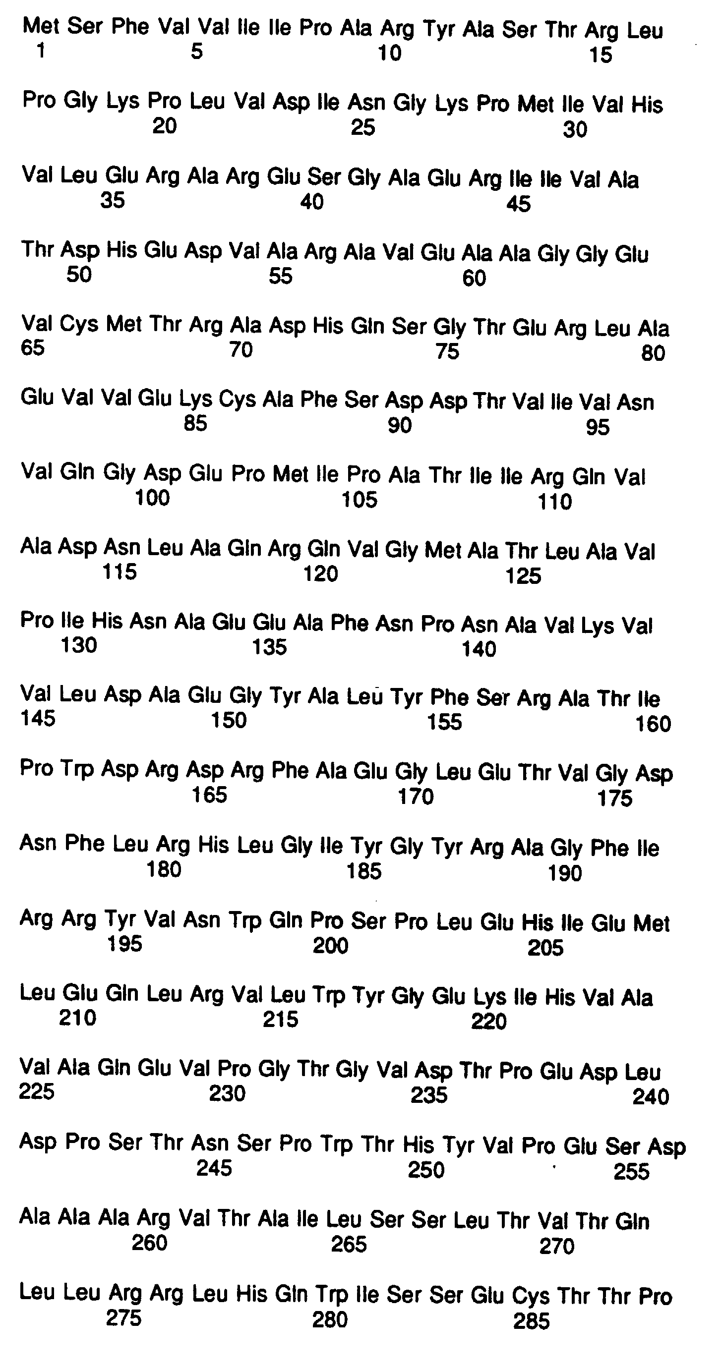
- E. coli derived recombinant antigens encoded by HCV sequences, designated as pHCV45, (HCV NS5, a.a. 1932-2191, SEQ. ID. NO. 1) was employed as immunogen for the generation of murine monoclonal antibodies specific for HCV NS5.
- Eight individual oligonucleotides representing HCV a.a.1932-2191 were ligated together and cloned as a 793 base pair EcoRI-BamHI fragment into the CKS ( E . coli CMP-KDO Synthetase) fusion vector pJ0200.
- CKS E . coli CMP-KDO Synthetase
- mice received 50 ⁇ g of purified pHCV45 in 200 ⁇ l of Freund's complete adjuvant injected intraperitoneally (i.p.) at two sites. A second immunization was done after 21 and 35 days with 50 ⁇ g of pHCV45 in incomplete Freund's adjuvant. Mice were bled on day 41 and the immune response to pHCV45 was assessed by enzyme linked immunoassay (EIA) and Western blot analysis. Fusion was performed after allowing the mice to rest for at least eight weeks.
- EIA enzyme linked immunoassay
- EIA Enzyme-Linked Immunoassay
- the immune response to the immunizing antigen was assessed by microtiter EIA and Western blot analysis.
- Wells of microtiter plates were coated with 100 ⁇ l of purified recombinant protein (pHCV 45, SEQ. ID. NO. 1) on 0.1M bicarbonate buffer at pH 9.5. After washing with Phosphate Buffered Saline (PBS) which also contained 0.01% sodium dodecyl sulfate (SDS) and 0.05% Tween-20® (available from Bio-Rad Laboratories, Richmond, CA), free sites were overcoated with 1% BSA in bicarbonate buffer at pH 9.5. Plates were stored at 4°C following a final wash.
- PBS Phosphate Buffered Saline
- SDS sodium dodecyl sulfate
- Tween-20® available from Bio-Rad Laboratories, Richmond, CA
- Sera from native or immunized mice were serially diluted in 100 ⁇ l of dilution buffer which contained 20 mM sodium phosphate, pH 7.4, 0.15M NaCI, 20% normal goat serum, 10% fetal calf serum, 5 mM EDTA, 10 mM EGTA, 50 mM Tris, 0.2% Tween-20®, with sodium azide as a preservative (at pH 6.8).
- the diluted sera were reacted with the antigen for three (3) hours at 37°C.
- the plates were washed and 100 ⁇ l of appropriately diluted goat anti-mouse IgG (heavy [h] and light [I] chain) Horseradish Peroxidase (HRPO)-conjugated antibody (Jackson Immunochemicals, West Grove, PA) was added. The plates were incubated at 37°C for two (2) hours. After a final wash, 100 ⁇ l of o-phenylenediamine:2 HCL (OPD) color reagent was added. The reaction was carried out at room temperature for 10 to 30 minutes, and then stopped by the addition of 1 ml of 1 N H2S04. The absorbance at 492/600 nm was recorded, which was found to be directly proportional to the amount of specific antibody bound to the respective antigen.
- HRPO Horseradish Peroxidase
- rHCV-45 purified rHCV protein
- SEQ. ID. NO. 1 Approximately 300 ⁇ g of purified rHCV protein (pHCV-45) (SEQ. ID. NO. 1) were treated with SDS and 2-mercaptoethanol at 95°C, and electrophoresed in a 12% polyacrylamide-SDS gel (Laemmli et al., Nature 227:680-685 (1970). Proteins were transferred overnight from the gel to nitrocellulose by electrophoresis at 100 mamp, or transferred in 1-2 hours at 1.0 amp, in a standard transfer buffer which comprised 25 mM Tris [(Hydroxymethyl) Aminomethane] 192 mM glycine, and 2.0% methanol, pH 8.3. (Towbin et al., Proc. Natl. Acad. Sci.
- reaction mixtures consisted of a nitrocellulose strip incubated with an appropriate amount of test sample in 2.0 ml of buffer (20 mM Tris, 1 mM EDTA, 0.2 M NaCI, 0.3% Triton X-100® and 2 mg.ml bovine serum albumin (BSA), pH 7.5, 5% E. coli lysate amd 3% CKS lysate overnight at 4°C.
- buffer 20 mM Tris, 1 mM EDTA, 0.2 M NaCI, 0.3% Triton X-100® and 2 mg.ml bovine serum albumin (BSA), pH 7.5, 5% E. coli lysate amd 3% CKS lysate overnight at 4°C.
- the strips were washed with buffered detergent (10 mM phosphate buffered saline (PBS) pH 7.5, containing 0.1% SDS and 0.5% Triton X-100®), followed by addition of goat anti-mouse IgG antibody conjugated to HRPO.
- the strips were incubated for one to two hours at room temperature, followed by washing with buffered. detergent.
- antibody bound to the protein was visualized by addition of freshly prepared HRP color reagent (Bio-Rad Laboratories, Richmond CA) (120 mg dissolved in 40 ml ice-cold methanol, then diluted into 200 ml Tris buffered saline [TBS] pH 7.8, containing 120 ⁇ l of 30% hydrogen peroxide.
- HRP color reagent Bio-Rad Laboratories, Richmond CA
- mice Upon demonstration of specific anti-HCV antibody present at reasonable titers in sera of immunized mice, the mice were allowed to rest for at least eight weeks prior to a pre-fusion boost of antigen.
- the pre-fusion antigen boost then was performed by intravenous (IV) tail vein injection of approximately 40 ⁇ g of respective purified HCV recombinant protein.
- IV intravenous
- mice Three days later the mice were sacrificed, and their spleens which contained anti-HCV antibody-producing cells were disrupted into single cells. These single cell suspensions were treated with 0.83% NH 4 CI to remove red blood cells, and then these suspensions were mixed with SP2/0 cells at a 10:1 (SP2/0:spleen cells) ratio.
- the mixed cells were centrifuged, washed once with serum-free medium, and again centrifuged.
- the fusogen polyethylene glycol (PEG) was used to form hybrids of the immune donor spleen cells with the myeloma cell line SP2/0 (HRPT neg.). Kohler and Milstein, Nature 356:494 (1975), and reviewed in J.G.R. Hurrel, ed., Monoclonal Hybridoma Antibodies: Techniques and Applications , CRC Press, Inc., Boco Ratan, FL (1982).
- fusion of the spleen and SP2/0 cells was accomplished by exposing the pellet to 40% PEG (ATCC, mw 1300-1600) In serum-free Iscoe's Modified Dulbecco's Medium (IMDM) for two minutes.
- PEG PEG
- IMDM Iscoe's Modified Dulbecco's Medium
- the PEG and cell suspension was diluted slowly by the addition of 20 ml of serum-free IMDM over a period of five minutes, followed by collection of the cells by centrifugation.
- the supernatant was decanted and replaced with 30 ml IMDM containing 20% fetal bovine serum (FBS) (Hyclone Laboratories, Logan, Utah) with HAT (hypoxanthene, aminopterin and thymidine) media in order to select for hybridomas.
- FBS fetal bovine serum
- HAT hyperxanthene, aminopterin and thymidine
- Spleen cells from one non-immune BALB/c mouse also were added as a feeder layer.
- the cells were plated at 0.1 ml/well in three 96-well tissue culture plates. An additional 0.1 ml of HAT media was added to each well three days later. At weekly intervals thereafter, one-half the media was replaced with IMDM containing 20% FBS with HT (hypoxanthene and thymidine), and hybrids were allowed to grow for an additional seven to fourteen days.
- IMDM containing 20% FBS with HT (hypoxanthene and thymidine
- the hybrids were composed of spleen cells making antibody to HCV fused with SP2/0 cells. Briefly, the fusogen promoted fusion of spleen cell and SP2/0 cell membranes, which formed a heterokaryon containing nuclei of both cells. Eventually, the dissimilar nuclei fuse produced a single nucleus capable of synchronous mitosis. As the fused cells divided, the hybrid stabilized by losing chromosomes of each nucleus. The fused cells were plated into multiple 96-well plates at 10 5 to 10 6 cells per well. The hybrid cells formed from SP2/0:spleen cell fusions were selectively propagated by culturing in HAT medium.
- SP2/0 or SP2/0:SP2/0 fused cells were prevented from growing aminopterin, and unfused spleen cells on spleen:spleen fused cells died off in culture. Only SP2/0:spleen cell hybrids grew in the HAT selective medium.
- culture fluids from wells containing hybridoma cell growth were screened for the presence of a monospecific antibody as follows.
- Each of the hybridoma culture fluids was tested on a plate coated with the immunogen as well as on a plate coated with CKS protein (fusion partner used for HCV protein SEQ. ID. NO. 1) by the EIA procedure described in Example 1.
- Hybridoma culture fluids reacting specifically to the immunogen, i.e., HCV protein, and not the CKS fusion partner were selected for further analysis by Western blot analysis.
- EIA-positive hybridoma culture fluids were tested for their reactivity to the HCV protein pHCV 45 (SEQ. ID. NO. 1) as well as CKS by Western blot analysis as described in Example 1.
- Hybrid samples reacting specifically with the HCV protein but not with the CKS protein by both EIA and Western blot were identified, and selected for cloning by the limiting dilution method, using the guidelines outlines by J. W. Goding, Monoclonal Antibodies: Principles and Practices, Academic Press, New York (1983).
- Culture supernatant of cloned samples were tested again by EIA with the immunogen and the CKS protein as described above in Example 1, for the confirmation of monospecific reactivity to HCV protein sequence SEQ. ID. NO. 1. Clones with strongest reactivity specifically to the protein were selected for expansion and further analysis.
- cloned cells of the desired hybridoma cell line were inoculated into a BALB/c mouse previously treated i.p. with 0.5 ml pristane (2,6,10,14-tetramethylpentadecane) by the method outlined in J.G.R. Hurrel, ed., Monoclonal Hybridoma Antibodies: Techniques and Applications , CRC Press, Inc., Boco Ratan, FL (1982).
- Pristane treatment enhanced growth of mouse myeloma hybrids within the peritoneum of the mouse, and the ascites fluids which formed were rich in the monoclonal antibody secreted by the hybrid cells.
- mice After formation of adequate ascites fluid (approximately seven days), the mice were sacrificed and the ascites were withdrawn from the peritoneum, clarified by centrifugation and stored at -20°C. Monoclonal antibodies from ascites fluid were purified using protein-A sepharose (according to J.G.R. Hurrell ed., supra) . All characterization procedures described herein were performed with either culture supernatants, ascites fluids or protein-A purified IgG.
- Enzyme-linked immunoassay as described in Example 1 was used to determine the specificity of each of the monoclonal antibodies to NS5. Briefly, clarified ascites fluids or protein-A purified IgG were reacted in serial dilutions in microtiter plates coated with either a) the immunogen pHCV45 (SEQ. ID. NO. 1), b) CKS protein (fusion partners used for cloning and expression of the immunogen. Specificity of each of the monoclonal antibodies for the pHCV45 protein was confirmed by specific activity of the monoclonal antibody to the immunogen but not with CKS protein.
- Example 2 The general protocol for Western blot analysis is as described in Example 1, except that CKS lysate was excluded from the dilution buffer. Briefly, approximately 300 ⁇ g of either a) the immunogen pHCV45 or b) CKS protein (fusion partner for the immunogen) were electrophoresed and transferred to the nicrocellulose. After blocking free sites on nitrocellulose, 2 mm side strips were cut. Each of the monoclonal antibodies was tested for reactivity against the immunogen and CKS. Specificity of each of the monoclonal antibodies was confirmed as described for EIA analysis. These data are illustrated in FIG.2. Referring to FIG.2A and 2B, the photographs show mono-specific binding of each monoclonal antibody of the invention to its specific protein. FIG.
- FIG. 2A shows the binding of monoclonal antibody H12C65, wherein lane 1 contains pHCV45 (SEQ.ID.NO.1) and lane 2 contains CKS vector protein only.
- FIG. 2B shows the binding of monoclonal antibody H18C141, wherein lane 1 contains pHCV45 (SEQ.ID.NO.1) and lane 2 contains CKS vector protein only.
- the isotypes of each of the monoclonal antibodies was determined by using an isotyping kit (Amersham, Arlington Heights, IL) and following the instructions included with it. Briefly, the tissue culture supernatant of each monoclonal antibody and appropriate controls were reacted at a 1:5 dilution with strips coated with specific anti-isotype antibody, provided in the kit described above. Assay protocol was followed exactly according to the manufacturer's instructions.
- a competition assay was performed as follows. Each of the monoclonal antibodies was tested in an assay where the monoclonal antibody competed with a human sera seropositive for antibody to NS5 (SEQ. ID. NO. 1) for the binding to the respective antigen. Briefly, a human serum from an individual infected with NANBH and strongly seropositive for antibodies to NS5 protein of HCV was included in the reaction mixture with each of the monoclonal antibodies at a final concentration of 10%. Microtiter EIA was carried out as described in Example 1. A greater than 50% inhibition in the binding of the monoclonal antibody to the respective protein by the immune human sera was considered as competitive.
- Monoclonal antibodies to HCV proteins were mapped to the specific region of the protein by (a) Western blot reactivity of each of the monoclonal antibodies with subfragments of the respective HCV proteins and (b) reactivity with several synthetic peptides selected for respective protein sequences, by microtiter EIA. Specific additional details for mapping are detailed where applicable for an individual monoclonal antibody.
- each of the peptides tested was coated on microtiter wells at a concentration of 10 ⁇ g/ml in bicarbonate buffer at pH 9.5.
- EIA was performed in the manner described in Example 1. Monoclonal antibody showing reactivity four times the negative control was considered positive.
- Young rabbits (3-4 months old and weighing approximately 2-3 kg) (available from Hazelton Labs, Denver PA) are immunized with 100-150 ⁇ g of highly purified HCV NS5 protein as described in Example 1 in Freund's complete adjuvant by intra-muscular (i.m.) injection at four different sites. Subsequently, two immunizations are carried out at two week intervals in similar fashion in Freund's incomplete adjuvant. Immune response of the rabbits is monitored by EIA and Western blot analysis. Rabbits are bled when acceptable immune response to the protein is achieved. IgG from the immune rabbit sera is purified by Protein-A sepharose affinity chromatography, by methods known to those in the art.
- Rabbit IgG is prepared as herein described and then is coated on polystyrene beads as the solid support for capture of NS5 antigens in test samples.
- the polystyrene beads are washed with distilled water and incubated at 40°C for two hours with 5-10 ⁇ g/ml of purified HCV NS5 synthetic peptide rabbit IgG in a buffer solution (0.1 M Tris, 0.5 M NaCI, 0.0022% Triton X-100®, pH 8.5).
- the beads are washed once with PBS and then soaked in 0.1% Triton X-100® in PBS for approximately one hour at 40°C.
- the beads After washing twice with PBS, the beads are overcoated with 3% bovine serum albumin (BSA) in PBS for approximately one hour at 40°C. Finally, the beads are overcoated with 5% sucrose solution in PBS and dried under nitrogen.
- BSA bovine serum albumin
- Anti-HCV human polyclonal IgG purified from sera of individuals seropositive for HCV antibodies to NS5 also is coated in similar fashion.
- Monoclonal antibodies specific for HCV NS5 are screened for use as the probe for detection of HCV proteins in a test sample by EIA. Briefly, each of the monoclonal antibodies is incubated with the NS5 antigen in the presence of polysytrene beads coated with anti-HCV rabbit polyclonal IgG. The protocol for EIA is similar to that described hereinbelow.
- test specimen suspected of containing antigen to HCV NS5 protein is incubated in a reaction tray with 50 ⁇ l of monoclonal antibody of the invention (at a final protein concentration of about 5-10 ⁇ g/ml diluted in a buffer containing 20 mM Tris, 0.1 mM NaCl, 1 mM EDTA, 3.0% BSA, 0.3% Tween-20® and 10% FBS at pH 7.5), and a bead coated with HCV rabbit IgG (prepared as described hereinabove).
- a negative control previously screened and confirmed to be negative for NANBH infection is included in the experiment.
- the positive control consists of a solution of synthetic peptide to NS5 in the buffer solution described hereinabove. Triplicates of both positive and negative control are included with each set of experiments.
- HCV NS5 In order to-determine the efficiency of the antigen capture assay for the detection of HCV NS5 in a sample, various concentrations of recombinant HCV NS5 protein, ranging from 100 ng protein/ml to 100 pg protein/ml are diluted in the buffer mentioned hereinabove.
- the EIA procedure described above is performed with each of the diluted panel members.
- each of the panel members is tested with (a) anti-HCV rabbit polyclonal antibody on the solid phase and (b) anti-HCV human polyclonal antibody on the solid phase.
- the efficiency of the assay then is determined by evaluating data obtained.
- hybridomas which produce the monoclonal antibodies of the invention are identified as hybridoma H12C65 producing monoclonal antibody H12C65, and hybridoma H18C141 producing monoclonal antibody H18C141.
- Hybridomas H12C65 and H18C141 were deposited at the American Type Culture Collection (ATCC), 12301 Parklawn Drive, Rockville, Maryland 20852 as of August 20, 1991, and have been accorded the following deposit numbers: hybridoma H12C65 was accorded ATCC deposit number HB 10855, and hybridoma H18C141 was accorded ATCC deposit number HB 10854.
- novel monoclonal antibodies of the invention can be used in a variety of ways. These monoclonal antibodies can be used for immunoprecipitation of amplified product and detection of HCV nucleic acid microparticles or carrier coated with anti-HCV monoclonal antibody used to capture virus or viral protein associated with HCV RNA. Then detection methodology for RNA may be used.
- monoclonal antibodies also can be used for localization of HCV antigens within the cell using HCV monoclonal antibody tagged directly (fluorescence, colloidal gold, etc.) or using secondary tagged anti-mouse antibody. Histopathology of disease may be tracked. Further, the detection of native or recombinant HCV antigens in sera, tissue, cells, culture media, or body fluid using individual monoclonal antibodies in a sandwich configuration or a cocktail of monoclonal antibodies on the solid phase and in the detection system.
- One step antigen assays using monoclonal antibodies against non overlapping epitopes may also be performed.
- Some monoclonal antibodies may recognize antigenic epitopes not recognized by the infected individual and therefore may be possible to recognize serum Ag both free and bound with human antibody.
- "cryptic" or hidden antigens or antigenic determinants may be uncovered by treatment of specimen with detergent or reducing agent or both.
- NS5 antigen may exist in a capsid form covered by the virus envelope. Stripping the envelope with detergent should expose NS5 antigen.
- Monoclonal antibodies may also offer pragmatic advantages over high titer polyclonal antibody in giving greater sensitivity in assay or allowing shorter incubation times.
- antibody immunoassays one or two step competitive assays, in which anti-HCV competed with labeled anti-HCV monoclonal antibody for binding to a limited number of antigenic sites.
- a more sensitive competitive assay may be developed in which human anti-HCV binds to HCV antigen in solution blocking or inhibiting the HCV antigen binding in HCV Antigen sandwich assay.
- Competitive assays using monoclonal antibodies allow a more precise mapping of human antibody epitopes and may be useful for determining virus neutralizing antibody epitopes. Some monoclonal antibodies may have virus neutralizing activity.
- monoclonal antibodies should be useful in immunoaffinity purification of native viral and recombinant HCV antigens and proteins.
Landscapes
- Chemical & Material Sciences (AREA)
- Life Sciences & Earth Sciences (AREA)
- Organic Chemistry (AREA)
- Health & Medical Sciences (AREA)
- Virology (AREA)
- Biophysics (AREA)
- General Health & Medical Sciences (AREA)
- Genetics & Genomics (AREA)
- Medicinal Chemistry (AREA)
- Molecular Biology (AREA)
- Proteomics, Peptides & Aminoacids (AREA)
- Biochemistry (AREA)
- Immunology (AREA)
- Gastroenterology & Hepatology (AREA)
- Peptides Or Proteins (AREA)
- Preparation Of Compounds By Using Micro-Organisms (AREA)
- Micro-Organisms Or Cultivation Processes Thereof (AREA)
- Medicines Containing Antibodies Or Antigens For Use As Internal Diagnostic Agents (AREA)
Abstract
Description
| Epitope Mapping with Synthetic Peptides | |||
| REGION OF HCV GENOME | MONOCLONAL TESTED | PEPTIDE a.a. | REACTIVITY OF EACH WITH PEPTIDE |
| NS5 | H12C65 | sp 1957-1971 | none |
| H18C141 | sp 2042-2056 | none | |
| sp. 2072-2091 |




Claims (11)
- A monoclonal antibody or fragment thereof which specifically binds to HCV NS5 antigen wherein said monoclonal antibody is the monoclonal antibody secreted by hybridoma cell line ATCC HB 10855.
- A monoclonal antibody or fragment thereof which specifically binds to HCV NS5 antigen wherein said monoclonal antibody is the monoclonal antibody secreted by hybridoma cell line ATCC HB 10854.
- A monoclonal antibody secreted by A.T.C.C. deposit No. HB 10855.
- A monoclonal antibody secreted by A.T.C.C. deposit No. HB 10854.
- A hybridoma cell line that secretes a monoclonal antibody which specifically binds to HCV NS5 antigen wherein said hybridoma cell line is A.T.C.C. deposit No. HB 10855.
- A hybridoma cell line that secretes a monoclonal antibody which specifically binds to HCV NS5 antigen wherein said hybridoma cell line is A.T.C.C. deposit No. HB 10854.
- A sandwich assay method for determining the presence of HCV in a test sample which may contain HCV, comprising:a. contacting the test sample with at least one anti-HCV NS5 antibody attached to a solid phase which antibody specifically binds to HCV NS5 antigen, to form a mixture, said antibody being a monoclonal antibody, or fragment thereof, or a combination of monoclonal antibodies specifically binding to HCV NS5 antigen, said monoclonal antibodies having been secreted by the hybridoma A.T.C.C. deposit No. HB 10855 or A.T.C.C. deposit No. HB 10554;b. incubating said mixture for a time and under conditions sufficient to form antigen/antibody complexes;c. contacting said complexes with an indicator reagent comprising a signal generating compound capable of generating a measurable detectable signal attached to an anti-HCV NS5 antibody, to form a second mixture, said antibody being a monoclonal antibody, or fragment thereof, or a combination of monoclonal antibodies specifically binding to HCV NS5 antigen, said monoclonal antibodies having been secreted by the hybridoma A.T.C.C. deposit No. HB 10855 or A.T.C.C. deposit No. HB 10854;d. incubating said second mixture for a time and under conditions sufficient to form antibody/antigen complexes; ande. determining the presence of HCV in the test sample by detecting the measuring signal generated, wherein the amount of HCV present in the test sample is proportional to said measurable signal.
- The method of claim 7 wherein the signal generating compound is selected from the group consisting of a luminescent compound, a chemiluminescent compound, an enzyme and a radioactive element.
- A sandwich assay method for determining the presence and amount of HCV which may be present in a test sample, comprising:a. contacting a test sample with a monoclonal anti-HCV NS5 antibody attached to a solid phase and an indicator reagent comprising a monoclonal antibody which specifically binds to HCV NS5 attached to a signal generating compound, to form a mixture, said antibodies being monoclonal antibodies, or fragment thereof, or a combination of monoclonal antibodies specifically binding to HCV NS5 antigen, said monoclonal antibodies having been secreted by the hybridoma A.T.C.C. deposit No. HB 10855 or A.T.C.C. deposit No. HB 10854;b. incubating said mixture for a time and under conditions sufficient to form antibody/antigen/antibody complexes;c. determining the presence of HCV present in the test sample by detecting the measurable signal as an indication of the presence of HCV in the test sample, wherein the amount of HCV present in the test sample is proportional to the measurable signal generated.
- A competitive assay method for determining the presence and amount of HCV antibody which may be present in a test sample, comprising:a. contacting a test sample suspected of containing HCV antibodies with both a solid phase coated with HCV NS5 proteins and an indicator reagent comprising a signal generating compound and a monoclonal antibody which specifically binds to HCV NS5 proteins, for a time and under conditions sufficient to form antigen/antibody complexes of the test sample and solid phase and/or indicator reagent and solid phase, said monoclonal antibody, or fragment thereof, or a combination of said monoclonal antibodies which specifically binds to HCV NS5 proteins, said monoclonal antibody having been secreted by the hybridoma A.T.C.C. deposit No. HB 10855 or A.T.C.C. deposit No. HB 10854;b. determining the presence of HCV antibody present in the test sample by detecting the reduction in binding of the indicator reagent to the solid phase as compared to the signal generated from a negative test sample to indicate the presence of HCV antibody in the test sample.
- An assay kit for determining the presence of HCV antigen or antibody in a test sample comprising:a container containing at least one monoclonal antibody, or fragment thereof, which specifically binds to HCV NS5 antigen wherein said monoclonal antibody is the monoclonal antibody secreted by the cell line A.T.C.C. deposit No. HB 10855 or A.T.C.C. deposit No. HB 10854.
Applications Claiming Priority (3)
| Application Number | Priority Date | Filing Date | Title |
|---|---|---|---|
| US74856391A | 1991-08-21 | 1991-08-21 | |
| US748563 | 1991-08-21 | ||
| PCT/US1992/006965 WO1993004084A1 (en) | 1991-08-21 | 1992-08-21 | Monoclonal antibodies to putative hcv ns5 proteins and methos for using same |
Publications (3)
| Publication Number | Publication Date |
|---|---|
| EP0599913A1 EP0599913A1 (en) | 1994-06-08 |
| EP0599913A4 EP0599913A4 (en) | 1995-01-18 |
| EP0599913B1 true EP0599913B1 (en) | 1998-12-02 |
Family
ID=25009975
Family Applications (1)
| Application Number | Title | Priority Date | Filing Date |
|---|---|---|---|
| EP92917270A Expired - Lifetime EP0599913B1 (en) | 1991-08-21 | 1992-08-21 | Monoclonal antibodies to putative hcv ns5 proteins and methos for using same |
Country Status (10)
| Country | Link |
|---|---|
| EP (1) | EP0599913B1 (en) |
| JP (1) | JPH06510188A (en) |
| AT (1) | ATE174060T1 (en) |
| AU (1) | AU2501392A (en) |
| CA (1) | CA2112667C (en) |
| DE (1) | DE69227776T2 (en) |
| DK (1) | DK0599913T3 (en) |
| ES (1) | ES2126598T3 (en) |
| GR (1) | GR3029320T3 (en) |
| WO (1) | WO1993004084A1 (en) |
Cited By (1)
| Publication number | Priority date | Publication date | Assignee | Title |
|---|---|---|---|---|
| US8551484B2 (en) | 2007-02-21 | 2013-10-08 | University Of Massachusetts | Human antibodies against hepatitis C virus (HCV) and uses thereof |
Families Citing this family (3)
| Publication number | Priority date | Publication date | Assignee | Title |
|---|---|---|---|---|
| JP3217600B2 (en) * | 1994-07-12 | 2001-10-09 | 株式会社先端生命科学研究所 | Immunoassay for non-A non-B hepatitis virus-related antigen, monoclonal antibody used therein, and hybridoma producing this antibody |
| CN1044384C (en) * | 1994-08-25 | 1999-07-28 | 中国药品生物制品检定所 | Establishment of Anti-HCV Antigen Monoclonal Antibody Cell Line and Its Monoclonal Antibody |
| CN110320364B (en) * | 2018-03-30 | 2023-03-31 | 洛阳普泰生物技术有限公司 | Double-antibody sandwich colloidal gold detection kit, and preparation method and application thereof |
Family Cites Families (7)
| Publication number | Priority date | Publication date | Assignee | Title |
|---|---|---|---|---|
| BR8807310A (en) * | 1987-11-18 | 1990-03-13 | Chiron Corp | POLINUCLEOTIDIO, POLINUCLEOTIDIO RECOMBINANT, RECOMBINANT VECTOR, CELL HOSTESS, EXPRESSION SYSTEM RECOMBINANT, CELL TRANSFORMED, polypeptide, HCV, DE polypeptide PREPARATION, polypeptide OF HCV polypeptide PURIFIED, polypeptide RECOMBINANT, DE FUSION polypeptide, MONOCLONAL ANTIBODY, PREPARATION PURIFIED ANTIBODY POLYCLONAL, PARTICLE, POLYNUCLEOTID PROBE, ANTIBODY KIT FOR HCV EPETOPE, METHOD FOR POLYPEPTIDY PRODUCTION, METHOD FOR DETECTING HCV NUCLEIC ACIDS, IMMUNOENSAIO TO DETECT HCV ANTIGENE, IMMUNOENSAIO DETECTORIGIO ANTIGENE OF HCV, CELL, CELL INFECTED WITH HCV, METHOD FOR THE PRODUCTION OF ANTIBODIES FOR HCV AND METHOD FOR ISOLATING CDNA DERIVED FROM THE GENOME FROM AN UNIDENTIFIED INFECTIOUS AGENT |
| HU225068B1 (en) * | 1989-03-17 | 2006-05-29 | Chiron Corp | Process for producing diagnostics and vaccine of nanbh |
| US5120662A (en) * | 1989-05-09 | 1992-06-09 | Abbott Laboratories | Multilayer solid phase immunoassay support and method of use |
| AU638304B2 (en) * | 1989-12-22 | 1993-06-24 | Abbott Laboratories | Hepatitis c assay |
| EP0464287A1 (en) * | 1990-06-25 | 1992-01-08 | The Research Foundation for Microbial Diseases of Osaka University | Non-A, non-B hepatitis virus genomic cDNA and antigen polypeptide |
| CA2047792C (en) * | 1990-07-26 | 2002-07-02 | Chang Y. Wang | Synthetic peptides specific for the detection of antibodies to hcv, diagnosis of hcv infection and prevention thereof as vaccines |
| CA2049679C (en) * | 1990-08-24 | 2005-06-21 | Sushil G. Devare | Hepatitis c assay utilizing recombinant antigens |
-
1992
- 1992-08-21 AU AU25013/92A patent/AU2501392A/en not_active Abandoned
- 1992-08-21 ES ES92917270T patent/ES2126598T3/en not_active Expired - Lifetime
- 1992-08-21 WO PCT/US1992/006965 patent/WO1993004084A1/en not_active Ceased
- 1992-08-21 EP EP92917270A patent/EP0599913B1/en not_active Expired - Lifetime
- 1992-08-21 DE DE69227776T patent/DE69227776T2/en not_active Expired - Lifetime
- 1992-08-21 CA CA002112667A patent/CA2112667C/en not_active Expired - Lifetime
- 1992-08-21 AT AT92917270T patent/ATE174060T1/en not_active IP Right Cessation
- 1992-08-21 JP JP5504551A patent/JPH06510188A/en active Pending
- 1992-08-21 DK DK92917270T patent/DK0599913T3/en active
-
1999
- 1999-02-10 GR GR990400406T patent/GR3029320T3/en unknown
Cited By (1)
| Publication number | Priority date | Publication date | Assignee | Title |
|---|---|---|---|---|
| US8551484B2 (en) | 2007-02-21 | 2013-10-08 | University Of Massachusetts | Human antibodies against hepatitis C virus (HCV) and uses thereof |
Also Published As
| Publication number | Publication date |
|---|---|
| AU2501392A (en) | 1993-03-16 |
| EP0599913A1 (en) | 1994-06-08 |
| WO1993004084A1 (en) | 1993-03-04 |
| CA2112667C (en) | 2004-02-17 |
| ES2126598T3 (en) | 1999-04-01 |
| DE69227776T2 (en) | 1999-07-01 |
| JPH06510188A (en) | 1994-11-17 |
| DE69227776D1 (en) | 1999-01-14 |
| CA2112667A1 (en) | 1993-03-04 |
| DK0599913T3 (en) | 1999-08-16 |
| EP0599913A4 (en) | 1995-01-18 |
| ATE174060T1 (en) | 1998-12-15 |
| GR3029320T3 (en) | 1999-05-28 |
Similar Documents
| Publication | Publication Date | Title |
|---|---|---|
| EP0603307B1 (en) | Monoclonal antibodies to putative hcv e2/ns1 proteins and methods for using same | |
| US5854001A (en) | Hepatitis C virus fusion proteins | |
| CA2032907C (en) | Hepatitis c assay | |
| US5753430A (en) | Monoclonal antibodies to hepatitis C virus and method for using same | |
| CA2101534C (en) | Monoclonal antibodies to putative hcv envelope region and methods for using same | |
| CA2112667C (en) | Monoclonal antibodies to putative hcv ns5 proteins and methods for using same | |
| WO1995017501A1 (en) | Monoclonal antibodies against hev orf-2 and methods for using same | |
| EP0556320B1 (en) | Monoclonal antibodies to hepatitis c virus and method for using the same | |
| US5830636A (en) | HEV orf-2 monoclonal antibodies and methods for using same | |
| US5667966A (en) | Mouse monoclonal antibodies to Hepatitis E virus and methods for using same | |
| WO1998029747A1 (en) | Reagents and methods for detecting hepatitis gb virus antigen | |
| HK1012838B (en) | Monoclonal antibodies to putative hcv envelope region and methods for using same | |
| WO1995002070A1 (en) | Confirmatory assay and reagents for hepatitis e virus |
Legal Events
| Date | Code | Title | Description |
|---|---|---|---|
| PUAI | Public reference made under article 153(3) epc to a published international application that has entered the european phase |
Free format text: ORIGINAL CODE: 0009012 |
|
| 17P | Request for examination filed |
Effective date: 19940302 |
|
| AK | Designated contracting states |
Kind code of ref document: A1 Designated state(s): AT BE CH DE DK ES FR GB GR IT LI NL SE |
|
| RHK1 | Main classification (correction) |
Ipc: C12P 21/08 |
|
| A4 | Supplementary search report drawn up and despatched |
Effective date: 19941128 |
|
| AK | Designated contracting states |
Kind code of ref document: A4 Designated state(s): AT BE CH DE DK ES FR GB GR IT LI NL SE |
|
| 17Q | First examination report despatched |
Effective date: 19960613 |
|
| GRAG | Despatch of communication of intention to grant |
Free format text: ORIGINAL CODE: EPIDOS AGRA |
|
| GRAG | Despatch of communication of intention to grant |
Free format text: ORIGINAL CODE: EPIDOS AGRA |
|
| GRAG | Despatch of communication of intention to grant |
Free format text: ORIGINAL CODE: EPIDOS AGRA |
|
| GRAH | Despatch of communication of intention to grant a patent |
Free format text: ORIGINAL CODE: EPIDOS IGRA |
|
| GRAH | Despatch of communication of intention to grant a patent |
Free format text: ORIGINAL CODE: EPIDOS IGRA |
|
| GRAA | (expected) grant |
Free format text: ORIGINAL CODE: 0009210 |
|
| AK | Designated contracting states |
Kind code of ref document: B1 Designated state(s): AT BE CH DE DK ES FR GB GR IT LI NL SE |
|
| REF | Corresponds to: |
Ref document number: 174060 Country of ref document: AT Date of ref document: 19981215 Kind code of ref document: T |
|
| REG | Reference to a national code |
Ref country code: CH Ref legal event code: EP |
|
| REF | Corresponds to: |
Ref document number: 69227776 Country of ref document: DE Date of ref document: 19990114 |
|
| ITF | It: translation for a ep patent filed | ||
| ET | Fr: translation filed | ||
| REG | Reference to a national code |
Ref country code: CH Ref legal event code: NV Representative=s name: A. BRAUN, BRAUN, HERITIER, ESCHMANN AG PATENTANWAE |
|
| REG | Reference to a national code |
Ref country code: ES Ref legal event code: FG2A Ref document number: 2126598 Country of ref document: ES Kind code of ref document: T3 |
|
| REG | Reference to a national code |
Ref country code: DK Ref legal event code: T3 |
|
| PLBE | No opposition filed within time limit |
Free format text: ORIGINAL CODE: 0009261 |
|
| 26N | No opposition filed | ||
| REG | Reference to a national code |
Ref country code: GB Ref legal event code: IF02 |
|
| PGFP | Annual fee paid to national office [announced via postgrant information from national office to epo] |
Ref country code: AT Payment date: 20040707 Year of fee payment: 13 |
|
| PGFP | Annual fee paid to national office [announced via postgrant information from national office to epo] |
Ref country code: DK Payment date: 20040709 Year of fee payment: 13 |
|
| PGFP | Annual fee paid to national office [announced via postgrant information from national office to epo] |
Ref country code: NL Payment date: 20040713 Year of fee payment: 13 |
|
| PGFP | Annual fee paid to national office [announced via postgrant information from national office to epo] |
Ref country code: GR Payment date: 20040727 Year of fee payment: 13 |
|
| PGFP | Annual fee paid to national office [announced via postgrant information from national office to epo] |
Ref country code: SE Payment date: 20040805 Year of fee payment: 13 |
|
| PGFP | Annual fee paid to national office [announced via postgrant information from national office to epo] |
Ref country code: BE Payment date: 20040901 Year of fee payment: 13 |
|
| PGFP | Annual fee paid to national office [announced via postgrant information from national office to epo] |
Ref country code: CH Payment date: 20040914 Year of fee payment: 13 |
|
| PG25 | Lapsed in a contracting state [announced via postgrant information from national office to epo] |
Ref country code: AT Free format text: LAPSE BECAUSE OF NON-PAYMENT OF DUE FEES Effective date: 20050821 |
|
| PG25 | Lapsed in a contracting state [announced via postgrant information from national office to epo] |
Ref country code: SE Free format text: LAPSE BECAUSE OF NON-PAYMENT OF DUE FEES Effective date: 20050822 |
|
| PG25 | Lapsed in a contracting state [announced via postgrant information from national office to epo] |
Ref country code: LI Free format text: LAPSE BECAUSE OF NON-PAYMENT OF DUE FEES Effective date: 20050831 Ref country code: DK Free format text: LAPSE BECAUSE OF NON-PAYMENT OF DUE FEES Effective date: 20050831 Ref country code: CH Free format text: LAPSE BECAUSE OF NON-PAYMENT OF DUE FEES Effective date: 20050831 Ref country code: BE Free format text: LAPSE BECAUSE OF NON-PAYMENT OF DUE FEES Effective date: 20050831 |
|
| PG25 | Lapsed in a contracting state [announced via postgrant information from national office to epo] |
Ref country code: NL Free format text: LAPSE BECAUSE OF NON-PAYMENT OF DUE FEES Effective date: 20060301 |
|
| PG25 | Lapsed in a contracting state [announced via postgrant information from national office to epo] |
Ref country code: GR Free format text: LAPSE BECAUSE OF NON-PAYMENT OF DUE FEES Effective date: 20060302 |
|
| REG | Reference to a national code |
Ref country code: CH Ref legal event code: PL |
|
| REG | Reference to a national code |
Ref country code: DK Ref legal event code: EBP |
|
| EUG | Se: european patent has lapsed | ||
| NLV4 | Nl: lapsed or anulled due to non-payment of the annual fee |
Effective date: 20060301 |
|
| BERE | Be: lapsed |
Owner name: *ABBOTT LABORATORIES Effective date: 20050831 |
|
| PGFP | Annual fee paid to national office [announced via postgrant information from national office to epo] |
Ref country code: DE Payment date: 20110831 Year of fee payment: 20 Ref country code: GB Payment date: 20110722 Year of fee payment: 20 Ref country code: ES Payment date: 20110804 Year of fee payment: 20 Ref country code: FR Payment date: 20110811 Year of fee payment: 20 |
|
| PGFP | Annual fee paid to national office [announced via postgrant information from national office to epo] |
Ref country code: IT Payment date: 20110816 Year of fee payment: 20 |
|
| REG | Reference to a national code |
Ref country code: DE Ref legal event code: R071 Ref document number: 69227776 Country of ref document: DE |
|
| REG | Reference to a national code |
Ref country code: DE Ref legal event code: R071 Ref document number: 69227776 Country of ref document: DE |
|
| REG | Reference to a national code |
Ref country code: GB Ref legal event code: PE20 Expiry date: 20120820 |
|
| PG25 | Lapsed in a contracting state [announced via postgrant information from national office to epo] |
Ref country code: DE Free format text: LAPSE BECAUSE OF EXPIRATION OF PROTECTION Effective date: 20120822 Ref country code: GB Free format text: LAPSE BECAUSE OF EXPIRATION OF PROTECTION Effective date: 20120820 |
|
| REG | Reference to a national code |
Ref country code: ES Ref legal event code: FD2A Effective date: 20130729 |
|
| PG25 | Lapsed in a contracting state [announced via postgrant information from national office to epo] |
Ref country code: ES Free format text: LAPSE BECAUSE OF EXPIRATION OF PROTECTION Effective date: 20120822 |