EP0587866B1 - Canule - Google Patents
Canule Download PDFInfo
- Publication number
- EP0587866B1 EP0587866B1 EP93908870A EP93908870A EP0587866B1 EP 0587866 B1 EP0587866 B1 EP 0587866B1 EP 93908870 A EP93908870 A EP 93908870A EP 93908870 A EP93908870 A EP 93908870A EP 0587866 B1 EP0587866 B1 EP 0587866B1
- Authority
- EP
- European Patent Office
- Prior art keywords
- cannula
- actuating member
- tubular element
- tongues
- conduit
- Prior art date
- Legal status (The legal status is an assumption and is not a legal conclusion. Google has not performed a legal analysis and makes no representation as to the accuracy of the status listed.)
- Expired - Lifetime
Links
Images
Classifications
-
- A—HUMAN NECESSITIES
- A61—MEDICAL OR VETERINARY SCIENCE; HYGIENE
- A61M—DEVICES FOR INTRODUCING MEDIA INTO, OR ONTO, THE BODY; DEVICES FOR TRANSDUCING BODY MEDIA OR FOR TAKING MEDIA FROM THE BODY; DEVICES FOR PRODUCING OR ENDING SLEEP OR STUPOR
- A61M1/00—Suction or pumping devices for medical purposes; Devices for carrying-off, for treatment of, or for carrying-over, body-liquids; Drainage systems
- A61M1/84—Drainage tubes; Aspiration tips
Definitions
- the present invention relates to a cannula for the evacuation of a rinsing liquid during an intervention or an endoscopic examination.
- the position of the aspiration cannula is preferably located in an area of the joint capsule where the visual field of the camera and the surgical instruments are not located so as not to hinder the surgeon's operating movements. , nor reduce the visual field. Therefore, the most frequently used area to place the cannula is the sub-quadricipital cul-de-sac. Given its position, the cannula is frequently isolated from the irrigation area in certain knee positions.
- US-2,541,691 has already proposed a drainage instrument which has at its front end a plurality of parallel tongues cut from an elastic sheet material, then wound around a cylinder with an axis parallel to the tongues, so that those -these deviate towards their ends.
- the tongues are brought manually to the axis of the cylinder and retained in a cap fixed to the end of a rod. Once the drain is in the vein, the rod is pushed axially forward so as to release the tabs from the cap.
- the present invention proposes to remedy, at least in part, this drawback.
- the subject of this invention is a cannula for the evacuation of a rinsing liquid during an intervention or an endoscopic examination, in particular arthroscopic, according to claim 1.
- the main advantage of the invention comes from the fact that the cannula acts as a retractor when it is introduced into the sub-quadricipital cul-de-sac. It thus keeps the joint capsule open to allow good circulation of the liquid through the cavity, between the arrival of said liquid at the arthroscope and its exit at the level of said cannula.
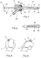
- Figure 1 is a side elevational view with partial section of this cannula, in a first working position.
- Figure 2 is a partial view of Figure 1, in a second position.
- Figure 3 is a side view of a knee in the extended position with a cannula of the prior art seen in section.
- Figure 4 is a side view of the same knee in the bending position.
- Figures 3 and 4 illustrate a knee in which we see a cannula 10 'inserted in the anterior sub-quadricipital cul-de-sac 31, the intervention area being identified by the reference 30.
- the cannula communicates with the intervention zone 30 when the knee is in extension (FIG. 3)
- the communication is interrupted at the location of the arrow F when it is in flexion, so that the rinsing liquid introduced at the level of the arthroscope (not shown) located in the intervention zone 30 can no longer be evacuated by the cannula 10 ′ situated in the zone 31.
- the pump supplying the arthroscope is provided with a pressure control, so that 'it stops and the liquid which no longer circulates quickly becomes cloudy.
- the cannula 10 which is the subject of the invention, illustrated by FIGS. 1 and 2, comprises a tubular element 11 whose internal channel 12 serves as an evacuation channel.
- This tubular element 10 has, at one end, a flange 13 which makes it possible to removably fix it on a connection element 14 between a channel 12a, forming a bend inclined relative to the axis of the channel 12 and intended to be connected to a suction pump (not shown), and a passage 15 which extends coaxially to the channel 12.
- the tubular element 11 comprises two slots 17 extending in the same diametral plane and thus forming two tongues 18 and 19 on either side of the channel 12. The latter stops before the conical end 20 of this element and an island of material 21 is reserved to form a connection between the respective ends of these tongues.
- this tubular element 11 is made of plastic, in particular PVC.
- the rear end of this piston is secured to an operating rod 23 which ends with a second piston 24 provided with an O-ring seal 25.
- a gripping member 26, integral with the piston 24, serves to actuate the piston 22 and its conical spacer end 22a.
- the tubular element 11 is first introduced into the appropriate cavity .
- the piston 22 is then pushed forward: its conical end 22a encounters resistance when arriving at the reduced section end of the channel 12 adjacent to the free end of the tubular element.
- the connection element 14 is held with one hand and, with the other, the gripping member 26 is pushed.
- the force thus developed by the cone 22a on the free ends of the tongues 18.19 breaks the connection formed by the plastic island 21 and then allows the tongues 18,19 to be separated from one another as illustrated in FIG. 2.
- This spacing of the tongues 18 and 19 makes it possible to act on the wall of the sub-quadricipital bag by moving it away from the bone to re-establish the connection between the zones 30 and 31 in the knee flexion position.
- the piston 24 and its O-ring seal 25 are adjusted in the passage 15 and thus ensure sealing, so that the evacuation of the rinsing liquid takes place through the channel 12a that is preferably inclined relative to the common axis of the passage 15 and the channel 12.
- the illustrated solution is an example of possible execution of the invention and other means could be used in particular to separate the tabs 18 and 19.
- This is how one could imagine replacing the piston 22 by an inflatable balloon and the rod 23 by a supply conduit, one end of which would open inside the balloon and the other end of which could, for example, be connected to a bellows capable of being actuated manually or by a pedal to inflate the spacer in order to separate the tabs 18 and 19 from each other.
- the number of tabs 18 and 19 could be greater than two, for example three or four.
Landscapes
- Health & Medical Sciences (AREA)
- Heart & Thoracic Surgery (AREA)
- Biomedical Technology (AREA)
- Vascular Medicine (AREA)
- Engineering & Computer Science (AREA)
- Anesthesiology (AREA)
- Surgery (AREA)
- Hematology (AREA)
- Life Sciences & Earth Sciences (AREA)
- Animal Behavior & Ethology (AREA)
- General Health & Medical Sciences (AREA)
- Public Health (AREA)
- Veterinary Medicine (AREA)
- Infusion, Injection, And Reservoir Apparatuses (AREA)
Abstract
Description
- La présente invention se rapporte à une canule pour l'évacuation d'un liquide de rinçage lors d'une intervention ou d'un examen endoscopique.
- Il est bien connu que le bon déroulement des interventions chirurgicales sous endoscopies et plus particulièrement les arthroscopies par irrigation sous pression constante de sérum physiologique au travers d'un arthroscope, est lié à la bonne circulation du liquide de rinçage de l'arthroscope vers la canule qui donne la transparence, garante de la visibilité dans la cavité opératoire, en éliminant en permanence les débris ou le sang. L'arrêt de cette circulation, consécutif à l'obturation de la canule d'évacuation qui est raccordée à une aspiration, crée une opacité quasi immédiate du champ opératoire.
- Dans une arthroscopie du genou, la position de la canule d'aspiration se situe de préférence dans une zone de la capsule articulaire où ne se trouvent pas le champ visuel de la caméra et les instruments chirurgicaux afin de ne pas entraver les gestes opératoires du chirurgien, ni réduire le champ visuel. De ce fait, la zone la plus fréquemment utilisée pour placer la canule est le cul-de-sac sous-quadricipital. Compte tenu de sa position, la canule est fréquemment isolée de la zone d'irrigation dans certaines positions du genou.
- Dans ces conditions particulières et suivant l'importance de la synoviale, il peut arriver que le liquide d'irrigation ne puisse plus migrer de la poche ou se situe l'arthroscope à celle ou se situe la canule. A ce moment là, la circulation qui permet le rinçage est interrompue, le liquide se trouble rapidement et rend le champ opératoire opaque, empêchant toute intervention.
- On a déjà proposé dans le US-2 541 691 un instrument de drainage qui présente à son extrémité avant une pluralité de languettes parallèles découpées dans un matériau élastique en feuille, enroulé ensuite selon un cylindre d'axe parallèle aux languettes, de manière que celles-ci s'écartent en direction de leurs extrémités.
- Pour permettre d'introduire le drain dans une veine, les languettes sont ramenées manuellement vers l'axe du cylindre et retenues dans un capuchon fixé à l'extrémité d'une tige. Une fois le drain dans la veine on pousse axialement vers l'avant la tige de manière à dégager les languettes du capuchon.
- L'écartement des languettes est obtenu sous l'effet de leur propre élasticité. Aucune force n'est appliquée sur elles pour les écarter. Un tel instrument ne permettrait pas de jouer le rôle d'écarteur en vue de rétablir la circulation de liquide entre la zone d'irrigation et la canule l'évacuation.
- La présente invention se propose de remédier, au moins en partie, à cet inconvénient.
- A cet effet, cette invention a pour objet une canule pour l'évacuation d'un liquide de rinçage lors d'une intervention ou d'un examen endoscopique, en particulier arthroscopique, selon la revendication 1.
- L'avantage principal de l'invention provient du fait que la canule joue le rôle d'un écarteur quand elle est introduite dans le cul-de-sac sous-quadricipital. Elle maintient ainsi la capsule articulaire ouverte pour permettre la bonne circulation du liquide au travers de la cavité, entre l'arrivée dudit liquide au niveau de l'arthroscope et sa sortie au niveau de ladite canule.
- D'autres avantages apparaîtront à l'aide de la description qui va suivre ainsi que des dessins annexés qui illustrent, schématiquement et à titre d'exemple, une forme d'exécution de la canule objet de l'invention.
- La figure 1 est une vue en élévation latérale avec coupe partielle de cette canule, dans une première position de travail.
- La figure 2 est une vue partielle de la figure 1, dans une seconde position.
- La figure 3 est une vue latérale d'un genou en position d'extension avec une canule de l'état de la technique vue en coupe.
- La figure 4 est une vue latérale du même genou en position de flexion.
- Les figures 3 et 4 illustrent un genou dans lequel on voit une canule 10' insérée dans le cul-de-sac sous-quadricipital antérieur 31, la zone d'intervention étant identifiée par la référence 30. Comme on le voit, alors que la canule communique avec la zone d'intervention 30 lorsque le genou est en extension (figure 3), la communication est interrompue à l'endroit de la flèche F lorsqu'il est en flexion, de sorte que le liquide de rinçage introduit au niveau de l'arthroscope (non représenté) se trouvant dans la zone d'intervention 30 ne peut plus être évacué par la canule 10' située dans la zone 31. La pompe alimentant l'arthroscope est munie d'un asservissement en pression, de sorte qu'elle s'arrête et que le liquide qui ne circule plus se trouble rapidement.
- La canule 10 objet de l'invention, illustrée par les figures 1 et 2, comporte un élément tubulaire 11 dont le canal intérieur 12 sert de canal d'évacuation. Cet élément tubulaire 10 présente, à une extrémité, une collerette 13 qui permet de le fixer de façon amovible sur un élément de connexion 14 entre un canal 12a, formant un coude incliné par rapport à l'axe du canal 12 et destiné à être relié à une pompe d'aspiration (non représentée), et un passage 15 qui s'étend coaxialement au canal 12. Une bague de fixation 16, vissée sur l'élément de connexion 14, applique la collerette 13 contre une face de cet élément 14 dans laquelle débouche une extrémité du passage 15. Des moyens d'étanchéité (non représentés) sont prévus pour rendre étanche la fixation de l'élément tubulaire 11 sur l'élément de connexion 14.
- L'élément tubulaire 11 comporte deux fentes 17 s'étendant dans un même plan diamétral et formant ainsi deux languettes 18 et 19 de part et d'autre du canal 12. Celui-ci s'arrête avant l'extrémité conique 20 de cet élément et un îlot de matière 21 est réservé pour former une liaison entre les extrémités respectives de ces languettes. De préférence, cet élément tubulaire 11 est en matière plastique, notamment en PVC.
- Un piston 22, se terminant par une extrèmité conique d'écartement 22a, est engagé dans le passage 15. L'extrémité arrière de ce piston est solidaire d'une tige de manoeuvre 23 qui se termine par un second piston 24 muni d'un joint O-ring 25. Un organe de préhension 26, solidaire du piston 24, sert à actionner le piston 22 et son extrémité conique d'écartement 22a.
- Lorsque la canule 10, illustrée par la figure 1, est mise en place en vue de l'intervention, par exemple, dans la position illustrée par les figures 3 et 4, on introduit d'abord l'élément tubulaire 11 dans la cavité appropriée. On pousse ensuite le piston 22 vers l'avant : son extrémité conique 22a rencontre une résistance en arrivant à l'extrémité de section réduite du canal 12 adjacent à l'extrémité libre de l'élément tubulaire. A ce moment, on tient d'une main l'élément de connexion 14 et, de l'autre, on pousse l'organe de préhension 26. La force ainsi développée par le cône 22a sur les extrémités libres des languettes 18,19 rompt la liaison formée par l'îlot de matière plastique 21 et permet alors d'écarter les languettes 18,19 l'une de l'autre comme illustré par la figure 2. Cet écartement des languettes 18 et 19 permet d'agir sur la paroi du sac sous-quadricipital en l'écartant de l'os pour rétablir la liaison entre les zones 30 et 31 en position de flexion du genou. Dans la position extrême antérieure du piston 22 illustrée par la figure 2, le piston 24 et son joint O-ring 25 sont ajustés dans le passage 15 et assurent ainsi l'étanchéité, de sorte que l'évacuation du liquide de rinçage s'effectue à travers le canal 12a que est de préférence incliné par rapport à l'axe commun du passage 15 et du canal 12.
- Si, dans la position illustrée par la figure 2, le canal d'évacuation 12 venait à se boucher par obstruction d'un corps solide, il suffirait de retirer le piston 22 en arrière en tirant l'organe de préhension 26 pour extraire ce corps hors du canal et de ramener ensuite le piston 22 dans sa position d'écartement des languettes 18 et 19 en repoussant l'organe de préhension 26.
- Bien entendu, la solution illustrée est un exemple d'exécution possible de l'invention et d'autres moyens pourraient être utilisés en particulier pour écarter les languettes 18 et 19. C'est ainsi que l'on pourrait imaginer de remplacer le piston 22 par un ballonnet gonflable et la tige 23 par un conduit d'alimentation dont une extrémité s'ouvrirait à l'intérieur du ballonnet et dont l'autre extrémité pourrait, par exemple, être reliée à un soufflet susceptible d'être actionné manuellement ou par une pédale pour gonfler l'élément d'écartement afin d'écarter les languettes 18 et 19 l'une de l'autre. Par ailleurs, le nombre des languettes 18 et 19 pourrait être supérieur à deux, par exemple trois ou quatre.
Claims (7)
- Canule pour l'évacuation d'un liquide de rinçage lors d'une intervention ou d'un examen endoscopique, en particulier arthroscopique comprenant une élément tubulaire (11) formant au moins en partie canal d'évacuation (12) dudit liquide de rinçage,un passage (15) pour relier ledit canal (12) à l'extérieur,un organe d'actionnement (22) et des moyens de commande (23, 26) de cet organe d'actionnement (22) susceptibles d'être manoeuvrés de l'extérieur dudit canal à travers ledit passage (15) pour déplacer longitudinalement ledit organe d'actionnementdes moyens d'étanchéité (25) pour permettre le passage étanche desdits moyens de commande de l'extérieur de la canule audit organe d'actionnement (22), au moins en position avancée de celui-ci, caractérisée par le faitqu'au moins deux fentes latérales allongées (17) sont ménagées de part et d'autre de la partie du canal d'évacuation (12) formée par ledit élément tubulaire (11), partant d'une extrémité libre de cet élément tubulaire (11) et s'étendant sur une partie de sa longueur formant deux languettes (18, 19) solidaires de cet élément tubulaire (11) etque ledit organe d'actionnement (22) est conformé pour appliquer auxdites languettes (18, 19) des forces tendant à les écarter l'une de l'autre dans la position avancée dudit organe d'actionnement.
- Canule selon la revendication 1, caractérisée en ce que ledit élément tubulaire (11) est monté de façon amovible sur un élément de connexion entre ledit canal d'évacuation (12) et ledit passage (15).
- Canule selon la revendication 2, caractérisée en ce que ledit élément tubulaire (11) et ledit passage (15) sont coaxiaux.
- Canule selon la revendication 3, caractérisée en ce que la section droite dudit canal (12) est réduite au voisinage de l'extrémité libre desdites languettes, que l'organe d'actionnement (22) comporte un élément conique (22a), dont le sommet est tourné vers l'extrémité libre des languettes (18, 19) et dont la base a un diamètre sensiblement égal à celui dudit canal (12) et que les moyens de commande comportent une tige (23) solidaire dudit élément conique (22a) pour amener la base de celui-ci dans la portion de section droite réduite dudit canal (12).
- Canule selon la revendication 4, caractérisée en ce que l'extrémité de ladite tige (23) opposée à celle solidaire dudit organe d'actionnement (22) se termine par un organe de manoeuvre (26) et par une surface de piston (24) associée à un joint O-ring (25) pour s'ajuster dans ledit passage (15).
- Canule selon la revendication 1, caractérisée en ce que des moyens de liaison (21), susceptibles d'être rompus par la force engendrée par ledit organe d'actionnement (22), relient les extrémités libres desdites languettes (18, 19), l'une à l'autre.
- Canule selon la revendication 6, caractérisée en ce que ledit élément tubulaire (11) est en matière plastique et que les moyens de liaison (21)sont formés par au moins un îlot de matière compris entre lesdites extrémités libres de languettes (18, 19).
Applications Claiming Priority (3)
| Application Number | Priority Date | Filing Date | Title |
|---|---|---|---|
| FR9204149A FR2689401A1 (fr) | 1992-04-02 | 1992-04-02 | Canule. |
| FR9204149 | 1992-04-02 | ||
| PCT/EP1993/000792 WO1993019790A1 (fr) | 1992-04-02 | 1993-03-31 | Canule |
Publications (2)
| Publication Number | Publication Date |
|---|---|
| EP0587866A1 EP0587866A1 (fr) | 1994-03-23 |
| EP0587866B1 true EP0587866B1 (fr) | 1996-09-11 |
Family
ID=9428510
Family Applications (1)
| Application Number | Title | Priority Date | Filing Date |
|---|---|---|---|
| EP93908870A Expired - Lifetime EP0587866B1 (fr) | 1992-04-02 | 1993-03-31 | Canule |
Country Status (6)
| Country | Link |
|---|---|
| US (1) | US5411517A (fr) |
| EP (1) | EP0587866B1 (fr) |
| AU (1) | AU3950193A (fr) |
| DE (1) | DE69304645D1 (fr) |
| FR (1) | FR2689401A1 (fr) |
| WO (1) | WO1993019790A1 (fr) |
Families Citing this family (16)
| Publication number | Priority date | Publication date | Assignee | Title |
|---|---|---|---|---|
| WO1998029026A2 (fr) * | 1996-12-30 | 1998-07-09 | Imagyn Medical Technologies, Inc. | Dispositif d'acces extensible et son procede de fabrication et d'utilisation |
| DE19823755A1 (de) * | 1998-05-27 | 1999-12-02 | Thomas Neuhann | Vorrichtung zur Aufweitung und Wiederherstellung des Tränenkanals am menschlichen Auge, durch den Tränenflüssigkeit vom Auge in den Nasenraum gelangt |
| US7597698B2 (en) * | 1999-08-10 | 2009-10-06 | Maquet Cardiovascular Llc | Apparatus and method for endoscopic encirclement of pulmonary veins for epicardial ablation |
| US20040102804A1 (en) * | 1999-08-10 | 2004-05-27 | Chin Albert K. | Apparatus and methods for endoscopic surgical procedures |
| US20060287574A1 (en) * | 1999-08-25 | 2006-12-21 | Chin Albert K | Longitudinal dilator |
| US8974462B2 (en) * | 2008-06-13 | 2015-03-10 | Pivot Medical, Inc. | Devices and methods for minimally invasive access into a joint |
| WO2009152470A1 (fr) | 2008-06-13 | 2009-12-17 | The Foundry, Llc. | Procédés et appareil pour une distraction d'articulation |
| US10426453B2 (en) | 2009-03-17 | 2019-10-01 | Pivot Medical, Inc. | Method and apparatus for distracting a joint |
| US9186181B2 (en) | 2009-03-17 | 2015-11-17 | Pivot Medical, Inc. | Method and apparatus for distracting a joint |
| US12035902B2 (en) | 2009-03-17 | 2024-07-16 | Stryker Corporation | Method and apparatus for distracting a joint |
| AU2010226598A1 (en) * | 2009-03-17 | 2011-10-27 | Pivot Medical, Inc. | Method and apparatus for distracting a joint, including the provision and use of a novel joint-spacing balloon catheter and a novel inflatable perineal post |
| US8568308B2 (en) | 2009-08-14 | 2013-10-29 | Alan M. Reznik | Customizable, self holding, space retracting arthroscopic/endoscopic cannula system |
| US8721649B2 (en) | 2009-12-04 | 2014-05-13 | Pivot Medical, Inc. | Hip joint access using a circumferential wire and balloon |
| US8298185B2 (en) * | 2010-09-14 | 2012-10-30 | Suremka Medical, Llc | Retractable cannula for surgical procedures |
| AU2011326601A1 (en) | 2010-11-08 | 2013-06-06 | Pivot Medical, Inc. | Method and apparatus for distracting a joint |
| CN106691385A (zh) * | 2016-12-29 | 2017-05-24 | 天津恒宇医疗科技有限公司 | 一种支撑架oct成像导管 |
Family Cites Families (9)
| Publication number | Priority date | Publication date | Assignee | Title |
|---|---|---|---|---|
| DE73751C (de) * | R. NYBLAD in Papenburg, Prov. Hannover | Beschickungsvorrichtung für Generatoren | ||
| US765879A (en) * | 1904-05-13 | 1904-07-26 | Wilber A K Campbell | Dilator. |
| US1637264A (en) * | 1925-11-06 | 1927-07-26 | Stuebner Gustav | Syringe |
| US1827497A (en) * | 1929-05-22 | 1931-10-13 | Varney William Thomas | Speculum |
| US2042900A (en) * | 1935-06-08 | 1936-06-02 | Floyd M James | Veinous drain tube |
| US2201749A (en) * | 1939-02-15 | 1940-05-21 | Vandegrift Middleton | Expanding vein tube |
| US2541691A (en) * | 1949-06-24 | 1951-02-13 | Clarence D Eicher | Embalmer's drainage instrument |
| MX144742A (es) * | 1975-08-29 | 1981-11-19 | Kendall & Co | Mejoras en dispositivo aspirador de liquido hiperbarico usado como medio de contraste en mielografias |
| US4692153A (en) * | 1986-04-03 | 1987-09-08 | Berlin Richard B | Surgical wound drain device |
-
1992
- 1992-04-02 FR FR9204149A patent/FR2689401A1/fr not_active Withdrawn
-
1993
- 1993-03-31 US US08/157,021 patent/US5411517A/en not_active Expired - Fee Related
- 1993-03-31 WO PCT/EP1993/000792 patent/WO1993019790A1/fr not_active Ceased
- 1993-03-31 EP EP93908870A patent/EP0587866B1/fr not_active Expired - Lifetime
- 1993-03-31 DE DE69304645T patent/DE69304645D1/de not_active Expired - Lifetime
- 1993-03-31 AU AU39501/93A patent/AU3950193A/en not_active Abandoned
Also Published As
| Publication number | Publication date |
|---|---|
| EP0587866A1 (fr) | 1994-03-23 |
| DE69304645D1 (de) | 1996-10-17 |
| US5411517A (en) | 1995-05-02 |
| AU3950193A (en) | 1993-11-08 |
| WO1993019790A1 (fr) | 1993-10-14 |
| FR2689401A1 (fr) | 1993-10-08 |
Similar Documents
| Publication | Publication Date | Title |
|---|---|---|
| FR2689401A1 (fr) | Canule. | |
| FR2521014A1 (fr) | Instrument chirurgical tubulaire, notamment catheter de thrombectomie | |
| EP0605351B1 (fr) | Instrument, destiné à l'extraction des tronçons veineux | |
| EP1335672B1 (fr) | Dispositif de forage comprenant un trepan recuperateur d'os | |
| CA1243247A (fr) | Instrument pour l'extraction de corps etrangers dans les canaux physiologiques de l'organisme | |
| EP1027091B1 (fr) | Catheter universel | |
| EP0490714B1 (fr) | Instrument chirurgical endoscopique pour le déplacement de tissus ou organes | |
| EP0223179B1 (fr) | Cathéter-guide à commande à distance | |
| CH670391A5 (fr) | ||
| EP0616793A1 (fr) | Instrument médical pour l'élimination de dépôts formés sur les parois intérieures des artères ou des veines | |
| WO1997009086A1 (fr) | Catheter multiconduits, notamment d'hemodialyse | |
| EP0065054A1 (fr) | Pince à biopsie | |
| FR2734145A1 (fr) | Instrument medical comportant un element d'extremite distal pouvant etre coude | |
| EP0485278A1 (fr) | Instrument chirurgical endoscopique pour la saisie de tissus | |
| FR2774895A1 (fr) | Dispositif medical comprenant une tige munie d'un moyen d'absorption des contraintes axiales | |
| EP0223176A2 (fr) | Cathéter à commande à distance à micro-ballonnet | |
| EP0071538A1 (fr) | Appareil de transfert d'éléments de reproduction animale, tels que des embryons | |
| EP0264419A1 (fr) | Dispositif ophtalmique d'aspiration/irrigation. | |
| EP3906960B1 (fr) | Catheter bi-fonctionnel a deux gaines coulissantes | |
| EP0836504B1 (fr) | Catheter de dilatation du type guide sur un fil | |
| FR2872696A1 (fr) | Introducteur pour intervention endoluminale | |
| CA2148110A1 (fr) | Trocart a protection de pointe | |
| FR2667780A1 (fr) | Dispositif de laparoscopie operatoire multifonctionnel a canule jetable. | |
| EP4021526A1 (fr) | Dispositif chirurgical comportant une tige présentant à son extrémité distale un manchon déformable entourant ladite tige | |
| FR2635971A1 (fr) | Dispositif ophtalmologique |
Legal Events
| Date | Code | Title | Description |
|---|---|---|---|
| PUAI | Public reference made under article 153(3) epc to a published international application that has entered the european phase |
Free format text: ORIGINAL CODE: 0009012 |
|
| 17P | Request for examination filed |
Effective date: 19931130 |
|
| AK | Designated contracting states |
Kind code of ref document: A1 Designated state(s): BE CH DE FR LI NL SE |
|
| 17Q | First examination report despatched |
Effective date: 19950804 |
|
| GRAH | Despatch of communication of intention to grant a patent |
Free format text: ORIGINAL CODE: EPIDOS IGRA |
|
| GRAH | Despatch of communication of intention to grant a patent |
Free format text: ORIGINAL CODE: EPIDOS IGRA |
|
| GRAA | (expected) grant |
Free format text: ORIGINAL CODE: 0009210 |
|
| AK | Designated contracting states |
Kind code of ref document: B1 Designated state(s): BE CH DE FR LI NL SE |
|
| PG25 | Lapsed in a contracting state [announced via postgrant information from national office to epo] |
Ref country code: NL Free format text: LAPSE BECAUSE OF FAILURE TO SUBMIT A TRANSLATION OF THE DESCRIPTION OR TO PAY THE FEE WITHIN THE PRESCRIBED TIME-LIMIT Effective date: 19960911 |
|
| REF | Corresponds to: |
Ref document number: 69304645 Country of ref document: DE Date of ref document: 19961017 |
|
| PG25 | Lapsed in a contracting state [announced via postgrant information from national office to epo] |
Ref country code: DE Effective date: 19961212 |
|
| REG | Reference to a national code |
Ref country code: CH Ref legal event code: NV Representative=s name: MOINAS KIEHL SAVOYE & CRONIN |
|
| NLV1 | Nl: lapsed or annulled due to failure to fulfill the requirements of art. 29p and 29m of the patents act | ||
| PG25 | Lapsed in a contracting state [announced via postgrant information from national office to epo] |
Ref country code: BE Effective date: 19970331 |
|
| PLBE | No opposition filed within time limit |
Free format text: ORIGINAL CODE: 0009261 |
|
| 26N | No opposition filed | ||
| BERE | Be: lapsed |
Owner name: GUIGNARD MIREILLE Effective date: 19970331 |
|
| PGFP | Annual fee paid to national office [announced via postgrant information from national office to epo] |
Ref country code: SE Payment date: 19990422 Year of fee payment: 7 |
|
| PG25 | Lapsed in a contracting state [announced via postgrant information from national office to epo] |
Ref country code: SE Free format text: LAPSE BECAUSE OF NON-PAYMENT OF DUE FEES Effective date: 20000401 |
|
| EUG | Se: european patent has lapsed |
Ref document number: 93908870.4 |
|
| REG | Reference to a national code |
Ref country code: FR Ref legal event code: CL |
|
| REG | Reference to a national code |
Ref country code: CH Ref legal event code: PLI Owner name: PROMEPLA SAM Free format text: GUIGNARD, MIREILLE#"LE VEZELY", SERGY GARE#F-01630 SAINT-GENIS POUILLY (FR) -TRANSFER TO- PROMEPLA SAM#9, AVENUE PRINCE HEREDITIAIRE ALBERT#98000 MONACO (MC) |
|
| PGFP | Annual fee paid to national office [announced via postgrant information from national office to epo] |
Ref country code: CH Payment date: 20110316 Year of fee payment: 19 |
|
| PGFP | Annual fee paid to national office [announced via postgrant information from national office to epo] |
Ref country code: FR Payment date: 20110412 Year of fee payment: 19 |
|
| REG | Reference to a national code |
Ref country code: CH Ref legal event code: PL |
|
| REG | Reference to a national code |
Ref country code: FR Ref legal event code: ST Effective date: 20121130 |
|
| PG25 | Lapsed in a contracting state [announced via postgrant information from national office to epo] |
Ref country code: LI Free format text: LAPSE BECAUSE OF NON-PAYMENT OF DUE FEES Effective date: 20120331 Ref country code: CH Free format text: LAPSE BECAUSE OF NON-PAYMENT OF DUE FEES Effective date: 20120331 Ref country code: FR Free format text: LAPSE BECAUSE OF NON-PAYMENT OF DUE FEES Effective date: 20120402 |