EP0580507B1 - A method for the monitoring of multiple anticancer drug resistance - Google Patents
A method for the monitoring of multiple anticancer drug resistance Download PDFInfo
- Publication number
- EP0580507B1 EP0580507B1 EP93401884A EP93401884A EP0580507B1 EP 0580507 B1 EP0580507 B1 EP 0580507B1 EP 93401884 A EP93401884 A EP 93401884A EP 93401884 A EP93401884 A EP 93401884A EP 0580507 B1 EP0580507 B1 EP 0580507B1
- Authority
- EP
- European Patent Office
- Prior art keywords
- ornithine decarboxylase
- cells
- resistance
- increase
- assay
- Prior art date
- Legal status (The legal status is an assumption and is not a legal conclusion. Google has not performed a legal analysis and makes no representation as to the accuracy of the status listed.)
- Expired - Lifetime
Links
- 239000002246 antineoplastic agent Substances 0.000 title claims description 33
- 238000000034 method Methods 0.000 title claims description 32
- 229940041181 antineoplastic drug Drugs 0.000 title claims description 26
- 206010059866 Drug resistance Diseases 0.000 title claims description 8
- 238000012544 monitoring process Methods 0.000 title description 3
- 210000004027 cell Anatomy 0.000 claims description 82
- 102000052812 Ornithine decarboxylases Human genes 0.000 claims description 67
- 108700005126 Ornithine decarboxylases Proteins 0.000 claims description 67
- 230000000694 effects Effects 0.000 claims description 46
- 230000035945 sensitivity Effects 0.000 claims description 23
- 230000001413 cellular effect Effects 0.000 claims description 15
- 229940127089 cytotoxic agent Drugs 0.000 claims description 14
- 239000002254 cytotoxic agent Substances 0.000 claims description 13
- 231100000599 cytotoxic agent Toxicity 0.000 claims description 13
- 210000004962 mammalian cell Anatomy 0.000 claims description 13
- 229940079593 drug Drugs 0.000 claims description 11
- 239000003814 drug Substances 0.000 claims description 11
- 210000002966 serum Anatomy 0.000 claims description 11
- RJURFGZVJUQBHK-UHFFFAOYSA-N actinomycin D Natural products CC1OC(=O)C(C(C)C)N(C)C(=O)CN(C)C(=O)C2CCCN2C(=O)C(C(C)C)NC(=O)C1NC(=O)C1=C(N)C(=O)C(C)=C2OC(C(C)=CC=C3C(=O)NC4C(=O)NC(C(N5CCCC5C(=O)N(C)CC(=O)N(C)C(C(C)C)C(=O)OC4C)=O)C(C)C)=C3N=C21 RJURFGZVJUQBHK-UHFFFAOYSA-N 0.000 claims description 8
- 239000000284 extract Substances 0.000 claims description 7
- 230000005764 inhibitory process Effects 0.000 claims description 7
- 230000001086 cytosolic effect Effects 0.000 claims description 6
- 231100000673 dose–response relationship Toxicity 0.000 claims description 6
- 108010092160 Dactinomycin Proteins 0.000 claims description 4
- 229940122803 Vinca alkaloid Drugs 0.000 claims description 4
- RJURFGZVJUQBHK-IIXSONLDSA-N actinomycin D Chemical compound C[C@H]1OC(=O)[C@H](C(C)C)N(C)C(=O)CN(C)C(=O)[C@@H]2CCCN2C(=O)[C@@H](C(C)C)NC(=O)[C@H]1NC(=O)C1=C(N)C(=O)C(C)=C2OC(C(C)=CC=C3C(=O)N[C@@H]4C(=O)N[C@@H](C(N5CCC[C@H]5C(=O)N(C)CC(=O)N(C)[C@@H](C(C)C)C(=O)O[C@@H]4C)=O)C(C)C)=C3N=C21 RJURFGZVJUQBHK-IIXSONLDSA-N 0.000 claims description 4
- 230000003432 anti-folate effect Effects 0.000 claims description 4
- 229940127074 antifolate Drugs 0.000 claims description 4
- 229960000640 dactinomycin Drugs 0.000 claims description 4
- VLCYCQAOQCDTCN-UHFFFAOYSA-N eflornithine Chemical compound NCCCC(N)(C(F)F)C(O)=O VLCYCQAOQCDTCN-UHFFFAOYSA-N 0.000 claims description 4
- 238000000684 flow cytometry Methods 0.000 claims description 4
- 239000004052 folic acid antagonist Substances 0.000 claims description 4
- 230000001900 immune effect Effects 0.000 claims description 4
- 230000000638 stimulation Effects 0.000 claims description 4
- 238000002965 ELISA Methods 0.000 claims description 3
- 229940045799 anthracyclines and related substance Drugs 0.000 claims description 3
- 238000005457 optimization Methods 0.000 claims description 3
- 238000004448 titration Methods 0.000 claims description 3
- 238000006243 chemical reaction Methods 0.000 claims description 2
- 238000000799 fluorescence microscopy Methods 0.000 claims description 2
- 239000003102 growth factor Substances 0.000 claims description 2
- 239000005556 hormone Substances 0.000 claims description 2
- 229940088597 hormone Drugs 0.000 claims description 2
- 238000010166 immunofluorescence Methods 0.000 claims description 2
- 230000002401 inhibitory effect Effects 0.000 claims description 2
- 239000013038 irreversible inhibitor Substances 0.000 claims description 2
- 238000003127 radioimmunoassay Methods 0.000 claims description 2
- 238000003556 assay Methods 0.000 description 26
- IAKHMKGGTNLKSZ-INIZCTEOSA-N (S)-colchicine Chemical compound C1([C@@H](NC(C)=O)CC2)=CC(=O)C(OC)=CC=C1C1=C2C=C(OC)C(OC)=C1OC IAKHMKGGTNLKSZ-INIZCTEOSA-N 0.000 description 24
- 230000036457 multidrug resistance Effects 0.000 description 16
- 229960001338 colchicine Drugs 0.000 description 12
- 239000012737 fresh medium Substances 0.000 description 10
- 238000000338 in vitro Methods 0.000 description 9
- 239000002609 medium Substances 0.000 description 9
- AHLPHDHHMVZTML-BYPYZUCNSA-N L-Ornithine Chemical compound NCCC[C@H](N)C(O)=O AHLPHDHHMVZTML-BYPYZUCNSA-N 0.000 description 7
- 206010028980 Neoplasm Diseases 0.000 description 7
- 238000009643 clonogenic assay Methods 0.000 description 6
- 231100000096 clonogenic assay Toxicity 0.000 description 6
- 230000003021 clonogenic effect Effects 0.000 description 5
- 231100000433 cytotoxic Toxicity 0.000 description 5
- 230000001472 cytotoxic effect Effects 0.000 description 5
- 230000003211 malignant effect Effects 0.000 description 5
- VQJHOPSWBGJHQS-UHFFFAOYSA-N metoprine, methodichlorophen Chemical compound CC1=NC(N)=NC(N)=C1C1=CC=C(Cl)C(Cl)=C1 VQJHOPSWBGJHQS-UHFFFAOYSA-N 0.000 description 5
- AHLPHDHHMVZTML-UHFFFAOYSA-N Orn-delta-NH2 Natural products NCCCC(N)C(O)=O AHLPHDHHMVZTML-UHFFFAOYSA-N 0.000 description 4
- 229960003104 ornithine Drugs 0.000 description 4
- 125000003831 tetrazolyl group Chemical group 0.000 description 4
- 231100000111 LD50 Toxicity 0.000 description 3
- 229940034982 antineoplastic agent Drugs 0.000 description 3
- 239000000872 buffer Substances 0.000 description 3
- 230000003833 cell viability Effects 0.000 description 3
- 238000002474 experimental method Methods 0.000 description 3
- 230000012010 growth Effects 0.000 description 3
- 238000011534 incubation Methods 0.000 description 3
- 230000007246 mechanism Effects 0.000 description 3
- 230000009467 reduction Effects 0.000 description 3
- IJGRMHOSHXDMSA-UHFFFAOYSA-N Atomic nitrogen Chemical compound N#N IJGRMHOSHXDMSA-UHFFFAOYSA-N 0.000 description 2
- 206010006187 Breast cancer Diseases 0.000 description 2
- AOJJSUZBOXZQNB-TZSSRYMLSA-N Doxorubicin Chemical compound O([C@H]1C[C@@](O)(CC=2C(O)=C3C(=O)C=4C=CC=C(C=4C(=O)C3=C(O)C=21)OC)C(=O)CO)[C@H]1C[C@H](N)[C@H](O)[C@H](C)O1 AOJJSUZBOXZQNB-TZSSRYMLSA-N 0.000 description 2
- 102100021079 Ornithine decarboxylase Human genes 0.000 description 2
- 229930012538 Paclitaxel Natural products 0.000 description 2
- 230000009471 action Effects 0.000 description 2
- 239000003242 anti bacterial agent Substances 0.000 description 2
- 229940088710 antibiotic agent Drugs 0.000 description 2
- 230000008901 benefit Effects 0.000 description 2
- 201000011510 cancer Diseases 0.000 description 2
- 238000004113 cell culture Methods 0.000 description 2
- 230000004663 cell proliferation Effects 0.000 description 2
- 239000003153 chemical reaction reagent Substances 0.000 description 2
- 238000002512 chemotherapy Methods 0.000 description 2
- 210000001072 colon Anatomy 0.000 description 2
- 238000007398 colorimetric assay Methods 0.000 description 2
- 238000001514 detection method Methods 0.000 description 2
- 230000002255 enzymatic effect Effects 0.000 description 2
- 239000007788 liquid Substances 0.000 description 2
- 210000004072 lung Anatomy 0.000 description 2
- 230000002503 metabolic effect Effects 0.000 description 2
- 229960001592 paclitaxel Drugs 0.000 description 2
- 238000007747 plating Methods 0.000 description 2
- 230000004983 pleiotropic effect Effects 0.000 description 2
- RXWNCPJZOCPEPQ-NVWDDTSBSA-N puromycin Chemical compound C1=CC(OC)=CC=C1C[C@H](N)C(=O)N[C@H]1[C@@H](O)[C@H](N2C3=NC=NC(=C3N=C2)N(C)C)O[C@@H]1CO RXWNCPJZOCPEPQ-NVWDDTSBSA-N 0.000 description 2
- 230000002285 radioactive effect Effects 0.000 description 2
- 239000011541 reaction mixture Substances 0.000 description 2
- 238000011160 research Methods 0.000 description 2
- 230000004044 response Effects 0.000 description 2
- 230000004083 survival effect Effects 0.000 description 2
- RCINICONZNJXQF-MZXODVADSA-N taxol Chemical compound O([C@@H]1[C@@]2(C[C@@H](C(C)=C(C2(C)C)[C@H](C([C@]2(C)[C@@H](O)C[C@H]3OC[C@]3([C@H]21)OC(C)=O)=O)OC(=O)C)OC(=O)[C@H](O)[C@@H](NC(=O)C=1C=CC=CC=1)C=1C=CC=CC=1)O)C(=O)C1=CC=CC=C1 RCINICONZNJXQF-MZXODVADSA-N 0.000 description 2
- 238000012360 testing method Methods 0.000 description 2
- IQFYYKKMVGJFEH-OFKYTIFKSA-N 1-[(2r,4s,5r)-4-hydroxy-5-(tritiooxymethyl)oxolan-2-yl]-5-methylpyrimidine-2,4-dione Chemical compound C1[C@H](O)[C@@H](CO[3H])O[C@H]1N1C(=O)NC(=O)C(C)=C1 IQFYYKKMVGJFEH-OFKYTIFKSA-N 0.000 description 1
- QKNYBSVHEMOAJP-UHFFFAOYSA-N 2-amino-2-(hydroxymethyl)propane-1,3-diol;hydron;chloride Chemical compound Cl.OCC(N)(CO)CO QKNYBSVHEMOAJP-UHFFFAOYSA-N 0.000 description 1
- 125000000972 4,5-dimethylthiazol-2-yl group Chemical group [H]C([H])([H])C1=C(N=C(*)S1)C([H])([H])[H] 0.000 description 1
- VJXSSYDSOJBUAV-UHFFFAOYSA-N 6-(2,5-dimethoxy-benzyl)-5-methyl-pyrido[2,3-d]pyrimidine-2,4-diamine Chemical compound COC1=CC=C(OC)C(CC=2C(=C3C(N)=NC(N)=NC3=NC=2)C)=C1 VJXSSYDSOJBUAV-UHFFFAOYSA-N 0.000 description 1
- STQGQHZAVUOBTE-UHFFFAOYSA-N 7-Cyan-hept-2t-en-4,6-diinsaeure Natural products C1=2C(O)=C3C(=O)C=4C(OC)=CC=CC=4C(=O)C3=C(O)C=2CC(O)(C(C)=O)CC1OC1CC(N)C(O)C(C)O1 STQGQHZAVUOBTE-UHFFFAOYSA-N 0.000 description 1
- 102100033350 ATP-dependent translocase ABCB1 Human genes 0.000 description 1
- 208000031261 Acute myeloid leukaemia Diseases 0.000 description 1
- 229920001817 Agar Polymers 0.000 description 1
- VHUUQVKOLVNVRT-UHFFFAOYSA-N Ammonium hydroxide Chemical compound [NH4+].[OH-] VHUUQVKOLVNVRT-UHFFFAOYSA-N 0.000 description 1
- 108091003079 Bovine Serum Albumin Proteins 0.000 description 1
- 208000026310 Breast neoplasm Diseases 0.000 description 1
- 108010078791 Carrier Proteins Proteins 0.000 description 1
- 241000699802 Cricetulus griseus Species 0.000 description 1
- KCXVZYZYPLLWCC-UHFFFAOYSA-N EDTA Chemical compound OC(=O)CN(CC(O)=O)CCN(CC(O)=O)CC(O)=O KCXVZYZYPLLWCC-UHFFFAOYSA-N 0.000 description 1
- 102000004190 Enzymes Human genes 0.000 description 1
- 108090000790 Enzymes Proteins 0.000 description 1
- 102000009024 Epidermal Growth Factor Human genes 0.000 description 1
- 101800003838 Epidermal growth factor Proteins 0.000 description 1
- WQZGKKKJIJFFOK-GASJEMHNSA-N Glucose Natural products OC[C@H]1OC(O)[C@H](O)[C@@H](O)[C@@H]1O WQZGKKKJIJFFOK-GASJEMHNSA-N 0.000 description 1
- 208000002250 Hematologic Neoplasms Diseases 0.000 description 1
- 101001041245 Homo sapiens Ornithine decarboxylase Proteins 0.000 description 1
- UFHFLCQGNIYNRP-UHFFFAOYSA-N Hydrogen Chemical compound [H][H] UFHFLCQGNIYNRP-UHFFFAOYSA-N 0.000 description 1
- FBOZXECLQNJBKD-ZDUSSCGKSA-N L-methotrexate Chemical compound C=1N=C2N=C(N)N=C(N)C2=NC=1CN(C)C1=CC=C(C(=O)N[C@@H](CCC(O)=O)C(O)=O)C=C1 FBOZXECLQNJBKD-ZDUSSCGKSA-N 0.000 description 1
- 108010047230 Member 1 Subfamily B ATP Binding Cassette Transporter Proteins 0.000 description 1
- 208000034578 Multiple myelomas Diseases 0.000 description 1
- 102000016943 Muramidase Human genes 0.000 description 1
- 108010014251 Muramidase Proteins 0.000 description 1
- 208000033776 Myeloid Acute Leukemia Diseases 0.000 description 1
- 108010062010 N-Acetylmuramoyl-L-alanine Amidase Proteins 0.000 description 1
- 208000015914 Non-Hodgkin lymphomas Diseases 0.000 description 1
- UTJLXEIPEHZYQJ-UHFFFAOYSA-N Ornithine Natural products OC(=O)C(C)CCCN UTJLXEIPEHZYQJ-UHFFFAOYSA-N 0.000 description 1
- 206010033128 Ovarian cancer Diseases 0.000 description 1
- 206010061535 Ovarian neoplasm Diseases 0.000 description 1
- 229910019142 PO4 Inorganic materials 0.000 description 1
- 206010035226 Plasma cell myeloma Diseases 0.000 description 1
- GLNADSQYFUSGOU-GPTZEZBUSA-J Trypan blue Chemical compound [Na+].[Na+].[Na+].[Na+].C1=C(S([O-])(=O)=O)C=C2C=C(S([O-])(=O)=O)C(/N=N/C3=CC=C(C=C3C)C=3C=C(C(=CC=3)\N=N\C=3C(=CC4=CC(=CC(N)=C4C=3O)S([O-])(=O)=O)S([O-])(=O)=O)C)=C(O)C2=C1N GLNADSQYFUSGOU-GPTZEZBUSA-J 0.000 description 1
- JXLYSJRDGCGARV-WWYNWVTFSA-N Vinblastine Natural products O=C(O[C@H]1[C@](O)(C(=O)OC)[C@@H]2N(C)c3c(cc(c(OC)c3)[C@]3(C(=O)OC)c4[nH]c5c(c4CCN4C[C@](O)(CC)C[C@H](C3)C4)cccc5)[C@@]32[C@H]2[C@@]1(CC)C=CCN2CC3)C JXLYSJRDGCGARV-WWYNWVTFSA-N 0.000 description 1
- 239000002253 acid Substances 0.000 description 1
- 230000001464 adherent effect Effects 0.000 description 1
- 210000004100 adrenal gland Anatomy 0.000 description 1
- 239000008272 agar Substances 0.000 description 1
- 238000013019 agitation Methods 0.000 description 1
- 239000000908 ammonium hydroxide Substances 0.000 description 1
- 239000003098 androgen Substances 0.000 description 1
- 229940030486 androgens Drugs 0.000 description 1
- 230000000340 anti-metabolite Effects 0.000 description 1
- 229940100197 antimetabolite Drugs 0.000 description 1
- 239000002256 antimetabolite Substances 0.000 description 1
- 239000003080 antimitotic agent Substances 0.000 description 1
- 230000009286 beneficial effect Effects 0.000 description 1
- 230000001851 biosynthetic effect Effects 0.000 description 1
- 230000015572 biosynthetic process Effects 0.000 description 1
- 230000010261 cell growth Effects 0.000 description 1
- 239000013592 cell lysate Substances 0.000 description 1
- 210000000170 cell membrane Anatomy 0.000 description 1
- 239000001913 cellulose Substances 0.000 description 1
- 229920002678 cellulose Polymers 0.000 description 1
- 210000003169 central nervous system Anatomy 0.000 description 1
- 239000003795 chemical substances by application Substances 0.000 description 1
- 230000035572 chemosensitivity Effects 0.000 description 1
- 230000000973 chemotherapeutic effect Effects 0.000 description 1
- 230000005757 colony formation Effects 0.000 description 1
- 230000001332 colony forming effect Effects 0.000 description 1
- 230000002596 correlated effect Effects 0.000 description 1
- 239000003246 corticosteroid Substances 0.000 description 1
- 229960001334 corticosteroids Drugs 0.000 description 1
- 238000002425 crystallisation Methods 0.000 description 1
- 230000008025 crystallization Effects 0.000 description 1
- 229960000975 daunorubicin Drugs 0.000 description 1
- STQGQHZAVUOBTE-VGBVRHCVSA-N daunorubicin Chemical compound O([C@H]1C[C@@](O)(CC=2C(O)=C3C(=O)C=4C=CC=C(C=4C(=O)C3=C(O)C=21)OC)C(C)=O)[C@H]1C[C@H](N)[C@H](O)[C@H](C)O1 STQGQHZAVUOBTE-VGBVRHCVSA-N 0.000 description 1
- 230000003247 decreasing effect Effects 0.000 description 1
- 230000001419 dependent effect Effects 0.000 description 1
- 238000011161 development Methods 0.000 description 1
- 238000000502 dialysis Methods 0.000 description 1
- LOKCTEFSRHRXRJ-UHFFFAOYSA-I dipotassium trisodium dihydrogen phosphate hydrogen phosphate dichloride Chemical compound P(=O)(O)(O)[O-].[K+].P(=O)(O)([O-])[O-].[Na+].[Na+].[Cl-].[K+].[Cl-].[Na+] LOKCTEFSRHRXRJ-UHFFFAOYSA-I 0.000 description 1
- VHJLVAABSRFDPM-QWWZWVQMSA-N dithiothreitol Chemical compound SC[C@@H](O)[C@H](O)CS VHJLVAABSRFDPM-QWWZWVQMSA-N 0.000 description 1
- 229960004679 doxorubicin Drugs 0.000 description 1
- 229940000406 drug candidate Drugs 0.000 description 1
- 238000007877 drug screening Methods 0.000 description 1
- 238000001035 drying Methods 0.000 description 1
- 229960001484 edetic acid Drugs 0.000 description 1
- 230000008030 elimination Effects 0.000 description 1
- 238000003379 elimination reaction Methods 0.000 description 1
- 238000007824 enzymatic assay Methods 0.000 description 1
- 229940088598 enzyme Drugs 0.000 description 1
- 229940116977 epidermal growth factor Drugs 0.000 description 1
- VJJPUSNTGOMMGY-MRVIYFEKSA-N etoposide Chemical compound COC1=C(O)C(OC)=CC([C@@H]2C3=CC=4OCOC=4C=C3[C@@H](O[C@H]3[C@@H]([C@@H](O)[C@@H]4O[C@H](C)OC[C@H]4O3)O)[C@@H]3[C@@H]2C(OC3)=O)=C1 VJJPUSNTGOMMGY-MRVIYFEKSA-N 0.000 description 1
- 229960005420 etoposide Drugs 0.000 description 1
- 230000007717 exclusion Effects 0.000 description 1
- 230000001747 exhibiting effect Effects 0.000 description 1
- 239000012894 fetal calf serum Substances 0.000 description 1
- 230000008014 freezing Effects 0.000 description 1
- 238000007710 freezing Methods 0.000 description 1
- 230000006870 function Effects 0.000 description 1
- 239000008103 glucose Substances 0.000 description 1
- 239000000122 growth hormone Substances 0.000 description 1
- 239000001963 growth medium Substances 0.000 description 1
- 239000001257 hydrogen Substances 0.000 description 1
- 229910052739 hydrogen Inorganic materials 0.000 description 1
- 230000002209 hydrophobic effect Effects 0.000 description 1
- 230000005918 in vitro anti-tumor Effects 0.000 description 1
- 238000000099 in vitro assay Methods 0.000 description 1
- 239000003112 inhibitor Substances 0.000 description 1
- 230000002427 irreversible effect Effects 0.000 description 1
- 230000000155 isotopic effect Effects 0.000 description 1
- 210000003734 kidney Anatomy 0.000 description 1
- 208000032839 leukemia Diseases 0.000 description 1
- 238000005567 liquid scintillation counting Methods 0.000 description 1
- 210000004185 liver Anatomy 0.000 description 1
- 229960000274 lysozyme Drugs 0.000 description 1
- 235000010335 lysozyme Nutrition 0.000 description 1
- 239000004325 lysozyme Substances 0.000 description 1
- 230000036210 malignancy Effects 0.000 description 1
- 239000003550 marker Substances 0.000 description 1
- 201000001441 melanoma Diseases 0.000 description 1
- 239000012528 membrane Substances 0.000 description 1
- 229960000485 methotrexate Drugs 0.000 description 1
- WHLATLJVYMNVTJ-UHFFFAOYSA-N methyl n-(1,2,3,10-tetramethoxy-9-oxo-6,7-dihydro-5h-benzo[a]heptalen-7-yl)carbamate Chemical compound C1=C(OC)C(=O)C=C2C(NC(=O)OC)CCC3=CC(OC)=C(OC)C(OC)=C3C2=C1 WHLATLJVYMNVTJ-UHFFFAOYSA-N 0.000 description 1
- 238000007392 microtiter assay Methods 0.000 description 1
- 238000012986 modification Methods 0.000 description 1
- 230000004048 modification Effects 0.000 description 1
- VMGAPWLDMVPYIA-HIDZBRGKSA-N n'-amino-n-iminomethanimidamide Chemical compound N\N=C\N=N VMGAPWLDMVPYIA-HIDZBRGKSA-N 0.000 description 1
- 229930014626 natural product Natural products 0.000 description 1
- 239000002547 new drug Substances 0.000 description 1
- 229910052757 nitrogen Inorganic materials 0.000 description 1
- 102000039446 nucleic acids Human genes 0.000 description 1
- 108020004707 nucleic acids Proteins 0.000 description 1
- 150000007523 nucleic acids Chemical class 0.000 description 1
- 230000002611 ovarian Effects 0.000 description 1
- 210000001672 ovary Anatomy 0.000 description 1
- 238000012261 overproduction Methods 0.000 description 1
- NBIIXXVUZAFLBC-UHFFFAOYSA-K phosphate Chemical compound [O-]P([O-])([O-])=O NBIIXXVUZAFLBC-UHFFFAOYSA-K 0.000 description 1
- 239000010452 phosphate Substances 0.000 description 1
- 239000002953 phosphate buffered saline Substances 0.000 description 1
- 229950001030 piritrexim Drugs 0.000 description 1
- 210000002826 placenta Anatomy 0.000 description 1
- 229920000768 polyamine Polymers 0.000 description 1
- 230000001376 precipitating effect Effects 0.000 description 1
- 239000002243 precursor Substances 0.000 description 1
- 239000000047 product Substances 0.000 description 1
- 102000004169 proteins and genes Human genes 0.000 description 1
- 108090000623 proteins and genes Proteins 0.000 description 1
- 229950010131 puromycin Drugs 0.000 description 1
- NGVDGCNFYWLIFO-UHFFFAOYSA-N pyridoxal 5'-phosphate Chemical compound CC1=NC=C(COP(O)(O)=O)C(C=O)=C1O NGVDGCNFYWLIFO-UHFFFAOYSA-N 0.000 description 1
- 235000007682 pyridoxal 5'-phosphate Nutrition 0.000 description 1
- 239000011589 pyridoxal 5'-phosphate Substances 0.000 description 1
- 230000002829 reductive effect Effects 0.000 description 1
- BOLDJAUMGUJJKM-LSDHHAIUSA-N renifolin D Natural products CC(=C)[C@@H]1Cc2c(O)c(O)ccc2[C@H]1CC(=O)c3ccc(O)cc3O BOLDJAUMGUJJKM-LSDHHAIUSA-N 0.000 description 1
- 238000012552 review Methods 0.000 description 1
- 238000012216 screening Methods 0.000 description 1
- 239000002356 single layer Substances 0.000 description 1
- 239000000243 solution Substances 0.000 description 1
- 238000001228 spectrum Methods 0.000 description 1
- 238000012799 strong cation exchange Methods 0.000 description 1
- 239000000758 substrate Substances 0.000 description 1
- 239000006228 supernatant Substances 0.000 description 1
- 238000010257 thawing Methods 0.000 description 1
- 231100000331 toxic Toxicity 0.000 description 1
- 230000002588 toxic effect Effects 0.000 description 1
- NOYPYLRCIDNJJB-UHFFFAOYSA-N trimetrexate Chemical compound COC1=C(OC)C(OC)=CC(NCC=2C(=C3C(N)=NC(N)=NC3=CC=2)C)=C1 NOYPYLRCIDNJJB-UHFFFAOYSA-N 0.000 description 1
- 229960001099 trimetrexate Drugs 0.000 description 1
- 210000004881 tumor cell Anatomy 0.000 description 1
- 230000007306 turnover Effects 0.000 description 1
- VBEQCZHXXJYVRD-GACYYNSASA-N uroanthelone Chemical compound C([C@@H](C(=O)N[C@H](C(=O)N[C@@H](CS)C(=O)N[C@@H](CC(N)=O)C(=O)N[C@@H](CS)C(=O)N[C@H](C(=O)N[C@@H]([C@@H](C)CC)C(=O)NCC(=O)N[C@@H](CC=1C=CC(O)=CC=1)C(=O)N[C@@H](CO)C(=O)NCC(=O)N[C@@H](CC(O)=O)C(=O)N[C@@H](CCCNC(N)=N)C(=O)N[C@@H](CS)C(=O)N[C@@H](CCC(N)=O)C(=O)N[C@@H]([C@@H](C)O)C(=O)N[C@@H](CCCNC(N)=N)C(=O)N[C@@H](CC(O)=O)C(=O)N[C@@H](CC(C)C)C(=O)N[C@@H](CCCNC(N)=N)C(=O)N[C@@H](CC=1C2=CC=CC=C2NC=1)C(=O)N[C@@H](CC=1C2=CC=CC=C2NC=1)C(=O)N[C@@H](CCC(O)=O)C(=O)N[C@@H](CC(C)C)C(=O)N[C@@H](CCCNC(N)=N)C(O)=O)C(C)C)[C@@H](C)O)NC(=O)[C@H](CO)NC(=O)[C@H](CC(O)=O)NC(=O)[C@H](CC(C)C)NC(=O)[C@H](CO)NC(=O)[C@H](CCC(O)=O)NC(=O)[C@@H](NC(=O)[C@H](CC=1NC=NC=1)NC(=O)[C@H](CCSC)NC(=O)[C@H](CS)NC(=O)[C@@H](NC(=O)CNC(=O)CNC(=O)[C@H](CC(N)=O)NC(=O)[C@H](CC(C)C)NC(=O)[C@H](CS)NC(=O)[C@H](CC=1C=CC(O)=CC=1)NC(=O)CNC(=O)[C@H](CC(O)=O)NC(=O)[C@H](CC=1C=CC(O)=CC=1)NC(=O)[C@H](CO)NC(=O)[C@H](CO)NC(=O)[C@H]1N(CCC1)C(=O)[C@H](CS)NC(=O)CNC(=O)[C@H]1N(CCC1)C(=O)[C@H](CC=1C=CC(O)=CC=1)NC(=O)[C@H](CO)NC(=O)[C@@H](N)CC(N)=O)C(C)C)[C@@H](C)CC)C1=CC=C(O)C=C1 VBEQCZHXXJYVRD-GACYYNSASA-N 0.000 description 1
- 229960003048 vinblastine Drugs 0.000 description 1
- JXLYSJRDGCGARV-XQKSVPLYSA-N vincaleukoblastine Chemical compound C([C@@H](C[C@]1(C(=O)OC)C=2C(=CC3=C([C@]45[C@H]([C@@]([C@H](OC(C)=O)[C@]6(CC)C=CCN([C@H]56)CC4)(O)C(=O)OC)N3C)C=2)OC)C[C@@](C2)(O)CC)N2CCC2=C1NC1=CC=CC=C21 JXLYSJRDGCGARV-XQKSVPLYSA-N 0.000 description 1
- 229960004528 vincristine Drugs 0.000 description 1
- OGWKCGZFUXNPDA-UHFFFAOYSA-N vincristine Natural products C1C(CC)(O)CC(CC2(C(=O)OC)C=3C(=CC4=C(C56C(C(C(OC(C)=O)C7(CC)C=CCN(C67)CC5)(O)C(=O)OC)N4C=O)C=3)OC)CN1CCC1=C2NC2=CC=CC=C12 OGWKCGZFUXNPDA-UHFFFAOYSA-N 0.000 description 1
- OGWKCGZFUXNPDA-XQKSVPLYSA-N vincristine Chemical compound C([N@]1C[C@@H](C[C@]2(C(=O)OC)C=3C(=CC4=C([C@]56[C@H]([C@@]([C@H](OC(C)=O)[C@]7(CC)C=CCN([C@H]67)CC5)(O)C(=O)OC)N4C=O)C=3)OC)C[C@@](C1)(O)CC)CC1=C2NC2=CC=CC=C12 OGWKCGZFUXNPDA-XQKSVPLYSA-N 0.000 description 1
- 238000005406 washing Methods 0.000 description 1
Images



Classifications
-
- G—PHYSICS
- G01—MEASURING; TESTING
- G01N—INVESTIGATING OR ANALYSING MATERIALS BY DETERMINING THEIR CHEMICAL OR PHYSICAL PROPERTIES
- G01N33/00—Investigating or analysing materials by specific methods not covered by groups G01N1/00 - G01N31/00
- G01N33/48—Biological material, e.g. blood, urine; Haemocytometers
- G01N33/50—Chemical analysis of biological material, e.g. blood, urine; Testing involving biospecific ligand binding methods; Immunological testing
- G01N33/53—Immunoassay; Biospecific binding assay; Materials therefor
- G01N33/573—Immunoassay; Biospecific binding assay; Materials therefor for enzymes or isoenzymes
-
- G—PHYSICS
- G01—MEASURING; TESTING
- G01N—INVESTIGATING OR ANALYSING MATERIALS BY DETERMINING THEIR CHEMICAL OR PHYSICAL PROPERTIES
- G01N33/00—Investigating or analysing materials by specific methods not covered by groups G01N1/00 - G01N31/00
- G01N33/48—Biological material, e.g. blood, urine; Haemocytometers
- G01N33/50—Chemical analysis of biological material, e.g. blood, urine; Testing involving biospecific ligand binding methods; Immunological testing
- G01N33/5005—Chemical analysis of biological material, e.g. blood, urine; Testing involving biospecific ligand binding methods; Immunological testing involving human or animal cells
- G01N33/5008—Chemical analysis of biological material, e.g. blood, urine; Testing involving biospecific ligand binding methods; Immunological testing involving human or animal cells for testing or evaluating the effect of chemical or biological compounds, e.g. drugs, cosmetics
- G01N33/5011—Chemical analysis of biological material, e.g. blood, urine; Testing involving biospecific ligand binding methods; Immunological testing involving human or animal cells for testing or evaluating the effect of chemical or biological compounds, e.g. drugs, cosmetics for testing antineoplastic activity
Definitions
- the present invention relates to an assay for the identification and determination of drug sensitivity. More particularly, the invention relates to an assay in-vitro for the identification and determination of multidrug resistance levels to various anticancer drugs in mammalian cells.
- the clinical testing of primary tumor sensitivity and determination of drug dosage to be administered to individual cancer patients should maximize the elimination of malignant cells and would be useful in avoiding undesired toxic side effects exerted on healthy cells.
- P-170 P-glycoprotein
- This membrane functions as an energy-dependent efflux transporter of some antitumor agents known as pleiotropic drugs.
- Increased P-170 expression has been detected in both normal tissues, including adrenal glands, kidney, lung, liver, colon and placenta as well as in a number of hematological malignancies including acute myeloid leukemia, multiple myeloma and non-Hodgkin's lymphoma and in untreated and treated breast cancer.
- Taxol has shown very promising activity against ovarian cancer, advanced breast cancer, and other tumor types and has also been approved as an investigational drug by the F.D.A.
- the disadvantages as mentioned above for the cytotoxic agents, should be taken in account.
- clonogenic cytotoxic assay
- the widely used clonogenic cytotoxic assay was applied to non-adherent malignant cells by utilizing a soft agar-based colony formation technique.
- the clonogenicity-independent methods include a cell counting, after trypan blue exclusion, or using a radioactive nucleic acid precursor, for example, tritiated thymidine.
- a microtiter assay based on cellular proliferation was specially developed for the assessment of anticancer drug sensitivity of mammalian cells that bear poor plating efficiencies.
- High-flux in-vitro antitumor drug screening methods that evaluate cell viability include the wellknown colorimetric assay based on tetrazolium. According to this method, hydrogen acceptor reagent such as MTT [3,(4,5-dimethylthiazol-2-yl)] and its derivatives, when are present in viable cells become metabolically reduced, thus precipitating as insoluble coloured formazans, which are subsequently dissolved and determined by a spectrophotometer.
- clonogenic assays suffer from a number of disadvantages, the major one being the very low plating efficiency of tumor-derived cells as well as their tendency to form cell clumps. These limitations result in unreliable drug survival curves.
- Cellular viability assessed by colorimetric assays such as the metabolic reduction of soluble tetrazolium salt derivatives to insoluble formazans, has several limitations including major variabilities in formazan formation between different cell lines, variabilities that result from cell culture age as well as from differences in glucose concentration at the time of assay, i.e. at the metabolic reduction of the tetrazolium salts.
- the invention relates to a method for assessing clinical sensitivity and resistance to multiple anticancer drugs of mammalian cells, based on a dose-dependent inhibition with cytotoxic agents of the cellular rise in ornithine decarboxylase activity,following serum stimulation, which comprises the steps of: (a) determining by an immunological technique the rise in the content of ornithine decarboxylase protein levels, after adding serum-containing medium to quiescent mammalian cells, and (b) obtaining the levels of the anticancer drug resistance from the ratio of 50% inhibitory concentration of ornithine decarboxylase obtained with anticancer drug resistant cells and drug sensitive cells.
- the invention is based on the discovery that ornithine decarboxylase activity possesses the ability of an early enzymatic growth marker and thus could serve as a quantitative and diagnostic tool in monitoring cellular sensitivity or resistance to anticancer agents.
- the method is characterized by a number of advantageous features which are in particular required for this determination, such as reliability, sensitivity, reproducibility, independency of clonogenicity, rapidity and simplicity.
- a particular characteristic property of ornithine decarboxylase which was discovered according to the present invention, is its strikingly short turnover time in mammalian cells which is in the order of minutes and generally about 15 minutes, whereby its activity rapidly rises in-vitro in response to a variety of growth stimuli.
- hormones such as growth hormones,corticosteroids and androgens
- growth factors such as epidermal growth factor as well as the addition of fresh serum-containing medium.Other reagents required are polyamines, which are the biosynthetic products of ornithine decarboxylase, being used for optimal growth of mammalian cells.
- the cell lysates were then centrifuged at 15,000g for 15 minutes at 4°C, the supernatant liquid was collected and used in the assay for the ornithine decarboxylase activity.
- the assay was based on the Djurhuss method that utilizes a strong cation-exchange paper (Anal. Biochem. 113:352-5, 1981).
- reaction mixture of (0.1 ml) in ornithine decarboxylase buffer with 50uM of non-isotopic L-ornithine, 43 nM of (0.2uCi of L-[2,3- 3 H] ornithine, specific activity 46.5 Ci/mmol, radioactive concentration lmCi/ml, New England Nuclear) and 80 ul of crude cytosolic cell extract. After incubation for 60 minutes at 37°C under agitation, the reaction mixture was spotted onto a strip of P81 cellulose phosphate paper and dried.After washing in a solution of 0.1M ammonium hydroxide and drying,liquid scintillation counting was performed. In a similar manner, blank values were determined by using 80 ul of ornithine decarboxylase buffer, instead of the cytosolic cell extract.
- the protein determinations were carried out according to the method of Bradford (Anal.Biochem. 72, 248-54, 1976), using lysozyme, triple crystallization and dialysis, as a standard.
- ornithine decarboxylase derived from T19 multidrug resistance cells and their wild type parental AA8 cells were examined.
- the apparent Michaelis constants of ornithine decarboxylase from AA8 and from multidrug resistance T19 cells for L-ornithine were 48uM and 46uM, respectively.
- the mean specific activities of ornithine decarboxylase from AA8 and T19 cells were 154 pmoles CO 2 /hour/10 6 cells and 80 pmoles CO 2 /h per 10 6 cells respectively.
- ornithine decarboxylase activity in multidrug resistance T19 cells which received the fresh medium show a mean ornithine decarboxylase activity of 252 pmoles CO 2 /hr/10 cm petri dish, while in the tested T19 cells which did not received said fresh medium, the mean activity was only 50 pmoles CO 2 /hr/10 cm petri dish.
- the increase in ornithine decarboxylase activity was found to be a result of a parallel increase in the number of ornithine decarboxylase molecules. This was done by a titration of the actual number of catalytically-active ornithine decarboxylase molecules per cell using the irreversible inhibitor DL-o-difluoromethylornithine (DFMO).
- DFMO DL-o-difluoromethylornithine
- Clonogenic cytotoxic assays were performed with AA8 and T19 cells exposed to various concentrations of colchicine for 6 hours, 18 hours, 48 hours or continuously, and each time the clonal surviving fractions were determined. It was found that the LD 50 (50% lethal doses) of colchicine to AA8 cells, at the above periods were: 848nM, 426nM, 245nM and 100nM, respectively, while the LD 50 for T19 cells treated continuously with colchicine was 6.7uM, as can be noticed from Figure 3B. The differences in the IC 50 values between T19 and AA8 cells reflect correctly the differences in the colchicine resistance levels of T19 cells as compared to parental AA8 cells.
- this assay was tested with various anticancer drugs of various structure and distinct mechanisms of action.
- the following cytotoxic agents were tested: vinca alkaloids, anthracyclines, epipodophyllotoxins, actinomycin D and antifolates.
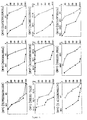
- Typical dose-response curves of decreasing the ornithine decarboxylase activity versus increasing concentrations of some chemotherapeutic agents, are presented in Figure 5 and in the following Table 1. Sensitivity of wild type AA8 cells and multidrug resistance levels of T19 cells to various cytotoxic agents as determined by the ODC assay and clonogenic method.
- the ornithine decarboxylase activity is suitable for the prediction of cellular resistance to antineoplastic agents.
- the rise in the activity of ornithine decarboxylase after adding a serum containing medium to quiescent mammalian cells may be determined by any sensitive immunological technique known in the art, such as ELISA, radioimmunoassay,immunofluorescence, fluorescence microscopy and flow cytometry.In particular preferably will be the ELISA technique, which is now frequently used in many clinical laboratories.
- the flow cytometry analysis of malignant cells stained with fluorescent antibodies directed to human ornithine decarboxylase may detect elevated levels of ornithine decarboxylase in individual cells, thus minimizing the number of cells to 10 3 as required for a reliable flow cytometric analysis as compared to the two orders of magnitude higher cell number generally used for ornithine decarboxylase activity assay.
- the assay for the identification and determination of multidrug resistance is characterized by a number of beneficial characteristics over the currently used chemosensitivity methods. Among the main important advantages the following are mentioned: versatility, sensitivity, reproducibility, reliability, rapidity, simplicity and independence of clonogenicity.
- the versatility is a very important advantage, since the assay is equally efficient in monitoring cellular sensitivity or resistance to a wide-spectrum of anticancer drugs of dissimilar structure and of diverse mechanisms of action, thus being not restricted to a certain group of antineoplastic agents.
Landscapes
- Health & Medical Sciences (AREA)
- Life Sciences & Earth Sciences (AREA)
- Engineering & Computer Science (AREA)
- Immunology (AREA)
- Biomedical Technology (AREA)
- Molecular Biology (AREA)
- Chemical & Material Sciences (AREA)
- Hematology (AREA)
- Urology & Nephrology (AREA)
- Food Science & Technology (AREA)
- General Physics & Mathematics (AREA)
- Cell Biology (AREA)
- Biotechnology (AREA)
- Medicinal Chemistry (AREA)
- Physics & Mathematics (AREA)
- Analytical Chemistry (AREA)
- Biochemistry (AREA)
- General Health & Medical Sciences (AREA)
- Microbiology (AREA)
- Pathology (AREA)
- Bioinformatics & Cheminformatics (AREA)
- Toxicology (AREA)
- Tropical Medicine & Parasitology (AREA)
- Measuring Or Testing Involving Enzymes Or Micro-Organisms (AREA)
- Medicines That Contain Protein Lipid Enzymes And Other Medicines (AREA)
- Enzymes And Modification Thereof (AREA)
Description
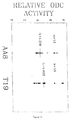
- Figure 1,
- shows the extent of ornithine decarboxylase activity increase in parental AA8 cells and in their multidrug resistance T19 subline following the addition of fresh serum-containing medium.
- Figure 2,
- illustrates the dose-dependent inhibition with difluoromethylornithine of ornithine decarboxylase activity from wild type AA8 cell grown in the absence or presence of fresh serum-containing medium.
- Figure 3,
- illustrates the inhibition of cellular ornithine decarboxylase activity and of clonal survival in AA8 cells following colchicine exposure for various periods of time.
- Figure 4,
- illustrates the reproducibility of ornithine decarboxylase assay in repeated independent experiments of cellular sensitivity to colchicine.
- Figure 5,
- shows the determination by the ornithine decarboxylase assay of cellular sensitivity and resistance to various anticancer drugs in AA8 and multidrug resistance T19 cells.
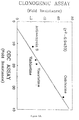
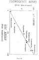
- Figure 6,
- illustrates the correlation of anticancer drug resistance levels determined by the clonogenic assay versus resistance levels obtained by the ornithine decarboxylase assay (6A after 8 hours and 6B after 48 hours).
- Figure 7,
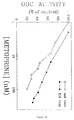
- illustrates the sensitivity of the ornithine decarboxylase activity assay in detection of low levels of cellular resistance to the antifolate metoprine as compared to the clonogenic assay (7A after 8 days and 7B after 48 hours).
Monolayer cells grown in the absence and presence of a known cytotoxic agent, were washed twice with 10 mls of phosphate-buffered saline and suspended in a petri dish (0.5 ml/10 cm) in an ice-cold ornithine decarboxylase buffer containinig 50 mM of Tris-HCl,pH = 7.3, 0.lmM ethylene diamine tetraacetic acid, 50uM pyridoxal 5'-phosphate (Sigma) and 5mM of dithiothreitol.The cells were lysed under three cycles of freezing in liquid nitrogen and thawing at 37°C. The cell lysates were then centrifuged at 15,000g for 15 minutes at 4°C, the supernatant liquid was collected and used in the assay for the ornithine decarboxylase activity. The assay was based on the Djurhuss method that utilizes a strong cation-exchange paper (Anal. Biochem. 113:352-5, 1981). The reaction mixture of (0.1 ml) in ornithine decarboxylase buffer with 50uM of non-isotopic L-ornithine, 43 nM of (0.2uCi of L-[2,3-3H] ornithine, specific activity 46.5 Ci/mmol, radioactive concentration lmCi/ml, New England Nuclear) and 80 ul of crude cytosolic cell extract. After incubation for 60 minutes at 37°C under agitation, the reaction mixture was spotted onto a strip of P81 cellulose phosphate paper and dried.After washing in a solution of 0.1M ammonium hydroxide and drying,liquid scintillation counting was performed. In a similar manner, blank values were determined by using 80 ul of ornithine decarboxylase buffer, instead of the cytosolic cell extract.
| Sensitivity of wild type AA8 cells and multidrug resistance levels of T19 cells to various cytotoxic agents as determined by the ODC assay and clonogenic method. | |||||||
| ODC ASSAY | CLONOGENIC ASSAY | ||||||
| Antimetabolites | |||||||
| Folio acid analogs | Incubation Time(hr) | AA8 | T19 | Resistance (-fold) | AA3 | T19 | Resistance (-fold) |
| Methotrexate | 48 | 56.5 | 63.8 | 1.1 | 25.1 | 30.6 | 1.2 |
| Trimetrexate | 48 | 2.9 | 72.4 | 25.0 | 2.4 | 68.6 | 28.6 |
| Piritrexim | 48 | 5.8 | 127.3 | 22.0 | 46.6 | 113 | 24.5 |
| Metoprine | 48 | 13.8 | 53.2 | 3.9 | 12.6 | 33.7 | 2.7 |
| Natural Products | |||||||
| Vinca alkaloids | |||||||
| Vinblastine | 18 | 15.9 | 275 | 17.3 | 3.0 | 80 | 26.7 |
| Vincristine | 18 | 79.6 | 1534 | 19.2 | 8.2 | 229 | 28.1 |
| Epipodophyllotoxins | |||||||
| Etoposide | 48 | 366 | 4176 | 11.4 | 265 | 4200 | 15.9 |
| Antibiotics | |||||||
| Actinomycin D | 18 | 3.7 | 42 | 11.3 | 2.2 | 49.7 | 22.6 |
| Doxorubicin | 48 | 55 | 3581 | 65.0 | 40 | 2450 | 61.3 |
| Daunorubicin | 48 | 29.6 | 1864 | 63.0 | 8.6 | 726 | 84.1 |
| Miscellaneous | |||||||
| Puromycin | 48 | 10.5 | 315 | 30.0 | 45 | 116 | 25.8 |
| Colchicine | 18 | 404 | 13800 | 34.2 | 100 | 6700 | 67.0 |
Claims (8)
- A method for assessing clinical sensitivity and resistance to multiple anticancer drugs of mammalian cells, based on a dose-dependent inhibition with cytotoxic agents of the cellular rise in ornithine decarboxylase activity, following serum stimulation, which comprises the steps of:(a) determining by an immunological technique the rise in the ornithine decarboxylase protein levels, after adding serum containing medium to quiescent mammalian cells, and(b) obtaining the levels of the anticancer drug resistance from the ratio of 50% inhibitory concentration of ornithine decarboxylase obtained with anticancer drug resistant cells and drug sensitive cells.
- The method according to Claim 1, wherein prior to said determination in step (a), an optimization of the reaction conditions is carried out by an increase in the volume of the crude cytosolic extract.
- The method according to Claim 1, wherein said stimulation is obtained by adding hormones and growth factors.
- The method according to Claims 1 to 3, wherein said immunological technique is selected from ELISA, radioimmunoassay, immunofluorescence, flow cytometry and fluorescence microscopy.
- The method according to any one of Claims 1 to 4, wherein the increase in the ornithine decarboxylase activity resulted from an increase in the number of ornithine decarboxylase molecules.
- The method according tc Claim 5, wherein the deterrmination of said increase is carried out by a titration of the actual number of catalytically-active ornithine decarboxylase molecules per cell.
- The method according to Claim 6, wherein said titration is carried out by using the irreversible inhibitor DL-o-difluoromethylornithine (DFMO).
- The method according to Claims 1 to 7, wherein the cytotoxic agents are selected from vinca alkaloids, anthracyclines, epipodophyllotoxins, actinomycin D and antifolates.
Applications Claiming Priority (2)
| Application Number | Priority Date | Filing Date | Title |
|---|---|---|---|
| IL10261792A IL102617A (en) | 1992-07-23 | 1992-07-23 | Method for assessing clinical sensitivity and resistance to multiple anticancer drugs of mammalian cells |
| IL102617 | 1992-07-23 |
Publications (2)
| Publication Number | Publication Date |
|---|---|
| EP0580507A1 EP0580507A1 (en) | 1994-01-26 |
| EP0580507B1 true EP0580507B1 (en) | 1998-04-15 |
Family
ID=11063851
Family Applications (1)
| Application Number | Title | Priority Date | Filing Date |
|---|---|---|---|
| EP93401884A Expired - Lifetime EP0580507B1 (en) | 1992-07-23 | 1993-07-21 | A method for the monitoring of multiple anticancer drug resistance |
Country Status (6)
| Country | Link |
|---|---|
| US (1) | US5455161A (en) |
| EP (1) | EP0580507B1 (en) |
| JP (1) | JPH07146296A (en) |
| CA (1) | CA2100835A1 (en) |
| DE (1) | DE69317953T2 (en) |
| IL (1) | IL102617A (en) |
Families Citing this family (7)
| Publication number | Priority date | Publication date | Assignee | Title |
|---|---|---|---|---|
| US5789189A (en) * | 1993-09-24 | 1998-08-04 | The Regents Of The University Of California | Inhibition of cyst formation by cytoskeletal specific drugs |
| US5851537A (en) * | 1996-02-02 | 1998-12-22 | Cancer Technologies, Inc. | Topical application of α-DFMO for preventing skin cancer |
| IL119066A0 (en) * | 1996-08-14 | 1996-11-14 | Technion R & D Foundation Ltd | Transmembrane movement and multidrug resistance |
| EP0871441B1 (en) * | 1996-10-04 | 2003-08-13 | Ilex Oncology, Inc. | Dfmo and taxol for the treatment or prevention of breast cancer |
| CA2385528C (en) | 1999-10-01 | 2013-12-10 | Immunogen, Inc. | Compositions and methods for treating cancer using immunoconjugates and chemotherapeutic agents |
| DE602006005333D1 (en) * | 2006-03-06 | 2009-04-09 | Gen Electric | Automatic calibration of a person's sensitivity to a drug |
| CA3044030A1 (en) | 2016-11-17 | 2018-05-24 | Nantbio, Inc. | Validation of inferred anticancer pathways |
Family Cites Families (1)
| Publication number | Priority date | Publication date | Assignee | Title |
|---|---|---|---|---|
| US4637984A (en) * | 1983-10-11 | 1987-01-20 | Wistar Institute Of Anatomy And Biology | Monoclonal anti-ornithine decarboxylase antibody and method of producing same |
-
1992
- 1992-07-23 IL IL10261792A patent/IL102617A/en not_active IP Right Cessation
-
1993
- 1993-07-19 CA CA002100835A patent/CA2100835A1/en not_active Abandoned
- 1993-07-20 US US08/095,141 patent/US5455161A/en not_active Expired - Fee Related
- 1993-07-21 EP EP93401884A patent/EP0580507B1/en not_active Expired - Lifetime
- 1993-07-21 DE DE69317953T patent/DE69317953T2/en not_active Expired - Fee Related
- 1993-07-23 JP JP5202653A patent/JPH07146296A/en active Pending
Also Published As
| Publication number | Publication date |
|---|---|
| JPH07146296A (en) | 1995-06-06 |
| DE69317953D1 (en) | 1998-05-20 |
| IL102617A (en) | 1996-01-19 |
| US5455161A (en) | 1995-10-03 |
| DE69317953T2 (en) | 1998-10-22 |
| EP0580507A1 (en) | 1994-01-26 |
| CA2100835A1 (en) | 1994-01-24 |
| IL102617A0 (en) | 1993-01-14 |
Similar Documents
| Publication | Publication Date | Title |
|---|---|---|
| Cook et al. | Cellular glutathione and thiol measurements from surgically resected human lung tumor and normal lung tissue | |
| Adan et al. | Cell proliferation and cytotoxicity assays | |
| Sevin et al. | Application of an ATP-bioluminescence assay in human tumor chemosensitivity testing | |
| Naccache et al. | Selective inhibition of human neutrophil functional responsiveness by erbstatin, an inhibitor of tyrosine protein kinase | |
| McGrath et al. | Mechanisms of multidrug resistance in HL60 cells: evidence that a surface membrane protein distinct from P-glycoprotein contributes to reduced cellular accumulation of drug | |
| EP1800127B1 (en) | Whole blood preparation for cytometric analysis of cell signaling pathways | |
| US5871946A (en) | Method for determining activity of enzymes in metabolically active whole cells | |
| US20050042694A1 (en) | Detection of protein conformations in single cells | |
| Finlay et al. | The use of human cancer cell lines as a primary screening system for antineoplastic compounds | |
| US11874281B2 (en) | Assays for detecting modified compounds | |
| Barhoumi et al. | Kinetic analysis of glutathione in anchored cells with monochlorobimane | |
| US5455161A (en) | Method for the monitoring of multiple anticancer drug resistance | |
| Hegardt et al. | Rapid caspase‐dependent cell death in cultured human breast cancer cells induced by the polyamine analogue N1, N11‐diethylnorspermine | |
| Green et al. | Development and validation of a drug activity biomarker that shows target inhibition in cancer patients receiving enzastaurin, a novel protein kinase C-β inhibitor | |
| US9739765B2 (en) | Metal-based covalent viability reagent for single cell analysis | |
| Yang et al. | In vitro cytotoxicity testing with fluorescence-based assays in cultured human lung and dermal cells | |
| Kumaragurubaran et al. | Development of an activity-based ratiometric electrochemical substrate for measuring circulating dipeptidyl peptidase-IV/CD26 in whole blood samples | |
| Cook et al. | [17] Measurement of thiols in cell populations fromtumor and normal tissue | |
| Wu et al. | Assaying Bcr-Abl kinase activity and inhibition in whole cell extracts by phosphorylation of substrates immobilized on agarose beads | |
| Rabinovitch et al. | Measurements of cell physiology: Ionized calcium, pH and glutathione | |
| Traganos et al. | Effects of a Prospective Antitumor Agent, 1, 4-bis (2′-Chloroethyl)-1, 4-diazabicyclo-[2.2. 1] Heptane Diperchlorate, on Cultured Mammalian Cells | |
| Lelkes et al. | Direct fluorometric assay of catecholamine secretion from isolated bovine adrenal chromaffin cells | |
| Russo | Cellular Glutathione and Thiol Measurements from Surgically Resected Human Lung Tumor and Normal Lung Tissue | |
| CN120084704A (en) | A method for detecting mitochondrial ATP at the level of a single organelle and its application | |
| Hamilton et al. | Novel use of the fluorescent dye 5‐(and‐6)‐chloromethyl SNARF‐1 acetate for the measurement of intracellular glutathione in leukemic cells and primary lymphocytes |
Legal Events
| Date | Code | Title | Description |
|---|---|---|---|
| PUAI | Public reference made under article 153(3) epc to a published international application that has entered the european phase |
Free format text: ORIGINAL CODE: 0009012 |
|
| AK | Designated contracting states |
Kind code of ref document: A1 Designated state(s): CH DE FR GB IT LI |
|
| 17P | Request for examination filed |
Effective date: 19940707 |
|
| GRAG | Despatch of communication of intention to grant |
Free format text: ORIGINAL CODE: EPIDOS AGRA |
|
| 17Q | First examination report despatched |
Effective date: 19970411 |
|
| GRAG | Despatch of communication of intention to grant |
Free format text: ORIGINAL CODE: EPIDOS AGRA |
|
| GRAH | Despatch of communication of intention to grant a patent |
Free format text: ORIGINAL CODE: EPIDOS IGRA |
|
| GRAH | Despatch of communication of intention to grant a patent |
Free format text: ORIGINAL CODE: EPIDOS IGRA |
|
| GRAA | (expected) grant |
Free format text: ORIGINAL CODE: 0009210 |
|
| AK | Designated contracting states |
Kind code of ref document: B1 Designated state(s): CH DE FR GB IT LI |
|
| REG | Reference to a national code |
Ref country code: CH Ref legal event code: EP |
|
| REF | Corresponds to: |
Ref document number: 69317953 Country of ref document: DE Date of ref document: 19980520 |
|
| ITF | It: translation for a ep patent filed | ||
| PGFP | Annual fee paid to national office [announced via postgrant information from national office to epo] |
Ref country code: FR Payment date: 19980724 Year of fee payment: 6 |
|
| ET | Fr: translation filed | ||
| PG25 | Lapsed in a contracting state [announced via postgrant information from national office to epo] |
Ref country code: LI Free format text: LAPSE BECAUSE OF NON-PAYMENT OF DUE FEES Effective date: 19980731 Ref country code: CH Free format text: LAPSE BECAUSE OF NON-PAYMENT OF DUE FEES Effective date: 19980731 |
|
| PGFP | Annual fee paid to national office [announced via postgrant information from national office to epo] |
Ref country code: GB Payment date: 19980917 Year of fee payment: 6 |
|
| PGFP | Annual fee paid to national office [announced via postgrant information from national office to epo] |
Ref country code: DE Payment date: 19980929 Year of fee payment: 6 |
|
| REG | Reference to a national code |
Ref country code: CH Ref legal event code: NV Representative=s name: BUGNION S.A. |
|
| PLBE | No opposition filed within time limit |
Free format text: ORIGINAL CODE: 0009261 |
|
| STAA | Information on the status of an ep patent application or granted ep patent |
Free format text: STATUS: NO OPPOSITION FILED WITHIN TIME LIMIT |
|
| REG | Reference to a national code |
Ref country code: CH Ref legal event code: PL |
|
| 26N | No opposition filed | ||
| PG25 | Lapsed in a contracting state [announced via postgrant information from national office to epo] |
Ref country code: GB Free format text: LAPSE BECAUSE OF NON-PAYMENT OF DUE FEES Effective date: 19990721 |
|
| PG25 | Lapsed in a contracting state [announced via postgrant information from national office to epo] |
Ref country code: FR Free format text: THE PATENT HAS BEEN ANNULLED BY A DECISION OF A NATIONAL AUTHORITY Effective date: 19990731 |
|
| GBPC | Gb: european patent ceased through non-payment of renewal fee |
Effective date: 19990721 |
|
| PG25 | Lapsed in a contracting state [announced via postgrant information from national office to epo] |
Ref country code: DE Free format text: LAPSE BECAUSE OF NON-PAYMENT OF DUE FEES Effective date: 20000503 |
|
| REG | Reference to a national code |
Ref country code: FR Ref legal event code: ST |
|
| PG25 | Lapsed in a contracting state [announced via postgrant information from national office to epo] |
Ref country code: IT Free format text: LAPSE BECAUSE OF NON-PAYMENT OF DUE FEES;WARNING: LAPSES OF ITALIAN PATENTS WITH EFFECTIVE DATE BEFORE 2007 MAY HAVE OCCURRED AT ANY TIME BEFORE 2007. THE CORRECT EFFECTIVE DATE MAY BE DIFFERENT FROM THE ONE RECORDED. Effective date: 20050721 |