EP0571538B1 - Structural gene of pneumococcal protein - Google Patents
Structural gene of pneumococcal protein Download PDFInfo
- Publication number
- EP0571538B1 EP0571538B1 EP92907062A EP92907062A EP0571538B1 EP 0571538 B1 EP0571538 B1 EP 0571538B1 EP 92907062 A EP92907062 A EP 92907062A EP 92907062 A EP92907062 A EP 92907062A EP 0571538 B1 EP0571538 B1 EP 0571538B1
- Authority
- EP
- European Patent Office
- Prior art keywords
- pspa
- protein
- region
- truncated
- strain
- Prior art date
- Legal status (The legal status is an assumption and is not a legal conclusion. Google has not performed a legal analysis and makes no representation as to the accuracy of the status listed.)
- Expired - Lifetime
Links
Images


Classifications
-
- C—CHEMISTRY; METALLURGY
- C12—BIOCHEMISTRY; BEER; SPIRITS; WINE; VINEGAR; MICROBIOLOGY; ENZYMOLOGY; MUTATION OR GENETIC ENGINEERING
- C12N—MICROORGANISMS OR ENZYMES; COMPOSITIONS THEREOF; PROPAGATING, PRESERVING, OR MAINTAINING MICROORGANISMS; MUTATION OR GENETIC ENGINEERING; CULTURE MEDIA
- C12N15/00—Mutation or genetic engineering; DNA or RNA concerning genetic engineering, vectors, e.g. plasmids, or their isolation, preparation or purification; Use of hosts therefor
- C12N15/09—Recombinant DNA-technology
- C12N15/63—Introduction of foreign genetic material using vectors; Vectors; Use of hosts therefor; Regulation of expression
- C12N15/74—Vectors or expression systems specially adapted for prokaryotic hosts other than E. coli, e.g. Lactobacillus, Micromonospora
-
- A—HUMAN NECESSITIES
- A61—MEDICAL OR VETERINARY SCIENCE; HYGIENE
- A61P—SPECIFIC THERAPEUTIC ACTIVITY OF CHEMICAL COMPOUNDS OR MEDICINAL PREPARATIONS
- A61P31/00—Antiinfectives, i.e. antibiotics, antiseptics, chemotherapeutics
- A61P31/04—Antibacterial agents
-
- C—CHEMISTRY; METALLURGY
- C07—ORGANIC CHEMISTRY
- C07K—PEPTIDES
- C07K14/00—Peptides having more than 20 amino acids; Gastrins; Somatostatins; Melanotropins; Derivatives thereof
- C07K14/195—Peptides having more than 20 amino acids; Gastrins; Somatostatins; Melanotropins; Derivatives thereof from bacteria
- C07K14/28—Peptides having more than 20 amino acids; Gastrins; Somatostatins; Melanotropins; Derivatives thereof from bacteria from Vibrionaceae (F)
-
- C—CHEMISTRY; METALLURGY
- C07—ORGANIC CHEMISTRY
- C07K—PEPTIDES
- C07K14/00—Peptides having more than 20 amino acids; Gastrins; Somatostatins; Melanotropins; Derivatives thereof
- C07K14/195—Peptides having more than 20 amino acids; Gastrins; Somatostatins; Melanotropins; Derivatives thereof from bacteria
- C07K14/315—Peptides having more than 20 amino acids; Gastrins; Somatostatins; Melanotropins; Derivatives thereof from bacteria from Streptococcus (G), e.g. Enterococci
- C07K14/3156—Peptides having more than 20 amino acids; Gastrins; Somatostatins; Melanotropins; Derivatives thereof from bacteria from Streptococcus (G), e.g. Enterococci from Streptococcus pneumoniae (Pneumococcus)
-
- C—CHEMISTRY; METALLURGY
- C07—ORGANIC CHEMISTRY
- C07K—PEPTIDES
- C07K14/00—Peptides having more than 20 amino acids; Gastrins; Somatostatins; Melanotropins; Derivatives thereof
- C07K14/435—Peptides having more than 20 amino acids; Gastrins; Somatostatins; Melanotropins; Derivatives thereof from animals; from humans
- C07K14/705—Receptors; Cell surface antigens; Cell surface determinants
- C07K14/715—Receptors; Cell surface antigens; Cell surface determinants for cytokines; for lymphokines; for interferons
- C07K14/7151—Receptors; Cell surface antigens; Cell surface determinants for cytokines; for lymphokines; for interferons for tumor necrosis factor [TNF], for lymphotoxin [LT]
-
- C—CHEMISTRY; METALLURGY
- C12—BIOCHEMISTRY; BEER; SPIRITS; WINE; VINEGAR; MICROBIOLOGY; ENZYMOLOGY; MUTATION OR GENETIC ENGINEERING
- C12N—MICROORGANISMS OR ENZYMES; COMPOSITIONS THEREOF; PROPAGATING, PRESERVING, OR MAINTAINING MICROORGANISMS; MUTATION OR GENETIC ENGINEERING; CULTURE MEDIA
- C12N15/00—Mutation or genetic engineering; DNA or RNA concerning genetic engineering, vectors, e.g. plasmids, or their isolation, preparation or purification; Use of hosts therefor
- C12N15/09—Recombinant DNA-technology
- C12N15/63—Introduction of foreign genetic material using vectors; Vectors; Use of hosts therefor; Regulation of expression
- C12N15/74—Vectors or expression systems specially adapted for prokaryotic hosts other than E. coli, e.g. Lactobacillus, Micromonospora
- C12N15/746—Vectors or expression systems specially adapted for prokaryotic hosts other than E. coli, e.g. Lactobacillus, Micromonospora for lactic acid bacteria (Streptococcus; Lactococcus; Lactobacillus; Pediococcus; Enterococcus; Leuconostoc; Propionibacterium; Bifidobacterium; Sporolactobacillus)
-
- A—HUMAN NECESSITIES
- A61—MEDICAL OR VETERINARY SCIENCE; HYGIENE
- A61K—PREPARATIONS FOR MEDICAL, DENTAL OR TOILETRY PURPOSES
- A61K39/00—Medicinal preparations containing antigens or antibodies
-
- C—CHEMISTRY; METALLURGY
- C07—ORGANIC CHEMISTRY
- C07K—PEPTIDES
- C07K2319/00—Fusion polypeptide
-
- C—CHEMISTRY; METALLURGY
- C07—ORGANIC CHEMISTRY
- C07K—PEPTIDES
- C07K2319/00—Fusion polypeptide
- C07K2319/01—Fusion polypeptide containing a localisation/targetting motif
- C07K2319/02—Fusion polypeptide containing a localisation/targetting motif containing a signal sequence
Definitions
- the present invention is concerned with the development of an improved vaccine against pneumococcal infections.
- Streptococcus Pneumoniae is an important cause of otitis media, meningitis, bacteremia and pneumonia. Despite the use of antibodies and vaccines, the prevalence of pneumococcal infections has declined little over the last twenty-five years.
- the PspA protein type is independent of capsular type. It would seem that genetic mutation or exchange in the environment has allowed for the development of a large pool of strains which are highly diverse with respect to capsule, PspA, and possibly other molecules with variable structures. Variability of PspA's from different strains also is evident in their molecular weights, which range from 67 to 99 kD. The observed differences are stably inherited and are not the result of protein degradation.
- the present invention relates to the preparation of mutants of S. pneumoniae that secrete an immunogenic truncated form of the PspA protein, and isolation and purification of the secreted protein.
- the truncated form of the PspA protein is immunoprotective and contains at least one protective epitope of PspA.
- the PspA protein is soluble in physiologic solution and lacking at least the functional cell membrane anchor region.
- a purified immunoprotective pneumococcal surface protein comprising a truncated form of PspA which contains at least one immunoprotective epitope of the protein and up to about 90% of the whole PspA protein and from which the functional cell membrane anchor region is absent.
- the molecular size of the purified truncated PspA protein obtained may be varied by directing the point of insertion, which determines the termination of gene expression, to different points in the pspA gene. For example, an N-terminal fragment of apparent molecular weight of 43 kD, constituting approximately one-half of the native protein, has been found useful.
- the truncated segment which is produced by this procedure is capable of eliciting protection in mice from fatal challenge with type 3 S. pneumoniae, demonstrating for the first time that a purified PspA can elicit protection and that this truncated segment of the protein contains protective epitopes of PspA.
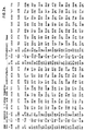
- Amino acid sequence information was obtained on the N-terminal 45 amino acids of the truncated segment of PspA. This sequence is shown in Figure 2. Predictive secondary structural analysis shows that this sequence has a very strong alpha-helical formation, with no non-helical inserts. About 51% of the segment is composed only of two amino acids, namely lysine, a charged amino acid, and alanine, a non-polar amino acid.
- the DNA sequence of the pspA gene is contained on a Hin dIII - Kpn I fragment that is 2086 base pairs in length.
- the pspA gene itself represents approximately 1985 base pairs of this fragment, and comprises an initial region containing transcription and translational signals with translation starting at the ATG/met (nucleotide position 127, codon position -31), followed by a leader sequence extending from the ATG/met (nucleotide position 127, codon position -31) to CGA/ala (nucleotide position 217, codon -1).
- Mature Pspa starts with the Glu amino acid at nucleotide position 220 (codon +1) and ends at the translational stop TAA/OCH at nucleotide position 1984. This translational stop codon is followed by transcription termination signals.
- the amino terminal of the protein sequence contains a 31 amino acid leader sequence and a 45 amino acid sequence identical to the 45 amino acid sequence of the N-terminal of PspA ( Figure 2).
- the amino end of the predicted protein sequence is highly charged and ⁇ -helical in nature. This region has homology with tropomyosin at the amino acid level (approximately 22% identity and 50% similarity). This homology is due largely to a repeating seven residue periodicity where the first and fourth amino acids are hydrophobic, the intervening amino acids are helix-promoting and the seventh amino acid is charged.
- proline-rich region Following the charged helical region is a proline-rich region in which 23 of 81 amino acids are prolines. Immediately carboxy to the proline-rich region is the first of ten highly homologous twenty amino acid repeats. The only significantly hydrophobic region in the sequenced portion of the molecule begins at the last repeat. This potential membrane-spanning region contains several charged amino acids preceding the translational stop codon.
- the insertionally-inactivated mutants of S.pneumoniae lacking the C-terminal anchor regions are capable of growth in chemically-defined medium and secrete the N-terminal portion of the PspA protein into the medium.
- the N-terminal region of PspA is highly soluble in the culture medium and is much easier to isolate than the entire molecule.
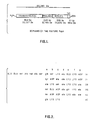
- Soluble truncated molecules have been produced using insertional duplicational mutagenesis directed by the cloned PspA DNA fragments shown in Figure 4. Expression of the same truncated construct (with the pneumococcal promoter) in E.coli results in the same PspA fragment being secreted into the periplasm of E.coli. PspA is readily released from the periplasm by hypotonic lysis.
- Truncated PspA is isolated from culture medium of mutant pneumococci in any convenient manner, such as by tangential flow filtration. Ion-exchange chromatography then is performed on an anionic resin to purify the protein. In this procedure, the solution containing PspA is dialyzed to pH6 in 0.02 M salt solution and passed over the resin. The PspA is eluted from the resin with a gradient of 0.08 to 2.0 M ionic strength and is collected in the fraction between 0.34 and 0.87 M ionic strength, depending on the nature of the column used.
- the PspA may be further purified by sodium dodecyl sulfate polyacrylamide gel (SDS-PAGE) electrophoresis.
- SDS-PAGE sodium dodecyl sulfate polyacrylamide gel
- the electrophoresis purification is convenient when only small quantities of PspA are being handled.
- the protein may be purified by size chromatography in a pH7 phosphate buffer.
- fusion protein is a fusion of the soluble N-terminal region of PspA and the B-subunit of cholera toxin. Fusion proteins also may be formed by chemical attachment of the truncated PspA protein to other proteins.
- Another aspect of the present invention provides a method for the production of cloned proteins, which comprises fusing a pspA gene coding for a truncated form of PspA protein with the gene coding for another protein to form a fusion protein clone, transforming S.pneumoniae , E.coli or other bacteria with the fusion protein clone, growing the transformed bacterium to effect expression of a fusion protein comprising truncated PspA and the other protein into the culture medium, and isolating the fusion protein.
- gram positive bacteria such as pneumococci, for example, S.pneumoniae
- mycobacteria for example, Bacille Calmette-Guerin (BCG).
- BCG Bacille Calmette-Guerin
- a gene fusion of a pspA gene coding for a truncated form of PspA protein with a ctxB gene coding for the B-subunit of cholera toxin is effected.
- the PspA and CTB may be cleaved one from another by dilute acid at an asparagine-proline sequence, known to be labile to dilute acid, engineered at the fusion site of the two proteins.
- the fusion PspA-CTB protein may be isolated from the culture medium by adsorption to a G M1 affinity column, from which the fusion protein subsequently may be eluted at low pH.
- the PspA-CTB fusion protein finds considerable utility in solid phase immunoadsorbant assays.
- solid supports such as microtitration plates
- PspA fragments without having first to isolate the PspA fragments. This may be done by adding bacterial extract containing the fusion protein to plates coated with G M1 .
- the PspA-CTB fusion protein then binds to G M1 through the CTB moiety, thereby coating the solid support with PspA.
- the resulting coated product then may be used in a solid phase immunoadsorbant assay for the detection of PspA antibody and/or antigen in test samples.
- Such immunoadsorbant assays constitute an additional aspect of this invention.
- the PspA attachment/anchor region containing the proline-rich region, the repeat region and/or the C-terminus of PspA, also may be employed to effect expression of heterologous proteins in pneumococci, or other gram positive or gram negative bacteria in the which the attachment/anchor region is functional. Generally, expression is effected on bacterial membrane, cell walls or cell surfaces in gram positive bacteria and in the periplasm of gram negative bacteria.
- An example of such heterologous protein is the B-subunit of cholera toxin.
- the truncated form of PspA provided herein contains the immunoprotective epitopes of the protein and hence is useful in a vaccine against pneumococcal infection. Accordingly, a yet further aspect of the present invention provides a vaccine against pneumococcal infection comprising, as an immunogenically-active component, the purified immunoprotective pneumococcal surface protein provided herein.
- the PspA protein may be employed as one component of a multicomponent vaccine which is effective in providing protection from a variety of infections.
- gram positive bacteria which have been transformed to express the pspA gene coding for the truncated soluble PspA protein may be employed, in a live-attenuated or killed form, as an immunologically-active component of a vaccine against pneumococcal infection.
- pspA gene may be fused to a gene coding for another protein.
- an additional aspect of this invention provides a vaccine against pneumococcal infection comprising, as an immunologically-active component, a live-attenuated or killed bacteria containing a gene coding for the truncated form of PspA.
- the truncated form of PspA also may be employed in conjugates with normally weakly-immunogenic or non-immunogenic protection-eliciting molecules, such as various polysaccharides, to achieve immunogenic potentiation thereof.
- An additional aspect of the invention therefore, provides a vaccine comprising, as an immunogenically-active component, a conjugate of the purified immunoprotective pneumococcal surface protein provided herein and a normally weakly-immunogenic or non-immunogenic protection-eliciting molecule.
- conserved sequences of pspA may be employed as probes to detect the presence of pneumococci of various strains, through detection of pneumococcal DNA, in tissues, body fluids and/or secretions.
- portions of the pspA gene may be used in diagnostic kits for the detection of pneumococcal infections.
- primers made based on conserved sequences of pspA may be used to assay for the presence of pneumococci in tissues, body fluids and/or secretions, through amplification of pneumococcal DNA.
- a single primer pair derived from the nucleotide sequence of the pspA gene of S.pneumoniae may be employed in an assay using the polymerase chain reaction (PCR) for the specific detection of Streptococcus pneumoniae .
- PCR analysis using the primers described herein is performed in accordance with conventional PCR techniques, such as are described in the literature, for example, as described in Arnhem et al at C&EN Special Report, 36, October 1, 1990.
- the primer may be labelled or labelled nucleotide triphosphates may be included in the PCR reaction to label the PCR amplification product.
- the PCR primers may be prepared by well-known methods, for example, by oligonucleotide synthesis or by fragmentation of a larger nucleotide sequence using suitable restriction enzymes.
- the ability to use a single primer capable of detecting a large number of S.pneumoniae strains enables a universal PCR detection kit to be provided which is able to diagnose pneumococcal infection in mammals, including humans, independent of the strain which has caused the disease.
- This Example illustrates the preparation and growth of novel strains of S. pneumoniae.
- S. pneumoniae strain Rx1 which is a non-encapsulated derivative of capsular type 2 strain D39 (National Collection of Type Cultures, London, NCTC #7466), was subjected to insertional inactivation (as described in McDaniel et al (III) 1987, Crain et al 1990, Talkington et al 1991, with 10 different cloned fragments of PspA (see Figure 4). These fragments have all been obtained from restriction digests of cloned PspA DNA on a plasmid in E.coli strain JY4313 (deposited with the American Type Culture Collection on January 31, 1991 under ATCC accession number 68529). This insertional duplication mutagenesis (see Figure 4) results in the termination of gene expression near the 3' end of the cloned fragment.
- the expected molecular size is based on the deduced amino acid sequence and the apparent molecular size is based on migration in SDS-PAGE. The difference between expected and apparent molecular size is due to the conformation of the PspA fragment.
- the proline and repeats/anchor regions were deleted and the resulting protein was unable to attach to cell due to their absence.
- the unattached protein then may be isolated from culture supernatants, as described below.
- the pneumococcal strain JY2008 was grown in 6 liters of a chemically defined medium (see Inf. Imm. 27:444) supplemented with 0.10% choline chloride, 0.075% L-cysteine hydrochloride and 0.25% NaHCO 3 .
- the supernatant fluid of the mid-log phase culture of JY2008 was harvested using a 0.22 ⁇ m membrane tangential flow filter and concentrated 60 fold.
- the concentrated supernatant fluid produced as described in Example 1, was washed in 0.1 M PBS, pH 7.2, and ultracentrifuged at 196,000 xg.
- the supernatant fluid was diluted 1:5 in 20 mM L-histidine buffer-NaCl, the pH adjusted to 6.0 and the injected into a DEAE-fibered Isonet-D2 TM an ion exchange column.
- a stepwise NaCl gradient from 80 mM to 2 M was applied to the column and PspA-containing fractions (0.32 to 0.64 M ionic strength) were pooled and separated on an SDA-polyacrylamide gel.
- the proteins on a representative section of the gel were stained with Comassie Blue R-250 to identify PspA.
- the fraction containing PspA was excised from the remainder of the SDS-gel and electroeluted from the excised gel.
- the eluted protein was precipitated in a 50:50 methanol:acetone solvent and resuspended in PBS. Purity of product was confirmed by silver staining and Western Immunoblotting with mAb Xi126 (IgG 2b, k, see McDaniel et al (I), supra).
- This Example illustrates the isolation of PspA from the bacterial periplasmic space.
- the pellet was discarded and the supernatant, which contained the PspA was saved. Concentration of the supernatant was by standard procedures such as concentration against solid sucrose or ultrafiltration. Purification of the protein isolated from E. coli proceeded by the same chromatography techniques used for the isolation of the 43 kDa (truncated) PspA from the media of growing pneumococci.
- This Example illustrates the immunogenic properties of the PspA protein.
- mice carrying the Xid mutation were bled via the periorbital sinus to establish pre-exposure levels of antibody to PspA.
- Purified PspA prepared as described in Example 2, was emulsified in complete Freund's adjuvant and injected subcutaneously into the inguinal and auxiliary regions, delivering approximately 5 ⁇ g of protein per mouse.
- the mice were injected intraperitoneally with 5 ⁇ g of PspA prepared as described in Example 2.
- Control mice were immunized via the same routes with sterile SDS buffer. Seven days after the last immunization, all mice were bled via the periorbital sinus and were challenged intravenously with 300 CFU of the type 3 strain WU2, grown as described in Example 1.
- Preimmunization and prechallenge sera were analyzed by Western immunoblots to establish baseline and postimmunization response to the truncated protein.
- the PspA of strain WU2 was electrophoresed and transferred to nitrocellulose membranes.
- the membranes were separated into strips and probed with the appropriate mouse antisera at a 1:50 dilution for 2 hours, incubated with biotinylated goat anti-mouse immunoglobulin for 1 hr, washed and incubated with Strepavidin-conjugated phosphatase.
- the membranes were developed with 5-bromo-4-chloro-3-indoyl phosphate toludine salt with 0.01% into blue tetrazolium.
- mice immunized with the purified fragment of PspA all were still alive 14 days after challenge with strain WU2 and none showed any signs of illness following challenge.
- mice immunized with buffer controls six were dead by two days post challenge, while the two remaining control mice appeared very sick, with ruffled fur, arched back and decreased movement, two to three days following challenge but survived. Chi-square analysis indicated that there was a significant difference (P ⁇ 0.003) in survival between the immunized and control groups.
- Preimmunization and prechallenge sera were analyzed by Western immunoblotting. None of the preimmunization sera contained antibody to truncated PspA. Postimmunization sera from eight of eight mice contained detectable antibodies to PspA, and six mice had very strong anti-PspA reactions. When the challenge strain WU2 was probed with the antisera, all the immunized mice had antibodies that were highly cross-reactive with the WU2 PspA epitopes. No control mice developed antibodies to PspA.
- This Example illustrates sequencing of the PspA protein.
- Purified PspA prepared as described in Example 2, was electrophoresed through 9% resolving gels containing recrystallized SDS with the Laemmli buffer system (Nature 227:680). The gels were soaked twice in a 10mM 3-(cyclohexylamino)-1-propanesulfonic acid, pH 11.0, containing 10% methanol for 10 minutes. A polyvinylidene difluoride membrane (PVDF) was wetted completely for several seconds in 100% methanol, then washed in CAPS buffer for 10 min. PspA was electrotransferred to the PVDF membrane in CAPS buffer at 0.5 A for 1 hr.
- PVDF polyvinylidene difluoride membrane
- the membrane was washed two times in deionized water for 5 min, and stained with 0.1% Coomassie Blue R-250 in 50% methanol for 20 minutes.
- the section of the membrane containing PspA was excised and destained in 40% methanol and 10% acetic acid for 5 min.
- the membrane was cut into small segments and stored in sterile Eppendorf TM tubes until sequencing.
- the isolated PspA was sequenced directly from the PVDF membranes.
- Figure 2 depicts the N-terminal 45 residue amino acid sequence and
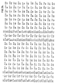
- Figure 5 depicts the amino acid sequence for the whole alpha-helical region.
- the DNA sequence of the whole pspA gene and the deduced amino acid sequence for the PspA protein are shown in Figure 3.
- This Example illustrates the use of the pspA 5'-sequence and/or the PspA N-terminal region to serve as an expression and leader sequence for expressing and/or excreting/secreting heterologous proteins from S.pneumoniae and E.coli.
- this Example there is described the expression of the N-terminal of the PspA protein fused to the B-subunit of cholera toxin (CTB) through a genetic fusion and the excretion of the fused protein from pneumococci and its secretion into the periplasmic space of E.coli .
- CTB cholera toxin
- a fusion protein consisting of CTB and the N-terminal half of PspA was constructed and expressed in E.coli.
- the Hin dIII/ Dra l pspA gene fragment used contained all the pspA transcription and translation initiation signals and the PspA signal peptide leader sequence for transport across the cell membrane.
- the mature PspA encoded by this fragment is predicted to be a product of 29 kDa (observed molecular weight of 42 kDa), encompassing more than 90% of the ⁇ -helical coiled-coil domain.
- the PspA-CTB fusion protein was stably expressed in both high and low copy number plasmids (pUC18, more than 100 copies/cell; pJY4163, about 15 to 30 copies/cell) in E.coli.
- the fusion products were of the expected size (about 54 kDa) and reacted with antibody to both PspA and CTB. That the CTB product retained its functionality was demonstrated by the ability of the fusion protein to bind ganglioside G M1 , a property of CTB.
- the high level of expression of the fusion product apparently resulted in a reduced rate of processing and/or conformational changes that prevented the protein from being completely transported to the periplasm.
- about 60% of the fusion protein was localized in the periplasm, where large quantities were readily released from E. coli by osmotic shock.
- the fusion protein In addition to expression in E.coli , the fusion protein also was expressed in S.pneumoniae by transformation of the low copy number construct into the avirulent S.pneumoniae Rx1 to generate an insertion-duplication mutant. In this way, the gene encoding the fusion protein was integrated into the S.pneumoniae chromosome, from which it was stably expressed. As in the case of Example 1, the truncated PspA molecule lacking the attachment/anchor region, this time in the form of the PspA-CTB fusion protein, was excreted into the culture supernatant. The fusion protein product was of the expected molecular size (54 kDa), reacted with antibody to PspA and CTB, and bound G M1 .
- This Example illustrates the use of PspA attachment or anchor region to permit expression of heterologous proteins on the surface of S.pneumoniae or other bacteria in which the attachment/anchor sequence is functional in particular the expression of a PspA-CTB (cholera toxin B subunit) fusion expressed on the surface of pneumococci.
- PspA-CTB cholera toxin B subunit
- the N-terminal encoding region of PspA including its transcription and translation initiation signals and its signal peptide leader sequence, is linked via a translationally in-frame genetic fusion to the CTB-encoding ctxB fragment that lacks transcription and translation initiation and termination signals.
- This sequence is followed in-frame by the PspA attachment/anchor domain, including part or all of the proline, repeat and C-terminal domains.
- the resulting fusion protein is directed to the outside of the cell via the PspA leader sequence, which is cleaned following transport across the membrane, and then attached to cell by the PspA attachment/anchor sequence.
- the heterologous protein, located between the two PspA fragments is expressed on the outside surface of the membrane and, in S.pneumoniae, on the surface of the cell.
- This Example illustrates the expression of truncated and full length PspA by the Mycobacterium tuberculosis strain Bacille Calmette-Guerin (BCG).
- BCG was chromosomically modified to incorporate the pspA gene coding for the truncated PspA protein.
- the 43 kDa truncated PspA protein was expressed from the modified BCG to approximately 15% of total BCG protein. This result was achieved with an expression vector construct carrying the pspA gene segment encoding the 43 kDa region without its 5'-secretion signal. Expression was only about 1% of BCG protein when the PspA or mycobacterial signal sequences were included. In either case, a significant portion of the expressed PspA was excreted into the medium. Expression of the 43 kDa PspA protein in a fusion with the mycobacterial lipoprotein signal sequence resulted in the expression of the recombinant PspA in the membrane fraction of BCG.
- This Example illustrates the close homology of portions of the pspA gene among different strains of pneumococci and the potential that such homology may be useful for molecular (DNA) approaches to detect pneumococci in tissues, body fluids and/or secretions and to identify pneumococci grown from patient samples.
- DNA molecular
- each of these probes detected two discrete bands whose exact size was dependent on the strain examined.
- the two bands were 4.0 and 9.1 kb.
- the 4.0 kb band corresponded to pspA and was altered or absent in pspA mutants.
- the other band shares some homology with the coding regions for both the N-terminal and C-terminal halves of PspA but is not affected by pspA mutations.
- JY4323 and JY4262 probes failed to react with another gram positive bacterium, Streptococcus pyogenes , and a gram negative bacterium, Salmonella typhimurium.
- the N-terminal probe, JY4306, recognized about one-third of the strains of pneumococci tested.
- This Example illustrates the detection and determination of the location of epitopes in the ⁇ -helical N-terminal region of PspA.
- Monoclonal antibodies protective against pneumococcal infection in a mouse model denoted by an asterisk in Figures 5, 6 and 7, were used to determine the location of epitopes for each antibody in the ⁇ -helical N-terminal region of PspA. The sites were mapped to fragments of PspA.
- the results are illustrated in Figures 5 to 7, with Figure 5 showing the deduced amino acid sequence for the N-terminal region of PspA and the general location of epitopes recognized by monoclonal antibodies, Figure 6 showing antibody reactivity with PspA fragments produced by various pspA gene segments, and Figure 7 showing the mapped location of epitopes in the PspA fragments produced by the different pspA gene segments.
- Numbers 138, 193 and 261 in Figure 5 indicate break positions in the PspA fragments used to map the location of epitopes detected by monoclonal antibodies Xi1526, Xi126, XiR35, XiR38, XiR1224, XiR16, Xi64, XiR278, Xi1325 and Xi1323.
- the asterisk (*) after some of the antibodies denotes those which are able to protect against fatal pneumococcal infection with strain WU2 pneumococci.
- the vertical lines to the right of the Figure indicate those areas predicted to have coiled-coil ⁇ -helical structure.
- the divisions to the left of the Figure indicate the mapped location of the epitopes for each antibody.
- the present invention relates to a truncated PspA molecule capable of eliciting an immunoprotective response and hence containing the protective epitopes of PspA protein. Modifications are possible within the scope of this invention.
Landscapes
- Health & Medical Sciences (AREA)
- Chemical & Material Sciences (AREA)
- Life Sciences & Earth Sciences (AREA)
- Genetics & Genomics (AREA)
- Organic Chemistry (AREA)
- Engineering & Computer Science (AREA)
- General Health & Medical Sciences (AREA)
- Zoology (AREA)
- Biophysics (AREA)
- Biochemistry (AREA)
- Molecular Biology (AREA)
- Biomedical Technology (AREA)
- Wood Science & Technology (AREA)
- Bioinformatics & Cheminformatics (AREA)
- General Engineering & Computer Science (AREA)
- Biotechnology (AREA)
- Medicinal Chemistry (AREA)
- Gastroenterology & Hepatology (AREA)
- Proteomics, Peptides & Aminoacids (AREA)
- Microbiology (AREA)
- Plant Pathology (AREA)
- Physics & Mathematics (AREA)
- Toxicology (AREA)
- Cell Biology (AREA)
- Pulmonology (AREA)
- Immunology (AREA)
- Communicable Diseases (AREA)
- Animal Behavior & Ethology (AREA)
- Public Health (AREA)
- Veterinary Medicine (AREA)
- Nuclear Medicine, Radiotherapy & Molecular Imaging (AREA)
- General Chemical & Material Sciences (AREA)
- Oncology (AREA)
- Chemical Kinetics & Catalysis (AREA)
- Pharmacology & Pharmacy (AREA)
- Preparation Of Compounds By Using Micro-Organisms (AREA)
- Peptides Or Proteins (AREA)
- Medicines Containing Antibodies Or Antigens For Use As Internal Diagnostic Agents (AREA)
- Measuring Or Testing Involving Enzymes Or Micro-Organisms (AREA)
- Micro-Organisms Or Cultivation Processes Thereof (AREA)
Abstract
Description
| AMINO ACID ABBREVIATIONS | |
| A = Ala = Alanine | M = Met = Methionine |
| C = Cys = Cysteine | N = Asn = Asparagine |
| D = Asp = Aspartic Acid | P = Pro = Proline |
| E = Glu = Glutamic Acid | Q = Gln = Glutamine |
| F = Phe = Phenylalanine | R = Arg = Arginine |
| G = Gly = Glycine | S = Ser = Serine |
| H = His = Histidine | T = Thr = Threonine |
| I = Ile = Isoleucine | V = Val = Valine |
| K = Lys = Lysine | W = Try = Tryptophan |
| L = Leu = Leucine | Y = Try = Tyrosine |


The pneumococcal strain JY2008 was grown in 6 liters of a chemically defined medium (see Inf. Imm. 27:444) supplemented with 0.10% choline chloride, 0.075% L-cysteine hydrochloride and 0.25% NaHCO3. The supernatant fluid of the mid-log phase culture of JY2008 was harvested using a 0.22 µm membrane tangential flow filter and concentrated 60 fold.
| Immunogen | Detection of Antibody to PspA | Alive at 2 days post challenge | Alive at 14 days post challenge |
| Isolated PspA (Example 2) | 8/8 | 8/8 | 8/8 |
| Sterile SDS (control) | 0/8 | 2/8 | 2/8 |
Claims (28)
- A purified immunoprotective pneumococcal surface protein (PspA), comprising a truncated form of PspA which contains at least one immunoprotective epitope of the protein and up to 90% of the whole PspA protein and from which the functional cell membrane anchor region is absent.
- The protein claimed in Claim 1, comprising approximately 50% of the whole PspA protein from which the cell membrane anchor region, the repeat region and the proline region are absent and comprising the 43 kD (apparent molecular size) N-terminal region of an 84 kD PspA protein.
- The protein claimed in Claim 1 or Claim 2, comprising at least the N-terminal, protective epitope-containing region of the PspA protein.
- The protein claimed in any one of Claims 1 to 3, comprising at least the N-terminal, α-helical protective epitope-containing region of the PspA protein and having a seven residue periodicity.
- A biologically-pure, recombinant DNA molecule coding for a truncated form of PspA which contains up to 90% of the whole PspA protein and from which the functional cell membrane anchor region is absent, said DNA molecule having a coding sequence included in that set forth below:-


- A DNA molecule claimed in Claim 5, coding for at least part of an α-helical coiled coil region of the PspA protein, at least part of a proline rich region of the PspA protein, or at least part of a repeat region of the PspA protein.
- A DNA molecule claimed in Claim 5 or Claim 6, in the form of a DNA probe for the detection of pneumococcal DNA in a sample characterised by a conserved sequence of the pspA gene, said sequence selected from the group consisting of a nucleic acid sequence encoding a proline-rich region of PspA, a nucleic acid sequence encoding a repeats region of PspA and a nucleic acid sequence encoding a proline-rich region and a repeats region of PspA.
- A DNA molecule claimed in Claims 5 or 6, in the form of a DNA primer for effecting polymerase chain reaction of pneumococcal DNA, characterised by a conserved sequence of the pspA gene.
- A DNA molecule claimed in Claim 8, in which said conserved sequence is located in the proline-rich region of the gene, the repeat region in the gene or both.
- The DNA molecule claimed in Claim 7, which is DNA probe JY4262, consisting of bases 1221 to 1832 of the sequence shown in Claim 5.
- The DNA molecule claimed in Claim 8, in the form of a DNA primer, in which said primer has the nucleotide sequence:or


- A DNA molecule claimed in Claim 5, which codes for a truncated form of the PspA protein which contains approximately 50% of the whole PspA protein from which the cell membrane anchor region, the repeats region and the proline region are absent.
- A DNA molecule claimed in Claim 5 or Claim 12, which is linked by a translationally in-frame genetic fusion to a gene coding for another protein.
- A DNA molecule claimed in Claim 13, in which said gene coding for another protein is a ctxB gene.
- A method for forming an immunoprotective truncated PspA protein, characterised by:(a) effecting insertion-duplication mutagenesis of a bacterium with a pspA gene resulting in the coding of a truncated expressible pneumococcal surface protein (PspA) from which the functional cell membrane anchor region is absent, or(b) fusing a pspA gene coding for a truncated form of PspA protein from which the functional cell membrane anchor region is absent with a gene coding for another protein to form a fusion protein clone and transforming a bacterium with said fusion protein clone,growing said bacterium to effect expression of a truncated PspA protein or a fusion protein comprising truncated PspA and said other protein, andisolating the expressed protein.
- A method as claimed in Claim 15, in which said bacterium is an S.pneumoniae strain, an E.coli strain or a Mycobacteria strain.
- A method as claimed in Claims 15 or 16, in which said bacterium is S.pneumoniae strain JY2008 (ATCC 55143), or an S.pneumoniae strain producing a truncated PspA protein corresponding to an N-terminal portion of PspA protein of 21 kDa molecular weight, or a truncated PspA protein corresponding to an N-terminal portion of PspA protein of 18 kDa molecular weight, or strain JY4306 (ATCC 68522), or BCG.
- A method as claimed in any one of Claims 15 or 16, in which said gene coding for another protein is the ctxB gene coding for the B-subunit of cholera toxin (CTB).
- A method as claimed in Claim 18, in which said fusion protein comprises an acid labile sequence joining said truncated form of PspA protein and CTB and said PspA protein and CTB are separated from each other by dilute acid treatment.
- A method as claimed in any one of Claims 15 to 17, in which said fusion protein is isolated from culture medium by adsorption to a GM1 affinity column, from which said fusion protein is eluted.
- A mutated strain of Streptococcus pneumoniae, Eschericia coli or Mycobacterium characterised by a pspA gene coding for a truncated expressible pneumococcal surface protein (PspA) from which the cell membrane anchor region is absent.
- A strain as claimed in Claim 21, which is the JY2008 (ATCC 55143) strain of S.pneumoniae, or an S.pneumoniae strain producing a truncated PspA protein corresponding to an N-terminal portion of PspA protein of 21 kDa molecular weight, or a truncated PspA protein corresponding to an N-terminal portion of PspA of 18 kDa molecular weight, or the strain JY4306 (ATCC 68522), or a mutated BCG.
- A strain as claimed in Claims 21 or 22, in which the pspA gene coding for a truncated PspA protein is linked to a gene coding for another protein.
- A strain as claimed in Claim 23, in which said gene coding for another protein is the ctxB gene coding for the B-subunit for cholera toxin.
- A vaccine against pneumococcal infection, characterised by, as an immunogenically-active component, the protein defined in any one of Claims 1 to 4, or the expression product of a DNA molecule defined in any one of Claims 5, 6, 12, 13 or 14, or a live-attenuated or killed gram positive bacteria containing a gene coding for the protein defined in any one of Claims 1 to 4 or a DNA molecule defined in any one of Claims 5, 6, 12, 13 or 14 or the protein produced by the method of any one of Claims 15 to 20.
- A vaccine as claimed in Claim 25, in which said protein is conjugated to a normally weakly-immunogenic or non-immunogenic protection-eliciting molecule.
- A vaccine claimed in Claim 25, in which said gram positive bacteria is Streptococcus pneumoniae or Mycobacterium tuberculosis.
- A solid phase immunoabsorbent assay, characterised by a coating of a truncated form of PspA protein as claimed in any one of Claims 1 to 4 on a solid substrate wherein said PspA protein is fused to the B-subunit of cholera toxin (CTB) which is bound to monosinloganglioside (GM1) coating said solid substrate.
Priority Applications (1)
| Application Number | Priority Date | Filing Date | Title |
|---|---|---|---|
| EP96119668A EP0786521A1 (en) | 1991-02-15 | 1992-02-12 | Nucleic acid encoding PspA |
Applications Claiming Priority (3)
| Application Number | Priority Date | Filing Date | Title |
|---|---|---|---|
| US65677391A | 1991-02-15 | 1991-02-15 | |
| US656773 | 1991-02-15 | ||
| PCT/US1992/000857 WO1992014488A1 (en) | 1991-02-15 | 1992-02-12 | Structural gene of pneumococcal protein |
Related Child Applications (1)
| Application Number | Title | Priority Date | Filing Date |
|---|---|---|---|
| EP96119668A Division EP0786521A1 (en) | 1991-02-15 | 1992-02-12 | Nucleic acid encoding PspA |
Publications (3)
| Publication Number | Publication Date |
|---|---|
| EP0571538A1 EP0571538A1 (en) | 1993-12-01 |
| EP0571538A4 EP0571538A4 (en) | 1994-08-24 |
| EP0571538B1 true EP0571538B1 (en) | 1998-07-08 |
Family
ID=24634503
Family Applications (2)
| Application Number | Title | Priority Date | Filing Date |
|---|---|---|---|
| EP92907062A Expired - Lifetime EP0571538B1 (en) | 1991-02-15 | 1992-02-12 | Structural gene of pneumococcal protein |
| EP96119668A Ceased EP0786521A1 (en) | 1991-02-15 | 1992-02-12 | Nucleic acid encoding PspA |
Family Applications After (1)
| Application Number | Title | Priority Date | Filing Date |
|---|---|---|---|
| EP96119668A Ceased EP0786521A1 (en) | 1991-02-15 | 1992-02-12 | Nucleic acid encoding PspA |
Country Status (15)
| Country | Link |
|---|---|
| US (1) | US5728387A (en) |
| EP (2) | EP0571538B1 (en) |
| JP (3) | JP3215109B2 (en) |
| AT (1) | ATE168132T1 (en) |
| AU (1) | AU692678C (en) |
| CA (1) | CA2104014C (en) |
| DE (1) | DE69226167T2 (en) |
| DK (1) | DK0571538T5 (en) |
| ES (1) | ES2118812T3 (en) |
| FI (1) | FI933580A7 (en) |
| IE (2) | IE920479A1 (en) |
| IL (1) | IL100946A0 (en) |
| NO (1) | NO313999B1 (en) |
| WO (1) | WO1992014488A1 (en) |
| ZA (1) | ZA921025B (en) |
Families Citing this family (58)
| Publication number | Priority date | Publication date | Assignee | Title |
|---|---|---|---|---|
| US5965141A (en) * | 1991-02-15 | 1999-10-12 | Uab Research Foundation | Epitopic regions of pneumococcal surface protein a |
| US6027734A (en) * | 1991-02-15 | 2000-02-22 | Uab Research Foundation | Mucosal administration of pneumococcal antigens |
| US5980909A (en) * | 1991-02-15 | 1999-11-09 | Uab Research Foundation | Epitopic regions of pneumococcal surface protein A |
| US6232116B1 (en) | 1991-02-15 | 2001-05-15 | University Of Alabama At Birmingham Research Foundation | Oral administration of pneumococcal antigens |
| US6592876B1 (en) | 1993-04-20 | 2003-07-15 | Uab Research Foundation | Pneumococcal genes, portions thereof, expression products therefrom, and uses of such genes, portions and products |
| US6231870B1 (en) * | 1995-06-02 | 2001-05-15 | Uab Research Foundation | Oral administration of pneumoccal antigens |
| CA2116261A1 (en) * | 1993-04-20 | 1994-10-21 | David E. Briles | Epitopic regions of pneumococcal surface protein a |
| US5928900A (en) * | 1993-09-01 | 1999-07-27 | The Rockefeller University | Bacterial exported proteins and acellular vaccines based thereon |
| AU734488B2 (en) * | 1994-05-20 | 2001-06-14 | Uab Research Foundation | Mucosal administration of pneumococcal antigens |
| US6001564A (en) * | 1994-09-12 | 1999-12-14 | Infectio Diagnostic, Inc. | Species specific and universal DNA probes and amplification primers to rapidly detect and identify common bacterial pathogens and associated antibiotic resistance genes from clinical specimens for routine diagnosis in microbiology laboratories |
| US20020055101A1 (en) | 1995-09-11 | 2002-05-09 | Michel G. Bergeron | Specific and universal probes and amplification primers to rapidly detect and identify common bacterial pathogens and antibiotic resistance genes from clinical specimens for routine diagnosis in microbiology laboratories |
| AU709368B2 (en) * | 1995-06-02 | 1999-08-26 | Uab Research Foundation | Oral administration of pneumococcal antigens |
| US6004802A (en) * | 1995-06-02 | 1999-12-21 | University Of Alabama At Birmingham | Oral administration of pneumococcal antigens |
| ZA964896B (en) | 1995-06-07 | 1997-01-08 | Connaught Lab | Expression of lipoproteins |
| AU700080B2 (en) * | 1995-06-07 | 1998-12-17 | Biochem Vaccines Inc. | Streptococcal heat shock proteins members of the HSP70 family |
| US6251405B1 (en) | 1995-06-07 | 2001-06-26 | Connaught Laboratories, Inc. | Immunological combination compositions and methods |
| CA2704354A1 (en) * | 1995-06-07 | 1996-12-19 | Connaught Laboratories, Inc. | Expression of lipoproteins |
| US5994066A (en) * | 1995-09-11 | 1999-11-30 | Infectio Diagnostic, Inc. | Species-specific and universal DNA probes and amplification primers to rapidly detect and identify common bacterial pathogens and associated antibiotic resistance genes from clinical specimens for routine diagnosis in microbiology laboratories |
| US7078042B2 (en) | 1995-09-15 | 2006-07-18 | Uab Research Foundation | Pneumococcal surface protein C (PspC), epitopic regions and strain selection thereof, and uses therefor |
| US5949537A (en) | 1996-04-18 | 1999-09-07 | American Air Liquide Inc. | In-line cell for absorption spectroscopy |
| US6165989A (en) | 1996-05-14 | 2000-12-26 | Smithkline Beecham Corporation | Era of Streptococcus pneumoniae |
| JP2002512501A (en) | 1996-07-03 | 2002-04-23 | メリアル インコーポレイテッド | Recombinant canine adenovirus (CAV) containing exogenous DNA |
| US5928895A (en) * | 1996-09-24 | 1999-07-27 | Smithkline Beecham Corporation | IgA Fc binding protein |
| US5882896A (en) | 1996-09-24 | 1999-03-16 | Smithkline Beecham Corporation | M protein |
| US20030049636A1 (en) | 1999-05-03 | 2003-03-13 | Bergeron Michel G. | Species-specific, genus-specific and universal DNA probes and amplification primers to rapidly detect and identify common bacterial and fungal pathogens and associated antibiotic resistance genes from clinical specimens for diagnosis in microbiology laboratories |
| US20100267012A1 (en) | 1997-11-04 | 2010-10-21 | Bergeron Michel G | Highly conserved genes and their use to generate probes and primers for detection of microorganisms |
| IL130291A0 (en) * | 1996-12-04 | 2000-06-01 | Univ Alabama | Compositions and methods for administering pneumococcal DNA |
| KR19980067137A (en) * | 1997-01-31 | 1998-10-15 | 박원훈 | Vaccine against pneumococcal pneumoniae using alginate ultrafine granulocytes |
| AU9574598A (en) * | 1997-09-18 | 1999-04-05 | Pasteur Merieux Connaught | Improved method of production of pneumococcal surface proteins |
| US6224882B1 (en) | 1997-11-07 | 2001-05-01 | Protein Science Corp. | Insect cells or fractions as adjuvant for antigens |
| US20010016200A1 (en) | 1998-04-23 | 2001-08-23 | Briles David E. | Pneumococcal surface protein C (PspC), epitopic regions and strain selection thereof, and uses therefor |
| EP1246935B1 (en) | 1999-09-28 | 2013-08-14 | Geneohm Sciences Canada Inc. | Highly conserved genes and their use to generate probes and primers for detection of microorganisms |
| GB0022742D0 (en) | 2000-09-15 | 2000-11-01 | Smithkline Beecham Biolog | Vaccine |
| AT410412B (en) | 2000-11-10 | 2003-04-25 | Inteco Int Techn Beratung | METHOD OF ELECTRIC SLACKING METHODS OF MELTING METALS |
| US8691243B2 (en) | 2002-04-02 | 2014-04-08 | Ben-Gurion University Of The Negev Research And Development Authority | Protein-based Streptococcus pneumoniae vaccine |
| EP2275120A3 (en) * | 2002-04-02 | 2011-06-08 | Ben-Gurion University Of The Negev Research And Development Authority | Protein-based streptococcus pneumoniae vaccines |
| TWI457133B (en) | 2005-12-13 | 2014-10-21 | Glaxosmithkline Biolog Sa | Novel composition |
| BRPI0620460B8 (en) | 2005-12-22 | 2021-05-25 | Glaxosmithkline Biologicals Sa | use of immunogenic composition for babies, and process to produce a vaccine |
| GB0607088D0 (en) | 2006-04-07 | 2006-05-17 | Glaxosmithkline Biolog Sa | Vaccine |
| EP1973933B1 (en) | 2006-01-17 | 2020-06-24 | Forsgren, Arne | A NOVEL SURFACE EXPOSED HAEMOPHILUS INFLUENZAE PROTEIN (PROTEIN E; pE) |
| BRPI0813644B8 (en) | 2007-06-26 | 2021-05-25 | Glaxosmithkline Biologicals Sa | immunogenic composition, vaccine, process to manufacture the same, and use of the immunogenic composition or vaccine |
| US20120164178A1 (en) | 2008-04-16 | 2012-06-28 | Ballou Jr William Ripley | Vaccine |
| CN102170903B (en) | 2008-05-02 | 2016-04-06 | 亮点医疗有限公司 | For product and the method for immune response stimulating |
| EP2545071A2 (en) | 2010-03-09 | 2013-01-16 | Maksyutov, Amir | Polyepitope constructs and methods for their preparation and use |
| GB201003924D0 (en) | 2010-03-09 | 2010-04-21 | Glaxosmithkline Biolog Sa | Immunogenic composition |
| MX340739B (en) | 2010-05-14 | 2016-07-22 | Brookhaven Science Ass Llc | Ospa chimeras and use thereof in vaccines. |
| WO2012023033A2 (en) | 2010-08-18 | 2012-02-23 | Purdue Pharma L.P. | Improved peptide immunogens |
| SG194733A1 (en) | 2011-05-17 | 2013-12-30 | Glaxosmithkline Biolog Sa | Vaccine against streptococcus pneumoniae |
| WO2014018274A1 (en) | 2012-07-27 | 2014-01-30 | Baxter International, Inc. | Compositions comprising chimeric ospa molecules and methods of use thereof |
| JP5854537B2 (en) | 2012-09-19 | 2016-02-09 | 国立大学法人大阪大学 | Pneumococcal vaccine containing pneumococcal surface protein A |
| US9815886B2 (en) | 2014-10-28 | 2017-11-14 | Adma Biologics, Inc. | Compositions and methods for the treatment of immunodeficiency |
| GB201518684D0 (en) | 2015-10-21 | 2015-12-02 | Glaxosmithkline Biolog Sa | Vaccine |
| US11806392B2 (en) | 2016-12-03 | 2023-11-07 | The Uab Research Foundation | Pneumococcal vaccine combining selected alpha helical domains and proline rich domains of pneumococcal surface protein A |
| US10259865B2 (en) | 2017-03-15 | 2019-04-16 | Adma Biologics, Inc. | Anti-pneumococcal hyperimmune globulin for the treatment and prevention of pneumococcal infection |
| GB201711635D0 (en) | 2017-07-19 | 2017-08-30 | Glaxosmithkline Biologicals Sa | Immunogenic composition |
| CN110499375B (en) * | 2019-08-28 | 2023-03-28 | 首钢医院有限公司 | Method for detecting streptococcus pneumoniae by combining multi-cross displacement amplification and gold nanoparticles |
| WO2023240124A1 (en) | 2022-06-07 | 2023-12-14 | Regeneron Pharmaceuticals, Inc. | Pseudotyped viral particles for targeting tcr-expressing cells |
| AU2023285085A1 (en) | 2022-06-07 | 2024-12-19 | Regeneron Pharmaceuticals, Inc. | Multispecific molecules for modulating t-cell activity, and uses thereof |
Family Cites Families (2)
| Publication number | Priority date | Publication date | Assignee | Title |
|---|---|---|---|---|
| US4673574A (en) * | 1981-08-31 | 1987-06-16 | Anderson Porter W | Immunogenic conjugates |
| US4496538A (en) * | 1982-07-06 | 1985-01-29 | Connaught Laboratories, Inc. | Haemophilus influenzae b polysaccharide-diphtheria toxoid conjugate vaccine |
-
1992
- 1992-02-12 AT AT92907062T patent/ATE168132T1/en not_active IP Right Cessation
- 1992-02-12 EP EP92907062A patent/EP0571538B1/en not_active Expired - Lifetime
- 1992-02-12 CA CA002104014A patent/CA2104014C/en not_active Expired - Fee Related
- 1992-02-12 EP EP96119668A patent/EP0786521A1/en not_active Ceased
- 1992-02-12 JP JP50675592A patent/JP3215109B2/en not_active Expired - Fee Related
- 1992-02-12 ZA ZA921025A patent/ZA921025B/en unknown
- 1992-02-12 WO PCT/US1992/000857 patent/WO1992014488A1/en not_active Ceased
- 1992-02-12 DK DK92907062T patent/DK0571538T5/en active
- 1992-02-12 FI FI933580A patent/FI933580A7/en not_active Application Discontinuation
- 1992-02-12 ES ES92907062T patent/ES2118812T3/en not_active Expired - Lifetime
- 1992-02-12 DE DE69226167T patent/DE69226167T2/en not_active Expired - Fee Related
- 1992-02-14 IE IE047992A patent/IE920479A1/en not_active IP Right Cessation
- 1992-02-14 IL IL100946A patent/IL100946A0/en unknown
- 1992-02-14 IE IE20000062A patent/IE20000062A1/en not_active IP Right Cessation
-
1993
- 1993-08-13 NO NO19932890A patent/NO313999B1/en not_active IP Right Cessation
-
1994
- 1994-03-17 US US08/214,164 patent/US5728387A/en not_active Expired - Lifetime
-
1996
- 1996-03-08 AU AU47980/96A patent/AU692678C/en not_active Ceased
- 1996-07-02 JP JP8172110A patent/JPH09224680A/en active Pending
-
2000
- 2000-12-18 JP JP2000383712A patent/JP2001204482A/en active Pending
Also Published As
| Publication number | Publication date |
|---|---|
| JPH06504446A (en) | 1994-05-26 |
| AU692678C (en) | 2002-08-29 |
| JP3215109B2 (en) | 2001-10-02 |
| ATE168132T1 (en) | 1998-07-15 |
| NO313999B1 (en) | 2003-01-13 |
| CA2104014C (en) | 2000-05-02 |
| JP2001204482A (en) | 2001-07-31 |
| IL100946A0 (en) | 1992-11-15 |
| DK0571538T5 (en) | 2002-03-04 |
| ZA921025B (en) | 1992-11-25 |
| DK0571538T3 (en) | 1999-04-19 |
| US5728387A (en) | 1998-03-17 |
| IE20000062A1 (en) | 2000-12-13 |
| DE69226167T2 (en) | 1998-11-12 |
| EP0571538A4 (en) | 1994-08-24 |
| WO1992014488A1 (en) | 1992-09-03 |
| FI933580L (en) | 1993-10-13 |
| AU1445392A (en) | 1992-09-15 |
| EP0786521A1 (en) | 1997-07-30 |
| JPH09224680A (en) | 1997-09-02 |
| HK1013629A1 (en) | 1999-11-26 |
| FI933580A0 (en) | 1993-08-13 |
| AU4798096A (en) | 1996-06-27 |
| NO932890D0 (en) | 1993-08-13 |
| AU667668B2 (en) | 1996-04-04 |
| IE920479A1 (en) | 1992-08-26 |
| ES2118812T3 (en) | 1998-10-01 |
| EP0571538A1 (en) | 1993-12-01 |
| FI933580A7 (en) | 1993-10-13 |
| DE69226167D1 (en) | 1998-08-13 |
| CA2104014A1 (en) | 1992-08-16 |
| NO932890L (en) | 1993-10-14 |
| AU692678B2 (en) | 1998-06-11 |
Similar Documents
| Publication | Publication Date | Title |
|---|---|---|
| US5476929A (en) | Structural gene of pneumococcal protein | |
| CA2104014C (en) | Structural gene of pneumococcal protein | |
| US6291649B1 (en) | Anti-bodies binding adhesin-derived antigens | |
| CN100398653C (en) | novel streptococcal antigen | |
| AU682018B2 (en) | Epitopic regions of pneumococcal surface protein A | |
| US5908629A (en) | Conjugate vaccine for group B streptococcus | |
| CA2098448A1 (en) | Dna fragments which encode the transferrin receptor subunits of neisseria meningitidis | |
| US5997882A (en) | Epitopic regions of pneumococcal surface protein A | |
| AU689452B2 (en) | Conjugate vaccine against group B streptococcus | |
| US5679768A (en) | Epitopic regions of pneumococcal surface protein A | |
| EP0500736B1 (en) | Recombinant vaccine for porcine pleuropneumonia | |
| US6027734A (en) | Mucosal administration of pneumococcal antigens | |
| AU667668C (en) | Structural gene of pneumococcal protein | |
| Kolberg et al. | Monoclonal antibodies that recognize a common pneumococcal protein with similarities to streptococcal group A surface glyceraldehyde-3-phosphate dehydrogenase | |
| HK1013629B (en) | Structual gene of pneumococcal protein | |
| PT100193A (en) | Structural gene of pneumococcal protein |
Legal Events
| Date | Code | Title | Description |
|---|---|---|---|
| PUAI | Public reference made under article 153(3) epc to a published international application that has entered the european phase |
Free format text: ORIGINAL CODE: 0009012 |
|
| 17P | Request for examination filed |
Effective date: 19930817 |
|
| AK | Designated contracting states |
Kind code of ref document: A1 Designated state(s): AT BE CH DE DK ES FR GB GR IT LI LU MC NL SE |
|
| RHK1 | Main classification (correction) |
Ipc: C12N 15/31 |
|
| A4 | Supplementary search report drawn up and despatched | ||
| AK | Designated contracting states |
Kind code of ref document: A4 Designated state(s): AT BE CH DE DK ES FR GB GR IT LI LU MC NL SE |
|
| 17Q | First examination report despatched |
Effective date: 19950711 |
|
| GRAG | Despatch of communication of intention to grant |
Free format text: ORIGINAL CODE: EPIDOS AGRA |
|
| GRAG | Despatch of communication of intention to grant |
Free format text: ORIGINAL CODE: EPIDOS AGRA |
|
| GRAG | Despatch of communication of intention to grant |
Free format text: ORIGINAL CODE: EPIDOS AGRA |
|
| GRAH | Despatch of communication of intention to grant a patent |
Free format text: ORIGINAL CODE: EPIDOS IGRA |
|
| GRAH | Despatch of communication of intention to grant a patent |
Free format text: ORIGINAL CODE: EPIDOS IGRA |
|
| GRAA | (expected) grant |
Free format text: ORIGINAL CODE: 0009210 |
|
| ITF | It: translation for a ep patent filed | ||
| AK | Designated contracting states |
Kind code of ref document: B1 Designated state(s): AT BE CH DE DK ES FR GB GR IT LI LU MC NL SE |
|
| REF | Corresponds to: |
Ref document number: 168132 Country of ref document: AT Date of ref document: 19980715 Kind code of ref document: T |
|
| XX | Miscellaneous (additional remarks) |
Free format text: TEILANMELDUNG 96119668.0 EINGEREICHT AM 07/12/96. |
|
| REG | Reference to a national code |
Ref country code: CH Ref legal event code: NV Representative=s name: ROTTMANN, ZIMMERMANN + PARTNER AG Ref country code: CH Ref legal event code: EP |
|
| REF | Corresponds to: |
Ref document number: 69226167 Country of ref document: DE Date of ref document: 19980813 |
|
| ET | Fr: translation filed | ||
| REG | Reference to a national code |
Ref country code: ES Ref legal event code: FG2A Ref document number: 2118812 Country of ref document: ES Kind code of ref document: T3 |
|
| PG25 | Lapsed in a contracting state [announced via postgrant information from national office to epo] |
Ref country code: LU Free format text: LAPSE BECAUSE OF NON-PAYMENT OF DUE FEES Effective date: 19990212 |
|
| REG | Reference to a national code |
Ref country code: DK Ref legal event code: T3 |
|
| PLBE | No opposition filed within time limit |
Free format text: ORIGINAL CODE: 0009261 |
|
| 26N | No opposition filed | ||
| PG25 | Lapsed in a contracting state [announced via postgrant information from national office to epo] |
Ref country code: MC Free format text: LAPSE BECAUSE OF NON-PAYMENT OF DUE FEES Effective date: 19990831 |
|
| REG | Reference to a national code |
Ref country code: GB Ref legal event code: IF02 |
|
| REG | Reference to a national code |
Ref country code: DK Ref legal event code: T5 |
|
| PGFP | Annual fee paid to national office [announced via postgrant information from national office to epo] |
Ref country code: ES Payment date: 20040226 Year of fee payment: 13 |
|
| PGFP | Annual fee paid to national office [announced via postgrant information from national office to epo] |
Ref country code: DE Payment date: 20040227 Year of fee payment: 13 |
|
| PGFP | Annual fee paid to national office [announced via postgrant information from national office to epo] |
Ref country code: FR Payment date: 20040302 Year of fee payment: 13 |
|
| PGFP | Annual fee paid to national office [announced via postgrant information from national office to epo] |
Ref country code: SE Payment date: 20040303 Year of fee payment: 13 |
|
| PGFP | Annual fee paid to national office [announced via postgrant information from national office to epo] |
Ref country code: GB Payment date: 20040312 Year of fee payment: 13 Ref country code: AT Payment date: 20040312 Year of fee payment: 13 |
|
| PGFP | Annual fee paid to national office [announced via postgrant information from national office to epo] |
Ref country code: DK Payment date: 20040315 Year of fee payment: 13 |
|
| PGFP | Annual fee paid to national office [announced via postgrant information from national office to epo] |
Ref country code: NL Payment date: 20040316 Year of fee payment: 13 |
|
| PGFP | Annual fee paid to national office [announced via postgrant information from national office to epo] |
Ref country code: GR Payment date: 20040326 Year of fee payment: 13 |
|
| PGFP | Annual fee paid to national office [announced via postgrant information from national office to epo] |
Ref country code: BE Payment date: 20040518 Year of fee payment: 13 |
|
| PGFP | Annual fee paid to national office [announced via postgrant information from national office to epo] |
Ref country code: CH Payment date: 20040519 Year of fee payment: 13 |
|
| PG25 | Lapsed in a contracting state [announced via postgrant information from national office to epo] |
Ref country code: IT Free format text: LAPSE BECAUSE OF NON-PAYMENT OF DUE FEES;WARNING: LAPSES OF ITALIAN PATENTS WITH EFFECTIVE DATE BEFORE 2007 MAY HAVE OCCURRED AT ANY TIME BEFORE 2007. THE CORRECT EFFECTIVE DATE MAY BE DIFFERENT FROM THE ONE RECORDED. Effective date: 20050212 Ref country code: GB Free format text: LAPSE BECAUSE OF NON-PAYMENT OF DUE FEES Effective date: 20050212 Ref country code: AT Free format text: LAPSE BECAUSE OF NON-PAYMENT OF DUE FEES Effective date: 20050212 |
|
| PG25 | Lapsed in a contracting state [announced via postgrant information from national office to epo] |
Ref country code: SE Free format text: LAPSE BECAUSE OF NON-PAYMENT OF DUE FEES Effective date: 20050213 |
|
| PG25 | Lapsed in a contracting state [announced via postgrant information from national office to epo] |
Ref country code: ES Free format text: LAPSE BECAUSE OF NON-PAYMENT OF DUE FEES Effective date: 20050214 |
|
| PG25 | Lapsed in a contracting state [announced via postgrant information from national office to epo] |
Ref country code: LI Free format text: LAPSE BECAUSE OF NON-PAYMENT OF DUE FEES Effective date: 20050228 Ref country code: DK Free format text: LAPSE BECAUSE OF NON-PAYMENT OF DUE FEES Effective date: 20050228 Ref country code: CH Free format text: LAPSE BECAUSE OF NON-PAYMENT OF DUE FEES Effective date: 20050228 Ref country code: BE Free format text: LAPSE BECAUSE OF NON-PAYMENT OF DUE FEES Effective date: 20050228 |
|
| BERE | Be: lapsed |
Owner name: THE *UAB RESEARCH FOUNDATION Effective date: 20050228 |
|
| PG25 | Lapsed in a contracting state [announced via postgrant information from national office to epo] |
Ref country code: NL Free format text: LAPSE BECAUSE OF NON-PAYMENT OF DUE FEES Effective date: 20050901 Ref country code: DE Free format text: LAPSE BECAUSE OF NON-PAYMENT OF DUE FEES Effective date: 20050901 |
|
| PG25 | Lapsed in a contracting state [announced via postgrant information from national office to epo] |
Ref country code: GR Free format text: LAPSE BECAUSE OF NON-PAYMENT OF DUE FEES Effective date: 20050905 |
|
| EUG | Se: european patent has lapsed | ||
| GBPC | Gb: european patent ceased through non-payment of renewal fee |
Effective date: 20050212 |
|
| REG | Reference to a national code |
Ref country code: CH Ref legal event code: PL |
|
| PG25 | Lapsed in a contracting state [announced via postgrant information from national office to epo] |
Ref country code: FR Free format text: LAPSE BECAUSE OF NON-PAYMENT OF DUE FEES Effective date: 20051031 |
|
| NLV4 | Nl: lapsed or anulled due to non-payment of the annual fee |
Effective date: 20050901 |
|
| REG | Reference to a national code |
Ref country code: FR Ref legal event code: ST Effective date: 20051031 |
|
| REG | Reference to a national code |
Ref country code: ES Ref legal event code: FD2A Effective date: 20050214 |
|
| REG | Reference to a national code |
Ref country code: DK Ref legal event code: EBP |
|
| BERE | Be: lapsed |
Owner name: THE *UAB RESEARCH FOUNDATION Effective date: 20050228 |