EP0567585B1 - Hepatic growth factor receptor is the met proto-oncogene - Google Patents
Hepatic growth factor receptor is the met proto-oncogene Download PDFInfo
- Publication number
- EP0567585B1 EP0567585B1 EP92905178A EP92905178A EP0567585B1 EP 0567585 B1 EP0567585 B1 EP 0567585B1 EP 92905178 A EP92905178 A EP 92905178A EP 92905178 A EP92905178 A EP 92905178A EP 0567585 B1 EP0567585 B1 EP 0567585B1
- Authority
- EP
- European Patent Office
- Prior art keywords
- hgf
- met
- sample
- complex
- proto
- Prior art date
- Legal status (The legal status is an assumption and is not a legal conclusion. Google has not performed a legal analysis and makes no representation as to the accuracy of the status listed.)
- Expired - Lifetime
Links
Images


Classifications
-
- G—PHYSICS
- G01—MEASURING; TESTING
- G01N—INVESTIGATING OR ANALYSING MATERIALS BY DETERMINING THEIR CHEMICAL OR PHYSICAL PROPERTIES
- G01N33/00—Investigating or analysing materials by specific methods not covered by groups G01N1/00 - G01N31/00
- G01N33/48—Biological material, e.g. blood, urine; Haemocytometers
- G01N33/50—Chemical analysis of biological material, e.g. blood, urine; Testing involving biospecific ligand binding methods; Immunological testing
- G01N33/68—Chemical analysis of biological material, e.g. blood, urine; Testing involving biospecific ligand binding methods; Immunological testing involving proteins, peptides or amino acids
- G01N33/6872—Intracellular protein regulatory factors and their receptors, e.g. including ion channels
-
- A—HUMAN NECESSITIES
- A61—MEDICAL OR VETERINARY SCIENCE; HYGIENE
- A61K—PREPARATIONS FOR MEDICAL, DENTAL OR TOILETRY PURPOSES
- A61K38/00—Medicinal preparations containing peptides
- A61K38/16—Peptides having more than 20 amino acids; Gastrins; Somatostatins; Melanotropins; Derivatives thereof
- A61K38/17—Peptides having more than 20 amino acids; Gastrins; Somatostatins; Melanotropins; Derivatives thereof from animals; from humans
- A61K38/19—Cytokines; Lymphokines; Interferons
- A61K38/193—Colony stimulating factors [CSF]
-
- A—HUMAN NECESSITIES
- A61—MEDICAL OR VETERINARY SCIENCE; HYGIENE
- A61P—SPECIFIC THERAPEUTIC ACTIVITY OF CHEMICAL COMPOUNDS OR MEDICINAL PREPARATIONS
- A61P35/00—Antineoplastic agents
-
- C—CHEMISTRY; METALLURGY
- C07—ORGANIC CHEMISTRY
- C07K—PEPTIDES
- C07K14/00—Peptides having more than 20 amino acids; Gastrins; Somatostatins; Melanotropins; Derivatives thereof
- C07K14/435—Peptides having more than 20 amino acids; Gastrins; Somatostatins; Melanotropins; Derivatives thereof from animals; from humans
- C07K14/475—Growth factors; Growth regulators
- C07K14/4753—Hepatocyte growth factor; Scatter factor; Tumor cytotoxic factor II
-
- C—CHEMISTRY; METALLURGY
- C07—ORGANIC CHEMISTRY
- C07K—PEPTIDES
- C07K14/00—Peptides having more than 20 amino acids; Gastrins; Somatostatins; Melanotropins; Derivatives thereof
- C07K14/435—Peptides having more than 20 amino acids; Gastrins; Somatostatins; Melanotropins; Derivatives thereof from animals; from humans
- C07K14/705—Receptors; Cell surface antigens; Cell surface determinants
- C07K14/71—Receptors; Cell surface antigens; Cell surface determinants for growth factors; for growth regulators
-
- C—CHEMISTRY; METALLURGY
- C07—ORGANIC CHEMISTRY
- C07K—PEPTIDES
- C07K14/00—Peptides having more than 20 amino acids; Gastrins; Somatostatins; Melanotropins; Derivatives thereof
- C07K14/82—Translation products from oncogenes
-
- C—CHEMISTRY; METALLURGY
- C07—ORGANIC CHEMISTRY
- C07K—PEPTIDES
- C07K16/00—Immunoglobulins [IG], e.g. monoclonal or polyclonal antibodies
- C07K16/18—Immunoglobulins [IG], e.g. monoclonal or polyclonal antibodies against material from animals or humans
- C07K16/22—Immunoglobulins [IG], e.g. monoclonal or polyclonal antibodies against material from animals or humans against growth factors ; against growth regulators
-
- C—CHEMISTRY; METALLURGY
- C07—ORGANIC CHEMISTRY
- C07K—PEPTIDES
- C07K16/00—Immunoglobulins [IG], e.g. monoclonal or polyclonal antibodies
- C07K16/18—Immunoglobulins [IG], e.g. monoclonal or polyclonal antibodies against material from animals or humans
- C07K16/32—Immunoglobulins [IG], e.g. monoclonal or polyclonal antibodies against material from animals or humans against translation products of oncogenes
-
- C—CHEMISTRY; METALLURGY
- C12—BIOCHEMISTRY; BEER; SPIRITS; WINE; VINEGAR; MICROBIOLOGY; ENZYMOLOGY; MUTATION OR GENETIC ENGINEERING
- C12Q—MEASURING OR TESTING PROCESSES INVOLVING ENZYMES, NUCLEIC ACIDS OR MICROORGANISMS; COMPOSITIONS OR TEST PAPERS THEREFOR; PROCESSES OF PREPARING SUCH COMPOSITIONS; CONDITION-RESPONSIVE CONTROL IN MICROBIOLOGICAL OR ENZYMOLOGICAL PROCESSES
- C12Q1/00—Measuring or testing processes involving enzymes, nucleic acids or microorganisms; Compositions therefor; Processes of preparing such compositions
- C12Q1/68—Measuring or testing processes involving enzymes, nucleic acids or microorganisms; Compositions therefor; Processes of preparing such compositions involving nucleic acids
- C12Q1/6813—Hybridisation assays
-
- C—CHEMISTRY; METALLURGY
- C12—BIOCHEMISTRY; BEER; SPIRITS; WINE; VINEGAR; MICROBIOLOGY; ENZYMOLOGY; MUTATION OR GENETIC ENGINEERING
- C12Q—MEASURING OR TESTING PROCESSES INVOLVING ENZYMES, NUCLEIC ACIDS OR MICROORGANISMS; COMPOSITIONS OR TEST PAPERS THEREFOR; PROCESSES OF PREPARING SUCH COMPOSITIONS; CONDITION-RESPONSIVE CONTROL IN MICROBIOLOGICAL OR ENZYMOLOGICAL PROCESSES
- C12Q1/00—Measuring or testing processes involving enzymes, nucleic acids or microorganisms; Compositions therefor; Processes of preparing such compositions
- C12Q1/68—Measuring or testing processes involving enzymes, nucleic acids or microorganisms; Compositions therefor; Processes of preparing such compositions involving nucleic acids
- C12Q1/6876—Nucleic acid products used in the analysis of nucleic acids, e.g. primers or probes
- C12Q1/6883—Nucleic acid products used in the analysis of nucleic acids, e.g. primers or probes for diseases caused by alterations of genetic material
- C12Q1/6886—Nucleic acid products used in the analysis of nucleic acids, e.g. primers or probes for diseases caused by alterations of genetic material for cancer
-
- G—PHYSICS
- G01—MEASURING; TESTING
- G01N—INVESTIGATING OR ANALYSING MATERIALS BY DETERMINING THEIR CHEMICAL OR PHYSICAL PROPERTIES
- G01N33/00—Investigating or analysing materials by specific methods not covered by groups G01N1/00 - G01N31/00
- G01N33/48—Biological material, e.g. blood, urine; Haemocytometers
- G01N33/50—Chemical analysis of biological material, e.g. blood, urine; Testing involving biospecific ligand binding methods; Immunological testing
- G01N33/53—Immunoassay; Biospecific binding assay; Materials therefor
- G01N33/575—Immunoassay; Biospecific binding assay; Materials therefor for cancer
- G01N33/5753—Immunoassay; Biospecific binding assay; Materials therefor for cancer of the stomach or small intestine
-
- C—CHEMISTRY; METALLURGY
- C12—BIOCHEMISTRY; BEER; SPIRITS; WINE; VINEGAR; MICROBIOLOGY; ENZYMOLOGY; MUTATION OR GENETIC ENGINEERING
- C12Q—MEASURING OR TESTING PROCESSES INVOLVING ENZYMES, NUCLEIC ACIDS OR MICROORGANISMS; COMPOSITIONS OR TEST PAPERS THEREFOR; PROCESSES OF PREPARING SUCH COMPOSITIONS; CONDITION-RESPONSIVE CONTROL IN MICROBIOLOGICAL OR ENZYMOLOGICAL PROCESSES
- C12Q2600/00—Oligonucleotides characterized by their use
- C12Q2600/158—Expression markers
Definitions
- the present invention relates to a complex comprising hepatocyte growth factor (HGF) and met proto-oncogene protein.
- HGF hepatocyte growth factor
- the present invention also relates to methods for detecting the presence of HGF ligand, met proto-oncogene receptor and methods for isolating either the ligand, receptor or complex comprising both.
- the present invention further relates to methods of diagnosing and treating conditions proliferative disorders such as hepatitis, hepatocarcinogenesis, carcinogenesis and wound healing.
- the present methods involve detection of the ligand-receptor pairs.
- Hepatocyte growth factor was first purified from human and rabbit plasma and rat platelets on the basis of its ability to stimulate mitogenesis of rat hepatocytes (E. Gohoda et al., J. Clin. Invest. 81 , 414 (1988); R. Zarnegar and G. Michalopoulos, CancerRes. 49 , 3314 (1989); T. Nakamura et al. FEBS Lett . 224 , 311 (1987)).
- HGF may act as a humoral factor promoting liver regeneration after partial hepatectomy or liver injury (G.K. Michalopoulos, FASEB J. 4 , 176 (1990)).
- HGF may also act as a paracrine mediator of proliferation for a broad spectrum of cell types.
- Molecular cloning of HGF revealed a remarkable structural homology to plasminogen and related serine proteases (J.S. Rubin et al., Proc. Natl. Acad. Sci. U.S.A. 88 , 415 (1990); T. Nakamura et al., Nature 342 , 440 (1989); K. Miyazawa et al., Biophys. Res. Comm.
- HGF is structurally related to the family of serine proteases that includes plasminogen, prothrombin, urokinase, and tissue plasminogen activator (J.S. Rubin et al., Proc. Natl. Acad. Sci. U.S.A . 88 , 415 (1990)); T. Nakamura et al., Nature 342 , 440 (1989)).
- HGF includes a variant of HGF previously characterized as a broad-spectrum mitogen called plasminogen like growth factor (PLGF).
- PLGF plasminogen like growth factor
- proteases including members of the serine protease family, stimulate DNA synthesis presumably through a proteolytic mechanism similar to tryptic activation of the insulin receptor (S.E. Shoelson et al. J. Biol. Chem. 263 , 4852 (1988)). Only urokinase has been found to associate with a specific cell-surface receptor, which itself bears no homology to any known tyrosine kinase receptors (A.L. Roldan et al., EMBO J. 9 , 467 (1990)).
- the present invention describes the complex comprising HGF and met proto-oncogene protein and identifies the met proto-oncogene as the receptor for HGF.
- the met protooncogene protein is a member of the tyrosine kinase growth factor receptor family. Knowledge of this receptor/ligand relationship should facilitate the study of proliferative disorders in which expression of these molecules may play an important role. Additionally, identification of the met proto-oncogene receptor HGF complex provides a means for identifying tissues other than liver tissue affected by factor binding.
- HGF hepatocyte growth factor
- the present invention relates to a complex of a HGF ligand and met proto-oncogene receptor protein wherein said complex is free of protein with which it is naturally associated.
- the present invention relates to a complex comprising a HGF ligand and met proto-oncogene receptor protein wherein one member of said complex is bound to a solid support.
- the present invention relates to a method of detecting a HGF:met proto-oncogene receptor protein complex in a sample comprising reacting said sample with an antibody that binds specifically with either HGF or met proto-oncogene receptor protein on the complex. A positive immunological reaction is indicative of the presence of the complex in the sample.
- the present invention relates to a method of diagnosing a proliferative disorder in a patient suspected of having the disorder comprising reacting a biological sample from the patient with an antibody that binds with a HGF-met proto-oncogene receptor protein complex.
- the present invention relates to a method of diagnosing a tissue undergoing regeneration in a patient comprising, reacting a biological sample from the patient with an antibody that binds to a HGF-met proto-oncogene receptor protein complex.
- a further embodiment of the present invention relates to a method of diagnosing a diseased state in a patient suspected of having the stated disease comprising reacting a biological sample from the patient with an antibody that binds with a HGF- met proto-oncogene receptor protein complex.
- the present invention relates to a method for detecting HGF in a sample comprising contacting the sample with met proto-oncogene receptor protein under conditions such that binding of HGF present in the sample to the receptor is effected and detecting the presence of bound HGF.
- the present invention relates to a method for detecting met proto-oncogene receptor protein in a sample comprising the steps of contacting the sample with HGF under conditions such that binding of said receptor present in the sample to HGF is effected and detecting the presence of bound receptor.
- kits for measuring would healing and proliferative disorders.
- One type of kit comprises labeled HGF in one container and ancillary reagents suitable for use in detecting the presence or absence of met proto-oncogene receptor in a biological sample.
- a second type of kit comprises labeled met proto-oncogene receptor protein in one container and ancillary reagents suitable for use in detecting the presence or absence of HGF in a biological sample.
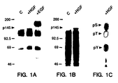
- Figures 1A-1C show the tyrosine phosphorylation of p145 in B5/589 human mammary epithelial cells in response to HGF.
- Fig. 1A is an immunoblot of phosphotyrosyl proteins from untreated control cells (C), cells treated with HGF, and with EGF (Collaborative Research). HGF was purified as described (J.S. Rubin et al., Proc. Natl. Acad. Sci . U.S.A. 88, 415 1990)).
- Serum-starved cells were exposed to growth factor (100 ng/ml) for 10 min at 37°C as indicated, detergent-solubilized on ice, and immunoprecipitated with monoclonal anti-pTyr (Upstate Biotechnology). Immunoprecipitated proteins were resolved by 7.5% SDS polyacrylamide gel electrophoresis (SDS-PAGE) (U.K. Laemmli Nature 227, 680 (1970)), and immunoblotted with the same antibody as described (D.P. Bottaro et al., J. Biol. Chem. 265, 12767 (1990)).
- Fig. 1B is an autoradiogram of 32 P-labeled phosphoproteins from control (C) and HGF-treated cells.
- Serum-starved cells were metabolically labeled with 32 P-orthophosphate (1.0 mCi/ml) as described (M.F. White and C.R. Kahn, in Insulin Receptors, Part A: Methods for the Study of Structure and Function, C.R. Kahn and L. Harrison, Eds. (Liss, New York, 1988) pp. 125-147).
- the cells were treated with HGF (100 ng/ml) for 10 min at 37°C as indicated, and detergent-solubilized on ice.
- Phosphotyrosyl proteins were immunoprecipitated with anti-pTyr and resolved by 7.5% SDS-PAGE. Fig.
- FIG. 1C shows a phosphoamino acid analysis of p145 from lane 2 of Fig. 1B which was performed as described (M.F. White and C.R. Kahn, in Insulin Receptors, Part A: Methods for the Study of Structure and Function , C.R. Kahn and L. Harrison, Eds. Liss, New York, 1988, pp. 125-147).
- the dotted circles indicate the migration of unlabeled phosphoserine (pS), phosphothreonine (pT), and phosphotyrosine (pY).
- Figures 2A-2B show the identification of p145 as the ⁇ -subunit of the c-met proto-oncogene product.
- Fig. 2A is an anti c-met immunoblot of anti-pTyr immunoprecipitates from control (C) and HGF-treated B5/589 cells. Samples for immunoprecipitation (2 mg protein) were prepared as described in Fig. 1A, resolved by 7.5% SDS-PAGE, transferred to Immobilon (Millipore) membranes and detected with monoclonal anti -c-met and [ 125 I]-protein-A.
- Fig. 2B is an autoradiogram of 32 P-labeled phosphoproteins from control (C) and HGF-treated B5/589 cells resolved by 7.5% SDS-PAGE under reduced (R) and non-reduced (NR) conditions. Serum-starved cells were metabolically labeled with 32 P-ortho-phosphate, left untreated (C) or treated with HGF, and immunoprecipitated with anti-pTyr as described in Fig. 1B. Samples were reduced with 100 mM ⁇ -mercaptoethanol before electrophoresis as indicated.
- Figures 3A-3C demonstrate the covalent affinity cross-linking of 125 I-labeled HGFp28 to the c-met protein-tyrosine kinase.
- Fig. 3A is an immunoblot of lysates (200 ⁇ g protein) prepared from M426 human lung fibroblasts and B5/589 cells using monoclonal antibody to the cytoplasmic domain of c-met protein.
- Fig. 3B shows crosslinking of 125 I-labeled HGFp28 to M426 and B5/589 cells resolved by 6.5% SDS-PAGE under non-reduced (NR) and reduced (R) conditions.
- HGFp28 was purified as described and radiolabeled with [ 125 I]-Na by the chloramine-T method (W.M. Hunter and F.C. Greenwood, Nature 194, 495 (1962)). Cells were incubated with HEPES binding buffer (D.P. Bottaro et al., J. Biol. Chem. 265, 12767 (1990) containing 125 I-labeled HGFp28 (5 x 10 5 cpm) for 45 min at 25°C, washed with cold HEPES-buffered saline (pH 7.4), and treated with disuccinimidyl suberate (D.P. Bottaro et al., J. Biol. Chem. 265, 12767 (1990).
- Fig. 3C shows the immunoprecipitation of [ 125 I]-HGFp28-cross-linked complexes from B5/589 cells with c-met peptide antiserum (A. Gonzatti-Haces et al., Proc. Natl. Acad. Sci . U.S.A. 85, 21 (1988)). Sample preparation and cross-linking prior to immunoprecipitation, performed as described in reference to Fig.
- the present invention relates to a complex comprising hepatocyte growth factor (HGF) and met proto-oncogene protein.
- HGF hepatocyte growth factor
- the present invention further relates to methods of utilizing the complex.
- One embodiment of the present invention relates to a complex formed by the interaction of HGF with its receptor, the met proto-oncogene protein.
- the complex is free of protein with which it is naturally associated.
- the binding of HGF to its receptor, the met-proto-oncogene protein regulates the intrinsic tyrosine kinase activity of the receptor.
- HGF c-met receptor tyrosine kinase
- the present invention also relates to detection and quantitation methods that may be used in diagnostics to identify HGF (ligand), met-proto-oncogene receptor or the ligand-receptor complex. Since the met-proto-oncogene receptor is expressed on many cell types and tissues including the liver, the methods described herein provide a means for identifying tissues other than liver affected by HGF binding. The methods of the present invention also aid in understanding the role of the interaction between receptor and ligand in regulating biochemical and physiological mechanisms in a broad spectrum of tissues.
- the present invention further relates to a method of detecting and quantitating HGF receptor in a biological sample using labeled HGF as a probe.
- Suitable labels include, for example, radiolabels such as 125 I, and flourescein.
- a biological sample can be extracted with a non-ionic detergent and incubated with labeled HGF in the presence or absence of unlabeled HGF.
- the resulting complex can be separated from the uncomplexed (or unbound) labeled material, for example, by immunoprecipitating the complex with a specific polyclonal or monoclonal antibody that recognizes the met-proto-oncogene receptor protein or the HGF-met proto oncogene receptor complex.
- the overall signal resulting from the presence of label associated with the resulting complex is compared with the signal from a mock sample.
- the mock sample is prepared using purified met-oncogene receptor protein in a known quantity treated the same way as the biological sample.
- the complex may be separated from uncomplexed material by precipitating with polyethylene glycol.
- the amount of label that is immunoprecipitated or precipitated is directly related to the amount of complex in the biological sample.
- the present invention also relates to a method for detecting and quantitating HGF in a biological sample using labeled HGF receptor as a probe.
- the method is carried out as a reciprocal binding assay following the methodology described above except substituting as antibody, one that specifically recognizes HGF or the HGF-met proto-oncogene receptor complex.
- the present invention also relates to further methods of detecting and quantitating HGF-met proto-oncogene receptor complexes in a sample.
- complexes are detected and quantitated using antibodies.
- Antibodies utilized in this embodiment can be directed against HGF, met-proto-oncogene receptor protein or the HGF-receptor complex. Antibodies can be either polyclonal or monoclonal.
- a sample can be extracted with non-ionic detergent and incubated with labeled HGF or met - proto-oncogene receptor protein. After incubation, the sample is covalently cross-linked with a bifunctional reagent such as a chemical cross-linker, for example, disuccinimidil suberate (DSS). After quenching the reaction with a quenching agent, the sample is immunoprecipitated with specific antibody or precipitated with polyethylene glycol. Quantitation requires chromatographic separation by, for example, gel electrophoresis, followed by autoradiography.
- HGF-met proto-oncogene receptor complexes in another method for detecting HGF-met proto-oncogene receptor complexes in a sample, the simultaneous expression of HGF and met protooncogene receptor mRNAs are determined. Simultaneous co-expression of HGF and met proto-oncogene receptor can be determined by Northern analysis using oligo- or cDNA probes derived from the sequence of either gene to identify mRNA or using the polymerase chain reaction (PCR) or any combination. Northern analysis and the PCR technology are methods well known to those skilled in the art.
- the present invention further relates to diagnostic methodologies using the methods described above.
- the disorders which diagnosed by the methods of the present invention include, for example, proliferative disorders such as hepatocellular carcinoma or other carcinomas of tissues that normally express met proto-oncogene receptor.
- Such tissues can be derived from epithelial cells such as skin, lung, stomach, kidney or colon, liver or endothelial cells, such as those contained in the vascular lining or bone marrow, or hematopoietic stem cells.
- the present diagnostic methods can also be used to measure wound repair in tissues derived from the cells described above, and in cells that normally express HGF such as platelets, fibroblasts (stromal tissue of skin and other organs) and spleen.
- Inactivation of the HGF/met mitogenic pathway provides the basis for-therapeutic methodologies designed to diminish or arrest normal or pathological cell proliferation.
- These methodologies include the production of genetically engineered HGF species that lack or possess an impaired met-binding domain, or that lack or possess an impaired activating domain, but that otherwise retain the structural and biochemical characteristics of HGF.
- These methodologies also include the production of a water-soluble form of met protein consisting of the extracellular HGF-binding domain that can act as an antagonist of normal met protein activation by HGF.
- the delivery of the genetically engineered HGF or met protein species described above to the selected site of action may be achieved using conventional methods of drug delivery, gene transfer, or any combination thereof.
- HGF/met mitogenic pathway provides the basis for therapeutic methodologies designed to restore, replace, or enhance naturally occurring wound repair mechanisms. These methodologies include application to the wound site of genetically engineered HGF or met species that enhance the binding interaction between met and HGF and thereby create an artificially sustained HGF/met interaction. For example, site-directed mutagenesis of the HGF-binding domain of met, or the met-binding domain of HGF (or both) may be used to create a member of the HGF/met pair with higher binding affinity for the other member of the pair and thus affect accelerated growth or regeneration of the wounded tissue.
- conventional recombinant DNA techniques could be used to enhance or sustain the kinase activity of the met protein normally regulated by HGF binding, including met mutations possessing a constitutively activated tyrosine kinase.
- the delivery of the genetically engineered HGF or met protein species described above to the selected site of action can be achieved using conventional methods of drug delivery, gene transfer, or any combination thereof.
- Activation of the HGF/met mitogenic pathway by means of supplementing the natural expression of met by recombinant DNA techniques in combination with exogenously administered HGF is also included.
- the human mammary epithelial cell line B5/589 is particularly sensitive to the mitogenic effects of HGF (J.S. Rubin et al., Proc. Natl. Acad . Sci. U.S.A . 88 , 415 (1990)).
- HGF approximately 100 ng/ml
- Phosphotyrosyl proteins were isolated from cell lysates by immunoprecipitation with antibody to phosphotyrosine (anti-pTyr). These proteins were resolved by SDS polyacrylamide gel electrophoresis (SDS-PAGE) and immunoblotted with the same antibody.
- FIG. 1A Several phosphotyrosyl proteins were detected in untreated cells by this method (Fig. 1A).
- HGF HGF induced phosphorylation of a 145-kD protein (p145)
- FIG. 1A center lane
- B5/589 cells exposed to epidermal growth factor (EFG) displayed tyrosine phosphorylation of the EGF receptor, but not p145 (Fig. 1a, right lane).
- EGF epidermal growth factor
- FIG. 1B When lysates from control and HGF-treated cells that had been labeled with 32 P-orthophosphate were used for immunoprecipitation with anti-pTyr, phosphorylation of p145 was specifically detected in HGF-treated cells (Fig. 1B).
- Phosphoamino acid analysis of 32 P-labeled p145 confirmed the presence of phosphotyrosine, and revealed the presence of phosphoserine as well (Fig. 1C).
- the HGF-stimulated phosphorylation of p145 on tyrosine and its apparent molecular weight were consistent with the possibility that p145 represented the receptor tyrosine kinase for HGF.
- Example 2 Identification of p145 as the ⁇ subunit of the c-met proto oncogene product.
- c-met proto oncogene product which is a receptor-like tyrosine kinase comprised of disulfide-linked subunits of 50-kD ( ⁇ ) and 145-kD ( ⁇ ) (P.R. Tempest et al. Br. J. Cancer 58 , 3 (1988); S. Giordano et al., Oncogene 4 , 1383 (1989)).
- the a subunit is extracellular, and the ⁇ subunit has extracellular, transmembrane, and tyrosine kinase domains as well as sites of tyrosine phosphorylation (S. Giordano et al., Oncogene 4 , 1383 (1989); A. A. Gonzatti-Haces et al., Proc. Natl. Acad. Sci. U.S.A . 85 , 21 (1988).
- proteins immunoprecipitated by anti-pTyr from control and HGF-treated B5/589 cells were immunoblotted with a monoclonal antibody directed against the cytoplasmic domain of the c-met product.
- a mouse monoclonal IgG raised against recombinant human c-met protein cytoplasmic domain was used. Recognition of human c-met protein by immunoprecipitation or immunoblotting can be specifically blocked by incubating in the presence of the recombinant protein fragment.
- Fig. 2A The prominent 145-kD protein observed specifically in HGF-treated cells (Fig. 2A) provided direct evidence that this mitogen induced phosphorylation of the c-met protein on tyrosine residues.
- Fig. 2A the percentage of c-met protein phosphorylated on tyrosine in response to HGF could be quantitated (Fig. 2A). It is estimated that at least 10% of the total cellular c-met protein content was immunoprecipitated by anti-pTyr after HGF stimulation.
- Example 3 125 I-HGFp28 is physically associated with the c-met protein-tyrosine kinase.
- HGFp28 was labeled with [ 125 I]Na by the chloramine-T method as follows: HGFp28 (3 ⁇ g in 50 ⁇ l of 20 mM phosphate buffer containing 1.0 M NaCl, pH 7.4) was reacted with chloramine-T (1.2 ⁇ g in 4 ⁇ l of phosphate buffer) and [ 125 I]Na (1 ⁇ Ci) at 24°C for 1 min. The reaction was terminated by addition of sodium metabisulfite (10 ⁇ g in 8 ⁇ l of phosphate buffer).
- the mixture was diluted with phosphate buffer containing 0.1% bovine serum albumin (200 ⁇ l) and applied to a column (300 ⁇ l packed volume) of heparin-Sepharose CL-6B that had been equilibrated in phosphate-buffered saline containing 0.1% BSA (PBS/BSA).
- the column was washed with 30 ml of PBS/BSA and eluted with PBS/BSA containing 1.0 M NaCl (200 ⁇ l/fraction), removing 98% of trichloroacetic acid-precipitable radioactivity from the column. Peak fractions (specific activity: 150 to 250 ⁇ Ci/ ⁇ g) were 99% trichloroacetic acid-precipitable, and migrated as a single band on SDS-PAGE.
Landscapes
- Health & Medical Sciences (AREA)
- Chemical & Material Sciences (AREA)
- Life Sciences & Earth Sciences (AREA)
- Organic Chemistry (AREA)
- Molecular Biology (AREA)
- Immunology (AREA)
- Proteomics, Peptides & Aminoacids (AREA)
- Engineering & Computer Science (AREA)
- General Health & Medical Sciences (AREA)
- Biochemistry (AREA)
- Medicinal Chemistry (AREA)
- Genetics & Genomics (AREA)
- Zoology (AREA)
- Biophysics (AREA)
- Analytical Chemistry (AREA)
- Gastroenterology & Hepatology (AREA)
- Biotechnology (AREA)
- Cell Biology (AREA)
- Hematology (AREA)
- Urology & Nephrology (AREA)
- Physics & Mathematics (AREA)
- Biomedical Technology (AREA)
- Pathology (AREA)
- Wood Science & Technology (AREA)
- Microbiology (AREA)
- Bioinformatics & Cheminformatics (AREA)
- Oncology (AREA)
- Food Science & Technology (AREA)
- General Engineering & Computer Science (AREA)
- Toxicology (AREA)
- General Physics & Mathematics (AREA)
- Animal Behavior & Ethology (AREA)
- Veterinary Medicine (AREA)
- Public Health (AREA)
- Pharmacology & Pharmacy (AREA)
- Hospice & Palliative Care (AREA)
- Epidemiology (AREA)
- Nuclear Medicine, Radiotherapy & Molecular Imaging (AREA)
- General Chemical & Material Sciences (AREA)
- Chemical Kinetics & Catalysis (AREA)
Abstract
Description
- The present invention relates to a complex comprising hepatocyte growth factor (HGF) and met proto-oncogene protein. The present invention also relates to methods for detecting the presence of HGF ligand, met proto-oncogene receptor and methods for isolating either the ligand, receptor or complex comprising both.
- The present invention further relates to methods of diagnosing and treating conditions proliferative disorders such as hepatitis, hepatocarcinogenesis, carcinogenesis and wound healing. In particular, the present methods involve detection of the ligand-receptor pairs.
- Hepatocyte growth factor (HGF) was first purified from human and rabbit plasma and rat platelets on the basis of its ability to stimulate mitogenesis of rat hepatocytes (E. Gohoda et al., J. Clin. Invest. 81, 414 (1988); R. Zarnegar and G. Michalopoulos, CancerRes. 49, 3314 (1989); T. Nakamura et al. FEBS Lett. 224, 311 (1987)). Thus, HGF may act as a humoral factor promoting liver regeneration after partial hepatectomy or liver injury (G.K. Michalopoulos, FASEB J. 4, 176 (1990)). The same factor was purified from human fibroblast culture medium and shown to act on melanocytes and a variety of epithelial and endothelial cells (J.S. Rubin et al., Proc. Natl. Acad. Sci. U.S.A. 88, 415 (1990)). Together with evidence of HGF expression in several organs (J.S. Rubin et al., Proc. Natl. Acad. Sci. U.S.A. 88, 415 (1990); K. Tashiro et al. Proc. Natl. Acad. Sci. U.S.A. 87, 3200 (1990); R. Zarnegar et al., Proc. Natl. Acad. Sci. U.S.A. 87, 1252 (1990); T. Kinoshita et al. Biochem. Biophys. Res. Comm. 165, 1229 (1989)), these findings indicate that HGF may also act as a paracrine mediator of proliferation for a broad spectrum of cell types. Molecular cloning of HGF revealed a remarkable structural homology to plasminogen and related serine proteases (J.S. Rubin et al., Proc. Natl. Acad. Sci. U.S.A.88, 415 (1990); T. Nakamura et al., Nature 342, 440 (1989); K. Miyazawa et al., Biophys. Res. Comm. 163, 967 (1989)). Recent evidence that HGF induces rapid tyrosine phosphorylation of proteins in intact target cells suggests that a tyrosine kinase receptor might mediate its mitogenic signal (J.S. Rubin et al., Proc. Natl. Acad. Sci. U.S.A. 88, 415 (1990)).
- HGF is structurally related to the family of serine proteases that includes plasminogen, prothrombin, urokinase, and tissue plasminogen activator (J.S. Rubin et al., Proc. Natl. Acad. Sci. U.S.A. 88, 415 (1990)); T. Nakamura et al., Nature 342, 440 (1989)). As defined in the present invention, HGF includes a variant of HGF previously characterized as a broad-spectrum mitogen called plasminogen like growth factor (PLGF). Several proteases, including members of the serine protease family, stimulate DNA synthesis presumably through a proteolytic mechanism similar to tryptic activation of the insulin receptor (S.E. Shoelson et al. J. Biol. Chem. 263, 4852 (1988)). Only urokinase has been found to associate with a specific cell-surface receptor, which itself bears no homology to any known tyrosine kinase receptors (A.L. Roldan et al., EMBO J. 9 , 467 (1990)).
- It is clear that a need exists to identify the receptor of HGF. The present invention describes the complex comprising HGF and met proto-oncogene protein and identifies the met proto-oncogene as the receptor for HGF. The met protooncogene protein is a member of the tyrosine kinase growth factor receptor family. Knowledge of this receptor/ligand relationship should facilitate the study of proliferative disorders in which expression of these molecules may play an important role. Additionally, identification of the met proto-oncogene receptor HGF complex provides a means for identifying tissues other than liver tissue affected by factor binding.
- It is an object of the present invention to provide a complex comprising a hepatocyte growth factor (HGF) ligand and met proto-oncogene protein receptor and methods of utilizing the complex.
- Various other objects and advantages of the present invention will become apparent from the drawings and the following description of the invention.
- In one embodiment, the present invention relates to a complex of a HGF ligand and met proto-oncogene receptor protein wherein said complex is free of protein with which it is naturally associated.
- In another embodiment, the present invention relates to a complex comprising a HGF ligand and met proto-oncogene receptor protein wherein one member of said complex is bound to a solid support.
- In yet another embodiment, the present invention relates to a method of detecting a HGF:met proto-oncogene receptor protein complex in a sample comprising reacting said sample with an antibody that binds specifically with either HGF or met proto-oncogene receptor protein on the complex. A positive immunological reaction is indicative of the presence of the complex in the sample.
- In a further embodiment, the present invention relates to a method of diagnosing a proliferative disorder in a patient suspected of having the disorder comprising reacting a biological sample from the patient with an antibody that binds with a HGF-met proto-oncogene receptor protein complex.
- In yet another embodiment, the present invention relates to a method of diagnosing a tissue undergoing regeneration in a patient comprising, reacting a biological sample from the patient with an antibody that binds to a HGF-met proto-oncogene receptor protein complex.
- A further embodiment of the present invention relates to a method of diagnosing a diseased state in a patient suspected of having the stated disease comprising reacting a biological sample from the patient with an antibody that binds with a HGF- met proto-oncogene receptor protein complex.
- In another embodiment, the present invention relates to a method for detecting HGF in a sample comprising contacting the sample with met proto-oncogene receptor protein under conditions such that binding of HGF present in the sample to the receptor is effected and detecting the presence of bound HGF.
- In a further embodiment, the present invention relates to a method for detecting met proto-oncogene receptor protein in a sample comprising the steps of contacting the sample with HGF under conditions such that binding of said receptor present in the sample to HGF is effected and detecting the presence of bound receptor.
- Another embodiment of the present invention relates to a diagnostic kits for measuring would healing and proliferative disorders. One type of kit comprises labeled HGF in one container and ancillary reagents suitable for use in detecting the presence or absence of met proto-oncogene receptor in a biological sample.
- A second type of kit comprises labeled met proto-oncogene receptor protein in one container and ancillary reagents suitable for use in detecting the presence or absence of HGF in a biological sample.
- The entire contents of all publications mentioned herein are incorporated by reference.
- Figures 1A-1C show the tyrosine phosphorylation of p145 in B5/589 human mammary epithelial cells in response to HGF. Fig. 1A is an immunoblot of phosphotyrosyl proteins from untreated control cells (C), cells treated with HGF, and with EGF (Collaborative Research). HGF was purified as described (J.S. Rubin et al., Proc. Natl. Acad. Sci. U.S.A. 88, 415 1990)). Serum-starved cells were exposed to growth factor (100 ng/ml) for 10 min at 37°C as indicated, detergent-solubilized on ice, and immunoprecipitated with monoclonal anti-pTyr (Upstate Biotechnology). Immunoprecipitated proteins were resolved by 7.5% SDS polyacrylamide gel electrophoresis (SDS-PAGE) (U.K. Laemmli Nature 227, 680 (1970)), and immunoblotted with the same antibody as described (D.P. Bottaro et al., J. Biol. Chem. 265, 12767 (1990)). Fig. 1B is an autoradiogram of 32P-labeled phosphoproteins from control (C) and HGF-treated cells. Serum-starved cells were metabolically labeled with 32P-orthophosphate (1.0 mCi/ml) as described (M.F. White and C.R. Kahn, in Insulin Receptors, Part A: Methods for the Study of Structure and Function, C.R. Kahn and L. Harrison, Eds. (Liss, New York, 1988) pp. 125-147). The cells were treated with HGF (100 ng/ml) for 10 min at 37°C as indicated, and detergent-solubilized on ice. Phosphotyrosyl proteins were immunoprecipitated with anti-pTyr and resolved by 7.5% SDS-PAGE. Fig. 1C shows a phosphoamino acid analysis of p145 from lane 2 of Fig. 1B which was performed as described (M.F. White and C.R. Kahn, in Insulin Receptors, Part A: Methods for the Study of Structure and Function, C.R. Kahn and L. Harrison, Eds. Liss, New York, 1988, pp. 125-147).
- The dotted circles indicate the migration of unlabeled phosphoserine (pS), phosphothreonine (pT), and phosphotyrosine (pY).
- Figures 2A-2B show the identification of p145 as the β-subunit of the c-met proto-oncogene product. Fig. 2A is an anti c-met immunoblot of anti-pTyr immunoprecipitates from control (C) and HGF-treated B5/589 cells. Samples for immunoprecipitation (2 mg protein) were prepared as described in Fig. 1A, resolved by 7.5% SDS-PAGE, transferred to Immobilon (Millipore) membranes and detected with monoclonal anti-c-met and [125I]-protein-A. To quantify the percentage of c-met protein that was immuno-precipitable with anti-pTyr, 200 µg of B5/589 cell lysate (LYSATE) was resolved by SDS-PAGE and immunoblotted directly with monoclonal antibody to c-met. Fig. 2B is an autoradiogram of 32P-labeled phosphoproteins from control (C) and HGF-treated B5/589 cells resolved by 7.5% SDS-PAGE under reduced (R) and non-reduced (NR) conditions. Serum-starved cells were metabolically labeled with 32P-ortho-phosphate, left untreated (C) or treated with HGF, and immunoprecipitated with anti-pTyr as described in Fig. 1B. Samples were reduced with 100 mM β-mercaptoethanol before electrophoresis as indicated.
- Figures 3A-3C demonstrate the covalent affinity cross-linking of 125I-labeled HGFp28 to the c-met protein-tyrosine kinase. Fig. 3A is an immunoblot of lysates (200 µg protein) prepared from M426 human lung fibroblasts and B5/589 cells using monoclonal antibody to the cytoplasmic domain of c-met protein. Fig. 3B shows crosslinking of 125I-labeled HGFp28 to M426 and B5/589 cells resolved by 6.5% SDS-PAGE under non-reduced (NR) and reduced (R) conditions. HGFp28 was purified as described and radiolabeled with [125I]-Na by the chloramine-T method (W.M. Hunter and F.C. Greenwood, Nature 194, 495 (1962)). Cells were incubated with HEPES binding buffer (D.P. Bottaro et al., J. Biol. Chem. 265, 12767 (1990) containing 125I-labeled HGFp28 (5 x 10⁵ cpm) for 45 min at 25°C, washed with cold HEPES-buffered saline (pH 7.4), and treated with disuccinimidyl suberate (D.P. Bottaro et al., J. Biol. Chem. 265, 12767 (1990). The cells were then solubilized with SDS and boiled for 3 min in the presence 100 mM β-mercaptoethanol as indicated. 125I-labeled proteins were resolved by 6.5% SDS-PAGE and autoradiography at -70°C. Fig. 3C shows the immunoprecipitation of [125I]-HGFp28-cross-linked complexes from B5/589 cells with c-met peptide antiserum (A. Gonzatti-Haces et al., Proc. Natl. Acad. Sci. U.S.A. 85, 21 (1988)). Sample preparation and cross-linking prior to immunoprecipitation, performed as described in reference to Fig. 3B, yielded the electrophoretic pattern shown in the left lane (LYSATE) under reduced conditions. The adjacent lanes show immunoprecipitation of the cross-linked species with c-met peptide antiserum (1:100) in the absence (α-MET) or presence (+COMP) of competing peptide (10 µG/ml). Immunoprecipitated proteins were absorbed to immobilized protein-G (Genex) and eluted with SDS prior to electrophoresis and autoradiography as described in reference to Fig. 3B.
- The present invention relates to a complex comprising hepatocyte growth factor (HGF) and met proto-oncogene protein. The present invention further relates to methods of utilizing the complex.
- One embodiment of the present invention relates to a complex formed by the interaction of HGF with its receptor, the met proto-oncogene protein. The complex is free of protein with which it is naturally associated. The binding of HGF to its receptor, the met-proto-oncogene protein, regulates the intrinsic tyrosine kinase activity of the receptor.
- The direct interaction of HGF with the c-met receptor tyrosine kinase suggests a biochemical mechanism of mitogenic signal transduction similar to that of insulin, EGF and other peptide growth factors. This interaction represents a significant functional divergence from HGF's structurally related family of serine protease homologs.
- The present invention also relates to detection and quantitation methods that may be used in diagnostics to identify HGF (ligand), met-proto-oncogene receptor or the ligand-receptor complex. Since the met-proto-oncogene receptor is expressed on many cell types and tissues including the liver, the methods described herein provide a means for identifying tissues other than liver affected by HGF binding. The methods of the present invention also aid in understanding the role of the interaction between receptor and ligand in regulating biochemical and physiological mechanisms in a broad spectrum of tissues.
- The present invention further relates to a method of detecting and quantitating HGF receptor in a biological sample using labeled HGF as a probe. Suitable labels include, for example, radiolabels such as 125I, and flourescein.
- Using standard methodologies well known in the art, a biological sample can be extracted with a non-ionic detergent and incubated with labeled HGF in the presence or absence of unlabeled HGF. The resulting complex can be separated from the uncomplexed (or unbound) labeled material, for example, by immunoprecipitating the complex with a specific polyclonal or monoclonal antibody that recognizes the met-proto-oncogene receptor protein or the HGF-met proto oncogene receptor complex. The overall signal resulting from the presence of label associated with the resulting complex is compared with the signal from a mock sample. The mock sample is prepared using purified met-oncogene receptor protein in a known quantity treated the same way as the biological sample.
- Alternatively, the complex may be separated from uncomplexed material by precipitating with polyethylene glycol. In both methodologies, the amount of label that is immunoprecipitated or precipitated is directly related to the amount of complex in the biological sample.
- The present invention also relates to a method for detecting and quantitating HGF in a biological sample using labeled HGF receptor as a probe. The method is carried out as a reciprocal binding assay following the methodology described above except substituting as antibody, one that specifically recognizes HGF or the HGF-met proto-oncogene receptor complex.
- The present invention also relates to further methods of detecting and quantitating HGF-met proto-oncogene receptor complexes in a sample. In one aspect, complexes are detected and quantitated using antibodies. Antibodies utilized in this embodiment can be directed against HGF, met-proto-oncogene receptor protein or the HGF-receptor complex. Antibodies can be either polyclonal or monoclonal. A sample can be extracted with non-ionic detergent and incubated with labeled HGF or met - proto-oncogene receptor protein. After incubation, the sample is covalently cross-linked with a bifunctional reagent such as a chemical cross-linker, for example, disuccinimidil suberate (DSS). After quenching the reaction with a quenching agent, the sample is immunoprecipitated with specific antibody or precipitated with polyethylene glycol. Quantitation requires chromatographic separation by, for example, gel electrophoresis, followed by autoradiography.
- In another method for detecting HGF-met proto-oncogene receptor complexes in a sample, the simultaneous expression of HGF and met protooncogene receptor mRNAs are determined. Simultaneous co-expression of HGF and met proto-oncogene receptor can be determined by Northern analysis using oligo- or cDNA probes derived from the sequence of either gene to identify mRNA or using the polymerase chain reaction (PCR) or any combination. Northern analysis and the PCR technology are methods well known to those skilled in the art.
- The present invention further relates to diagnostic methodologies using the methods described above. The disorders which diagnosed by the methods of the present invention include, for example, proliferative disorders such as hepatocellular carcinoma or other carcinomas of tissues that normally express met proto-oncogene receptor. Such tissues can be derived from epithelial cells such as skin, lung, stomach, kidney or colon, liver or endothelial cells, such as those contained in the vascular lining or bone marrow, or hematopoietic stem cells. The present diagnostic methods can also be used to measure wound repair in tissues derived from the cells described above, and in cells that normally express HGF such as platelets, fibroblasts (stromal tissue of skin and other organs) and spleen.
- Inactivation of the HGF/met mitogenic pathway provides the basis for-therapeutic methodologies designed to diminish or arrest normal or pathological cell proliferation. These methodologies include the production of genetically engineered HGF species that lack or possess an impaired met-binding domain, or that lack or possess an impaired activating domain, but that otherwise retain the structural and biochemical characteristics of HGF. Similarly, production of genetically engineered met species that lack or possess an impaired HGF-binding domain, or lack or possess an impaired tyrosine kinase domain, but which otherwise retain the structural and biochemical characteristics of the met protein. These methodologies also include the production of a water-soluble form of met protein consisting of the extracellular HGF-binding domain that can act as an antagonist of normal met protein activation by HGF. The delivery of the genetically engineered HGF or met protein species described above to the selected site of action may be achieved using conventional methods of drug delivery, gene transfer, or any combination thereof.
- Artificial activation of the HGF/met mitogenic pathway provides the basis for therapeutic methodologies designed to restore, replace, or enhance naturally occurring wound repair mechanisms. These methodologies include application to the wound site of genetically engineered HGF or met species that enhance the binding interaction between met and HGF and thereby create an artificially sustained HGF/met interaction. For example, site-directed mutagenesis of the HGF-binding domain of met, or the met-binding domain of HGF (or both) may be used to create a member of the HGF/met pair with higher binding affinity for the other member of the pair and thus affect accelerated growth or regeneration of the wounded tissue. Similarly, conventional recombinant DNA techniques could be used to enhance or sustain the kinase activity of the met protein normally regulated by HGF binding, including met mutations possessing a constitutively activated tyrosine kinase. The delivery of the genetically engineered HGF or met protein species described above to the selected site of action can be achieved using conventional methods of drug delivery, gene transfer, or any combination thereof. Activation of the HGF/met mitogenic pathway by means of supplementing the natural expression of met by recombinant DNA techniques in combination with exogenously administered HGF is also included.
- The human mammary epithelial cell line B5/589 is particularly sensitive to the mitogenic effects of HGF (J.S. Rubin et al., Proc. Natl. Acad. Sci. U.S.A. 88, 415 (1990)). Intact serum-starved B5/589 cells were treated with HGF (approximately 100 ng/ml) for 10 min at 37°C and solubilized on ice. Phosphotyrosyl proteins were isolated from cell lysates by immunoprecipitation with antibody to phosphotyrosine (anti-pTyr). These proteins were resolved by SDS polyacrylamide gel electrophoresis (SDS-PAGE) and immunoblotted with the same antibody. Several phosphotyrosyl proteins were detected in untreated cells by this method (Fig. 1A). Treatment of intact cells with HGF induced phosphorylation of a 145-kD protein (p145) (Fig. 1A, center lane). B5/589 cells exposed to epidermal growth factor (EFG) displayed tyrosine phosphorylation of the EGF receptor, but not p145 (Fig. 1a, right lane). When lysates from control and HGF-treated cells that had been labeled with 32P-orthophosphate were used for immunoprecipitation with anti-pTyr, phosphorylation of p145 was specifically detected in HGF-treated cells (Fig. 1B). Phosphoamino acid analysis of 32P-labeled p145 confirmed the presence of phosphotyrosine, and revealed the presence of phosphoserine as well (Fig. 1C). The HGF-stimulated phosphorylation of p145 on tyrosine and its apparent molecular weight were consistent with the possibility that p145 represented the receptor tyrosine kinase for HGF.
- A number of receptor-like molecules have been described for which there are as yet no known ligands. One of these is the c-met proto oncogene product, which is a receptor-like tyrosine kinase comprised of disulfide-linked subunits of 50-kD (α) and 145-kD (β) (P.R. Tempest et al. Br. J. Cancer 58, 3 (1988); S. Giordano et al., Oncogene 4, 1383 (1989)). In the fully processed c-met product, the a subunit is extracellular, and the β subunit has extracellular, transmembrane, and tyrosine kinase domains as well as sites of tyrosine phosphorylation (S. Giordano et al., Oncogene 4, 1383 (1989); A. A. Gonzatti-Haces et al., Proc. Natl. Acad. Sci. U.S.A. 85, 21 (1988).
- To test the hypothesis that p145 might represent the c-met protein β subunit, proteins immunoprecipitated by anti-pTyr from control and HGF-treated B5/589 cells were immunoblotted with a monoclonal antibody directed against the cytoplasmic domain of the c-met product. Specifically, a mouse monoclonal IgG raised against recombinant human c-met protein cytoplasmic domain was used. Recognition of human c-met protein by immunoprecipitation or immunoblotting can be specifically blocked by incubating in the presence of the recombinant protein fragment.
- The prominent 145-kD protein observed specifically in HGF-treated cells (Fig. 2A) provided direct evidence that this mitogen induced phosphorylation of the c-met protein on tyrosine residues. When whole lysates prepared from identically treated cells were blotted directly with the c-met antibody, the percentage of c-met protein phosphorylated on tyrosine in response to HGF could be quantitated (Fig. 2A). It is estimated that at least 10% of the total cellular c-met protein content was immunoprecipitated by anti-pTyr after HGF stimulation. Analysis of the time course of HGF action revealed that the c-met protein could be recovered by immunoprecipitation with anti-pTyr within 1 min of treatment and that this effect persisted for at least 3 hours. Comparison of the electrophoretic mobility of p145 under reduced and non-reduced conditions confirmed that it was the B subunit of the c-met protein (Fig. 2C). Without reduction, the 50-kD α subunit of the c-met protein remains disulfide-linked to the β subunit and substantially retards its migration in SDS-PAGE (P.R. Tempest et al., Br. J. Cancer 58, 3 (1988); S. Giordano et al., Oncogene 4, 1383 (1989); P.R. Tempest et al., FEBS Lett. 209, 357 (1986); M. Park et al., Proc. Natl. Acad. Sci. U.S.A. 84, 6379 (1987); A. Gonzatti-Haces et al., Proc. Natl. Acad. Sci. U.S.A. 85, 21 (1988)). Similarly, p145 immunoprecipitated from 32P-labeled B5/589 cells that had been treated with HGF displayed a shift in mobility characteristic of the c-met proto oncogene product when subjected to reduced or non-reduced electrophoretic conditions (Fig. 2B). Together these results identified p145 as the c-met protein β subunit and established that HGF stimulated its phosphorylation on tyrosine residues.
- The rapidity and extent of c-met protein tyrosine phosphorylation in response to HGF supported the possibility that c-met protein was the cell-surface receptor for HGF. However, there is evidence that receptor kinases can phosphorylate other receptors (D.F. Stern and M.P. Kamps, EMBO J. 7, 995 (1988); C.R. King et al., EMBO J. 7, 1647 (1988)). Thus, conclusive identification of the C-met product as the HGF receptor required a demonstration of their direct interaction. 125I-labeled HGF was unsuitable for covalent affinity cross-linking because it consisted of a mixture of single chain and heterodimeric labeled species. A smaller form of HGF with similar binding properties, designated HGFp28, was 125I-labeled as a single entity and used to characterize the HGF receptor.
- HGFp28 was labeled with [125I]Na by the chloramine-T method as follows: HGFp28 (3 µg in 50 µl of 20 mM phosphate buffer containing 1.0 M NaCl, pH 7.4) was reacted with chloramine-T (1.2 µg in 4 µl of phosphate buffer) and [125I]Na (1 µCi) at 24°C for 1 min. The reaction was terminated by addition of sodium metabisulfite (10 µg in 8 µl of phosphate buffer). The mixture was diluted with phosphate buffer containing 0.1% bovine serum albumin (200 µl) and applied to a column (300 µl packed volume) of heparin-Sepharose CL-6B that had been equilibrated in phosphate-buffered saline containing 0.1% BSA (PBS/BSA). The column was washed with 30 ml of PBS/BSA and eluted with PBS/BSA containing 1.0 M NaCl (200 µl/fraction), removing 98% of trichloroacetic acid-precipitable radioactivity from the column. Peak fractions (specific activity: 150 to 250 µCi/µg) were 99% trichloroacetic acid-precipitable, and migrated as a single band on SDS-PAGE.
- Comparative cross-linking studies were performed using 125I-labeled HGF p28 on B5/589 cells and M426 human fibroblasts, an HGF-insensitive cell line which also lacks detectable amounts of c-met protein (Fig. 3A). The 125I-labeled HGFp28 crosslinked to its receptor on B5/589 cells migrated as a 210-kD protein complex under non-reduced conditions (Fig. 3B). Under reduced conditions, a major 170-kD complex was observed (Fig. 3B). These apparent molecular sizes were consistent with a direct interaction between the labeled HGFp28 and the 145-kD β subunit of the c-met protein. Under reduced conditions, two minor bands of 190-kD and about 300-kD were also detected (Fig.3B). Cross-linking of 125I-labeled HGFp28 to the species observed under reduced conditions was blocked by addition of either unlabeled HGFp28 or HGF-neutralizing antisera. Under identical conditions, 125I-labeled HGFp28 failed to cross-link to any large proteins in M426 cells (Fig. 3B).
- To establish that "'I-labeled HGFp28 was physically associated with the c-met protein, "'I-labeled HGFp28 cross-linked complexes were immunoprecipitated with a polyclonal antiserum (A. Gonzatti-Haces et al., Proc. Natl. Acad. Sci. U.S.A. 85, 21 (1988) specific to the carboxyl-terminal 28 amino acids of the β subunit of the c-met protein. The covalently cross-linked major 170-kD and minor 300-kD species detected under reduced conditions were immunoprecipitated by the antibody, and their detection was specifically blocked by competing peptide (Fig. 3C). These results demonstrate a direct molecular interaction between 125I-labeled HGFp28 and the c-met β subunit. The composition of the minor 300-kD cross-linked species remains to be determined. All of these findings establish that the c-met product is the cell surface receptor for HGF.
Claims (18)
- A complex comprising a HGF ligand and met proto-oncogene receptor protein wherein said complex is free of protein with which it is naturally associated.
- A complex comprising a HGF ligand and met proto-oncogene receptor protein wherein one member of said complex is bound to a solid support.
- A method of detecting a HGF:met proto-oncogene receptor protein complex in a sample comprising reacting a sample in which the presence of said complex is to be detected with an antibody that binds specifically to said complex, a positive immunological reaction being indicative of the presence of said complex in said sample.
- The method of detecting HGF:met -proto-oncogene receptor protein complex according to claim 3 wherein said sample is a biological sample.
- Use of antibody that binds with a HGF-met proto-oncogene receptor protein complex for diagnosing a proliferative disorder in a patient suspected of having said disorder wherein a biological sample from said patient is reacted with said antibody that binds with a HGF-met proto-oncogene receptor protein complex.
- Use of an antibody that binds with a HGF-met proto-oncogene receptor protein complex for diagnosing a tissue undergoing regeneration in a patient wherein a biological sample from said patient is reacted with said antibody that binds with a HGF-met proto-oncogene receptor protein complex.
- Use of an antibody that binds with a HGF-met proto-oncogene receptor complex for diagnosing a diseased state in a patient suspected of having said disease wherein a biological sample from said patient is reacted with an antibody that binds with a HGF-met proto-oncogene receptor protein complex.
- Use according to claim 5, wherein said biological sample is a solid tissue sample.
- Use according to claim 5, wherein said biological sample is serum sample.
- A method for detecting HGF in sample comprising the steps of:(i) contacting said sample with met proto-oncogene receptor protein under conditions such that binding of HGF present in said sample to said receptor is effected and(ii) detecting the presence of bound HGF.
- A method for detecting met proto-oncogene receptor protein in a sample comprising the steps of:(i) contacting said sample with HGF under conditions such that binding of said receptor present in said sample to HGF is effected and(ii) detecting the presence of bound receptor.
- A diagnostic kit for measuring wound healing comprising labeled HGF in one container and ancillary reagents suitable for use in detecting the presence or absence of met proto-oncogene receptor in a biological sample.
- A diagnostic kit for measuring wound healing comprising labeled met proto-oncogene receptor protein in one container and ancillary reagents suitable for use in detecting the presence or absence of HGF in a biological sample.
- An in vitro method of activating the HGF/met pathway in viable cells comprising delivering to the site of activation Hepatocyte Growth Factor or Hepatocyte Growth Factor agonist having an equal or greater binding efficiency for met than natural Hepatocyte Growth Factor.
- An in vitro method of inactivating the HGF/met pathway in viable cells comprising delivering to the site of activation or an HGF antagonist, which competes with natural HGF for met binding but which is unable to induce a biological response.
- An in vitro method of inactivating the HGF/met mitogenic pathway comprising delivering to the site of inactivation a met substitute antagonist which competes with natural met of HGF binding but which is unable to induce a biological response.
- An in vitro method of developing drugs for the regulation of the HGF/met pathway comprising (a) producing an HGF substitute which is either an HGF agonist that supplements, or antagonist that inhibits natural HGF/met interactions ; (b) contacting met with said HGF substitute ; and (c) determining whether said HGF substitute is an HGF agonist or HGF antagonist,
wherein said production of said HGF substitute is guided by molecular models of natural HGF/met interactions and/or by screening techniques which exploit analysis of the HGF/met interactions. - An in vitro method of developing drugs for the regulation of the HGF/met pathway comprising (a) producing a met substitute which is either an agonist that supplements or an antagonist that inhibits natural HGF/met interactions ; (b) contacting HGF with said met substitute ; and (c) determining whether said met substitute is a met agonist or met antagonist ;
wherein said production of said met substitute is guided by molecular models of natural HGF/met interactions and/or by screening techniques which exploit analysis of the HGF/met interactions.
Applications Claiming Priority (3)
| Application Number | Priority Date | Filing Date | Title |
|---|---|---|---|
| US642971 | 1991-01-18 | ||
| US07/642,971 US5648273A (en) | 1989-12-27 | 1991-01-18 | Hepatic growth factor receptor is the MET proto-oncogene |
| PCT/US1992/000071 WO1992013097A1 (en) | 1991-01-18 | 1992-01-15 | Hepatic growth factor receptor is the met proto-oncogene |
Publications (3)
| Publication Number | Publication Date |
|---|---|
| EP0567585A1 EP0567585A1 (en) | 1993-11-03 |
| EP0567585A4 EP0567585A4 (en) | 1994-08-03 |
| EP0567585B1 true EP0567585B1 (en) | 1999-08-04 |
Family
ID=24578815
Family Applications (1)
| Application Number | Title | Priority Date | Filing Date |
|---|---|---|---|
| EP92905178A Expired - Lifetime EP0567585B1 (en) | 1991-01-18 | 1992-01-15 | Hepatic growth factor receptor is the met proto-oncogene |
Country Status (11)
| Country | Link |
|---|---|
| US (1) | US5648273A (en) |
| EP (1) | EP0567585B1 (en) |
| JP (2) | JP2598362B2 (en) |
| AT (1) | ATE182902T1 (en) |
| AU (1) | AU652031B2 (en) |
| CA (2) | CA2176620C (en) |
| DE (1) | DE69229720T2 (en) |
| DK (1) | DK0567585T3 (en) |
| ES (1) | ES2136082T3 (en) |
| GR (1) | GR3031381T3 (en) |
| WO (1) | WO1992013097A1 (en) |
Cited By (5)
| Publication number | Priority date | Publication date | Assignee | Title |
|---|---|---|---|---|
| US6207152B1 (en) | 1995-06-02 | 2001-03-27 | Genentech, Inc. | Hepatocyte growth factor receptor antagonists and uses thereof |
| US7737115B2 (en) | 2005-04-15 | 2010-06-15 | Genetech, Inc. | HGF beta chain variants |
| US8637027B2 (en) | 2010-11-03 | 2014-01-28 | Argen-X B.V. | Anti c-Met antibodies |
| US9487589B2 (en) | 2011-06-30 | 2016-11-08 | Genentech, Inc. | Anti-c-met-antibody formulations |
| US9926364B2 (en) | 2011-11-03 | 2018-03-27 | Argen-X N.V. | Chimeric human-llama antigens and methods of use |
Families Citing this family (33)
| Publication number | Priority date | Publication date | Assignee | Title |
|---|---|---|---|---|
| US5871959A (en) * | 1989-12-27 | 1999-02-16 | The United States Of America As Represented By The Department Of Health And Human Services | Method of producing hepatocycte growth factor/scatter factor and related cell lines |
| US5362716A (en) * | 1989-12-27 | 1994-11-08 | The United States Of America As Represented By The Department Of Health And Human Services | Methods for stimulating hematopoietic progenitors using hepatocyte growth factor and lymphokines |
| ATE155525T1 (en) * | 1991-05-10 | 1997-08-15 | Pharmacia & Upjohn Spa | SHORTENED FORMS OF THE HEPATOCYTE GROWTH FACTOR (HGF) RECEPTOR |
| CA2144856A1 (en) * | 1992-09-18 | 1994-03-31 | Donna L. Faletto | A method of preventing tumor metastasis |
| US5646036A (en) * | 1995-06-02 | 1997-07-08 | Genentech, Inc. | Nucleic acids encoding hepatocyte growth factor receptor antagonist antibodies |
| US6214344B1 (en) | 1995-06-02 | 2001-04-10 | Genetech, Inc. | Hepatocyte growth factor receptor antagonists and uses thereof |
| WO1997009997A1 (en) * | 1995-09-12 | 1997-03-20 | Genentech, Inc. | Cystic fibrosis therapy |
| US5866329A (en) * | 1995-09-27 | 1999-02-02 | Demetriou; Achilles A. | Method and probe for detection of gene associated with liver neoplastic disease |
| WO1997012629A1 (en) * | 1995-10-05 | 1997-04-10 | Genentech, Inc. | Improved angiogenesis using hepatocyte growth factor |
| WO1997012628A1 (en) * | 1995-10-05 | 1997-04-10 | Genentech, Inc. | Methods and compositions for treating vascular stenosis |
| JP2000515735A (en) * | 1996-07-03 | 2000-11-28 | ジェネンテック インコーポレーテッド | Hepatocyte growth factor receptor agonist |
| JP4006058B2 (en) * | 1997-03-11 | 2007-11-14 | 第一三共株式会社 | Agent for preventing and / or treating multiple organ failure |
| ES2274567T3 (en) | 1997-03-14 | 2007-05-16 | Daiichi Pharmaceutical Co., Ltd. | USE OF TCF-II FOR THE TREATMENT OF LOSS OF BODY WEIGHT, ANEMIA AND THE ELEVATION OF TNF CAUSED BY CANCER. |
| ATE375168T1 (en) | 2000-08-28 | 2007-10-15 | Damavand Wound Ab | THE SYNERGISTIC EFFECTS OF HGF AND ANTIBACTERIAL TREATMENT |
| US20030118585A1 (en) * | 2001-10-17 | 2003-06-26 | Agy Therapeutics | Use of protein biomolecular targets in the treatment and visualization of brain tumors |
| TWI353991B (en) | 2003-05-06 | 2011-12-11 | Syntonix Pharmaceuticals Inc | Immunoglobulin chimeric monomer-dimer hybrids |
| HN2004000285A (en) | 2003-08-04 | 2006-04-27 | Pfizer Prod Inc | ANTIBODIES DIRECTED TO c-MET |
| DE602004019134D1 (en) * | 2003-09-29 | 2009-03-05 | Peas Inst Ab | FAST DETERMINATION OF VARIOUS FORMS OF HGF (HEPTOCYTE GROWTH FACTOR) IN BODY FLUIDS |
| EP2385069A3 (en) | 2003-11-12 | 2012-05-30 | Biogen Idec MA Inc. | Neonatal Fc rReceptor (FcRn)- binding polypeptide variants, dimeric Fc binding proteins and methods related thereto |
| PT1773885E (en) | 2004-08-05 | 2010-07-21 | Genentech Inc | Humanized anti-cmet antagonists |
| AU2006203889A1 (en) | 2005-01-05 | 2006-07-13 | Biogen Idec Ma Inc. | CRIPTO binding molecules |
| ES2440953T3 (en) | 2005-03-31 | 2014-01-31 | The General Hospital Corporation | Modulation of HGF / HGFR activity to treat lymphedema |
| EP1952826A4 (en) * | 2005-10-24 | 2011-05-18 | Fujifilm Ri Pharma Co Ltd | DIAGNOSTIC AND THERAPEUTIC AGENT FOR DISEASES ASSOCIATED WITH HEPATOCYTE GROWTH FACTOR RECEPTOR |
| CA2627446A1 (en) * | 2005-11-21 | 2007-05-31 | Laboratoires Serono S.A. | Compositions and methods of producing hybrid antigen binding molecules and uses thereof |
| KR101671378B1 (en) * | 2009-10-30 | 2016-11-01 | 삼성전자 주식회사 | c-Met specific antibodies and uses thereof |
| WO2011110642A2 (en) | 2010-03-10 | 2011-09-15 | Genmab A/S | Monoclonal antibodies against c-met |
| HRP20191305T1 (en) | 2010-07-09 | 2019-10-18 | Bioverativ Therapeutics Inc. | Processable single chain molecules and polypeptides made using same |
| US9738707B2 (en) | 2011-07-15 | 2017-08-22 | Biogen Ma Inc. | Heterodimeric Fc regions, binding molecules comprising same, and methods relating thereto |
| HUE039033T2 (en) | 2012-01-10 | 2018-12-28 | Biogen Ma Inc | Enhancement of transport of therapeutic molecules across the blood brain barrier |
| TWI828269B (en) | 2013-03-15 | 2024-01-01 | 美商百歐維拉提夫治療公司 | Factor ix polypeptide formulations |
| MX2016012285A (en) | 2014-03-24 | 2017-01-23 | Genentech Inc | Cancer treatment with c-met antagonists and correlation of the latter with hgf expression. |
| EP3254104B1 (en) | 2015-02-04 | 2021-08-25 | Bristol-Myers Squibb Company | Methods of selecting therapeutic molecules |
| SG11201706293XA (en) | 2015-02-04 | 2017-09-28 | Hoffmann La Roche | Tau antisense oligomers and uses thereof |
Family Cites Families (1)
| Publication number | Priority date | Publication date | Assignee | Title |
|---|---|---|---|---|
| US5030565A (en) * | 1983-08-17 | 1991-07-09 | Scripps Clinic And Research Foundation | Polypeptide-induced monoclonal receptors to protein ligands |
-
1991
- 1991-01-18 US US07/642,971 patent/US5648273A/en not_active Expired - Lifetime
-
1992
- 1992-01-15 AU AU12738/92A patent/AU652031B2/en not_active Expired
- 1992-01-15 WO PCT/US1992/000071 patent/WO1992013097A1/en not_active Ceased
- 1992-01-15 CA CA002176620A patent/CA2176620C/en not_active Expired - Lifetime
- 1992-01-15 AT AT92905178T patent/ATE182902T1/en not_active IP Right Cessation
- 1992-01-15 ES ES92905178T patent/ES2136082T3/en not_active Expired - Lifetime
- 1992-01-15 EP EP92905178A patent/EP0567585B1/en not_active Expired - Lifetime
- 1992-01-15 CA CA002099865A patent/CA2099865C/en not_active Expired - Lifetime
- 1992-01-15 JP JP4505290A patent/JP2598362B2/en not_active Expired - Lifetime
- 1992-01-15 DE DE69229720T patent/DE69229720T2/en not_active Expired - Lifetime
- 1992-01-15 DK DK92905178T patent/DK0567585T3/en active
-
1996
- 1996-07-18 JP JP20772896A patent/JP3387745B2/en not_active Expired - Lifetime
-
1999
- 1999-09-29 GR GR990402475T patent/GR3031381T3/en unknown
Cited By (11)
| Publication number | Priority date | Publication date | Assignee | Title |
|---|---|---|---|---|
| US6207152B1 (en) | 1995-06-02 | 2001-03-27 | Genentech, Inc. | Hepatocyte growth factor receptor antagonists and uses thereof |
| US7737115B2 (en) | 2005-04-15 | 2010-06-15 | Genetech, Inc. | HGF beta chain variants |
| US8110377B2 (en) | 2005-04-15 | 2012-02-07 | Genentech, Inc. | HGF β chain variants |
| US8637027B2 (en) | 2010-11-03 | 2014-01-28 | Argen-X B.V. | Anti c-Met antibodies |
| US9631027B2 (en) | 2010-11-03 | 2017-04-25 | Argen-X N.V. | Anti c-Met antibodies |
| US9688773B2 (en) | 2010-11-03 | 2017-06-27 | Argen-X N.V. | C-Met antibody combinations |
| US9688774B2 (en) | 2010-11-03 | 2017-06-27 | Argen-X N.V. | Anti c-Met antibodies |
| US9884917B2 (en) | 2010-11-03 | 2018-02-06 | Argen-X N.V. | Anti c-Met antibodies |
| US10676535B2 (en) | 2010-11-03 | 2020-06-09 | Argenx Bvba | Anti c-Met antibodies |
| US9487589B2 (en) | 2011-06-30 | 2016-11-08 | Genentech, Inc. | Anti-c-met-antibody formulations |
| US9926364B2 (en) | 2011-11-03 | 2018-03-27 | Argen-X N.V. | Chimeric human-llama antigens and methods of use |
Also Published As
| Publication number | Publication date |
|---|---|
| JP3387745B2 (en) | 2003-03-17 |
| ES2136082T3 (en) | 1999-11-16 |
| DE69229720D1 (en) | 1999-09-09 |
| US5648273A (en) | 1997-07-15 |
| CA2176620A1 (en) | 1992-07-19 |
| AU1273892A (en) | 1992-08-27 |
| AU652031B2 (en) | 1994-08-11 |
| EP0567585A1 (en) | 1993-11-03 |
| CA2176620C (en) | 2001-04-24 |
| WO1992013097A1 (en) | 1992-08-06 |
| EP0567585A4 (en) | 1994-08-03 |
| JPH06505968A (en) | 1994-07-07 |
| DK0567585T3 (en) | 1999-12-13 |
| DE69229720T2 (en) | 2000-04-27 |
| CA2099865A1 (en) | 1992-07-19 |
| GR3031381T3 (en) | 2000-01-31 |
| JP2598362B2 (en) | 1997-04-09 |
| CA2099865C (en) | 1997-11-25 |
| ATE182902T1 (en) | 1999-08-15 |
| JPH09127107A (en) | 1997-05-16 |
Similar Documents
| Publication | Publication Date | Title |
|---|---|---|
| US5648273A (en) | Hepatic growth factor receptor is the MET proto-oncogene | |
| US5362716A (en) | Methods for stimulating hematopoietic progenitors using hepatocyte growth factor and lymphokines | |
| Bottaro et al. | Identification of the hepatocyte growth factor receptor as the c-met proto-oncogene product | |
| Weindel et al. | Detection and quantification of vascular endothelial growth factor/vascular permeability factor in brain tumor tissue and cyst fluid: the key to angiogenesis? | |
| EP0836380B1 (en) | Fgf9 as a specific ligand for fgfr3 | |
| Leung et al. | Structural characterization of the 1, 4-dihydropyridine receptor of the voltage-dependent Ca2+ channel from rabbit skeletal muscle. Evidence for two distinct high molecular weight subunits. | |
| Naldini et al. | The tyrosine kinase encoded by the MET proto-oncogene is activated by autophosphorylation | |
| Giordano et al. | p145, a protein with associated tyrosine kinase activity in a human gastric carcinoma cell line | |
| Holgado-Madruga et al. | A Grb2-associated docking protein in EGF-and insulin-receptor signalling | |
| US6277585B1 (en) | Netrin receptors | |
| JPH07224089A (en) | Stimulator of neu receptor | |
| KR20210044244A (en) | GFRAL extracellular domain and method of use | |
| Epstein et al. | Modulation of a Mr 175,000 c-neu receptor isoform in G8/DHFR cells by serum starvation. | |
| AU672808B2 (en) | Method of determining metastatic potential of tumor cells | |
| JPH06505011A (en) | Ligand growth factor that binds to erbB-2 receptor protein and induces a cellular response | |
| US6045797A (en) | Treatment or diagnosis of diseases or conditions associated with a BLM domain | |
| KR100414637B1 (en) | Anti-human mitochondrial adenylate kinase isozyme antibody, diagnostic formulation and diagnositc kit for cardiac disease | |
| Allen et al. | Galaptin and galaptin-binding glycoconjugates in serum and effusions of carcinoma patients | |
| JPH08506654A (en) | Prostate disease detection method and treatment method | |
| WO1999038971A1 (en) | Protein binding to midkine | |
| Wedegaertner | Expression, purification, and characterization of the tyrosine kinase domain of the epidermal growth factor receptor | |
| JPWO1999038971A1 (en) | Midkine-binding proteins |
Legal Events
| Date | Code | Title | Description |
|---|---|---|---|
| PUAI | Public reference made under article 153(3) epc to a published international application that has entered the european phase |
Free format text: ORIGINAL CODE: 0009012 |
|
| 17P | Request for examination filed |
Effective date: 19930714 |
|
| AK | Designated contracting states |
Kind code of ref document: A1 Designated state(s): AT BE CH DE DK ES FR GB GR IT LI LU MC NL SE |
|
| A4 | Supplementary search report drawn up and despatched |
Effective date: 19940615 |
|
| AK | Designated contracting states |
Kind code of ref document: A4 Designated state(s): AT BE CH DE DK ES FR GB GR IT LI LU MC NL SE |
|
| RHK1 | Main classification (correction) |
Ipc: C07K 13/00 |
|
| 17Q | First examination report despatched |
Effective date: 19961007 |
|
| GRAG | Despatch of communication of intention to grant |
Free format text: ORIGINAL CODE: EPIDOS AGRA |
|
| GRAG | Despatch of communication of intention to grant |
Free format text: ORIGINAL CODE: EPIDOS AGRA |
|
| GRAG | Despatch of communication of intention to grant |
Free format text: ORIGINAL CODE: EPIDOS AGRA |
|
| GRAH | Despatch of communication of intention to grant a patent |
Free format text: ORIGINAL CODE: EPIDOS IGRA |
|
| GRAH | Despatch of communication of intention to grant a patent |
Free format text: ORIGINAL CODE: EPIDOS IGRA |
|
| GRAA | (expected) grant |
Free format text: ORIGINAL CODE: 0009210 |
|
| AK | Designated contracting states |
Kind code of ref document: B1 Designated state(s): AT BE CH DE DK ES FR GB GR IT LI LU MC NL SE |
|
| REF | Corresponds to: |
Ref document number: 182902 Country of ref document: AT Date of ref document: 19990815 Kind code of ref document: T |
|
| RTI1 | Title (correction) |
Free format text: HEPATIC GROWTH FACTOR RECEPTOR IS THE MET PROTO-ONCOGENE |
|
| REG | Reference to a national code |
Ref country code: CH Ref legal event code: EP |
|
| REF | Corresponds to: |
Ref document number: 69229720 Country of ref document: DE Date of ref document: 19990909 |
|
| RAP2 | Party data changed (patent owner data changed or rights of a patent transferred) |
Owner name: THE GOVERNMENT OF THE UNITED STATES OF AMERICA, AS |
|
| REG | Reference to a national code |
Ref country code: CH Ref legal event code: NV Representative=s name: KIRKER & CIE SA |
|
| ITF | It: translation for a ep patent filed | ||
| RAP2 | Party data changed (patent owner data changed or rights of a patent transferred) |
Owner name: THE GOVERNMENT OF THE UNITED STATES OF AMERICA, AS |
|
| REG | Reference to a national code |
Ref country code: ES Ref legal event code: FG2A Ref document number: 2136082 Country of ref document: ES Kind code of ref document: T3 |
|
| ET | Fr: translation filed | ||
| NLT2 | Nl: modifications (of names), taken from the european patent patent bulletin |
Owner name: THE GOVERNMENT OF THE UNITED STATES OF AMERICA, AS |
|
| REG | Reference to a national code |
Ref country code: DK Ref legal event code: T3 |
|
| NLT2 | Nl: modifications (of names), taken from the european patent patent bulletin |
Owner name: THE GOVERNMENT OF THE UNITED STATES OF AMERICA, AS |
|
| PLBE | No opposition filed within time limit |
Free format text: ORIGINAL CODE: 0009261 |
|
| STAA | Information on the status of an ep patent application or granted ep patent |
Free format text: STATUS: NO OPPOSITION FILED WITHIN TIME LIMIT |
|
| 26N | No opposition filed | ||
| REG | Reference to a national code |
Ref country code: GB Ref legal event code: IF02 |
|
| PGFP | Annual fee paid to national office [announced via postgrant information from national office to epo] |
Ref country code: MC Payment date: 20101220 Year of fee payment: 20 |
|
| PGFP | Annual fee paid to national office [announced via postgrant information from national office to epo] |
Ref country code: DK Payment date: 20110127 Year of fee payment: 20 |
|
| PGFP | Annual fee paid to national office [announced via postgrant information from national office to epo] |
Ref country code: CH Payment date: 20110125 Year of fee payment: 20 Ref country code: DE Payment date: 20110127 Year of fee payment: 20 Ref country code: AT Payment date: 20101221 Year of fee payment: 20 Ref country code: SE Payment date: 20110127 Year of fee payment: 20 Ref country code: LU Payment date: 20110201 Year of fee payment: 20 Ref country code: NL Payment date: 20110128 Year of fee payment: 20 Ref country code: FR Payment date: 20110301 Year of fee payment: 20 Ref country code: IT Payment date: 20110126 Year of fee payment: 20 |
|
| PGFP | Annual fee paid to national office [announced via postgrant information from national office to epo] |
Ref country code: GR Payment date: 20110128 Year of fee payment: 20 Ref country code: BE Payment date: 20110124 Year of fee payment: 20 |
|
| PGFP | Annual fee paid to national office [announced via postgrant information from national office to epo] |
Ref country code: ES Payment date: 20110126 Year of fee payment: 20 Ref country code: GB Payment date: 20110125 Year of fee payment: 20 |
|
| REG | Reference to a national code |
Ref country code: DE Ref legal event code: R071 Ref document number: 69229720 Country of ref document: DE |
|
| REG | Reference to a national code |
Ref country code: DE Ref legal event code: R071 Ref document number: 69229720 Country of ref document: DE |
|
| REG | Reference to a national code |
Ref country code: NL Ref legal event code: V4 Effective date: 20120115 |
|
| BE20 | Be: patent expired |
Owner name: THE GOVERNMENT OF THE *UNITED STATES OF AMERICA AS Effective date: 20120115 |
|
| REG | Reference to a national code |
Ref country code: CH Ref legal event code: PL |
|
| REG | Reference to a national code |
Ref country code: DK Ref legal event code: EUP |
|
| REG | Reference to a national code |
Ref country code: GB Ref legal event code: PE20 Expiry date: 20120114 |
|
| REG | Reference to a national code |
Ref country code: SE Ref legal event code: EUG |
|
| REG | Reference to a national code |
Ref country code: GR Ref legal event code: MA Ref document number: 990402475 Country of ref document: GR Effective date: 20120116 |
|
| REG | Reference to a national code |
Ref country code: ES Ref legal event code: FD2A Effective date: 20120411 |
|
| REG | Reference to a national code |
Ref country code: AT Ref legal event code: MK07 Ref document number: 182902 Country of ref document: AT Kind code of ref document: T Effective date: 20120115 |
|
| PG25 | Lapsed in a contracting state [announced via postgrant information from national office to epo] |
Ref country code: ES Free format text: LAPSE BECAUSE OF EXPIRATION OF PROTECTION Effective date: 20120116 Ref country code: DE Free format text: LAPSE BECAUSE OF EXPIRATION OF PROTECTION Effective date: 20120116 |
|
| PG25 | Lapsed in a contracting state [announced via postgrant information from national office to epo] |
Ref country code: GB Free format text: LAPSE BECAUSE OF EXPIRATION OF PROTECTION Effective date: 20120114 |