EP0532587B1 - Methods for trans-destabilization of specific proteins in vivo - Google Patents
Methods for trans-destabilization of specific proteins in vivo Download PDFInfo
- Publication number
- EP0532587B1 EP0532587B1 EP91910688A EP91910688A EP0532587B1 EP 0532587 B1 EP0532587 B1 EP 0532587B1 EP 91910688 A EP91910688 A EP 91910688A EP 91910688 A EP91910688 A EP 91910688A EP 0532587 B1 EP0532587 B1 EP 0532587B1
- Authority
- EP
- European Patent Office
- Prior art keywords
- protein
- βgal
- peptide
- degradation
- interest
- Prior art date
- Legal status (The legal status is an assumption and is not a legal conclusion. Google has not performed a legal analysis and makes no representation as to the accuracy of the status listed.)
- Expired - Lifetime
Links
- 108090000623 proteins and genes Proteins 0.000 title claims abstract description 305
- 102000004169 proteins and genes Human genes 0.000 title claims abstract description 275
- 238000000034 method Methods 0.000 title claims abstract description 45
- 238000001727 in vivo Methods 0.000 title abstract description 50
- 238000006731 degradation reaction Methods 0.000 claims abstract description 183
- 230000015556 catabolic process Effects 0.000 claims abstract description 178
- 108090000765 processed proteins & peptides Proteins 0.000 claims abstract description 106
- 230000008685 targeting Effects 0.000 claims abstract description 53
- 230000000368 destabilizing effect Effects 0.000 claims abstract description 39
- 230000017854 proteolysis Effects 0.000 claims abstract description 19
- 108010005774 beta-Galactosidase Proteins 0.000 claims description 145
- 210000004027 cell Anatomy 0.000 claims description 74
- 108090000848 Ubiquitin Proteins 0.000 claims description 26
- 102000044159 Ubiquitin Human genes 0.000 claims description 26
- 125000000539 amino acid group Chemical group 0.000 claims description 10
- 108020001507 fusion proteins Proteins 0.000 claims description 10
- 102000037865 fusion proteins Human genes 0.000 claims description 10
- 108020004414 DNA Proteins 0.000 claims description 8
- 108091028043 Nucleic acid sequence Proteins 0.000 claims description 7
- 102000053602 DNA Human genes 0.000 claims description 5
- 102000005936 beta-Galactosidase Human genes 0.000 claims description 5
- 210000003527 eukaryotic cell Anatomy 0.000 claims description 5
- 108020004511 Recombinant DNA Proteins 0.000 claims description 4
- 230000001687 destabilization Effects 0.000 claims description 4
- 238000011144 upstream manufacturing Methods 0.000 claims description 3
- 108010054278 Lac Repressors Proteins 0.000 claims description 2
- 230000001131 transforming effect Effects 0.000 claims description 2
- 102100024746 Dihydrofolate reductase Human genes 0.000 claims 1
- 102000009661 Repressor Proteins Human genes 0.000 claims 1
- 108020001096 dihydrofolate reductase Proteins 0.000 claims 1
- 210000005253 yeast cell Anatomy 0.000 claims 1
- 125000003275 alpha amino acid group Chemical group 0.000 abstract description 13
- 235000018102 proteins Nutrition 0.000 description 235
- WQZGKKKJIJFFOK-FPRJBGLDSA-N beta-D-galactose Chemical compound OC[C@H]1O[C@@H](O)[C@H](O)[C@@H](O)[C@H]1O WQZGKKKJIJFFOK-FPRJBGLDSA-N 0.000 description 143
- 239000000284 extract Substances 0.000 description 35
- 230000037361 pathway Effects 0.000 description 35
- 239000012634 fragment Substances 0.000 description 34
- 102000004196 processed proteins & peptides Human genes 0.000 description 32
- 125000000729 N-terminal amino-acid group Chemical group 0.000 description 29
- 230000002503 metabolic effect Effects 0.000 description 27
- 210000001995 reticulocyte Anatomy 0.000 description 24
- 230000006870 function Effects 0.000 description 21
- 230000004927 fusion Effects 0.000 description 21
- 239000000758 substrate Substances 0.000 description 21
- 238000002474 experimental method Methods 0.000 description 20
- 239000013612 plasmid Substances 0.000 description 19
- 238000012217 deletion Methods 0.000 description 17
- 230000037430 deletion Effects 0.000 description 17
- 238000010586 diagram Methods 0.000 description 17
- KDXKERNSBIXSRK-UHFFFAOYSA-N Lysine Natural products NCCCCC(N)C(O)=O KDXKERNSBIXSRK-UHFFFAOYSA-N 0.000 description 16
- FAPWRFPIFSIZLT-UHFFFAOYSA-M Sodium chloride Chemical compound [Na+].[Cl-] FAPWRFPIFSIZLT-UHFFFAOYSA-M 0.000 description 16
- 230000000694 effects Effects 0.000 description 16
- 241000588724 Escherichia coli Species 0.000 description 15
- 240000004808 Saccharomyces cerevisiae Species 0.000 description 15
- 238000013459 approach Methods 0.000 description 15
- 229920001184 polypeptide Polymers 0.000 description 14
- 101000969080 Saccharomyces cerevisiae (strain ATCC 204508 / S288c) Mating-type protein ALPHA2 Proteins 0.000 description 13
- 101001047921 Saccharomyces cerevisiae (strain ATCC 204508 / S288c) Silenced mating-type protein ALPHA2 Proteins 0.000 description 13
- 238000012360 testing method Methods 0.000 description 13
- 230000001419 dependent effect Effects 0.000 description 12
- 125000003588 lysine group Chemical group [H]N([H])C([H])([H])C([H])([H])C([H])([H])C([H])([H])C([H])(N([H])[H])C(*)=O 0.000 description 12
- 238000004458 analytical method Methods 0.000 description 11
- 125000002924 primary amino group Chemical group [H]N([H])* 0.000 description 11
- 230000002797 proteolythic effect Effects 0.000 description 11
- 239000004472 Lysine Substances 0.000 description 10
- XSQUKJJJFZCRTK-UHFFFAOYSA-N Urea Chemical compound NC(N)=O XSQUKJJJFZCRTK-UHFFFAOYSA-N 0.000 description 10
- 230000006378 damage Effects 0.000 description 10
- 238000010494 dissociation reaction Methods 0.000 description 10
- 230000005593 dissociations Effects 0.000 description 10
- 230000000087 stabilizing effect Effects 0.000 description 9
- 230000034512 ubiquitination Effects 0.000 description 9
- 238000010798 ubiquitination Methods 0.000 description 9
- 102000004190 Enzymes Human genes 0.000 description 8
- 108090000790 Enzymes Proteins 0.000 description 8
- 108010022394 Threonine synthase Proteins 0.000 description 8
- 238000003556 assay Methods 0.000 description 8
- 230000027455 binding Effects 0.000 description 8
- 230000015572 biosynthetic process Effects 0.000 description 8
- 238000003776 cleavage reaction Methods 0.000 description 8
- 102000004419 dihydrofolate reductase Human genes 0.000 description 8
- 239000013604 expression vector Substances 0.000 description 8
- 230000002068 genetic effect Effects 0.000 description 8
- 238000000338 in vitro Methods 0.000 description 8
- 239000000047 product Substances 0.000 description 8
- 230000007017 scission Effects 0.000 description 8
- 239000011780 sodium chloride Substances 0.000 description 8
- 239000000872 buffer Substances 0.000 description 7
- 230000007246 mechanism Effects 0.000 description 7
- OPIFSICVWOWJMJ-AEOCFKNESA-N 5-bromo-4-chloro-3-indolyl beta-D-galactoside Chemical compound O[C@@H]1[C@@H](O)[C@@H](O)[C@@H](CO)O[C@H]1OC1=CNC2=CC=C(Br)C(Cl)=C12 OPIFSICVWOWJMJ-AEOCFKNESA-N 0.000 description 6
- PEDCQBHIVMGVHV-UHFFFAOYSA-N Glycerine Chemical compound OCC(O)CO PEDCQBHIVMGVHV-UHFFFAOYSA-N 0.000 description 6
- FFEARJCKVFRZRR-BYPYZUCNSA-N L-methionine Chemical compound CSCC[C@H](N)C(O)=O FFEARJCKVFRZRR-BYPYZUCNSA-N 0.000 description 6
- 235000001014 amino acid Nutrition 0.000 description 6
- 230000014509 gene expression Effects 0.000 description 6
- 239000003112 inhibitor Substances 0.000 description 6
- 239000000203 mixture Substances 0.000 description 6
- 238000002415 sodium dodecyl sulfate polyacrylamide gel electrophoresis Methods 0.000 description 6
- 241000894007 species Species 0.000 description 6
- 239000013598 vector Substances 0.000 description 6
- 101000824035 Homo sapiens Serum response factor Proteins 0.000 description 5
- 102100022056 Serum response factor Human genes 0.000 description 5
- 239000004202 carbamide Substances 0.000 description 5
- 230000007547 defect Effects 0.000 description 5
- 230000002950 deficient Effects 0.000 description 5
- 239000000539 dimer Substances 0.000 description 5
- 238000001962 electrophoresis Methods 0.000 description 5
- 239000000499 gel Substances 0.000 description 5
- 238000001114 immunoprecipitation Methods 0.000 description 5
- 230000005764 inhibitory process Effects 0.000 description 5
- 239000003550 marker Substances 0.000 description 5
- 230000001404 mediated effect Effects 0.000 description 5
- 230000035772 mutation Effects 0.000 description 5
- 229920000936 Agarose Polymers 0.000 description 4
- 241000206602 Eukaryota Species 0.000 description 4
- TWRXJAOTZQYOKJ-UHFFFAOYSA-L Magnesium chloride Chemical compound [Mg+2].[Cl-].[Cl-] TWRXJAOTZQYOKJ-UHFFFAOYSA-L 0.000 description 4
- 229920002684 Sepharose Polymers 0.000 description 4
- 150000001413 amino acids Chemical class 0.000 description 4
- VHJLVAABSRFDPM-QWWZWVQMSA-N dithiothreitol Chemical compound SC[C@@H](O)[C@H](O)CS VHJLVAABSRFDPM-QWWZWVQMSA-N 0.000 description 4
- 108091006104 gene-regulatory proteins Proteins 0.000 description 4
- 102000034356 gene-regulatory proteins Human genes 0.000 description 4
- 239000000710 homodimer Substances 0.000 description 4
- 230000002401 inhibitory effect Effects 0.000 description 4
- 238000010243 pulse-chase analysis Methods 0.000 description 4
- 230000002829 reductive effect Effects 0.000 description 4
- 238000012163 sequencing technique Methods 0.000 description 4
- 239000000126 substance Substances 0.000 description 4
- PLUBXMRUUVWRLT-UHFFFAOYSA-N Ethyl methanesulfonate Chemical compound CCOS(C)(=O)=O PLUBXMRUUVWRLT-UHFFFAOYSA-N 0.000 description 3
- 239000007995 HEPES buffer Substances 0.000 description 3
- 108091005804 Peptidases Proteins 0.000 description 3
- 239000004365 Protease Substances 0.000 description 3
- 230000008859 change Effects 0.000 description 3
- 230000021615 conjugation Effects 0.000 description 3
- 238000010276 construction Methods 0.000 description 3
- 230000007423 decrease Effects 0.000 description 3
- 238000001914 filtration Methods 0.000 description 3
- 230000009760 functional impairment Effects 0.000 description 3
- 230000001939 inductive effect Effects 0.000 description 3
- 230000003834 intracellular effect Effects 0.000 description 3
- 238000002955 isolation Methods 0.000 description 3
- BPHPUYQFMNQIOC-NXRLNHOXSA-N isopropyl beta-D-thiogalactopyranoside Chemical compound CC(C)S[C@@H]1O[C@H](CO)[C@H](O)[C@H](O)[C@H]1O BPHPUYQFMNQIOC-NXRLNHOXSA-N 0.000 description 3
- 238000002372 labelling Methods 0.000 description 3
- 101150066555 lacZ gene Proteins 0.000 description 3
- 108020004999 messenger RNA Proteins 0.000 description 3
- 238000002156 mixing Methods 0.000 description 3
- 230000008569 process Effects 0.000 description 3
- 230000008684 selective degradation Effects 0.000 description 3
- 238000003786 synthesis reaction Methods 0.000 description 3
- 238000013519 translation Methods 0.000 description 3
- BSYNRYMUTXBXSQ-UHFFFAOYSA-N Aspirin Chemical compound CC(=O)OC1=CC=CC=C1C(O)=O BSYNRYMUTXBXSQ-UHFFFAOYSA-N 0.000 description 2
- 230000004568 DNA-binding Effects 0.000 description 2
- 108010085220 Multiprotein Complexes Proteins 0.000 description 2
- 102000007474 Multiprotein Complexes Human genes 0.000 description 2
- 102000007999 Nuclear Proteins Human genes 0.000 description 2
- 108010089610 Nuclear Proteins Proteins 0.000 description 2
- 108091034117 Oligonucleotide Proteins 0.000 description 2
- 241000283973 Oryctolagus cuniculus Species 0.000 description 2
- 102100037486 Reverse transcriptase/ribonuclease H Human genes 0.000 description 2
- 229960001138 acetylsalicylic acid Drugs 0.000 description 2
- 238000001042 affinity chromatography Methods 0.000 description 2
- 230000001580 bacterial effect Effects 0.000 description 2
- 239000011324 bead Substances 0.000 description 2
- 125000003178 carboxy group Chemical group [H]OC(*)=O 0.000 description 2
- 230000002759 chromosomal effect Effects 0.000 description 2
- 238000010367 cloning Methods 0.000 description 2
- 238000000975 co-precipitation Methods 0.000 description 2
- YPHMISFOHDHNIV-FSZOTQKASA-N cycloheximide Chemical compound C1[C@@H](C)C[C@H](C)C(=O)[C@@H]1[C@H](O)CC1CC(=O)NC(=O)C1 YPHMISFOHDHNIV-FSZOTQKASA-N 0.000 description 2
- 238000013461 design Methods 0.000 description 2
- 238000001514 detection method Methods 0.000 description 2
- 230000009504 deubiquitination Effects 0.000 description 2
- 238000002224 dissection Methods 0.000 description 2
- 230000008030 elimination Effects 0.000 description 2
- 238000003379 elimination reaction Methods 0.000 description 2
- 230000002255 enzymatic effect Effects 0.000 description 2
- 239000011521 glass Substances 0.000 description 2
- 125000003630 glycyl group Chemical group [H]N([H])C([H])([H])C(*)=O 0.000 description 2
- 239000000833 heterodimer Substances 0.000 description 2
- 230000000977 initiatory effect Effects 0.000 description 2
- 239000000543 intermediate Substances 0.000 description 2
- 238000009630 liquid culture Methods 0.000 description 2
- 229910001629 magnesium chloride Inorganic materials 0.000 description 2
- 230000035800 maturation Effects 0.000 description 2
- MYWUZJCMWCOHBA-VIFPVBQESA-N methamphetamine Chemical compound CN[C@@H](C)CC1=CC=CC=C1 MYWUZJCMWCOHBA-VIFPVBQESA-N 0.000 description 2
- 238000010369 molecular cloning Methods 0.000 description 2
- 239000002773 nucleotide Substances 0.000 description 2
- 125000003729 nucleotide group Chemical group 0.000 description 2
- 230000002018 overexpression Effects 0.000 description 2
- 229940094443 oxytocics prostaglandins Drugs 0.000 description 2
- 238000002360 preparation method Methods 0.000 description 2
- 150000003180 prostaglandins Chemical class 0.000 description 2
- 230000004844 protein turnover Effects 0.000 description 2
- 238000005204 segregation Methods 0.000 description 2
- 230000006641 stabilisation Effects 0.000 description 2
- 238000011105 stabilization Methods 0.000 description 2
- 239000012536 storage buffer Substances 0.000 description 2
- 239000006228 supernatant Substances 0.000 description 2
- 239000000725 suspension Substances 0.000 description 2
- 230000009897 systematic effect Effects 0.000 description 2
- LWIHDJKSTIGBAC-UHFFFAOYSA-K tripotassium phosphate Chemical compound [K+].[K+].[K+].[O-]P([O-])([O-])=O LWIHDJKSTIGBAC-UHFFFAOYSA-K 0.000 description 2
- 230000007306 turnover Effects 0.000 description 2
- MRXDGVXSWIXTQL-HYHFHBMOSA-N (2s)-2-[[(1s)-1-(2-amino-1,4,5,6-tetrahydropyrimidin-6-yl)-2-[[(2s)-4-methyl-1-oxo-1-[[(2s)-1-oxo-3-phenylpropan-2-yl]amino]pentan-2-yl]amino]-2-oxoethyl]carbamoylamino]-3-phenylpropanoic acid Chemical compound C([C@H](NC(=O)N[C@H](C(=O)N[C@@H](CC(C)C)C(=O)N[C@@H](CC=1C=CC=CC=1)C=O)C1NC(N)=NCC1)C(O)=O)C1=CC=CC=C1 MRXDGVXSWIXTQL-HYHFHBMOSA-N 0.000 description 1
- QKNYBSVHEMOAJP-UHFFFAOYSA-N 2-amino-2-(hydroxymethyl)propane-1,3-diol;hydron;chloride Chemical compound Cl.OCC(N)(CO)CO QKNYBSVHEMOAJP-UHFFFAOYSA-N 0.000 description 1
- QFVHZQCOUORWEI-UHFFFAOYSA-N 4-[(4-anilino-5-sulfonaphthalen-1-yl)diazenyl]-5-hydroxynaphthalene-2,7-disulfonic acid Chemical compound C=12C(O)=CC(S(O)(=O)=O)=CC2=CC(S(O)(=O)=O)=CC=1N=NC(C1=CC=CC(=C11)S(O)(=O)=O)=CC=C1NC1=CC=CC=C1 QFVHZQCOUORWEI-UHFFFAOYSA-N 0.000 description 1
- SEHFUALWMUWDKS-UHFFFAOYSA-N 5-fluoroorotic acid Chemical compound OC(=O)C=1NC(=O)NC(=O)C=1F SEHFUALWMUWDKS-UHFFFAOYSA-N 0.000 description 1
- 102100033647 Activity-regulated cytoskeleton-associated protein Human genes 0.000 description 1
- 108700028369 Alleles Proteins 0.000 description 1
- 108010087765 Antipain Proteins 0.000 description 1
- 108010039627 Aprotinin Proteins 0.000 description 1
- 108010018239 Arginyltransferase Proteins 0.000 description 1
- 241000894006 Bacteria Species 0.000 description 1
- 241000283690 Bos taurus Species 0.000 description 1
- 238000009010 Bradford assay Methods 0.000 description 1
- OLVPQBGMUGIKIW-UHFFFAOYSA-N Chymostatin Natural products C=1C=CC=CC=1CC(C=O)NC(=O)C(C(C)CC)NC(=O)C(C1NC(N)=NCC1)NC(=O)NC(C(O)=O)CC1=CC=CC=C1 OLVPQBGMUGIKIW-UHFFFAOYSA-N 0.000 description 1
- 108091026890 Coding region Proteins 0.000 description 1
- 108020004705 Codon Proteins 0.000 description 1
- 108050006400 Cyclin Proteins 0.000 description 1
- 102000016736 Cyclin Human genes 0.000 description 1
- XUJNEKJLAYXESH-UHFFFAOYSA-N Cysteine Chemical compound SCC(N)C(O)=O XUJNEKJLAYXESH-UHFFFAOYSA-N 0.000 description 1
- 241000196324 Embryophyta Species 0.000 description 1
- 230000005526 G1 to G0 transition Effects 0.000 description 1
- 108010001496 Galectin 2 Proteins 0.000 description 1
- 102100021735 Galectin-2 Human genes 0.000 description 1
- WQZGKKKJIJFFOK-GASJEMHNSA-N Glucose Natural products OC[C@H]1OC(O)[C@H](O)[C@@H](O)[C@@H]1O WQZGKKKJIJFFOK-GASJEMHNSA-N 0.000 description 1
- BCCRXDTUTZHDEU-VKHMYHEASA-N Gly-Ser Chemical compound NCC(=O)N[C@@H](CO)C(O)=O BCCRXDTUTZHDEU-VKHMYHEASA-N 0.000 description 1
- 101150009006 HIS3 gene Proteins 0.000 description 1
- 108010048671 Homeodomain Proteins Proteins 0.000 description 1
- 102000009331 Homeodomain Proteins Human genes 0.000 description 1
- 206010061218 Inflammation Diseases 0.000 description 1
- 108091092195 Intron Proteins 0.000 description 1
- FFEARJCKVFRZRR-UHFFFAOYSA-N L-Methionine Natural products CSCCC(N)C(O)=O FFEARJCKVFRZRR-UHFFFAOYSA-N 0.000 description 1
- ROHFNLRQFUQHCH-YFKPBYRVSA-N L-leucine Chemical compound CC(C)C[C@H](N)C(O)=O ROHFNLRQFUQHCH-YFKPBYRVSA-N 0.000 description 1
- 229930195722 L-methionine Natural products 0.000 description 1
- HSQGMTRYSIHDAC-BQBZGAKWSA-N Leu-Ala Chemical compound CC(C)C[C@H](N)C(=O)N[C@@H](C)C(O)=O HSQGMTRYSIHDAC-BQBZGAKWSA-N 0.000 description 1
- ROHFNLRQFUQHCH-UHFFFAOYSA-N Leucine Natural products CC(C)CC(N)C(O)=O ROHFNLRQFUQHCH-UHFFFAOYSA-N 0.000 description 1
- GDBQQVLCIARPGH-UHFFFAOYSA-N Leupeptin Natural products CC(C)CC(NC(C)=O)C(=O)NC(CC(C)C)C(=O)NC(C=O)CCCN=C(N)N GDBQQVLCIARPGH-UHFFFAOYSA-N 0.000 description 1
- 241000124008 Mammalia Species 0.000 description 1
- 102000018697 Membrane Proteins Human genes 0.000 description 1
- 108010052285 Membrane Proteins Proteins 0.000 description 1
- 241001465754 Metazoa Species 0.000 description 1
- 239000007832 Na2SO4 Substances 0.000 description 1
- 102000004316 Oxidoreductases Human genes 0.000 description 1
- 108090000854 Oxidoreductases Proteins 0.000 description 1
- 239000002033 PVDF binder Substances 0.000 description 1
- 229930182555 Penicillin Natural products 0.000 description 1
- JGSARLDLIJGVTE-MBNYWOFBSA-N Penicillin G Chemical compound N([C@H]1[C@H]2SC([C@@H](N2C1=O)C(O)=O)(C)C)C(=O)CC1=CC=CC=C1 JGSARLDLIJGVTE-MBNYWOFBSA-N 0.000 description 1
- 102000035195 Peptidases Human genes 0.000 description 1
- 102000005877 Peptide Initiation Factors Human genes 0.000 description 1
- 108010044843 Peptide Initiation Factors Proteins 0.000 description 1
- 108010076504 Protein Sorting Signals Proteins 0.000 description 1
- 102000052575 Proto-Oncogene Human genes 0.000 description 1
- 108700020978 Proto-Oncogene Proteins 0.000 description 1
- 108700008625 Reporter Genes Proteins 0.000 description 1
- 101100394989 Rhodopseudomonas palustris (strain ATCC BAA-98 / CGA009) hisI gene Proteins 0.000 description 1
- 102000002278 Ribosomal Proteins Human genes 0.000 description 1
- 108010000605 Ribosomal Proteins Proteins 0.000 description 1
- 108020004682 Single-Stranded DNA Proteins 0.000 description 1
- PMZURENOXWZQFD-UHFFFAOYSA-L Sodium Sulfate Chemical compound [Na+].[Na+].[O-]S([O-])(=O)=O PMZURENOXWZQFD-UHFFFAOYSA-L 0.000 description 1
- 238000002105 Southern blotting Methods 0.000 description 1
- 108091081024 Start codon Proteins 0.000 description 1
- HNEGCRMUYSKRRR-UHFFFAOYSA-N Trichodermin Natural products CC(=O)OC1CC2OC3C=C(C)CCC3(C)C1(C)C21CO1 HNEGCRMUYSKRRR-UHFFFAOYSA-N 0.000 description 1
- 229920004890 Triton X-100 Polymers 0.000 description 1
- 239000013504 Triton X-100 Substances 0.000 description 1
- 102000003431 Ubiquitin-Conjugating Enzyme Human genes 0.000 description 1
- 108060008747 Ubiquitin-Conjugating Enzyme Proteins 0.000 description 1
- 241000700605 Viruses Species 0.000 description 1
- 235000018936 Vitellaria paradoxa Nutrition 0.000 description 1
- JLCPHMBAVCMARE-UHFFFAOYSA-N [3-[[3-[[3-[[3-[[3-[[3-[[3-[[3-[[3-[[3-[[3-[[5-(2-amino-6-oxo-1H-purin-9-yl)-3-[[3-[[3-[[3-[[3-[[3-[[5-(2-amino-6-oxo-1H-purin-9-yl)-3-[[5-(2-amino-6-oxo-1H-purin-9-yl)-3-hydroxyoxolan-2-yl]methoxy-hydroxyphosphoryl]oxyoxolan-2-yl]methoxy-hydroxyphosphoryl]oxy-5-(5-methyl-2,4-dioxopyrimidin-1-yl)oxolan-2-yl]methoxy-hydroxyphosphoryl]oxy-5-(6-aminopurin-9-yl)oxolan-2-yl]methoxy-hydroxyphosphoryl]oxy-5-(6-aminopurin-9-yl)oxolan-2-yl]methoxy-hydroxyphosphoryl]oxy-5-(6-aminopurin-9-yl)oxolan-2-yl]methoxy-hydroxyphosphoryl]oxy-5-(6-aminopurin-9-yl)oxolan-2-yl]methoxy-hydroxyphosphoryl]oxyoxolan-2-yl]methoxy-hydroxyphosphoryl]oxy-5-(5-methyl-2,4-dioxopyrimidin-1-yl)oxolan-2-yl]methoxy-hydroxyphosphoryl]oxy-5-(4-amino-2-oxopyrimidin-1-yl)oxolan-2-yl]methoxy-hydroxyphosphoryl]oxy-5-(5-methyl-2,4-dioxopyrimidin-1-yl)oxolan-2-yl]methoxy-hydroxyphosphoryl]oxy-5-(5-methyl-2,4-dioxopyrimidin-1-yl)oxolan-2-yl]methoxy-hydroxyphosphoryl]oxy-5-(6-aminopurin-9-yl)oxolan-2-yl]methoxy-hydroxyphosphoryl]oxy-5-(6-aminopurin-9-yl)oxolan-2-yl]methoxy-hydroxyphosphoryl]oxy-5-(4-amino-2-oxopyrimidin-1-yl)oxolan-2-yl]methoxy-hydroxyphosphoryl]oxy-5-(4-amino-2-oxopyrimidin-1-yl)oxolan-2-yl]methoxy-hydroxyphosphoryl]oxy-5-(4-amino-2-oxopyrimidin-1-yl)oxolan-2-yl]methoxy-hydroxyphosphoryl]oxy-5-(6-aminopurin-9-yl)oxolan-2-yl]methoxy-hydroxyphosphoryl]oxy-5-(4-amino-2-oxopyrimidin-1-yl)oxolan-2-yl]methyl [5-(6-aminopurin-9-yl)-2-(hydroxymethyl)oxolan-3-yl] hydrogen phosphate Polymers Cc1cn(C2CC(OP(O)(=O)OCC3OC(CC3OP(O)(=O)OCC3OC(CC3O)n3cnc4c3nc(N)[nH]c4=O)n3cnc4c3nc(N)[nH]c4=O)C(COP(O)(=O)OC3CC(OC3COP(O)(=O)OC3CC(OC3COP(O)(=O)OC3CC(OC3COP(O)(=O)OC3CC(OC3COP(O)(=O)OC3CC(OC3COP(O)(=O)OC3CC(OC3COP(O)(=O)OC3CC(OC3COP(O)(=O)OC3CC(OC3COP(O)(=O)OC3CC(OC3COP(O)(=O)OC3CC(OC3COP(O)(=O)OC3CC(OC3COP(O)(=O)OC3CC(OC3COP(O)(=O)OC3CC(OC3COP(O)(=O)OC3CC(OC3COP(O)(=O)OC3CC(OC3COP(O)(=O)OC3CC(OC3COP(O)(=O)OC3CC(OC3CO)n3cnc4c(N)ncnc34)n3ccc(N)nc3=O)n3cnc4c(N)ncnc34)n3ccc(N)nc3=O)n3ccc(N)nc3=O)n3ccc(N)nc3=O)n3cnc4c(N)ncnc34)n3cnc4c(N)ncnc34)n3cc(C)c(=O)[nH]c3=O)n3cc(C)c(=O)[nH]c3=O)n3ccc(N)nc3=O)n3cc(C)c(=O)[nH]c3=O)n3cnc4c3nc(N)[nH]c4=O)n3cnc4c(N)ncnc34)n3cnc4c(N)ncnc34)n3cnc4c(N)ncnc34)n3cnc4c(N)ncnc34)O2)c(=O)[nH]c1=O JLCPHMBAVCMARE-UHFFFAOYSA-N 0.000 description 1
- 230000001594 aberrant effect Effects 0.000 description 1
- 230000002159 abnormal effect Effects 0.000 description 1
- 239000002253 acid Substances 0.000 description 1
- 150000007513 acids Chemical class 0.000 description 1
- 239000002671 adjuvant Substances 0.000 description 1
- 238000001261 affinity purification Methods 0.000 description 1
- 238000000246 agarose gel electrophoresis Methods 0.000 description 1
- 230000004075 alteration Effects 0.000 description 1
- 230000000692 anti-sense effect Effects 0.000 description 1
- 239000000935 antidepressant agent Substances 0.000 description 1
- 229940005513 antidepressants Drugs 0.000 description 1
- SDNYTAYICBFYFH-TUFLPTIASA-N antipain Chemical compound NC(N)=NCCC[C@@H](C=O)NC(=O)[C@H](C(C)C)NC(=O)[C@H](CCCN=C(N)N)NC(=O)N[C@H](C(O)=O)CC1=CC=CC=C1 SDNYTAYICBFYFH-TUFLPTIASA-N 0.000 description 1
- 229960004405 aprotinin Drugs 0.000 description 1
- 125000000637 arginyl group Chemical group N[C@@H](CCCNC(N)=N)C(=O)* 0.000 description 1
- 230000033228 biological regulation Effects 0.000 description 1
- 210000004556 brain Anatomy 0.000 description 1
- 230000006369 cell cycle progression Effects 0.000 description 1
- 230000003915 cell function Effects 0.000 description 1
- 239000006285 cell suspension Substances 0.000 description 1
- 230000001413 cellular effect Effects 0.000 description 1
- 238000006243 chemical reaction Methods 0.000 description 1
- 108010086192 chymostatin Proteins 0.000 description 1
- 230000009918 complex formation Effects 0.000 description 1
- 230000008878 coupling Effects 0.000 description 1
- 238000010168 coupling process Methods 0.000 description 1
- 238000005859 coupling reaction Methods 0.000 description 1
- 239000000287 crude extract Substances 0.000 description 1
- ATDGTVJJHBUTRL-UHFFFAOYSA-N cyanogen bromide Chemical group BrC#N ATDGTVJJHBUTRL-UHFFFAOYSA-N 0.000 description 1
- 230000009089 cytolysis Effects 0.000 description 1
- 230000001086 cytosolic effect Effects 0.000 description 1
- 230000003247 decreasing effect Effects 0.000 description 1
- 230000000593 degrading effect Effects 0.000 description 1
- 238000000502 dialysis Methods 0.000 description 1
- LRPQMNYCTSPGCX-UHFFFAOYSA-N dimethyl pimelimidate Chemical compound COC(=N)CCCCCC(=N)OC LRPQMNYCTSPGCX-UHFFFAOYSA-N 0.000 description 1
- ZPWVASYFFYYZEW-UHFFFAOYSA-L dipotassium hydrogen phosphate Chemical compound [K+].[K+].OP([O-])([O-])=O ZPWVASYFFYYZEW-UHFFFAOYSA-L 0.000 description 1
- 229910000396 dipotassium phosphate Inorganic materials 0.000 description 1
- 229940042399 direct acting antivirals protease inhibitors Drugs 0.000 description 1
- 230000008034 disappearance Effects 0.000 description 1
- 238000009826 distribution Methods 0.000 description 1
- 230000013020 embryo development Effects 0.000 description 1
- 230000003631 expected effect Effects 0.000 description 1
- 238000000605 extraction Methods 0.000 description 1
- 238000011049 filling Methods 0.000 description 1
- 230000005714 functional activity Effects 0.000 description 1
- 239000008103 glucose Substances 0.000 description 1
- 229960000789 guanidine hydrochloride Drugs 0.000 description 1
- PJJJBBJSCAKJQF-UHFFFAOYSA-N guanidinium chloride Chemical compound [Cl-].NC(N)=[NH2+] PJJJBBJSCAKJQF-UHFFFAOYSA-N 0.000 description 1
- 230000001900 immune effect Effects 0.000 description 1
- 238000003119 immunoblot Methods 0.000 description 1
- 238000010185 immunofluorescence analysis Methods 0.000 description 1
- 238000003125 immunofluorescent labeling Methods 0.000 description 1
- 230000001771 impaired effect Effects 0.000 description 1
- 230000002779 inactivation Effects 0.000 description 1
- 238000011534 incubation Methods 0.000 description 1
- 230000006698 induction Effects 0.000 description 1
- 230000004054 inflammatory process Effects 0.000 description 1
- ZPNFWUPYTFPOJU-LPYSRVMUSA-N iniprol Chemical compound C([C@H]1C(=O)NCC(=O)NCC(=O)N[C@H]2CSSC[C@H]3C(=O)N[C@@H](CCCCN)C(=O)N[C@@H](C)C(=O)N[C@@H](CCCNC(N)=N)C(=O)N[C@H](C(N[C@H](C(=O)N[C@@H](CCCNC(N)=N)C(=O)N[C@@H](CC=4C=CC(O)=CC=4)C(=O)N[C@@H](CC=4C=CC=CC=4)C(=O)N[C@@H](CC=4C=CC(O)=CC=4)C(=O)N[C@@H](CC(N)=O)C(=O)N[C@@H](C)C(=O)N[C@@H](CCCCN)C(=O)N[C@@H](C)C(=O)NCC(=O)N[C@@H](CC(C)C)C(=O)N[C@@H](CSSC[C@H](NC(=O)[C@H](CC(O)=O)NC(=O)[C@H](CCC(O)=O)NC(=O)[C@H](C)NC(=O)[C@H](CO)NC(=O)[C@H](CCCCN)NC(=O)[C@H](CC=4C=CC=CC=4)NC(=O)[C@H](CC(N)=O)NC(=O)[C@H](CC(N)=O)NC(=O)[C@H](CCCNC(N)=N)NC(=O)[C@H](CCCCN)NC(=O)[C@H](C)NC(=O)[C@H](CCCNC(N)=N)NC2=O)C(=O)N[C@@H](CCSC)C(=O)N[C@@H](CCCNC(N)=N)C(=O)N[C@@H]([C@@H](C)O)C(=O)N[C@@H](CSSC[C@H](NC(=O)[C@H](CC=2C=CC=CC=2)NC(=O)[C@H](CC(O)=O)NC(=O)[C@H]2N(CCC2)C(=O)[C@@H](N)CCCNC(N)=N)C(=O)N[C@@H](CC(C)C)C(=O)N[C@@H](CCC(O)=O)C(=O)N2[C@@H](CCC2)C(=O)N2[C@@H](CCC2)C(=O)N[C@@H](CC=2C=CC(O)=CC=2)C(=O)N[C@@H]([C@@H](C)O)C(=O)NCC(=O)N2[C@@H](CCC2)C(=O)N3)C(=O)NCC(=O)NCC(=O)N[C@@H](C)C(O)=O)C(=O)N[C@@H](CCC(N)=O)C(=O)N[C@H](C(=O)N[C@@H](CC=2C=CC=CC=2)C(=O)N[C@H](C(=O)N1)C(C)C)[C@@H](C)O)[C@@H](C)CC)=O)[C@@H](C)CC)C1=CC=C(O)C=C1 ZPNFWUPYTFPOJU-LPYSRVMUSA-N 0.000 description 1
- 230000010354 integration Effects 0.000 description 1
- 230000010039 intracellular degradation Effects 0.000 description 1
- 230000037041 intracellular level Effects 0.000 description 1
- 231100000518 lethal Toxicity 0.000 description 1
- 230000001665 lethal effect Effects 0.000 description 1
- 108010071185 leucyl-alanine Proteins 0.000 description 1
- GDBQQVLCIARPGH-ULQDDVLXSA-N leupeptin Chemical compound CC(C)C[C@H](NC(C)=O)C(=O)N[C@@H](CC(C)C)C(=O)N[C@H](C=O)CCCN=C(N)N GDBQQVLCIARPGH-ULQDDVLXSA-N 0.000 description 1
- 108010052968 leupeptin Proteins 0.000 description 1
- 210000004185 liver Anatomy 0.000 description 1
- 238000000464 low-speed centrifugation Methods 0.000 description 1
- 230000002132 lysosomal effect Effects 0.000 description 1
- 210000001161 mammalian embryo Anatomy 0.000 description 1
- 238000004519 manufacturing process Methods 0.000 description 1
- 238000013507 mapping Methods 0.000 description 1
- 229960004452 methionine Drugs 0.000 description 1
- 239000000178 monomer Substances 0.000 description 1
- 238000002703 mutagenesis Methods 0.000 description 1
- 231100000350 mutagenesis Toxicity 0.000 description 1
- ZIUHHBKFKCYYJD-UHFFFAOYSA-N n,n'-methylenebisacrylamide Chemical compound C=CC(=O)NCNC(=O)C=C ZIUHHBKFKCYYJD-UHFFFAOYSA-N 0.000 description 1
- 238000011587 new zealand white rabbit Methods 0.000 description 1
- 229940046166 oligodeoxynucleotide Drugs 0.000 description 1
- 230000008520 organization Effects 0.000 description 1
- 238000012261 overproduction Methods 0.000 description 1
- 230000001717 pathogenic effect Effects 0.000 description 1
- 239000008188 pellet Substances 0.000 description 1
- 230000000149 penetrating effect Effects 0.000 description 1
- 229940049954 penicillin Drugs 0.000 description 1
- 108010091212 pepstatin Proteins 0.000 description 1
- FAXGPCHRFPCXOO-LXTPJMTPSA-N pepstatin A Chemical compound OC(=O)C[C@H](O)[C@H](CC(C)C)NC(=O)[C@H](C)NC(=O)C[C@H](O)[C@H](CC(C)C)NC(=O)[C@H](C(C)C)NC(=O)[C@H](C(C)C)NC(=O)CC(C)C FAXGPCHRFPCXOO-LXTPJMTPSA-N 0.000 description 1
- 239000000137 peptide hydrolase inhibitor Substances 0.000 description 1
- 230000000737 periodic effect Effects 0.000 description 1
- 230000003094 perturbing effect Effects 0.000 description 1
- 239000000825 pharmaceutical preparation Substances 0.000 description 1
- 229940127557 pharmaceutical product Drugs 0.000 description 1
- 230000000144 pharmacologic effect Effects 0.000 description 1
- 230000026731 phosphorylation Effects 0.000 description 1
- 238000006366 phosphorylation reaction Methods 0.000 description 1
- 230000004983 pleiotropic effect Effects 0.000 description 1
- 229920002401 polyacrylamide Polymers 0.000 description 1
- 229920002981 polyvinylidene fluoride Polymers 0.000 description 1
- 229910000160 potassium phosphate Inorganic materials 0.000 description 1
- 235000011009 potassium phosphates Nutrition 0.000 description 1
- 238000011533 pre-incubation Methods 0.000 description 1
- 239000002244 precipitate Substances 0.000 description 1
- 238000001556 precipitation Methods 0.000 description 1
- 238000012545 processing Methods 0.000 description 1
- 230000012846 protein folding Effects 0.000 description 1
- 230000004850 protein–protein interaction Effects 0.000 description 1
- 230000002285 radioactive effect Effects 0.000 description 1
- 238000011160 research Methods 0.000 description 1
- 230000000717 retained effect Effects 0.000 description 1
- 210000003705 ribosome Anatomy 0.000 description 1
- 239000000523 sample Substances 0.000 description 1
- 239000012723 sample buffer Substances 0.000 description 1
- 230000035945 sensitivity Effects 0.000 description 1
- 238000002741 site-directed mutagenesis Methods 0.000 description 1
- 229910052938 sodium sulfate Inorganic materials 0.000 description 1
- 239000007787 solid Substances 0.000 description 1
- 239000000243 solution Substances 0.000 description 1
- 239000002904 solvent Substances 0.000 description 1
- 238000001228 spectrum Methods 0.000 description 1
- 230000002269 spontaneous effect Effects 0.000 description 1
- 230000028070 sporulation Effects 0.000 description 1
- 238000010186 staining Methods 0.000 description 1
- 230000004083 survival effect Effects 0.000 description 1
- 231100000331 toxic Toxicity 0.000 description 1
- 230000002588 toxic effect Effects 0.000 description 1
- 231100000419 toxicity Toxicity 0.000 description 1
- 230000001988 toxicity Effects 0.000 description 1
- 239000003204 tranquilizing agent Substances 0.000 description 1
- 230000002936 tranquilizing effect Effects 0.000 description 1
- 238000013518 transcription Methods 0.000 description 1
- 230000035897 transcription Effects 0.000 description 1
- 230000037426 transcriptional repression Effects 0.000 description 1
- 108091006107 transcriptional repressors Proteins 0.000 description 1
- 230000001052 transient effect Effects 0.000 description 1
- 230000007704 transition Effects 0.000 description 1
- HNEGCRMUYSKRRR-IKIFYQGPSA-N trichodermin Chemical compound C([C@@]12[C@@]3(C)[C@@]4(C)CCC(C)=C[C@H]4O[C@@H]1C[C@H]3OC(=O)C)O2 HNEGCRMUYSKRRR-IKIFYQGPSA-N 0.000 description 1
- GPRLSGONYQIRFK-MNYXATJNSA-N triton Chemical compound [3H+] GPRLSGONYQIRFK-MNYXATJNSA-N 0.000 description 1
- 238000003260 vortexing Methods 0.000 description 1
- XLYOFNOQVPJJNP-UHFFFAOYSA-N water Substances O XLYOFNOQVPJJNP-UHFFFAOYSA-N 0.000 description 1
Images











Classifications
-
- C—CHEMISTRY; METALLURGY
- C07—ORGANIC CHEMISTRY
- C07K—PEPTIDES
- C07K14/00—Peptides having more than 20 amino acids; Gastrins; Somatostatins; Melanotropins; Derivatives thereof
-
- C—CHEMISTRY; METALLURGY
- C07—ORGANIC CHEMISTRY
- C07K—PEPTIDES
- C07K1/00—General methods for the preparation of peptides, i.e. processes for the organic chemical preparation of peptides or proteins of any length
- C07K1/107—General methods for the preparation of peptides, i.e. processes for the organic chemical preparation of peptides or proteins of any length by chemical modification of precursor peptides
-
- C—CHEMISTRY; METALLURGY
- C07—ORGANIC CHEMISTRY
- C07K—PEPTIDES
- C07K14/00—Peptides having more than 20 amino acids; Gastrins; Somatostatins; Melanotropins; Derivatives thereof
- C07K14/37—Peptides having more than 20 amino acids; Gastrins; Somatostatins; Melanotropins; Derivatives thereof from fungi
- C07K14/39—Peptides having more than 20 amino acids; Gastrins; Somatostatins; Melanotropins; Derivatives thereof from fungi from yeasts
- C07K14/395—Peptides having more than 20 amino acids; Gastrins; Somatostatins; Melanotropins; Derivatives thereof from fungi from yeasts from Saccharomyces
-
- C—CHEMISTRY; METALLURGY
- C12—BIOCHEMISTRY; BEER; SPIRITS; WINE; VINEGAR; MICROBIOLOGY; ENZYMOLOGY; MUTATION OR GENETIC ENGINEERING
- C12N—MICROORGANISMS OR ENZYMES; COMPOSITIONS THEREOF; PROPAGATING, PRESERVING, OR MAINTAINING MICROORGANISMS; MUTATION OR GENETIC ENGINEERING; CULTURE MEDIA
- C12N15/00—Mutation or genetic engineering; DNA or RNA concerning genetic engineering, vectors, e.g. plasmids, or their isolation, preparation or purification; Use of hosts therefor
- C12N15/09—Recombinant DNA-technology
- C12N15/11—DNA or RNA fragments; Modified forms thereof; Non-coding nucleic acids having a biological activity
-
- C—CHEMISTRY; METALLURGY
- C07—ORGANIC CHEMISTRY
- C07K—PEPTIDES
- C07K2319/00—Fusion polypeptide
-
- C—CHEMISTRY; METALLURGY
- C07—ORGANIC CHEMISTRY
- C07K—PEPTIDES
- C07K2319/00—Fusion polypeptide
- C07K2319/61—Fusion polypeptide containing an enzyme fusion for detection (lacZ, luciferase)
-
- C—CHEMISTRY; METALLURGY
- C07—ORGANIC CHEMISTRY
- C07K—PEPTIDES
- C07K2319/00—Fusion polypeptide
- C07K2319/80—Fusion polypeptide containing a DNA binding domain, e.g. Lacl or Tet-repressor
-
- C—CHEMISTRY; METALLURGY
- C07—ORGANIC CHEMISTRY
- C07K—PEPTIDES
- C07K2319/00—Fusion polypeptide
- C07K2319/95—Fusion polypeptide containing a motif/fusion for degradation (ubiquitin fusions, PEST sequence)
Definitions
- Ubiquitin a 76-residue protein
- Ubiquitin a 76-residue protein
- Ubiquitin-conjugating enzymes also called E2 enzymes.
- E2 enzymes The fact that the amino acid sequence of ubiquitin is conserved among eukaryotes to an extent unparalleled among known proteins suggested that ubiquitin mediates a basic cellular function. However, the biological role of ubiquitin remained a mystery until relatively recently.
- Ubiquitin has been found to be one of several factors required for ATP-dependent protein degradation in eukaryotic cells.
- Another is to confer short half-lives on undamaged proteins whose concentrations in the cell must vary as functions of time, as is the case, for example, with many regulatory proteins.
- Many other proteins, while long-lived as components of larger macromolecular complexes such as ribosomes and oligomeric proteins, are metabolically unstable in a free, unassociated state.
- metabolic instability implies a relatively short half-life of a protein as a physical entity in vivo , with the adjective “metabolic” being used to distinguish this property from “stability” as such, which often means either conformational or chemical stability of a protein, but not its existence as a physical entity in vivo .
- the first degradation signal comprises two distinct determinants: the protein's amino-terminal residue and a specific internal lysine residue (Bachmair et al ., Science 234 :179-186 (1986); Bachmair and Varshavsky, Cell 56 :1013-1032 (1989)).
- the N-end rule a code that relates the protein's metabolic stability to the identity of its amino-terminal residue (Bachmair et al ., Science 234 :179-186 (1986)), is universal in that different versions of the N-end rule operate in all of the eukaryotic organisms examined, from yeast to mammals (Gonda et al ., J. Biol. Chem. 264 :16700-16712 (1989)).
- the second essential determinant of the N-end rule-based degradation signal is a specific internal lysine residue in the substrate protein that serves as the site of attachment of a multiubiquitin chain. Formation of the multiubiquitin chain on a targeted short-lived protein is essential for the protein's subsequent degradation ( Figure 1).
- the enzymatic conjugation of ubiquitin to other proteins involves formation of an isopeptide bond between the carboxyl-terminal glycine residue of ubiquitin and the ⁇ -amino group of a lysine residue in an acceptor protein.
- ubiquitin itself serves as an acceptor, with several ubiquitin moieties attached sequentially to an intital acceptor protein to form a chain of branched ubiquitin-ubiquitin conjugates (Chau et al ., Science 243 :1576-1583 (1989)).
- This invention pertains to methods and compositions for metabolically destabilizing a protein or peptide of interest (i.e., targeting a protein or peptide for degradation).
- the protein or peptide of interest must contain a second determinant of the N-end rule-based degradation signal.
- the method comprises contacting the protein or peptide of interest with a targeting protein or peptide which interacts specifically with the protein or peptide of interest.
- the targeting peptide or protein is characterized as having a destabilizing amino-terminal amino acid according to the N-end rule of protein degradation, but lacking a second determinant of the N-end rule-based degradation signal.
- a recombinant DNA molecule according to claim 1 In accordance with a first aspect of the present invention, there is provided a recombinant DNA molecule according to claim 1. In accordance with a second aspect of the present invention, then provided the method of claim 2. In accordance with a third aspect of the present invention, there is provided the use of a recombinant DNA molecule according to claim 7.
- the protein or peptide of interest and the targeting peptide or protein are subunits or portion of subunits of the same oligomeric protein in a living cell.
- the cell is transformed with an expressible DNA construct encoding a targeting peptide or protein having a destabilizing amino-terminal amino acid according to the N-end rule of protein degradation, but lacking a second determinant of the N-end rule-based degradation signal.
- One method for generating a targeting peptide or protein having a destabilizing amino-terminal amino acid residue is to transform a eukaryotic cell with an expressible DNA construct comprising ubiquitin fused in frame to a DNA sequence encoding a peptide or protein which interacts specifically with the protein or peptide of interest.
- the methods described herein facilitate the metabolic destabilization of a protein or peptide of interest.
- a requirement of the method is that it is necessary to identify a peptide or protein which interacts specifically with the protein or peptide of interest. Because nearly all proteins specifically interact with other proteins, this is a broadly applicable method for metabolically destabilizing a protein or polypeptide of interest.
- the invention disclosed in this application relates to a new class of methods for manipulating metabolic stability of a protein or polypeptide of interest.
- protein protein
- polypeptide and “peptide” are often used interchangeably in the art, with “peptides” sometimes, but not always, referring to relatively short polypeptides, on the order of ⁇ 50 residues or less.
- protein is meant to encompass both proteins and polypeptides, whereas the term peptide is meant to encompass relatively short polypeptides.
- trans recognition described in the present patent application, to destabilize metabolically long-lived proteins by targeting them for selective degradation in trans .
- both determinants of the degradation signal are located, by definition, within the same polypeptide chain ( cis recognition).
- the first and second determinants of the signal might reside within different subunits of the substrate ( trans recognition).
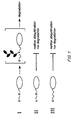
- a question which could be addressed is whether the targeting and degradation components of a proteolytic pathway distinguish between the subunits of an oligomeric protein that carry the degradation signals from those that do not, or whether the entire oligomeric protein is targeted for destruction even if only some of its subunits carry degradation signals. If either trans recognition of proteolytic substrates or their subunit-specific degradation, or both were possible ( Figure 2), this would have important functional and practical implications.
- N-end rule proteolytic pathway has been the subject of numerous scientific articles over the past several years. These studies have shown that the N-end rule-based degradation signal is composed of two distinct determinants: the protein's amino-terminal residue and a specific internal lysine residue (Figure 1).
- the first determinant reported initially by Bachmair et al . ( Science 234 :179-186 (1986)), is the amino-terminal amino acid residue of the protein (shown in Figure 1 as “d” for destabilizing or "s” for stabilizing ).
- the initial report by Bachmair et al . describes an assay used to study the effect of the amino-terminal residue on metabolic stability of a test protein.
- the group of destabilizing amino-terminal residues consisted of Ile, Glu, His, Tyr, Gln, Phe, Leu, Asp, Asn, Lys, Arg and Trp. Subsequent work by Gonda et al . ( J.
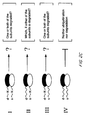
- ubiquitin-X- ⁇ gal (Ub-X- ⁇ gal) fusion proteins are precisely deubiquitinated either in vivo or in cell-free extracts by an endogenous processing protease to yield X- ⁇ gal test proteins bearing the (predetermined) residue X at the amino terminus ( Figure 3) (Bachmair et al ., Science 234 :179-186 (1986)).
- the X- ⁇ gal proteins are either long-lived or metabolically unstable, with destabilizing amino-terminal residues conferring short half-lives on the corresponding X- ⁇ gals. This amino-terminal degradation signal is manifested as the N-end rule.
- the exact form of the N-end rule varies between different types of cells.
- the methods for determining the exact form of the N-end rule in any given cell type are technically straightforward, and involve expressing, in a cell type of interest, using standard expression vectors, a set of Ub-X- ⁇ gal fusion proteins described above and determining, using standard pulse-chase protocols and immunoprecipitation, the half-lives of each of the 20 different X- ⁇ gal proteins.
- an X- ⁇ gal protein (derived from a Ub-X- ⁇ gal by deubiquitination) is pulse-labeled in vivo with a radioactive amino acid, followed by a cold chase, using which the metabolic fate of the pulse-labeled species is determined.
- the second, also essential, determinant of this signal is a specific internal lysine residue in the same protein (shown as "K" in Figure 1).
- K internal lysine residue
- the available evidence indicates that the relevant features of the second-determinant lysine are its spatial (but not necessarily “linear") proximity to the protein's amino-terminal residue and segmental mobility of the lysine-containing region.
- each lysine of a proteolytic substrate can be assigned a probability of being utilized as a ubiquitination site, depending on the time-average spatial location and mobility of the lysine.
- the probability of serving as a multiubiquitination site would be either small or infinitesimal because of their spatial remoteness from the protein's amino-terminal residue, lack of mobility or both (Bachmair and Varshavsky, Cell 56 :1013-1032 (1989)).
- the information about exactly where the second determinant-lysine (the multiubiquitination site) is located within a protein or peptide of interest can be obtained by direct chemical mapping (Chau et al ., Science 243 :1576-1583 (1989)) or other means. However, this additional information is in most cases unnecessary for the purposes of the present invention.
- null or nearly null phenotypes of specific genes is a recurring theme in both basic and applied biological research.
- the underlying goals range from suppressing the functions of toxic and otherwise undesirable proteins, such as those of pathogenic viruses or aberrant proto-oncogene products, to analyzing the function of a protein of interest by selectively eliminating the function and observing the effects of such an elimination.
- Deletions of predetermined genes directly yield the desired null phenotypes. Such targeted deletions are technically straightforward for some single-cell organisms such as bacteria and yeast, but are either difficult or impossible, at present, with most animals and plants.
- null or nearly null phenotypes of specific genes include "conventional" pharmacology, e.g., the use of inhibitors of specific enzymes. For instance, penicillin, by specifically inhibiting one of the enzymes required for the synthesis of bacterial wall, generates, in effect, a (partially) null phenotype of the gene for this enzyme. Similarly, inhibitors of brain monoamino oxidase, by producing, in effect, a (partially) null phenotype of the gene for this enzyme, act as tranquilizers or antidepressants.
- Acetylsalicylic acid by inhibiting a specific enzyme involved in the synthesis of one of the prostaglandins, produces a (partially) null phenotype of the gene for this enzyme, thereby inhibiting the synthesis of specific prostaglandins and, as a result, reducing inflammation.
- antisense oligonucleotides by binding, via Watson-Crick base pairing, to a region of the "sense" strand of a specific mRNA, interfere with its translation, for instance, by inhibiting the binding of initiation factors to the 5'-region of the mRNA.
- This inhibition in effect, produces a (partially) null phenotype of a gene whose mRNA is targeted by the oligonucleotide.
- null phenotypes are based on constructing dominant negative mutations (reviewed by Herskowitz, I., Nature 329 :219-222 (1987)).
- the latter by definition, perturb the function of a protein of interest even in the presence of its wild-type version, for instance, by introducing a mutant form of a protein that forms a functionally inactive complex with the protein of interest.
- trans recognition in the N-end rule pathway and the subunit specificity of selective protein degradation allow construction of a new class of dominant negative mutations that is the subject of the present patent application.
- the distinguishing feature of this new class is selective metabolic destabilization of an otherwise long-lived protein of interest by targeting it for degradation in trans . Decreasing metabolic stability of a protein lowers its concentration in the cell, thereby providing an alternative route to the null phenotype.
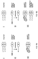
- One version of the trans -degradation approach, illustrated in Figure 4 requires a gene for a protein (or a portion thereof) that specifically interacts with the protein or peptide of interest.
- the gene for a targeting protein or peptide that specifically interacts with the protein or peptide of interest is modified, for example, by using the previously developed ubiquitin fusion technique ( Figure 3; Bachmair et al ., Science 234 :179-186 (1966); Bachmair and Varshavsky, Cell 56 :1013-1032 (1989)), to encode a variant of the targeting protein or peptide that, upon its in vivo expression, bears a destabilizing amino-terminal residue but lacks an efficient second determinant (multiubiquitination site) of the N-end rule-based degradation signal.
- Figure 3 Bachmair et al ., Science 234 :179-186 (1966); Bachmair and Varshavsky, Cell 56 :1013-1032 (1989)
- ovals A" and "B” represent polypeptide subunits.
- the first determinant of the N-end rule-based degradation signal (an amino-terminal amino acid residue) is designated “s” for stabilizing, or “d” for destabilizing.
- the second determinant of the N-end rule-based degradation signal (a specific lysine residue) is designated “K”.
- Ubiquitin is designated as "Ub” in Figure 4.
- the trans -degradation technique is applicable to either homomeric (Figure 4b) or heteromeric ( Figure 4a) targets.
- a modified subunit B bearing a destabilizing amino-terminal residue and lacking an efficient second determinant of the degradation signal is introduced into a cell, for instance, by expressing it from a vector encoding the modified subunit B ( Figure 4a).
- Targeting for degradation a long-lived homodimer AA is similar to the above method except that the targeting subunit is additionally modified to encode a variant (A*) of the subunit A that is functionally inactive at least as a homodimer (d-A*/d-A*).
- the N-end rule-based degradation signal is a multi-component one, and can function either in cis or in trans , can readily be adapted to dissect, in a similar manner, other degradation signals in short-lived proteins, and to establish whether the cis / trans recognition, and hence, the methods of the present invention, are relevant to these signals as well.
- subunit specificity in protein degradation is not confined to the N-end rule pathway, inasmuch as at least one of the degradation signals in a naturally short-lived yeast ⁇ 2 repressor operates in a subunit-specific manner.
- the present invention relates not only to the targeting proteins or peptides that interact with mature forms of a protein of interest, but also to the targeting proteins or peptides that interact with incompletely folded forms of a protein or peptide of interest in vivo .
- Every in vivo - synthesized protein must undergo conformational maturation, a process that converts a relatively disordered polypeptide chain of a protein as it emerges from the ribosome into a well-defined folded structure that is characteristic of a mature protein.
- the actual folding pathways are known for very few proteins in vitro , and for virtually no proteins in vivo , where folding pathways may not be identical or even similar to those in vitro .
- our method in addition to being in vivo -based and protein degradation-oriented, provides for a systematic genetic approach to ascertain directly, for any protein of interest, which of its fragments are the ones that most effectively interfere with the folding, function and/or metabolic stability of the full-length protein.
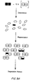
- Figure 5 illustrates the use of a folding-interfering peptide to target a protein of interest for degradation.
- the interference can occur either during the folding of the full-length protein, as shown in Figure 5, or even during folding of the amino terminus-proximal region of the protein as it emerges from the ribosome.
- a folding-interfering targeting peptide interrupts the folding of a protein (from which the peptide had been derived) either irreversibly or at least for a relatively long period of time before a dissociation event and the resumption of folding, the resulting functional impairment of the protein of interest will be obtained in vivo even in the absence of the accompanying metabolic destabilization (destruction) of the targeted peptide-protein complex.
- a trapped complex would be likely to resemble a "misfolded" protein to the intracellular "surveillance" proteolytic pathways. it is likely that the effective decrease in the concentration of an active protein species in a cell would be caused by a combination of functional impairment and actual degradation of a targeted complex by the above proteolytic pathways.
- a folding-interfering targeting peptide can be designed to be produced as a ubiquitin-peptide fusion whose efficient in vivo deubiquitination (Figure 3) would generate a predetermined destabilizing residue at the peptide's amino terminus ( Figure 5).
- Figure 3 When such a peptide traps a folding protein of interest, its destabilizing residue makes possible trans targeting of the (partially folded) trapped protein of interest for degradation via the N-end rule pathway, as discussed above ( Figure 2).
- a protein species trapped by the targeting peptide is by definition partially folded (Figure 5).
- the inevitable conformational instability of this species is expected to provide additional conformationally mobile lysine residues that could serve as second determinants (ubiquitination sites) of the N-end rule-based degradation signal.
- the N-end rule-mediated trans targeting of a trapped peptide-protein complex is expected to be unusually efficient because of the lack of conformational stabilization of lysine residues (potential second determinants of the degradation signal) within an incompletely folded protein of interest.
- ubiquitin fusions will temporarily stabilize these peptides against intracellular degradation, as has previously been found for several otherwise short-lived peptides fused to ubiquitin moieties (Finley et al ., Nature 338 :394-401 (1989)).
- a multiubiquitin chain is a signal for protein degradation (see e.g., Figures 1 and 2)
- an amino-terminal monoubiquitin moiety can increase (under certain conditions identified by Finley et al .) the in vivo half-life of a protein or peptide moiety located "downstream" of the ubiquitin moiety.
- the method of this invention should provide for a systematic, assumption-free way to distinguish between efficiently and inefficiently acting folding-interfering targeting peptides, and to identify positively the former for any given protein of interest.
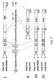
- a genetically based, generally applicable method that provides for such an identification is presented in Figure 6.
- a fusion of a protein of interest with a marker protein such as ⁇ -galactosidase ( ⁇ gal)
- a marker protein such as ⁇ -galactosidase ( ⁇ gal)
- a construct encoding such a fusion can be made, for example, by fusing a gene encoding a protein of interest to a gene encoding a marker protein such as ⁇ gal to yield a fusion protein containing the moiety of the protein of interest in front of the ⁇ gal moiety ( Figure 6-I).
- a DNA library encoding ubiquitin fusions to various fragments of the protein of interest is constructed ( Figure 6-II).
- One way to construct such a library is as follows:
- the method illustrated in Figure 6 makes possible direct detection and identification of those fragments of a protein of interest that target the protein of interest, in its fusion to ⁇ gal, for degradation in vivo .
- This method is applicable to any protein for which a gene is available, is essentially assumption-free in its execution, and is technically straightforward, in that all relevant procedures are well known to those skilled in the art.
- the method illustrated in Figure 6 cannot detect folding-interfering targeting peptides that, while perturbing the folding of the protein, do not target it for degradation. Although such fragments are unnecessary for the purposes of this invention (since folding-interfering, degradation-targeting fragments of the protein of interest are expected to be not infrequent), these fragments could also be identified by the approach illustrated in Figure 6 if a functional activity of the protein of interest could be adapted to either a selection-type or a screen-type test, thereby bypassing the need for a marker protein moiety. In this case, a protein itself (rather than its fusion to ⁇ gal or another marker protein) would serve as a marker, with the rest of the Figure 6 method intact.
- second-generation targeting peptides could also be designed that, while equivalent in their folding-interfering capacity to the originally identified peptides, would be capable of penetrating cells directly, thereby obviating the necessity of expressing these peptides via DNA constructs from within cells.
- the N-end rule-based degradation signal in a short-lived protein comprises a destabilizing amino-terminal residue and a specific internal lysine residue (Bachmair, A. et al ., Science 234 :179-186 (1986); Varshavsky, A. et al ., in Ubiquitin (ed. Rechsteiner, M.) 287-324 (Plenum, New York, 1988); Bachmair, A. and A. Varshavsky, Cell 56 :1019-1032 (1989); Chau, V. et al ., Science 243 :1576-1583 (1989); and Gonda, D.K. et al ., J. Biol. Chem.
- both determinants of the degradation signal are located, by definition, within the same polypeptide chain, and are therefore said to be recognized in cis ( Figures 1 and 2a).
- the targeting of an individual X- ⁇ gal subunit might be influenced by the presence of other subunits in the tetramer.
- the possibility of trans recognition in which the first and second determinants of the degradation signal reside within different subunits of X- ⁇ gal ( Figure 2b, c).
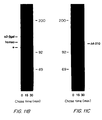
- Figures 8 and 9 illustrate the ATP-dependent ubiquitination and degration of X- ⁇ gal homotetramers and heterotetramers, respectively.
- the results show the kinetics of degration in ATP supplemented reticulocyte extract of 35 S-labeled X- ⁇ gal proteins bearing either a stabilizing (Val) or destabilizing (Arg) amino-terminal residue, and either containing or lacking the second determinant of the degradation signal.
- V ⁇ K ⁇ gal*/ V ⁇ K ⁇ gal tetramer (the 35 S-labeled subunit is designated with an asterisk) was degraded in reticulocyte extract at a slightly higher rate than the initial V ⁇ K ⁇ gal tetramer that had not been subjected to the dissociation/reassociation treatment (t 1 ⁇ 2 of ⁇ 20 h and ⁇ 50 h, respectively; Figure 8a, curve 3; Figure 9b, curve 3).
- V ⁇ K ⁇ gal subunit When the labeled V ⁇ K ⁇ gal subunit was present within the V ⁇ K ⁇ gal*/R ⁇ K ⁇ gal heterotetramer, it was multiply ubiquitinated and degraded in reticulocyte extract with a t 1 ⁇ 2 of ⁇ 3.3 h ( Figure 9b, curve 1). In contrast, when the same V ⁇ K ⁇ gal subunit resided in the V ⁇ K ⁇ gal*/V ⁇ K ⁇ gal tetramer, its t 1 ⁇ 2 was 20 h, and it showed very little ubiquitination (Figure 9b, curve 3).
- V ⁇ K ⁇ gal was mixed with an excess of the unlabeled R ⁇ K ⁇ gal homotetramer, and incubated in reticulocyte extract without the dissociation/reassociation pretreatment.
- V ⁇ K ⁇ gal was long-lived, with a t 1 ⁇ 2 of ⁇ 50 h, identical to the t 1 ⁇ 2 , of V ⁇ K ⁇ gal in the absence of R ⁇ K ⁇ gal ( Figure 8a, curve 3).
- the metabolic instability of the V ⁇ K ⁇ gal*/R ⁇ K ⁇ gal heterotetramer did not depend on the degradation of the tetramer's intrinsically short-lived R ⁇ K ⁇ gal subunits: the V ⁇ K ⁇ gal subunit had a t 1 ⁇ 2 of ⁇ 3.3 h in reticulocyte extract within either V ⁇ K ⁇ gal*/ R ⁇ K ⁇ gal or V ⁇ K ⁇ gal*/R ⁇ K ⁇ gal ( Figure 9b, curves 1 and 2).
- amino-terminal Glu (E) and Asp are secondary destabilizing residues in that they function by virtue of their ability to be conjugated, via an Arg-tRNA-protein transferase, to a primary destabilizing residue, Arg (Gonda, D.K. et al ., J. Biol. Chem. 264 :16700-16712 (1989); Ferber, S. and A. Ciechanover, Nature 326 :808-811 (1987)).
- the Arg conjugation, and the resultant destabilizing property of a secondary destabilizing residue can be selectively inhibited by a tRNA-depleting pretreatment of reticulocyte extract with RNAase (Gonda, D.K. et al ., J. Biol. Chem. 264 :16700-16712 (1989)).
- RNAase RNAase
- the labeled V ⁇ K ⁇ gal subunit was short-lived within either the V ⁇ K ⁇ gal*/R ⁇ K ⁇ gal or the V ⁇ K ⁇ gal*/E ⁇ K ⁇ gal tetramer (the latter bearing E, a secondary destabilizing residue (Bachmair, A.
- the differential degradation of individual subunits within X- ⁇ gal heterotetramers was not due to a rapid dissociation/reassociation of X- ⁇ gal subunits in reticulocyte extract that might allow short-lived subunits to be targeted and degraded while in a transient monomeric state.
- the ⁇ gal tetramer is known to be highly stable, requiring strongly denaturing solvents for its dissociation (Zipser, D., J. Mol. Biol. 7 :113-121 (1963); Givol, D. et al ., Biochim. Biophys. Acta. 113 : 120-125 (1966)).
- V ⁇ K ⁇ gal homotetramers and an excess of unlabeled R ⁇ K ⁇ gal homotetramers were incubated together in reticulocyte extract, the V ⁇ K ⁇ gal subunits were long-lived (t 1 ⁇ 2 of ⁇ 50 h), in contrast to the metabolic instability of the V ⁇ K ⁇ gal subunit in the V ⁇ K ⁇ gal*/R ⁇ K ⁇ gal heterotetramer (t 1 ⁇ 2 of ⁇ 3.3 h; Figure 9b, curve 2). It was concluded that the degradation of an oligomeric protein by the N-end rule pathway is subunit-specific.
- X ⁇ K ⁇ gal subunits bearing a stabilizing amino-terminal residue can be targeted for degradation only in trans (see above and Figure 2b).
- the R ⁇ K ⁇ gal subunit which contains both determinants of the degradation signal, is targetable either in cis or in trans within R ⁇ K ⁇ gal*/R ⁇ K ⁇ gal and R ⁇ K ⁇ gal*/R ⁇ K ⁇ gal, but only in cis within R ⁇ K ⁇ gal*/V ⁇ K ⁇ gal and R ⁇ K ⁇ gal*/V ⁇ K ⁇ gal ( Figures 2 and 9c). Nonetheless, the degradation time courses (as well as ubiquitination levels) of the R ⁇ K ⁇ gal subunit in reticulocyte extract were indistinguishable for all of these tetramer backgrounds ( Figure 9c, curves 7-10).
- trans targeting in the N-end rule pathway ( Figures 2, 9 and 10) indicates that substrate recognition by this proteolytic system does not involve a linear "tracking" mechanism which might be used to ascertain, before the ubiquitination step, whether both determinants of the degradation signal are located within the same polypeptide chain.
- Trans targeting is therefore consistent with the previously proposed model (Bachmair, A. and A. Varshavsky, Cell 56 :1019-1032 (1989)) in which an N-end-recognizing (E3) protein (either alone or complexed with a specific ubiquitin-conjugating (E2) enzyme) has a binding site for a substrate's destabilizing amino-terminal residue, and a lysine-binding site.
- E3 protein either alone or complexed with a specific ubiquitin-conjugating (E2) enzyme
- Both of these sites must be occupied for the multiubiquitination to commence at the bound lysine of a proteolytic substrate. If the two binding sites of the recognition component are spatially fixed relative to one another, binding of the substrate would require either a specific spatial arrangement of the substrate's two determinants or a conformational mobility of these determinants relative to one another, so that their correct spatial arrangement is achieved transiently, but frequently enough for both of them to be bound by the recognition complex (Bachmair, A. and A. Varshavsky, Cell 56 :1019-1032 (1989)).
- Segmental mobility of the determinant-containing region (or regions) can account for both the cis and trans recognition in a short-lived X- ⁇ gal.
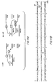
- the non- ⁇ gal extension at the amino termini of the X- ⁇ gal proteins ( Figure 7), being derived from an internal region of the lac repressor, is likely to be disordered (segmentally mobile) when present in an unnatural (amino-terminal) location within an unrelated protein such as ⁇ gal (Bachmair, A. and A. Varshavsky, Cell 56 :1019-1032 (1989)).
- the dissociation and degradation need not be temporally coupled: a mechanochemical process analogous to the one above might be used to dissociate the multiubiquitin chain-containing subunit from an oligomeric substrate, with the degradation of the subunit following rather than accompanying its dissociation. (It has been shown above that spontaneous dissociation/reassociation of X- ⁇ gal subunits cannot account for the observed subunit specificity of X- ⁇ gal degradation by the N-end rule pathway.)
- Subunit specificity is likely to emerge as a general feature of selective protein degradation because alteration of the functions of protein complexes via combinatorial variation of their subunit composition is a common theme in biological regulation (Abel, T. and T. Maniatis, Nature 341 :24-25 (1989); Goutte, C. and A.D. Johnson, Cell 52 :875-882 (1988)).
- many regulatory proteins are expressed in distinct but over-lapping regions of a developing embryo, with cell determination depending at least in part on the exact combination of these proteins (many of which are short-lived) present in a given region at different stages of embryo-genesis (Ingham, P.W., Nature 335 :25-34 (1988); Scott, M.P. and S.B.
- Subunit-specific degradation may also underlie the periodic destruction of cyclin, a subunit of the multiprotein complex that regulates cell cycle progression in eukaryotes (Evans, T. et al ., Cell 33 :389-396 (1983); Murray, A.W. et al ., Nature 339 :280-286 (1989)).
- the two-determinant nature of a degradation signal, and the possibility of either cis or trans recognition may afford additional flexibility to subunit-specific degradation, for instance, by making the accessibility of one of the determinants dependent upon specific phosphorylation events within a multisubunit substrate.
- the constructs encoding Ub-R ⁇ K ⁇ gal and Ub-V ⁇ K ⁇ gal were made as follows.
- the small Sal I/ Mst II fragment of pKKUb-Val- ⁇ gal was subcloned into M13mp18 ⁇ (a derivative of M13mp18 (Ausubel, F.M.
- UB-X- ⁇ gal 35 :35-42 (1969)) of UB-X- ⁇ gal, a mixture of 18 ⁇ g [ 35 S]Ub-X- ⁇ gal and 180 ⁇ g of unlabeled Ub-X- ⁇ gal (whose amino acid sequence was either identical to or different from that of the labeled Ub-X- ⁇ gal), at a total protein concentration of 1.5 to 3 mg/ml in storage buffer (50% (v/v) glycerol, 0.1 mM Na-EDTA, 1 mM dithiothreitol (DTT), 40 mM Tris-HCl (pH 7.5)), was diluted to a protein concentration of 80 ⁇ g/ml with buffer A (10 mM Na-EDTA, 5 mM DTT, 20 mM Na-HEPES (pH 7.2)) containing 2 M NaCl and 8 M (deionized) urea.
- buffer A 10 mM Na-EDTA, 5 mM DTT, 20 m
- the solution was dialyzed against changes of buffer A containing, successively, 8 M urea, 4 M urea and 10 mM NaCl, 2 M urea and 10 mM NaCl, 1 M urea and 10 mM NaCl, and finally, against two changes of buffer A plus 10 mM NaCl.
- Dialysis was carried out with ten Ub-X-ßgal samples at a time, at 4°C, in a Multiple Dialyzer (Spectrum), for ⁇ 14 h per change of buffer.
- Each of the dialyzed samples was brought to 1.6 M in NaCl and 10 mM in MgCl 2 , added to ⁇ 0.4 ml of APTG-agarose (Gonda, D.K. et al ., J. Biol. Chem. 264 :16700-16712 (1989)) and rocked overnight at 4°C. The suspension was then loaded into columns, which were processed for affinity purification of Ub-X- ⁇ gal as described (Gonda, D.K. et al ., J. Biol. Chem. 264 :16700-16712 (1989)).
- the enzymatic activity of the reconstituted Ub-X- ⁇ gal proteins varied between different preparations from 50 to 75% of the activity of their initial (homotetramer) counterparts.
- Degradation assays in reticulocyte extract and SDS-PAGE were carried out as described above. Less than 0.4% of either the initial or reconstituted X- ⁇ gal tetramers were degraded in 2 h in ATP-depleted reticulocyte extract.
- the pKKUb-Glu ⁇ Lys ⁇ gal expression vector encoding Ub-E ⁇ K ⁇ gal was constructed as described for pKKUb-Arg ⁇ Lys ⁇ gal above.
- Ub-X- ⁇ gal heterotetramers were prepared from the corresponding homotetramers as described above.
- the RNAase treatment (Gonda, D.K. et al ., J. Biol. Chem.
- RNAase A-agarose Sigma
- RNasin RNAase inhibitor (Promega Biotech)
- Reactions supplemented with tRNA included 50 ⁇ g/ml of purified total tRNA from bovine liver (Boehringer).
- Metabolic instability is characteristic of regulatory proteins whose in vivo concentrations must vary as function of time.
- the cell type-specific ⁇ 2 repressor of the yeast S . cerevisiae is shown in this Example to have a half-life of only ⁇ 5 minutes.
- Each of the two structural domains of ⁇ 2 carries a sequence that can independently target a normally long-lived protein for rapid destruction.
- these two degradation signals are shown here to operate via distinct mechanisms. Mutants deficient in the degradation of ⁇ 2 have been isolated and found to have a number of additional defects, indicating that the pathways responsible for ⁇ 2 turnover include components with multiple functions.
- E . coli ⁇ -galactosidase ( ⁇ gal) was used as an immunological tag, allowing immunoprecipitation of ⁇ 2- ⁇ gal fusion proteins with a monoclonal antibody to ⁇ gal.
- Cells carrying a high copy plasmid encoding ⁇ 2- ⁇ gal were labeled with [ 35 S] methionine for 7 minutes at 30°C, followed by a chase in the presence of translation inhibitors, extraction, precipitation with antibody to ⁇ gal, and SDS-PAGE.
- ⁇ gal that carried only the first 3 residues of ⁇ 2 at its amino terminus was metabolically stable, with no detectable degradation during the chase period (Figure 11c).
- attachment of the full-length 210-residue ⁇ 2 sequence to ⁇ gal yielded a short-lived protein ( Figure 11b).
- the Intact ⁇ 2 Repressor is Extremely Short-lived
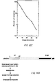
- the ⁇ 2 repressor was found by pulse-chase analysis to be extremely short-lived in vivo ( Figures 12b, 12c). Its half-life of ⁇ 5 minutes at 30°C was ⁇ 3-fold shorter than the "t 1/2 " of the ⁇ 2- ⁇ gal protein ( Figure 12b). Moreover, unlike ⁇ 2- ⁇ gal, the intact ⁇ 2 repressor was degraded with apparent first-order kinetics, indicating that most of the ⁇ 2 molecules in the cell were equally susceptible to degradation. No discrete ⁇ 2 degradation intermediates accumulated to detectable levels during the chase (labeling with either [ 35 S]methionine or [ 3 H] leucine), implying either rapid destruction of intermediates or a highly processive mode of proteolysis.
- the metabolic instability of ⁇ 2 could be a consequence of either some global property of the protein, such as low conformational stability (Parsell, D.A. and R.T. Sauer, J. Biol. Chem. 264 :7590-7595 (1989)), or a more localized structural feature.
- the latter would be analogous to the signal sequences that target proteins to distinct subcellular compartments (Dingwall, C. and R.A. Laskey, Ann. Rev. Cell Biol. 2 :367-390 (1986); Colman, A. and C. Robinson, Cell 46 :321-322 (1986); Warren, G., Nature 327 :17-18 (1987)).
- FIG. 13 shows two series of deletion derivatives of ⁇ 2- ⁇ gal, initiating at either the amino or the carboxyl terminus of ⁇ 2 (the junction between the ⁇ 2 and ⁇ gal sequences) that wore expressed in S . cerevisiae , and rates of in vivo degradation.
- ⁇ 68-210 which contains only the first 67 residues of ⁇ 2, was about as short-lived ("t 1 ⁇ 2 " of ⁇ 10 minutes) as the full-length ⁇ 2- ⁇ gal protein.
- an entirely different region of ⁇ 2 also can function as a degradation signal.
- the ⁇ 2- ⁇ gal derivative obtained, ⁇ 2-135 was approximately as short-lived ("t 1 ⁇ 2 " of ⁇ 15 minutes) as the full-length ⁇ 2- ⁇ gal ( Figure 13).
- t 1 ⁇ 2 of ⁇ 15 minutes
- ⁇ 2- ⁇ gal Figure 13
- carboxyl-terminal 75-residue region of ⁇ 2 can confer metabolic instability on an otherwise long-lived protein, and information within residues 136-140 is essential for this property.
- the two independent degradation signals uncovered in the ⁇ 2 repressor by the above analysis reside in its amino-terminal and carboxyl-terminal regions, respectively. These same regions have previously been shown to exist as structurally and functionally distinct globular domains in the repressor (Hall, M.N. and A.D. Johnson, Science 237 :1007-1012 (1987); Sauer, R.T. et al ., Genes Dev. 2 :807-816 (1988)). Moreover, as shown below, the two degradation signals in ⁇ 2 operate via genetically distinguishable pathways.
- ⁇ 68-210 In addition to bypassing potential complications caused by the presence of both signals in the full-length protein, the use of ⁇ 68-210 also avoided formation of the relatively long-lived, ⁇ gal-containing cleavage product that occurs with the full-length ⁇ 2- ⁇ gal. Presence of this product would have greatly reduced the sensitivity of the screen to changes in the steady state level of ⁇ 2- ⁇ gal.
- Figure 14a summarizes the genetic screen for mutants defective in degrading the ⁇ 68-210 derivative of ⁇ 2- ⁇ gal that carries one, but not both of the ⁇ 2 degradation signals.
- Figure 14b shows results of pulse-chase analysis of the ⁇ 68-210 protein in wild-type cells and in doa mutants at 36°C.
- Figure 14c shows results from tests measuring the ability of various strains to degrade the intact (not fused to ⁇ gal) ⁇ 2 repressor at 36°C.
- the ⁇ 2-135 derivative of ⁇ 2- ⁇ gal which carries only the carboxyl-terminal degradation signal of ⁇ 2 ( Figure 13), was introduced into three of the doa mutants identified through their defects in the degradation of an ⁇ 2- ⁇ gal derivative containing only the amino-terminal signal.
- the ⁇ 2-135 protein continued to be degraded at wild-type rates in the mutants ( Figure 15a). It was concluded that the two degradation signals detectable in ⁇ 2 using the ⁇ 2- ⁇ gal approach operate via genetically distinguishable pathways.
- the amino-terminal degradation signal is active not only within a ⁇ 2- ⁇ gal but within the intact (unfused) ⁇ 2 repressor as well.
- MCM1 GMM, PRTF
- MCM1 is present in all three cell types of S . cerevisiae and forms complexes with several different regulatory proteins. Thus, MCM1 is likely to be a more abundant and more metabolically stable protein than ⁇ 2. This raises the question of whether the fate of MCM1 in a tetrameric complex with the short-lived ⁇ 2 protein is different from that of ⁇ 2 itself. More generally, it could be asked whether the mechanisms of in vivo protein degradation are such that an oligomeric protein can contain both long-lived and short-lived subunits. The design of in vivo "subunit mixing" experiments that address this question is shown in Figure 16.
- S . cerevisiae strains used in this study were DBY1705 (MAT ⁇ leu 2-3,112 ura 3-52 lys 2-801 gal 2), DBY1826 (MATa leu 2-3, 112 ura 3-52 trp 1 his 3- ⁇ 200 ade 2-101), and HR125-5Dalf( ⁇ (MAT)::CAN1-14 leu 2-3, 112 ura 3-52 trp 1 his 3 his 4) (Hall, M.N. and A.D. Johnson, Science 237 :1007-1012 (1987)).
- Expression vectors pKK ⁇ 2- lac Z and pKK ⁇ 2 for producing ⁇ 2- ⁇ gal and ⁇ 2 in E . coli were constructed as follows. A Bam HI- Hind III fragment of the E . coli expression vector pKK233-2 carrying the P trc promoter (Amann, E. and J. Brosius, Gene 40 :183-190 (1985)), was ligated to a Hind III- Sac I fragment of M13mp19 carrying ⁇ 2- lac Z'. A 38-mer synthetic oligodeoxynucleotide was hybridized to the single-stranded DNA of the resulting M13mp19 phage derivative, causing the intervening sequence between Ptrc and the start codon of ⁇ 2 to loop out.
- the presence of the correct ⁇ 2 moiety was confirmed by amino-terminal microsequencing (see below) of the purified fusion protein.
- the pKK ⁇ 2 expression vector was produced from pKK ⁇ 2- lac Z by ligating its Bam HI- Xba I fragment carrying the P trc promoter and ⁇ 75% of the ⁇ 2 coding region to a pUC19-based XbaI-XhoI fragment of the ⁇ X152 derivative of MAT ⁇ (Tatchell, K. et al ., Cell 27 :25-35 (1981)), carrying the rest of the ⁇ 2 reading frame. The resulting fragment was then ligated to the larger Bam HI- Hind III fragment of pKK233-2, yielding pKK ⁇ 2.
- the ⁇ 53-210 ⁇ 2- ⁇ gal derivative ( Figure 13) was constructed by subcloning a ⁇ 3 kb SalI fragment of pMC1871 (Casadaban, M.J. et al ., Meth. Enzymol. 100 :293-308 (1983)) that encodes E . coli ⁇ gal into the XhoI linker site of mat ⁇ 2:: ⁇ X182 carried in the plasmid YRp7 (Tatchell, K. et al ., Cell 27 :25-35 (1981)).
- the resulting ⁇ 53-210 construct encoded a fusion of the first 52 residues of ⁇ 2 to ⁇ gal.
- Hind III fragment containing ⁇ 53-210 was then subcloned into both high copy (YEp13) and low copy (YCp50) plasmids (Parent, S.A. et al ., Yeast 1 :83-138 (1985)). Only the low copy plasmid encoding ⁇ 53-210 yielded transformants in S . cerevisiae . Hall et al . ( Cell 36 :1057-1065 (1984)) noted that the ⁇ 14-210 and ⁇ 26-210 proteins ( Figure 13) were lethal if expressed from the MAT ⁇ 2 promoter in high copy plasmids in the HR125-5D ⁇ strain.
- Cells from a 10-ml culture were harvested by filtration on a Millipore microtiter filtration plate, washed several times with methionine-free SD medium, resuspended in 0.3 ml of 0.5% glucose, 40 mM potassium phosphate (pH 7.4), and labeled for 5 minutes at 30°C (unless stated otherwise) with 0.15 mCi of 35 S-Translabel (ICN; ⁇ 80% [ 35 S]methionine, ⁇ 20% [ 35 S]cysteine).
- ICN 35 S-Translabel
- Labeled cells were collected by filtration and resuspended in SD medium supplemented with 10 mM L-methionine, 0.2 mg/ml cycloheximide, and 50 ⁇ g/ml trichodermin (a gift from Leo Pharmaceutical Products, Ballerup, Denmark).
- Acid-insoluble 35 S in the supernatants was determined, and samples containing equal amounts of acid-insoluble 35 S were immunoprecipitated with a molar excess of either an affinity-purified antibody of ⁇ 2 (see below) or a monoclonal antibody of ⁇ gal (a gift from J. Partaledis and T. Mason, University of Massachusetts, Amherst), obtained via J. Paul and R. Hynes (M.I.T.); see Bachmair, A. et al ., Science 234 :179-186 (1986)).
- the ⁇ 2- ⁇ gal protein was purified from isopropylthio-galactoside (IPTG)-induced E. coli carrying pKK ⁇ 2- lac Z by subjecting an extract prepared as in Gonda et al ., ( J. Biol. Chem. 264 :16700-16712 (1989)) to affinity chromatography on aminophenyithiopyranogalactoside-Sepharose (APTG-Sepharose) (Ullmann, A., Gene 29 :27-31 (1984)).
- the affinity-purified ⁇ 2- ⁇ gal was purified further by electrophoresis in a preparative-scale 6% polyacrylamide-SDS gel.
- the ⁇ 2- ⁇ gal band was excised, and macerated gel slices, suspended in Freund's complete adjuvant, were injected subcutaneously into female New Zealand white rabbits (either 0.1 or 0.2 mg of ⁇ 2- ⁇ gal per rabbit) (Carroll, S.B. and A. Laughton, DNA Cloning : A Practical Approach , D.M. Glover, ed. (IRL Press, Oxford), pp. 89-111, 1987).
- the antiserum to ⁇ 2- ⁇ gal was precipitated with 18% (w/v) Na 2 SO 4 , yielding an IgG-enriched fraction.
- the first affinity column contained E .
- the second column contained ⁇ 2- ⁇ gal crosslinked with dimethylpimelimidate (Pierce) to anti- ⁇ gal-Sepharose (the latter was made using the eluate of the first column).
- the flow-through from the first column was applied to the second one, and the bound fraction was eluted with 4 M guanidine hydrochloride.
- the eluate was dialyzed against 0.15 M NaCl, 10 mM K 2 HPO 4 (pH 7.2) at 4°C and passed through the ⁇ gal-Sepharose column to remove any residual antibodies to ⁇ gal.
- the affinity-purified anti- ⁇ 2 was concentrated in Centriprep tubes (Amicon), made 40% (v/v) in glycerol, and stored at either -20°C or -85°C. Immunoprecipitation with this antibody of extracts from [ 35 S]methionine-labeled S . cerevisiae yielded, upon SDS-PAGE, a single band of predicted molecular mass with ⁇ but not a cells ( Figure 1d). Furthermore, electrophoretic bands of the expected molecular mass were seen by immunoblot analyses of extracts from E . coli expressing either ⁇ 2 or ⁇ 2- ⁇ gal but not in control extracts. The anti- ⁇ 2 also precipitated the same ⁇ 2- ⁇ gal derivatives that were detectable using antibody to ⁇ gal, provided the derivative retained the requisite epitope(s) of ⁇ 2 ( Figure 13).
- DBY1705 cells carrying ⁇ 68-210 ( Figure 3) in the low copy YCp50 vector were grown to stationary phase in minimal medium, mutagenized with EMS to ⁇ 20% survival, and spread onto minimal plates ( ⁇ 40,000 survivors).
- Cells were then replica-plated to plates containing the chromogenic ⁇ gal substrate X-Gal (Rose, M. et al ., Proc. Natl. Acad. Sci. USA 78 :2460-2464 (1981)) and incubated at 23°C for 3 days, and then at 36°C for 3 days.
- Colonies that turned blue at either 23°C or 36°C were picked and rescreened by dispersing cells in water in 96-well microtiter plates and stamping cells onto fresh X-Gal indicator plates.
- Strains that passed the rescreening test were grown in liquid cultures and quantitatively assayed for ⁇ gal activity using ortho-nitrophenyl galactoside (ONPG) as substrate.
- ONPG ortho-nitrophenyl galactoside
- Sixty potential mutants (with ⁇ gal activity levels at least 3-fold higher than wild-type) were isolated.
- Pulse-chase analyses of these mutants identified 25 strains that showed a significant (at least 2-fold) increase in the "t 1 ⁇ 2 " of the ⁇ 68-210 fusion protein. Immunofluorescence analysis indicated that, in all of these doa ( D egradation O f A lpha) mutants, the ⁇ 68-210 protein was concentrated in the nucleus.
- the plasmid-cured doa mutants were backcrossed to MHY101, a strain derived from DBY1826 by integrating the ⁇ 68-210 fusion construct into the LEU2 locus, using the YIp33 vector (Parent, S.A. et al ., Yeast 1 :83-138 (1985)); Orr-Weaver, T.L. et al ., Proc. Natl. Acad. Sci. USA 78 :6354-6358 (1981)). The site of integration was verified by Southern hybridization analysis and allelism tests. Segregants from sporulated diploids carrying both the integrated reporter gene encoding ⁇ 68-210 and the doa degradation defect were then identified.
Landscapes
- Chemical & Material Sciences (AREA)
- Health & Medical Sciences (AREA)
- Life Sciences & Earth Sciences (AREA)
- Organic Chemistry (AREA)
- Genetics & Genomics (AREA)
- Molecular Biology (AREA)
- Engineering & Computer Science (AREA)
- Biochemistry (AREA)
- Biophysics (AREA)
- General Health & Medical Sciences (AREA)
- Biomedical Technology (AREA)
- Medicinal Chemistry (AREA)
- Proteomics, Peptides & Aminoacids (AREA)
- Gastroenterology & Hepatology (AREA)
- General Engineering & Computer Science (AREA)
- Mycology (AREA)
- Zoology (AREA)
- Wood Science & Technology (AREA)
- Bioinformatics & Cheminformatics (AREA)
- Microbiology (AREA)
- Biotechnology (AREA)
- Plant Pathology (AREA)
- Physics & Mathematics (AREA)
- Chemical Kinetics & Catalysis (AREA)
- General Chemical & Material Sciences (AREA)
- Analytical Chemistry (AREA)
- Peptides Or Proteins (AREA)
- Medicines That Contain Protein Lipid Enzymes And Other Medicines (AREA)
- Preparation Of Compounds By Using Micro-Organisms (AREA)
- Accessory Devices And Overall Control Thereof (AREA)
- Control Or Security For Electrophotography (AREA)
- Facsimiles In General (AREA)
Abstract
Description
- Ubiquitin, a 76-residue protein, is present in eukaryotes either free or covalently joined, through its carboxyl-terminal glycine residue, to various cytoplasmic, nuclear, and integral membrane proteins. The coupling of ubiquitin to such proteins is catalyzed by a family of ubiquitin-conjugating enzymes (also called E2 enzymes). The fact that the amino acid sequence of ubiquitin is conserved among eukaryotes to an extent unparalleled among known proteins suggested that ubiquitin mediates a basic cellular function. However, the biological role of ubiquitin remained a mystery until relatively recently.
- Ubiquitin has been found to be one of several factors required for ATP-dependent protein degradation in eukaryotic cells. One function of intracellular protein degradation, most of which is ATP-dependent, is selective elimination of damaged and otherwise abnormal proteins. Another is to confer short half-lives on undamaged proteins whose concentrations in the cell must vary as functions of time, as is the case, for example, with many regulatory proteins. Many other proteins, while long-lived as components of larger macromolecular complexes such as ribosomes and oligomeric proteins, are metabolically unstable in a free, unassociated state. (The term "metabolic instability" implies a relatively short half-life of a protein as a physical entity in vivo, with the adjective "metabolic" being used to distinguish this property from "stability" as such, which often means either conformational or chemical stability of a protein, but not its existence as a physical entity in vivo.)
- Recent work has shown that selective degradation of many short-lived proteins requires a preliminary step of ubiquitin conjugation to a targeted proteolytic substrate. It was proposed that one role of ubiquitin is to serve as a signal for attack by proteases specific for ubiquitin-protein conjugates (reviewed by Finley and Varshavsky, Trends Biochem. Sci. 10:343-348 (1985)).
- This understanding, however, left unsolved the problem of targeting: how are intracellular proteins initially recognized as proteolytic substrates? At least some short-lived proteins are recognized as such because they contain sequences (degradation signals) which make these proteins substrates of specific proteolytic pathways. The first degradation signal to be understood in some detail comprises two distinct determinants: the protein's amino-terminal residue and a specific internal lysine residue (Bachmair et al., Science 234:179-186 (1986); Bachmair and Varshavsky, Cell 56:1013-1032 (1989)). The N-end rule, a code that relates the protein's metabolic stability to the identity of its amino-terminal residue (Bachmair et al., Science 234:179-186 (1986)), is universal in that different versions of the N-end rule operate in all of the eukaryotic organisms examined, from yeast to mammals (Gonda et al., J. Biol. Chem. 264:16700-16712 (1989)).
- The second essential determinant of the N-end rule-based degradation signal, referred to herein as the second determinant, is a specific internal lysine residue in the substrate protein that serves as the site of attachment of a multiubiquitin chain. Formation of the multiubiquitin chain on a targeted short-lived protein is essential for the protein's subsequent degradation (Figure 1). The enzymatic conjugation of ubiquitin to other proteins involves formation of an isopeptide bond between the carboxyl-terminal glycine residue of ubiquitin and the ε-amino group of a lysine residue in an acceptor protein. In a multiubiquitin chain, ubiquitin itself serves as an acceptor, with several ubiquitin moieties attached sequentially to an intital acceptor protein to form a chain of branched ubiquitin-ubiquitin conjugates (Chau et al., Science 243:1576-1583 (1989)).
- The elucidation of the fundamental rules governing the metabolic stability of proteins in cells, and especially the deciphering of the N-end rule-based degradation signal, has made possible the manipulation of proteins to vary their half-lives in vivo (Bachmair and Varshavsky, Cell 56:1019-1032 (1989)). A more detailed understanding of these degradation signals, their components, and their interrelationships is necessary in order to realize the full potential of this powerful methodology.
- This invention pertains to methods and compositions for metabolically destabilizing a protein or peptide of interest (i.e., targeting a protein or peptide for degradation). The protein or peptide of interest must contain a second determinant of the N-end rule-based degradation signal. The method comprises contacting the protein or peptide of interest with a targeting protein or peptide which interacts specifically with the protein or peptide of interest. The targeting peptide or protein is characterized as having a destabilizing amino-terminal amino acid according to the N-end rule of protein degradation, but lacking a second determinant of the N-end rule-based degradation signal.
- In accordance with a first aspect of the present invention, there is provided a recombinant DNA molecule according to
claim 1. In accordance with a second aspect of the present invention, then provided the method ofclaim 2. In accordance with a third aspect of the present invention, there is provided the use of a recombinant DNA molecule according toclaim 7. - In a preferred embodiment, the protein or peptide of interest and the targeting peptide or protein are subunits or portion of subunits of the same oligomeric protein in a living cell. The cell is transformed with an expressible DNA construct encoding a targeting peptide or protein having a destabilizing amino-terminal amino acid according to the N-end rule of protein degradation, but lacking a second determinant of the N-end rule-based degradation signal.
- One method for generating a targeting peptide or protein having a destabilizing amino-terminal amino acid residue is to transform a eukaryotic cell with an expressible DNA construct comprising ubiquitin fused in frame to a DNA sequence encoding a peptide or protein which interacts specifically with the protein or peptide of interest.
- The methods described herein facilitate the metabolic destabilization of a protein or peptide of interest. A requirement of the method is that it is necessary to identify a peptide or protein which interacts specifically with the protein or peptide of interest. Because nearly all proteins specifically interact with other proteins, this is a broadly applicable method for metabolically destabilizing a protein or polypeptide of interest.
-
- Figure 1 is a diagram showing the two-determinant organization of the N-end rule-based degradation signal in a monomeric protein, with d and s denoting, respectively, a destabilizing and a stabilizing amino-terminal residue, and the chain of black ovals joined to the second-determinant lysine (K) denoting a multiubiquitin chain.
- Figure 2 is a diagram illustrating the cis and trans recognition in the N-end rule pathway.
- Figure 3 is a diagram illustrating the use of ubiquitin-protein fusions, exemplified by Ub-X-βgal, to generate test proteins bearing predetermined amino-terminal residues.
- Figure 4 is a diagram illustrating the use of trans recognition to metabolically destabilize a protein of interest.
- Figure 5 is a diagram illustrating the use of peptides having an amino acid sequence identical or substantially homologous to a portion of the amino acid sequence of a protein of interest to metabolically destabilize a protein of interest in vivo.
- Figure 6 is a diagram illustrating the use of a genetic screen to identify polypeptides having an amino acid sequence identical or substantially homologous to a portion of the amino acid sequence of a protein of interest that metabolically destabilize the protein of interest in vivo.
- Figure 7 is a diagram illustrating several genetic constructs and their use in dissecting the N-end rule-based degradation signal.
- Figure 8 is a diagram illustrating ATP-dependent ubiquitination and degradation of X-βgal homotetramers in reticulocyte extract.
- Figure 9 is a diagram illustrating ATP-dependent ubiquitination and degradation of X-βgal heterotetramers in reticulocyte extract.
- Figure 10 is a diagram illustrating trans recognition-dependent degradation of X-βgal subunits bearing a stabilizing amino-terminal residue.
- Figure 11 is a diagram illustrating the metabolic instability of an α2-βgal fusion protein.
- Figure 12 is a diagram illustrating that the α2 repressor is short-lived in vivo.
- Figure 13 is a diagram illustrating the results of deletion analysis of degradation signals in α2-βgal.
- Figure 14 is a diagram illustrating the isolation of S. cerevisiae mutants defective in α2 degradation.
- Figure 15 is a diagram illustrating that two degradation signals in α2 are targeted by different pathways.
- Figure 16 is a diagram illustrating the design of in vivo subunit mixing experiments.
- Figure 17 is a diagram illustrating that the degradation of an oligomeric protein is subunit-specific.
-
- The invention disclosed in this application relates to a new class of methods for manipulating metabolic stability of a protein or polypeptide of interest. The terms "protein", "polypeptide" and "peptide" are often used interchangeably in the art, with "peptides" sometimes, but not always, referring to relatively short polypeptides, on the order of ∼50 residues or less. As used herein, the term protein is meant to encompass both proteins and polypeptides, whereas the term peptide is meant to encompass relatively short polypeptides. As discussed below, a common feature of these methodologies is the use of trans recognition, described in the present patent application, to destabilize metabolically long-lived proteins by targeting them for selective degradation in trans.
- In a monomeric protein substrate, both determinants of the degradation signal are located, by definition, within the same polypeptide chain (cis recognition). However, in the case of a multisubunit protein substrate, it is conceivable that the first and second determinants of the signal might reside within different subunits of the substrate (trans recognition). A question which could be addressed is whether the targeting and degradation components of a proteolytic pathway distinguish between the subunits of an oligomeric protein that carry the degradation signals from those that do not, or whether the entire oligomeric protein is targeted for destruction even if only some of its subunits carry degradation signals. If either trans recognition of proteolytic substrates or their subunit-specific degradation, or both were possible (Figure 2), this would have important functional and practical implications.
- The N-end rule proteolytic pathway has been the subject of numerous scientific articles over the past several years. These studies have shown that the N-end rule-based degradation signal is composed of two distinct determinants: the protein's amino-terminal residue and a specific internal lysine residue (Figure 1).
- The first determinant, reported initially by Bachmair et al. (Science 234:179-186 (1986)), is the amino-terminal amino acid residue of the protein (shown in Figure 1 as "d" for destabilizing or "s" for stabilizing ). The initial report by Bachmair et al. describes an assay used to study the effect of the amino-terminal residue on metabolic stability of a test protein. In the yeast S. cervisiae, it was determined that the group of destabilizing amino-terminal residues consisted of Ile, Glu, His, Tyr, Gln, Phe, Leu, Asp, Asn, Lys, Arg and Trp. Subsequent work by Gonda et al. (J. Biol. Chem. 264:16700-16712 (1989)) served to elucidate the set of destabilizing residues in mammalian reticulocytes. This set consisted of Asn, Gln, Asp, Glu, Cys, Arg, Lys, His, Phe, Leu, Trp, Tyr, Ala, Ser and Thr. As described below, the assays which were used to determine the destabilizing residues in these two systems can be readily and directly adapted to make an analogous determination in any eukaryotic system.
- In all eukaryotes examined, ubiquitin-X-βgal (Ub-X-βgal) fusion proteins are precisely deubiquitinated either in vivo or in cell-free extracts by an endogenous processing protease to yield X-βgal test proteins bearing the (predetermined) residue X at the amino terminus (Figure 3) (Bachmair et al., Science 234:179-186 (1986)). Depending on the nature of X, the X-βgal proteins are either long-lived or metabolically unstable, with destabilizing amino-terminal residues conferring short half-lives on the corresponding X-βgals. This amino-terminal degradation signal is manifested as the N-end rule.
- It has previously been found that the exact form of the N-end rule varies between different types of cells. The methods for determining the exact form of the N-end rule in any given cell type are technically straightforward, and involve expressing, in a cell type of interest, using standard expression vectors, a set of Ub-X-βgal fusion proteins described above and determining, using standard pulse-chase protocols and immunoprecipitation, the half-lives of each of the 20 different X-βgal proteins. Specifically, an X-βgal protein (derived from a Ub-X-βgal by deubiquitination) is pulse-labeled in vivo with a radioactive amino acid, followed by a cold chase, using which the metabolic fate of the pulse-labeled species is determined. The way to implement this latter portion of the protocol is also well known to those skilled in the art, and consists of immunoprecipitation of each of the X-βgal proteins from pulse-labeled and chased samples of the corresponding crude extracts using an antibody to βgal, followed by electrophoretic analysis of immunoprecipitated X-βgals, which completes this direct determination of the N-end rule in a cell type of interest (Bachmair et al. , Science 234:179-186 (1986); Bachmair and Varshavsky, Cell 56:1019-1032 (1989)).
- While the first determinant of the N-end rule-based degradation signal is the protein's amino-terminal residue, the second, also essential, determinant of this signal is a specific internal lysine residue in the same protein (shown as "K" in Figure 1). The available evidence indicates that the relevant features of the second-determinant lysine are its spatial (but not necessarily "linear") proximity to the protein's amino-terminal residue and segmental mobility of the lysine-containing region. In a stochastic view of the second determinant, each lysine of a proteolytic substrate can be assigned a probability of being utilized as a ubiquitination site, depending on the time-average spatial location and mobility of the lysine. For most or all of the lysine residues in a given protein, the probability of serving as a multiubiquitination site would be either small or infinitesimal because of their spatial remoteness from the protein's amino-terminal residue, lack of mobility or both (Bachmair and Varshavsky, Cell 56:1013-1032 (1989)).
- While the understanding, in precise physio-chemical terms, of what exactly makes a lysine residue in a protein a second determinant (ubiquitination site) of the N-end rule-based degradation signal is not yet complete, the procedure for determining, in each specific case, whether a given protein or polypeptide of interest has an efficient second determinant is both well defined and technically straightforward. Indeed, to determine whether a given protein of interest has an efficient multiubiquitination site (the second determinant of the N-end rule-based degradation signal), it is sufficient to determine, whether the in vivo half-life of this protein is a function of its amino-terminal residue. Since the presence of both the first and the second determinant is essential for the function of the N-end rule-based degradation signal, one could directly determine whether the second determinant is present in a protein of interest by asking whether the in vivo half-life of this protein is short when its amino-terminal residue is destabilizing according to the N-end rule. In other words, if a protein's metabolic stability is a strong function of its amino-terminal residue (as is the case for instance, with X-βgals, where Arg-βgal is ∼500-fold shorter-lived than Val-βgal), this protein, by definition, has an efficient second determinant of the N-end rule-based degradation signal. Either the absence of or a weak dependence of a protein's half-life on the identity of its amino-terminal residue define this protein as lacking an efficient second determinant.
- The information about exactly where the second determinant-lysine (the multiubiquitination site) is located within a protein or peptide of interest can be obtained by direct chemical mapping (Chau et al., Science 243:1576-1583 (1989)) or other means. However, this additional information is in most cases unnecessary for the purposes of the present invention.
- Selective destruction of long-lived proteins that is made possible by the methodology of the present invention could be viewed, in a larger context, as a new way to generate specific null phenotypes. Producing null or nearly null phenotypes of specific genes is a recurring theme in both basic and applied biological research. The underlying goals range from suppressing the functions of toxic and otherwise undesirable proteins, such as those of pathogenic viruses or aberrant proto-oncogene products, to analyzing the function of a protein of interest by selectively eliminating the function and observing the effects of such an elimination. Deletions of predetermined genes directly yield the desired null phenotypes. Such targeted deletions are technically straightforward for some single-cell organisms such as bacteria and yeast, but are either difficult or impossible, at present, with most animals and plants.
- Alternative approaches to producing null or nearly null phenotypes of specific genes include "conventional" pharmacology, e.g., the use of inhibitors of specific enzymes. For instance, penicillin, by specifically inhibiting one of the enzymes required for the synthesis of bacterial wall, generates, in effect, a (partially) null phenotype of the gene for this enzyme. Similarly, inhibitors of brain monoamino oxidase, by producing, in effect, a (partially) null phenotype of the gene for this enzyme, act as tranquilizers or antidepressants. Acetylsalicylic acid (aspirin), by inhibiting a specific enzyme involved in the synthesis of one of the prostaglandins, produces a (partially) null phenotype of the gene for this enzyme, thereby inhibiting the synthesis of specific prostaglandins and, as a result, reducing inflammation.
- In a different example, "antisense" oligonucleotides, by binding, via Watson-Crick base pairing, to a region of the "sense" strand of a specific mRNA, interfere with its translation, for instance, by inhibiting the binding of initiation factors to the 5'-region of the mRNA. This inhibition, in effect, produces a (partially) null phenotype of a gene whose mRNA is targeted by the oligonucleotide.
- The above "null phenotype" terminology, while not yet widely used, makes possible a unifying description of a variety of pharmacological (inhibitor-based) and genetic (gene manipulation-based) interventions whose common feature is inhibition of the functions of specific proteins or protein complexes.
- An alternative approach to specific null phenotypes is based on constructing dominant negative mutations (reviewed by Herskowitz, I., Nature 329:219-222 (1987)). The latter, by definition, perturb the function of a protein of interest even in the presence of its wild-type version, for instance, by introducing a mutant form of a protein that forms a functionally inactive complex with the protein of interest.
- As shown below, the existence of trans recognition in the N-end rule pathway and the subunit specificity of selective protein degradation allow construction of a new class of dominant negative mutations that is the subject of the present patent application. The distinguishing feature of this new class is selective metabolic destabilization of an otherwise long-lived protein of interest by targeting it for degradation in trans. Decreasing metabolic stability of a protein lowers its concentration in the cell, thereby providing an alternative route to the null phenotype. One version of the trans-degradation approach, illustrated in Figure 4, requires a gene for a protein (or a portion thereof) that specifically interacts with the protein or peptide of interest. (Most natural proteins exist as either dimers or higher oligomers; even those proteins that occur as monomers in vitro, in most cases interact with other proteins during their functioning in vivo.) The gene for a targeting protein or peptide that specifically interacts with the protein or peptide of interest is modified, for example, by using the previously developed ubiquitin fusion technique (Figure 3; Bachmair et al., Science 234:179-186 (1966); Bachmair and Varshavsky, Cell 56:1013-1032 (1989)), to encode a variant of the targeting protein or peptide that, upon its in vivo expression, bears a destabilizing amino-terminal residue but lacks an efficient second determinant (multiubiquitination site) of the N-end rule-based degradation signal. In vivo production of the targeting protein or peptide would result in complex formation with the protein or peptide of interest, which could then be targeted for degradation in trans (Figure 4). A remarkable feature of this new approach, stemming from the subunit specificity of protein degradation, is that the targeting protein, lacking the second determinant of the degradation signal, would not be destroyed. It therefore acts catalytically rather than stoichiometrically in trans-targeting the protein of interest for selective degradation.
- In Figure 4, ovals A" and "B" represent polypeptide subunits. The first determinant of the N-end rule-based degradation signal (an amino-terminal amino acid residue) is designated "s" for stabilizing, or "d" for destabilizing. The second determinant of the N-end rule-based degradation signal (a specific lysine residue) is designated "K". Ubiquitin is designated as "Ub" in Figure 4. As illustrated in Figure 4, the trans-degradation technique is applicable to either homomeric (Figure 4b) or heteromeric (Figure 4a) targets. Specifically, to destroy a subunit A in a long-lived AB heterodimer, a modified subunit B bearing a destabilizing amino-terminal residue and lacking an efficient second determinant of the degradation signal is introduced into a cell, for instance, by expressing it from a vector encoding the modified subunit B (Figure 4a). Targeting for degradation a long-lived homodimer AA is similar to the above method except that the targeting subunit is additionally modified to encode a variant (A*) of the subunit A that is functionally inactive at least as a homodimer (d-A*/d-A*). Introduction of a thus modified subunit A* into a cell leads either to a heterodimer in which the modified subunit d-A* trans-targets the subunit A for degradation or to a long-lived but functionally inactive d-A*/d-A* homodimer (Figure 4b).
- Since protein-protein interactions often involve relatively short contiguous amino acid sequences that retain their binding specificity as peptides (O'shea et al., Science 245:646-648 (1989)), the latter, or their cell-penetrating chemical analogs, should also be of use in the trans-degradation approach. Furthermore, while the experimental demonstration of the possibility of trans recognition is confined, at present, to the N-end rule-based degradation signal, trans recognition is likely also to be characteristic of other degradation signals in proteins. Features of a degradation signal that should make it a candidate for the cis/trans recognition are the presence of more than one distinct determinant and the lack of a strict "linear" distance constraint on the determinants' arrangement. The experimental approaches that have been used to show that the N-end rule-based degradation signal is a multi-component one, and can function either in cis or in trans, can readily be adapted to dissect, in a similar manner, other degradation signals in short-lived proteins, and to establish whether the cis/trans recognition, and hence, the methods of the present invention, are relevant to these signals as well. As shown below, subunit specificity in protein degradation, another independent element of the trans-degradation approach, is not confined to the N-end rule pathway, inasmuch as at least one of the degradation signals in a naturally short-lived yeast α2 repressor operates in a subunit-specific manner.
- With the analysis of the N-end rule-based degradation signal and of the degradation signals present in α2 repressor having established a way to approach the above questions, those skilled in the art can determine directly, for any degradation signal encountered, whether the trans-degradation approach of the present invention is a relevant and applicable one.
- In another aspect, the present invention relates not only to the targeting proteins or peptides that interact with mature forms of a protein of interest, but also to the targeting proteins or peptides that interact with incompletely folded forms of a protein or peptide of interest in vivo. Every in vivo- synthesized protein must undergo conformational maturation, a process that converts a relatively disordered polypeptide chain of a protein as it emerges from the ribosome into a well-defined folded structure that is characteristic of a mature protein. Although some aspects of this complex process are understood, the actual folding pathways are known for very few proteins in vitro, and for virtually no proteins in vivo, where folding pathways may not be identical or even similar to those in vitro. The reasons for this latter complication include the presence of many other proteins in a cell, some of which may interact with the nascent protein of interest, and also the fact that, while in vitro folding assays usually start with an unfolded full-length protein, the folding in vivo starts while the protein's carboxyl-terminal region is still emerging from the ribosome.
- A recent study by Hall and Frieden (Proc. Natl. Acad. Sci. USA 86:3060-3064 (1989)), using a model protein dihydrofolate reductase (DHFR), has shown that some fragments of DHFR can inhibit the in vitro refolding of the same full-length DHFR from its initially unfolded (denatured) state. Furthermore, these authors showed that some DHFR fragments inhibited the folding of the full-length DHFR much more effectively than others, i.e., that the inhibition effect is extremely sensitive to fragment sequence and composition. These results have led Hall and Frieden to propose the use of protein fragments as probes to study the mechanisms of protein folding in vitro.
- The discovery of trans targeting and subunit-specific degradation of mature (folded) proteins makes possible a new application of the in vitro findings of Hall and Frieden. Specifically, folding-interfering targeting peptides whose amino acid sequence is identical or substantially homologous to a portion of the amino acid sequence of a protein or peptide of interest, can be used to target the protein of interest, in trans, for either degradation or functional impairment in vivo. In the in vitro approach of Hall and Frieden, the few fragments of DHFR that have actually been used were chosen for reasons of their relative ease of preparation rather than for their expected effect on the DHFR folding. In contrast, our method, in addition to being in vivo-based and protein degradation-oriented, provides for a systematic genetic approach to ascertain directly, for any protein of interest, which of its fragments are the ones that most effectively interfere with the folding, function and/or metabolic stability of the full-length protein.
- Figure 5 illustrates the use of a folding-interfering peptide to target a protein of interest for degradation. The interference can occur either during the folding of the full-length protein, as shown in Figure 5, or even during folding of the amino terminus-proximal region of the protein as it emerges from the ribosome.
- If a folding-interfering targeting peptide interrupts the folding of a protein (from which the peptide had been derived) either irreversibly or at least for a relatively long period of time before a dissociation event and the resumption of folding, the resulting functional impairment of the protein of interest will be obtained in vivo even in the absence of the accompanying metabolic destabilization (destruction) of the targeted peptide-protein complex. However, since such a trapped complex would be likely to resemble a "misfolded" protein to the intracellular "surveillance" proteolytic pathways. it is likely that the effective decrease in the concentration of an active protein species in a cell would be caused by a combination of functional impairment and actual degradation of a targeted complex by the above proteolytic pathways. Whether a trapped peptide-protein complex (Figure 5) would be degraded or whether it would remain long-lived but functionally inactive will depend on the amino acid sequences of the protein of interest and the folding-interfering targeting peptide (whose sequence is either identical or homologous to a subset of the sequence of the protein of interest).
- To increase the probability that a trapped peptide-protein complex will be short-lived in vivo, a folding-interfering targeting peptide can be designed to be produced as a ubiquitin-peptide fusion whose efficient in vivo deubiquitination (Figure 3) would generate a predetermined destabilizing residue at the peptide's amino terminus (Figure 5). When such a peptide traps a folding protein of interest, its destabilizing residue makes possible trans targeting of the (partially folded) trapped protein of interest for degradation via the N-end rule pathway, as discussed above (Figure 2). Moreover, a protein species trapped by the targeting peptide is by definition partially folded (Figure 5). The inevitable conformational instability of this species (relative to the folded species of the same protein) is expected to provide additional conformationally mobile lysine residues that could serve as second determinants (ubiquitination sites) of the N-end rule-based degradation signal. Thus, the N-end rule-mediated trans targeting of a trapped peptide-protein complex is expected to be unusually efficient because of the lack of conformational stabilization of lysine residues (potential second determinants of the degradation signal) within an incompletely folded protein of interest. Furthermore, once the best (i.e., most efficient) of the folding-interfering targeting peptides are identified for a given protein of interest, (see Figure 6 and discussion below), it is a routine matter to substitute the peptides' lysine residues (if any are present) for homologous nonubiquitinatable (arginine) residues, so that no cis-recognition of the peptide itself is possible by the N-end rule pathway. Expressing targeting peptides as ubiquitin fusions will temporarily stabilize these peptides against intracellular degradation, as has previously been found for several otherwise short-lived peptides fused to ubiquitin moieties (Finley et al., Nature 338:394-401 (1989)). Thus, while a multiubiquitin chain is a signal for protein degradation (see e.g., Figures 1 and 2), an amino-terminal monoubiquitin moiety can increase (under certain conditions identified by Finley et al.) the in vivo half-life of a protein or peptide moiety located "downstream" of the ubiquitin moiety.
- Since the number of possible fragments of even a moderately sized protein of interest is quite large, the method of this invention, to be generally applicable, should provide for a systematic, assumption-free way to distinguish between efficiently and inefficiently acting folding-interfering targeting peptides, and to identify positively the former for any given protein of interest. A genetically based, generally applicable method that provides for such an identification is presented in Figure 6.
- Initially, a fusion of a protein of interest with a marker protein, such as β-galactosidase (βgal), is provided in a host cell. A construct encoding such a fusion can be made, for example, by fusing a gene encoding a protein of interest to a gene encoding a marker protein such as βgal to yield a fusion protein containing the moiety of the protein of interest in front of the βgal moiety (Figure 6-I). Thereafter, a DNA library encoding ubiquitin fusions to various fragments of the protein of interest is constructed (Figure 6-II). One way to construct such a library is as follows:
- (i) A set of genes encoding carboxyl-terminally truncated derivatives of the protein of interest is obtained by making stepwise deletions from the 3'-end of a gene encoding the protein of interest. Such "nested" deletion techniques are veil known to those skilled in the art (see e.g., Sambrook et al., Molecular Cloning, Cold Spring Harbor Laboratory, NY, 1989).
- (ii) In a similar manner, a set of stepwise amino-terminal deletions of the protein of interest is constructed. The total number of different deletion derivatives that retain one mature end of the protein of interest (either the carboxyl-terminal or the amino-terminal end) is made as close as possible to twice the number of amino acid residues in the protein of interest (there are as many possible carboxyl-terminally truncated derivatives of a protein as there are amino acid residues in the protein; the same is true of amino-terminally truncated derivatives).
- (iii) In a separate cloning step, genes encoding the amino-terminally truncated derivatives of the protein of interest are subjected to stepwise carboxyl-terminal deletions, to yield a set of genes encoding internal fragments of the protein of interest.
- (iv) All three classes of the genes encoding deletion derivatives thus produced (internal, carboxyl-terminal, and amino-terminal) are fused to a gene encoding the upstream ubiquitin moiety, either with an additional (predetermined) residue X at the ubiquitin-peptide junction, or without such a residue.
- (v) Either yeast (e.g., S. cerevisiae) or bacterial (e.g., E. coli) cells lacking β-galactosidase are transformed with an expression vector encoding the target protein of interest fused to βgal (Figure 6-I), and thereafter transformed with the above-described library of genes encoding different fragments of the protein of interest and carried in the background of an expression vector with an inducible promoter (for instance, GAL promoter in the yeast S. cerevisiae or Ptrc promoter in E. coli).
- (vi) Transformants from step (v) are grown on a solid medium containing X-Gal, a color indicator for βgal activity (Sambrook et al., Molecular Cloning, Cold Spring Harbor Laboratory, NY, 1989), and screened for light blue or white colonies (i.e., those producing little or no βgal) under conditions where an inducible promoter of step (v) is active.
- (vii) If any such colonies are found, the corresponding transformants are retested under conditions where an inducible promoter of step (v) is inactive (little or no expression of folding-interfering targeting peptide). If the transformants thus tested are blue in the absence of peptide expression but white or light blue in the presence of peptide expression, the corresponding cell clones are subjected to a more detailed analysis. In particular, plasmids encoding specific ubiquitin-peptide fusions are isolated from the above transformants, and the sequence of the corresponding peptide(s) is established by determining nucleotide sequence(s) of the corresponding gene(s). These ubiquitin-peptide fusions are then independently transformed into cells that express the original protein-βgal target (Figure 6-I), to confirm that the peptide indeed strongly decreases βgal activity in the recipient cells. More detailed experiments could also be carried out to characterize the folding-interfering peptide(s), for instance, a direct determination of the metabolic fate of a test βgal fusion (Figure 6-I) in the presence or absence of the folding-interfering targeting peptide's expression. This determination is carried out by pulse-chase analysis (Bachmair et al., Science 234:179-186 (1986)), a method well known to those skilled in the art.
-
- The rationale for experimental steps described above and in Figure 6 is as follows. If an efficient folding-interfering peptide prevents or otherwise interferes with the conformational maturation of a protein of interest in vivo, the resulting (trapped) peptide-protein complex and downstream βgal moiety would be likely to be targeted for degradation for the reasons indicated above. As a consequence, the steady-state level of βgal would decrease either moderately or strongly, depending on the efficiency of a folding-interfering targeting peptide, and the resulting change in βgal activity could be detected by the X-Gal-based screen described above (Figure 6). The method illustrated in Figure 6 makes possible direct detection and identification of those fragments of a protein of interest that target the protein of interest, in its fusion to βgal, for degradation in vivo. This method is applicable to any protein for which a gene is available, is essentially assumption-free in its execution, and is technically straightforward, in that all relevant procedures are well known to those skilled in the art.
- The method illustrated in Figure 6 cannot detect folding-interfering targeting peptides that, while perturbing the folding of the protein, do not target it for degradation. Although such fragments are unnecessary for the purposes of this invention (since folding-interfering, degradation-targeting fragments of the protein of interest are expected to be not infrequent), these fragments could also be identified by the approach illustrated in Figure 6 if a functional activity of the protein of interest could be adapted to either a selection-type or a screen-type test, thereby bypassing the need for a marker protein moiety. In this case, a protein itself (rather than its fusion to βgal or another marker protein) would serve as a marker, with the rest of the Figure 6 method intact.
- Once the most efficient folding-interfering targeting peptides are identified through the method of Figure 6 or its equivalents, these targeting peptides could be modified to make them more resistant to degradation in vivo. Moreover, "second-generation" targeting peptides could also be designed that, while equivalent in their folding-interfering capacity to the originally identified peptides, would be capable of penetrating cells directly, thereby obviating the necessity of expressing these peptides via DNA constructs from within cells.
- The invention is illustrated further by the following examples.
- The N-end rule-based degradation signal in a short-lived protein comprises a destabilizing amino-terminal residue and a specific internal lysine residue (Bachmair, A. et al., Science 234:179-186 (1986); Varshavsky, A. et al., in Ubiquitin (ed. Rechsteiner, M.) 287-324 (Plenum, New York, 1988); Bachmair, A. and A. Varshavsky, Cell 56:1019-1032 (1989); Chau, V. et al., Science 243:1576-1583 (1989); and Gonda, D.K. et al., J. Biol. Chem. 264:16700-16712 (1989); see also Figure 7). In this example, it is shown that, in a multisubunit protein, these two determinants can be located on different subunits and still target the protein for destruction. Moreover, in this case (trans recognition) only the subunit that bears the second (lysine) determinant is actually degraded. Thus, an oligomeric protein can contain both short-lived and long -lived subunits.
- In a monomeric substrate of the N-end rule pathway, both determinants of the degradation signal are located, by definition, within the same polypeptide chain, and are therefore said to be recognized in cis (Figures 1 and 2a). However, in the case of a multisubunit protein, such as an X-βgal tetramer, the targeting of an individual X-βgal subunit might be influenced by the presence of other subunits in the tetramer. In particular, one can consider the possibility of trans recognition, in which the first and second determinants of the degradation signal reside within different subunits of X-βgal (Figure 2b, c). One can also ask whether the targeting and degradation components of the N-end rule pathway distinguish between those subunits of an X-βgal heterotetramer that carry the degradation signal from those that do not, or whether an entire tetramer is targeted for destruction even if only some of its subunits carry degradation signals (Figure 2c). (To simplify the discussion, the cis and trans concepts are illustrated in Figure 2 for a dimeric protein. The D2 symmetry and related properties (Edwards, L.A. et al., J. Biol. Chem. 263:1848-1858 (1988); Kania, J. and D.T. Brown, Proc. Natl. Acad. Sci. USA 73:3529-3533 (1976)) of the βgal tetramer suggest that its amino-terminal regions are spatially close within each pair of dimers forming the tetramer, but distant between the dimers.) To follow the fate of individual X-βgal subunits within an X-βgal tetramer, we prepared radioactively labeled Ub-X-βgal heterotetramers the bulk of which contained one labeled and three unlabeled subunits per tetramer (Figure 9a), and used these proteins to address the questions stated above.
- Figures 8 and 9 illustrate the ATP-dependent ubiquitination and degration of X-βgal homotetramers and heterotetramers, respectively. The results show the kinetics of degration in ATP supplemented reticulocyte extract of 35S-labeled X-βgal proteins bearing either a stabilizing (Val) or destabilizing (Arg) amino-terminal residue, and either containing or lacking the second determinant of the degradation signal. Before describing the results of the mixed oligomer approach, it is noted that the reconstituted V∼K∼βgal*/ V∼K∼βgal tetramer (the 35S-labeled subunit is designated with an asterisk) was degraded in reticulocyte extract at a slightly higher rate than the initial V∼K∼βgal tetramer that had not been subjected to the dissociation/reassociation treatment (t½ of ∼20 h and ∼50 h, respectively; Figure 8a,
curve 3; Figure 9b, curve 3). - This degradation, while ATP-dependent, was not mediated by the N-end rule pathway, because the labeled subunit within the reconstituted V∼ΔK∼βgal*/V∼ΔK∼βgal tetramer (all of whose subunits lacked both determinants of the degradation signal) was degraded in reticulocyte extract at a similar rate (Figure 9b, curves 3 and 6). This additional slow degradation was therefore the background of our assays with reconstituted X-βgal tetramers. All of the time courses in Figure 9 were quantitatively reproducible both within and between different experiments.
- When the labeled V∼K∼βgal subunit was present within the V∼K∼βgal*/R∼K∼βgal heterotetramer, it was multiply ubiquitinated and degraded in reticulocyte extract with a t½ of ∼3.3 h (Figure 9b, curve 1). In contrast, when the same V∼K∼βgal subunit resided in the V∼K∼βgal*/V∼K∼βgal tetramer, its t½ was 20 h, and it showed very little ubiquitination (Figure 9b, curve 3). In a control experiment, the labeled V∼K∼βgal homotetramer was mixed with an excess of the unlabeled R∼K∼βgal homotetramer, and incubated in reticulocyte extract without the dissociation/reassociation pretreatment. Under these conditions, V∼K∼βgal was long-lived, with a t½ of ∼50 h, identical to the t½, of V∼K∼βgal in the absence of R∼K∼βgal (Figure 8a, curve 3).
- Taken together, these results indicated that a subunit bearing a stabilizing amino-terminal residue can be metabolically destabilized if it is physically associated with subunits containing the N-end rule-based degradation signal. These results also suggested the existence of trans recognition in the N-end rule pathway (Figure 2b), i.e., that recognition of a destabilizing amino-terminal residue in an R∼K∼βgal subunit of the V∼K∼βgal*/R∼K∼βgal tetramer could promote multiubiquitination of the second-determinant lysine (
Lys 15 or Lys 17; Figure 7) in the V∼K∼βgal subunit of the same heterotetramer, resulting in the degradation of this otherwise long-lived subunit. - Remarkably, the metabolic instability of the V∼K∼βgal*/R∼K∼βgal heterotetramer did not depend on the degradation of the tetramer's intrinsically short-lived R∼K∼βgal subunits: the V∼K∼βgal subunit had a t½ of ∼3.3 h in reticulocyte extract within either V∼K∼βgal*/ R∼K∼βgal or V∼K∼βgal*/R∼ΔK∼βgal (Figure 9b, curves 1 and 2). The R∼ΔK∼βgal subunits of the latter heterotetramer lacked the second-
determinant Lys 15/17 (Figures 1 and 7) and were, therefore, much longer-lived in reticulocyte extract than the otherwise identical R∼K∼βgal subunits (Figure 8a, curves 1 and 2). Taken together, these results (Figure 9b, curves 1-3) directly demonstrated the existence of trans recognition of short-lived subunits within X-βgal tetramers. - In an additional control experiment, we confirmed that the trans recognition-dependent degradation of X-βgal subunits bearing a stabilizing amino-terminal residue was, in fact, mediated by the N-end rule pathway. To do so, we exploited the previously established existence of secondary destabilizing residues in the N-end rule (Bachmair, A. et al., Science 234:179-186 (1986); Gonda, D.K. et al., J. Biol. Chem. 264:16700-16712 (1989); Ferber, S. and A. Ciechanover, Nature 326:808-811 (1987)). Specifically, amino-terminal Glu (E) and Asp (as well as Cys in reticulocytes) are secondary destabilizing residues in that they function by virtue of their ability to be conjugated, via an Arg-tRNA-protein transferase, to a primary destabilizing residue, Arg (Gonda, D.K. et al., J. Biol. Chem. 264:16700-16712 (1989); Ferber, S. and A. Ciechanover, Nature 326:808-811 (1987)). The Arg conjugation, and the resultant destabilizing property of a secondary destabilizing residue, can be selectively inhibited by a tRNA-depleting pretreatment of reticulocyte extract with RNAase (Gonda, D.K. et al., J. Biol. Chem. 264:16700-16712 (1989)). As shown in Figure 10, the labeled V∼K∼βgal subunit was short-lived within either the V∼K∼βgal*/R∼ΔK∼βgal or the V∼K∼βgal*/E∼ΔK∼βgal tetramer (the latter bearing E, a secondary destabilizing residue (Bachmair, A. et al., Science 234:179-186 (1986); Gonda, D.K. et al., J. Biol. Chem. 264:16700-16712 (1989); Ferber, S. and A. Ciechanover, Nature 326:808-811 (1987)) when they were incubated in reticulocyte extract. However, when the same assay was carried out in a tRNA-depleted reticulocyte extract, the V∼K∼βgal subunit in V∼K∼βgal*/E∼ΔK∼βgal became long-lived (Figure 10b), in contrast to the same subunit in V∼K∼βgal*/R∼ΔK∼βgal, where it remained metabolically unstable (Figure 10a). This result directly confirmed that the trans recognition-dependent degradation of the V∼K∼βgal subunit in a tetramer whose other X-βgal subunits bear destabilizing amino-terminal residues is mediated by the N-end rule pathway.
- While the labeled V∼K∼βgal subunit was short-lived within the V∼K∼βgal*/R∼K∼βgal tetramer, the otherwise identical V∼ΔK∼βgal subunit lacking the second-
determinant Lys 15/17 (Figure 7) was much more stable in the same tetramer background (Figure 9b, curves 1 and 4). One implication of this result is that the trans-recognition-mediated degradation of a subunit bearing a stabilizing amino-terminal residue depends on the presence of the second determinant (multiubiquitination site) within the trans-targeted subunit. Remarkably, this result also indicated that an oligomeric protein can contain both short-lived and long-lived subunits. Indeed, while the V∼ΔK∼βgal subunit of V∼ΔK∼βgal*/R∼K∼ βgal was long-lived in reticulocyte extract (t½ of ∼11.5 h; Figure 9b,curve 4, the R∼K∼βgal subunits of this heterotetramer were much more unstable, with the R∼K∼βgal subunit of R∼K∼βgal*/V∼ΔK∼βgal having a t½ of ∼2.0 h (Figure 9c, curve 10). Another implication of these results is that the degradation of short-lived subunits is not inhibited by the presence of long-lived subunits within the same oligomeric protein. Specifically, the t½ of 2.0 h for the R∼K∼βgal subunit in reticulocyte extract was the same irrespective of whether this subunit was residing within a homotetramer (Figure 9c, curve 7) or surrounded by the long-lived V∼ΔK∼βgal subunits (Figure 9c, curve 10). - The differential degradation of individual subunits within X-βgal heterotetramers was not due to a rapid dissociation/reassociation of X-βgal subunits in reticulocyte extract that might allow short-lived subunits to be targeted and degraded while in a transient monomeric state. The βgal tetramer is known to be highly stable, requiring strongly denaturing solvents for its dissociation (Zipser, D., J. Mol. Biol. 7:113-121 (1963); Givol, D. et al., Biochim. Biophys. Acta. 113 : 120-125 (1966)). Furthermore, when labeled V∼K∼βgal homotetramers and an excess of unlabeled R∼ΔK∼βgal homotetramers were incubated together in reticulocyte extract, the V∼K∼βgal subunits were long-lived (t½ of ∼50 h), in contrast to the metabolic instability of the V∼K∼βgal subunit in the V∼K∼βgal*/R∼ΔK∼βgal heterotetramer (t½ of ∼3.3 h; Figure 9b, curve 2). It was concluded that the degradation of an oligomeric protein by the N-end rule pathway is subunit-specific.
- X∼K∼βgal subunits bearing a stabilizing amino-terminal residue can be targeted for degradation only in trans (see above and Figure 2b). On the other hand, the R∼K∼βgal subunit, which contains both determinants of the degradation signal, is targetable either in cis or in trans within R∼K∼βgal*/R∼K∼βgal and R∼K∼βgal*/R∼ΔK∼βgal, but only in cis within R∼K∼βgal*/V∼K∼βgal and R∼K∼βgal*/V∼ΔK∼βgal (Figures 2 and 9c). Nonetheless, the degradation time courses (as well as ubiquitination levels) of the R∼K∼βgal subunit in reticulocyte extract were indistinguishable for all of these tetramer backgrounds (Figure 9c, curves 7-10).
- These results indicated that the possibility of trans recognition does not increase the rate of degradation of an X-βgal subunit if the cis recognition mode is available as well. At the same time, for an X-βgal, trans recognition alone is comparable in efficiency to cis recognition alone, because the V∼K∼βgal subunit (targetable only in trans) was degraded within either V∼K∼βgal*/R∼K∼βgal or V∼K∼βgal*/R∼ΔK∼βgal at ∼65% of the rate observed for the R∼K∼βgal subunits which could be targeted at least in cis (t½ of ∼3.3 h and ∼2.0 h, respectively; Figure 9b, curves 1 and 2; Figure 9c, curves 7-10). The lack of influence of trans recognition in the presence of the cis recognition option is not due to inhibition of the trans mode of recognition by the cis node, inasmuch as the V∼K∼βgal subunit was degraded at indistinguishable rates within either V∼K∼βgal*/R∼K∼βgal or V∼K∼βgal*/R∼ΔK∼βgal (Figure 9b, curves 1 and 2). A likely explanation of these results is that the cis recognition alone may be sufficient for the degradation of an X-βgal subunit to proceed at a rate limited by some other step in the N-end rule pathway.
- The existence of trans targeting in the N-end rule pathway (Figures 2, 9 and 10) indicates that substrate recognition by this proteolytic system does not involve a linear "tracking" mechanism which might be used to ascertain, before the ubiquitination step, whether both determinants of the degradation signal are located within the same polypeptide chain. Trans targeting is therefore consistent with the previously proposed model (Bachmair, A. and A. Varshavsky, Cell 56:1019-1032 (1989)) in which an N-end-recognizing (E3) protein (either alone or complexed with a specific ubiquitin-conjugating (E2) enzyme) has a binding site for a substrate's destabilizing amino-terminal residue, and a lysine-binding site. Both of these sites must be occupied for the multiubiquitination to commence at the bound lysine of a proteolytic substrate. If the two binding sites of the recognition component are spatially fixed relative to one another, binding of the substrate would require either a specific spatial arrangement of the substrate's two determinants or a conformational mobility of these determinants relative to one another, so that their correct spatial arrangement is achieved transiently, but frequently enough for both of them to be bound by the recognition complex (Bachmair, A. and A. Varshavsky, Cell 56:1019-1032 (1989)).
- Segmental mobility of the determinant-containing region (or regions) can account for both the cis and trans recognition in a short-lived X-βgal. Indeed, the non-βgal extension at the amino termini of the X-βgal proteins (Figure 7), being derived from an internal region of the lac repressor, is likely to be disordered (segmentally mobile) when present in an unnatural (amino-terminal) location within an unrelated protein such as βgal (Bachmair, A. and A. Varshavsky, Cell 56:1019-1032 (1989)). Because of segmental mobility of the extension, its
Lys 15/17 residues (Figure 7) could occur transiently in spatial proximity to a destabilizing amino-terminal residue of either their own or a different subunit within the same X-βgal tetramer, the latter allowing trans recognition. - We have shown (Figure 9 and discussion above) that only those subunits of a metabolically unstable X-βgal tetramer that contain the second (but not necessarily the first) determinant of the degradation signal are actually degraded. In other words, the degradation is confined to those subunits that can be ubiquitinated. What might be the mechanism underlying this novel aspect of protein turnover? In one class of models, the selective (and apparently processive) (Chau, V. et al., Science 243:1576-1583 (1989); Hershko, A., J. Biol. Chem. 263:15237-15240 (1988)) degradation of a multiubiquitin chain-bearing subunit would be temporarily coupled to its dissociation from adjacent subunits within an oligomeric substrate. For this to occur, the ATP-dependent "down-stream" protease (Hershko, A., J. Biol. Chem. 263:15237-15240 (1988)) would recognize the multiubiquitin chain (Chau, V. et al., Science 243:1576-1583 (1989)), use it to identify the subunit to destroy, and thereafter track along the subunit's polypeptide chain, presumably unfolding it before degradation. A priori, the dissociation and degradation need not be temporally coupled: a mechanochemical process analogous to the one above might be used to dissociate the multiubiquitin chain-containing subunit from an oligomeric substrate, with the degradation of the subunit following rather than accompanying its dissociation. (It has been shown above that spontaneous dissociation/reassociation of X-βgal subunits cannot account for the observed subunit specificity of X-βgal degradation by the N-end rule pathway.)
- Subunit specificity is likely to emerge as a general feature of selective protein degradation because alteration of the functions of protein complexes via combinatorial variation of their subunit composition is a common theme in biological regulation (Abel, T. and T. Maniatis, Nature 341:24-25 (1989); Goutte, C. and A.D. Johnson, Cell 52:875-882 (1988)). For instance, many regulatory proteins are expressed in distinct but over-lapping regions of a developing embryo, with cell determination depending at least in part on the exact combination of these proteins (many of which are short-lived) present in a given region at different stages of embryo-genesis (Ingham, P.W., Nature 335:25-34 (1988); Scott, M.P. and S.B. Carroll, Cell 51:689-698 (1987)). Subunit-specific degradation may also underlie the periodic destruction of cyclin, a subunit of the multiprotein complex that regulates cell cycle progression in eukaryotes (Evans, T. et al., Cell 33:389-396 (1983); Murray, A.W. et al., Nature 339:280-286 (1989)). The two-determinant nature of a degradation signal, and the possibility of either cis or trans recognition may afford additional flexibility to subunit-specific degradation, for instance, by making the accessibility of one of the determinants dependent upon specific phosphorylation events within a multisubunit substrate.
- The generality of this new aspect of protein turnover is further supported by the recent finding that subunit-specific degradation is also characteristic of at least one of the two degradation signals present in the naturally short-lived transcriptional repressor MATα2 of the yeast S. cerevisiae. Neither of these signals operates via the N-end rule pathway (see Example 2).
- The pKK233-2-based E. coli expression vectors pKKUb-Arg-βgal and pKKUb-Val-βgal encoding, respectively, Ub-R∼K∼βgal and Ub-V∼K∼βgal, have been described (Gonda, D.K. et al., J. Biol. Chem. 264:16700-16712 (1989)). The constructs encoding Ub-R∼ΔK∼βgal and Ub-V∼ΔK∼βgal were made as follows. The small SalI/MstII fragment of pKKUb-Val-βgal was subcloned into M13mp18Δ (a derivative of M13mp18 (Ausubel, F.M. et al., Current Protocols in Molecular Biology. Wiley-Interscience, New York, 1987) lacking the region between the AvaII and EcoRI sites), and oligonucleotide-directed mutagenesis (Ausubel, F.M. et al., Current Protocols in Molecular Biology, Wiley-Interscience, New York, 1987) was employed to generate a BglII site at the
position encompassing codons 39 and 40 of the V∼K∼βgal reading frame. The small SalI/MstII fragment of the resulting construct was ligated to the vector-containing SalI/MstII fragment of pKKUb-Val-βgal. The large BamHI/BglII fragment of the construct obtained was ligated to the small BamHI fragment of the previously described Leu-DHFR construct V (Bachmair, A. and A. Varshavsky, Cell 56:1019-1032 (1989)), yielding pKKUb-Val∼ΔLys-∼βgal. Owing to the construction route taken, Leu-Ala at the positions 39-40 of the original V∼K∼βgal was replaced, in V∼ΔK∼βgal, by Gly-Ser. These changes were confined to the E. coli lacI-encoded 45-residue extension present at the amino termini of our X-βgal test proteins (Bachmair, A. and A. Varshavaky, Cell 56:1019-1032 (1989)). To construct pKKUb-Arg-ΔLys∼βgal, the large SalI/BamHI fragment of pKKUb-Val∼ΔLys∼βgal was ligated to the small SalI/BamHI fragment of pKKUb-Arg-βgal. Ub-X-βgal proteins were expressed in E. coli, metabolically labeled with [35 S]methionine, and purified by affinity chromatography on aminophenylthiopyranogalactoside-(APTG)-agarose as described (Gonda, D.K. et al., J. Biol. Chem. 264:16700-16712 (1989)), except that 3 mCi of 35S-Translabel (ICN) were used for labeling, and that MgCl2 was omitted from the final storage buffer. The specific radioactivity of Ub-X-βgal was 6-8x10⁵ cpm/µg. Rabbit reticulocyte extract was prepared and assayed for the degradation of X-βgal proteins as previously described (Gonda, D.K. et al., J. Biol. Chem. 264:16700-16712 (1989)), including a 10-min preincubation in ATP-depleted extract (Gonda, D.K. et al., J. Biol. Chem. 264:16700-16712 (1989)) to deubiquitinate Ub-X-βgal. SDS-PAGE was carried out in 6.5% polyacrylamide, 0.18% bisacrylamide gels, with subsequent fluorography. The ATP- and ubiquitin-dependent degradation of X-βgal in reticulocyte extract obeyed first-order kinetics for at least the first 2 hours, making it possible to compare the degradation of different χ-βgal proteins by comparing their half-lives in the extract (Gonda, D.K. et al., J. Biol. Chem. 264:16700-16712 (1989)). The t½ of R∼K∼βgal did not change when its initial concentration in the extract was reduced 20-fold. - Variants of Ub-X-βgal subunits were constructed, prepared (as homotetramers), and purified as described above. Total protein concentration was measured using the Bradford assay (BioRad). To prepare heterotetramers (Ullman, A. and J. Monod, Biochem. Biophys. Res. Commun. 35:35-42 (1969)) of UB-X-βgal, a mixture of 18 µg [35S]Ub-X-βgal and 180 µg of unlabeled Ub-X-βgal (whose amino acid sequence was either identical to or different from that of the labeled Ub-X-βgal), at a total protein concentration of 1.5 to 3 mg/ml in storage buffer (50% (v/v) glycerol, 0.1 mM Na-EDTA, 1 mM dithiothreitol (DTT), 40 mM Tris-HCl (pH 7.5)), was diluted to a protein concentration of 80 µg/ml with buffer A (10 mM Na-EDTA, 5 mM DTT, 20 mM Na-HEPES (pH 7.2)) containing 2 M NaCl and 8 M (deionized) urea. After 1 hour at 0°C, the solution was dialyzed against changes of buffer A containing, successively, 8 M urea, 4 M urea and 10 mM NaCl, 2 M urea and 10 mM NaCl, 1 M urea and 10 mM NaCl, and finally, against two changes of buffer A plus 10 mM NaCl. Dialysis was carried out with ten Ub-X-ßgal samples at a time, at 4°C, in a Multiple Dialyzer (Spectrum), for ∼14 h per change of buffer. Each of the dialyzed samples was brought to 1.6 M in NaCl and 10 mM in MgCl2, added to ∼0.4 ml of APTG-agarose (Gonda, D.K. et al., J. Biol. Chem. 264:16700-16712 (1989)) and rocked overnight at 4°C. The suspension was then loaded into columns, which were processed for affinity purification of Ub-X-βgal as described (Gonda, D.K. et al., J. Biol. Chem. 264:16700-16712 (1989)). At a 1:10 ratio of labeled of unlabeled Ub-X-βgal subunits, random reassociation should result in 75% of the labeled subunits residing in tetramers containing one labeled subunit per tetramer, with most of the rest of the labeled subunits (23%) present at two subunits per tetramer. In a test of this prediction, a 1:10 mixture of two βgal-based homotetramers containing amino-terminal extensions of different sizes was subjected to the dissociation/reassociation protocol. Electrophoresis of the resulting tetramers in a non-denaturing gel produced the expected distribution of heterotetramer compositions. The enzymatic activity of the reconstituted Ub-X-βgal proteins (determined as described, Guarente, L., Meth. Enzymol. 101:181-182 (1983)) varied between different preparations from 50 to 75% of the activity of their initial (homotetramer) counterparts. Degradation assays in reticulocyte extract and SDS-PAGE were carried out as described above. Less than 0.4% of either the initial or reconstituted X-βgal tetramers were degraded in 2 h in ATP-depleted reticulocyte extract.
- The pKKUb-Glu∼ΔLys∼βgal expression vector encoding Ub-E∼ΔK∼βgal was constructed as described for pKKUb-Arg∼ΔLys∼βgal above. Ub-X-βgal heterotetramers were prepared from the corresponding homotetramers as described above. The RNAase treatment (Gonda, D.K. et al., J. Biol. Chem. 264:16700-16712 (1989)) was carried out by incubating reticulocyte extract for 45 min at 37°C with 3 units/ml of RNAase A-agarose (Sigma), centrifuging to remove the immobilized RNAase, and adding RNasin (RNAase inhibitor (Promega Biotech)) to 1500 units/ml. Reactions supplemented with tRNA included 50 µg/ml of purified total tRNA from bovine liver (Boehringer).
- Metabolic instability is characteristic of regulatory proteins whose in vivo concentrations must vary as function of time. The cell type-specific α2 repressor of the yeast S. cerevisiae is shown in this Example to have a half-life of only ∼5 minutes. Each of the two structural domains of α2 carries a sequence that can independently target a normally long-lived protein for rapid destruction. Moreover, these two degradation signals are shown here to operate via distinct mechanisms. Mutants deficient in the degradation of α2 have been isolated and found to have a number of additional defects, indicating that the pathways responsible for α2 turnover include components with multiple functions. These insights have also allowed the demonstration that a short-lived subunit of an oligomeric protein can be degraded in vivo without destabilizing other, long-lived subunits of the same protein. In other words, it is shown below that at least one of the two degradation signals in α2 (both of which are distinct from the N-end rule-based signal) operates in a subunit-specific manner, thereby indicating that subunit-specific degradation is not confined to the N-end rule pathway.
- To study the in vivo degradation of α2, E. coli β-galactosidase (βgal) was used as an immunological tag, allowing immunoprecipitation of α2-βgal fusion proteins with a monoclonal antibody to βgal. Cells carrying a high copy plasmid encoding α2-βgal were labeled with [35S] methionine for 7 minutes at 30°C, followed by a chase in the presence of translation inhibitors, extraction, precipitation with antibody to βgal, and SDS-PAGE. βgal that carried only the first 3 residues of α2 at its amino terminus was metabolically stable, with no detectable degradation during the chase period (Figure 11c). In contrast, attachment of the full-length 210-residue α2 sequence to βgal yielded a short-lived protein (Figure 11b).
- The in vivo degradation of α2-βgal did not follow first-order kinetics but instead, slowed with time. A similar phenomenon has been observed with βgal derivatives designed to be degraded via the N-end rule pathway (Bachmair, A. et al., Science 234:179-186 (1986)). Since a single half-life cannot be assigned to a protein degraded with non-first order kinetics, "initial half-lives" were calculated (denoted below with quotation marks) by assuming first-order kinetics between the pulse (zero time) and the earliest chase time points (10 or 15 minutes). For example, the "t½" of α2-βgal was ∼15 minutes at 30° C.
- A portion of the short-lived α2-βgal was converted to a smaller protein (homeo in Figure 11b) that accumulated during the chase and was therefore likely to be the product of an in vivo cleavage. Amino acid sequencing of the purified cleavage product placed the major cleavage site within the DNA-binding homeodomain at α2 (Laughon, A. and M.P. Scott, Nature 310:25-31 (1984); Shepherd, J.C.W. et al., Nature 310:70-71 (1984); Porter, S.D. and M. Smith, Nature 320:766-768 (1986); Hall, M.N. and A.D. Johnson, Science 237:1007-1012 (1987)), between residues 165 and 166 (Figure 11d) in the positioning helix of the helix-turn-helix motif. The steady state concentration of the cleavage product was a much as 6-fold higher than that of the intact α2-βgal, at least in part because of its longer half-life (∼2 hours). Since this cleavage is expected to inactivate the repressor, it may provide a mechanism for clearing α2 from its DNA binding sites during a switch from an α to an a cell; this possibility remains to be tested.
- To examine the in vivo degradation of the unmodified α2 repressor, a polyclonal antibody to α2 was generated. This affinity-purified antibody specifically immunoprecipitated the 24-kD α2 protein from extracts of yeast α cells (Figure 12a). By immunofluorescent staining using this antibody, it was also determined that α2 is a predominantly nuclear protein.
- The α2 repressor was found by pulse-chase analysis to be extremely short-lived in vivo (Figures 12b, 12c). Its half-life of ∼5 minutes at 30°C was ∼3-fold shorter than the "t1/2" of the α2-βgal protein (Figure 12b). Moreover, unlike α2-βgal, the intact α2 repressor was degraded with apparent first-order kinetics, indicating that most of the α2 molecules in the cell were equally susceptible to degradation. No discrete α2 degradation intermediates accumulated to detectable levels during the chase (labeling with either [35S]methionine or [3H] leucine), implying either rapid destruction of intermediates or a highly processive mode of proteolysis. Because of the low abundance of α2 in α cells, a pulse-chase experiment in Figure 12b, c was done using cells that carried MATα on a high copy plasmid. However, the rapid degradation of α2 was not due to its overproduction (by analogy, for example, to the metabolic instability of ribosomal proteins synthesized in stoichiometric excess (Maicas, E. et al., Mol. Cell Biol. 8:169-175 (1988); Tsay, Y-F., et al., Genes Dev. 2:664-676 (1988)) insofar as the half-life of α2 expressed from a single chromosomal copy of MATα was ∼4 minutes at 30°C, close to the t1/2 value above. Another implication of this result is that the pathway(s) responsible for α2 degradation operates well below saturation at normal cellular levels of the repressor.
- The metabolic instability of α2 could be a consequence of either some global property of the protein, such as low conformational stability (Parsell, D.A. and R.T. Sauer, J. Biol. Chem. 264:7590-7595 (1989)), or a more localized structural feature. The latter would be analogous to the signal sequences that target proteins to distinct subcellular compartments (Dingwall, C. and R.A. Laskey, Ann. Rev. Cell Biol. 2:367-390 (1986); Colman, A. and C. Robinson, Cell 46:321-322 (1986); Warren, G., Nature 327:17-18 (1987)). A search for degradation signals in α2 was conducted by examining a set of α2-βgal fusions with deletions in the α2 moiety (Figure 13). Two series of deletion derivatives of α2-βgal, initiating either of the amino terminus of α2 or at the carboxyl terminus (the junction between the α2 and βgal sequences) were expressed in S. cerevisiae, and the rates of in vivo degradation were determined. All constructs share the same wild-type coding sequences of the E. coli βgal gene (lacZ), subcloned from pMC1871. Metabolically stable (S) and unstable (U) proteins are operationally defined as having initial in vivo half lives of more than ∼3 hours (little or no detectable degradation during the chase period) and less than ∼20 minutes, respectively.
- Over two-thirds of the carboxyl-terminal region of the 210-residue α2 moiety could be deleted without significantly altering the rate of degradation of the fusion protein. Figure 13 shows two series of deletion derivatives of α2-βgal, initiating at either the amino or the carboxyl terminus of α2 (the junction between the α2 and βgal sequences) that wore expressed in S. cerevisiae, and rates of in vivo degradation. For instance, Δ68-210, which contains only the first 67 residues of α2, was about as short-lived ("t½" of ∼10 minutes) as the full-length α2-βgal protein. However, deleting 15 additional residues toward the amino terminus (Δ53-210) yielded a long-lived α2-βgal derivative (Figure 13). Deletions extending further toward the amino terminus also resulted in metabolically stable proteins. It was concluded that the amino-terminal region of α2 carries a degradation signal capable of targeting a normally long-lived protein such as βgal for rapid degradation in vivo, and an essential component of this signal lies within residues 53-67 of the repressor. Genetic evidence (see below) indicated that this degradation signal, uncovered using α2-βgal fusions, is also relevant to the degradation of the intact (unfused) α2.
- Remarkably, an entirely different region of α2 also can function as a degradation signal. When nearly two-thirds of the amino-terminal region of the α2 sequence was deleted, the α2-βgal derivative obtained, Δ2-135, was approximately as short-lived ("t½" of ∼15 minutes) as the full-length α2-βgal (Figure 13). As with the carboxyl-terminal deletions, a sharp transition between metabolically unstable and stable α2-βgal derivatives was also observed: removing just 5 more residues beyond residue 135 yielded a long-lived protein (Δ4-140, Figure 13). Thus, the carboxyl-terminal 75-residue region of α2 can confer metabolic instability on an otherwise long-lived protein, and information within residues 136-140 is essential for this property.
- As a prelude to a more detailed dissection of the carboxyl-terminal degradation signal in α2, we deleted residues 158-210 from the short-lived Δ2-135 protein. The derivative obtained, Δ2-135/158-210, was metabolically stable (Figure 13). Therefore, information both downstream of
residue 157 and upstream ofresidue 141 is required for the degradation signal to function. - The two independent degradation signals uncovered in the α2 repressor by the above analysis reside in its amino-terminal and carboxyl-terminal regions, respectively. These same regions have previously been shown to exist as structurally and functionally distinct globular domains in the repressor (Hall, M.N. and A.D. Johnson, Science 237:1007-1012 (1987); Sauer, R.T. et al., Genes Dev. 2:807-816 (1988)). Moreover, as shown below, the two degradation signals in α2 operate via genetically distinguishable pathways.
- The half-life of α2 remained unchanged in S. cerevisiae mutants defective in either vacuolar (lysosomal) proteolysis or the N-end rule pathway. Therefore, a genetic screen was devised for mutants impaired in α2 degradation. Since the intracellular level of α2-βgal is a function of its half-life, a plate assay with the chromogenic βgal substrate X-Gal should allow the detection of mutants in which α2-βgal is metabolically stabilized. The actual screen utilized the short-lived Δ68-210 protein (Figure 13), which carries the amino-terminal but not the carboxyl-terminal degradation signal of α2. In addition to bypassing potential complications caused by the presence of both signals in the full-length protein, the use of Δ68-210 also avoided formation of the relatively long-lived, βgal-containing cleavage product that occurs with the full-length α2-βgal. Presence of this product would have greatly reduced the sensitivity of the screen to changes in the steady state level of α2-βgal.
- To date, 20 mutants in which the Δ68-210 protein was metabolically stabilized have been obtained from a screen of ∼40,000 survivors of ethyl methanesulfonate (EMS) mutagenesis (Experimental Procedures). The corresponding mutations involved at least 4 complementation groups. Pulse-chase experiments with representative alleles of two of the doa (degradation of alpha) mutants, doa1 and doa2, were conducted. The metabolic stability of Δ68-210 was greatly increased in these mutants, with little or no degradation detectable over one hour of chase, in contrast to the wild type "t½" of ∼10 minutes (Figures 13 and 14).
- Figure 14a summarizes the genetic screen for mutants defective in degrading the Δ68-210 derivative of α2-βgal that carries one, but not both of the α2 degradation signals. Figure 14b shows results of pulse-chase analysis of the Δ68-210 protein in wild-type cells and in doa mutants at 36°C. Figure 14c shows results from tests measuring the ability of various strains to degrade the intact (not fused to βgal) α2 repressor at 36°C.
- The doa mutants were then examined for their ability to degrade the intact (unfused) α2 repressor (Figure 14c). In every case tested (5 mutants representing at least 3 complementation groups), the metabolic stability of α2 was enhanced, with half-life increases relative to wild type of between 2- and 7-fold. It was concluded that the amino-terminal degradation signal in α2 identified through the analysis of α2-βgal fusions operates in the bona fide repressor as well. These results validate both the use of α2-βgal fusions to locate putative degradation signals in α2 and the use of a βgal-based screen to isolate mutants in α2 degradation.
- While a detailed description of the doa mutants is beyond the scope of this application, we note that, in tetrad analyses, a number of additional phenotypes cosegregated with the α2 degradation defect (see Experimental Procedures). Some of the doa mutants were unable to sporulate as homozygous doa/doa diploids. Growth of at least two of the mutants was temperature-sensitive, and several of them had reduced growth rates at normal temperatures (23-30°C). Inasmuch as these pleiotropic effects are unlikely to be due to metabolic stabilization of α2 as such (Nasmyth, K. and D. Shore, Science 237:1162-1170 (1987); Herskowitz, I., Microbiol. Rev. 52:536-553 (1988)), they indicate that the pathways which degrade α2 have multiple functions in yeast. These functions presumably include the degradation of other proteins whose rapid turnover is physiologically essential.
- The Δ2-135 derivative of α2-βgal, which carries only the carboxyl-terminal degradation signal of α2 (Figure 13), was introduced into three of the doa mutants identified through their defects in the degradation of an α2-βgal derivative containing only the amino-terminal signal. The Δ2-135 protein continued to be degraded at wild-type rates in the mutants (Figure 15a). It was concluded that the two degradation signals detectable in α2 using the α2-βgal approach operate via genetically distinguishable pathways. As discussed above (Figure 14), the amino-terminal degradation signal is active not only within a α2-βgal but within the intact (unfused) α2 repressor as well. Whether the carboxyl-terminal signal actually contributes to the metabolic instability of intact α2 remains to be determined. This uncertainty, however, does not influence the conclusion about the mechanistic difference between the amino- and carboxyl-terminal degradation signals in α2. Independent evidence for this conclusion is presented below.
- Recent work has shown that the α2 homodimer, while capable of selective binding to the operators of a cell-specific genes, is by itself insufficient for their transcriptional repression (Hall, M.N. and A.D. Johnson, Science 237:1007-1012 (1987)). A second protein, MCM1 (GRM, PRTF), is also required (Keleher, C.A. et al., Cell 53:927-936 (1988); Passmore, S. et al., Genes Dev., 3:921-935 (1989); Jarvis, E.E. et al., Genes Dev. 3:936-945 (1989)). An α2 dimer and an MCM1 dimer apparently form a tetrameric repressor that turns off transcription of a cell-specific genes. MCM1 is present in all three cell types of S. cerevisiae and forms complexes with several different regulatory proteins. Thus, MCM1 is likely to be a more abundant and more metabolically stable protein than α2. This raises the question of whether the fate of MCM1 in a tetrameric complex with the short-lived α2 protein is different from that of α2 itself. More generally, it could be asked whether the mechanisms of in vivo protein degradation are such that an oligomeric protein can contain both long-lived and short-lived subunits. The design of in vivo "subunit mixing" experiments that address this question is shown in Figure 16.
- As described above, the initial α2-βgal fusion and some of its deletion variants exist in vivo as extremely short-lived homotetramers (Figures 13 and 16a), while other homotetrameric derivatives of α2-βgal are metabolically stable (Figures 13 and 16b). What would be the metabolic fate of individual subunits within an α2-βgal heterotetramer in which a majority of subunits is either of a short-lived or a long-lived type (Figure 16c,d)? For example, if an α2-βgal subunit that is long-lived within a homotetramer (Figure 16b) is present instead within a heterotetramer containing an excess of short-lived α2-βgal derivatives (Figure 16c), will the long-lived subunit remain long-lived? In other words, it was asked whether the in vivo degradation machinery can distinguish those subunits of an oligomeric protein that carry a degradation signal from those that do not, or whether an entire oligomer is targeted for destruction even if only a subset of its subunits carry degradation signals.
- Mixed tetramers with biased subunit ratios were formed in vivo by expressing one of the α2-βgal subunits from a high copy plasmid and the other from a low copy plasmid in the same cell (Hall, M.N. et al., Cell 36:1057-1065 (1984); Hall, M.N. and A.D. Johnson, Science 237:1007-1012 (1987)). Because the short-lived (degradation signal-containing) α2-βgal subunits are larger than their long-lived counterparts (Figures 13 and 16), they could be resolved by SDS-PAGE, allowing the metabolic stabilities of both species to be determined simultaneously. In two such experiments, in which an excess of the degradation signal-containing Δ68-210 subunit was coexpressed with either the long-lived Δ4-210 or Δ14-210 subunits, the subunits that were long-lived in a homotetramer remained long-lived even within a heterotetramer containing an excess of subunits that carried the amino-terminal degradation signal of α2 (Figure 17a). An alternative interpretation of this result (Figure 17a) is that in cells coexpressing both types of α2-βgal subunits, homotetramers might have formed in strong preference to heterotetramers. Another possibility is that long-lived subunits such as Δ14-210 could have inhibited the degradation of otherwise short-lived subunits such as Δ68-210 when present within the same heterotetramer.
- Control experiments demonstrated that the results described above could not have resulted from lack of heterotetramer formation in vivo. These control experiments were made possible by the fact that the affinity-purified antibody to α2 (see Experimental Procedures) does not precipitate α2-βgal fusions with fewer than 26 amino-terminal residues of the α2 moiety. For instance, the Δ26-210 protein (Figure 13) was precipitated by the antibody to βgal but was not precipitated from the same extract by the antibody to α2 (Figure 17b). In contrast, when cells coexpressed Δ26-210 (from a high copy plasmid) and Δ68-210 (from a low copy plasmid), the Δ26-210 subunit could be precipitated by either anti-βgal or anti-α2 (Figure 17c). Thus, Δ26-210/Δ68-210 heterotetramers actually did form in vivo, allowing coprecipitation of Δ26-210 by the antibody to α2. Such coprecipitation was also obtained with Δ14-210 substituted for Δ26-210 and was abolished by pretreatment of the samples with SDS.
- When anti-α2 was used for immunoprecipitation, the Δ26-210 subunit was found to disappear along with Δ68-210 during the chase (Figure 17c). In contrast, when anti-βgal was used, the same experiment showed that Δ26-210 did not become short-lived in the presence of an excess of the degradation signal-bearing Δ68-210 subunit (Figure 17). The apparent disappearance of Δ26-210 during the chase (Figure 17c) must therefore have been due to the degradation of Δ68-210 present in mixed tetramers with Δ26-210, the latter subunit being precipitable with anti-α2 only via the Δ68-210 subunit within the same heterotetramer. Thus, subunits lacking the amino-terminal degradation signal did not inhibit the destruction of the signal-bearing subunits, even when the latter were outnumbered within the heterotetramer. We conclude that the in vivo degradation of a multisubunit protein such as α2-βgal is subunit-specific, i.e., that an oligomeric protein can contain both long-lived and short-lived subunits.
- A series of in vivo subunit mixing experiments analogous to those in Figure 17a was performed using Δ2-135, an α2-βgal derivative carrying only the carboxyl-terminal degradation signal (Figure. 13 and 15b,c). As with the amino-terminal signal (Figure 17a), over-expression of Δ2-135 was found not to affect the long half-life of the coexpressed Δ26-210 subunit that lacks a degradation signal (Figures 13 and 15b). Control experiments analogous to those with the amino-terminal degradation signal, using the antibody to α2, confirmed the formation of mixed tetramers in cells coexpressing Δ2-135 and Δ26-210. Interestingly, however, over-expression of Δ26-210 was found to inhibit degradation of the carboxyl-terminal signal-bearing Δ2-135 subunits expressed in the same cells (Figure 15c). This effect is unlikely to be due to a steric shielding of the carboxyl-terminal degradation signal in (Δ2-135)-containing heterotetramers, since long-lived deletion derivatives of α2-βgal bearing only short segments from either end of the α2 moiety caused the same trans inactivation of the degradation signal. It is, therefore, probable that, unlike the amino-terminal degradation signal, the carboxyl-terminal signal of α2 is inactive when present in only a single subunit within an oligomeric assembly. The D2 symmetry of the tetrameric βgal moiety (Langley, K.E. et al., Proc. Natl. Acad. Sci. USA 72:1254-1257 (1975); Kania, J. and D.T. Brown, Proc. Natl. Acad. Sci. USA 73:3529-3533 (1976)) and the dimeric nature of the α2 repressor (Hall, M.N. and A.D. Johnson, Science 237:1007-1012 (1987); Sauer, R.T. et al., Genes Dev. 2:807-816 (1988)) lead to the suggestion that a dimeric state of the carboxyl-terminal domain of α2 is required for the activity of its degradation signal. The subunit-autonomous nature of the amino-terminal degradation signal in α2 and the lack of this property in the carboxyl-terminal degradation signal support the genetic evidence that these two signals are mechanistically distinct.
- S. cerevisiae strains used in this study were DBY1705 (MATαleu2-3,112 ura3-52 lys2-801 gal2), DBY1826 (MATaleu2-3, 112 ura3-52 trp1 his3-Δ200 ade2-101), and HR125-5Dalf(Δ(MAT)::CAN1-14 leu2-3, 112 ura3-52 trp1 his3 his4) (Hall, M.N. and A.D. Johnson, Science 237:1007-1012 (1987)).
- Expression vectors pKKα2-lacZ and pKKα2 for producing α2-βgal and α2 in E. coli were constructed as follows. A BamHI-HindIII fragment of the E. coli expression vector pKK233-2 carrying the Ptrc promoter (Amann, E. and J. Brosius, Gene 40:183-190 (1985)), was ligated to a HindIII-SacI fragment of M13mp19 carrying α2-lacZ'. A 38-mer synthetic oligodeoxynucleotide was hybridized to the single-stranded DNA of the resulting M13mp19 phage derivative, causing the intervening sequence between Ptrc and the start codon of α2 to loop out. After filling in the gapped duplex and transforming into E. coli BMH71-18mutS (Kramer, W. et al., Nucl. Acids Res. 12:9441-9456 (1984)), phage DNAs were screened for the desired deletion by agarose gel electrophoresis. The (Ptrc-α2-lacZ')-containing SalI-SacI fragment was then ligated to a SalI-SacI vector fragment of pKK233-2 that carried the region of lacZ 3' to the SacI site. The resulting pKKα2-lacZ vector produced a βgal fusion protein of the expected size in E. coli JM101 upon induction with IPTG. The presence of the correct α2 moiety was confirmed by amino-terminal microsequencing (see below) of the purified fusion protein. The pKKα2 expression vector was produced from pKKα2-lacZ by ligating its BamHI-XbaI fragment carrying the Ptrc promoter and ∼75% of the α2 coding region to a pUC19-based XbaI-XhoI fragment of the αX152 derivative of MATα (Tatchell, K. et al., Cell 27:25-35 (1981)), carrying the rest of the α2 reading frame. The resulting fragment was then ligated to the larger BamHI-HindIII fragment of pKK233-2, yielding pKKα2.
- The Δ53-210 α2-βgal derivative (Figure 13) was constructed by subcloning a ∼3 kb SalI fragment of pMC1871 (Casadaban, M.J. et al., Meth. Enzymol. 100:293-308 (1983)) that encodes E. coli βgal into the XhoI linker site of matα2::αX182 carried in the plasmid YRp7 (Tatchell, K. et al., Cell 27:25-35 (1981)). The resulting Δ53-210 construct encoded a fusion of the first 52 residues of α2 to βgal. The HindIII fragment containing Δ53-210 was then subcloned into both high copy (YEp13) and low copy (YCp50) plasmids (Parent, S.A. et al., Yeast 1:83-138 (1985)). Only the low copy plasmid encoding Δ53-210 yielded transformants in S. cerevisiae. Hall et al. (Cell 36:1057-1065 (1984)) noted that the Δ14-210 and Δ26-210 proteins (Figure 13) were lethal if expressed from the MATα2 promoter in high copy plasmids in the HR125-5Dα strain. This effect was not observed in DBY1705, nor could we confirm, with our yeast strains, the previously reported (Hall, M.N. et al., Cell 36:1057-1065 (1984)) toxicity of Δ68-210 upon its expression from high copy plasmids. The Δ2-135/158-210 derivative (Figure 13) was constructed as follows: Δ2-135 in YEp13 (Hall, M.N. and A.D. Johnson, Science 237:1007-1012 (1987)), was digested with XbaI, treated with exoVII, and then digested with HindIII. The resulting ∼2kb fragment encoding the amino terminus of α2 from Δ2-135 was ligated to the SmaI-SacI lacZ' fragment from pMC1871 and to the SacI-HindIII fragment of Δ2-135 encoding the carboxyl-terminal region of βgal. Nucleotide sequencing of the final plasmid construct (Kraft, R. et al., Biotechniques 6:544-546 (1988)), showed that it encoded the Δ2-135/158-210 protein (Figure 13).
- S. cerevisiae cells transformed (Ito, H. et al., J. Bacteriol. 153:163 (1983)) with plasmids of interest were grown at 30°C in a minimal SD medium (Sherman, F. et al., Methods in Yeast Genetics, Cold Spring Harbor Laboratory, Cold Spring Harbor, NY (1986) to an A600 of approximately 1 (mid-exponential phase). Cells from a 10-ml culture were harvested by filtration on a Millipore microtiter filtration plate, washed several times with methionine-free SD medium, resuspended in 0.3 ml of 0.5% glucose, 40 mM potassium phosphate (pH 7.4), and labeled for 5 minutes at 30°C (unless stated otherwise) with 0.15 mCi of 35S-Translabel (ICN; ∼80% [35S]methionine, ∼20% [35S]cysteine). Labeled cells were collected by filtration and resuspended in SD medium supplemented with 10 mM L-methionine, 0.2 mg/ml cycloheximide, and 50 µg/ml trichodermin (a gift from Leo Pharmaceutical Products, Ballerup, Denmark). (In some experiments, translation inhibitors were omitted; a slight increase in labeling during the case was observed, resulting in a less than 10% apparent increase in half-life values (data not shown).) Samples (0.1 ml) were withdrawn at indicated times and mixed with 0.8 ml of cold buffer A (1% Triton X-100, 0.15 M NaCl, 5 mM Na-EDTA, 50 mM Na-HEPES, pH 7.5) containing leupeptin, pepstatin A, chymostatin, antipain and aprotinin (Sigma) (each at 20 µg/ml), in addition to 0.4 ml of 0.5 mm glass beads. Unless indicated otherwise, cells were disrupted by
vortexing 3 times for 1 minute intervals at 4°C; the extracts were centrifuged at 12,000g for 10 minutes. (An alternative lysis procedure was used in some experiments as indicated in the figure legends. Aliquots of cell suspensions were mixed with an equal volume of 2% SDS, 30 mM dithiothreitol, 90 mM Na-HEPES, pH 7.5 and incubated at 100°C for 3 minutes. The extracts were diluted 10-fold with buffer A plus protease inhibitors, and centrifuged as above.) Acid-insoluble 35S in the supernatants was determined, and samples containing equal amounts of acid-insoluble 35S were immunoprecipitated with a molar excess of either an affinity-purified antibody of α2 (see below) or a monoclonal antibody of βgal (a gift from J. Partaledis and T. Mason, University of Massachusetts, Amherst), obtained via J. Paul and R. Hynes (M.I.T.); see Bachmair, A. et al., Science 234:179-186 (1986)). After incubation at 4°C for ∼2 hours, Protein A-agarose (Repligen or Pharmacia) was added, and the suspensions incubated with rocking at 4°C for ∼1 hour, followed by a 15-second, low speed centrifugation. The pellets were washed three times in buffer A containing 0.1% SDS, resuspended in electrophoresis sample buffer (Laemmli, U.K., Nature 227:680-685 (1970)), incubated at 100°C for 3 minutes, centrifuged as above, and subjected to electrophoresis in 6% polyacrylamide-SDS gels, with subsequent fluorography. - Approximately 1011 DBY1705 cells carrying a high copy, YEp13-based plasmid that expressed α2-βgal from the MATα2 promoter (Hall, M.N. et al., Cell 36:1057-1065 (1984)), were grown to late exponential phase in SD medium, and disrupted by the Triton/glass bead method. The extract was centrifuged at 13,000g for 1 hour at 4°C, and the supernatant was immunoprecipitated with antibody to βgal. The precipitated α2-βgal and its in vivo cleavage product was subjected to SDS-PAGE and electro-blotted onto a PVDF filter (Millipore) (Matsudaira, P., J. Biol. Chem. 262:10035-10038 (1987)). After briefly staining with Coomassie Blue, the α2-βgal cleavage product was excised and subjected to amino acid sequencing by Edman degradation for 6 cycles using an Applied Biosystems 470A Protein Sequencer equipped with an online 120A PTH Analyzer.
- The α2-βgal protein was purified from isopropylthio-galactoside (IPTG)-induced E. coli carrying pKKα2-lacZ by subjecting an extract prepared as in Gonda et al., (J. Biol. Chem. 264:16700-16712 (1989)) to affinity chromatography on aminophenyithiopyranogalactoside-Sepharose (APTG-Sepharose) (Ullmann, A., Gene 29:27-31 (1984)). The affinity-purified α2-βgal was purified further by electrophoresis in a preparative-
scale 6% polyacrylamide-SDS gel. The α2-βgal band was excised, and macerated gel slices, suspended in Freund's complete adjuvant, were injected subcutaneously into female New Zealand white rabbits (either 0.1 or 0.2 mg of α2-βgal per rabbit) (Carroll, S.B. and A. Laughton, DNA Cloning: A Practical Approach, D.M. Glover, ed. (IRL Press, Oxford), pp. 89-111, 1987). The antiserum to α2-βgal was precipitated with 18% (w/v) Na2SO4, yielding an IgG-enriched fraction. The first affinity column contained E. coli βgal (Sigma) bound to CNBr-activated Sepharose; the second column contained α2-βgal crosslinked with dimethylpimelimidate (Pierce) to anti-βgal-Sepharose (the latter was made using the eluate of the first column). The flow-through from the first column was applied to the second one, and the bound fraction was eluted with 4 M guanidine hydrochloride. The eluate was dialyzed against 0.15 M NaCl, 10 mM K2HPO4 (pH 7.2) at 4°C and passed through the βgal-Sepharose column to remove any residual antibodies to βgal. The affinity-purified anti-α2 was concentrated in Centriprep tubes (Amicon), made 40% (v/v) in glycerol, and stored at either -20°C or -85°C. Immunoprecipitation with this antibody of extracts from [35S]methionine-labeled S. cerevisiae yielded, upon SDS-PAGE, a single band of predicted molecular mass with α but not a cells (Figure 1d). Furthermore, electrophoretic bands of the expected molecular mass were seen by immunoblot analyses of extracts from E. coli expressing either α2 or α2-βgal but not in control extracts. The anti-α2 also precipitated the same α2-βgal derivatives that were detectable using antibody to βgal, provided the derivative retained the requisite epitope(s) of α2 (Figure 13). - DBY1705 cells carrying Δ68-210 (Figure 3) in the low copy YCp50 vector (Hall, M.N. et al., Cell 36:1057-1065 (1984)) were grown to stationary phase in minimal medium, mutagenized with EMS to ∼20% survival, and spread onto minimal plates (∼40,000 survivors). Cells were then replica-plated to plates containing the chromogenic βgal substrate X-Gal (Rose, M. et al., Proc. Natl. Acad. Sci. USA 78:2460-2464 (1981)) and incubated at 23°C for 3 days, and then at 36°C for 3 days. Colonies that turned blue at either 23°C or 36°C (wild-type colonies are white) were picked and rescreened by dispersing cells in water in 96-well microtiter plates and stamping cells onto fresh X-Gal indicator plates. Strains that passed the rescreening test were grown in liquid cultures and quantitatively assayed for βgal activity using ortho-nitrophenyl galactoside (ONPG) as substrate. Sixty potential mutants (with βgal activity levels at least 3-fold higher than wild-type) were isolated. Pulse-chase analyses of these mutants identified 25 strains that showed a significant (at least 2-fold) increase in the "t½" of the Δ68-210 fusion protein. Immunofluorescence analysis indicated that, in all of these doa (Degradation Of Alpha) mutants, the Δ68-210 protein was concentrated in the nucleus.
- These strains were streaked onto 5-fluoro-orotic acid plates (Boeke, J.D. et al., Mol. Gen. Genet. 197:345-346 (1984)) to isolate colonies from which the Δ68-210 bearing plasmid had been lost. Cells from these colonies were then retransformed with the wild-type YCp50::Δ68-210 plasmid. Assays for βgal activity in both plate and liquid cultures reduced to 20 the number of strains that continued to show higher than wild-type levels of Δ68-210, indicating that the corresponding mutations were chromosomal and not plasmid-linked.
- The plasmid-cured doa mutants were backcrossed to MHY101, a strain derived from DBY1826 by integrating the Δ68-210 fusion construct into the LEU2 locus, using the YIp33 vector (Parent, S.A. et al., Yeast 1:83-138 (1985)); Orr-Weaver, T.L. et al., Proc. Natl. Acad. Sci. USA 78:6354-6358 (1981)). The site of integration was verified by Southern hybridization analysis and allelism tests. Segregants from sporulated diploids carrying both the integrated reporter gene encoding Δ68-210 and the doa degradation defect were then identified. These segregants were used for subsequent backcrossing, complementation, and segregation analyses. Complementation tests between different doa mutants were done on X-Gal plates. In ambiguous cases, tetrad analysis was used to test the segregation pattern of each pair of mutations. Back-crosses between doa mutants and MHY101, followed by sporulation and tetrad dissections, allowed tests of cosegregation of the α2 degradation defect with other mutant phenotypes, such as inability to sporulate.
Claims (7)
- A recombinant DNA molecule comprising a DNA sequence encoding ubiquitin fused in frame to a DNA sequence encoding a targeting peptide or protein which interacts specifically with a protein or peptide of interest, the targeting peptide or protein lacking a second determinant of the N-end rule-based degradation signal, and having a destabilizing amino-terminal amino acid residue according to the N-end rule of protein degradation, wherein the protein or peptide of interest and the targeting peptide or protein are subunits of a single oligomeric protein other than β-galactosidase and the targeting peptide or protein does not comprise a fusion protein of amino acid residues 318-355 of the lac repressor protein fused upstream of DHFR.
- A method for metabolically destabilizing a protein or peptide of interest in a eukaryotic cell, the protein or peptide of interest containing a second determinant for the N-end rule of protein degradation, the method comprising transforming the cell with an expressible DNA construct comprising a DNA sequence encoding ubiquitin fused in frame with and immediately five-prime to a DNA sequence encoding a targeting protein or peptide having a destabilizing amino acid residue according to the N-end rule of protein degradation immediately following ubiquitin, but lacking a second determinant of the N-end rule-based degradation signal, wherein the protein or peptide of interest and the targeting peptide or protein are subunits or portions of subunits of a single oligomeric protein, and wherein the association of the targeting protein or peptide with the protein or peptide of interest results in destabilization of the protein or peptide of interest.
- A method of Claim 2, wherein the eukaryotic cell is a yeast cell.
- A method of Claim 3, wherein destabilizing amino acid residue is selected from the group consisting of Ile, Glu, His, Tyr, Gln, Phe, Leu, Asp, Asn, Lys, Arg and Trp.
- A method of Claim 2, wherein the single oligomeric protein is a homomeric protein.
- A method of Claim 2, wherein the single oligomeric protein is a heteromeric protein.
- Use of a recombinant DNA molecule comprising a DNA sequence encoding ubiquitin fused in frame to a DNA sequence encoding a targeting peptide or protein which interacts specifically a protein or peptide of interest, the targeting peptide or protein lacking a second determinant of the N-end rule-based degradation signal, and having a destabilizing amino-terminal amino acid residue according to the N-end rule of protein degradation, wherein the protein or peptide of interest and the targeting peptide or protein are subunits of a single oligomeric protein for metabolically destabilizing a protein or peptide of interest in a eukaryotic cell, wherein the protein or peptide of interest contains a second determinant for the N-end rule of protein degradation.
Applications Claiming Priority (3)
| Application Number | Priority Date | Filing Date | Title |
|---|---|---|---|
| US07/525,150 US5122463A (en) | 1990-05-17 | 1990-05-17 | Methods for trans-destabilization of specific proteins in vivo and dna molecules useful therefor |
| PCT/US1991/003366 WO1991018096A1 (en) | 1990-05-17 | 1991-05-14 | Methods for trans-destabilization of specific proteins in vivo |
| US525150 | 2000-03-14 |
Publications (2)
| Publication Number | Publication Date |
|---|---|
| EP0532587A1 EP0532587A1 (en) | 1993-03-24 |
| EP0532587B1 true EP0532587B1 (en) | 2000-01-19 |
Family
ID=24092138
Family Applications (1)
| Application Number | Title | Priority Date | Filing Date |
|---|---|---|---|
| EP91910688A Expired - Lifetime EP0532587B1 (en) | 1990-05-17 | 1991-05-14 | Methods for trans-destabilization of specific proteins in vivo |
Country Status (10)
| Country | Link |
|---|---|
| US (1) | US5122463A (en) |
| EP (1) | EP0532587B1 (en) |
| JP (1) | JP3406314B2 (en) |
| AT (1) | ATE188994T1 (en) |
| CA (1) | CA2079584C (en) |
| DE (1) | DE69131929T2 (en) |
| DK (1) | DK0532587T3 (en) |
| ES (1) | ES2140390T3 (en) |
| GR (1) | GR3033094T3 (en) |
| WO (1) | WO1991018096A1 (en) |
Families Citing this family (25)
| Publication number | Priority date | Publication date | Assignee | Title |
|---|---|---|---|---|
| US6068994A (en) * | 1989-08-07 | 2000-05-30 | Chiron Corporation | Ubiquitin expression system |
| US5565352A (en) * | 1993-11-24 | 1996-10-15 | Arch Development Corporation | Deubiquitinating enzyme: compositions and methods |
| US5538862A (en) * | 1994-02-04 | 1996-07-23 | California Institute Of Technology | Heat-inducible N-degron module |
| US5763212A (en) * | 1994-02-04 | 1998-06-09 | California Institute Of Technology | Inhibiting degradation of a degron-bearing protein |
| US6551618B2 (en) | 1994-03-15 | 2003-04-22 | University Of Birmingham | Compositions and methods for delivery of agents for neuronal regeneration and survival |
| JPH11502407A (en) * | 1995-03-01 | 1999-03-02 | カリフオルニア・インステイテユート・オブ・テクノロジー | Toxins mediated by co-dominance |
| US5932425A (en) * | 1997-02-18 | 1999-08-03 | Signal Pharmaceuticals, Inc. | Compositions and methods for modulating cellular NF-κB activation |
| US20030215421A1 (en) * | 1999-07-21 | 2003-11-20 | Mcdonald John R. | Methods and compositions for treating secondary tissue damage and other inflammatory conditions and disorders |
| US7157418B1 (en) | 1998-07-22 | 2007-01-02 | Osprey Pharmaceuticals, Ltd. | Methods and compositions for treating secondary tissue damage and other inflammatory conditions and disorders |
| WO2000042185A1 (en) * | 1999-01-11 | 2000-07-20 | Mindset Biopharmaceuticals (Usa), Inc. | Methods for regulating the stability of recombinant proteins and products useful therein |
| US6306663B1 (en) | 1999-02-12 | 2001-10-23 | Proteinex, Inc. | Controlling protein levels in eucaryotic organisms |
| US7262005B1 (en) | 2000-02-04 | 2007-08-28 | Aurora Biosciences Corporation | Methods of protein destabilization and uses thereof |
| CN101684158A (en) | 2001-01-17 | 2010-03-31 | 特鲁比昂药品公司 | Binding domain-immunoglobulin fusion proteins |
| US7829084B2 (en) | 2001-01-17 | 2010-11-09 | Trubion Pharmaceuticals, Inc. | Binding constructs and methods for use thereof |
| AUPR446801A0 (en) | 2001-04-18 | 2001-05-17 | University Of Queensland, The | Novel compositions and uses therefor |
| BRPI0617057A2 (en) | 2005-08-30 | 2011-07-12 | Univ Miami | isolated antibody, tumor necrosis factor 25 receptor-specific toxin (tnfr25), method for activating tumor necrosis factor 25 receptor (tnfr25), method for inhibiting tumor necrosis factor 25 receptor (tnfr25) signaling in a cell , antitumor vaccine, method for immunizing a patient against tumor, method for treating cancer in a patient, method for treating and / or preventing intestinal inflammation, therapeutic composition for facilitating an organ transplant, method for transplanting tissue from a donor to a host, method for inhibiting clonal expression of a cognate cd8 t cell population, method for treating and / or preventing pulmonary inflammation, isolated tnfr25 antagonist, composition and expression vector |
| US8173792B2 (en) | 2007-02-09 | 2012-05-08 | The Board Of Trustees Of The Leland Stanford Junior University | Method for regulating protein function in cells using synthetic small molecules |
| US9593340B2 (en) * | 2007-10-15 | 2017-03-14 | Admedus Vaccines Pty Ltd. | Expression system for modulating an immune response |
| US8530636B2 (en) * | 2008-05-07 | 2013-09-10 | The Board Of Trustees Of The Leland Stanford Junior University | Method for regulating protein function in cells in vivo using synthetic small molecules |
| CA2741373A1 (en) | 2008-10-30 | 2010-06-03 | Peixuan Guo | Membrane-integrated viral dna-packaging motor protein connector biosensor for dna sequencing and other uses |
| DK2462165T3 (en) | 2009-08-03 | 2016-08-29 | Univ Miami | Method for in vivo proliferation of regulatory T cells |
| US9795658B2 (en) | 2010-04-20 | 2017-10-24 | Admedus Vaccines Pty Ltd | Expression system for modulating an immune response |
| WO2012177499A1 (en) | 2011-06-23 | 2012-12-27 | The Trustees Of Princeton University | Systems for induction of gene expression and protein depletion in yeast |
| CA2897826C (en) | 2013-01-09 | 2022-09-27 | Taylor H. Schreiber | Compositions and methods for the regulation of t regulatory cells using tl1a-ig fusion protein |
| WO2018018082A1 (en) | 2016-07-26 | 2018-02-01 | The Australian National University | Immunostimulatory compositions and uses therefor |
Family Cites Families (3)
| Publication number | Priority date | Publication date | Assignee | Title |
|---|---|---|---|---|
| CA1266639C (en) * | 1985-10-16 | 1990-03-13 | Cable-pulling system | |
| US5132213A (en) * | 1986-10-02 | 1992-07-21 | Massachusetts Institute Of Technology | Method for producing proteins and polypeptides using ubiquitin fusions |
| DE3731103C1 (en) * | 1987-09-16 | 1988-11-24 | Schoeller & Co Elektrotech | Keypad composed of key switches |
-
1990
- 1990-05-17 US US07/525,150 patent/US5122463A/en not_active Expired - Lifetime
-
1991
- 1991-05-14 JP JP51043091A patent/JP3406314B2/en not_active Expired - Fee Related
- 1991-05-14 DK DK91910688T patent/DK0532587T3/en active
- 1991-05-14 EP EP91910688A patent/EP0532587B1/en not_active Expired - Lifetime
- 1991-05-14 CA CA002079584A patent/CA2079584C/en not_active Expired - Fee Related
- 1991-05-14 WO PCT/US1991/003366 patent/WO1991018096A1/en not_active Ceased
- 1991-05-14 DE DE69131929T patent/DE69131929T2/en not_active Expired - Fee Related
- 1991-05-14 AT AT91910688T patent/ATE188994T1/en not_active IP Right Cessation
- 1991-05-14 ES ES91910688T patent/ES2140390T3/en not_active Expired - Lifetime
-
2000
- 2000-03-29 GR GR20000400783T patent/GR3033094T3/en not_active IP Right Cessation
Non-Patent Citations (2)
| Title |
|---|
| JOURNAL OF BIOLOGICAL CHEMISTRY, vol. 264, no. 28, 05 October 1989, Baltimore, MD (US); D.K. GONDA et al., pp. 16700-16712 * |
| NATURE, vol. 364, 19 July 1990, London (GB); E.S. JOHNSON et al., pp., 287-291 * |
Also Published As
| Publication number | Publication date |
|---|---|
| ATE188994T1 (en) | 2000-02-15 |
| GR3033094T3 (en) | 2000-08-31 |
| DK0532587T3 (en) | 2000-04-17 |
| DE69131929D1 (en) | 2000-02-24 |
| JP3406314B2 (en) | 2003-05-12 |
| WO1991018096A1 (en) | 1991-11-28 |
| JPH05506360A (en) | 1993-09-22 |
| CA2079584A1 (en) | 1991-11-18 |
| DE69131929T2 (en) | 2000-08-17 |
| CA2079584C (en) | 1999-04-13 |
| ES2140390T3 (en) | 2000-03-01 |
| EP0532587A1 (en) | 1993-03-24 |
| US5122463A (en) | 1992-06-16 |
Similar Documents
| Publication | Publication Date | Title |
|---|---|---|
| US5122463A (en) | Methods for trans-destabilization of specific proteins in vivo and dna molecules useful therefor | |
| Hochstrasser et al. | In vivo degradation of a transcriptional regulator: the yeast α2 repressor | |
| Papa et al. | Interaction of the Doa4 deubiquitinating enzyme with the yeast 26S proteasome | |
| Dohmen et al. | An essential yeast gene encoding a homolog of ubiquitin-activating enzyme | |
| Bartel et al. | The recognition component of the N‐end rule pathway. | |
| Nocker et al. | The multiubiquitin-chain-binding protein Mcb1 is a component of the 26S proteasome in Saccharomyces cerevisiae and plays a nonessential, substrate-specific role in protein turnover | |
| Sung et al. | Yeast RAD6 encoded ubiquitin conjugating enzyme mediates protein degradation dependent on the N‐end‐recognizing E3 enzyme. | |
| US5847097A (en) | Methods of generating desired amino-terminal residues in peptides | |
| Swaminathan et al. | The Doa4 deubiquitinating enzyme is required for ubiquitin homeostasis in yeast | |
| Cyert et al. | Regulatory subunit (CNB1 gene product) of yeast Ca2+/calmodulin-dependent phosphoprotein phosphatases is required for adaptation to pheromone | |
| Baker et al. | Yeast N-terminal amidase: a new enzyme and component of the N-end rule pathway | |
| US5093242A (en) | Methods of generating desired amino-terminal residues in proteins | |
| Fried et al. | Ubiquitin has intrinsic proteolytic activity: implications for cellular regulation. | |
| JP2894357B2 (en) | Methods for generating desired amino terminal residues in proteins | |
| DeMarini et al. | The yeast SEN3 gene encodes a regulatory subunit of the 26S proteasome complex required for ubiquitin-dependent protein degradation in vivo | |
| Sawaya et al. | Genetic and biochemical analysis of the functional domains of yeast tRNA ligase | |
| Wang et al. | The RAD7, RAD16, and RAD23 genes of Saccharomyces cerevisiae: requirement for transcription-independent nucleotide excision repair in vitro and interactions between the gene products | |
| US5766927A (en) | Inhibition of protein degradation in living cells with dipeptides | |
| Huang et al. | Expression in yeast of the T‐urf13 protein from Texas male‐sterile maize mitochondria confers sensitivity to methomyl and to Texas‐cytoplasm‐specific fungal toxins. | |
| Mullen et al. | Wss1 is a SUMO-dependent isopeptidase that interacts genetically with the Slx5-Slx8 SUMO-targeted ubiquitin ligase | |
| Wilkinson et al. | Distinct domains within yeast Sec61p involved in post-translational translocation and protein dislocation | |
| Banerjee et al. | Characterization of a Dominant Negative Mutant of the Cell Cycle Ubiquitin-conjugating Enzyme Cdc34 (∗) | |
| Büchler et al. | Proteinase yscD (oligopeptidase yscD) Structure, function and relationship of the yeast enzyme with mammalian thimet oligopeptidase (metalloendopeptidase, EP 24.15) | |
| Pitluk et al. | Novel CDC34 (UBC3) ubiquitin-conjugating enzyme mutants obtained by charge-to-alanine scanning mutagenesis | |
| US5763212A (en) | Inhibiting degradation of a degron-bearing protein |
Legal Events
| Date | Code | Title | Description |
|---|---|---|---|
| PUAI | Public reference made under article 153(3) epc to a published international application that has entered the european phase |
Free format text: ORIGINAL CODE: 0009012 |
|
| 17P | Request for examination filed |
Effective date: 19921023 |
|
| AK | Designated contracting states |
Kind code of ref document: A1 Designated state(s): AT BE CH DE DK ES FR GB GR IT LI LU NL SE |
|
| 17Q | First examination report despatched |
Effective date: 19960229 |
|
| GRAG | Despatch of communication of intention to grant |
Free format text: ORIGINAL CODE: EPIDOS AGRA |
|
| GRAG | Despatch of communication of intention to grant |
Free format text: ORIGINAL CODE: EPIDOS AGRA |
|
| GRAH | Despatch of communication of intention to grant a patent |
Free format text: ORIGINAL CODE: EPIDOS IGRA |
|
| GRAH | Despatch of communication of intention to grant a patent |
Free format text: ORIGINAL CODE: EPIDOS IGRA |
|
| GRAA | (expected) grant |
Free format text: ORIGINAL CODE: 0009210 |
|
| ITF | It: translation for a ep patent filed | ||
| AK | Designated contracting states |
Kind code of ref document: B1 Designated state(s): AT BE CH DE DK ES FR GB GR IT LI LU NL SE |
|
| REF | Corresponds to: |
Ref document number: 188994 Country of ref document: AT Date of ref document: 20000215 Kind code of ref document: T |
|
| REG | Reference to a national code |
Ref country code: CH Ref legal event code: EP |
|
| REF | Corresponds to: |
Ref document number: 69131929 Country of ref document: DE Date of ref document: 20000224 |
|
| REG | Reference to a national code |
Ref country code: ES Ref legal event code: FG2A Ref document number: 2140390 Country of ref document: ES Kind code of ref document: T3 |
|
| REG | Reference to a national code |
Ref country code: DK Ref legal event code: T3 |
|
| REG | Reference to a national code |
Ref country code: CH Ref legal event code: NV Representative=s name: PATENTANWAELTE SCHAAD, BALASS, MENZL & PARTNER AG |
|
| ET | Fr: translation filed | ||
| PLBE | No opposition filed within time limit |
Free format text: ORIGINAL CODE: 0009261 |
|
| STAA | Information on the status of an ep patent application or granted ep patent |
Free format text: STATUS: NO OPPOSITION FILED WITHIN TIME LIMIT |
|
| 26N | No opposition filed | ||
| REG | Reference to a national code |
Ref country code: GB Ref legal event code: IF02 |
|
| PGFP | Annual fee paid to national office [announced via postgrant information from national office to epo] |
Ref country code: NL Payment date: 20040416 Year of fee payment: 14 |
|
| PGFP | Annual fee paid to national office [announced via postgrant information from national office to epo] |
Ref country code: AT Payment date: 20040421 Year of fee payment: 14 |
|
| PGFP | Annual fee paid to national office [announced via postgrant information from national office to epo] |
Ref country code: GB Payment date: 20040505 Year of fee payment: 14 |
|
| PGFP | Annual fee paid to national office [announced via postgrant information from national office to epo] |
Ref country code: FR Payment date: 20040519 Year of fee payment: 14 |
|
| PGFP | Annual fee paid to national office [announced via postgrant information from national office to epo] |
Ref country code: SE Payment date: 20040521 Year of fee payment: 14 |
|
| PGFP | Annual fee paid to national office [announced via postgrant information from national office to epo] |
Ref country code: DK Payment date: 20040524 Year of fee payment: 14 Ref country code: CH Payment date: 20040524 Year of fee payment: 14 |
|
| PGFP | Annual fee paid to national office [announced via postgrant information from national office to epo] |
Ref country code: GR Payment date: 20040526 Year of fee payment: 14 |
|
| PGFP | Annual fee paid to national office [announced via postgrant information from national office to epo] |
Ref country code: LU Payment date: 20040528 Year of fee payment: 14 |
|
| PGFP | Annual fee paid to national office [announced via postgrant information from national office to epo] |
Ref country code: ES Payment date: 20040609 Year of fee payment: 14 |
|
| PGFP | Annual fee paid to national office [announced via postgrant information from national office to epo] |
Ref country code: BE Payment date: 20040622 Year of fee payment: 14 |
|
| PGFP | Annual fee paid to national office [announced via postgrant information from national office to epo] |
Ref country code: DE Payment date: 20040630 Year of fee payment: 14 |
|
| PG25 | Lapsed in a contracting state [announced via postgrant information from national office to epo] |
Ref country code: LU Free format text: LAPSE BECAUSE OF NON-PAYMENT OF DUE FEES Effective date: 20050514 Ref country code: IT Free format text: LAPSE BECAUSE OF NON-PAYMENT OF DUE FEES;WARNING: LAPSES OF ITALIAN PATENTS WITH EFFECTIVE DATE BEFORE 2007 MAY HAVE OCCURRED AT ANY TIME BEFORE 2007. THE CORRECT EFFECTIVE DATE MAY BE DIFFERENT FROM THE ONE RECORDED. Effective date: 20050514 Ref country code: GB Free format text: LAPSE BECAUSE OF NON-PAYMENT OF DUE FEES Effective date: 20050514 Ref country code: AT Free format text: LAPSE BECAUSE OF NON-PAYMENT OF DUE FEES Effective date: 20050514 |
|
| PG25 | Lapsed in a contracting state [announced via postgrant information from national office to epo] |
Ref country code: SE Free format text: LAPSE BECAUSE OF NON-PAYMENT OF DUE FEES Effective date: 20050515 |
|
| PG25 | Lapsed in a contracting state [announced via postgrant information from national office to epo] |
Ref country code: ES Free format text: LAPSE BECAUSE OF NON-PAYMENT OF DUE FEES Effective date: 20050516 |
|
| PG25 | Lapsed in a contracting state [announced via postgrant information from national office to epo] |
Ref country code: LI Free format text: LAPSE BECAUSE OF NON-PAYMENT OF DUE FEES Effective date: 20050531 Ref country code: DK Free format text: LAPSE BECAUSE OF NON-PAYMENT OF DUE FEES Effective date: 20050531 Ref country code: CH Free format text: LAPSE BECAUSE OF NON-PAYMENT OF DUE FEES Effective date: 20050531 Ref country code: BE Free format text: LAPSE BECAUSE OF NON-PAYMENT OF DUE FEES Effective date: 20050531 |
|
| BERE | Be: lapsed |
Owner name: MASSACHUSETTS *INSTITUTE OF TECHNOLOGY Effective date: 20050531 |
|
| PG25 | Lapsed in a contracting state [announced via postgrant information from national office to epo] |
Ref country code: NL Free format text: LAPSE BECAUSE OF NON-PAYMENT OF DUE FEES Effective date: 20051201 Ref country code: DE Free format text: LAPSE BECAUSE OF NON-PAYMENT OF DUE FEES Effective date: 20051201 |
|
| PG25 | Lapsed in a contracting state [announced via postgrant information from national office to epo] |
Ref country code: GR Free format text: LAPSE BECAUSE OF NON-PAYMENT OF DUE FEES Effective date: 20051205 |
|
| REG | Reference to a national code |
Ref country code: CH Ref legal event code: PL |
|
| EUG | Se: european patent has lapsed | ||
| GBPC | Gb: european patent ceased through non-payment of renewal fee |
Effective date: 20050514 |
|
| PG25 | Lapsed in a contracting state [announced via postgrant information from national office to epo] |
Ref country code: FR Free format text: LAPSE BECAUSE OF NON-PAYMENT OF DUE FEES Effective date: 20060131 |
|
| NLV4 | Nl: lapsed or anulled due to non-payment of the annual fee |
Effective date: 20051201 |
|
| REG | Reference to a national code |
Ref country code: DK Ref legal event code: EBP |
|
| REG | Reference to a national code |
Ref country code: FR Ref legal event code: ST Effective date: 20060131 |
|
| REG | Reference to a national code |
Ref country code: ES Ref legal event code: FD2A Effective date: 20050516 |
|
| BERE | Be: lapsed |
Owner name: MASSACHUSETTS *INSTITUTE OF TECHNOLOGY Effective date: 20050531 |