EP0526577B1 - Methods for removing ligands from a particle surface - Google Patents
Methods for removing ligands from a particle surface Download PDFInfo
- Publication number
- EP0526577B1 EP0526577B1 EP91909347A EP91909347A EP0526577B1 EP 0526577 B1 EP0526577 B1 EP 0526577B1 EP 91909347 A EP91909347 A EP 91909347A EP 91909347 A EP91909347 A EP 91909347A EP 0526577 B1 EP0526577 B1 EP 0526577B1
- Authority
- EP
- European Patent Office
- Prior art keywords
- ligand
- cell
- cells
- antibody
- bacteria
- Prior art date
- Legal status (The legal status is an assumption and is not a legal conclusion. Google has not performed a legal analysis and makes no representation as to the accuracy of the status listed.)
- Expired - Lifetime
Links
- 239000003446 ligand Substances 0.000 title claims abstract description 109
- 238000000034 method Methods 0.000 title claims abstract description 33
- 239000002245 particle Substances 0.000 title abstract description 71
- 210000004027 cell Anatomy 0.000 claims description 97
- 108090001008 Avidin Proteins 0.000 claims description 28
- 241000894006 Bacteria Species 0.000 claims description 12
- 241000233866 Fungi Species 0.000 claims description 12
- 241000700605 Viruses Species 0.000 claims description 12
- 244000045947 parasite Species 0.000 claims description 12
- 239000012581 transferrin Substances 0.000 claims description 11
- 108010002350 Interleukin-2 Proteins 0.000 claims description 10
- 102000004338 Transferrin Human genes 0.000 claims description 10
- 108090000901 Transferrin Proteins 0.000 claims description 10
- 102000004169 proteins and genes Human genes 0.000 claims description 5
- 108090000623 proteins and genes Proteins 0.000 claims description 5
- 210000003958 hematopoietic stem cell Anatomy 0.000 claims description 4
- 101710120037 Toxin CcdB Proteins 0.000 claims description 3
- 102100031573 Hematopoietic progenitor cell antigen CD34 Human genes 0.000 claims description 2
- 101000777663 Homo sapiens Hematopoietic progenitor cell antigen CD34 Proteins 0.000 claims description 2
- 210000003719 b-lymphocyte Anatomy 0.000 claims description 2
- 230000000844 anti-bacterial effect Effects 0.000 claims 3
- 230000002141 anti-parasite Effects 0.000 claims 3
- 230000002155 anti-virotic effect Effects 0.000 claims 3
- 239000000499 gel Substances 0.000 description 13
- 239000011324 bead Substances 0.000 description 12
- XLYOFNOQVPJJNP-UHFFFAOYSA-N water Substances O XLYOFNOQVPJJNP-UHFFFAOYSA-N 0.000 description 11
- 102000000588 Interleukin-2 Human genes 0.000 description 9
- YBJHBAHKTGYVGT-ZKWXMUAHSA-N (+)-Biotin Chemical compound N1C(=O)N[C@@H]2[C@H](CCCCC(=O)O)SC[C@@H]21 YBJHBAHKTGYVGT-ZKWXMUAHSA-N 0.000 description 8
- 239000000203 mixture Substances 0.000 description 7
- 102000005962 receptors Human genes 0.000 description 7
- 108020003175 receptors Proteins 0.000 description 7
- 239000000758 substrate Substances 0.000 description 7
- 238000013019 agitation Methods 0.000 description 6
- 229960002685 biotin Drugs 0.000 description 5
- 239000011616 biotin Substances 0.000 description 5
- DHMQDGOQFOQNFH-UHFFFAOYSA-N Glycine Chemical compound NCC(O)=O DHMQDGOQFOQNFH-UHFFFAOYSA-N 0.000 description 4
- 108010033576 Transferrin Receptors Proteins 0.000 description 4
- 102000007238 Transferrin Receptors Human genes 0.000 description 4
- 239000000427 antigen Substances 0.000 description 4
- 102000036639 antigens Human genes 0.000 description 4
- 108091007433 antigens Proteins 0.000 description 4
- 235000020958 biotin Nutrition 0.000 description 4
- 239000008367 deionised water Substances 0.000 description 4
- 229910021641 deionized water Inorganic materials 0.000 description 4
- 230000001419 dependent effect Effects 0.000 description 4
- 238000010494 dissociation reaction Methods 0.000 description 4
- 230000005593 dissociations Effects 0.000 description 4
- 229940088598 enzyme Drugs 0.000 description 4
- 230000006870 function Effects 0.000 description 4
- 238000002360 preparation method Methods 0.000 description 4
- 108010062580 Concanavalin A Proteins 0.000 description 3
- 102000004190 Enzymes Human genes 0.000 description 3
- 108090000790 Enzymes Proteins 0.000 description 3
- HEMHJVSKTPXQMS-UHFFFAOYSA-M Sodium hydroxide Chemical compound [OH-].[Na+] HEMHJVSKTPXQMS-UHFFFAOYSA-M 0.000 description 3
- 210000001744 T-lymphocyte Anatomy 0.000 description 3
- 230000001464 adherent effect Effects 0.000 description 3
- 239000012153 distilled water Substances 0.000 description 3
- 230000000694 effects Effects 0.000 description 3
- 239000011521 glass Substances 0.000 description 3
- LMDZBCPBFSXMTL-UHFFFAOYSA-N 1-ethyl-3-(3-dimethylaminopropyl)carbodiimide Chemical compound CCN=C=NCCCN(C)C LMDZBCPBFSXMTL-UHFFFAOYSA-N 0.000 description 2
- YYROPELSRYBVMQ-UHFFFAOYSA-N 4-toluenesulfonyl chloride Chemical compound CC1=CC=C(S(Cl)(=O)=O)C=C1 YYROPELSRYBVMQ-UHFFFAOYSA-N 0.000 description 2
- 102000014914 Carrier Proteins Human genes 0.000 description 2
- 102000000844 Cell Surface Receptors Human genes 0.000 description 2
- 108010001857 Cell Surface Receptors Proteins 0.000 description 2
- 238000012413 Fluorescence activated cell sorting analysis Methods 0.000 description 2
- 239000004471 Glycine Substances 0.000 description 2
- CDBYLPFSWZWCQE-UHFFFAOYSA-L Sodium Carbonate Chemical compound [Na+].[Na+].[O-]C([O-])=O CDBYLPFSWZWCQE-UHFFFAOYSA-L 0.000 description 2
- PXIPVTKHYLBLMZ-UHFFFAOYSA-N Sodium azide Chemical compound [Na+].[N-]=[N+]=[N-] PXIPVTKHYLBLMZ-UHFFFAOYSA-N 0.000 description 2
- UIIMBOGNXHQVGW-UHFFFAOYSA-M Sodium bicarbonate Chemical compound [Na+].OC([O-])=O UIIMBOGNXHQVGW-UHFFFAOYSA-M 0.000 description 2
- 230000004913 activation Effects 0.000 description 2
- 230000008859 change Effects 0.000 description 2
- 238000001727 in vivo Methods 0.000 description 2
- 229920002401 polyacrylamide Polymers 0.000 description 2
- 238000003756 stirring Methods 0.000 description 2
- MZOFCQQQCNRIBI-VMXHOPILSA-N (3s)-4-[[(2s)-1-[[(2s)-1-[[(1s)-1-carboxy-2-hydroxyethyl]amino]-4-methyl-1-oxopentan-2-yl]amino]-5-(diaminomethylideneamino)-1-oxopentan-2-yl]amino]-3-[[2-[[(2s)-2,6-diaminohexanoyl]amino]acetyl]amino]-4-oxobutanoic acid Chemical compound OC[C@@H](C(O)=O)NC(=O)[C@H](CC(C)C)NC(=O)[C@H](CCCN=C(N)N)NC(=O)[C@H](CC(O)=O)NC(=O)CNC(=O)[C@@H](N)CCCCN MZOFCQQQCNRIBI-VMXHOPILSA-N 0.000 description 1
- FPQQSJJWHUJYPU-UHFFFAOYSA-N 3-(dimethylamino)propyliminomethylidene-ethylazanium;chloride Chemical compound Cl.CCN=C=NCCCN(C)C FPQQSJJWHUJYPU-UHFFFAOYSA-N 0.000 description 1
- 102100024222 B-lymphocyte antigen CD19 Human genes 0.000 description 1
- 108091003079 Bovine Serum Albumin Proteins 0.000 description 1
- 108010078791 Carrier Proteins Proteins 0.000 description 1
- 206010057248 Cell death Diseases 0.000 description 1
- CMSMOCZEIVJLDB-UHFFFAOYSA-N Cyclophosphamide Chemical compound ClCCN(CCCl)P1(=O)NCCCO1 CMSMOCZEIVJLDB-UHFFFAOYSA-N 0.000 description 1
- WQZGKKKJIJFFOK-QTVWNMPRSA-N D-mannopyranose Chemical compound OC[C@H]1OC(O)[C@@H](O)[C@@H](O)[C@@H]1O WQZGKKKJIJFFOK-QTVWNMPRSA-N 0.000 description 1
- WQZGKKKJIJFFOK-GASJEMHNSA-N Glucose Natural products OC[C@H]1OC(O)[C@H](O)[C@@H](O)[C@@H]1O WQZGKKKJIJFFOK-GASJEMHNSA-N 0.000 description 1
- SXRSQZLOMIGNAQ-UHFFFAOYSA-N Glutaraldehyde Chemical compound O=CCCCC=O SXRSQZLOMIGNAQ-UHFFFAOYSA-N 0.000 description 1
- 108010017080 Granulocyte Colony-Stimulating Factor Proteins 0.000 description 1
- 102000004269 Granulocyte Colony-Stimulating Factor Human genes 0.000 description 1
- 108010017213 Granulocyte-Macrophage Colony-Stimulating Factor Proteins 0.000 description 1
- 102000004457 Granulocyte-Macrophage Colony-Stimulating Factor Human genes 0.000 description 1
- 102100039620 Granulocyte-macrophage colony-stimulating factor Human genes 0.000 description 1
- 102000006354 HLA-DR Antigens Human genes 0.000 description 1
- 108010058597 HLA-DR Antigens Proteins 0.000 description 1
- 101000980825 Homo sapiens B-lymphocyte antigen CD19 Proteins 0.000 description 1
- 101000746373 Homo sapiens Granulocyte-macrophage colony-stimulating factor Proteins 0.000 description 1
- 102000008394 Immunoglobulin Fragments Human genes 0.000 description 1
- 108010021625 Immunoglobulin Fragments Proteins 0.000 description 1
- 102100037850 Interferon gamma Human genes 0.000 description 1
- 102000006992 Interferon-alpha Human genes 0.000 description 1
- 108010047761 Interferon-alpha Proteins 0.000 description 1
- 108010074328 Interferon-gamma Proteins 0.000 description 1
- 108010002386 Interleukin-3 Proteins 0.000 description 1
- 102000000646 Interleukin-3 Human genes 0.000 description 1
- 108090000978 Interleukin-4 Proteins 0.000 description 1
- 102000004388 Interleukin-4 Human genes 0.000 description 1
- 108010002616 Interleukin-5 Proteins 0.000 description 1
- 102000000743 Interleukin-5 Human genes 0.000 description 1
- 108090001005 Interleukin-6 Proteins 0.000 description 1
- 102000004889 Interleukin-6 Human genes 0.000 description 1
- 108010002586 Interleukin-7 Proteins 0.000 description 1
- 102000000704 Interleukin-7 Human genes 0.000 description 1
- 108010074338 Lymphokines Proteins 0.000 description 1
- 102000008072 Lymphokines Human genes 0.000 description 1
- 108010046938 Macrophage Colony-Stimulating Factor Proteins 0.000 description 1
- 102000007651 Macrophage Colony-Stimulating Factor Human genes 0.000 description 1
- 102000014842 Multidrug resistance proteins Human genes 0.000 description 1
- 108050005144 Multidrug resistance proteins Proteins 0.000 description 1
- OVRNDRQMDRJTHS-UHFFFAOYSA-N N-acelyl-D-glucosamine Natural products CC(=O)NC1C(O)OC(CO)C(O)C1O OVRNDRQMDRJTHS-UHFFFAOYSA-N 0.000 description 1
- MBLBDJOUHNCFQT-LXGUWJNJSA-N N-acetylglucosamine Natural products CC(=O)N[C@@H](C=O)[C@@H](O)[C@H](O)[C@H](O)CO MBLBDJOUHNCFQT-LXGUWJNJSA-N 0.000 description 1
- 102000016979 Other receptors Human genes 0.000 description 1
- 108010090804 Streptavidin Proteins 0.000 description 1
- 230000006044 T cell activation Effects 0.000 description 1
- 108060008682 Tumor Necrosis Factor Proteins 0.000 description 1
- 102000000852 Tumor Necrosis Factor-alpha Human genes 0.000 description 1
- 108010046516 Wheat Germ Agglutinins Proteins 0.000 description 1
- 229940022698 acetylcholinesterase Drugs 0.000 description 1
- 238000001261 affinity purification Methods 0.000 description 1
- 239000000956 alloy Substances 0.000 description 1
- 229910045601 alloy Inorganic materials 0.000 description 1
- -1 among others Substances 0.000 description 1
- 230000001130 anti-lysozyme effect Effects 0.000 description 1
- 230000009830 antibody antigen interaction Effects 0.000 description 1
- 108091008324 binding proteins Proteins 0.000 description 1
- 238000004166 bioassay Methods 0.000 description 1
- 210000004369 blood Anatomy 0.000 description 1
- 239000008280 blood Substances 0.000 description 1
- 150000001718 carbodiimides Chemical class 0.000 description 1
- PFKFTWBEEFSNDU-UHFFFAOYSA-N carbonyldiimidazole Chemical compound C1=CN=CN1C(=O)N1C=CN=C1 PFKFTWBEEFSNDU-UHFFFAOYSA-N 0.000 description 1
- 230000021523 carboxylation Effects 0.000 description 1
- 238000006473 carboxylation reaction Methods 0.000 description 1
- 230000003915 cell function Effects 0.000 description 1
- 230000001413 cellular effect Effects 0.000 description 1
- 238000005119 centrifugation Methods 0.000 description 1
- 230000001268 conjugating effect Effects 0.000 description 1
- ATDGTVJJHBUTRL-UHFFFAOYSA-N cyanogen bromide Chemical compound BrC#N ATDGTVJJHBUTRL-UHFFFAOYSA-N 0.000 description 1
- 229960004397 cyclophosphamide Drugs 0.000 description 1
- 230000009089 cytolysis Effects 0.000 description 1
- 231100000433 cytotoxic Toxicity 0.000 description 1
- 230000001472 cytotoxic effect Effects 0.000 description 1
- 230000004069 differentiation Effects 0.000 description 1
- YGANSGVIUGARFR-UHFFFAOYSA-N dipotassium dioxosilane oxo(oxoalumanyloxy)alumane oxygen(2-) Chemical compound [O--].[K+].[K+].O=[Si]=O.O=[Al]O[Al]=O YGANSGVIUGARFR-UHFFFAOYSA-N 0.000 description 1
- 210000002889 endothelial cell Anatomy 0.000 description 1
- 238000011067 equilibration Methods 0.000 description 1
- 239000012091 fetal bovine serum Substances 0.000 description 1
- 230000001605 fetal effect Effects 0.000 description 1
- 239000000835 fiber Substances 0.000 description 1
- 238000001914 filtration Methods 0.000 description 1
- GNBHRKFJIUUOQI-UHFFFAOYSA-N fluorescein Chemical compound O1C(=O)C2=CC=CC=C2C21C1=CC=C(O)C=C1OC1=CC(O)=CC=C21 GNBHRKFJIUUOQI-UHFFFAOYSA-N 0.000 description 1
- MHMNJMPURVTYEJ-UHFFFAOYSA-N fluorescein-5-isothiocyanate Chemical compound O1C(=O)C2=CC(N=C=S)=CC=C2C21C1=CC=C(O)C=C1OC1=CC(O)=CC=C21 MHMNJMPURVTYEJ-UHFFFAOYSA-N 0.000 description 1
- 238000000799 fluorescence microscopy Methods 0.000 description 1
- 238000012757 fluorescence staining Methods 0.000 description 1
- 239000012634 fragment Substances 0.000 description 1
- 239000008103 glucose Substances 0.000 description 1
- 239000003102 growth factor Substances 0.000 description 1
- 239000008241 heterogeneous mixture Substances 0.000 description 1
- 239000012510 hollow fiber Substances 0.000 description 1
- 108091008039 hormone receptors Proteins 0.000 description 1
- 210000005260 human cell Anatomy 0.000 description 1
- 239000000017 hydrogel Substances 0.000 description 1
- 230000003100 immobilizing effect Effects 0.000 description 1
- 230000028993 immune response Effects 0.000 description 1
- 230000002998 immunogenetic effect Effects 0.000 description 1
- 238000011534 incubation Methods 0.000 description 1
- 108040006849 interleukin-2 receptor activity proteins Proteins 0.000 description 1
- 210000004153 islets of langerhan Anatomy 0.000 description 1
- 210000004698 lymphocyte Anatomy 0.000 description 1
- 210000002540 macrophage Anatomy 0.000 description 1
- 239000000463 material Substances 0.000 description 1
- 230000008774 maternal effect Effects 0.000 description 1
- 239000011159 matrix material Substances 0.000 description 1
- 239000012528 membrane Substances 0.000 description 1
- 210000001616 monocyte Anatomy 0.000 description 1
- 229950006780 n-acetylglucosamine Drugs 0.000 description 1
- OHDXDNUPVVYWOV-UHFFFAOYSA-N n-methyl-1-(2-naphthalen-1-ylsulfanylphenyl)methanamine Chemical compound CNCC1=CC=CC=C1SC1=CC=CC2=CC=CC=C12 OHDXDNUPVVYWOV-UHFFFAOYSA-N 0.000 description 1
- 210000000822 natural killer cell Anatomy 0.000 description 1
- 230000037361 pathway Effects 0.000 description 1
- 239000008188 pellet Substances 0.000 description 1
- 230000002572 peristaltic effect Effects 0.000 description 1
- 239000011148 porous material Substances 0.000 description 1
- 239000003755 preservative agent Substances 0.000 description 1
- 230000002335 preservative effect Effects 0.000 description 1
- 238000000746 purification Methods 0.000 description 1
- 229920006395 saturated elastomer Polymers 0.000 description 1
- 238000000926 separation method Methods 0.000 description 1
- 235000017557 sodium bicarbonate Nutrition 0.000 description 1
- 229910000030 sodium bicarbonate Inorganic materials 0.000 description 1
- 235000017550 sodium carbonate Nutrition 0.000 description 1
- 229910000029 sodium carbonate Inorganic materials 0.000 description 1
- 239000007787 solid Substances 0.000 description 1
- 230000001225 therapeutic effect Effects 0.000 description 1
- 238000002560 therapeutic procedure Methods 0.000 description 1
- 210000004881 tumor cell Anatomy 0.000 description 1
- 108091009357 vitamin binding proteins Proteins 0.000 description 1
- 102000028728 vitamin binding proteins Human genes 0.000 description 1
- 238000005406 washing Methods 0.000 description 1
Classifications
-
- G—PHYSICS
- G01—MEASURING; TESTING
- G01N—INVESTIGATING OR ANALYSING MATERIALS BY DETERMINING THEIR CHEMICAL OR PHYSICAL PROPERTIES
- G01N33/00—Investigating or analysing materials by specific methods not covered by groups G01N1/00 - G01N31/00
- G01N33/48—Biological material, e.g. blood, urine; Haemocytometers
- G01N33/50—Chemical analysis of biological material, e.g. blood, urine; Testing involving biospecific ligand binding methods; Immunological testing
- G01N33/5005—Chemical analysis of biological material, e.g. blood, urine; Testing involving biospecific ligand binding methods; Immunological testing involving human or animal cells
-
- C—CHEMISTRY; METALLURGY
- C12—BIOCHEMISTRY; BEER; SPIRITS; WINE; VINEGAR; MICROBIOLOGY; ENZYMOLOGY; MUTATION OR GENETIC ENGINEERING
- C12Q—MEASURING OR TESTING PROCESSES INVOLVING ENZYMES, NUCLEIC ACIDS OR MICROORGANISMS; COMPOSITIONS OR TEST PAPERS THEREFOR; PROCESSES OF PREPARING SUCH COMPOSITIONS; CONDITION-RESPONSIVE CONTROL IN MICROBIOLOGICAL OR ENZYMOLOGICAL PROCESSES
- C12Q1/00—Measuring or testing processes involving enzymes, nucleic acids or microorganisms; Compositions therefor; Processes of preparing such compositions
- C12Q1/02—Measuring or testing processes involving enzymes, nucleic acids or microorganisms; Compositions therefor; Processes of preparing such compositions involving viable microorganisms
- C12Q1/24—Methods of sampling, or inoculating or spreading a sample; Methods of physically isolating an intact microorganisms
-
- G—PHYSICS
- G01—MEASURING; TESTING
- G01N—INVESTIGATING OR ANALYSING MATERIALS BY DETERMINING THEIR CHEMICAL OR PHYSICAL PROPERTIES
- G01N33/00—Investigating or analysing materials by specific methods not covered by groups G01N1/00 - G01N31/00
- G01N33/48—Biological material, e.g. blood, urine; Haemocytometers
- G01N33/50—Chemical analysis of biological material, e.g. blood, urine; Testing involving biospecific ligand binding methods; Immunological testing
- G01N33/53—Immunoassay; Biospecific binding assay; Materials therefor
- G01N33/536—Immunoassay; Biospecific binding assay; Materials therefor with immune complex formed in liquid phase
- G01N33/537—Immunoassay; Biospecific binding assay; Materials therefor with immune complex formed in liquid phase with separation of immune complex from unbound antigen or antibody
- G01N33/538—Immunoassay; Biospecific binding assay; Materials therefor with immune complex formed in liquid phase with separation of immune complex from unbound antigen or antibody by sorbent column, particles or resin strip, i.e. sorbent materials
-
- G—PHYSICS
- G01—MEASURING; TESTING
- G01N—INVESTIGATING OR ANALYSING MATERIALS BY DETERMINING THEIR CHEMICAL OR PHYSICAL PROPERTIES
- G01N33/00—Investigating or analysing materials by specific methods not covered by groups G01N1/00 - G01N31/00
- G01N33/48—Biological material, e.g. blood, urine; Haemocytometers
- G01N33/50—Chemical analysis of biological material, e.g. blood, urine; Testing involving biospecific ligand binding methods; Immunological testing
- G01N33/53—Immunoassay; Biospecific binding assay; Materials therefor
- G01N33/569—Immunoassay; Biospecific binding assay; Materials therefor for microorganisms, e.g. protozoa, bacteria, viruses
-
- Y—GENERAL TAGGING OF NEW TECHNOLOGICAL DEVELOPMENTS; GENERAL TAGGING OF CROSS-SECTIONAL TECHNOLOGIES SPANNING OVER SEVERAL SECTIONS OF THE IPC; TECHNICAL SUBJECTS COVERED BY FORMER USPC CROSS-REFERENCE ART COLLECTIONS [XRACs] AND DIGESTS
- Y10—TECHNICAL SUBJECTS COVERED BY FORMER USPC
- Y10S—TECHNICAL SUBJECTS COVERED BY FORMER USPC CROSS-REFERENCE ART COLLECTIONS [XRACs] AND DIGESTS
- Y10S436/00—Chemistry: analytical and immunological testing
- Y10S436/824—Immunological separation techniques
-
- Y—GENERAL TAGGING OF NEW TECHNOLOGICAL DEVELOPMENTS; GENERAL TAGGING OF CROSS-SECTIONAL TECHNOLOGIES SPANNING OVER SEVERAL SECTIONS OF THE IPC; TECHNICAL SUBJECTS COVERED BY FORMER USPC CROSS-REFERENCE ART COLLECTIONS [XRACs] AND DIGESTS
- Y10—TECHNICAL SUBJECTS COVERED BY FORMER USPC
- Y10S—TECHNICAL SUBJECTS COVERED BY FORMER USPC CROSS-REFERENCE ART COLLECTIONS [XRACs] AND DIGESTS
- Y10S436/00—Chemistry: analytical and immunological testing
- Y10S436/828—Protein A
Definitions
- the present invention relates generally to methods for removing ligands which are bound to the surface of a particle.
- a ligand such as a fluoresceinated monocional antibody.
- a ligand such as a fluoresceinated monocional antibody.
- binding of an antibody to a cell may cause steric interference or a change in cell behavior.
- binding of anti-CD3 antibody leads to activation of T cells and hence a change in their behavior (see Fox et al., "Regulation of the Alternative Pathway of T Cell Activation by Anti-T3 Monoclonal Antibody," J. Immunology 136 :1945, 1986).
- Cells which have been labeled with an antibody may traffic differently in vivo , and may trigger an immune response such as antibody-dependent cell cytolysis (Perimann. "Cytotoxic Effects of Lymphoid Cells In Vivo .” .Adv. in Immun. Volume 11. Academy Press. 1969).
- ligands other than antibodies may be removed from a cell surface.
- growth factors such as IL-2 or GM-CSF may be used in order to grow cells for a study, but may themselves interfere with the purpose of the study.
- cells which are grown with IL-2 do not give normal Con-A suppressor activity (see Palacios and Moiler. "T Cell Growth Factor Abrogates Concanavalin-A Induced Suppressor Cell Function.” J. Exp. Med. 153 :1360. 1981).
- WO-A-87/04628 describes the use of a first antibody, on the surface of a target population of cells, with a second, immobilized antibody to separate the target population of cells from a heterogenous mixture of cells. Subsequent to separation, the remaining target cells are confirmed as such by the presence of the first antibody on their surface.
- the present invention provides a method for removing a ligand (e.g. an antibody) from a particle surface. This method overcomes the disadvantages of prior methods, and further provides other related advantages.
- a ligand e.g. an antibody
- the present invention is directed toward methods for removing a second ligand from a particle surface without substantially affecting the particle surface.
- the method comprises exposing the particle to a first ligand immobilized onto a support, wherein the particle is exposed under conditions and for a residence time sufficient to allow the second ligand to desorb from the particle surface, and wherein the first ligand has an affinity for the second ligand that is at least two orders of magnitude greater than the affinity of the second ligand for the particle surface, such that at least 25% of the second ligand is removed from the particle surface without substantially affecting the particle surface.
- the particle is selected from the group consisting of viruses, bacteria, fungi, parasites, and cells.
- the method may further comprise, subsequent to the step of exposing, agitating the support upon which the first ligand is immobilized.
- the first ligand is avidin and the second ligand is a biotinylated molecule such as biotinylated transferrin, biotinylated IL-2, a biotinylated anti-particle-surface antibody, or an anti-particle-surface antibody which is coupled to a biotinylated anti-immunoglobulin antibody.
- a biotinylated molecule such as biotinylated transferrin, biotinylated IL-2, a biotinylated anti-particle-surface antibody, or an anti-particle-surface antibody which is coupled to a biotinylated anti-immunoglobulin antibody.
- the first ligand is an anti-immunoglobulin antibody and the second ligand is an anti-particle-surface antibody.
- the first ligand is either protein A or protein G and the second ligand is an anti-particle-surface antibody.
- the present invention provides methods for the removal of ligands from particle surfaces.
- Many particles may be utilized within the context of the present invention, including among others, viruses, bacteria, fungi, parasites and cells.
- Cells include, among other types of cells, classes of human cells such as endothelial cells, tumor cells, pancreatic islet cells, macrophages, monocytes, NK cells, B lymphocytes, T lymphocytes, and hematopoietic stem cells.
- Representative T lymphocytes include CD4+ cells.
- Hematopoietic stem cells include cells with differentiation markers such as CD34.
- the ligand which is bound to the particle surface is referred to as the second ligand.
- the present invention provides methods for the removal of the second ligand from the particle surface by exposing the particle to a first ligand immobilized onto a support, wherein the particle is exposed under conditions and for a residence time sufficient to allow the second ligand to desorb from the particle surface.
- the first ligand has an affinity for the second ligand that is at least two orders of magnitude greater than the affinity of the second ligand for the particle surface, such that the second ligand is removed from the particle surface without substantially affecting the particle surface.
- the second ligand is bound to the particle surface prior to the methods of the present invention (see related application entitled “Immunoselection Device and Method,” PCT/US91/02780, and related application entitled “A Method for Enriching Fetal Cells from Maternal Blood,” PCT/US91/02789, both of which are incorporated herein by reference).
- the particles are incubated with a second ligand under conditions which permit binding of the ligand to the particle surface. Such incubations are performed to label cells for FACS analysis or fluorescence microscopy, to stimulate or suppress cell behavior, or in preparation for affinity purification.
- Second ligands which may be removed from a particle surface without substantially affecting the particle surface include receptor substrates to cell surface receptors.
- lymphokines such as IL-1 ⁇ , IL-1 ⁇ , IL-2, IL-3, IL-4, IL-5, IL-6, IL-7, GMCSF, G-CSF, M-CSF, IFN- ⁇ , IFN- ⁇ , TNF- ⁇ , TGF-B, and their respective cell surface receptors are suitable for use within the present invention and are well known in the art.
- second ligand-particle substrate pairs include: lectincarbohydrate (for example, wheat germ agglutinin - N-acetyl glucosamine: and concanavalin A - glucose or mannose); enzyme inhibitor-enzyme (for example, organophosphate-acetyl cholinesterase); binding protein-receptor (for example, fibronecrin-gplc, or collagen-gpla); substrate molecule transport protein (for example, cyclophosphamide - multi-drug resistance protein); hormone-receptor (for example, transferrin-transferrin receptor); and vitamin-binding protein (for example, biotin-avidin).
- lectincarbohydrate for example, wheat germ agglutinin - N-acetyl glucosamine: and concanavalin A - glucose or mannose
- enzyme inhibitor-enzyme for example, organophosphate-acetyl cholinesterase
- binding protein-receptor for example,
- second ligands which may be removed from a particle surface include antibodies to particle surface determinates.
- a monoclonal antibody suitable for use within the present invention may be made according to the method of Hansen et al. ( Immunogenetics 10 :247-260, 1980), or purchased from a conventional source such as the American Type Culture Collection, Rockville, Maryland (see ATCC Nos. HB55, HB96, and HB104) and utilized to bind Ia bearing cells.
- an entire antibody need not be bound to the particle surface. More specifically, only the binding region of the antibody is necessary to specifically bind to the particle surface.
- antibody fragments such as Fab or F(ab')2 fragments, or artificially constructed proteins with incorporated antibody binding proteins (see Reichmann et al., "Reshaping Human Antibodies for Therapy,” Nature 332 :323-327, 1988; Verhoeyen et al., “Reshaping Human Antibodies: Grafting an Antilysozyme Activity,” Science 239 :1534-1536, 1989; and Roberts et al, "Generation of an Antibody With Enhanced Affinity and Specificity for its Antigen by Protein Engineering,” Nature 328 :731-734, 1987) may also be removed from a particle surface within the present invention.
- the second ligand may also be a complex, for example, a biotinylated receptor substrate, a biotinylated antibody, or a biotinylated antibody coupled to an anti-particle surface antibody.
- Representative complexes include biotinylated transferrin, biotinylated IL-2, a biotinylated anti-particle-surface antibody, or an anti-particle-surface antibody which is coupled to a biotinylated anti-immunoglobulin antibody.
- avidinated complexes such as an avidinated receptor substrate, an avidinated antibody, or an avidinated antibody coupled to an anti-particle surface antibody may be utilized. Many such complexes may be constructed sufficient for use within the present invention, as long as all of the binding affinities within the second ligand complex are at least two orders of magnitude greater than the affinity of the second ligand for the particle surface.
- the first ligand is also selected such that it has an affinity for the second ligand of at least two orders of magnitude greater than the affinity of the second ligand for the particle surface.
- Choice of the first ligand is dependent on the second ligand. For example, if the second ligand has been biotinylated, avidin may function as the first ligand and be immobilized onto a support. Alternatively, if the second ligand has been avidinated, biotin may function as the first ligand and be immobilized onto the support. If the second ligand is an antibody, protein A, protein G, or an anti-immunoglobulin antibody may be immobilized onto the support and thus function as a first ligand.
- Various supports may be used to immobilize the first ligand including among others, hollow fibers (Amicon Corporation, Danvers, Mass.), beads (Polysciences, Warrington, Pa.), magnetic beads (Robbin Scientific, Mountain View, Calif.), plates, dishes and flasks (Corning Glass Works, Corning, N.Y.), meshes (Becton Dickinson, Mountain View, Calif.), screens and solid fibers (see Edeiman et al., U.S. Patent No. 3,843,324; see also Kuroda et al., U.S. Patent No. 4,416,777), membranes (Millipore Corp., Bedford, Mass.), and dipsticks.
- hollow fibers Amicon Corporation, Danvers, Mass.
- beads Polysciences, Warrington, Pa.
- magnetic beads Robot Scientific, Mountain View, Calif.
- plates dishes and flasks
- Meses Becton Dickinson, Mountain View, Calif.
- screens and solid fibers see Edeiman e
- Biogel P-60TM is a porous polyacrylamide hydrogel bead.
- the beads are generally spherical, on average about 250 microns in size, and have an average pore size which excludes molecules larger than approximately 60,000 daltons.
- a ligand such as an antibody
- a ligand may be directly coupled to the support by various methods (see J. K Inman, Methods In Enzymology , Vol. 34, Affinity Techniques, Enzyme Purification: Part B , W. B. Jakoby and M. Wilchek (eds.), Academic Press, New York, p. 30, 1974; see also M. Wilcheck and W. Bayer, "The Avidin-Biotin Complex in Bioanalytical Applications," Anal. Biochem. 171 :1-32, 1988).
- These methods include the use of glutaraldehyde, carbodiimide, carbonyl diimidazole, cyanogen bromide, tosyl chloride, biotin/avidin, and biotin/streptavidin.
- the particle is exposed to the immobilized first ligand under conditions, and for a residence time sufficient to allow the second ligand to desorb from the particle surface, such that the second ligand is removed from the particle surface without substantially affecting the particle surface.
- sufficient "residence time" for the second ligand to desorb from the particle surface and bind to the first ligand may be mathematically represented as where
- the dissociation constant for a second ligand desorbing from a surface can be readily measured using radiolabeled second ligand according to the method of Olson et al. ("Dissociation Kinetics of Antigen-Antibody interactions: Studies on a Panel of Anti-Albumin Monoclonal Antibodies," Molecular Immunology 26 :129-136, 1989). Briefly, the particle surface is first saturated with second ligand, a proportion of which is radiolabeled. The surface is then placed in fresh solution containing second ligand which is not radiolabeled. The rate of appearance of radiolabeled second ligand in the fresh solution can be used to calculate the rate constant for dissociation of second ligand from the particle surface. The other variables may also be readily determined.
- the number of cell surface sites may be determined through the use of a radiolabeled antibody, or by quantitative FACS analysis, and the number of avidin sites may be measured with radiolabeled biotin.
- the "average distance squared to site” may be calculated by squaring the distance between the ligand and each respective site, summing the squared distances over the total number of sites, and dividing by the number of sites.
- the second ligand is removed from the particle surface without substantially affecting the particle surface.
- at least 25%, and preferably 70% to 90% of the second ligand is removed from the particle surface.
- a particle surface is not "substantially affected” if greater than 90% of the cell surface antigens or receptors remain. This determination may be easily accomplished through many different techniques. For example, cells which were labeled with biotinylated IL-2 may be passed through an avidin-coated support material in order to remove the bound IL-2.
- the cells may then be labeled with a fluorescein-labeled anti-IL-2 receptor antibody and analyzed by FACS to determine if the intensity of fluorescence staining is within 90% of the fluorescence intensity of starting cells which were never exposed to the biotinylated IL-2.
- the support upon which the first ligand is immobilized is agitated.
- a support such as BiogelTM beads with an immobilized first ligand are contained within a column.
- a means for agitating the beads are contained within the column.
- the particle with bound second ligand is exposed to the immobilized first ligand, resulting in the binding of the first ligand to the second ligand, hence immobilizing the particles.
- the means for agitation agitates the beads.
- the weakest bonds are disrupted first; since the affinity of the first ligand for the second ligand is at least two orders of magnitude greater than the affinity of the second ligand for the particle surface, the particle - second ligand bond will be disrupted first, removing the second ligand from the particle surface.
- Means for agitating the support include magnetic impellers (Parker Industries Corporation, Chicago, Ill.), magnetic beads (Robbin Scientific, Mountain View, Calif.), weights (Astrolite Alloys, Camarillo, Calif.), magnetic weights, pipettes, and buoyant floats (Polysciences Corp., Warrington, Pa.).
- the external force which supplies energy to the means for agitating the support is generally located outside of the column and includes electromagnetic, gravitational and mechanical forces. Electromagnetic forces may act on means such as metallic or magnetic beads or weights, causing agitation and release of particles. Electromagnetic forces may be provided by, for example, magnetic stir plates (VWR Scientific, San Francisco, Calif.) or electromagnetic coils (Radio Shack).
- Gravitational forces may be provided by, for example, rocking plates or manual shaking of a column, such that the forces of gravitation agitate the support, thereby causing release of particles.
- a weight which is either denser or more buoyant than the support may be placed in the bed to enhance the effect of gravitational forces on bed agitation.
- Mechanical forces may be provided by, for example, hydrodynamic jets (VWR Scientific, San Francisco, Calif.), vortexes (VWR Scientific, San Francisco, Calif.), or sonicators (Curtin Matheson Scientific, Seattle, Wash.). To determine the proper level of agitation using these means, the level of force is gradually increased until the support is visibly moved. This level of agitation may then be fine-tuned ( e.g. , a lower force to remove only nonspecifically bound particles, or a higher force to remove the most tightly bound particles) to fit the specific application.
- the beads are washed several times with distilled or deionized water, followed by several washings of PBS using a coarse glass filter connected to a vacuum.
- the carboxylated gel may be stored in PBS at 4°C, and is stable for up to one year if sterilized or stored with a preservative.
- PBS is first removed from a measured amount of carboxylated Biogel by filtering with a coarse glass filter connected to a vacuum.
- the gel is then equilibrated in distilled or deionized water for 15-30 minutes. Equilibration in water causes an expansion of the gel to a volume of about 4 times its previously measured amount.
- the gel is resuspended in 10 ml of distilled or deionized water for each ml of gel (as originally measured in PBS).
- EDC-HCl 1-ethyl-3-(3-dimethylaminopropyl) carbodiimide
- Avidin International Enzymes, Inc., Fallbrook, Calif.
- 100 ⁇ g of avidin is rapidly added for each ml of gel (as originally measured in PBS). The mixture is stirred for 1.5 hours.
- 2 M glycine is added to give a final concentration of 0.2 M glycine in the mixture, and stirred for an additional 1 hour.
- the gel is washed with several volumes of PBS using a coarse glass filter and vacuum, and stored in PBS with 0.1% NaN3 at 4°C. The gel is stable for approximately one year.
- Daudi cells (ATCC No. CCL 213) are centrifuged at 280 g for 8 minutes and resuspended in PBS + 1% BSA at 1 x 10⁸ cells/ml. The cells are incubated with biotinylated anti-Ia antibody at a concentration of 25 ⁇ g/ml for 30 minutes at 4°C. The cells are resuspended in PBS + 1% BSA after the first spin, then resuspended in PBS + 5% BSA to a concentration of 5 x 10⁷ cells/ml following the second spin.
- Carboxylated Biogel P-30TM (prepared as described above) is allowed to equilibrate to room temperature and placed in a K9/15 column (Pharmacia, Piscataway, N.J.) to a total bed height of 1 cm. The column is washed with PBS, followed by washes with PBS plus 5% BSA. This column functions as a "pre-column.”
- the avidin column contains avidin-conjugated Biogel P-60TM, which is prepared as described above. The avidin-conjugated Biogel is allowed to equilibrate to room temperature, and placed in a K9/15 column to a total bed height of 4 cm. The column is then washed with several volumes of PBS, followed by washes with PBS plus 5% BSA.
- Cells which have been prepared as described above are gently transferred onto the top of the gel bed of the pre-column.
- the cells are allowed to filter through the pre-column and are washed with 1 ml PBS plus 5% BSA into the avidin column.
- a peristaltic pump (Cole-Parmer, Rockford, Ill.) controls flow from the avidin column to a rate of about 1 ml/minute.
- 1-2 ml of PBS plus 5% BSA is added to the top of the avidin column in order to wash out remaining cells.
- the column is washed out with 4-6 ml of PBS.
- the avidin column is placed on top of a 15 ml centrifuge tube.
- the valve of the column is opened and 15 ml of PBS is added to the column with a wide bore, 9-inch transfer pipette.
- the PBS is added to the column while the pipette is used for mechanically agitating and resuspending the cell bed, thus allowing cells to become detached from the gel matrix, and to filter into the centrifuge tube.
- the tube is then centrifuged at 280 g for 8 minutes and the pellet is resuspended in Iscoves' Modified Dulbecco's Medium plus 10% Fetal Bovine Serum.
- the total time of exposure of the cells to the avidin gel is about 15 minutes.
- the cells are labeled with fluorescein-tagged antibodies and analyzed with a FACScan for fluorescence intensity.
- the starting cells, nonadherent, and the cells which adhered to the avidin-conjugated Biogel are stained with fluoresceinated goat-anti-mouse IgG (Tago) to determine the amount of anti-Ia antibody remaining on the cell surface.
- adherent and starting cells are restained with a fluoresceinated anti-HLA-dr (Becton Dickinson, Mountain View, Calif.) antibody to determine if the antigen density had been altered.
- the adherent cells stained 16% brighter than the starting cells indicating that the Ia antigen (HLA-DR molecule) remained on the cells even though the anti-Ia antibody had been removed.
- CEM cells (ATCC #CCL 119) are centrifuged at 280 g for 8 minutes and 20 x 106 cells were resuspended in 100 ul of biotinylated transferrin (Terry Fox Laboratory, Vancouver, B.C.) at 0.3 mg/ml for 30 minutes at 4°C. The cells are washed by centrifugation at 280 g for 8 minutes, resuspended in PBS + 1% BSA and centrifuged again, then resuspended in PBS + 5% BSA to a concentration of 5 x 10⁷ cells/ml.
- a carboxylated Biogel P-30TM precolumn and a P60 avidin column are prepared and operated as described above in Example 3.
- the cells are labeled with fluorescein-tagged avidin (Vector, Burlingame, Calif.) to measure the amount of biotinylated transferrin on the cells.
- the cells are relabeled with biotinylated transferrin then fluorescein-tagged avidin to measure the amount of transferrin receptors on the cells.
- the amount of label is then analyzed with a FACScan for fluorescence intensity.
Landscapes
- Health & Medical Sciences (AREA)
- Life Sciences & Earth Sciences (AREA)
- Immunology (AREA)
- Engineering & Computer Science (AREA)
- Chemical & Material Sciences (AREA)
- Molecular Biology (AREA)
- Hematology (AREA)
- Biomedical Technology (AREA)
- Urology & Nephrology (AREA)
- Biochemistry (AREA)
- General Health & Medical Sciences (AREA)
- Microbiology (AREA)
- Biotechnology (AREA)
- Pathology (AREA)
- Analytical Chemistry (AREA)
- Physics & Mathematics (AREA)
- Medicinal Chemistry (AREA)
- Food Science & Technology (AREA)
- General Physics & Mathematics (AREA)
- Cell Biology (AREA)
- Organic Chemistry (AREA)
- Tropical Medicine & Parasitology (AREA)
- Wood Science & Technology (AREA)
- Zoology (AREA)
- Proteomics, Peptides & Aminoacids (AREA)
- Biophysics (AREA)
- Virology (AREA)
- Bioinformatics & Cheminformatics (AREA)
- General Engineering & Computer Science (AREA)
- Genetics & Genomics (AREA)
- Peptides Or Proteins (AREA)
- Treatments Of Macromolecular Shaped Articles (AREA)
- Physical Or Chemical Processes And Apparatus (AREA)
- Preparation Of Compounds By Using Micro-Organisms (AREA)
- Solid-Sorbent Or Filter-Aiding Compositions (AREA)
- Cleaning Or Drying Semiconductors (AREA)
- Mechanical Treatment Of Semiconductor (AREA)
Abstract
Description
- The present invention relates generally to methods for removing ligands which are bound to the surface of a particle.
- There are many circumstances in which it is desirable to remove a ligand from a cellular surface. For example, in order to sort cells with a Fluorescence-Activated Cell Sorter (FACS), cells must first be labeled with a ligand such as a fluoresceinated monocional antibody. Once tagged cells have been sorted they may be used within biological assays. Binding of an antibody to a cell, however, may cause steric interference or a change in cell behavior. For example, binding of anti-CD3 antibody leads to activation of T cells and hence a change in their behavior (see Fox et al., "Regulation of the Alternative Pathway of T Cell Activation by Anti-T3 Monoclonal Antibody," J. Immunology 136:1945, 1986).
- Similar problems with cell bound antibodies may be found in cells which are used for therapeutic purposes. Cells which have been labeled with an antibody may traffic differently in vivo, and may trigger an immune response such as antibody-dependent cell cytolysis (Perimann. "Cytotoxic Effects of Lymphoid Cells In Vivo." .Adv. in Immun. Volume 11. Academy Press. 1969).
- It is also desirable to remove ligands other than antibodies from a cell surface. For example, growth factors such as IL-2 or GM-CSF may be used in order to grow cells for a study, but may themselves interfere with the purpose of the study. For example, cells which are grown with IL-2 do not give normal Con-A suppressor activity (see Palacios and Moiler. "T Cell Growth Factor Abrogates Concanavalin-A Induced Suppressor Cell Function." J. Exp. Med. 153:1360. 1981).
- WO-A-87/04628 describes the use of a first antibody, on the surface of a target population of cells, with a second, immobilized antibody to separate the target population of cells from a heterogenous mixture of cells. Subsequent to separation, the remaining target cells are confirmed as such by the presence of the first antibody on their surface.
- The present invention provides a method for removing a ligand (e.g. an antibody) from a particle surface. This method overcomes the disadvantages of prior methods, and further provides other related advantages.
- Briefly stated, the present invention is directed toward methods for removing a second ligand from a particle surface without substantially affecting the particle surface. In one aspect, the method comprises exposing the particle to a first ligand immobilized onto a support, wherein the particle is exposed under conditions and for a residence time sufficient to allow the second ligand to desorb from the particle surface, and wherein the first ligand has an affinity for the second ligand that is at least two orders of magnitude greater than the affinity of the second ligand for the particle surface, such that at least 25% of the second ligand is removed from the particle surface without substantially affecting the particle surface. Within the context of the present invention, the particle is selected from the group consisting of viruses, bacteria, fungi, parasites, and cells. The method may further comprise, subsequent to the step of exposing, agitating the support upon which the first ligand is immobilized.
- Within one embodiment of the present invention, the first ligand is avidin and the second ligand is a biotinylated molecule such as biotinylated transferrin, biotinylated IL-2, a biotinylated anti-particle-surface antibody, or an anti-particle-surface antibody which is coupled to a biotinylated anti-immunoglobulin antibody.
- Within another embodiment of the present invention, the first ligand is an anti-immunoglobulin antibody and the second ligand is an anti-particle-surface antibody. Within yet another embodiment of the present invention the first ligand is either protein A or protein G and the second ligand is an anti-particle-surface antibody.
- These and other aspects of the present invention will become evident upon reference to the following detailed description.
- As noted above, the present invention provides methods for the removal of ligands from particle surfaces. Many particles may be utilized within the context of the present invention, including among others, viruses, bacteria, fungi, parasites and cells. Cells include, among other types of cells, classes of human cells such as endothelial cells, tumor cells, pancreatic islet cells, macrophages, monocytes, NK cells, B lymphocytes, T lymphocytes, and hematopoietic stem cells. Representative T lymphocytes include CD4⁺ cells. CD⁺8 cells, and specific subsets, such as IL-2R⁺, CD19⁺, and transferrin receptor (TrR)⁺ cells. Hematopoietic stem cells include cells with differentiation markers such as CD34.
- The ligand which is bound to the particle surface is referred to as the second ligand. The present invention provides methods for the removal of the second ligand from the particle surface by exposing the particle to a first ligand immobilized onto a support, wherein the particle is exposed under conditions and for a residence time sufficient to allow the second ligand to desorb from the particle surface. Within the present invention, the first ligand has an affinity for the second ligand that is at least two orders of magnitude greater than the affinity of the second ligand for the particle surface, such that the second ligand is removed from the particle surface without substantially affecting the particle surface.
- The second ligand is bound to the particle surface prior to the methods of the present invention (see related application entitled "Immunoselection Device and Method," PCT/US91/02780, and related application entitled "A Method for Enriching Fetal Cells from Maternal Blood," PCT/US91/02789, both of which are incorporated herein by reference). Briefly, the particles are incubated with a second ligand under conditions which permit binding of the ligand to the particle surface. Such incubations are performed to label cells for FACS analysis or fluorescence microscopy, to stimulate or suppress cell behavior, or in preparation for affinity purification. Second ligands which may be removed from a particle surface without substantially affecting the particle surface include receptor substrates to cell surface receptors. For example, lymphokines such as IL-1α, IL-1β, IL-2, IL-3, IL-4, IL-5, IL-6, IL-7, GMCSF, G-CSF, M-CSF, IFN-α, IFN-γ, TNF-α, TGF-B, and their respective cell surface receptors are suitable for use within the present invention and are well known in the art.
- Many other receptor substrates and their respective particle surface receptors may also be utilized, the choice of second ligand being dependent upon the target particle of interest. The binding of the second ligand to the particle is usually dependent on certain moieties or substrates which are present on the particle. Suitable second ligand-particle substrate pairs include: lectincarbohydrate (for example, wheat germ agglutinin - N-acetyl glucosamine: and concanavalin A - glucose or mannose); enzyme inhibitor-enzyme (for example, organophosphate-acetyl cholinesterase); binding protein-receptor (for example, fibronecrin-gplc, or collagen-gpla); substrate molecule transport protein (for example, cyclophosphamide - multi-drug resistance protein); hormone-receptor (for example, transferrin-transferrin receptor); and vitamin-binding protein (for example, biotin-avidin).
- Other second ligands which may be removed from a particle surface include antibodies to particle surface determinates. For example, a monoclonal antibody suitable for use within the present invention may be made according to the method of Hansen et al. (Immunogenetics 10:247-260, 1980), or purchased from a conventional source such as the American Type Culture Collection, Rockville, Maryland (see ATCC Nos. HB55, HB96, and HB104) and utilized to bind Ia bearing cells. As one of ordinary skill in the art will appreciate, an entire antibody need not be bound to the particle surface. More specifically, only the binding region of the antibody is necessary to specifically bind to the particle surface. Thus, antibody fragments such as Fab or F(ab')₂ fragments, or artificially constructed proteins with incorporated antibody binding proteins (see Reichmann et al., "Reshaping Human Antibodies for Therapy," Nature 332:323-327, 1988; Verhoeyen et al., "Reshaping Human Antibodies: Grafting an Antilysozyme Activity," Science 239:1534-1536, 1989; and Roberts et al, "Generation of an Antibody With Enhanced Affinity and Specificity for its Antigen by Protein Engineering," Nature 328:731-734, 1987) may also be removed from a particle surface within the present invention.
- The second ligand may also be a complex, for example, a biotinylated receptor substrate, a biotinylated antibody, or a biotinylated antibody coupled to an anti-particle surface antibody. Representative complexes include biotinylated transferrin, biotinylated IL-2, a biotinylated anti-particle-surface antibody, or an anti-particle-surface antibody which is coupled to a biotinylated anti-immunoglobulin antibody. Similarly, avidinated complexes such as an avidinated receptor substrate, an avidinated antibody, or an avidinated antibody coupled to an anti-particle surface antibody may be utilized. Many such complexes may be constructed sufficient for use within the present invention, as long as all of the binding affinities within the second ligand complex are at least two orders of magnitude greater than the affinity of the second ligand for the particle surface.
- The first ligand is also selected such that it has an affinity for the second ligand of at least two orders of magnitude greater than the affinity of the second ligand for the particle surface. Choice of the first ligand is dependent on the second ligand. For example, if the second ligand has been biotinylated, avidin may function as the first ligand and be immobilized onto a support. Alternatively, if the second ligand has been avidinated, biotin may function as the first ligand and be immobilized onto the support. If the second ligand is an antibody, protein A, protein G, or an anti-immunoglobulin antibody may be immobilized onto the support and thus function as a first ligand.
- Various supports may be used to immobilize the first ligand including among others, hollow fibers (Amicon Corporation, Danvers, Mass.), beads (Polysciences, Warrington, Pa.), magnetic beads (Robbin Scientific, Mountain View, Calif.), plates, dishes and flasks (Corning Glass Works, Corning, N.Y.), meshes (Becton Dickinson, Mountain View, Calif.), screens and solid fibers (see Edeiman et al., U.S. Patent No. 3,843,324; see also Kuroda et al., U.S. Patent No. 4,416,777), membranes (Millipore Corp., Bedford, Mass.), and dipsticks. A variety of different sources exist for supports other than those designated. Particularly preferred is a support such as Biogel P-60™ (BIORAD, Richmond, Calif.). Biogel P-60™ is a porous polyacrylamide hydrogel bead. The beads are generally spherical, on average about 250 microns in size, and have an average pore size which excludes molecules larger than approximately 60,000 daltons.
- Many methods may be used to immobilize the first ligand onto the support. For example, a ligand, such as an antibody, may be directly coupled to the support by various methods (see J. K Inman, Methods In Enzymology, Vol. 34, Affinity Techniques, Enzyme Purification: Part B, W. B. Jakoby and M. Wilchek (eds.), Academic Press, New York, p. 30, 1974; see also M. Wilcheck and W. Bayer, "The Avidin-Biotin Complex in Bioanalytical Applications," Anal. Biochem. 171:1-32, 1988). These methods include the use of glutaraldehyde, carbodiimide, carbonyl diimidazole, cyanogen bromide, tosyl chloride, biotin/avidin, and biotin/streptavidin.
- As noted above, the particle is exposed to the immobilized first ligand under conditions, and for a residence time sufficient to allow the second ligand to desorb from the particle surface, such that the second ligand is removed from the particle surface without substantially affecting the particle surface. Within the context of the present invention, sufficient "residence time" for the second ligand to desorb from the particle surface and bind to the first ligand may be mathematically represented as

where - t =
- required residence time
- k =
- the first order dissociation rate constant for the second ligand from the particle surface.
- P =
- probability of binding to a first ligand site as defined by the equation below:
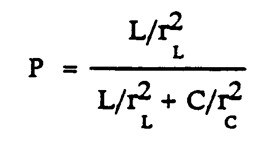
- L =
- # first ligand sites
- C =
- # cell surface sites
- r² =
- average distance squared to site.
- The dissociation constant for a second ligand desorbing from a surface can be readily measured using radiolabeled second ligand according to the method of Olson et al. ("Dissociation Kinetics of Antigen-Antibody interactions: Studies on a Panel of Anti-Albumin Monoclonal Antibodies," Molecular Immunology 26:129-136, 1989). Briefly, the particle surface is first saturated with second ligand, a proportion of which is radiolabeled. The surface is then placed in fresh solution containing second ligand which is not radiolabeled. The rate of appearance of radiolabeled second ligand in the fresh solution can be used to calculate the rate constant for dissociation of second ligand from the particle surface. The other variables may also be readily determined. For example, the number of cell surface sites may be determined through the use of a radiolabeled antibody, or by quantitative FACS analysis, and the number of avidin sites may be measured with radiolabeled biotin. The "average distance squared to site" may be calculated by squaring the distance between the ligand and each respective site, summing the squared distances over the total number of sites, and dividing by the number of sites.
- As noted above, the second ligand is removed from the particle surface without substantially affecting the particle surface. Through use of the present invention at least 25%, and preferably 70% to 90% of the second ligand is removed from the particle surface. Furthermore, within the context of the present invention, a particle surface is not "substantially affected" if greater than 90% of the cell surface antigens or receptors remain. This determination may be easily accomplished through many different techniques. For example, cells which were labeled with biotinylated IL-2 may be passed through an avidin-coated support material in order to remove the bound IL-2. The cells may then be labeled with a fluorescein-labeled anti-IL-2 receptor antibody and analyzed by FACS to determine if the intensity of fluorescence staining is within 90% of the fluorescence intensity of starting cells which were never exposed to the biotinylated IL-2.
- Within one aspect of the present invention, subsequent to the step of exposing the particle to the first ligand, the support upon which the first ligand is immobilized is agitated. Within a preferred embodiment, a support such as Biogel™ beads with an immobilized first ligand are contained within a column. Also contained within the column is a means for agitating the beads. The particle with bound second ligand is exposed to the immobilized first ligand, resulting in the binding of the first ligand to the second ligand, hence immobilizing the particles. Upon application of an external force, the means for agitation agitates the beads. The weakest bonds are disrupted first; since the affinity of the first ligand for the second ligand is at least two orders of magnitude greater than the affinity of the second ligand for the particle surface, the particle - second ligand bond will be disrupted first, removing the second ligand from the particle surface.
- Means for agitating the support include magnetic impellers (Parker Industries Corporation, Chicago, Ill.), magnetic beads (Robbin Scientific, Mountain View, Calif.), weights (Astrolite Alloys, Camarillo, Calif.), magnetic weights, pipettes, and buoyant floats (Polysciences Corp., Warrington, Pa.). The external force which supplies energy to the means for agitating the support is generally located outside of the column and includes electromagnetic, gravitational and mechanical forces. Electromagnetic forces may act on means such as metallic or magnetic beads or weights, causing agitation and release of particles. Electromagnetic forces may be provided by, for example, magnetic stir plates (VWR Scientific, San Francisco, Calif.) or electromagnetic coils (Radio Shack). Gravitational forces may be provided by, for example, rocking plates or manual shaking of a column, such that the forces of gravitation agitate the support, thereby causing release of particles. A weight which is either denser or more buoyant than the support may be placed in the bed to enhance the effect of gravitational forces on bed agitation. Mechanical forces may be provided by, for example, hydrodynamic jets (VWR Scientific, San Francisco, Calif.), vortexes (VWR Scientific, San Francisco, Calif.), or sonicators (Curtin Matheson Scientific, Seattle, Wash.). To determine the proper level of agitation using these means, the level of force is gradually increased until the support is visibly moved. This level of agitation may then be fine-tuned (e.g., a lower force to remove only nonspecifically bound particles, or a higher force to remove the most tightly bound particles) to fit the specific application.
- The following examples are offered by way of illustration and not by way of limitation.
- Seventeen grams of dry Biogel P-60™, (50-100 mesh (wet), coarse beads) (BIORAD, Catalog No. 150-1630, Richmond, Calif.) are added to 1.5 l of 0.5 M NaHCO₃/0.5 M Na₂CO₃. The pH is adjusted to 10.5 with NaOH and carefully stirred with a mixer (RZR1, Carfamo, Wiarton, Ontario, Canada) so as not to damage the beads for approximately 20 to 30 minutes. The mixture is then placed in a 60°C water bath. After the mixture reaches a temperature of 60°C, it is incubated for an additional 2 hours (at 60°C) with occasional stirring. The mixture is then removed from the water bath, and placed in an ice bath to bring the mixture temperature down to room temperature.
- The beads are washed several times with distilled or deionized water, followed by several washings of PBS using a coarse glass filter connected to a vacuum. The carboxylated gel may be stored in PBS at 4°C, and is stable for up to one year if sterilized or stored with a preservative.
- PBS is first removed from a measured amount of carboxylated Biogel by filtering with a coarse glass filter connected to a vacuum. The gel is then equilibrated in distilled or deionized water for 15-30 minutes. Equilibration in water causes an expansion of the gel to a volume of about 4 times its previously measured amount. The gel is resuspended in 10 ml of distilled or deionized water for each ml of gel (as originally measured in PBS).
- Ten mg of 1-ethyl-3-(3-dimethylaminopropyl) carbodiimide (EDC-HCl) (Sigma Chemical Co., Catalog No. E7750, St. Louis, Mo.) is added for each ml of gel as originally measured. The pH is rapidly adjusted to 5.5 by dropwise addition of HCl. Care is taken to maintain the pH at 5.5; pHs of less than 5.0 or greater than 6.0 result in significantly less activation of the Biogel. The mixture is stirred for five minutes.
- Avidin (International Enzymes, Inc., Fallbrook, Calif.) is dissolved at a concentration of between 10 and 100 mg/ml in deionized water. Next, 100 µg of avidin is rapidly added for each ml of gel (as originally measured in PBS). The mixture is stirred for 1.5 hours. Next, 2 M glycine is added to give a final concentration of 0.2 M glycine in the mixture, and stirred for an additional 1 hour.
- The gel is washed with several volumes of PBS using a coarse glass filter and vacuum, and stored in PBS with 0.1% NaN₃ at 4°C. The gel is stable for approximately one year.
- Daudi cells (ATCC No. CCL 213) are centrifuged at 280 g for 8 minutes and resuspended in PBS + 1% BSA at 1 x 10⁸ cells/ml. The cells are incubated with biotinylated anti-Ia antibody at a concentration of 25 µg/ml for 30 minutes at 4°C. The cells are resuspended in PBS + 1% BSA after the first spin, then resuspended in PBS + 5% BSA to a concentration of 5 x 10⁷ cells/ml following the second spin.
- Carboxylated Biogel P-30™ (prepared as described above) is allowed to equilibrate to room temperature and placed in a K9/15 column (Pharmacia, Piscataway, N.J.) to a total bed height of 1 cm. The column is washed with PBS, followed by washes with PBS plus 5% BSA. This column functions as a "pre-column." The avidin column contains avidin-conjugated Biogel P-60™, which is prepared as described above. The avidin-conjugated Biogel is allowed to equilibrate to room temperature, and placed in a K9/15 column to a total bed height of 4 cm. The column is then washed with several volumes of PBS, followed by washes with PBS plus 5% BSA.
- Cells which have been prepared as described above are gently transferred onto the top of the gel bed of the pre-column. The cells are allowed to filter through the pre-column and are washed with 1 ml PBS plus 5% BSA into the avidin column. A peristaltic pump (Cole-Parmer, Rockford, Ill.) controls flow from the avidin column to a rate of about 1 ml/minute. Once the cells have almost all entered the top of the avidin column bed, 1-2 ml of PBS plus 5% BSA is added to the top of the avidin column in order to wash out remaining cells. The column is washed out with 4-6 ml of PBS.
- The avidin column is placed on top of a 15 ml centrifuge tube. The valve of the column is opened and 15 ml of PBS is added to the column with a wide bore, 9-inch transfer pipette. The PBS is added to the column while the pipette is used for mechanically agitating and resuspending the cell bed, thus allowing cells to become detached from the gel matrix, and to filter into the centrifuge tube. The tube is then centrifuged at 280 g for 8 minutes and the pellet is resuspended in Iscoves' Modified Dulbecco's Medium plus 10% Fetal Bovine Serum. The total time of exposure of the cells to the avidin gel is about 15 minutes.
- The cells are labeled with fluorescein-tagged antibodies and analyzed with a FACScan for fluorescence intensity. The starting cells, nonadherent, and the cells which adhered to the avidin-conjugated Biogel are stained with fluoresceinated goat-anti-mouse IgG (Tago) to determine the amount of anti-Ia antibody remaining on the cell surface. The adherent cells bound 30% and the nonadherent cells bound 25% as much of the goat-anti-mouse FITC as the starting cells, indicating that 70%-75% of the anti-Ia antibody is removed from the cell surface. Next the adherent and starting cells are restained with a fluoresceinated anti-HLA-dr (Becton Dickinson, Mountain View, Calif.) antibody to determine if the antigen density had been altered. The adherent cells stained 16% brighter than the starting cells indicating that the Ia antigen (HLA-DR molecule) remained on the cells even though the anti-Ia antibody had been removed.
- CEM cells (ATCC #CCL 119) are centrifuged at 280 g for 8 minutes and 20 x 10⁶ cells were resuspended in 100 ul of biotinylated transferrin (Terry Fox Laboratory, Vancouver, B.C.) at 0.3 mg/ml for 30 minutes at 4°C. The cells are washed by centrifugation at 280 g for 8 minutes, resuspended in PBS + 1% BSA and centrifuged again, then resuspended in PBS + 5% BSA to a concentration of 5 x 10⁷ cells/ml.
- A carboxylated Biogel P-30™ precolumn and a P60 avidin column are prepared and operated as described above in Example 3.
- The cells are labeled with fluorescein-tagged avidin (Vector, Burlingame, Calif.) to measure the amount of biotinylated transferrin on the cells. The cells are relabeled with biotinylated transferrin then fluorescein-tagged avidin to measure the amount of transferrin receptors on the cells. The amount of label is then analyzed with a FACScan for fluorescence intensity.
- The cells which bind to the column and are agitated off after approximately 15 minutes residence time have lost 87% of the biotinylated transferrin. Those cells which remain on the column for 60 minutes before agitation have lost 90% of the transferrin on their surface. Restaining with biotinylated transferrin, followed by avidin-FITC shows that the cells averaged 97% as many transferrin receptors as the starting cells.
Claims (11)
- A method for removing a second ligand from a surface of a virus, bacteria, fungus, parasite or cell without substantially affecting the surface of the virus, bacteria, fungus, parasite or cell, comprising:
exposing the virus, bacteria, fungus, parasite or cell to a first ligand immobilized onto a support, characterized in that said virus, bacteria, fungus, parasite or cell is exposed under conditions and for a residence time sufficient to allow the second ligand to desorb from the surface of the virus, bacteria, fungus, parasite or cell, and wherein said first ligand has an affinity for the second ligand that is at least two orders of magnitude greater than the affinity of the second ligand for the surface of the virus, bacteria, fungus, parasite or cell, such that at least 25% of the second ligand is removed from the surface of the virus, bacteria, fungus, parasite or cell without substantially affecting the surface of the virus, bacteria, fungus, parasite or cell. - The method of claim 1, further comprising, subsequent to the step of exposing, agitating the support upon which the first ligand is immobilized.
- The method of Claims 1 or 2 wherein the first ligand is avidin and the second ligand is biotinylated transferrin.
- The method of Claims 1 or 2 wherein the first ligand is avidin and the second ligand is biotinylated IL-2.
- The method of Claims 1 or 2 wherein the first ligand is avidin and the second ligand is a biotinylated anti-virus, anti-bacteria, anti-fungus, anti-parasite or anti-cell surface antibody.
- The method of Claims 1 or 2 wherein the first ligand is protein A and the second ligand is an anti-virus, anti-bacteria, anti-fungus, anti-parasite or anti-cell surface antibody.
- The method of Claims 1 or 2 wherein the first ligand is protein G and the second ligand is an anti-virus, anti-bacteria, anti-fungus, anti-parasite or anti-cell surface antibody.
- A method for removing biotinylated antibody from a cell surface without substantially affecting the cell surface, comprising:
exposing the cell to immobilized avidin characterized in that said exposure is under conditions, and for a residence time sufficient to allow the biotinylated antibody to desorb from the cell, wherein the avidin has an affinity for the biotinylated antibody that is at least two orders of magnitude greater than the affinity of the biotinylated antibody for the cell surface, such that, at least 25% of the biotinylated antibody is removed from the cell surface without substantially affecting the cell surface. - A method according to any one of the preceding claims such that at least 70% of the second ligand is removed from the surface of the virus, bacteria, fungus, parasite or cell without substantially affecting the surface of the virus, bacteria, fungus, parasite or cell.
- A method of Claims 1 or 2 wherein said cell is a B-lymphocyte, T-lyphocyte or hematopoietic stem cell.
- A method of Claim 10 wherein an hematopoietic stem cell is a CD34 positive cell.
Applications Claiming Priority (3)
| Application Number | Priority Date | Filing Date | Title |
|---|---|---|---|
| US07/513,056 US5378624A (en) | 1990-04-23 | 1990-04-23 | Methods for removing ligands from a particle surface |
| PCT/US1991/002785 WO1991016088A1 (en) | 1990-04-23 | 1991-04-23 | Methods for removing ligands from a particle surface |
| US513056 | 2000-02-24 |
Publications (2)
| Publication Number | Publication Date |
|---|---|
| EP0526577A1 EP0526577A1 (en) | 1993-02-10 |
| EP0526577B1 true EP0526577B1 (en) | 1994-11-30 |
Family
ID=24041719
Family Applications (1)
| Application Number | Title | Priority Date | Filing Date |
|---|---|---|---|
| EP91909347A Expired - Lifetime EP0526577B1 (en) | 1990-04-23 | 1991-04-23 | Methods for removing ligands from a particle surface |
Country Status (9)
| Country | Link |
|---|---|
| US (1) | US5378624A (en) |
| EP (1) | EP0526577B1 (en) |
| JP (1) | JP2721040B2 (en) |
| AT (1) | ATE114484T1 (en) |
| CA (1) | CA2081202C (en) |
| DE (1) | DE69105497T2 (en) |
| DK (1) | DK0526577T3 (en) |
| ES (1) | ES2065686T3 (en) |
| WO (1) | WO1991016088A1 (en) |
Families Citing this family (23)
| Publication number | Priority date | Publication date | Assignee | Title |
|---|---|---|---|---|
| US5536475A (en) * | 1988-10-11 | 1996-07-16 | Baxter International Inc. | Apparatus for magnetic cell separation |
| US5635387A (en) * | 1990-04-23 | 1997-06-03 | Cellpro, Inc. | Methods and device for culturing human hematopoietic cells and their precursors |
| EP0553288B1 (en) * | 1990-10-18 | 1997-06-18 | Cellpro Incorporated | An apparatus and method for separating particles using a pliable vessel |
| US6825169B1 (en) | 1991-10-22 | 2004-11-30 | Trustees Of Tufts College | Inhibitors of dipeptidyl-aminopeptidase type IV |
| US6120735A (en) * | 1992-02-26 | 2000-09-19 | The Ohio States University | Fractional cell sorter |
| US5965532A (en) | 1996-06-28 | 1999-10-12 | Trustees Of Tufts College | Multivalent compounds for crosslinking receptors and uses thereof |
| US6100234A (en) | 1997-05-07 | 2000-08-08 | Tufts University | Treatment of HIV |
| US6040145A (en) | 1997-05-07 | 2000-03-21 | Tufts University | Potentiation of the immune response |
| CA2304206A1 (en) | 1997-09-29 | 1999-04-08 | Point Therapeutics, Inc. | Stimulation of hematopoietic cells in vitro |
| MY132924A (en) | 1998-05-04 | 2007-10-31 | Point Therapeutics Inc | Hematopoietic stimulation |
| WO1999062914A1 (en) | 1998-06-05 | 1999-12-09 | Point Therapeutics, Inc. | Cyclic boroproline compounds |
| US6890904B1 (en) | 1999-05-25 | 2005-05-10 | Point Therapeutics, Inc. | Anti-tumor agents |
| US20060014697A1 (en) * | 2001-08-22 | 2006-01-19 | Travis Mickle | Pharmaceutical compositions for prevention of overdose or abuse |
| US20030198967A1 (en) * | 2002-04-23 | 2003-10-23 | Matson Robert S. | Multi-functional microarrays and methods |
| US7439062B2 (en) * | 2004-12-23 | 2008-10-21 | Biocept, Inc. | Beads for capturing target cells from bodily fluid |
| US20090136982A1 (en) * | 2005-01-18 | 2009-05-28 | Biocept, Inc. | Cell separation using microchannel having patterned posts |
| US8158410B2 (en) | 2005-01-18 | 2012-04-17 | Biocept, Inc. | Recovery of rare cells using a microchannel apparatus with patterned posts |
| WO2006078470A2 (en) | 2005-01-18 | 2006-07-27 | Biocept, Inc. | Cell separation using microchannel having patterned posts |
| US20060252087A1 (en) * | 2005-01-18 | 2006-11-09 | Biocept, Inc. | Recovery of rare cells using a microchannel apparatus with patterned posts |
| US7695956B2 (en) * | 2006-01-12 | 2010-04-13 | Biocept, Inc. | Device for cell separation and analysis and method of using |
| KR102300886B1 (en) * | 2013-12-02 | 2021-09-10 | 삼성전자주식회사 | Methods of removing surface ligand compounds |
| US9492840B2 (en) * | 2013-12-02 | 2016-11-15 | Samsung Electronics Co., Ltd. | Methods of removing surface ligand compounds |
| US20190127697A1 (en) * | 2016-04-30 | 2019-05-02 | BioLegend, Inc. | Compositions and methods for performing magnetibuoyant separations |
Family Cites Families (16)
| Publication number | Priority date | Publication date | Assignee | Title |
|---|---|---|---|---|
| US4276206A (en) * | 1979-01-18 | 1981-06-30 | Scripps Clinic & Research Foundation | Immunochemical conjugates: method and composition |
| US4230685A (en) * | 1979-02-28 | 1980-10-28 | Northwestern University | Method of magnetic separation of cells and the like, and microspheres for use therein |
| JPS603367B2 (en) * | 1979-10-09 | 1985-01-28 | 旭化成株式会社 | Leukocyte separation method and leukocyte separation material |
| US4513088A (en) * | 1983-03-30 | 1985-04-23 | The Board Of Trustees Of The Leland Stanford Junior University | Assay for monoclonal antibody against surface Ig of a human B cell tumor |
| US4550075A (en) * | 1983-06-22 | 1985-10-29 | Kallestad Laboratories, Inc. | Method for ligand determination utilizing an immunoassay monitorable by biotin-containing enzymes, and compositions therefor |
| US4965204A (en) * | 1984-02-06 | 1990-10-23 | The Johns Hopkins University | Human stem cells and monoclonal antibodies |
| US4714680B1 (en) * | 1984-02-06 | 1995-06-27 | Univ Johns Hopkins | Human stem cells |
| US4935339A (en) * | 1985-05-07 | 1990-06-19 | Nichols Institute Diagnostics | Delayed solid phase immunologic assay |
| EP0260280B2 (en) * | 1986-01-30 | 1995-09-20 | Fred Hutchinson Cancer Research Center | Immunoselection method |
| US5225353A (en) * | 1986-01-30 | 1993-07-06 | Fred Hutchinson Cancer Research Center | Method for immunoselection of cells using avidin and biotin |
| US5215927A (en) * | 1986-01-30 | 1993-06-01 | Fred Hutchinson Cancer Research Center | Method for immunoselection of cells using avidin and biotin |
| US5262334A (en) * | 1986-01-30 | 1993-11-16 | Fred Hutchinson Cancer Research Center | Method for immunoselection of cells using avidin and biotin |
| US4798795A (en) * | 1987-02-17 | 1989-01-17 | Hoechst Celanese Corporation | Biotinylating agents |
| US5006464A (en) * | 1987-10-01 | 1991-04-09 | E-Y Laboratories, Inc. | Directed flow diagnostic device and method |
| US5256532A (en) * | 1988-05-02 | 1993-10-26 | Zynaxis Technologies, Inc. | Methods, reagents and test kits for determination of subpopulations of biological entities |
| US5081030A (en) * | 1989-04-25 | 1992-01-14 | The Johns Hopkins University | Release of cells from affinity matrices |
-
1990
- 1990-04-23 US US07/513,056 patent/US5378624A/en not_active Expired - Fee Related
-
1991
- 1991-04-23 DE DE69105497T patent/DE69105497T2/en not_active Expired - Fee Related
- 1991-04-23 JP JP3509004A patent/JP2721040B2/en not_active Expired - Lifetime
- 1991-04-23 DK DK91909347.6T patent/DK0526577T3/en active
- 1991-04-23 WO PCT/US1991/002785 patent/WO1991016088A1/en not_active Ceased
- 1991-04-23 CA CA002081202A patent/CA2081202C/en not_active Expired - Fee Related
- 1991-04-23 ES ES91909347T patent/ES2065686T3/en not_active Expired - Lifetime
- 1991-04-23 AT AT91909347T patent/ATE114484T1/en active
- 1991-04-23 EP EP91909347A patent/EP0526577B1/en not_active Expired - Lifetime
Also Published As
| Publication number | Publication date |
|---|---|
| JPH05507792A (en) | 1993-11-04 |
| ES2065686T3 (en) | 1995-02-16 |
| CA2081202C (en) | 2000-03-21 |
| JP2721040B2 (en) | 1998-03-04 |
| DK0526577T3 (en) | 1995-05-15 |
| ATE114484T1 (en) | 1994-12-15 |
| US5378624A (en) | 1995-01-03 |
| WO1991016088A1 (en) | 1991-10-31 |
| CA2081202A1 (en) | 1991-10-24 |
| DE69105497D1 (en) | 1995-01-12 |
| EP0526577A1 (en) | 1993-02-10 |
| DE69105497T2 (en) | 1995-04-06 |
Similar Documents
| Publication | Publication Date | Title |
|---|---|---|
| US5378624A (en) | Methods for removing ligands from a particle surface | |
| Abts et al. | CD20 positive human B lymphocytes separated with the magnetic cell sorter (MACS) can be induced to proliferation and antibody secretion in vitro | |
| US9739768B2 (en) | Methods and reagents for improved selection of biological materials | |
| JP4151986B2 (en) | Method for measuring lymphocyte function | |
| US20030143642A1 (en) | Secretions of proteins by encapsulated cells | |
| WO1991016116A1 (en) | Immunoselection device and method | |
| EP1207961B1 (en) | Methods of modification of selected cells in a magnetic cell separation column | |
| JPH0664064B2 (en) | Products for separation applicable to cells in immunopurification | |
| WO1992004961A1 (en) | Apparatus and methods for magnetic separation | |
| WO1988008538A1 (en) | Immune profile assay and device | |
| WO1994009117A9 (en) | Direct selection of cells by secretion product | |
| JP2000500337A (en) | Method and apparatus for bulk enrichment of a population or subpopulation of cells | |
| EP0819250A1 (en) | Multiparameter cell separation using releasable colloidal magnetic particles | |
| Sekine et al. | Panning of multiple subsets of leukocytes on antibody-decorated poly (ethylene) glycol-coated glass slides | |
| Hebell et al. | The isolation of B lymphocytes from human peripheral blood using antibodies coupled to paramagnetic particles and rosetting techniques | |
| US20020019004A1 (en) | Method for isolating molecules, cells and other particles which are specifically bound to a large particle | |
| EP0114528A2 (en) | Cell separation on plates using non-antibody ligands | |
| Andreoni | Immunomagnetic Particles for Cell Isolation | |
| Malsch et al. | Measurement of fluorescent-labeled LMM-heparin in biological fluids using protamine-linked microbeads | |
| Campbell et al. | The use of flow microfluorimetry in the analysis of the phenotype expression of mouse histocompatibility antigens | |
| Wang | Sorting of human peripheral blood T-cell subsets using immunomagnetic beads | |
| JPH04131764A (en) | Immunological measuring reagent for diagnosis of bernard-sourier syndrome and a one for disgnosis of blood platelet asthenia | |
| JPS6353511B2 (en) |
Legal Events
| Date | Code | Title | Description |
|---|---|---|---|
| PUAI | Public reference made under article 153(3) epc to a published international application that has entered the european phase |
Free format text: ORIGINAL CODE: 0009012 |
|
| 17P | Request for examination filed |
Effective date: 19921028 |
|
| AK | Designated contracting states |
Kind code of ref document: A1 Designated state(s): AT BE CH DE DK ES FR GB GR IT LI LU NL SE |
|
| 17Q | First examination report despatched |
Effective date: 19930416 |
|
| GRAA | (expected) grant |
Free format text: ORIGINAL CODE: 0009210 |
|
| AK | Designated contracting states |
Kind code of ref document: B1 Designated state(s): AT BE CH DE DK ES FR GB GR IT LI LU NL SE |
|
| REF | Corresponds to: |
Ref document number: 114484 Country of ref document: AT Date of ref document: 19941215 Kind code of ref document: T |
|
| ITF | It: translation for a ep patent filed | ||
| REF | Corresponds to: |
Ref document number: 69105497 Country of ref document: DE Date of ref document: 19950112 |
|
| REG | Reference to a national code |
Ref country code: ES Ref legal event code: FG2A Ref document number: 2065686 Country of ref document: ES Kind code of ref document: T3 |
|
| ET | Fr: translation filed | ||
| REG | Reference to a national code |
Ref country code: GR Ref legal event code: FG4A Free format text: 3014772 |
|
| REG | Reference to a national code |
Ref country code: DK Ref legal event code: T3 |
|
| PLBI | Opposition filed |
Free format text: ORIGINAL CODE: 0009260 |
|
| 26 | Opposition filed |
Opponent name: BAXTER HEALTHCARE CORPORATION Effective date: 19950830 |
|
| NLR1 | Nl: opposition has been filed with the epo |
Opponent name: BAXTER HEALTHCARE CORPORATION |
|
| PLBF | Reply of patent proprietor to notice(s) of opposition |
Free format text: ORIGINAL CODE: EPIDOS OBSO |
|
| PLBF | Reply of patent proprietor to notice(s) of opposition |
Free format text: ORIGINAL CODE: EPIDOS OBSO |
|
| PLBO | Opposition rejected |
Free format text: ORIGINAL CODE: EPIDOS REJO |
|
| PLBN | Opposition rejected |
Free format text: ORIGINAL CODE: 0009273 |
|
| STAA | Information on the status of an ep patent application or granted ep patent |
Free format text: STATUS: OPPOSITION REJECTED |
|
| 27O | Opposition rejected |
Effective date: 19970214 |
|
| NLR2 | Nl: decision of opposition | ||
| PGFP | Annual fee paid to national office [announced via postgrant information from national office to epo] |
Ref country code: LU Payment date: 20001013 Year of fee payment: 10 |
|
| PGFP | Annual fee paid to national office [announced via postgrant information from national office to epo] |
Ref country code: FR Payment date: 20010418 Year of fee payment: 11 |
|
| PGFP | Annual fee paid to national office [announced via postgrant information from national office to epo] |
Ref country code: NL Payment date: 20010419 Year of fee payment: 11 Ref country code: DE Payment date: 20010419 Year of fee payment: 11 |
|
| PGFP | Annual fee paid to national office [announced via postgrant information from national office to epo] |
Ref country code: SE Payment date: 20010420 Year of fee payment: 11 Ref country code: GB Payment date: 20010420 Year of fee payment: 11 Ref country code: DK Payment date: 20010420 Year of fee payment: 11 Ref country code: CH Payment date: 20010420 Year of fee payment: 11 Ref country code: AT Payment date: 20010420 Year of fee payment: 11 |
|
| PG25 | Lapsed in a contracting state [announced via postgrant information from national office to epo] |
Ref country code: LU Free format text: LAPSE BECAUSE OF NON-PAYMENT OF DUE FEES Effective date: 20010423 |
|
| PGFP | Annual fee paid to national office [announced via postgrant information from national office to epo] |
Ref country code: GR Payment date: 20010427 Year of fee payment: 11 |
|
| PGFP | Annual fee paid to national office [announced via postgrant information from national office to epo] |
Ref country code: ES Payment date: 20010511 Year of fee payment: 11 Ref country code: BE Payment date: 20010511 Year of fee payment: 11 |
|
| REG | Reference to a national code |
Ref country code: GB Ref legal event code: IF02 |
|
| PG25 | Lapsed in a contracting state [announced via postgrant information from national office to epo] |
Ref country code: GB Free format text: LAPSE BECAUSE OF NON-PAYMENT OF DUE FEES Effective date: 20020423 Ref country code: AT Free format text: LAPSE BECAUSE OF NON-PAYMENT OF DUE FEES Effective date: 20020423 |
|
| PG25 | Lapsed in a contracting state [announced via postgrant information from national office to epo] |
Ref country code: SE Free format text: LAPSE BECAUSE OF NON-PAYMENT OF DUE FEES Effective date: 20020424 Ref country code: ES Free format text: LAPSE BECAUSE OF NON-PAYMENT OF DUE FEES Effective date: 20020424 |
|
| PG25 | Lapsed in a contracting state [announced via postgrant information from national office to epo] |
Ref country code: LI Free format text: LAPSE BECAUSE OF NON-PAYMENT OF DUE FEES Effective date: 20020430 Ref country code: DK Free format text: LAPSE BECAUSE OF NON-PAYMENT OF DUE FEES Effective date: 20020430 Ref country code: CH Free format text: LAPSE BECAUSE OF NON-PAYMENT OF DUE FEES Effective date: 20020430 Ref country code: BE Free format text: LAPSE BECAUSE OF NON-PAYMENT OF DUE FEES Effective date: 20020430 |
|
| PG25 | Lapsed in a contracting state [announced via postgrant information from national office to epo] |
Ref country code: NL Free format text: LAPSE BECAUSE OF NON-PAYMENT OF DUE FEES Effective date: 20021101 Ref country code: DE Free format text: LAPSE BECAUSE OF NON-PAYMENT OF DUE FEES Effective date: 20021101 |
|
| PG25 | Lapsed in a contracting state [announced via postgrant information from national office to epo] |
Ref country code: GR Free format text: LAPSE BECAUSE OF NON-PAYMENT OF DUE FEES Effective date: 20021105 |
|
| EUG | Se: european patent has lapsed |
Ref document number: 91909347.6 |
|
| GBPC | Gb: european patent ceased through non-payment of renewal fee |
Effective date: 20020423 |
|
| REG | Reference to a national code |
Ref country code: CH Ref legal event code: PL |
|
| REG | Reference to a national code |
Ref country code: DK Ref legal event code: EBP |
|
| PG25 | Lapsed in a contracting state [announced via postgrant information from national office to epo] |
Ref country code: FR Free format text: LAPSE BECAUSE OF NON-PAYMENT OF DUE FEES Effective date: 20021231 |
|
| NLV4 | Nl: lapsed or anulled due to non-payment of the annual fee |
Effective date: 20021101 |
|
| REG | Reference to a national code |
Ref country code: FR Ref legal event code: ST |
|
| REG | Reference to a national code |
Ref country code: ES Ref legal event code: FD2A Effective date: 20030514 |
|
| PG25 | Lapsed in a contracting state [announced via postgrant information from national office to epo] |
Ref country code: IT Free format text: LAPSE BECAUSE OF NON-PAYMENT OF DUE FEES;WARNING: LAPSES OF ITALIAN PATENTS WITH EFFECTIVE DATE BEFORE 2007 MAY HAVE OCCURRED AT ANY TIME BEFORE 2007. THE CORRECT EFFECTIVE DATE MAY BE DIFFERENT FROM THE ONE RECORDED. Effective date: 20050423 |