EP0523769B1 - Gram-positive alkaliphilic microorganisms - Google Patents
Gram-positive alkaliphilic microorganisms Download PDFInfo
- Publication number
- EP0523769B1 EP0523769B1 EP92201883A EP92201883A EP0523769B1 EP 0523769 B1 EP0523769 B1 EP 0523769B1 EP 92201883 A EP92201883 A EP 92201883A EP 92201883 A EP92201883 A EP 92201883A EP 0523769 B1 EP0523769 B1 EP 0523769B1
- Authority
- EP
- European Patent Office
- Prior art keywords
- bacteria
- positive
- growth
- grows
- gives
- Prior art date
- Legal status (The legal status is an assumption and is not a legal conclusion. Google has not performed a legal analysis and makes no representation as to the accuracy of the status listed.)
- Expired - Lifetime
Links
Images

Classifications
-
- C—CHEMISTRY; METALLURGY
- C12—BIOCHEMISTRY; BEER; SPIRITS; WINE; VINEGAR; MICROBIOLOGY; ENZYMOLOGY; MUTATION OR GENETIC ENGINEERING
- C12N—MICROORGANISMS OR ENZYMES; COMPOSITIONS THEREOF; PROPAGATING, PRESERVING, OR MAINTAINING MICROORGANISMS; MUTATION OR GENETIC ENGINEERING; CULTURE MEDIA
- C12N9/00—Enzymes; Proenzymes; Compositions thereof; Processes for preparing, activating, inhibiting, separating or purifying enzymes
- C12N9/14—Hydrolases (3)
- C12N9/24—Hydrolases (3) acting on glycosyl compounds (3.2)
- C12N9/2402—Hydrolases (3) acting on glycosyl compounds (3.2) hydrolysing O- and S- glycosyl compounds (3.2.1)
- C12N9/2405—Glucanases
- C12N9/2408—Glucanases acting on alpha -1,4-glucosidic bonds
- C12N9/2411—Amylases
- C12N9/2414—Alpha-amylase (3.2.1.1.)
- C12N9/2417—Alpha-amylase (3.2.1.1.) from microbiological source
-
- C—CHEMISTRY; METALLURGY
- C12—BIOCHEMISTRY; BEER; SPIRITS; WINE; VINEGAR; MICROBIOLOGY; ENZYMOLOGY; MUTATION OR GENETIC ENGINEERING
- C12N—MICROORGANISMS OR ENZYMES; COMPOSITIONS THEREOF; PROPAGATING, PRESERVING, OR MAINTAINING MICROORGANISMS; MUTATION OR GENETIC ENGINEERING; CULTURE MEDIA
- C12N9/00—Enzymes; Proenzymes; Compositions thereof; Processes for preparing, activating, inhibiting, separating or purifying enzymes
-
- C—CHEMISTRY; METALLURGY
- C12—BIOCHEMISTRY; BEER; SPIRITS; WINE; VINEGAR; MICROBIOLOGY; ENZYMOLOGY; MUTATION OR GENETIC ENGINEERING
- C12N—MICROORGANISMS OR ENZYMES; COMPOSITIONS THEREOF; PROPAGATING, PRESERVING, OR MAINTAINING MICROORGANISMS; MUTATION OR GENETIC ENGINEERING; CULTURE MEDIA
- C12N9/00—Enzymes; Proenzymes; Compositions thereof; Processes for preparing, activating, inhibiting, separating or purifying enzymes
- C12N9/14—Hydrolases (3)
- C12N9/16—Hydrolases (3) acting on ester bonds (3.1)
- C12N9/18—Carboxylic ester hydrolases (3.1.1)
- C12N9/20—Triglyceride splitting, e.g. by means of lipase
-
- C—CHEMISTRY; METALLURGY
- C12—BIOCHEMISTRY; BEER; SPIRITS; WINE; VINEGAR; MICROBIOLOGY; ENZYMOLOGY; MUTATION OR GENETIC ENGINEERING
- C12N—MICROORGANISMS OR ENZYMES; COMPOSITIONS THEREOF; PROPAGATING, PRESERVING, OR MAINTAINING MICROORGANISMS; MUTATION OR GENETIC ENGINEERING; CULTURE MEDIA
- C12N9/00—Enzymes; Proenzymes; Compositions thereof; Processes for preparing, activating, inhibiting, separating or purifying enzymes
- C12N9/14—Hydrolases (3)
- C12N9/48—Hydrolases (3) acting on peptide bonds (3.4)
- C12N9/50—Proteinases, e.g. Endopeptidases (3.4.21-3.4.25)
- C12N9/52—Proteinases, e.g. Endopeptidases (3.4.21-3.4.25) derived from bacteria or Archaea
Definitions
- the present invention is in the field of microbiology and more particularly in the field of alkaliphilic microorganisms.
- Alkaliphiles are defined as organisms which exhibit optimum growth in an alkaline pH environment, particularly in excess of pH 8, and generally in the range between pH 9 and 10. Alkaliphiles may also be found living in environments having a pH as high as 12. Obligate alkaliphiles are incapable of growth at neutral pH.
- Alkaliphiles may be found in certain everyday environments such as garden soil, presumably due to transient alkaline conditions caused by biological activities including ammonification, sulphate reduction or photosynthesis. A much richer source of a greater variety of alkaliphilic organisms may be found in naturally occurring, stable alkaline environments such as soda lakes.
- Alkaliphiles the majority of which are Bacillus species, have been isolated from non-saline environments and are discussed by Horikoshi, K. and Akiba, T. in Alkalophilic Microorganisms (Springer-Verlag, Berlin, Heidelberg, N.Y., (1982)). However, alkaliphilic organisms from saline and alkaline environments such as lakes are not discussed therein. Strictly anaerobic bacteria from alkaline, hypersaline, environments have been recently described by Shiba, H. in Superbugs (eds. K. Horikoshi and W.D. Grant); Japan Scientific Societies Press, Tokyo and Springer-Verlag, Berlin, Heidelberg, N.Y., (1991), pp. 191-211; and by Nakatsugawa. N., ibid, pp. 212-220.
- Soda lakes which may be found in various locations around the world, are caused by a combination of geological, geographical and climatic conditions. They are characterized by the presence of large amounts of sodium carbonate (or complexes thereof) formed by evaporative concentration, as well as by the corresponding lack of Ca 2+ and Mg 2+ which would remove carbonate ions as insoluble salts. Other salts such as NaCl may also concentrate resulting in environments which are both alkaline and saline.
- soda lakes are nevertheless home to a large population of prokaryotes, a few types of which may dominate as permanent or seasonal blooms.
- the organisms range from alkaliphilic cyanobacteria to haloalkaliphilic archaeabacteria.
- alkaliphilic cyanobacteria haloalkaliphilic archaeabacteria.
- it is not unusual to find common types of alkaliphilic organisms inhabiting soda lakes in various widely dispersed locations throughout the world such as in the East African Rift Valley, in the western U.S., Cambodia, China and Hungary.
- natronobacteria have been isolated and identified in soda lakes located in China (Wang, D.
- Alkaliphiles have already made an impact in the application of biotechnology for the manufacture of consumer products.
- Alkalitolerant enzymes produced by alkaliphilic microorganisms have already found use in industrial processes and have considerable economic potential. For example, these enzymes are currently used in detergent compositions and in leather tanning, and are foreseen to find applications in the food, waste treatment and textile industries. Additionally, alkaliphiles and their enzymes are potentially useful for biotransformations, especially in the synthesis of pure enantiomers. Also, many of the microorganisms described herein are brightly pigmented and are potentially useful for the production of natural colorants.
- the present invention concerns novel aerobic, Gram-positive alkaliphilic bacteria. These bacteria have been isolated from samples of soil, water, sediment and a number of other sources, all of which were obtained from in and around alkaline soda lakes. These alkaliphiles have been analyzed according to the principles of numerical taxonomy with respect to each other and also to a collection of known bacteria in order to confirm their novelty. In addition, these bacterial taxa are further circumscribed by an analysis of the lipid components which serve as chemotaxonomic markers.
- the present invention also provides data as to the composition of the environments from which the samples containing the microorganisms were obtained, as well as the media required for their efficient isolation and culture such that one of ordinary skill may easily locate such an environment and be able to isolate the organisms of the present invention by following the procedures described herein.
- alkalitolerant enzymes are capable of performing their functions at high pH which makes them uniquely suited for applications requiring such extreme conditions.
- alkalitolerant enzymes may be employed in detergent compositions, in leather tanning and in the food, waste treatment and textile industries, as well as for biotransformations such as the production of pure enantiomers and the production of natural pigments.
- genes encoding these alkalitolerant enzymes may be isolated, cloned and brought to expression in compatible expression hosts to provide a source of larger yields of enzyme products which may be, if desired, more easily purified and used in various industrial applications, should the wild-type strain fail to produce sufficient amounts of the desired enzyme, or performs poorly under normal industrial fermentation conditions.
- the samples were collected in sterile plastic bags. Sampling was conducted at lakes Elmenteita, Nakuru, Bogoria, Crater (Sonachi), Little Naivasha (Oloidien) and Magadi, all of which are located in Kenya, East Africa. Alkaline soda lakes having similar environments may also be found in Cambodia, China, Hungary and the western U.S.. At each sampling site, the physical appearance of the site and the sample were described and physical parameters such as pH, conductivity and temperature were measured. Some of the samples were treated locally within 36 hours of collection of the samples but the majority were examined off-site, several weeks after collection.
- Table 1 lists various strains which have been isolated. The strains are listed according to the location from which the sample was taken and the physical appearance of the sample itself.
- Table 2 provides examples of typical chemical analyses of the lake waters at the sampling locations at the time of extraction of many of the samples. These data are consistent with earlier analyses (Grant, W.D. and Tindall, B.J., supra).
- Table 3 provides a list of the isolated strains arranged according to the results of the numerical taxonomic analysis ( Figure 1). Furthermore, Table 3 provides physical properties of the original sample, in particular the temperature, conductivity and alkaline pH, as well as the numerous isolation media required for obtaining pure cultures of these novel bacteria. These media are letter coded with reference to Appendix A.
- Tables 1, 2 and 3 provide data from which the environment of the sampling locations may be characterized.
- the chemical and physical analyses of the samples confirm the presence of alkaline pH, as well as the presence of unusually high levels of Na 2 CO 3 , coupled with low levels of Ca 2+ and Mg 2+ . It is known that the basic environments of soda lakes are stable with respect to their pH and ionic composition. Moreover, the microbial populations found at these sites remain largely stable. Thus, it is to be expected that the environment from which bacteria according to the present invention may be obtained can be determined from the data presented in Tables 1-3.
- the fresh soda-lake water samples were plated out on an alkaline nutrient medium (Medium A) soon after collection. Microscopic inspection of the soda lake samples showed a surprisingly high diversity of bacterial types. Considering the extremely alkaline nature of the environment, viable counts showed unexpectedly high numbers of organotrophic bacteria, in the range of 10 5 - 10 6 colony forming units per ml.
- the samples were stored either cooled or at ambient temperatures. After a few weeks' storage, the total numbers of bacteria in the sample rose, whereas the diversity of types decreased.
- wE1, wE2, wE4 Lake Elmenteita (east bay). Sediment and water, littoral zone. 60E.4 Lake Elmenteita (east bay). Mud, littoral zone. wN10, wN12, wN16 Lake Nakuru, north beach between Hippo Point and Njoro Point. Mud and water, littoral zone. wB3 Lake Bogoria, northern mud flats. Mud and water, littoral zone. 66B.4 Lake Bogoria (west shore), Loboru delta area. Soda crusts and mud (around hot spring). 69B.4 Lake Bogoria (south bay). Water column and sediment, littoral zone.
- the different nutrient media employed for the isolation of the new alkaliphilic bacteria are designated Medium A - Medium G.
- the composition of the various media employed is shown in Appendix A.
- the samples were suspended in an alkaline nutrient broth (Medium A) and the bacteria cultivated by shaking, preferably at 37°C for 2 - 3 days before spreading the broth onto an alkaline nutrient agar (Medium A) for the isolation of bacterial colonies.
- an alkaline nutrient broth Medium A
- the bacteria cultivated by shaking, preferably at 37°C for 2 - 3 days before spreading the broth onto an alkaline nutrient agar (Medium A) for the isolation of bacterial colonies.
- samples were spread onto alkaline nutrient agar containing specific substrates such as lactalbumin or casein or olive oil.
- specific substrates such as lactalbumin or casein or olive oil.
- the bacteria in the sample were enriched for 1 day up to several weeks in a non-specific alkaline nutrient broth such as Medium A before spreading the broth onto an alkaline nutrient agar specific for the detection of bacteria exhibiting enzyme activities such as lipolytic or proteolytic activity.
- Gram-positive bacteria Twenty strains of bacteria isolated from in and around alkaline lakes were assigned to the type of bacteria known as Gram-positive bacteria on the basis of (1) the Dussault modification of the Gram's staining reaction (Dussault, H.P., (1955), Journal of Bacteriology, 70 , 484-485); (2) the KOH sensitivity test (Gregersen, T., (1978), European Journal of Applied Microbiology and Biotechnology 5 , 123-127; Halebian, S.
- 17 known Gram-positive bacteria were subjected to the same analysis using the same conditions where appropriate. These reference bacteria included genera that are known to include facultative or obligate alkaliphilic species. These 17 known reference bacteria are recorded in Table 4 from which it will be seen that the "Type Strain" of the known species has been used where available. Thirteen of the strains are known alkaliphilic Bacillus species.
- RS7 (alkaliphilic) Bacillus species DSM 2514 (RS8) (alkaliphilic) Bacillus species DSM 2515 (RS10) (alkaliphilic) Bacillus species DSM 2517 (RS11) (alkaliphilic) Bacillus species DSM 2518 (RS12) (alkaliphilic) Bacillus species DSM 2519 (RS13) (alkaliphilic) Bacillus species DSM 2521 (RS14) (alkaliphilic) Bacillus species DSM 2523 (RS15) (alkaliphilic) Bacillus species DSM 2525 (RS16) Bacillus alcalophilus DSM 485 (RS17) Bacillus alcalophilus subsp.
- halodurans DSM 497 (AB30) (alkaliphilic) Bacillus species ATCC 21596 (AB42) (alkaliphilic) Bacillus species ATCC 21833 (AB49) (alkaliphilic) Bacillus species ATCC 21591 (Exig) Exiguobacterium aurantiacum NCIMB 11798 (BG114) Arthrobacter luteus ATCC 21596 (Br.li) Brevibacterium linens NCIMB 9904 (Micro) Micrococcus luteus NCTC 2665
- the phenetic data consisting of 200 unit characters was scored as indicated in Appendix C, and set out in the form of an "n x t" matrix, whose t columns represent the t bacterial strains to be grouped on the basis of resemblances, and whose n rows are the unit characters.
- Taxonomic resemblance of the bacterial strains was estimated by means of a similarity coefficient (Sneath, P.H.A. and Sokal, R.R., Numerical Taxonomy , supra, pp. 114-187). Although many different coefficients have been used for biological classification, only a few have found regular use in bacteriology. We have chosen to apply three association coefficients (Sneath, P.H.A.
- a similarity matrix was constructed for all pairs of strains using the Gower Coefficient (S G ) with the option of permitting negative matches (Sneath, P.H.A. and Sokal, R.R., supra, pp. 135-136) using the RTBNSIM program in TAXPAK.
- the Gower Coefficient was chosen over other coefficients for generating similarity matrices because it is applicable to all types of characters or data, namely, two-state, multistate (ordered and qualitative), and quantitative.
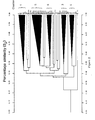
- the result of the cluster analysis is a dendrogram, a simplified version of which is provided in Figure 1.
- the dendrogram illustrates the levels of similarity between the bacterial strains.
- the dendrogram is obtained by using the DENDGR program in TAXPAK.
- S J Jaccard Coefficient
- S SM Simple Matching Coefficient
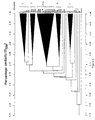
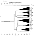
- a further two dendrograms were obtained by using the SMATCLST with UPGMA option and DENDGR sub-routines in TAXPAK. Simplified versions of these dendrograms are illustrated in Figure 2 and Figure 3 respectively.
- Figure 1 illustrates the results of the cluster analysis, based on the Gower Coefficient and the UPGMA method, of 20 Gram-positive, alkaliphilic bacteria isolated from in and around alkaline lakes, together with 17 known Gram-positive bacteria, including 14 alkaliphilic species.
- the cluster analysis groups the control Bacillus species in 3 distinct clusters which are separate from the novel alkaliphilic bacteria of the present invention. These results broadly concur with a taxonomic analysis of alkaliphilic Bacillus strains reported by Fritze, D., et al . (International Journal of Systematic Bacteriology, (1990), 40 , 92-97)). None of the known organisms are significantly related to any of the 3 clusters of new, Gram-positive alkaliphilic bacteria. A clear discrimination between these clusters is possible using the concept of the minimum discriminatory tests (see below) and chemotaxonomic information (see below).
- the cophenetic correlation is 0.804 which indicates the high degree of reliability with which this dendrogram represents the true taxonomic structure. (Sneath, P.H.A. and Sokal, R.R., supra, pp. 277-280, 304). Furthermore, the pattern of clusters obtained using the Jaccard Coefficient ( Figure 2 and below) and Simple Matching Coefficient ( Figure 3 and below) support the conclusions drawn here.
- the S J coefficient is a useful adjunct to the S G coefficient as it can be used to detect phenons in the latter that are based on negative matches or distortions owing to undue weight being put on potentially subjective qualitative data. Consequently, the S J coefficient is useful for confirming the validity of clusters defined initially by the use of the S G coefficient.
- the Jaccard Coefficient is particularly useful in comparing biochemically unreactive organisms (Austin, B. and Priest, F.G., supra, p. 37). There may be doubts about the admissability of matching negative character states (Sneath, P.H.A., and Sokal, R.R., supra, p. 131) in which case the Simple Matching Coefficient is a widely applied alternative.
- Chemotaxonomy is the study of the chemical variations of cells in relation to systematics.
- the analysis of chromosomal DNA, ribosomal RNA, proteins, cell walls and membranes, for example, can give valuable insights into taxonomic relationships and may be used as a further tool to classify or to verify the taxonomy of microorganisms (Goodfellow, M. and Minnikin, D.E. in Chemical Methods in Bacterial Systematics , (eds. Goodfellow, M. and Minnikin, D.E.), Academic Press, London and Orlando, FL, (1985), pp. 1-15).
- amphipathic polar lipids the major respiratory quinones, the fatty acids located in the bacterial membranes and the DNA base composition all have taxonomic significance for the classification of various bacteria (Lechevalier, H. and Lechevalier, M.P., in Microbial Lipids , volume 1 (eds. Ratledge, C. and Wilkinson, S.G.) Academic Press, London and San Diego, CA, (1988), pp. 869-902).
- lipids present on the chromatograms were visualized using a variety of differential stains (Ross, H.N.M., et al ., supra, p. 291, and Trincone, A., et al ., Journal of General Microbiology, (1990), 136 , pp. 2327-2331). The identity of components were confirmed by co-chromatography.
- glycolipid-containing strains contained a glycolipid also found in Gram-negative alkaliphiles isolated from soda lakes. Some of the other glycolipids appear to be common to certain clusters of Gram-positive alkaliphiles. It is possible therefore, that the chemical structures of the glycolipids will be chemotaxonomic markers for many obligate alkaliphiles in general and for specific groups in particular.
- the isoprenoid or respiratory quinones are characteristic components of the plasma membrane of aerobic bacteria. There are two types; menaquinones and ubiquinones.
- the value of isoprenoid quinones as taxonomic criteria lies in the variation in the length of the polyprenyl side-chain and the degree of saturation (Collins, M.D. and Jones, D. (1981), supra).
- Dry stationary phase bacterial cells were extracted, using a modified procedure of Collins, M.D. (in Chemical Methods in Bacterial Systematics, supra, pp. 267-284), in 1:1 (v/v) CHCl 3 :CH 3 OH at 50°C, for 16 hours.
- the quinones were examined by reverse phase thin layer chromatography as described by Collins, M.D. (supra).
- Freeze dried stationary phase cells (200-300 mg) were extracted for 16 hours at 75°C in toluene:methanol:conc. sulfuric acid (2.5 ml:2.5 ml:0.2 ml) and after cooling, the lipids were partitioned into hexane (twice times 1 ml). Residual acid was removed using NH 4 HCO 3 . Lipid extracts were concentrated under O 2 -free N 2 , dissolved in 300 ⁇ l hexane and applied to preparative silica gel plates (Merck F254, Type T). The plates were developed in hexane: diethyl ether 85:15 (v/v) and the fatty acid methyl esters scraped off, extracted with hexane and concentrated under a stream of O 2 -free N 2 .
- the fatty acid methyl esters were dissolved in heptane and analysed by gas chromatography using a Packard model 439 chromatograph equipped with flame ionization detectors. The samples were divided by a sample splitter and analyzed simultaneously over two columns, namely, CP-SIL-88 (Chrompack) (length 50 meter, internal diameter 0.22 mm) and Ultra-2 (Hewlett Packard) (length 50 m, internal diameter 0.20 mm). The carrier gas was nitrogen; the injection temperature 120°C; temperature gradient 2.5°C per minute to 240°C and isothermal at 240°C for 30 minutes. Fatty acid methyl esters were assigned by reference to known standard mixtures.
- the fatty acid composition of the individual Gram-positive alkaliphilic bacteria is set out in Table 8.
- Table 9 shows the unique fatty acid profiles of the individual clusters. Clusters 5 and 6 are typical for Bacillus species with a predominance of branched C15:0 and C17:0 fatty acids. In spite of the homgeneity of cluster 4 shown by the numerical taxonomy, fatty acid profiles clearly demonstrate that two subgroups of bacteria (designated 4A and 4B) exist within this cluster.
- CPN Coryneform-Mycobacterium-Nocardioform
- actinomycetes (Bennan, P.J., in Microbial Lipids , supra, pp 203-298), a designation supported by their characteristic cell habit, and the appearance of dihydromenaquinones.
- the bright orange-yellow colors of many actinomycetes is caused by the accumulation of carotenoid pigments, often induced by light. Many of these carotenoids are unique and important taxonomically.
- chemotaxonomic markers for these groups include a branched C19:0 fatty acid which is 10-methyloctadecanoic acid (tuberculostearic acid), an important criterion in the classification of the CMN group of bacteria. Branched, unsaturated fatty acids are also found in these bacteria. Branched C20:0 fatty acids, found in Clusters 3 and 4 are components of some Gram-positive cocci.
- the control bacteria for the coryneform group namely Arthrobacter luteus (now reclassified as Oerskovia xanthinolytica ) and Brevibacterium linens do not associate closely with any of the clusters.
- Arthrobacter luteus now reclassified as Oerskovia xanthinolytica
- Brevibacterium linens do not associate closely with any of the clusters.
- the known obligate alkaliphiles among the CMN class of bacteria are poorly characterized but are clearly different from the novel Gram-positive alkaliphile
- chromosomal DNA is unaffected by the growth conditions of the organism and an appropriate analysis may confirm or refute the taxonomic position of the organism.
- Chromosomal DNA may be analyzed by the determination of the base composition (G+C mol%) of individual strains.
- the guanine plus cytosine (G+C mol%) composition is constant for the chromosomal DNA from any given organism. Closely related organisms have similar G+C compositions. However, G+C results must be interpreted within the context of independent taxonomic data since similar G+C mol% of DNA samples from different organisms does not in itself imply biological relatedness.
- DNA was extracted from cells grown to exponential phase in Medium A by the chloroform:phenol method and was precipitated with ethanol.
- Base composition was determined by the thermal denaturation method (Marmur, J. and Doty, P. (1962), J. Mol. Biol., 3, 585-595) on a Phillips model PV8764 spectrophotometer with temperature programming.
- a second method involved HPLC analysis on a Beckman system gold using a Beckman ultrasphere ODS column and 0.04 M potassium dihydrogen phosphate plus acetonitrile (9 + 1, v/v) as eluent at a flow rate of 1.5 ml/min., after treatment of the DNA with nuclease P1 and alkaline phosphatase.
- centroid of each individual cluster generated by the S G /UPGMA method was computed using the RGROUPS program in TAXPAK.
- the centroid of a cluster of points representing real organisms projected into hyperspace represents a hypothetical average organism.
- the centroid seldom, if ever, represents a real organism. Therefore, the Euclidean distances of each of the members of the cluster from the centroid of the cluster were calculated in order to establish which organism was closest to the hypothetical average organism.
- the organism closest to the centroid was designated the "centrotype organism" (indicated with the superscript "CT").
- the centrotype organism can be thought of as the "Type Strain" which most closely represents the essential and discriminating features of each particular cluster.
- the centrotype strains are recorded in Table 11.
- Centrotype Strains Cluster Number Number of Strains in Cluster Mean Euclidean Distance of Strains from Centroid Standard Deviation Centrotype Strain Euclidean Distance from Centroid 1 5 6.24 0.65 3E.1 4.45 2 5 6.02 0.23 RS11 4.53 3 4 6.81 0.96 wN16 4.26 4 4 6.40 0.57 15LN.1 4.40 5 6 6.44 0.27 RS10 5.01 6 4 6.50 0.72 RS8 4.06
- the cells are almost spherical, 0.5-1.5 ⁇ m, usually in pairs or occasionally tetrads, forming short chains of up to 6 cells, or in irregular clusters.
- alkaline-broth In alkaline-broth (Medium A), growth (37°C) is moderate, evenly turbid, with the formation of a sediment but no surface pellicle.
- Optimum temperature for growth is about 30°C. Grows slowly at 15°C and 40°C. No growth at 45°C.
- Hydrolysis of Gelatin positive Hydrolysis of Starch weak positive.
- Major polar lipid components phosphatidylglycerol, diphosphatidylglycerol, phosphatidylglycerolphosphate. Three glycolipid ( ⁇ -naphthol positive) components present.
- Major menaquinones MK-3, MK-4.
- Major fatty acids C15:0 anteiso, C16:0, C17:0 anteiso, C18:0.
- Chemoorganotroph Grows on complex substrates such as yeast extract and peptones. Growth on simple sugars and organic acids very restricted. Growth is stimulated by glucose, acetate, some amino acids and pyrimidine nucleotides.
- the cells are short, thick, slightly irregular rods, 1.5-2.5 ⁇ m x 0.75-1.0 ⁇ m, occuring singly or in pairs, sometimes in short chains of up to 4 cells.
- alkaline-broth In alkaline-broth (Medium A), growth (37°C) is moderate, evenly turbid, forming a sediment but no surface pellicle.
- Chemoorganotroph Grows on complex substrates such as yeast extract, a range of sugars, amino- and organic acids.
- the cells are initially irregular, spherical or elongated, sometimes wedge-shaped, 1.5 - 2.0 ⁇ m x 0.75 - 1 ⁇ m, developing into short, thick, slightly curved rods, 1 - 3 ⁇ m x 0.5 - 1 ⁇ m.
- the cells often occur in pairs. Due to the characteristic snapping form of cell division, the cells are frequently found at an angle forming a V-shape, or in clusters of cells with a pallisade appearance.
- alkaline-broth In alkaline-broth (Medium A), growth (37°C) is slight to moderate, flocculent, with the formation of sediment and a surface ring or pellicle.
- Chemoorganotroph Grows on complex substrates such as yeast extract and peptones. Growth on simple sugars very restricted. Growth is stimulated by amino acids and fatty acids.
- strains which do not fall into the clusters defined here are also novel bacteria not previously known or described. These strains, coded wE2, wB3, 72C.4, 79LN.4 and 80LN.4, may represent rarer varieties of alkaliphilic bacteria. Some of these strains, such as wE2 and wB3 may represent intermediate forms, falling between closely related (and closely orientated in hyperspace) clusters as defined here. The other strains, 72C.4, 79LN.4 and 80LN.4, are probably members of clusters of bacteria representing new genera or species at present not defined. A description of these "non-clustering" strains has been made so as to be able to distinguish these organisms from all other bacteria previously known and described.
- the cells are irregular; mainly oval coccoid cells, 1 - 2 ⁇ m x 0.5 - 1 ⁇ m, or very short rods, occasionally in pairs, or slightly curved short rods. Due to the characteristic snapping form of cell division, the cells are frequently found at an angle forming a V-shape.
- Chemoorganotroph Grows on complex substrates such as yeast extract, peptones and carbohydrates (dextrin). Growth is stimulated by a variety of sugars, organic-, fatty- and amino-acids.
- Strain wE2 appears to be an intermediate form related to Cluster 4.
- the cells are short, straight or slightly curved rods, 1 - 2.5 ⁇ m x 0.5 ⁇ m, sometimes in pairs. Due to the characteristic snapping form of cell division, the cells are frequently found at an angle forming a V-shape.
- Chemoorganotroph Grows on complex substrates such as yeast extract. Growth on simple substrates (sugars, etc.) very restricted. Growth is stimulated by acetate and glucose. Strain wB3 appears to be an intermediate form related to Cluster 1.
- the cells are straight or slightly curved rods, 1.5 - 5 ⁇ m x 0.5 - 1 ⁇ m, often in pairs, sometimes in short chains of 2 to 4 cells.
- Chemoorganotroph Grows well on complex substrates, simple sugars, organic-, amino- and fatty acids, and pyrimidine nucleotides.
- the cells appear to have a distinct coccus-rod development cycle. Initially the cells are spherical or irregular coccobacillery in form which develop into short rods, 1 - 2.5 ⁇ m x 0.5 - 0.75 ⁇ m. Eventually some longer forms, 3 - 4 ⁇ m x 1 ⁇ m appear. The cells occur occasionally in pairs. Due to the characteristic snapping form of cell division, the cells are frequently found at an angle forming a V-shape.
- Chemoorganotroph Grows well on complex substrates (e.g. yeast extract) and a variety of sugars, organic acids and amino acids.
- the cells are nearly spherical or coccobacillery in form, developing into very short rods, 1 - 2 ⁇ m x 0.5 - 0.75 ⁇ m. Occasionally longer forms occur. The cells occur occasionally in pairs.
- Chemoorganotroph Grows well on complex substrates (e.g. yeast extract) and a variety of sugars, organic acids and amino acids.
- the classification test data can be used to determine the minimum set of tests which are required to define the clusters at the 79% (S G ) similarity level, and to identify those characters which are most diagnostic (predictive) for the individual clusters. In other words, the minimum number of tests which are required to assign an unknown organism to a pre-determined cluster with a high degree of predictability.
- a probability matrix can be constructed for the identification of unknown strains.
- the analysis is achieved by using a combination of the CHARSEP and DIACHAR programs in TAXPAK, supplemented by the MCHOICE program (not on TAXPAK, but available by Data-Mail from the University of Leicester, U.K.).
- An evaluation of the identification matrix is provided by using the MOSTTYP, OVERMAT and MATIDEN programs. Practical examples of the use of these programs for the probabilistic identification of bacteria have been published by Williams, S.T., et al ., (1983), Journal of General Microbiology, 129 , 1815-1830; and Priest, F.G. and Alexander, B., (1988), Journal of General Microbiology, 134 , 3011-3018; ibid, (1990), 136 , 367-376.
- the data matrix is first examined using the CHARSEP program which calculates separation indices and thus the diagnostic value of the individual characters for discriminating between the clusters.
- Character-states (tests) with a VSP index [(4 times variance) times strain potential] greater than 25% (Sneath, P.H.A., (1979), Computers and Geosciences, 5 , 349-357) are accepted, characters with a low diagnostic value (VSP ⁇ 25%) are rejected.
- VSP diagnostic value
- a preference is made for characters with the highest VSP indices, provided that the criteria in the DIACHAR and MCHOICE programs are also met. In this example, 63 tests have a VSP index >25%, and 16 of the 32 characters finally chosen have a VSP index >50% (Table 12).
- the data matrix is next re-examined by means of the DIACHAR program, which determines the most diagnostic character states of each of the clusters.
- the number of character states was set at 12. This result allows the choice of mutually exclusive character states between the clusters. As many of these tests as possible are retained in the final identification matrix of minimum discriminatory tests; in this example between 4 and 10 diagnostic characters per cluster. The remaining, unused tests are also noted and may be applied as additional tests for the confirmation of identification (Table 13).
- the MCHOICE program ranks the tests in groups which can be displayed in the form of a dendrogram using the MDEND subroutine.
- the groups identify tests with similar discriminatory value, thus allowing the rejection of tests which fail to make a significant discrimination as well as allowing choices to be made between tests of equal or very similar diagnostic value.
- Table 14 shows the set of 32 tests which is the minimum number required to define the clusters and which can be used for the assignment of unknown strains.
- Table 14 shows the identification matrix which consists of the percentage of positive characters which define the clusters on the basis of the 32 minimum discriminatory tests. This is computed by the IDMAT program.
- the evaluation of the discriminatory tests has two aspects. Firstly, the validity of the tests can be analysed using practical examples, which can be further evaluated using statistical theory, or the tests can be directly subjected to theoretical assessment using statistical methods.
- Illustration 1 The significance of the apparent clear cluster separation obtained in Illustration 1 (above) can be evaluated using the OVERMAT program which assesses cluster overlap between taxa in an identification matrix.
- This program examines the matrix constructed from the percentage positive values for the selected character states against a critical overlap value by considering the clusters defined by the coordinates of the centroid and cluster radius (twice root mean square of the distances of the strains of the cluster from the centroid). If there is significant overlap between the clusters, unknown strains may not identify with sufficient confidence to any one of them (Sneath, P.H.A. and Sokal, R.R., supra, p. 394-400). At a chosen critical overlap value of 2.5% (which is a more stringent condition than is used by most workers: see Priest, F.G.
- HMO The hypothetical median organism
- P.H.A. and Sokal, R.R., supra, pp. 195, et seq . A HMO is not a real strain but a hypothetical organism possessing the most common state for each character.
- the MOSTTYP program calculates HMO's for each cluster in the identification matrix and then attempts to identify them. In other words, MOSTTYP is a program to evaluate an identification matrix by calculating identification scores of the most typical strains against the clusters. A good identification matrix should give a high probability of a HMO being reassigned to its own cluster. The results of this analysis were very satisfactory.
- Identification of strains using the minimum set of discriminatory tests is achieved using the MATIDEN program in TAXPAK.
- the program compares presence-absence data for an unknown strain against each cluster in turn in an identification matrix of percentage positive characters. Identification coefficients are computed, namely Willcox probability, Taxonomic Distance and the Standard Error of the Taxonomic Distance.
- the results are displayed, showing the identification scores to the best cluster and to the two next best alternative clusters. Additionally, the atypical results ("characters against”) are recorded.
- the centrotypes were reassigned to their original clusters with Willcox probabilities of 1.000 (Table 18). The taxonomic distances were low, generally in the same range as the HMO's.
- the identification matrix was assessed for the ability to assign an unknown Gram-positive alkaliphile to the clusters defined herein.
- the criteria for a successful identification were:
- An unknown microorganism may be examined using the minimum tests listed in Table 14.
- the character states are determined and identification scores obtained using the MATIDEN program. This program compares the character states of the unknown with the identification matrix determined for all of the predetermined clusters, computes the best match and assigns the unknown to the most appropriate cluster.
- a Willcox probability is calculated to determine the acceptability of identification. Willcox probabilities of 0.85 and 0.95 have been accepted as criteria for a successful identification (Williams, S.T., et al ., (1983), supra; Priest, F.G. and Austin, B., (1988), supra).
- the taxonomic distance of the unknown from the cluster centroid is calculated and may be compared to the radius of the cluster. The standard error of the taxonomic distance should be less than the upper value of +3.0 suggested by Sneath, P.H.A. ((1979), pp. 195-213).
- physical characteristics, additional biochemical data and chemotaxomomic markers may be used to further confirm the identity of the unknown in a particular cluster.
- the alkaliphilic microorganisms of the present invention produce a variety of enzymes. These enzymes are capable of performing their functions at an extremely high pH, making them uniquely suited for their application in a variety of processes requiring such enzymatic activity in high pH environments or reaction conditions.
- alkalitolerant enzymes examples include detergent compositions, leather tanning, food treatment, waste treatment and in the textile industry. These enzymes may also be used for biotransformations, especially in the preparation of pure enantiomers.
- alkaliphiles may easily be screened for the production of alkalitolerant enzymes having lipolytic, proteolytic and/or starch-degrading activity using the methods described in Appendix B.
- the broth in which alkaliphilic bacteria are cultured typically contains one or more types of enzymatic activity.
- the broth containing the enzyme or enzymes may be used directly in the desired process after the removal of the bacteria therefrom by means of centrifugation or filtration, for example.
- the culture filtrate may be concentrated by freeze drying, before or after dialysis, or by ultrafiltration.
- the enzymes may also be recovered by precipitation and filtration.
- the enzyme or enzymes contained in the broth may be isolated and purified by chromatographic means or by gel electrophoresis, for example, before being applied to the desired process.
- genes encoding alkalitolerant enzymes of interest may be cloned and expressed in organisms capable of expressing the desired enzyme in a pure or easily recoverable form.
- the enzymatic preparation may be used in wash tests to determine the efficacy of the enzymatic activity.
- Enzyme preparations from the alkaliphilic bacteria may be tested in a specially developed mini-wash test using cotton swatches soiled, for example, with protein-, lipid- and/or starch-containing components.
- the swatches Prior to the wash test, the swatches can be pre-treated with a solution containing an anionic surfactant, sodium perborate and a bleach activator (TAED). After this treatment, the test swatches are rinsed in running demineralized water and air-dried. This treatment results in the fixation of the soil, making its removal more difficult.
- TAED bleach activator
- the washing tests may be performed using a defined detergent composition plus a specific amount of enzymatic activity in the presence of the test swatches. After washing, the swatches are rinsed in running demineralized water and air-dried. The reflectance of the test swatches is measured with a photometer.
- Strain ML207a is a Gram-positive, alkaliphilic bacterium isolated from Mono Lake, a hypersaline, alkaline lake situated in California, U.S.A. (Javor, B., in Hypersaline Environments ; Springer-Verlag, Berlin and Heidelberg, (1989), pp. 303-305) by plating out (on Medium A, Appendix A) mud and water samples collected in May, 1990.
- Strain ML207a is a coccus, forming bright yellow-orange, circular, entire, convex colonies on alkaline nutrient agar (Medium A).
- Strain ML207a was examined using 22 of the minimum tests listed in Table 14. The character states were determined and identification scores obtained using the MATIDEN program. The results are outlined in Table 19. These indicate a very satisfactory identification of strain ML207a to Cluster 1, despite assigning only 22 of the 32 character states from the minimum discriminatory tests.
- a Willcox probability of 0.9997 was calculated, which is significantly higher than the limit set at 0.95. Willcox probabilities of 0.85 and 0.95 have been accepted as criteria for a successful identification, (Williams, S.T., et al ., (1983), supra; Priest, F.G. and Austin, B., (1988), supra).
- a taxonomic distance from the cluster centroid of 0.423 is acceptable and within the cluster radius defined at 0.539 (99% level).
- the standard error of the taxonomic distance at 2.076 is less than the upper value of +3.0 suggested by Sneath, P.H.A. ((1979), pp. 195-213).
- the coccus-shaped cells and yellow-orange colony color of strain ML207a also conform with the characteristics of Cluster 1 (Table 13).
- the medium was sterilized at 121°C for 20 minutes and adjusted to pH 10.5 with sterile 40% Na 2 CO 3 solution.
- the flasks were placed in an orbital incubator rotating at 280 revolutions per minute at a constant temperature of 37°C. Samples of culture medium were removed from the flasks at intervals of 0-5.7 days for the determination of enzyme content which is expressed in Alkaline Delft Units (ADU - as described in U.S. Patent 4,002,572).
- Table 20 shows the enzyme yield and the pH of the cultivation medium at the moment at which the measurement of enzyme levels were made.
- Production of Proteolytic Enzymes Day Strain 60E.4 Strain 80LN.4 Strain 81LN.4 Strain wN10 Strain wN12 ADU/ml pH ADU/ml pH ADU/ml pH ADU/ml pH ADU/ml pH ADU/ml pH 0 0 10.5 0 10.5 0 10.5 0 10.5 0 10.5 1 137 9.5 2 10 16 10 0 10.5 2 10 2 2 9.5 18 10 0 10 3 122 10 3 10 17 10 2 10 4 101 10 5 88 10 3 10 15 10 5 10 5 10 7 74 10 14 10 9 58 10.5 5 10 14 10 5 10 3 10
- Enzyme preparations from the alkaliphilic bacteria were tested in a specially developed mini-wash test using swatches (2.5 x 2.5 cm) soiled with milk, blood and ink (obtained from EMPA, St. Gallen, Switzerland). Two types of fabric were tested; 100% cotton (designated EMPA 116) and polyester (35%)/cotton (65%) (designated EMPA 117).
- the test swatches were submitted to the mini-wash test either with or without a pretreatment ("pre-oxidized”).
- the pretreatment consisted of placing the swatches in a solution containing an anionic surfactant, sodium perborate and a bleach activator (TAED) and stirring at ambient temperature for 15 minutes. After this treatment the test swatches were rinsed in running demineralized water for 10 minutes and air-dried. This treatment results in the fixation of the remaining soil.
- the washing tests were performed in 100 ml Erlenmeyer flasks provided with a baffle and containing 30 ml of a defined detergent composition plus 300 ADU (Alkaline Delft Units - as described in U.S. Patent 4,002,572) protease to be tested.
- 300 ADU Alkaline Delft Units - as described in U.S. Patent 4,002,572
- protease to be tested.
- the flask were placed two EMPA test swatches.
- the flasks were placed in a reciprocal shaking water bath (2 cm stroke) and agitated at 200 revolutions per minute.
- the test were carried out at 40°C for 30 minutes.
- the swatches were rinsed in running demineralized water for 10 minutes and air-dried.
- the reflectance on both sides of the test swatches was measured at 680 nm with a Photovolt photometer (Model 577) equipped with a green filter.
- the wash performance of the supernatant fraction of cultures of several alkaliphilic bacteria in European powder detergents was determined according to the method specified above.
- the supernatant fractions were concentrated by ultrafiltration (Millipore CX Agitator or Amicon RA 2000 spiral ultrafiltrator) so as to produce an enzyme-containing preparation of at least 300 ADU/ml.
- composition of the powder detergent IEC was as follows: Component wt % Linear sodium alkyl benzene sulphonate 6.4 Ethoxylated tallow alcohol 2.3 Sodium soap 2.8 Sodium tripolyphosphate 35.0 Sodium silicate 6.0 Magnesium silicate 1.5 Carboxymethylcellulose 1.0 Sodium sulphate 16.8 Miscellaneous + water up to 100
- Standard tap water is composed of CaCl 2 ⁇ 2H 2 O, 0.291 g/l; MgCl ⁇ 6H 2 O, 0.140 g/l and NaHCO 3 , 0.210 g/l dissolved in demineralized water.
- the results of the trials demonstrate the efficacy of the proteolytic enzymes produced by the strains of the present invention in a detergent formulation and the improved washing performance obtained.
- the experiments were carried out in boiling tubes (2 x 20 cm) charged with 10 ml of alkaline medium Y.
- Medium Y had the following composition in g per liter demineralized water: yeast extract (Difco), 1.0; KNO 3 , 10.0; KH 2 PO 4 , 1.0; MgSO 4 ⁇ 7H 2 O, 0.2; Na 2 CO 3 , 10.0; NaCl, 40.0; soluble starch (Merck), 20.0.
- the tubes were inoculated (5%) with cells grown for 24 hours on medium A (see Appendix A) at 37°C. As controls, similar tubes of alkaline medium not containing starch were also inoculated.
- the tubes were placed in an orbital shaking incubator rotating at 280 revolutions per minute, at a constant temperature of 37°C for 72 hours.
- the fluid containing the enzyme activity was separated from the cells by centrifugation for 10 minutes at 4000 r.p.m.
- the enzyme activity of the supernatant fraction was assayed by measuring the reducing sugars released as glucose from waxy maize starch and quantified with para-hydroxybenzoic acid hydrazide by using a method based on that of Lever, M. (1973), Biochem. Med. 7 , 274-281.
- the reaction mixture (1.0 ml) contained 0.25% (w/v) waxy maize starch suspended in 0.1 M sodium carbonate buffer, pH 10 (0.9 ml) and enzyme-containing supernatant (0.1 ml).
- the assays were carried out at 25°C for 30 minutes and the reaction terminated by the addition of 3 ml para-hydroxybenzoic acid hydrazide reagent. After boiling for 5 minutes the absorbance at 410 nm was measured in a spectrophotometer. The reducing sugars were measured as glucose equivalents from a standard curve.
- One unit of starch degrading enzyme activity is defined as 1 ⁇ g of reducing sugars measured as glucose released per milliliter per minute at pH 10 and 25°C.
- starch degrading enzyme units formed The number of starch degrading enzyme units formed is shown in Table 23. Production of Starch Degrading Enzymes ENZYME units per liter MEDIUM Y Strain 60E.4 Strain wN12 Strain wN16 plus starch 33,333 9,088 6,158 no starch 404 140 246
- the flasks were incubated with shaking at 25°C for 2.5 hours at which time a second 0.1 ml sample was removed for the measurement of enzyme activity.
- Enzyme activity was determined using the reducing sugars method quantified by para-hydroxybenzoic acid hydrazide described previously.
- Exiguobacterium aurantiacum was cultivated at 25°C; Brevibacterium linens and Micrococcus luteus were cultivated at 30°C, all other strains were cultured at 37°C.
- L-fucose 40 D-sorbitol 41. L-arabinose 42. Propionate 43. Caprate 44. Valerate 45. Citrate 46. Histidine 47. 5-ketogluconate 48. Glycogen 49. 3-hydroxybenzonate 50. L-serine 51. 2-ketogluconate 52. 3-hydroxybutyrate 53. 4-hydroxybenzoate 54. L-proline 55-73 Enzymatic activity 55. Alkaline phosphatase 56. Esterase (C4) 57. Esterase lipase (C8) 58. Lipase (C14) 59. Leucine arylamidase 60. Valine arylamidase 61.
- D-alanine 170 L-alanine 171. L-alanylglycine 172. L-asparagine 173. L-aspartic acid 174. L-glutamic acid 175. Glycyl-L-aspartic acid 176. Glycyl-L-glutamic acid 177. L-histidine 178. Hydroxy L-proline 179. L-leucine 180. L-ornithine 181. L-phenylalanine 182. L-proline 183. L-pyroglutamic acid 184. D-serine 185. L-serine 186. L-threonine 187. DL-carnitine 188.
- gamma-aminobutyric acid 189 Urocanic acid 190. Inosine 191. Uridine 192. Thymidine 193. Phenylethylamine 194. Putrescine 195. 2-aminoethanol 196. 2,3-butanediol 197. Glycerol 198. DL- ⁇ -glycerol phosphate 199. Glucose-1-phosphate 200. Glucose-6-phosphate
Landscapes
- Life Sciences & Earth Sciences (AREA)
- Health & Medical Sciences (AREA)
- Chemical & Material Sciences (AREA)
- Engineering & Computer Science (AREA)
- Zoology (AREA)
- Organic Chemistry (AREA)
- Bioinformatics & Cheminformatics (AREA)
- Genetics & Genomics (AREA)
- Wood Science & Technology (AREA)
- Molecular Biology (AREA)
- Biomedical Technology (AREA)
- Biotechnology (AREA)
- Microbiology (AREA)
- Biochemistry (AREA)
- General Engineering & Computer Science (AREA)
- General Health & Medical Sciences (AREA)
- Medicinal Chemistry (AREA)
- Micro-Organisms Or Cultivation Processes Thereof (AREA)
- Medicines Containing Material From Animals Or Micro-Organisms (AREA)
- Enzymes And Modification Thereof (AREA)
- Measuring Or Testing Involving Enzymes Or Micro-Organisms (AREA)
- Preparation Of Compounds By Using Micro-Organisms (AREA)
- Cephalosporin Compounds (AREA)
Abstract
Description
- The present invention is in the field of microbiology and more particularly in the field of alkaliphilic microorganisms.
- Alkaliphiles are defined as organisms which exhibit optimum growth in an alkaline pH environment, particularly in excess of
pH 8, and generally in the range betweenpH 9 and 10. Alkaliphiles may also be found living in environments having a pH as high as 12. Obligate alkaliphiles are incapable of growth at neutral pH. - Alkaliphiles may be found in certain everyday environments such as garden soil, presumably due to transient alkaline conditions caused by biological activities including ammonification, sulphate reduction or photosynthesis. A much richer source of a greater variety of alkaliphilic organisms may be found in naturally occurring, stable alkaline environments such as soda lakes.
- A more detailed study of soda lakes and alkaliphilic organisms in general is provided in Grant, W.D., Mwatha, W.E. and Jones, B.E. ((1990) FEMS Microbiology Reviews, 75, 255-270), the text of which is hereby incorporated by reference. Lists of alkaline soda lakes may be found in the publications of Grant, W.D. and Tindall, B.J. in Microbes in Extreme Environments, (eds. R.A. Herbert and G.A. Codd); Academic Press, London, (1986), pp. 22-54); and Tindall, B.J. in Halophilic Bacteria,
Volume 1, (ed. F. Rodriguez-Valera); CRC Press Inc., Boca Raton, FL, (1988), pp. 31-70, both texts are also hereby incorporated by reference. - Alkaliphiles, the majority of which are Bacillus species, have been isolated from non-saline environments and are discussed by Horikoshi, K. and Akiba, T. in Alkalophilic Microorganisms (Springer-Verlag, Berlin, Heidelberg, N.Y., (1982)). However, alkaliphilic organisms from saline and alkaline environments such as lakes are not discussed therein. Strictly anaerobic bacteria from alkaline, hypersaline, environments have been recently described by Shiba, H. in Superbugs (eds. K. Horikoshi and W.D. Grant); Japan Scientific Societies Press, Tokyo and Springer-Verlag, Berlin, Heidelberg, N.Y., (1991), pp. 191-211; and by Nakatsugawa. N., ibid, pp. 212-220.
- Soda lakes, which may be found in various locations around the world, are caused by a combination of geological, geographical and climatic conditions. They are characterized by the presence of large amounts of sodium carbonate (or complexes thereof) formed by evaporative concentration, as well as by the corresponding lack of Ca2+ and Mg2+ which would remove carbonate ions as insoluble salts. Other salts such as NaCl may also concentrate resulting in environments which are both alkaline and saline.
- Despite this apparently harsh environment, soda lakes are nevertheless home to a large population of prokaryotes, a few types of which may dominate as permanent or seasonal blooms. The organisms range from alkaliphilic cyanobacteria to haloalkaliphilic archaeabacteria. Moreover, it is not unusual to find common types of alkaliphilic organisms inhabiting soda lakes in various widely dispersed locations throughout the world such as in the East African Rift Valley, in the western U.S., Tibet, China and Hungary. For example, natronobacteria have been isolated and identified in soda lakes located in China (Wang, D. and Tang, Q., "Natronobacterium from Soda Lakes of China" in Recent Advances in Microbial Ecology (Proceedings of the 5th International Symposium on Microbial Ecology, eds. T. Hattori et al.); Japan Scientific Societies Press, Tokyo, ((1989), pp. 68-72) and in the western U.S. (Morth, S. and Tindall, B.J. (1985) System. Appl. Microbiol., 6, pp. 247-250). Natronobacteria have also been found in soda lakes located in Tibet (W.D. Grant, unpublished observations) and India (Upasani, V. and Desai, S. (1990) Arch. Microbiol., 154, pp. 589-593).
- Alkaliphiles have already made an impact in the application of biotechnology for the manufacture of consumer products. Alkalitolerant enzymes produced by alkaliphilic microorganisms have already found use in industrial processes and have considerable economic potential. For example, these enzymes are currently used in detergent compositions and in leather tanning, and are foreseen to find applications in the food, waste treatment and textile industries. Additionally, alkaliphiles and their enzymes are potentially useful for biotransformations, especially in the synthesis of pure enantiomers. Also, many of the microorganisms described herein are brightly pigmented and are potentially useful for the production of natural colorants.
- The present invention concerns novel aerobic, Gram-positive alkaliphilic bacteria. These bacteria have been isolated from samples of soil, water, sediment and a number of other sources, all of which were obtained from in and around alkaline soda lakes. These alkaliphiles have been analyzed according to the principles of numerical taxonomy with respect to each other and also to a collection of known bacteria in order to confirm their novelty. In addition, these bacterial taxa are further circumscribed by an analysis of the lipid components which serve as chemotaxonomic markers.
- The present invention also provides data as to the composition of the environments from which the samples containing the microorganisms were obtained, as well as the media required for their efficient isolation and culture such that one of ordinary skill may easily locate such an environment and be able to isolate the organisms of the present invention by following the procedures described herein.
- It is also an object of the present invention to provide microorganisms which produce alkalitolerant enzymes. These enzymes are capable of performing their functions at high pH which makes them uniquely suited for applications requiring such extreme conditions. For example, alkalitolerant enzymes may be employed in detergent compositions, in leather tanning and in the food, waste treatment and textile industries, as well as for biotransformations such as the production of pure enantiomers and the production of natural pigments.
- The genes encoding these alkalitolerant enzymes may be isolated, cloned and brought to expression in compatible expression hosts to provide a source of larger yields of enzyme products which may be, if desired, more easily purified and used in various industrial applications, should the wild-type strain fail to produce sufficient amounts of the desired enzyme, or performs poorly under normal industrial fermentation conditions.
-
- Figure 1.
- Simplified dendrogram showing clusters (phenons) obtained with the SG coefficient and Unweighted Average Linkage procedure.
- Figure 2.
- Simplified dendrogram showing clusters (phenons) obtained with the SJ coefficient and Unweighted Average Linkage procedure.
- Figure 3.
- Simplified dendrogram showing clusters (phenons) obtained with the SSM coefficient and Unweighted Average Linkage procedure.
- Figure 4.
- Simplified dendrogram obtained with the SSM coefficient and Unweighted Average Linkage procedure using the derived minimum discriminatory tests.
- Several hundreds of strains of bacteria have been isolated from samples of soil, water, sediment and a number of other sources in and around alkaline lakes. These samples were obtained as part of an investigation over a period of three years. The isolated bacteria are non-phototrophic eubacteria. Until now, such bacteria have not been well characterized.
- The samples were collected in sterile plastic bags. Sampling was conducted at lakes Elmenteita, Nakuru, Bogoria, Crater (Sonachi), Little Naivasha (Oloidien) and Magadi, all of which are located in Kenya, East Africa. Alkaline soda lakes having similar environments may also be found in Tibet, China, Hungary and the western U.S.. At each sampling site, the physical appearance of the site and the sample were described and physical parameters such as pH, conductivity and temperature were measured. Some of the samples were treated locally within 36 hours of collection of the samples but the majority were examined off-site, several weeks after collection.
- Table 1 lists various strains which have been isolated. The strains are listed according to the location from which the sample was taken and the physical appearance of the sample itself.
- Table 2 provides examples of typical chemical analyses of the lake waters at the sampling locations at the time of extraction of many of the samples. These data are consistent with earlier analyses (Grant, W.D. and Tindall, B.J., supra).
- Table 3 provides a list of the isolated strains arranged according to the results of the numerical taxonomic analysis (Figure 1). Furthermore, Table 3 provides physical properties of the original sample, in particular the temperature, conductivity and alkaline pH, as well as the numerous isolation media required for obtaining pure cultures of these novel bacteria. These media are letter coded with reference to Appendix A.
- Tables 1, 2 and 3 provide data from which the environment of the sampling locations may be characterized. The chemical and physical analyses of the samples confirm the presence of alkaline pH, as well as the presence of unusually high levels of Na2CO3, coupled with low levels of Ca2+ and Mg2+. It is known that the basic environments of soda lakes are stable with respect to their pH and ionic composition. Moreover, the microbial populations found at these sites remain largely stable. Thus, it is to be expected that the environment from which bacteria according to the present invention may be obtained can be determined from the data presented in Tables 1-3.
- The fresh soda-lake water samples were plated out on an alkaline nutrient medium (Medium A) soon after collection. Microscopic inspection of the soda lake samples showed a surprisingly high diversity of bacterial types. Considering the extremely alkaline nature of the environment, viable counts showed unexpectedly high numbers of organotrophic bacteria, in the range of 105 - 106 colony forming units per ml. The samples were stored either cooled or at ambient temperatures. After a few weeks' storage, the total numbers of bacteria in the sample rose, whereas the diversity of types decreased.
Alkaliphilic Strains Arranged According to Their Place of Origin STRAINS SAMPLE LOCATION SAMPLE APPEARANCE 3E.1 Lake Elmenteita (east bay). Mud from dried up lake bed. wE1, wE2, wE4 Lake Elmenteita (east bay). Sediment and water, littoral zone. 60E.4 Lake Elmenteita (east bay). Mud, littoral zone. wN10, wN12, wN16 Lake Nakuru, north beach between Hippo Point and Njoro Point. Mud and water, littoral zone. wB3 Lake Bogoria, northern mud flats. Mud and water, littoral zone. 66B.4 Lake Bogoria (west shore), Loboru delta area. Soda crusts and mud (around hot spring). 69B.4 Lake Bogoria (south bay). Water column and sediment, littoral zone. 13C.1, 71C.4, 72C.4 Crater Lake (north point). Mud and water, littoral zone. 14LN.1, 15LN.1, 79LN.4 Little Lake Naivasha (south shore). Water column and sediment 80LN.4, 81LN.4 " Black mud, benthic zone. 23M.1 Lake Magadi (causeway, upper western arm). Mud and water. 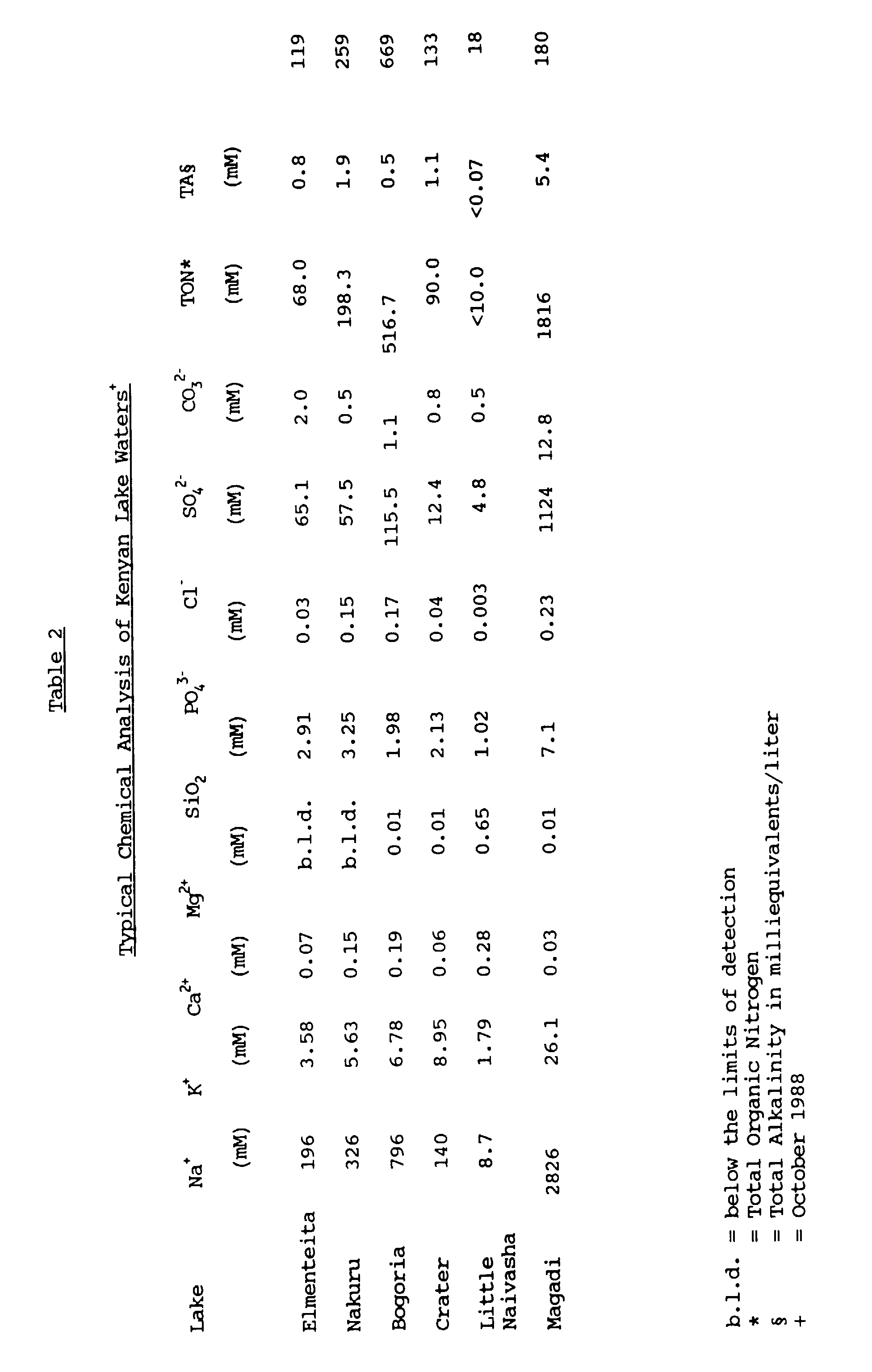
Origin of the Strains Arranged by Cluster CLUSTER STRAIN SAMPLE ISOLATION MEDIUM LOCATION pH Temp. 0° Conductivity mS/ cm 1 3E.1CT Elmenteita 9.5 35 2 A 1 71C.4 Crater 10 26 10.2 E 1 81LN.4 Little Naivasha 8.5-9 30 1.2 D 1 60E.4 Elmenteita 10 32 12.7 B 1 wE4 Elmenteita n.t. n.t. n.t. A 2 69B.4 Bogoria 10.5 33 44 C 2 RS11CT * 2 RS14 * 2 Exig. * 2 RS13 * 3 wE1 Elmenteita n.t. n.t. n.t. A 3 wN10 Nakuru n.t. n.t. n.t. A 3 wN12 Nakuru n.t. n.t. n.t. A 3 wN16CT Nakuru n.t. n.t. n.t. A 4 13C.1 Crater 9.0 30 10 A 4 23M.1 Magadi 11 36 100 A 4 14LN.1 Little Naivasha 8.5 26 1 A 4 15LN.1CT Little Naivasha 8.5 26 1 A - wE2 Elmenteita n.t. n.t. n.t. A - wB3 Bogoria n.t. n.t. n.t. A - BG114 * 5 66B.4 Bogoria n.t. n.t. n.t. F 5 AB30 * 5 RS10CT * 5 RS17 * 5 AB49 * 5 AB42 * 6 RS7 * 6 RS8CT * 6 RS15 * 6 RS16 * - 79LN.4 Little Naivasha 8.5-9 30 1.2 F - RS12 * - 72C.4 Crater 10 26 10.2 E - 80LN.4 Little Naivasha 8.5-9 30 1.2 G - Br.li * - Micro * n.t. = not tested
The letter codes given for the Isolation Media refer to Appendix A.
The asterisk (*) refers to a reference strain; the identity of which is provided in Table 4 (below). - A wide diversity of enrichment and isolation methods were applied. Some of the methods were specifically designed for the enrichment and isolation of alkaliphilic bacteria which exhibit specific types of enzyme activity at an alkaline pH. Other techniques of a more general nature were applied for the isolation of diverse sorts of alkaliphilic bacteria. In some cases, the specific conditions prevailing in the lakes (Table 2) were taken into account when experiments were performed for the isolation of bacteria.
- The different nutrient media employed for the isolation of the new alkaliphilic bacteria are designated Medium A - Medium G. The composition of the various media employed is shown in Appendix A.
- For the isolation of non-specific alkaliphilic organotrophic bacteria, soda-lake water samples or dilutions thereof were streaked out on an alkaline nutrient agar, pH 10 - pH 10.5 (Medium A). Samples of a more solid consistency, mud, sediment, etc. were first suspended in an alkaline nutrient broth (Medium A) before spreading on an alkaline nutrient agar (Medium A). The bacteria were cultivated in a heated incubator, preferably at 37°C. In some cases, the samples were suspended in an alkaline nutrient broth (Medium A) and the bacteria cultivated by shaking, preferably at 37°C for 2 - 3 days before spreading the broth onto an alkaline nutrient agar (Medium A) for the isolation of bacterial colonies.
- For the isolation of alkaliphilic bacteria exhibiting specific types of enzyme activity, samples were spread onto alkaline nutrient agar containing specific substrates such as lactalbumin or casein or olive oil. In some instances, the bacteria in the sample were enriched for 1 day up to several weeks in a non-specific alkaline nutrient broth such as Medium A before spreading the broth onto an alkaline nutrient agar specific for the detection of bacteria exhibiting enzyme activities such as lipolytic or proteolytic activity.
- Twenty strains of bacteria isolated from in and around alkaline lakes were assigned to the type of bacteria known as Gram-positive bacteria on the basis of (1) the Dussault modification of the Gram's staining reaction (Dussault, H.P., (1955), Journal of Bacteriology, 70, 484-485); (2) the KOH sensitivity test (Gregersen, T., (1978), European Journal of Applied Microbiology and
Biotechnology 5, 123-127; Halebian, S. et al., (1981), Journal of Clinical Microbiology, 13, 444-448); (3) the aminopeptidase reaction (Cerny, G., (1976), European Journal of Applied Microbiology, 3, 223-225; ibid, (1978), 5, 113-122); and in most cases, confirmation also on the basis of (4) a quinone analysis (Collins, M.D. and Jones, D., (1981), Microbiological Reviews, 45, 316-354) using the method described by Collins, M.D. in Chemical Methods in Bacterial Systematics (eds. Goodfellow, M. and Minnikin, D.) pp. 267-288, Academic Press, London, 1985. - The twenty strains were tested for 200 characters. The results were analyzed using the principles of numerical taxonomy (Sneath, P.H.A. and Sokal, R.R., in Numerical Taxonomy, W.H. Freeman & Co., San Francisco, 1973). The characters tested and manner of testing are compiled in Appendix B. In addition, Appendix C records how each character was coded for taxonomic analysis.
- As controls, 17 known Gram-positive bacteria were subjected to the same analysis using the same conditions where appropriate. These reference bacteria included genera that are known to include facultative or obligate alkaliphilic species. These 17 known reference bacteria are recorded in Table 4 from which it will be seen that the "Type Strain" of the known species has been used where available. Thirteen of the strains are known alkaliphilic Bacillus species.
Gram-Positive Reference Strains (RS7) (alkaliphilic) Bacillus species DSM 2514 (RS8) (alkaliphilic) Bacillus species DSM 2515 (RS10) (alkaliphilic) Bacillus species DSM 2517 (RS11) (alkaliphilic) Bacillus species DSM 2518 (RS12) (alkaliphilic) Bacillus species DSM 2519 (RS13) (alkaliphilic) Bacillus species DSM 2521 (RS14) (alkaliphilic) Bacillus species DSM 2523 (RS15) (alkaliphilic) Bacillus species DSM 2525 (RS16) Bacillus alcalophilus DSM 485 (RS17) Bacillus alcalophilus subsp. halodurans DSM 497 (AB30) (alkaliphilic) Bacillus species ATCC 21596 (AB42) (alkaliphilic) Bacillus species ATCC 21833 (AB49) (alkaliphilic) Bacillus species ATCC 21591 (Exig) Exiguobacterium aurantiacum NCIMB 11798 (BG114) Arthrobacter luteus ATCC 21596 (Br.li) Brevibacterium linens NCIMB 9904 (Micro) Micrococcus luteus NCTC 2665 - The phenetic data, consisting of 200 unit characters was scored as indicated in Appendix C, and set out in the form of an "n x t" matrix, whose t columns represent the t bacterial strains to be grouped on the basis of resemblances, and whose n rows are the unit characters. Taxonomic resemblance of the bacterial strains was estimated by means of a similarity coefficient (Sneath, P.H.A. and Sokal, R.R., Numerical Taxonomy, supra, pp. 114-187). Although many different coefficients have been used for biological classification, only a few have found regular use in bacteriology. We have chosen to apply three association coefficients (Sneath, P.H.A. and Sokal, R.R., ibid, p. 129 et seq.), namely, the Gower, Jaccard and Simple Matching coefficients. These have been frequently applied to the analysis of bacteriological data and have a wide acceptance by those skilled in the art since they have been shown to result in robust classifications.
- The coded data were analyzed using the TAXPAK program package (Sackin, M.J., "Programmes for classification and identification". In Methods in Microbiology, Volume 19 (eds. R.R. Colwell and R. Grigorova), pp. 459-494, Academic Press, London, (1987)) run on a DEC VAX computer at the University of Leicester, U.K.
- A similarity matrix was constructed for all pairs of strains using the Gower Coefficient (SG) with the option of permitting negative matches (Sneath, P.H.A. and Sokal, R.R., supra, pp. 135-136) using the RTBNSIM program in TAXPAK. As the primary instrument of analysis and the one upon which most of the arguments presented herein are based, the Gower Coefficient was chosen over other coefficients for generating similarity matrices because it is applicable to all types of characters or data, namely, two-state, multistate (ordered and qualitative), and quantitative.
- Cluster analysis of the similarity matrix was accomplished using the Unweighted Pair Group Method with Arithmetic Averages (UPGMA) algorithm, also known as the Unweighted Average Linkage procedure, by running the SMATCLST sub-routine in TAXPAK.
- The result of the cluster analysis is a dendrogram, a simplified version of which is provided in Figure 1. The dendrogram illustrates the levels of similarity between the bacterial strains. The dendrogram is obtained by using the DENDGR program in TAXPAK.
- The phenetic data, omitting multistate characters (characters 1-5, 12, 13; Appendix C) and thus consisting of 193 unit characters, and scored in binary notation (positive = 1, negative = 0) were re-analyzed using the Jaccard Coefficient (SJ) (Sneath, P.H.A. and Sokal, R.R., ibid, p. 131) and Simple Matching Coefficient (SSM) (Sneath, P.H.A. and Sokal, R.R., ibid, p. 132) by running the RTBNSIM program in TAXPAK. A further two dendrograms were obtained by using the SMATCLST with UPGMA option and DENDGR sub-routines in TAXPAK. Simplified versions of these dendrograms are illustrated in Figure 2 and Figure 3 respectively.
- Figure 1 illustrates the results of the cluster analysis, based on the Gower Coefficient and the UPGMA method, of 20 Gram-positive, alkaliphilic bacteria isolated from in and around alkaline lakes, together with 17 known Gram-positive bacteria, including 14 alkaliphilic species.
- Six natural clusters or phenons of alkaliphilic bacteria are generated at the 79% similarity level. These six clusters include 15 of the 20 alkaliphilic bacteria isolated from alkaline lakes. Although the choice of 79% for the level of delineation may seem arbitrary, it is in keeping with current practices in numerical taxonomy (Austin, B. and Priest, F., in Modern Bacterial Taxonomy, p. 37; Van Nostrand Reinhold; Wokingham, U.K., (1986)). Placing the delineation at a lower percentage would combine groups of clearly unrelated organisms whose definition is not supported by the data. At the 79% level, 3 of the clusters exclusively contain novel alkaliphilic bacteria representing 13 of the newly isolated strains, and these may represent new taxa.
- As expected, the cluster analysis groups the control Bacillus species in 3 distinct clusters which are separate from the novel alkaliphilic bacteria of the present invention. These results broadly concur with a taxonomic analysis of alkaliphilic Bacillus strains reported by Fritze, D., et al. (International Journal of Systematic Bacteriology, (1990), 40, 92-97)). None of the known organisms are significantly related to any of the 3 clusters of new, Gram-positive alkaliphilic bacteria. A clear discrimination between these clusters is possible using the concept of the minimum discriminatory tests (see below) and chemotaxonomic information (see below).
- The significance of the clustering at this level is supported by the results of the TESTDEN program. This program tests the significance of all dichotomous pairs of clusters (comprising 4 or more strains) in a UPGMA generated dendrogram with squared Euclidean distances, or their complement as a measurement and assuming that the clusters are hyperspherical. The critical overlap was set at 0.25%. As can be seen from Table 5, the separation of the clusters is highly significant.
Significance of the Clusters Generated by the SG/UPGMA Method Provided by TESTDEN CLUSTER separates from CLUSTER at Significance level 1 2 P = 0.99 2 3 0.99<P>0.95 3 4+wE2 P = 0.99 5 6 P = 0.99 - The cophenetic correlation is 0.804 which indicates the high degree of reliability with which this dendrogram represents the true taxonomic structure. (Sneath, P.H.A. and Sokal, R.R., supra, pp. 277-280, 304). Furthermore, the pattern of clusters obtained using the Jaccard Coefficient (Figure 2 and below) and Simple Matching Coefficient (Figure 3 and below) support the conclusions drawn here.
- Two of the newly isolated alkaliphiles, 69B.4 and 66B.4 cluster among the alkaliphilic Bacillus species and may properly be considered to belong to the genus Bacillus. However, five of the new alkaliphilic strains fall outside the major clusters. Two of these, wE2 and wB3, associate at the periphery of the clusters representing the major groups of novel alkaliphilic bacteria. Strain 79LN.4 and the related pair 72C.4 and 80LN.4 are also non-clustering. Their interrelationships are more difficult to define but they probably represent new phenons presently not described.
- The SJ coefficient is a useful adjunct to the SG coefficient as it can be used to detect phenons in the latter that are based on negative matches or distortions owing to undue weight being put on potentially subjective qualitative data. Consequently, the SJ coefficient is useful for confirming the validity of clusters defined initially by the use of the SG coefficient. The Jaccard Coefficient is particularly useful in comparing biochemically unreactive organisms (Austin, B. and Priest, F.G., supra, p. 37). There may be doubts about the admissability of matching negative character states (Sneath, P.H.A., and Sokal, R.R., supra, p. 131) in which case the Simple Matching Coefficient is a widely applied alternative.
- In the main, all of the clusters (especially the clusters of the new bacteria) generated by the SG/UPGMA method are recovered in the dendrograms produced by the SJ/UPGMA method (cophenetic correlation, 0.795) (Figure 2) and the SSM/UPGMA method (cophenetic correlation, 0.814) (Figure 3). The main effect of these transformations is to gather all the Bacillus strains in a single large cluster which further serves to emphasize the separation between the alkaliphilic Bacillus species and the new alkaliphilic bacteria, and the uniqueness of the latter.
- Chemotaxonomy is the study of the chemical variations of cells in relation to systematics. The analysis of chromosomal DNA, ribosomal RNA, proteins, cell walls and membranes, for example, can give valuable insights into taxonomic relationships and may be used as a further tool to classify or to verify the taxonomy of microorganisms (Goodfellow, M. and Minnikin, D.E. in Chemical Methods in Bacterial Systematics, (eds. Goodfellow, M. and Minnikin, D.E.), Academic Press, London and Orlando, FL, (1985), pp. 1-15). However, it is not always possible to decide a priori which type of chemical information will be most diagnostic for a given classification. The amphipathic polar lipids, the major respiratory quinones, the fatty acids located in the bacterial membranes and the DNA base composition all have taxonomic significance for the classification of various bacteria (Lechevalier, H. and Lechevalier, M.P., in Microbial Lipids, volume 1 (eds. Ratledge, C. and Wilkinson, S.G.) Academic Press, London and San Diego, CA, (1988), pp. 869-902).
- The extraction of polar lipids from bacteria and their analysis by two dimensional thin layer chromatography (2D-TLC) may yield patterns of diagnostic value. Stationary phase cells were extracted in 1:1 (v/v) CHCl3:CH3OH and examined by 2D-TLC as described by Ross, H.N.M., Grant, W.D. and Harris, J.E., in Chemical Methods in Bacterial Systematics, (eds. Goodfellow, M. and Minnikin, D.E.), Academic Press, London and Orlando, FL, (1985), pp. 289-300. The types of lipids present on the chromatograms were visualized using a variety of differential stains (Ross, H.N.M., et al., supra, p. 291, and Trincone, A., et al., Journal of General Microbiology, (1990), 136, pp. 2327-2331). The identity of components were confirmed by co-chromatography.
- The results of this analysis for representative strains of Gram-positive alkaliphiles are set out in Table 6. These show no clear polar lipid pattern which is distinct for any one cluster, although they do confirm that phosphatidylethanolamine is a characteristic phospholipid of many Bacillus species, (O'Leary, W.M. and Wilkinson, S.G., in Microbial Lipids,
volume 1, supra, p. 157). However, we were surprised to find that many of the bacteria contained one or several glycolipids. Glycolipids have not previously been demonstrated to be present in alkaliphilic bacteria (Krulwich, T.A., et al., CRC Critical Reviews in Microbiology, (1988), 16, 15-36). Furthermore, as judged by co-chromatography of several strains, all glycolipid-containing strains contained a glycolipid also found in Gram-negative alkaliphiles isolated from soda lakes. Some of the other glycolipids appear to be common to certain clusters of Gram-positive alkaliphiles. It is possible therefore, that the chemical structures of the glycolipids will be chemotaxonomic markers for many obligate alkaliphiles in general and for specific groups in particular.Polar Lipid Components of Gram-Positive Alkaliphilic Bacteria CLUSTER STRAIN PG DPG PGP PI PE GL 1 3E.1CT + + + 3+ 71C.4 + + + + + + 81LN.4 + + + + + 2 69B.4 + 3+ RS11CT + + 3+ RS14 + + + + + + 3 wE1 + + + + + wN10 + + + + + wN16CT + + + + + 4 13C.1 + + + + 3+ 23M.1 + + + + 3+ 14LN.1 + + + + 4+ 15LN.1CT + + + + 4+ - wE2 + + + + + 2+ 5 66B.4 + + + + + + RS17 + + + + + + AB49 + + + + + 6 RS7 + + + + 2+ RS15 + + + + 2+ - 72C.4 + + + + 3+ - 80LN.4 + + + + 3+ (PG) phosphatidylglycerol; (DPG) diphosphatidylglycerol;
(PGP) phosphatidylglycerolphosphate; (PI) phosphatidylinositol;
(PE) phosphatidylethanolamine (ninhydrin positive aminolipid);
(GL) unidentified glycolipid(s), α-naphthol positive (the number in the column gives the number of positive spots on the TLC plate). - The isoprenoid or respiratory quinones are characteristic components of the plasma membrane of aerobic bacteria. There are two types; menaquinones and ubiquinones. The value of isoprenoid quinones as taxonomic criteria lies in the variation in the length of the polyprenyl side-chain and the degree of saturation (Collins, M.D. and Jones, D. (1981), supra).
- Dry stationary phase bacterial cells were extracted, using a modified procedure of Collins, M.D. (in Chemical Methods in Bacterial Systematics, supra, pp. 267-284), in 1:1 (v/v) CHCl3:CH3OH at 50°C, for 16 hours. The quinones were examined by reverse phase thin layer chromatography as described by Collins, M.D. (supra).
- The results of quinone analyses of representative strains of Gram-positive alkaliphiles are illustrated in Table 7. However, there is no evidence to suggest that quinone composition is of value in the circumscription of the clusters, although the data do serve to confirm the status of these strains as Gram-positive. Furthermore, MK-7 as the major isoprenologue of Bacillus species, including alkaliphilic strains, is also confirmed (Lechevalier, H. and Lechevalier, M.P., supra, p. 881). Many of the novel, Gram-positive alkaliphilic bacteria of the present invention contain shorter molecules, especially MK-4 and MK-6, although no clear pattern emerges.
Menaquinone Components of Gram-Positive Alkaliphilic Bacteria CLUSTER STRAIN MENAQUINONE 1 3E.1CT 3 4 71C.4 4 5 5H 26 81LN.1 4 7 60E.4 3 4 8 wE4 4 5 2 69B.4 4 6 7 9 RS11 CT7 8 3 wE1 6 7 wN10 6 wN12 6 7 wN16 CT4 6 11 4 13C.1 4 6 7 9 23M.1 4 14LN.1 4 6 7 8 8H2 9 15LN.1CT 5 6 7 8H2 - wE2 7 - wB3 4 5 5 66B.4 4 6 7 RS17 4 7 8 AB49 7 6 RS7 4 5 7 8 - The analysis of fatty acid profiles has had a significant impact on bacterial classification especially in the assignment of genera and species among Gram-positive bacteria and actinomycetes (Kroppenstedt, R.M., in Chemical Methods in Bacterial Systematics (eds. M. Goodfellow and D.E. Minnikin), Academic Press; London and Orlando, FL, (1985), pp. 173-199); Lechevalier, H. and Lechevalier, M.P., supra.
- Freeze dried stationary phase cells (200-300 mg) were extracted for 16 hours at 75°C in toluene:methanol:conc. sulfuric acid (2.5 ml:2.5 ml:0.2 ml) and after cooling, the lipids were partitioned into hexane (
twice times 1 ml). Residual acid was removed using NH4HCO3. Lipid extracts were concentrated under O2-free N2, dissolved in 300 µl hexane and applied to preparative silica gel plates (Merck F254, Type T). The plates were developed in hexane: diethyl ether 85:15 (v/v) and the fatty acid methyl esters scraped off, extracted with hexane and concentrated under a stream of O2-free N2. - The fatty acid methyl esters were dissolved in heptane and analysed by gas chromatography using a Packard model 439 chromatograph equipped with flame ionization detectors. The samples were divided by a sample splitter and analyzed simultaneously over two columns, namely, CP-SIL-88 (Chrompack) (length 50 meter, internal diameter 0.22 mm) and Ultra-2 (Hewlett Packard) (length 50 m, internal diameter 0.20 mm). The carrier gas was nitrogen; the injection temperature 120°C; temperature gradient 2.5°C per minute to 240°C and isothermal at 240°C for 30 minutes. Fatty acid methyl esters were assigned by reference to known standard mixtures. The identity of some peaks was confirmed by means of gas chromatography-mass spectrometry using a Carlo Erba HRGC 5160 Mega series gas chromatograph equipped with a CP-SIL-88 column (length 50 meter, internal diameter 0.22 mm) with helium as carrier gas and direct injection into the source of a AMD 403 mass spectrometer.
- The fatty acid composition of the individual Gram-positive alkaliphilic bacteria is set out in Table 8. Table 9 shows the unique fatty acid profiles of the individual clusters.
Clusters cluster 4 shown by the numerical taxonomy, fatty acid profiles clearly demonstrate that two subgroups of bacteria (designated 4A and 4B) exist within this cluster. These profiles are typical of some members of the Coryneform-Mycobacterium-Nocardioform (CMN) groups of the actinomycetes (Bennan, P.J., in Microbial Lipids, supra, pp 203-298), a designation supported by their characteristic cell habit, and the appearance of dihydromenaquinones. Furthermore, the bright orange-yellow colors of many actinomycetes is caused by the accumulation of carotenoid pigments, often induced by light. Many of these carotenoids are unique and important taxonomically. Further chemotaxonomic markers for these groups include a branched C19:0 fatty acid which is 10-methyloctadecanoic acid (tuberculostearic acid), an important criterion in the classification of the CMN group of bacteria. Branched, unsaturated fatty acids are also found in these bacteria. Branched C20:0 fatty acids, found inClusters 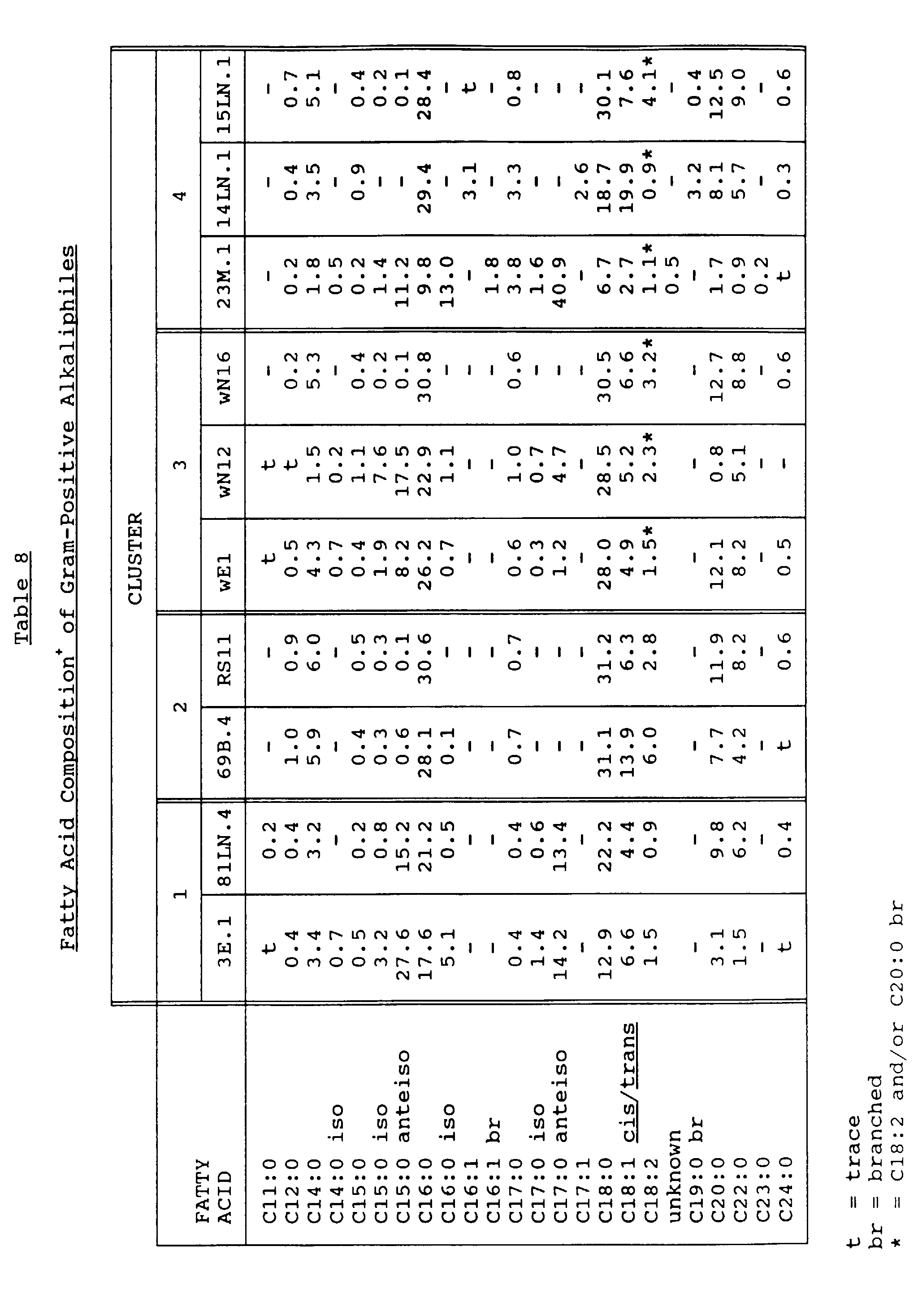


- An important component of a taxonomic study is an analysis of the genetic material - the nucleic acids. The composition of chromosomal DNA is unaffected by the growth conditions of the organism and an appropriate analysis may confirm or refute the taxonomic position of the organism. Chromosomal DNA may be analyzed by the determination of the base composition (G+C mol%) of individual strains. The guanine plus cytosine (G+C mol%) composition is constant for the chromosomal DNA from any given organism. Closely related organisms have similar G+C compositions. However, G+C results must be interpreted within the context of independent taxonomic data since similar G+C mol% of DNA samples from different organisms does not in itself imply biological relatedness.
- DNA was extracted from cells grown to exponential phase in Medium A by the chloroform:phenol method and was precipitated with ethanol. Base composition was determined by the thermal denaturation method (Marmur, J. and Doty, P. (1962), J. Mol. Biol., 3, 585-595) on a Phillips model PV8764 spectrophotometer with temperature programming. A second method involved HPLC analysis on a Beckman system gold using a Beckman ultrasphere ODS column and 0.04 M potassium dihydrogen phosphate plus acetonitrile (9 + 1, v/v) as eluent at a flow rate of 1.5 ml/min., after treatment of the DNA with nuclease P1 and alkaline phosphatase.
- The results of these analyses are set out in Table 10. These results are consistent with the grouping of the bacteria as defined by the numerical taxonomy analysis. The G+C mol% values for the new alkaliphilic bacteria (
clusters clusters DNA Base Composition of Gram-Positive Alkaliphilic Bacteria Cluster Strain G+C mol % HPLC TM Literature) 1 3E.1CT 52.3 wE4 63.1 2 RS11CT 35.0 35.2 RS14 39.6 RS13 37.2 3 wE1 49.3 wN10 34.1 wN12 40.1 36.6 wN16CT 46.0 40.2 4 23M.1 63.0 15LN.1CT 63.5 5 66B.4 42.1 RS10CT 39.5 RS17 42.5 AB49 42.1 42.8 AB42 36.1 6 RS7 43.2 RS8CT 39.9 43.6 RS15 43.5 RS16 36.5 - The centroid of each individual cluster generated by the SG/UPGMA method was computed using the RGROUPS program in TAXPAK. The centroid of a cluster of points representing real organisms projected into hyperspace represents a hypothetical average organism. The centroid seldom, if ever, represents a real organism. Therefore, the Euclidean distances of each of the members of the cluster from the centroid of the cluster were calculated in order to establish which organism was closest to the hypothetical average organism. The organism closest to the centroid was designated the "centrotype organism" (indicated with the superscript "CT").
- The centrotype organism can be thought of as the "Type Strain" which most closely represents the essential and discriminating features of each particular cluster. The centrotype strains are recorded in Table 11.
Centrotype Strains Cluster Number Number of Strains in Cluster Mean Euclidean Distance of Strains from Centroid Standard Deviation Centrotype Strain Euclidean Distance from Centroid 1 5 6.24 0.65 3E.1 4.45 2 5 6.02 0.23 RS11 4.53 3 4 6.81 0.96 wN16 4.26 4 4 6.40 0.57 15LN.1 4.40 5 6 6.44 0.27 RS10 5.01 6 4 6.50 0.72 RS8 4.06 - A description of each of the centrotype organisms from the clusters containing the novel bacteria has been made so as to be able to distinguish these organisms from all other bacteria previously known and described. In addition, the minimum number of discriminatory tests to define each cluster has been computed so that it may be clearly seen that the clusters containing these novel bacteria can be easily distinguished from each other and from all other known bacteria.
- An aerobic, Gram-positive coccoid bacterium. The cells are almost spherical, 0.5-1.5 µm, usually in pairs or occasionally tetrads, forming short chains of up to 6 cells, or in irregular clusters.
- Motility not observed.
- Obligate alkaliphile, grows optimally at about
pH 10. - On alkaline-agar, (Medium A) forms opaque, matte orange-colored colonies. The color development is influenced by light, beginning as cream developing through yellow to orange. The colonies are circular, convex, entire, 1-2 mm in diameter.
- In alkaline-broth (Medium A), growth (37°C) is moderate, evenly turbid, with the formation of a sediment but no surface pellicle.
- Optimum temperature for growth is about 30°C. Grows slowly at 15°C and 40°C. No growth at 45°C.
KOH test negative Aminopeptidase test negative Oxidase reaction negative Catalase reaction positive NaCl tolerance 0% to 12%. No growth at 15%. Hydrolysis of Gelatin positive Hydrolysis of Starch weak positive. Major polar lipid components phosphatidylglycerol, diphosphatidylglycerol, phosphatidylglycerolphosphate. Three glycolipid (α-naphthol positive) components present. Major menaquinones MK-3, MK-4. Major fatty acids C15:0 anteiso, C16:0, C17:0 anteiso, C18:0. G + C 52.3 mol % (HPLC) - Chemoorganotroph. Grows on complex substrates such as yeast extract and peptones. Growth on simple sugars and organic acids very restricted. Growth is stimulated by glucose, acetate, some amino acids and pyrimidine nucleotides.
- An aerobic, Gram-positive bacterium. The cells are short, thick, slightly irregular rods, 1.5-2.5 µm x 0.75-1.0 µm, occuring singly or in pairs, sometimes in short chains of up to 4 cells.
- No spores observed. Motility not observed.
- Obligate alkaliphile; no growth below
pH 8. - On alkaline-agar, (Medium A) forms smooth, cream-yellow colored colonies. The colonies are small, about 1 mm in diameter, circular, entire and convex.
- In alkaline-broth (Medium A), growth (37°C) is moderate, evenly turbid, forming a sediment but no surface pellicle.
- Grows well between 20°C and 40°C. Grows slowly at 10°C; no growth at 45°C.
KOH test negative Aminopeptidase test negative Oxidase reaction negative Catalase reaction negative NaCl tolerance 0% to 10%. Hydrolysis of Gelatin positive Hydrolysis of Starch positive. Major polar lipid components phosphatidylglycerol, diphosphatidylglycerol, phosphatidylglycerolphosphate, phosphatidylinositol, phosphatidylethanolamine. Major menaquinones MK-4, MK-6, MK-11. Major fatty acids C16:0, C18:0, C20:0 (fatty acids with even carbon numbers comprise >95%, branched fatty acids <1%). G + C 40.2 mol % (TM) - 46.0 mol % (HPLC) - Chemoorganotroph. Grows on complex substrates such as yeast extract, a range of sugars, amino- and organic acids.
- An aerobic, Gram-positive bacterium. The cells are initially irregular, spherical or elongated, sometimes wedge-shaped, 1.5 - 2.0 µm x 0.75 - 1 µm, developing into short, thick, slightly curved rods, 1 - 3 µm x 0.5 - 1 µm. The cells often occur in pairs. Due to the characteristic snapping form of cell division, the cells are frequently found at an angle forming a V-shape, or in clusters of cells with a pallisade appearance.
- No spores observed. No motility observed.
- Obligate alkaliphile; no growth below pH 7.5, optimum pH ≈ 9 - 10.
- On alkaline-agar, (Medium A) forms brightly colored, smooth, glistening colonies, initially orange developing into red. Color development is influenced by light. The colonies are circular, convex, entire, opaque, 1 - 2 mm in diameter.
- In alkaline-broth (Medium A), growth (37°C) is slight to moderate, flocculent, with the formation of sediment and a surface ring or pellicle.
- Grows well between 20°C and 40°C. Grows slowly at 45°C and 10°C. No growth at 50°C.
KOH test negative Aminopeptidase test negative Oxidase reaction negative Catalase reaction positive NaCl tolerance 0% to 8%. Hydrolysis of Gelatin negative Hydrolysis of Starch positive. Major polar lipid components phosphatidylglycerol, diphosphatidylglycerol, phosphatidylglycerolphosphate, phosphatidylinositol. Four glycolipid (α-naphthol positive) components present. Major menaquinones MK-5, MK-6, MK-7, MK-8(H 2 ) Major fatty acids C16:0, C18:0, C20:0 (fatty acids with even carbon numbers comprise >95%, branched fatty acids <1%). G + C 63.5 mol % (HPLC) - Chemoorganotroph. Grows on complex substrates such as yeast extract and peptones. Growth on simple sugars very restricted. Growth is stimulated by amino acids and fatty acids.
- The strains which do not fall into the clusters defined here are also novel bacteria not previously known or described. These strains, coded wE2, wB3, 72C.4, 79LN.4 and 80LN.4, may represent rarer varieties of alkaliphilic bacteria. Some of these strains, such as wE2 and wB3 may represent intermediate forms, falling between closely related (and closely orientated in hyperspace) clusters as defined here. The other strains, 72C.4, 79LN.4 and 80LN.4, are probably members of clusters of bacteria representing new genera or species at present not defined. A description of these "non-clustering" strains has been made so as to be able to distinguish these organisms from all other bacteria previously known and described.
- An aerobic, Gram-positive bacterium. The cells are irregular; mainly oval coccoid cells, 1 - 2 µm x 0.5 - 1 µm, or very short rods, occasionally in pairs, or slightly curved short rods. Due to the characteristic snapping form of cell division, the cells are frequently found at an angle forming a V-shape.
- No spores observed. No motility observed.
- Obligate alkaliphile; no growth below
pH 8. - On alkaline-agar, (Medium A) forms opaque, orange colored, punctiform or circular colonies, with a convex or domed elevation and entire margin, up to 1 mm in diameter.
- In alkaline-broth, (Medium A) growth (37°C) is slow, slight to moderate, flocculent turbidity, with the formation of a sediment and surface ring.
- Temperature: grows optimally at above 30°C, slowly at 10°C. No growth at 40°C.
KOH test negative Aminopeptidase test negative Oxidase reaction negative Catalase reaction positive NaCl tolerance 0% to 10%. No growth at 12%. Hydrolysis of Gelatin negative Hydrolysis of Starch negative Major polar lipid components phosphatidylglycerol, diphosphatidylglycerol, phosphatidylglycerolphosphate, phosphatidylinositol, phosphatidylethanolamine. Two glycolipids (α-naphthol positive) components present. Major menaquinones MK-7 - Chemoorganotroph. Grows on complex substrates such as yeast extract, peptones and carbohydrates (dextrin). Growth is stimulated by a variety of sugars, organic-, fatty- and amino-acids.
- Strain wE2 appears to be an intermediate form related to
Cluster 4. - An aerobic, Gram-positive bacterium. The cells are short, straight or slightly curved rods, 1 - 2.5 µm x 0.5 µm, sometimes in pairs. Due to the characteristic snapping form of cell division, the cells are frequently found at an angle forming a V-shape.
- Motility not observed. No spores observed.
- Obligate alkaliphile; no growth below
pH 8. On alkaline-agar, (Medium A) forms opaque yellow/ochre, circular, convex, entire colonies, 2 mm in diameter. - In alkaline-broth, (Medium A) growth (37°C) is slight to moderate with an even turbidity and the formation of a sediment, but no surface pellicle.
- Temperature range for growth: 10°C to 40°C. No growth at 45°C.
KOH test negative Aminopeptidase test negative Oxidase reaction negative Catalase reaction positive NaCl tolerance 0% to 12%. No growth at 15%. Hydrolysis of Gelatin positive Hydrolysis of Starch negative Major menaquinones MK-4, MK-5. Major fatty acids C16:0, C18:0 (fatty acids with even carbon numbers >90%, branched fatty acids <10%). - Chemoorganotroph. Grows on complex substrates such as yeast extract. Growth on simple substrates (sugars, etc.) very restricted. Growth is stimulated by acetate and glucose. Strain wB3 appears to be an intermediate form related to
Cluster 1. - An aerobic, motile, Gram-positive bacterium. The cells are straight or slightly curved rods, 1.5 - 5 µm x 0.5 - 1 µm, often in pairs, sometimes in short chains of 2 to 4 cells.
- No spores observed. Motility not observed.
- Obligate alkaliphile, no growth below pH 7.5.
- On alkaline-agar, (Medium A) forms opaque, cream colored colonies, 2 mm in diameter. The colonies are circular, umbonate in elevation, with an entire margin becoming undulate with age.
- In alkaline-broth, (Medium A) growth (37°C) is moderate to heavy, evenly turbid, with the formation of a sediment and eventually a surface ring.
- Temperature range: grows well at 20°C to 40°C. Grows slowly at 10°C and 45°C. No growth at 50°C.
KOH test negative Aminopeptidase test negative Oxidase reaction weak positive Catalase reaction positive NaCl tolerance 0% to 15% Hydrolysis of Gelatin positive Hydrolysis of Starch positive - Chemoorganotroph. Grows well on complex substrates, simple sugars, organic-, amino- and fatty acids, and pyrimidine nucleotides.
- An aerobic, Gram-positive bacterium. The cells appear to have a distinct coccus-rod development cycle. Initially the cells are spherical or irregular coccobacillery in form which develop into short rods, 1 - 2.5 µm x 0.5 - 0.75 µm. Eventually some longer forms, 3 - 4 µm x 1 µm appear. The cells occur occasionally in pairs. Due to the characteristic snapping form of cell division, the cells are frequently found at an angle forming a V-shape.
- No motility observed. No spores observed.
- Obligate alkaliphile; no growth below
pH 8. - On alkaline-agar, (Medium A) forms circular, convex, entire, opaque colonies, 1 - 2 mm in diameter. The colony color is initially orange developing with age and the influence of light into a deep salmon pink.
- In alkaline-broth, (Medium A) growth (37°C) is moderate, evenly turbid, with the formation of a sediment but no surface pellicle.
- Temperature range: grows well at 20°C to 37°C. Grows slowly at 10°C. No growth at 40°C.
KOH test negative Aminopeptidase test positive Oxidase reaction negative Catalase reaction positive NaCl tolerance 0% to 12% Hydrolysis of Gelatin positive Hydrolysis of Starch negative Major polar lipid components phosphatidylglycerol, diphosphatidylglycerol, phosphatidylglycerolphosphate, phosphatidylinositol. Three glycolipid (α-naphthol positive) components present. Major fatty acids C16:0, C18:0 (fatty acids with even carbon numbers >95%, branched fatty acids <5%). - Chemoorganotroph. Grows well on complex substrates (e.g. yeast extract) and a variety of sugars, organic acids and amino acids.
- An aerobic, Gram-positive bacterium. The cells are nearly spherical or coccobacillery in form, developing into very short rods, 1 - 2 µm x 0.5 - 0.75 µm. Occasionally longer forms occur. The cells occur occasionally in pairs.
- No motility observed. No spores observed.
- Obligate alkaliphile, no growth at
pH 8. - On alkaline-agar, (Medium A) forms circular, convex to umbonate, entire, opaque colonies, 1 - 2 mm in diameter. The colony color is initially orange developing with age and the influence of light into a deep salmon pink.
- In alkaline-broth, (Medium A) growth (37°C) is moderate, evenly turbid, with the formation of sediment and surface pellicle.
- Temperature range: grows well at 20°C to 37°C. Grows slowly at 10°C. No growth at 40°C.
KOH test negative Aminopeptidase test positive Oxidase reaction negative Catalase reaction positive NaCl tolerance 0% to 12%. Grows weakly at 15%. Hydrolysis of Gelatin positive Hydrolysis of Starch negative Major polar lipid components phosphatidylglycerol, diphosphatidylglycerol, phosphatidylglycerolphosphate, phosphatidylinositol. Three glycolipid (α-naphthol positive) components present. - Chemoorganotroph. Grows well on complex substrates (e.g. yeast extract) and a variety of sugars, organic acids and amino acids.
- One of the purposes of a numerical classification study is to use the phenetic data, which define the clusters at a selected similarity level, for the assignment or identification of unknown strains. The classification test data can be used to determine the minimum set of tests which are required to define the clusters at the 79% (SG) similarity level, and to identify those characters which are most diagnostic (predictive) for the individual clusters. In other words, the minimum number of tests which are required to assign an unknown organism to a pre-determined cluster with a high degree of predictability.
- From the minimum discriminatory tests, a probability matrix can be constructed for the identification of unknown strains. The analysis is achieved by using a combination of the CHARSEP and DIACHAR programs in TAXPAK, supplemented by the MCHOICE program (not on TAXPAK, but available by Data-Mail from the University of Leicester, U.K.). An evaluation of the identification matrix is provided by using the MOSTTYP, OVERMAT and MATIDEN programs. Practical examples of the use of these programs for the probabilistic identification of bacteria have been published by Williams, S.T., et al., (1983), Journal of General Microbiology, 129, 1815-1830; and Priest, F.G. and Alexander, B., (1988), Journal of General Microbiology, 134, 3011-3018; ibid, (1990), 136, 367-376.
- A "n x t" table was constructed using the two-state characters from the test data. In other words, using
characters 6 to 11 and 14 to 200 (Appendix C) scored in binary notation (positive = 1, negative = 0). - The data matrix is first examined using the CHARSEP program which calculates separation indices and thus the diagnostic value of the individual characters for discriminating between the clusters. Character-states (tests) with a VSP index [(4 times variance) times strain potential] greater than 25% (Sneath, P.H.A., (1979), Computers and Geosciences, 5, 349-357) are accepted, characters with a low diagnostic value (VSP <25%) are rejected. A preference is made for characters with the highest VSP indices, provided that the criteria in the DIACHAR and MCHOICE programs are also met. In this example, 63 tests have a VSP index >25%, and 16 of the 32 characters finally chosen have a VSP index >50% (Table 12).
- The data matrix is next re-examined by means of the DIACHAR program, which determines the most diagnostic character states of each of the clusters. The number of character states was set at 12. This result allows the choice of mutually exclusive character states between the clusters. As many of these tests as possible are retained in the final identification matrix of minimum discriminatory tests; in this example between 4 and 10 diagnostic characters per cluster. The remaining, unused tests are also noted and may be applied as additional tests for the confirmation of identification (Table 13).
- The MCHOICE program ranks the tests in groups which can be displayed in the form of a dendrogram using the MDEND subroutine. The groups identify tests with similar discriminatory value, thus allowing the rejection of tests which fail to make a significant discrimination as well as allowing choices to be made between tests of equal or very similar diagnostic value.
- Table 14 shows the set of 32 tests which is the minimum number required to define the clusters and which can be used for the assignment of unknown strains. In addition, Table 14 shows the identification matrix which consists of the percentage of positive characters which define the clusters on the basis of the 32 minimum discriminatory tests. This is computed by the IDMAT program.
Separation Values of Characters used for the Minimum Discriminatory Tests CHARACTER VSP Index [10] Gelatin 30.6 [14] Fumarate 35.4 [15] Fructose 35.2 [19] Galactose 34.8 [24] N-acetylglucosamine 58.1 [27] D-saccharose 74.1 [28] Maltose 70.7 [32] Acetate 56.8 [36] D-glucose 63.1 [37] Salicin 51.3 [38] D-melibiose 45.0 [42] Propionate 72.4 [44] Valerate 31.7 [48] Glycogen 85.3 [50] L-serine 38.1 [63] Chymotrypsin 44.4 [70] β-glucosidase 67.1 [74] Serine 58.6 [77] Arginine 65.3 [80] Methionine 54.0 [90] Penicillin G 55.9 [94] Methicillin 56.5 [96] Streptomycin 28.5 [97] Tetracyclin 51.8 [105] Bacitracin 32.8 [112] N-acetyl-D-glucosamine 32.8 [116] Cellobiose 45.0 [137] Turanose 55.5 [139] Methyl pyruvate 41.1 [140] Mono-methylsuccinate 39.1 [192] Thymidine 34.9 [197] Glycerol 34.0 Discriminatory Tests for Each of the Six Clusters (SG) Cluster 1: matte orange colored circular colonies; coccoid cells. Positive Negative [10] Gelatin hydrolysis [24] N-acetylglucosamine [90] Penicillin G [27] D-saccharose [94] Methicillin [37] Salicin [105] Bacitracin [38] D-melibiose [42] Propionate [48] Glycogen [52] 3-hydroxybutyrate [74] Serine [77] Arginine [80] Methionine [83] Valine [116] Cellobiose Cluster 2: Positive Negative [10] Gelatin hydrolysis [24] N-acetylglucosamine [15] Fructose [42] Propionate [28] Maltose [44] Valerate [37] Salicin [50] L-serine [70] β-glucosidase [89] Trimethoprim [74] Serine [123] m-inositol [80] Methionine [197] Glycerol [86] Ampicillin [94] Methicillin [96] Streptomycin [105] Bacitracin [137] Turanose Cluster 3: yellow colonies; rod-shaped cells in chains. Positive Negative [10] Gelatin hydrolysis [28] Maltose [19] Galactose [32] Acetate [70] β-glucosidase [36] D-glucose [77] Arginine [38] D-melibiose [105] Bacitracin [42] Propionate [112] N-acetyl-D-glucosamine [44] Valerate [116] Cellobiose [48] Glycogen [190] Inosine [74] Serine [191] Uridine [86] Ampicillin [90] Penicillin G [94] Methicillin [97] Tetracyclin [139] Methyl pyruvate [140] Mono-methylsuccinate [155] α-ketobutyric acid Cluster 4: glistening bright orange/red colonies; cells coccobacillery Positive Negative [77] Arginine [10] Gelatin hydrolysis [80] Methionine [14] Fumarate [82] Glycine [15] Fructose [83] Valine [19] Galactose [94] Methicillin [27] D-saccharose [97] Tetracyclin [38] D-melibiose [105] Bacitracin [48] Glycogen [106] α-cyclodextrin [50] L-serine [109] Tween 40 [70] β-glucosidase [139] Methyl pyruvate [112] N-acetyl-D-glucosamine [140] Mono-methylsuccinate [116] Cellobiose [151] β-hydroxybutyric acid [134] D-sorbitol [165] Bromo-succinic acid [136] D-trehalose [137] Turanose Cluster 5: beige or dull cream colonies; straight rod-shaped cells. Positive Negative [10] Gelatin hydrolysis [140] Mono-methylsuccinate [24] N-acetylglucosamine [157] α-ketovaleric acid [27] D-saccharose [28] Maltose [32] Acetate [36] D-glucose [48] Glycogen [63] Chymotrypsin [86] Ampicillin [94] Methicillin [97] Tetracyclin [137] Turanose Cluster 6: cream colonies; straight rod-shaped cells. Positive Negative [10] Gelatin hydrolysis [19] Galactose [15] Fructose [63] Chymotrypsin [24] N-acetylglucosamine [74] Serine [27] D-saccharose [75] Proline [28] Maltose [77] Arginine [32] Acetate [96] Streptomycin [36] D-glucose [99] Oleandomycin [37] Salicin [105] Bacitracin [38] D-melibiose [116] Cellobiose [42] Propionate [140] Mono-methylsuccinate [45] Citrate [192] Thymidine [48] Glycogen [50] L-serine [66] α-galactosidase [67] β-galactosidase [68] β-glucuronidase [70] β-glucosidase [123] m-inositol [137] Turanose [197] Glycerol Note: The numbers in square brackets proceeding the character state refers to the character states and unit tests in Appendices B and C. Identification Matrix: Percentage of Positive Discriminatory Characters which Define the Clusters of Gram-Positive Alkaliphilic Bacteria at the 79% Level (SG) TEST CLUSTER 1 2 3 4 5 6 [10] Gelatin 100 100 100 0 100 100 [14] Fumarate 20 25 75 0 83 50 [15] Fructose 60 100 75 0 83 100 [19] Galactose 20 25 100 0 17 0 [24] N-acetylglucosamine 0 0 50 25 100 100 [27] D-saccharose 0 75 25 0 100 100 [28] Maltose 20 100 0 25 100 100 [32] Acetate 20 25 0 75 100 100 [36] D-glucose 20 75 0 25 100 100 [37] Salicin 0 100 50 25 67 100 [38] D-melibiose 0 50 0 0 50 100 [42] Propionate 0 0 0 75 83 100 [44] Valerate 20 0 0 50 83 50 [48] Glycogen 0 75 0 0 100 100 [50] L-serine 40 0 25 0 17 100 [63] Chymotrypsin 40 25 75 25 100 0 [70] β-glucosidase 20 100 100 0 33 100 [74] Serine 0 100 0 75 50 0 [77] Arginine 0 50 100 100 33 0 [80] Methionine 0 100 nc 100 33 25 [90] Penicillin G 100 75 0 50 83 0 [94] Methicillin 100 100 0 100 100 25 [96] Streptomycin 40 100 50 75 67 0 [97] Tetracyclin 80 75 0 100 100 25 [105] Bacitracin 100 100 100 100 83 0 [112] N-acetyl-D-glucosamine 40 25 100 0 50 50 [116] Cellobiose 0 50 100 0 50 0 [137] Turanose 40 100 25 0 100 100 [139] Methyl pyruvate 60 75 0 100 33 75 [140] Mono-methylsuccinate 40 25 0 100 0 0 [192] Thymidine 80 25 50 50 83 0 [197] Glycerol 80 0 50 25 33 100 nc = not computed - The evaluation of the discriminatory tests has two aspects. Firstly, the validity of the tests can be analysed using practical examples, which can be further evaluated using statistical theory, or the tests can be directly subjected to theoretical assessment using statistical methods.
- Many workers assess the accuracy of the discriminatory tests only by redetermining the character states of selected cluster representatives. This approach has been used here for the centrotype strains (see below). A far more stringent approach which is seldom applied, is to examine all the strains which were used in the original numerical taxonomic analysis. When subjected to cluster analysis using only the data acquired from the derived set of minimum discriminatory tests, the reconstructed dendrogram can be compared with the original. Using only the 32 discriminatory tests previously described (Table 14), the data (two-state, binary form) for all 20 of the new Gram-positive alkaliphilic bacteria and 13 known alkaliphilic Bacillus species isolated by Japanese workers, were subjected to cluster analysis by the SG/UPGMA method (equivalent in this case to the SSM/UPGMA method). The reconstructed dendrogram is reproduced in Figure 4. This reconstructed dendrogram compares very favorably with the original dendrograms (Figures 1 and 2).
- Although there has been some rearrangement of position of the clusters, their composition is largely unchanged and there is a clear separation between the clusters of novel alkaliphilic bacteria of the present invention and the alkaliphilic Bacillus species.
- This evidence, together with the statistical data provided by the numerical taxonomic analysis and the chemotaxonomic data, indicates a robust classification which identifies three major groups of novel Gram-positive alkaliphilic bacteria.
- The significance of the apparent clear cluster separation obtained in Illustration 1 (above) can be evaluated using the OVERMAT program which assesses cluster overlap between taxa in an identification matrix. This program examines the matrix constructed from the percentage positive values for the selected character states against a critical overlap value by considering the clusters defined by the coordinates of the centroid and cluster radius (twice root mean square of the distances of the strains of the cluster from the centroid). If there is significant overlap between the clusters, unknown strains may not identify with sufficient confidence to any one of them (Sneath, P.H.A. and Sokal, R.R., supra, p. 394-400). At a chosen critical overlap value of 2.5% (which is a more stringent condition than is used by most workers: see Priest, F.G. and Alexander, B., (1988), supra; and Williams, S.T. et al. (supra), there was no significant overlap (99% confidence level) between most of the clusters (Table 15). Even at a 1% critical overlap value there was no significant cluster overlap (Table 16) except between
Cluster 2 andCluster 5, but since these both represent Bacillus strains this is not considered to have any practical significance for the correct identification of the new Gram-positive alkaliphiles.

- The hypothetical median organism (HMO) is another estimate of the "average" organism in a cluster (Sneath, P.H.A. and Sokal, R.R., supra, pp. 195, et seq.). A HMO is not a real strain but a hypothetical organism possessing the most common state for each character. The MOSTTYP program calculates HMO's for each cluster in the identification matrix and then attempts to identify them. In other words, MOSTTYP is a program to evaluate an identification matrix by calculating identification scores of the most typical strains against the clusters. A good identification matrix should give a high probability of a HMO being reassigned to its own cluster. The results of this analysis were very satisfactory. Each HMO was reassigned to its original cluster with Willcox probabilities of 1.000 (Willcox, W.R. et al., (1973) Journal of General Microbiology, 77, 317-330). The taxonomic distances were all low and the standard errors of the taxonomic distance were all negative, indicating that the HMO's were all closer to the centroid of the cluster than the average for the cluster (Table 17).
Identification Scores for the Hypothetical Median Organism of each cluster provided by the MOSTTYP Program Identification Score CLUSTER Willcox Probability Taxonomic Distance Standard Error of Taxonomic Distance 1 1.000 0.229 - 3.086 2 1.000 0.221 - 3.200 3 1.000 0.242 - 2.288 4 1.000 0.207 - 3.090 5 1.000 0.251 - 2.701 6 1.000 0.177 - 2.690 - Identification of strains using the minimum set of discriminatory tests is achieved using the MATIDEN program in TAXPAK. The program compares presence-absence data for an unknown strain against each cluster in turn in an identification matrix of percentage positive characters. Identification coefficients are computed, namely Willcox probability, Taxonomic Distance and the Standard Error of the Taxonomic Distance. The results are displayed, showing the identification scores to the best cluster and to the two next best alternative clusters. Additionally, the atypical results ("characters against") are recorded. In an analysis using data from real strains, the centrotypes were reassigned to their original clusters with Willcox probabilities of 1.000 (Table 18). The taxonomic distances were low, generally in the same range as the HMO's. The standard errors of the taxonomic distance were all negative indicating that the centrotypes were closer to the centroid of the cluster than the average for the cluster. The exception was Bacillus reference strain RS10 but this was well within the acceptable limits of +3.0 (Sneath, P.H.A. (1979), pp. 195-213).
Identification Scores for the Centrotype Organisms of Each Cluster Provided by the MATIDEN Program Identification Score Cluster Strain Assigned to Cluster Willcox Probability Taxonomic Distance Standard (D) Error of D 1 3E.1CT 1 1.000 0.289 - 1.088 2 RS11 CT2 1.000 0.273 - 0.584 3 wN16 CT3 1.000 0.229 - 1.767 4 15LN.1CT 4 1.000 0.217 - 0.519 5 RS10CT 5 1.000 0.421 + 1.678 6 RS8 CT6 1.000 0.221 - 2.468 - The identification matrix was assessed for the ability to assign an unknown Gram-positive alkaliphile to the clusters defined herein. The criteria for a successful identification were:
- (a) a bacterium isolated from a similar habitat to, but geographically separate from, the East African soda lakes;
- (b) a Willcox probability greater than 0.95 and low values for taxonomic distance and its standard error (< 3);
- (c) an identification score to the best cluster significantly better than those against the two next best alternatives;
- (d) "characters against" the best cluster should be zero or few in number.
-
- An unknown microorganism may be examined using the minimum tests listed in Table 14. The character states are determined and identification scores obtained using the MATIDEN program. This program compares the character states of the unknown with the identification matrix determined for all of the predetermined clusters, computes the best match and assigns the unknown to the most appropriate cluster.
- A Willcox probability is calculated to determine the acceptability of identification. Willcox probabilities of 0.85 and 0.95 have been accepted as criteria for a successful identification (Williams, S.T., et al., (1983), supra; Priest, F.G. and Austin, B., (1988), supra). The taxonomic distance of the unknown from the cluster centroid is calculated and may be compared to the radius of the cluster. The standard error of the taxonomic distance should be less than the upper value of +3.0 suggested by Sneath, P.H.A. ((1979), pp. 195-213). Moreover, physical characteristics, additional biochemical data and chemotaxomomic markers may be used to further confirm the identity of the unknown in a particular cluster.
- The alkaliphilic microorganisms of the present invention produce a variety of enzymes. These enzymes are capable of performing their functions at an extremely high pH, making them uniquely suited for their application in a variety of processes requiring such enzymatic activity in high pH environments or reaction conditions.
- Examples of the various applications for alkalitolerant enzymes are in detergent compositions, leather tanning, food treatment, waste treatment and in the textile industry. These enzymes may also be used for biotransformations, especially in the preparation of pure enantiomers.
- The alkaliphiles may easily be screened for the production of alkalitolerant enzymes having lipolytic, proteolytic and/or starch-degrading activity using the methods described in Appendix B.
- The broth in which alkaliphilic bacteria are cultured typically contains one or more types of enzymatic activity. The broth containing the enzyme or enzymes may be used directly in the desired process after the removal of the bacteria therefrom by means of centrifugation or filtration, for example.
- If desired, the culture filtrate may be concentrated by freeze drying, before or after dialysis, or by ultrafiltration. The enzymes may also be recovered by precipitation and filtration. Alternatively, the enzyme or enzymes contained in the broth may be isolated and purified by chromatographic means or by gel electrophoresis, for example, before being applied to the desired process.
- The genes encoding alkalitolerant enzymes of interest may be cloned and expressed in organisms capable of expressing the desired enzyme in a pure or easily recoverable form.
- In one embodiment, the enzymatic preparation may be used in wash tests to determine the efficacy of the enzymatic activity.
- Enzyme preparations from the alkaliphilic bacteria may be tested in a specially developed mini-wash test using cotton swatches soiled, for example, with protein-, lipid- and/or starch-containing components. Prior to the wash test, the swatches can be pre-treated with a solution containing an anionic surfactant, sodium perborate and a bleach activator (TAED). After this treatment, the test swatches are rinsed in running demineralized water and air-dried. This treatment results in the fixation of the soil, making its removal more difficult.
- The washing tests may be performed using a defined detergent composition plus a specific amount of enzymatic activity in the presence of the test swatches. After washing, the swatches are rinsed in running demineralized water and air-dried. The reflectance of the test swatches is measured with a photometer.
- The following example is provided to further illustrate the present invention and is not intended to limit the scope of the invention in any way.
- Strain ML207a is a Gram-positive, alkaliphilic bacterium isolated from Mono Lake, a hypersaline, alkaline lake situated in California, U.S.A. (Javor, B., in Hypersaline Environments; Springer-Verlag, Berlin and Heidelberg, (1989), pp. 303-305) by plating out (on Medium A, Appendix A) mud and water samples collected in May, 1990. Strain ML207a is a coccus, forming bright yellow-orange, circular, entire, convex colonies on alkaline nutrient agar (Medium A).
- Strain ML207a was examined using 22 of the minimum tests listed in Table 14. The character states were determined and identification scores obtained using the MATIDEN program. The results are outlined in Table 19. These indicate a very satisfactory identification of strain ML207a to
Cluster 1, despite assigning only 22 of the 32 character states from the minimum discriminatory tests. - A Willcox probability of 0.9997 was calculated, which is significantly higher than the limit set at 0.95. Willcox probabilities of 0.85 and 0.95 have been accepted as criteria for a successful identification, (Williams, S.T., et al., (1983), supra; Priest, F.G. and Austin, B., (1988), supra). A taxonomic distance from the cluster centroid of 0.423 is acceptable and within the cluster radius defined at 0.539 (99% level). The standard error of the taxonomic distance at 2.076 is less than the upper value of +3.0 suggested by Sneath, P.H.A. ((1979), pp. 195-213). In addition, the coccus-shaped cells and yellow-orange colony color of strain ML207a also conform with the characteristics of Cluster 1 (Table 13).


- Five alkaliphilic strains (60E.4, 80LN.4, 81LN.4, wN10 and wN12) were tested for the production of proteolytic enzyme(s) in a medium poised at an alkaline pH. The experiments were carried out in 2 liter shake flasks provided with a baffle. Each of the flasks contained 400 ml of Medium S. Medium S had the following composition in g per liter: fresh yeast, 8.25; glucose, 1.32; K2HPO4, 1.6; CaCl2, 0.05; MgSO4·7H2O, 0.05; FeSO4, 0.005; MnSO4, 0.0066; NaCl, 40.0. The medium was sterilized at 121°C for 20 minutes and adjusted to pH 10.5 with sterile 40% Na2CO3 solution. The flasks were placed in an orbital incubator rotating at 280 revolutions per minute at a constant temperature of 37°C. Samples of culture medium were removed from the flasks at intervals of 0-5.7 days for the determination of enzyme content which is expressed in Alkaline Delft Units (ADU - as described in U.S. Patent 4,002,572).
- Table 20 shows the enzyme yield and the pH of the cultivation medium at the moment at which the measurement of enzyme levels were made.
Production of Proteolytic Enzymes Day Strain 60E.4 Strain 80LN.4 Strain 81LN.4 Strain wN10 Strain wN12 ADU/ml pH ADU/ml pH ADU/ml pH ADU/ml pH ADU/ml pH 0 0 10.5 0 10.5 0 10.5 0 10.5 0 10.5 1 137 9.5 2 10 16 10 0 10.5 2 10 2 2 9.5 18 10 0 10 3 122 10 3 10 17 10 2 10 4 101 10 5 88 10 3 10 15 10 5 10 5 10 7 74 10 14 10 9 58 10.5 5 10 14 10 5 10 3 10 - The results of the test, together with the results shown in Appendix E clearly indicate the presence of proteolytic enzymes, produced by the alkaliphilic bacteria of the present invention, in the culture broth.
- Enzyme preparations from the alkaliphilic bacteria were tested in a specially developed mini-wash test using swatches (2.5 x 2.5 cm) soiled with milk, blood and ink (obtained from EMPA, St. Gallen, Switzerland). Two types of fabric were tested; 100% cotton (designated EMPA 116) and polyester (35%)/cotton (65%) (designated EMPA 117). The test swatches were submitted to the mini-wash test either with or without a pretreatment ("pre-oxidized"). The pretreatment consisted of placing the swatches in a solution containing an anionic surfactant, sodium perborate and a bleach activator (TAED) and stirring at ambient temperature for 15 minutes. After this treatment the test swatches were rinsed in running demineralized water for 10 minutes and air-dried. This treatment results in the fixation of the remaining soil.
- The washing tests were performed in 100 ml Erlenmeyer flasks provided with a baffle and containing 30 ml of a defined detergent composition plus 300 ADU (Alkaline Delft Units - as described in U.S. Patent 4,002,572) protease to be tested. In each flask were placed two EMPA test swatches. The flasks were placed in a reciprocal shaking water bath (2 cm stroke) and agitated at 200 revolutions per minute. The test were carried out at 40°C for 30 minutes. After washing, the swatches were rinsed in running demineralized water for 10 minutes and air-dried. The reflectance on both sides of the test swatches was measured at 680 nm with a Photovolt photometer (Model 577) equipped with a green filter.
- The wash performance of the supernatant fraction of cultures of several alkaliphilic bacteria in European powder detergents was determined according to the method specified above. The supernatant fractions were concentrated by ultrafiltration (Millipore CX Agitator or Amicon RA 2000 spiral ultrafiltrator) so as to produce an enzyme-containing preparation of at least 300 ADU/ml.
- 100 ml Erlenmeyer flasks were charged with powder detergent IEC dissolved in standard tap water of 15° German Hardness so as to give a final concentration of 4 g per liter, or IEC (3.2 g per liter) plus sodium perborate (0.74 g per liter) and TAED (0.6 g per liter) final concentrations in standard tap water.
- The composition of the powder detergent IEC was as follows:
Component wt % Linear sodium alkyl benzene sulphonate 6.4 Ethoxylated tallow alcohol 2.3 Sodium soap 2.8 Sodium tripolyphosphate 35.0 Sodium silicate 6.0 Magnesium silicate 1.5 Carboxymethylcellulose 1.0 Sodium sulphate 16.8 Miscellaneous + water up to 100 - Standard tap water is composed of CaCl2·2H2O, 0.291 g/l; MgCl·6H2O, 0.140 g/l and NaHCO3, 0.210 g/l dissolved in demineralized water.
- To each flask, two EMPA test swatches were added and sufficient enzyme-containing preparations to give a final activity of 300 ADU. The final volume of the sud was 30 ml. By way of comparision, one flask contained no enzyme preparation, which was replaced with sterile bacterial culture medium. The results are shown in Tables 21 and 22.
Application Washing Trials Performance of Proteolytic Enzyme-Containing Preparation from Alkaliphilic Bacterium 60E.4.
Average Remission of Test SwatchesIEC IEC + PERBORATE + TAED TEST SWATCH CONTROL ENZYME PREP. % IMPROVEMENT CONTROL ENZYME PREP. % IMPROVEMENT EMPA 116 22.7 34.3 51.0 EMPA 116 (oxidized) 12.5 12.9 3.2 12.0 12.7 5.8 EMPA 117 20.2 48.1 138.2 EMPA 117 (oxidized) 12.8 14.5 13.3 13.0 13.1 1.0 Application Washing Trials Performance of Proteolytic Enzyme-Containing Preparation from Alkaliphilic Bacterium 81LN.4.
Average Remission of Test SwatchesIEC IEC + PERBORATE + TAED TEST SWATCH CONTROL ENZYME PREP. % IMPROVEMENT CONTROL ENZYME PREP. % IMPROVEMENT EMPA 116 20.3 25.9 27.9 EMPA 116 (oxidized) 11.4 13.7 19.7 EMPA 117 18.8 31.1 65.22 EMPA 117 (oxidized) 12.4 13.0 5.3 - The results of the trials demonstrate the efficacy of the proteolytic enzymes produced by the strains of the present invention in a detergent formulation and the improved washing performance obtained.
- Three alkaliphilic strains (60E.4, wN12 and wN16) were tested for the production of starch degrading enzymes on a starch containing medium poised at an alkaline pH.
- The experiments were carried out in boiling tubes (2 x 20 cm) charged with 10 ml of alkaline medium Y. Medium Y had the following composition in g per liter demineralized water: yeast extract (Difco), 1.0; KNO3, 10.0; KH2PO4, 1.0; MgSO4·7H2O, 0.2; Na2CO3, 10.0; NaCl, 40.0; soluble starch (Merck), 20.0. The tubes were inoculated (5%) with cells grown for 24 hours on medium A (see Appendix A) at 37°C. As controls, similar tubes of alkaline medium not containing starch were also inoculated.
- The tubes were placed in an orbital shaking incubator rotating at 280 revolutions per minute, at a constant temperature of 37°C for 72 hours. The fluid containing the enzyme activity was separated from the cells by centrifugation for 10 minutes at 4000 r.p.m.
- The enzyme activity of the supernatant fraction was assayed by measuring the reducing sugars released as glucose from waxy maize starch and quantified with para-hydroxybenzoic acid hydrazide by using a method based on that of Lever, M. (1973), Biochem. Med. 7, 274-281. The reaction mixture (1.0 ml) contained 0.25% (w/v) waxy maize starch suspended in 0.1 M sodium carbonate buffer, pH 10 (0.9 ml) and enzyme-containing supernatant (0.1 ml). The assays were carried out at 25°C for 30 minutes and the reaction terminated by the addition of 3 ml para-hydroxybenzoic acid hydrazide reagent. After boiling for 5 minutes the absorbance at 410 nm was measured in a spectrophotometer. The reducing sugars were measured as glucose equivalents from a standard curve.
- One unit of starch degrading enzyme activity is defined as 1 µg of reducing sugars measured as glucose released per milliliter per minute at
pH 10 and 25°C. - The number of starch degrading enzyme units formed is shown in Table 23.
Production of Starch Degrading Enzymes ENZYME units per liter MEDIUM Y Strain 60E.4 Strain wN12 Strain wN16 plus starch 33,333 9,088 6,158 no starch 404 140 246 - The results of the test, together with the results shown in Appendix E clearly indicate the presence of starch degrading enzymes, produced by the alkaliphilic bacteria of the present invention.
- The ability of starch degrading enzymes from strains 60E4 and wN12 to withstand detergents, which is essential for their application in textile desizing, is demonstrated.
- 100 ml Erlenmeyer flasks provided with a baffle were each charged with 30 ml of 0.1 M Na2CO3/NaHCO3 buffer, pH 10.1 containing 0.12 g of sodium dodecyl sulphate (equivalent to 4 g per liter). To one half of the flasks 0.3 g potato starch (equivalent to 1%) was added.
- Each flask was dosed with enzyme-containing supernatant from the test strain by adding 0.5, 1.0 or 2.0 ml (see Table 24). As a control, the supernatant fluid was replaced with 1.0 ml water. Immediately after adding the enzyme, a 0.1 ml sample was removed (time = zero hours) for the measurement of enzyme activity using the para-hydroxybenzoic acid hydrazide method.
- The flasks were incubated with shaking at 25°C for 2.5 hours at which time a second 0.1 ml sample was removed for the measurement of enzyme activity.
- As a comparison the experiment was repeated using a conventional α-amylase (MaxamylR) from Bacillus amyloliquifaciens.
- Enzyme activity was determined using the reducing sugars method quantified by para-hydroxybenzoic acid hydrazide described previously.
- The results are recorded in Table 24.
Stability of Starch Degrading Enzymes in Detergent STRAIN ENZYME-CONTAINING SUPERNATANT ADDED (ml) CONDITIONS ENZYME UNITS RECOVERED 0 hours 2.5 hours 0 SDS+STARCH 0 0 60E4 0.5 SDS + STARCH <1 32 1.0 13 77 2.0 36 132 66B4 0.5 SDS + STARCH 3 33 1.0 6 65 2.0 13 119 Standard ) SDS 27 27 Standard ) SDS+STARCH 29 48 - The results of this test clearly demonstrate the stability of starch degrading enzymes, produced by the alkaliphilic enzyme of the present invention, in the presence of detergent.
-
MEDIUM A Glucose 10.0 gl-1 Peptone (Difco: Detroit, MI, USA) 5.0 gl-1 Yeast Extract (Difco) 5.0 gl-1 K2HPO4 1.0 gl-1 MgSO4·7H2O 0.2 gl-1 Na2CO3 10.0 gl-1 Agar 20.0 gl-1 MEDIUM B Glucose 10.0 gl-1 Peptone (Difco) 5.0 gl-1 Yeast Extract (Difco) 1.0 gl-1 K2HPO4 1.0 gl-1 MgSO4·7H2O 0.2 gl-1 NaCl 40.0 gl-1 Na2CO3 10.0 gl-1 Lactalbumin 10.0 gl-1 Agar 20.0 gl-1 MEDIUM C Glucose 10.0 gl-1 Peptone (Difco) 5.0 gl-1 Yeast Extract (Difco) 5.0 gl-1 K2HPO4 1.0 gl-1 MgSO4·7H2O 0.2 gl-1 NaCl 40.0 gl-1 Na2CO3 10.0 gl-1 Casein 20.0 gl-1 Agar 20.0 gl-1 MEDIUM D Soluble Starch 10.0 gl-1 Peptone (Difco) 5.0 gl-1 Yeast Extract (Difco) 5.0 gl-1 K2HPO4 1.0 gl-1 MgSO4·7H2O 0.2 gl-1 NaCl 40.0 gl-1 Na2CO3 10.0 gl-1 Lactalbumin 10.0 gl-1 Agar 20.0 gl-1 MEDIUM E Soluble Starch 10.0 gl-1 Peptone (Difco) 5.0 gl-1 Yeast Extract (Difco) 5.0 gl-1 K2HPO4 1.0 gl-1 MgSO4·7H2O 0.2 gl-1 NaCl 40.0 gl-1 Na2CO3 10.0 gl-1 Casein 20.0 gl-1 Agar 20.0 gl-1 MEDIUM F Oxbile (Oxoid: Basingstoke, U.K.) 10.0 gl-1 (NH4)2SO4 5.0 gl-1 MgSO4·7H2O 0.2 gl-1 Yeast Extract (Difco) 0.5 gl-1 Lactalbumin 10.0 gl-1 Agar 20.0 gl-1 Adjusted to pH 8.5 with 50% Na2Co3 solution MEDIUM G Oxbile (Oxoid) 10.0 gl-1 (NH4)2SO4 5.0 gl-1 MgSO4·7H2O 0.2 gl-1 Yeast Extract (Difco) 0.5 gl-1 Casein 20.0 gl-1 Agar 20.0 gl-1 Adjusted to pH 8.5 with 50% Na2Co3 solution -
- 1.
Character numbers 1 to 5
Colony color, form, elevation, margin, size
A suspension of bacteria was spread over an alkaline nutrient agar (Medium A) and cultivated at 37°C. Colonies were examined after 48 hours. - 2.
Character numbers
Cell morphologic Gram's strain reaction
Bacteria cells grown in alkaline nutrient broth (Medium A, without agar) for 24 hours were spun down in a centrifuge and resuspended in a small amount of alkaline nutrient broth and allowed to air-dry on a microscope slide. Or, bacteria were cultivated for 24 -48 hours on an alkaline nutrient agar (Medium A) so as to form colonies. Colonies of bacteria were suspended in physiological saline and a few drops allowed to air-dry on a microscope slide. The Grams's straining test was performed using the Dussault modification (Dussault, H.P. (1955) Journal of Bacteriology, 70, pp. 484-485) with safranin as counterstain. - 3.
Character number 8
Oxidase reaction
Filter paper moistened with a 1% aqueous solution of N,N,N',N'-tetramethyl-p-phenylenediamine or oxidase identification discs (bioMérieux: Charbonières-les-Bains, France) were smeared with a young bacterial culture from alkaline nutrient agar. A purple color within 1 minute was recorded as a positive reaction. E. coli, used as a control, did not give a positive reaction within one minute. - 4. Character number 9
Aminopeptidase reaction
The test was performed using the diagnostic test strips BactidentR Aminopeptidase (E. Merck; Darmstadt, Germany). A yellow color within 30 minutes was recorded as a positive reaction. - 5.
Character number 10
Gelatin hydrolysis
Charcoal-gelatin discs (bioMérieux) or "chargels" (Oxoid) were incubated at 37°C in an alkaline nutrient broth (Medium A) together with bacteria. A black sediment indicated a positive reaction. - 6.
Character number 11
Skim milk test
A minimal medium composed (g/l distilled water) of yeast extract, 1.0; KNO3, 10.0; K2HPO4, 1.0;MgSO4·7H2O, 0.2; NaCl, 40.0; Na2CO3, 10.0; agar, 20.0 was supplemented with 5.0 g/l skim milk powder, sterilised by autoclaving and poured into Petri dishes. Bacteria were inoculated and incubated at 37°C. Areas of clearing around bacterial colonies in an otherwise opaque agar were recorded as a positive reaction.
Non-alkaliphilic reference strains were tested in an identical fashion using media of the same composition but without Na2CO3 so as to give a pH of 6.8-7.0. - 7.
Character number 12
NaCl tolerance
Two methods were applied. - (a) Bacterial strains were cultivated at 37°C on an alkaline nutrient agar (Medium A) containing 0%, 4%, 8%, 12% or 15% (w/v) NaCl. The agar plates were examined for bacterial growth after 48 hours.
- (b) Bacterial strains were cultivated at 37°C in an alkaline nutrient broth (Medium A) containing 0%, 4%, 8%, 12%, 15% or 25% NaCl. Bacterial growth was monitored by optical density measurements using a Klett meter (green filter) at 0, 12, 24, 48, 72 and 144 hours.
- 8.
Character number 13
Minimum pH for growth
Nutrient agar, pH 6.8-7.0 (Medium A without sodium carbonate) was poured into square Petri dishes. A strip of solidified agar was removed from one end and replaced with molten 4% (w/v) agar containing 3.6% (w/v) Na2CO3 and 0.8% (w/v) NaOH. A pH gradient from pH 10.5 topH 7 across the plate was allowed to develop overnight. Bacteria were incoculated by streaking along the pH gradient and cultivated at 37°C for 48 hours. The pH at the point where bacterial growth ceased was measured with a flat head electrode and with "Alkalite" pH strips (Merck: Darmstadt, W. Germany). - 9. Character numbers 14 - 22
Carbohydrate utilisation
A minimal medium composed (g/l distilled water) of yeast extract, 1.0; KNO3, 10.0; K2HPO4, 1.0; MgSO4·7H2O, 0.2; NaCl, 40.0; Na2CO3, 10.0; agar, 20.0 was supplemented with 2.0 g/l of the carbohydrate under test and poured into square Petri dishes. Bacteria were inoculated, using a 25 point multi-point inoculator, from 1.0 ml of a bacterial suspension cultivated for 48 hours in an alkaline nutrient broth (Medium A). The agar-plates were incubated at 37°C for 48 hours. The results were recording by comparing bacterial growth on minimal nutrient medium containing a carbohydrate supplement with growth on a minimal medium without the carbohydrate under test.
Non-alkaliphilic reference strains were tested in an identical fashion using media of the same composition but without Na2CO3 so as to give a pH of 6.8-7.0. - 10. Character numbers 23 - 54
Growth on carbon substrates
Use was made of the commercially availabletest strip ATB 32 GN (API-bioMérieux: La Balme les Grottes, France). The strips were used according to the manufacturer's instructions but with an addition of 1.0 ml of a solution containing 4% NaCl and 1% Na2CO3 to the vials of basal medium provided. The strips were incubated at 37°C for 48 hours.
Non-alkaliphilic reference strains were incubated in the standard basal medium. - 11. Character numbers 55 - 73
Enzymatic activities
Use was made of the commercially available test strip APIZYM (API-bioMérieux) which was used according to the manufacturer's instructions, except that the alkaliphilic bacterial cells were suspended in alkaline nutrient broth (Medium A). The strips were incubated at 37°C for 24 hours. - 12. Character numbers 74 - 83
Amino acids as carbon and nitrogen source
The same technique was employed as for tests 14 - 21 except that KNO3 was omitted from the minimal nutrient medium. - 13. Character numbers 84 - 105
Antibiotic sensitivity
A light suspension of bacteria in alkaline nutrient broth was spread on the surface of alkaline nutrient agar (Medium A) and allowed to dry. Commercially available antibiotic susceptibility test discs (Oxoid or Mast Laboratories: Merseydide, U.K.) were applied to the agar surface. The bacteria were cultivated at 37°C for 48 hours. Clear zones around the antibiotic discs indicated sensitivity. - 14. Character numbers 106 - 200
Biolog GN system
Use was made of the commercially available bacterial identification system, GN MicroPlates™ (Biolog, Inc., Hayward, California, U.S.A.) which permits the performance of 95 carbon source utilization reactions. The plates were used according to the manufacturer's instructions except that alkaliphilic bacterial cells were suspended in a fluid composed of (g/l); NaCl, 40.0; KNO3, 1.0; which was adjusted to pH 8.5-9 with a 40% w/v solution of Na2CO3, and sterilised by filtration. The bacterial cell suspensions were adjusted to an optical density of 80 - 100 units on a model 900-3 Klett-Summerson photoelectric colorimeter (green filter) (approximately equivalent to 2 on the MacFarland scale). The plates were inoculated and incubated at 37°C in a moist chamber. The plates were read at 6 hours and 24 hours, and the results recorded by visual inspection and on a microplate reader (Anthos model 2001 at 620 nm or a Dynatech model MR600 at 570 nm). -
- Note: in the above tests, Exiguobacterium aurantiacum was cultivated at 25°C; Brevibacterium linens and Micrococcus luteus were cultivated at 30°C, all other strains were cultured at 37°C.
-
COMPUTER CODING CHARACTER NUMBER TEST DESCRIPTION Multistate Two- state 1. Colony color white = 1 not cream = 2 used beige = 3 yellow = 4 orange = 5 pink = 6 brown = 7 red = 8 2. Colony form circular = 1 not irregular = 2 used punctiform = 3 3. Colony elevation convex = 1 not raised = 2 used umbonate = 3 flat = 4 4. Colony margin entire = 1 not undulate = 2 used lobate = 3 fimbriate = 4 5. Colony size diameter in not millimeters used 6. Cell morphology rod = 1 = 0 coccobacillus = 1 = 0 coccus = 2 = 1 7. Gram's stain negative = 1 = 0 positive = 2 = 1 8. Oxidase reaction negative = 1 = 0 positive = 2 = 1 9. Aminopeptidase negative = 1 = 0 reaction positive = 2 = 1 10. Gelatin negative = 1 = 0 hydrolysis positive = 2 = 1 11. Skim milk test negative = 1 = 0 positive = 2 = 1 12. NaCl tolerance growth at 0-4% = 1 not used growth at 0-8% = 2 growth at 0-12% = 3 growth at 0-15% = 4 growth only at 0% = 5 growth only at 4-15% = 6 13. Minimum pH for growth on nutrient agar pH 7.0 = 7 not used pH 7.5 = 7.5 pH 8.0 = 8 pH 8.5 = 8.5 pH 9.0 = 9 pH 9.5 = 9.5 pH 10.0 = 10 pH 10.5 = 10.5 14-22 Carbohydrate utilization 14. Fumarate 15. Fructose 16. Succinate 17. Formate enhanced growth = 2 = 1 18. Lactose equal growth = 1 = 0 19. Galactose growth inhibited= 0 = 0 20. Xylose 21. Pyruvate 22. Starch 23-54 Growth on carbon substrates 23. Rhamnose 24. N-acetylglucosamine 25. D-ribose 26. Inositol positive = 2 = 1 27. D-saccharose negative = 1 = 0 28. Maltose 29. Itaconate 30. Suberate 31. Malonate 32. Acetate 33. DL-lactate 34. L-alanine 35. Mannitol 36. D-glucose 37. Salicin 38. D-melibiose 39. L-fucose 40. D-sorbitol 41. L-arabinose 42. Propionate 43. Caprate 44. Valerate 45. Citrate 46. Histidine 47. 5-ketogluconate 48. Glycogen 49. 3-hydroxybenzonate 50. L-serine 51. 2-ketogluconate 52. 3-hydroxybutyrate 53. 4-hydroxybenzoate 54. L-proline 55-73 Enzymatic activity 55. Alkaline phosphatase 56. Esterase (C4) 57. Esterase lipase (C8) 58. Lipase (C14) 59. Leucine arylamidase 60. Valine arylamidase 61. Cystine arylamidase positive = 2 = 1 62. Trypsin negative = 1 = 0 63. Chymotrypsin 64. Acid phosphatase 65. Naphthol-AS-BI-phosphohydrolase 66. α-galactosidase 67. β-galactosidase 68. β- glucuronidase 69. α-glucosidase 70. β- glucosidase 71. N-acetyl-β- glucosaminidase 72. α-mannosidase 73. α-fucosidase 74-83 Amino acids as carbon and nitrogen source 74. Serine 75. Proline 76. Asparagine 77. Arginine enhanced growth = 2 = 1 78. Alanine equal growth = 1 = 0 79. Lysine no growth = 0 = 0 80. Methionine 81. Phenylalanine 82. Glycine 83. Valine 84-105 Antibiotic sensitivity 84. Gentamycin 10 µg 85. Nitrofurantoin 50 µg 86. Ampicillin 25 µg 87. Nalidixic acid 30 µg 88. Sulphamethoxazole 50 µg 89. Trimethoprim 2.5 µg 90. Penicillin G 1 IU91. Chloramphenicol 25 µg 92. Erythromycin 5 µg antibiotic sensitive inhibition of 93. Fusidic acid 10 µg 94. Methicillin 10 µggrowth = 2 = 1 95. Novobiocin 5 µg96. Streptomycin 10 µgantibiotic sensitive no growth 97. Tetracyclin 25 µg 98. Sulphafurazole 100 µg inhibition = 1 = 0 99. Oleandomycin 5 µg100. Polymyxin 300 IU 101. Rifampicin 2 µg102. Neomycin 30 µg 103. Vancomycin 30 µg 104. Kanmycin 30 µg 105. Bacitracin 10 IU 106-200. Biolog GN system 106. α-cyclodextrin 107. Dextrin 108. Glycogen 109. Tween 40 110. Tween 80111. N-acetyl-D-galactosamine 112. N-acetyl-D-glucosamine 113. Adonitol 114. L-arabinose 115. D-arabitol 116. Cellobiose 117. i-erythritol 118. D-fructose 119. L-fucose 120. D-galactose 121. Gentiobiose negative = 1 = 0 122. α-D-glucose positve = 2 = 1 123. m-inositol weak positive = 3 = 1 124. α-lactose 125. Lactulose 126. Maltose 127. D-mannitol 128. D-mannose 129. D-melibiose 130. β-methylglucoside 131. Psicose 132. D-raffinose 133. L-rhamnose 134. D-sorbitol 135. Sucrose 136. D-trehalose 137. Turanose 138. Xylitol 139. Methyl pyruvate 140. Mono-methylsuccinate 141. Acetic acid 142. Cis-aconitic acid 143. Citric acid 144. Formic acid 145. D-galactonic acid lactone 146. D-galacturonic acid 147. D-gluconic acid 148. D-glucosaminic acid 149. D-glucuronic acid 150. α-hydroxybutyric acid 151. β-hydroxybutyric acid 152. gamma-hydroxybutyric acid 153. p-hydroxyphenylacetic acid 154. Itaconic acid 155. α-ketobutyric acid 156. α-ketoglutaric acid 157. α-ketovaleric acid 158. DL-lactic acid 159. Malonic acid 160. Propionic acid 161. Quinic acid 162. D-saccharic acid 163. Sebacic acid 164. Succinic acid 165. Bromo-succinic acid 166. Succinamic acid 167. Glucuronamide 168. Alaninamide 169. D-alanine 170. L-alanine 171. L-alanylglycine 172. L-asparagine 173. L-aspartic acid 174. L-glutamic acid 175. Glycyl-L-aspartic acid 176. Glycyl-L-glutamic acid 177. L-histidine 178. Hydroxy L-proline 179. L-leucine 180. L-ornithine 181. L-phenylalanine 182. L-proline 183. L-pyroglutamic acid 184. D-serine 185. L-serine 186. L-threonine 187. DL-carnitine 188. gamma-aminobutyric acid 189. Urocanic acid 190. Inosine 191. Uridine 192. Thymidine 193. Phenylethylamine 194. Putrescine 195. 2-aminoethanol 196. 2,3-butanediol 197. Glycerol 198. DL-α-glycerol phosphate 199. Glucose-1-phosphate 200. Glucose-6-phosphate -
CHARACTER CLUSTER 1 2 3 4 5 6 [6] Cell morphology 60 0 0 0 0 0 [7] Gram's stain 100 100 100 100 100 100 [8] Oxidase reaction 20 60 0 25 83 75 [9] Aminopeptidase 40 0 0 25 0 0 [10] Gelatin 100 100 100 0 100 100 [11] Skim milk 20 60 50 0 40 25 [14] Fumarate 20 20 75 0 83 50 [15] Fructose 60 80 75 0 83 100 [16] Succinate 60 20 100 50 50 75 [17] Formate 0 0 25 0 0 25 [18] Lactose 0 20 0 0 17 0 [19] Galactose 20 20 100 0 17 0 [20] Xylose 0 40 0 0 17 25 [21] Pyruvate 40 40 75 0 50 50 [22] Starch 80 100 75 75 100 100 [23] Rhamnose 20 20 0 0 17 75 [24] N-acetylglucosamine 0 20 50 25 100 100 [25] D-ribose 0 20 25 0 67 75 [26] Inositol 20 40 25 0 33 75 [27] D-saccharose 0 80 25 0 100 100 [28] Maltose 20 100 0 25 100 100 [29] Itaconate 0 20 0 0 0 0 [30] Suberate 40 20 0 0 50 50 [31] Malonate 60 20 0 25 67 25 [32] Acetate 20 40 0 75 100 100 [33] DL-lactate 60 40 25 25 83 100 [34] L-alanine 20 20 0 25 83 50 [35] Mannitol 20 60 25 0 83 100 [36] D-glucose 20 80 0 25 100 100 [37] Salicin 0 100 50 25 67 100 [38] D-melibiose 0 40 0 0 50 100 [39] L-fucose 0 20 0 0 0 25 [40] D-sorbitol 20 20 25 0 17 75 [41] L-arabinose 0 20 0 0 50 75 [42] Propionate 0 0 0 75 83 100 [43] Caprate 0 0 0 0 50 50 [44] Valerate 20 0 0 50 83 50 [45] Citrate 20 20 0 25 83 100 [46] Histidine 0 0 0 25 50 0 [47] 5-ketogluconate 0 0 0 0 17 50 [48] Glycogen 0 80 0 0 100 100 [49] 3-hydroxybenzoate 20 0 0 0 0 0 [50] L-serine 40 0 25 0 17 100 [51] 2-ketogluconate 20 20 0 0 67 75 [52] 3-hydroxybutyrate 0 40 0 50 67 75 [53] 4-hydroxybenzoate 20 20 0 0 0 0 [54] L-proline 40 40 0 25 83 100 [55] Alkaline phosphatase 80 100 100 100 83 100 [56] Esterase (C4) 100 100 100 100 100 100 [57] Esterase lipase(C8) 100 100 100 100 100 100 [58] Lipase (C14) 0 20 0 50 0 0 [59] Leucine arylamidase 40 80 100 100 33 100 [60] Valine arylamidase 20 20 50 75 0 0 [61] Cystine arylamidase 0 0 25 50 17 0 [62] Trypsin 20 0 50 25 0 75 [63] Chymotrypsin 40 20 75 25 100 0 [64] Acid phosphatase 100 60 100 100 100 100 [65] Naphthol phosphohydrolase 20 60 50 50 50 100 [66] α-galactosidase 20 60 25 0 17 100 [67] β-galactosidase 40 20 25 0 50 100 [68] β-glucuronidase 20 20 0 0 17 100 [69] α-glucosidase 100 80 75 100 83 100 [70] β-glucosidase 20 100 100 0 33 100 [71] N-acetyl glucosaminidase 20 20 0 0 0 75 [72] α-mannosidase 0 40 25 0 0 75 [73] α-fucosidase 0 0 0 0 0 0 [74] Serine 0 100 0 75 50 0 [75] Proline 80 80 100 100 83 0 [76] Asparagine 60 40 100 100 50 0 [77] Arginine 0 40 100 100 33 0 [78] Alanine 40 40 25 75 33 50 [79] Lysine 20 60 50 75 50 25 [80] Methionine 0 100 nc 100 33 25 [81] Phenylalanine 80 60 100 100 50 25 [82] Glycine 20 60 0 100 33 0 [83] Valine 0 60 25 100 17 0 [84] Gentamycin 60 0 50 50 50 25 [85] Nitrofurantoin 50 20 0 50 0 0 [86] Ampicillin 80 100 0 100 100 25 [87] Nalidixic acid 0 20 0 0 0 0 [88] Sulphamethoxazole 0 0 0 0 0 0 [89] Trimethoprim 25 0 25 50 83 50 [90] Penicillin G 100 60 0 50 83 0 [91] Chloramphenicol 80 80 100 100 100 75 [92] Erythromycin 100 100 100 100 100 25 [93] Fusidic acid 100 100 50 100 100 75 [94] Methicillin 100 100 0 100 100 25 [95] Novobiocin 0 25 0 0 0 25 [96] Streptomycin 40 80 50 75 67 0 [97] Tetracyclin 80 60 0 100 100 25 [98] Sulphafurazole 25 25 nc 100 33 25 [99] Oleandomycin 100 100 100 100 83 0 [100] Polymixin 40 20 0 0 0 0 [101] Rifampicin 100 100 100 100 100 100 [102] Neomycin 0 0 0 50 17 0 [103] Vancomycin 100 80 100 100 100 50 [104] Kanamycin 0 0 0 0 17 0 [105] Bacitracin 100 100 100 100 83 0 [106] α-cyclodextrin 0 20 25 100 17 25 [107] Dextrin 100 60 75 75 100 75 [108] Glycogen 60 60 50 75 83 75 [109] Tween 40 0 0 0 100 0 0 [110] Tween 80 0 0 0 75 0 0 [111] N-acetyl galactosamine 0 0 0 0 0 0 [112] N-acetylglucosamine 40 40 100 0 50 50 [113] Adonitol 0 20 50 0 0 0 [114] L-arabinose 0 20 50 0 17 0 [115] D-arabitol 0 0 0 0 0 0 [116] Cellobiose 0 40 100 0 50 0 [117] i-erythritol 0 0 25 50 0 0 [118] D-fructose 100 100 100 100 100 100 [119] L-fucose 0 60 50 25 33 75 [120] D-galactose 20 0 50 0 17 25 [121] Gentiobiose 40 60 100 25 67 100 [122] D-glucose 100 100 100 100 83 100 [123] m-inositol 0 0 50 0 33 100 [124] α-lactose 0 0 0 0 17 0 [125] Lactulose 0 0 0 0 0 0 [126] Maltose 100 100 100 50 100 100 [127] D-mannitol 0 40 50 0 33 50 [128] D-mannose 100 100 100 25 100 100 [129] D-melibiose 0 0 0 0 0 0 [130] β-methylglucoside 0 40 75 0 33 0 [131] Psicose 80 80 50 50 83 100 [132] D-raffinose 0 0 0 0 0 0 [133] L-rhamnose 0 0 50 0 0 0 [134] D-sorbitol 60 60 75 0 83 100 [135] Sucrose 100 80 100 25 83 50 [136] D-trehalose 100 100 100 0 100 100 [137] Turanose 40 80 25 0 100 100 [138] Xylitol 0 20 0 0 0 0 [139] Methyl pyrurate 60 80 0 100 33 75 [140] Monomethylsuccinate 40 20 0 100 0 0 [141] Acetic acid 100 60 25 75 50 75 [142] Cis-aconitic acid 0 0 0 0 0 0 [143] Citric acid 0 0 0 0 0 0 [144] Formic acid 0 0 0 0 0 0 [145] D-galactonic acid lactone 0 0 25 0 0 0 [146] Galacturonic acid 0 0 0 0 0 0 [147] D-gluconic acid 0 0 50 0 0 0 [148] D-glucosaminic acid 0 0 50 0 0 0 [149] D-glucuronic acid 0 0 0 0 0 0 [150] α-hydroxybutyric acid 0 0 0 50 0 0 [151] β-hydroxybutyric acid 0 0 25 100 17 0 [152] gamma-hydroxybutyric acid 0 40 50 75 17 25 [153] p-hydroxyphenyl acetic acid 0 0 0 0 0 0 [154] Itaconic acid 0 0 0 0 0 0 [155] α-ketobutyric acid 100 60 0 100 83 75 [156] α-ketoglutaric acid 0 0 0 25 0 0 [157] α-ketovaleric acid 80 20 0 75 0 0 [158] DL-lactic acid 60 20 0 50 17 0 [159] Malonic acid 0 0 0 25 0 0 [160] Propionic acid 100 60 50 75 67 75 [161] Quinic acid 0 0 0 0 0 25 [162] D-saccharic acid 0 0 0 0 0 0 [163] Sebacic acid 0 0 0 25 0 0 [164] Succinic acid 0 0 25 75 17 0 [165] Bromosuccinic acid 0 0 0 100 17 0 [166] Succinamic acid 40 0 25 75 17 25 [167] Glucuronamide 0 0 0 0 0 0 [168] Alaninamide 80 0 0 0 33 0 [169] D-alanine 0 0 0 0 0 0 [170] L-alanine 0 0 0 0 0 0 [171] L-alanylglycine 0 0 25 0 0 0 [172] L-asparagine 0 0 25 0 17 25 [173] L-aspartic acid 0 0 25 0 17 0 [174] L-glutamic acid 0 0 0 0 17 0 [175] Glycyl-L-aspartic acid 0 0 0 0 0 0 [176] Glycyl-L-glutamic acid 0 0 0 0 0 0 [177] L-histidine 0 0 0 0 0 0 [178] Hydroxy L-proline 0 0 0 0 0 0 [179] L-leucine 20 0 0 0 0 0 [180] L-ornithine 0 0 0 0 0 0 [181] L-phenylalanine 0 0 0 0 0 0 [182] L-proline 0 0 0 0 0 25 [183] L-pyroglutamic acid 0 0 0 0 0 0 [184] D-serine 0 0 0 0 0 0 [185] L-serine 0 0 0 0 0 0 [186] L-threonine 0 0 0 0 0 0 [187] DL-carnitine 0 0 0 0 0 0 [188] gamma-aminobutyric acid 0 0 25 0 0 0 [189] Urocanic acid 0 0 0 0 0 0 [190] Inosine 40 20 100 0 33 0 [191] Uridine 60 40 100 0 50 0 [192] Thymidine 80 20 50 50 83 0 [193] Phenylethylamine 0 0 0 0 0 0 [194] Putrescine 0 0 0 0 0 [195] 2-aminoethanol 0 0 0 0 0 0 [196] 2,3-butanediol 0 0 25 0 0 0 [197] Glycerol 80 20 50 25 33 100 [198] DL-α-glycerol phosphate 0 0 0 0 0 0 [199] Glucose-1-phosphate 0 0 0 0 0 0 [200] Glucose-6-phosphate 0 0 0 0 0 0 -
PROTEOLYTIC STRAIN LACTALBUMIN CASEIN GELATIN STARCH CELLULOSE Cluster 1 3E.1CT - + + + - 71C.4 n.t. + + + - 81LN.4 + n.t. + - - 60E.4 + n.t. + + - wE4 - + + - - Cluster 269B.4 n.t. + + + + RS11CT n.t. n.t. + + - RS14 n.t. n.t. + + - RS13 n.t. n.t. + + + Cluster 3wE1 - - + + - wN10 - - + + - wN12 - - + + - wN16CT - - + + - Cluster 413C.1 - - - + - 23M.1 - + - - - 14LN.1 - - - + - 15LN.1CT - - - + - Cluster 566B.4 + n.t. + + - AB30 n.t. n.t. n.t. n.t. n.t. RS10CT n.t. n.t. + + - RS17 n.t. n.t. + + - AB49 n.t. n.t. n.t. n.t. n.t. AB42 n.t. n.t. n.t. n.t. n.t. Cluster 6RS7 n.t. n.t. + + - RS8CT n.t. n.t. + + - RS15 n.t. n.t. + + - RS16 n.t. n.t. + - - non-clustering wE2 - - - - - wB3 - - + - - 79LN.4 + n.t. + + - RS12 n.t. n.t. + + - 72C.4 n.t. + + + - 80LN.4 n.t. + + - - n.t. = not tested
Claims (8)
- A pure bacterial culture useful for production of alkalitolerant enzymes wherein the bacteria consist of aerobic, Gram-positive, coccoid, obligate alkaliphilic bacteria having the following characteristics:a) forms orange-colored, circular colonies;b) grows optimally at about pH 10;c) gives a positive response to the following tests:1) Gelatin hydrolysis2) Penicillin G3) Methicillin4) Bacitracin;d) gives a negative response to the following tests:1) N-acetylglucosamine2) D-saccharose3) Salicin4) D-melibiose5) Propionate6) Glycogen7) 3-hydroxybutyrate8) Serine9) Arginine10) Methionine11) Valine12) Cellobiose.
- A pure bacterial culture useful for production of alkalitolerant enzymes wherein the bacteria consist of aerobic, Gram-positive, short, irregular, rod-shaped, obligate alkaliphilic bacteria having the following characteristics:a) forms cream-yellow, small, circular colonies;b) grows optimally above pH 8;c) gives a positive response to the following tests:1) Gelatin hydrolysis2) Galactose3) β-glucosidase4) Arginine5) Bacitracin6) N-acetyl-D-glucosamine7) Cellobiose8) Inosine9) Uridine;d) gives a negative response to the following tests:1) Maltose2) Acetate3) D-glucose4) D-melibiose5) Propionate6) Valerate7) Glycogen8) Serine9) Ampicillin10) Penicillin G11) Methicillin12) Tetracyclin13) Methyl pyruvate14) Mono-methylsuccinate15) α-ketobutyric acid.
- A pure bacterial culture useful for production of alkalitolerant enzymes wherein the bacteria consist of aerobic, Gram-positive, coryneform, obligate alkaliphilic bacteria having the following characteristics:a) mature colonies are glistening bright orange/red;b) grows optimally between about pH 9 and 10;c) gives a positive response to the following tests:1) Arginine2) Methionine3) Glycine4) Valine5) Methicillin6) Tetracyclin7) Bacitracin8) α-cyclodextrin9) Tween 4010) Methyl pyruvate11) Mono-methylsuccinate12) β-hydroxybutyric acid13) Bromo-succinic acid;d) gives a negative response to the following tests:1) Gelatin hydrolysis2) Fumarate3) Fructose4) Galactose5) D-saccharose6) D-melibiose7) Glycogen8) L-serine9) β-glucosidase10) N-acetyl-D-glucosamine11) Cellobiose12) D-sorbitol13) D-trehalose14) Turanose.
- A pure bacterial culture useful for production of alkalitolerant enzymes wherein the bacteria consist of aerobic, Gram-positive, coccoid or short rod-shaped, obligate alkaliphilic bacteria consisting essentially of the following characteristics:a) cells are often found in pairs, exhibiting a characteristic snapping form of cell division so as to give a V-form arrangement;b) no growth below pH 8;c) on alkaline-agar, forms opaque, orange colored, punctiform or circular colonies;d) in alkaline-broth, growth (37°C) is flocculent, with the formation of a sediment and surface ring;e) grows optimally at above 30°C, no growth above 40°C;f) grows at NaCl concentration of between 0-10%, no growth at a concentration of 12% NaCl;g) grows on yeast extract, peptones and carbohydrates;h) gives a positive response to the catalase reaction test;i) gives a negative response to the following tests:1) KOH2) Aminopeptidase3) Oxidase4) Gelatin hydrolysis5) Starch hydrolysis.
- A pure bacterial culture useful for production of alkalitolerant enzymes wherein the bacteria consist of aerobic, Gram-positive, straight or slightly curved rod-shaped, obligate alkaliphilic bacteria consisting essentially of the following characteristics:a) cells are sometimes found in pairs, exhibiting a characteristic snapping form of cell division so as to give a V-form arrangement;b) no growth below pH 8;c) on alkaline-agar forms opaque yellow/ochre-colored circular colonies;d) grows between 10°C and 40°C, no growth at 45°C;e) grows at NaCl concentration of 0-12%, no growth at NaCl concentration of 15%;f) grows on yeast extract;g) gives a positive response to the following tests:1) Catalase reaction2) Gelatin hydrolysis;h) gives a negative response to the following tests:1) KOH2) Aminopeptidase3) Oxidase4) Starch hydrolysis.
- A pure bacterial culture useful for production of alkalitolerant enzymes wherein the bacteria consist of aerobic, Gram-positive, straight or slightly curved rod-shaped, obligate alkaliphilic bacteria consisting essentially of the following characteristics:a) cells are often found in pairs, sometimes in short chains of 2 to 4 cells;b) no growth below pH 7.5;c) on alkaline-agar, forms circular, cream-colored colonies;d) grows between 10°C and 45°C, no growth at 50°C;e) grows at NaCl concentration of 0-15%;f) gives a positive response to the following tests:1) Catalase reaction2) Oxidase reaction3) Gelatin hydrolysis4) Starch hydrolysis;d) gives a negative response to the following tests:1) KOH2) Aminopeptidase.
- A pure bacterial culture useful for production of alkalitolerant enzymes wherein the bacteria consist of aerobic, Gram-positive, obligate alkaliphilic bacterium consisting essentially of the following characteristics:a) cells are initially spherical, developing into short rods, exhibiting a characteristic snapping form of cell division so as to give a V-form arrangement;b) no growth below pH 8;c) on alkaline-agar, forms circular, convex, entire, opaque, orange to deep salmon pink colonies;d) grows at 10°C to 37°C, no growth at 40°C;e) grows on yeast extract;f) grows at NaCl concentration of 0-12%;g) gives a positive response to the following tests:1) Catalase reaction2) Aminopeptidase3) Gelatin hydrolysis;h) gives a negative response to the following tests:1) KOH2) Oxidase reaction3) Starch hydrolysis.
- A method for the preparation of alkali-tolerant enzymes comprising:culturing the bacteria of any one of claims 1 to 7 in a suitable culture medium;separating the said bacteria from the culture medium and recovering enzyme activity from the culture medium.
Applications Claiming Priority (2)
| Application Number | Priority Date | Filing Date | Title |
|---|---|---|---|
| US71930791A | 1991-06-24 | 1991-06-24 | |
| US719307 | 1991-06-24 |
Publications (2)
| Publication Number | Publication Date |
|---|---|
| EP0523769A1 EP0523769A1 (en) | 1993-01-20 |
| EP0523769B1 true EP0523769B1 (en) | 1999-12-08 |
Family
ID=24889559
Family Applications (1)
| Application Number | Title | Priority Date | Filing Date |
|---|---|---|---|
| EP92201883A Expired - Lifetime EP0523769B1 (en) | 1991-06-24 | 1992-06-24 | Gram-positive alkaliphilic microorganisms |
Country Status (13)
| Country | Link |
|---|---|
| EP (1) | EP0523769B1 (en) |
| JP (1) | JPH06500704A (en) |
| AT (1) | ATE187489T1 (en) |
| AU (1) | AU644543B2 (en) |
| CA (1) | CA2089391C (en) |
| DE (1) | DE69230390T2 (en) |
| DK (1) | DK0523769T3 (en) |
| ES (1) | ES2142813T3 (en) |
| FI (1) | FI930704A7 (en) |
| IE (1) | IE922023A1 (en) |
| NO (1) | NO930634D0 (en) |
| NZ (1) | NZ243269A (en) |
| WO (1) | WO1993000422A1 (en) |
Families Citing this family (2)
| Publication number | Priority date | Publication date | Assignee | Title |
|---|---|---|---|---|
| MY120943A (en) * | 1996-08-19 | 2005-12-30 | Toyota Motor Co Ltd | Microorganism and method for environmental purification using the same |
| US6171844B1 (en) | 1996-08-19 | 2001-01-09 | Toyota Jidosha Kabushiki Kaisha | Microorganism and method for environmental purification using the same |
Family Cites Families (1)
| Publication number | Priority date | Publication date | Assignee | Title |
|---|---|---|---|---|
| JPS63128035A (en) * | 1986-11-17 | 1988-05-31 | Mitsui Toatsu Chem Inc | Air-permeable waterproof film |
-
1992
- 1992-06-24 DK DK92201883T patent/DK0523769T3/en active
- 1992-06-24 JP JP5501426A patent/JPH06500704A/en active Pending
- 1992-06-24 NZ NZ243269A patent/NZ243269A/en unknown
- 1992-06-24 CA CA002089391A patent/CA2089391C/en not_active Expired - Lifetime
- 1992-06-24 DE DE69230390T patent/DE69230390T2/en not_active Expired - Lifetime
- 1992-06-24 AU AU23637/92A patent/AU644543B2/en not_active Expired
- 1992-06-24 AT AT92201883T patent/ATE187489T1/en active
- 1992-06-24 EP EP92201883A patent/EP0523769B1/en not_active Expired - Lifetime
- 1992-06-24 ES ES92201883T patent/ES2142813T3/en not_active Expired - Lifetime
- 1992-06-24 FI FI930704A patent/FI930704A7/en not_active IP Right Cessation
- 1992-06-24 WO PCT/NL1992/000109 patent/WO1993000422A1/en not_active Ceased
- 1992-07-01 IE IE202392A patent/IE922023A1/en not_active Application Discontinuation
-
1993
- 1993-02-23 NO NO930634A patent/NO930634D0/en unknown
Also Published As
| Publication number | Publication date |
|---|---|
| DE69230390T2 (en) | 2000-03-30 |
| AU644543B2 (en) | 1993-12-09 |
| DE69230390D1 (en) | 2000-01-13 |
| ES2142813T3 (en) | 2000-05-01 |
| DK0523769T3 (en) | 2000-03-27 |
| WO1993000422A1 (en) | 1993-01-07 |
| CA2089391A1 (en) | 1992-12-25 |
| EP0523769A1 (en) | 1993-01-20 |
| NO930634L (en) | 1993-02-23 |
| IE922023A1 (en) | 1992-12-30 |
| NZ243269A (en) | 1995-12-21 |
| CA2089391C (en) | 2007-05-15 |
| NO930634D0 (en) | 1993-02-23 |
| FI930704A7 (en) | 1993-04-21 |
| JPH06500704A (en) | 1994-01-27 |
| FI930704A0 (en) | 1993-02-17 |
| ATE187489T1 (en) | 1999-12-15 |
| AU2363792A (en) | 1993-01-25 |
Similar Documents
| Publication | Publication Date | Title |
|---|---|---|
| Hoshino et al. | Isolation of a Pseudomonas species from fish intestine that produces a protease active at low temperature | |
| TAKEUCHI et al. | Proposals of Sphingobacterium faecium sp. nov., Sphingobacterium piscium sp. nov., Sphingobacterium heparinum comb. nov., Sphingobacterium thalpophilum comb. nov. and two genospecies of the genus Sphingobacterium, and synonymy of Flavobacterium yabuuchiae and Sphingobacterium spiritivorum | |
| Morita et al. | Cold-active enzymes from cold-adapted bacteria | |
| US5401657A (en) | Gram-positive alkaliphilic microorganisms | |
| US5858748A (en) | Gram-positive alkaliphilic microorganisms | |
| EP0540127B1 (en) | Haloalkaliphilic Microorganisms | |
| US5719047A (en) | Gram-negative alkaliphilic microorganisms | |
| AU644543B2 (en) | Gram-positive alkaliphilic microorganisms | |
| Arya et al. | Isolation, production and applications of alkaline protease from hot springs bacterial isolates | |
| KR100215259B1 (en) | Gram negative alkali microorganism | |
| US5571716A (en) | Gram-negative alkaliphilic microorganisms | |
| US6420147B1 (en) | Haloalkaliphilic microorganisms | |
| Ara et al. | Luteipulveratus mongoliensis gen. nov., sp. nov., an actinobacterial taxon in the family Dermacoccaceae | |
| Ara et al. | Pseudonocardia mongoliensis sp. nov. and Pseudonocardia khuvsgulensis sp. nov., isolated from soil | |
| Lee et al. | Agromyces soli sp. nov., isolated from farm soil | |
| Ara et al. | Actinoplanes toevensis sp. nov. and Actinoplanes tereljensis sp. nov., isolated from Mongolian soil | |
| KR100450687B1 (en) | Alkalophilic Bacillus amyloliquefaciens CMB01 producing extracellular alkaline proteases | |
| JPH07236482A (en) | Alkaline protease, method for producing the same, and microorganism producing the protease | |
| NZ239297A (en) | Gram negative alkaliphilic bacterial cultures and their use in producing alkali tolerant enzymes | |
| HK1011049B (en) | Novel alkaline proteinase and process for producing the same |
Legal Events
| Date | Code | Title | Description |
|---|---|---|---|
| PUAI | Public reference made under article 153(3) epc to a published international application that has entered the european phase |
Free format text: ORIGINAL CODE: 0009012 |
|
| AK | Designated contracting states |
Kind code of ref document: A1 Designated state(s): AT BE CH DE DK ES FR GB GR IT LI LU MC NL PT SE |
|
| 17P | Request for examination filed |
Effective date: 19921217 |
|
| 17Q | First examination report despatched |
Effective date: 19950410 |
|
| RAP1 | Party data changed (applicant data changed or rights of an application transferred) |
Owner name: GENENCOR INTERNATIONAL, INC. |
|
| GRAG | Despatch of communication of intention to grant |
Free format text: ORIGINAL CODE: EPIDOS AGRA |
|
| GRAG | Despatch of communication of intention to grant |
Free format text: ORIGINAL CODE: EPIDOS AGRA |
|
| GRAH | Despatch of communication of intention to grant a patent |
Free format text: ORIGINAL CODE: EPIDOS IGRA |
|
| GRAH | Despatch of communication of intention to grant a patent |
Free format text: ORIGINAL CODE: EPIDOS IGRA |
|
| GRAA | (expected) grant |
Free format text: ORIGINAL CODE: 0009210 |
|
| AK | Designated contracting states |
Kind code of ref document: B1 Designated state(s): AT BE CH DE DK ES FR GB GR IT LI LU MC NL PT SE |
|
| PG25 | Lapsed in a contracting state [announced via postgrant information from national office to epo] |
Ref country code: IT Free format text: LAPSE BECAUSE OF FAILURE TO SUBMIT A TRANSLATION OF THE DESCRIPTION OR TO PAY THE FEE WITHIN THE PRESCRIBED TIME-LIMIT;WARNING: LAPSES OF ITALIAN PATENTS WITH EFFECTIVE DATE BEFORE 2007 MAY HAVE OCCURRED AT ANY TIME BEFORE 2007. THE CORRECT EFFECTIVE DATE MAY BE DIFFERENT FROM THE ONE RECORDED. Effective date: 19991208 Ref country code: CH Free format text: LAPSE BECAUSE OF FAILURE TO SUBMIT A TRANSLATION OF THE DESCRIPTION OR TO PAY THE FEE WITHIN THE PRESCRIBED TIME-LIMIT Effective date: 19991208 Ref country code: SE Free format text: THE PATENT HAS BEEN ANNULLED BY A DECISION OF A NATIONAL AUTHORITY Effective date: 19991208 Ref country code: AT Free format text: LAPSE BECAUSE OF FAILURE TO SUBMIT A TRANSLATION OF THE DESCRIPTION OR TO PAY THE FEE WITHIN THE PRESCRIBED TIME-LIMIT Effective date: 19991208 Ref country code: GR Free format text: LAPSE BECAUSE OF NON-PAYMENT OF DUE FEES Effective date: 19991208 Ref country code: LI Free format text: LAPSE BECAUSE OF FAILURE TO SUBMIT A TRANSLATION OF THE DESCRIPTION OR TO PAY THE FEE WITHIN THE PRESCRIBED TIME-LIMIT Effective date: 19991208 Ref country code: BE Free format text: LAPSE BECAUSE OF FAILURE TO SUBMIT A TRANSLATION OF THE DESCRIPTION OR TO PAY THE FEE WITHIN THE PRESCRIBED TIME-LIMIT Effective date: 19991208 |
|
| REF | Corresponds to: |
Ref document number: 187489 Country of ref document: AT Date of ref document: 19991215 Kind code of ref document: T |
|
| REG | Reference to a national code |
Ref country code: CH Ref legal event code: EP |
|
| REF | Corresponds to: |
Ref document number: 69230390 Country of ref document: DE Date of ref document: 20000113 |
|
| PG25 | Lapsed in a contracting state [announced via postgrant information from national office to epo] |
Ref country code: PT Free format text: LAPSE BECAUSE OF FAILURE TO SUBMIT A TRANSLATION OF THE DESCRIPTION OR TO PAY THE FEE WITHIN THE PRESCRIBED TIME-LIMIT Effective date: 20000309 |
|
| ET | Fr: translation filed | ||
| REG | Reference to a national code |
Ref country code: DK Ref legal event code: T3 |
|
| REG | Reference to a national code |
Ref country code: ES Ref legal event code: FG2A Ref document number: 2142813 Country of ref document: ES Kind code of ref document: T3 |
|
| REG | Reference to a national code |
Ref country code: CH Ref legal event code: PL |
|
| PG25 | Lapsed in a contracting state [announced via postgrant information from national office to epo] |
Ref country code: LU Free format text: LAPSE BECAUSE OF NON-PAYMENT OF DUE FEES Effective date: 20000624 |
|
| PG25 | Lapsed in a contracting state [announced via postgrant information from national office to epo] |
Ref country code: MC Free format text: THE PATENT HAS BEEN ANNULLED BY A DECISION OF A NATIONAL AUTHORITY Effective date: 20000630 |
|
| PLBE | No opposition filed within time limit |
Free format text: ORIGINAL CODE: 0009261 |
|
| STAA | Information on the status of an ep patent application or granted ep patent |
Free format text: STATUS: NO OPPOSITION FILED WITHIN TIME LIMIT |
|
| 26N | No opposition filed | ||
| REG | Reference to a national code |
Ref country code: GB Ref legal event code: IF02 |
|
| PGFP | Annual fee paid to national office [announced via postgrant information from national office to epo] |
Ref country code: FR Payment date: 20110629 Year of fee payment: 20 Ref country code: ES Payment date: 20110628 Year of fee payment: 20 |
|
| PGFP | Annual fee paid to national office [announced via postgrant information from national office to epo] |
Ref country code: GB Payment date: 20110628 Year of fee payment: 20 Ref country code: NL Payment date: 20110630 Year of fee payment: 20 Ref country code: DK Payment date: 20110627 Year of fee payment: 20 |
|
| PGFP | Annual fee paid to national office [announced via postgrant information from national office to epo] |
Ref country code: DE Payment date: 20110629 Year of fee payment: 20 |
|
| REG | Reference to a national code |
Ref country code: DE Ref legal event code: R071 Ref document number: 69230390 Country of ref document: DE |
|
| REG | Reference to a national code |
Ref country code: DE Ref legal event code: R071 Ref document number: 69230390 Country of ref document: DE |
|
| REG | Reference to a national code |
Ref country code: NL Ref legal event code: V4 Effective date: 20120624 |
|
| REG | Reference to a national code |
Ref country code: DK Ref legal event code: EUP |
|
| REG | Reference to a national code |
Ref country code: GB Ref legal event code: PE20 Expiry date: 20120623 |
|
| PG25 | Lapsed in a contracting state [announced via postgrant information from national office to epo] |
Ref country code: DE Free format text: LAPSE BECAUSE OF EXPIRATION OF PROTECTION Effective date: 20120626 |
|
| PG25 | Lapsed in a contracting state [announced via postgrant information from national office to epo] |
Ref country code: GB Free format text: LAPSE BECAUSE OF EXPIRATION OF PROTECTION Effective date: 20120623 |
|
| REG | Reference to a national code |
Ref country code: ES Ref legal event code: FD2A Effective date: 20130801 |
|
| PG25 | Lapsed in a contracting state [announced via postgrant information from national office to epo] |
Ref country code: ES Free format text: LAPSE BECAUSE OF EXPIRATION OF PROTECTION Effective date: 20120625 |