EP0512334A2 - Methods for detecting a target nucleic acid in a sample - Google Patents
Methods for detecting a target nucleic acid in a sample Download PDFInfo
- Publication number
- EP0512334A2 EP0512334A2 EP92106989A EP92106989A EP0512334A2 EP 0512334 A2 EP0512334 A2 EP 0512334A2 EP 92106989 A EP92106989 A EP 92106989A EP 92106989 A EP92106989 A EP 92106989A EP 0512334 A2 EP0512334 A2 EP 0512334A2
- Authority
- EP
- European Patent Office
- Prior art keywords
- dna
- amplification
- pcr
- nucleic acid
- agent
- Prior art date
- Legal status (The legal status is an assumption and is not a legal conclusion. Google has not performed a legal analysis and makes no representation as to the accuracy of the status listed.)
- Granted
Links
Images


Classifications
-
- G—PHYSICS
- G01—MEASURING; TESTING
- G01N—INVESTIGATING OR ANALYSING MATERIALS BY DETERMINING THEIR CHEMICAL OR PHYSICAL PROPERTIES
- G01N21/00—Investigating or analysing materials by the use of optical means, i.e. using sub-millimetre waves, infrared, visible or ultraviolet light
- G01N21/62—Systems in which the material investigated is excited whereby it emits light or causes a change in wavelength of the incident light
- G01N21/63—Systems in which the material investigated is excited whereby it emits light or causes a change in wavelength of the incident light optically excited
- G01N21/64—Fluorescence; Phosphorescence
- G01N21/6428—Measuring fluorescence of fluorescent products of reactions or of fluorochrome labelled reactive substances, e.g. measuring quenching effects, using measuring "optrodes"
-
- C—CHEMISTRY; METALLURGY
- C12—BIOCHEMISTRY; BEER; SPIRITS; WINE; VINEGAR; MICROBIOLOGY; ENZYMOLOGY; MUTATION OR GENETIC ENGINEERING
- C12Q—MEASURING OR TESTING PROCESSES INVOLVING ENZYMES, NUCLEIC ACIDS OR MICROORGANISMS; COMPOSITIONS OR TEST PAPERS THEREFOR; PROCESSES OF PREPARING SUCH COMPOSITIONS; CONDITION-RESPONSIVE CONTROL IN MICROBIOLOGICAL OR ENZYMOLOGICAL PROCESSES
- C12Q1/00—Measuring or testing processes involving enzymes, nucleic acids or microorganisms; Compositions therefor; Processes of preparing such compositions
- C12Q1/68—Measuring or testing processes involving enzymes, nucleic acids or microorganisms; Compositions therefor; Processes of preparing such compositions involving nucleic acids
- C12Q1/6813—Hybridisation assays
- C12Q1/6816—Hybridisation assays characterised by the detection means
- C12Q1/6818—Hybridisation assays characterised by the detection means involving interaction of two or more labels, e.g. resonant energy transfer
-
- C—CHEMISTRY; METALLURGY
- C12—BIOCHEMISTRY; BEER; SPIRITS; WINE; VINEGAR; MICROBIOLOGY; ENZYMOLOGY; MUTATION OR GENETIC ENGINEERING
- C12Q—MEASURING OR TESTING PROCESSES INVOLVING ENZYMES, NUCLEIC ACIDS OR MICROORGANISMS; COMPOSITIONS OR TEST PAPERS THEREFOR; PROCESSES OF PREPARING SUCH COMPOSITIONS; CONDITION-RESPONSIVE CONTROL IN MICROBIOLOGICAL OR ENZYMOLOGICAL PROCESSES
- C12Q1/00—Measuring or testing processes involving enzymes, nucleic acids or microorganisms; Compositions therefor; Processes of preparing such compositions
- C12Q1/68—Measuring or testing processes involving enzymes, nucleic acids or microorganisms; Compositions therefor; Processes of preparing such compositions involving nucleic acids
- C12Q1/6844—Nucleic acid amplification reactions
- C12Q1/6851—Quantitative amplification
-
- C—CHEMISTRY; METALLURGY
- C12—BIOCHEMISTRY; BEER; SPIRITS; WINE; VINEGAR; MICROBIOLOGY; ENZYMOLOGY; MUTATION OR GENETIC ENGINEERING
- C12Q—MEASURING OR TESTING PROCESSES INVOLVING ENZYMES, NUCLEIC ACIDS OR MICROORGANISMS; COMPOSITIONS OR TEST PAPERS THEREFOR; PROCESSES OF PREPARING SUCH COMPOSITIONS; CONDITION-RESPONSIVE CONTROL IN MICROBIOLOGICAL OR ENZYMOLOGICAL PROCESSES
- C12Q1/00—Measuring or testing processes involving enzymes, nucleic acids or microorganisms; Compositions therefor; Processes of preparing such compositions
- C12Q1/68—Measuring or testing processes involving enzymes, nucleic acids or microorganisms; Compositions therefor; Processes of preparing such compositions involving nucleic acids
- C12Q1/6844—Nucleic acid amplification reactions
- C12Q1/686—Polymerase chain reaction [PCR]
-
- C—CHEMISTRY; METALLURGY
- C12—BIOCHEMISTRY; BEER; SPIRITS; WINE; VINEGAR; MICROBIOLOGY; ENZYMOLOGY; MUTATION OR GENETIC ENGINEERING
- C12Q—MEASURING OR TESTING PROCESSES INVOLVING ENZYMES, NUCLEIC ACIDS OR MICROORGANISMS; COMPOSITIONS OR TEST PAPERS THEREFOR; PROCESSES OF PREPARING SUCH COMPOSITIONS; CONDITION-RESPONSIVE CONTROL IN MICROBIOLOGICAL OR ENZYMOLOGICAL PROCESSES
- C12Q1/00—Measuring or testing processes involving enzymes, nucleic acids or microorganisms; Compositions therefor; Processes of preparing such compositions
- C12Q1/68—Measuring or testing processes involving enzymes, nucleic acids or microorganisms; Compositions therefor; Processes of preparing such compositions involving nucleic acids
- C12Q1/6876—Nucleic acid products used in the analysis of nucleic acids, e.g. primers or probes
- C12Q1/6879—Nucleic acid products used in the analysis of nucleic acids, e.g. primers or probes for sex determination
-
- C—CHEMISTRY; METALLURGY
- C12—BIOCHEMISTRY; BEER; SPIRITS; WINE; VINEGAR; MICROBIOLOGY; ENZYMOLOGY; MUTATION OR GENETIC ENGINEERING
- C12Q—MEASURING OR TESTING PROCESSES INVOLVING ENZYMES, NUCLEIC ACIDS OR MICROORGANISMS; COMPOSITIONS OR TEST PAPERS THEREFOR; PROCESSES OF PREPARING SUCH COMPOSITIONS; CONDITION-RESPONSIVE CONTROL IN MICROBIOLOGICAL OR ENZYMOLOGICAL PROCESSES
- C12Q1/00—Measuring or testing processes involving enzymes, nucleic acids or microorganisms; Compositions therefor; Processes of preparing such compositions
- C12Q1/70—Measuring or testing processes involving enzymes, nucleic acids or microorganisms; Compositions therefor; Processes of preparing such compositions involving virus or bacteriophage
- C12Q1/701—Specific hybridization probes
- C12Q1/702—Specific hybridization probes for retroviruses
- C12Q1/703—Viruses associated with AIDS
-
- B—PERFORMING OPERATIONS; TRANSPORTING
- B01—PHYSICAL OR CHEMICAL PROCESSES OR APPARATUS IN GENERAL
- B01L—CHEMICAL OR PHYSICAL LABORATORY APPARATUS FOR GENERAL USE
- B01L7/00—Heating or cooling apparatus; Heat insulating devices
- B01L7/52—Heating or cooling apparatus; Heat insulating devices with provision for submitting samples to a predetermined sequence of different temperatures, e.g. for treating nucleic acid samples
-
- Y—GENERAL TAGGING OF NEW TECHNOLOGICAL DEVELOPMENTS; GENERAL TAGGING OF CROSS-SECTIONAL TECHNOLOGIES SPANNING OVER SEVERAL SECTIONS OF THE IPC; TECHNICAL SUBJECTS COVERED BY FORMER USPC CROSS-REFERENCE ART COLLECTIONS [XRACs] AND DIGESTS
- Y10—TECHNICAL SUBJECTS COVERED BY FORMER USPC
- Y10S—TECHNICAL SUBJECTS COVERED BY FORMER USPC CROSS-REFERENCE ART COLLECTIONS [XRACs] AND DIGESTS
- Y10S435/00—Chemistry: molecular biology and microbiology
- Y10S435/81—Packaged device or kit
-
- Y—GENERAL TAGGING OF NEW TECHNOLOGICAL DEVELOPMENTS; GENERAL TAGGING OF CROSS-SECTIONAL TECHNOLOGIES SPANNING OVER SEVERAL SECTIONS OF THE IPC; TECHNICAL SUBJECTS COVERED BY FORMER USPC CROSS-REFERENCE ART COLLECTIONS [XRACs] AND DIGESTS
- Y10—TECHNICAL SUBJECTS COVERED BY FORMER USPC
- Y10T—TECHNICAL SUBJECTS COVERED BY FORMER US CLASSIFICATION
- Y10T436/00—Chemistry: analytical and immunological testing
- Y10T436/14—Heterocyclic carbon compound [i.e., O, S, N, Se, Te, as only ring hetero atom]
- Y10T436/142222—Hetero-O [e.g., ascorbic acid, etc.]
- Y10T436/143333—Saccharide [e.g., DNA, etc.]
Definitions
- the present invention provides improved methods for detecting a target nucleic acid in a sample and for monitoring the increase in a double-stranded DNA during amplification of a target nucleic acid in a sample as also a kit for amplifying a target nucleic acid.
- the novel methods for simultaneous nucleic acid amplification and detection enhance the speed and accuracy of prior detection methods and eliminate the need for sample processing following amplification.
- the method provides a modification of the polymerase chain reaction and utilizes agents whose fluorescence is enhanced upon binding double-stranded DNA.
- the methods provided herein have numerous applications, particularly in the fields of molecular biology, medical diagnostics and forensic sciences.
- nucleic acid detection methods offer the advantages of speed and simplicity over prior methods for detecting amplified nucleic acids.
- Nucleic acid detection techniques in general are particularly useful in medical diagnostic assays.
- U.S. Patent No. 4,358,535 discloses a method for detecting pathogens by spotting a sample (e.g., blood, cells, saliva, etc.) on a filter, lysing the cells and fixing the DNA through chemical denaturation and heating. Then, labeled DNA probes are added and allowed to hybridize with the fixed sample DNA. Hybridization indicates the presence of the pathogen's DNA.
- Nucleic acid detection using oligonucleotide probes has become a standard method for specific target detection. Numerous modifications of the method have been described, including culturing the target cells or organisms in situ on the filter, increasing the amount of target nucleic acid available for detection. Generally, these methods require that the DNA sample is noncovalently bound onto a solid support such as nitrocellulose or nylon and then hybridized to a labeled target-speciflc probe.
- PCR polymerase chain reaction
- Alterative methods for detecting amplified nucleic acids describe simultaneous amplification and labeling of a target nucleic acid.
- the methods require that at least one amplification primer is labeled.
- the amplification primer can be labeled with, for example, a radioisotope for direct detection of the amplified product or labeled with a reagent suitable for capturing the product onto a solid support for subsequent detection.
- fragment length polymorphism hybridization allele-specific oligonucleotide (ASO) probes (Saiki et al., 1986, Nature 324 :163), or direct sequencing via the dideoxy method using amplified DNA rather than cloned DNA.
- ASO allele-specific oligonucleotide
- the fragment length polymorphism method detects insertions and deletions between PCR primers resulting in PCR products of different lengths, detectable by sizing.
- ASO methods are useful for detecting allelic sequence variations.
- the amplified DNA is fixed to a nylon filter by, for example, UV irradiation in a series of "dot blots," then allowed to hybridize with an oligonucleotide probe under stringent conditions.
- the probe may be labeled with, for example, horseradish peroxidase (HRP) and detected by the presence of a blue precipitate following treatment with suitable oxidation reagents.
- HRP horseradish peroxidase
- an alterative assay method for detecting amplified nucleic acids employs the 5' to 3' nuclease activity of a nucleic acid polymerase to cleave annealed, labeled oligonucleotides from hybridized duplexes and release labeled oligonucleotide fragments for detection.
- the method is suitable for detecting PCR products and requires a primer pair and a labeled oligonucleotide probe having a blocked 3'-0H terminus to prevent extension by the polymerase.
- Suitable labels may provide signals detectable by fluorescence, radioactivity, colorimetry, X-ray diffraction or absorption, magnetism or enzymatic activity and include, for example, fluorophores, chromophores, radioactive isotopes (particularly 32P and 125I) electron-dense reagents, enzymes, and ligands having specific binding partners.
- Labeling is achieved by a number of means, such as chemical modification of a primer or probe to incorporate a label or the use of polymerizing agents to incorporate a modified nucleoside triphosphate into an extension product.
- Intercalating agents noncovalently bind the stacked bases of nucleic acids and as a result the fluorescence of the agent either increases or shifts to a different wavelength.
- U.S. Patent No. 4,582,789 describes several intercalating moieties including psoralens.
- Fluorescent dyes are suitable for detecting nucleic acids.
- ethidium bromide is an intercalating agent that displays increased fluorescence when bound to double-stranded DNA rather than when in free solution (Sharp et al., 1973, Biochemistry 12 :3055).
- Ethidium bromide can be used to detect both single- and double-stranded nucleic acids, although the affinity of ethidium bromide for singlestranded nucleic acid is relatively low.
- Ethidium bromide is routinely used to detect nucleic acids following gel electrophoresis. Following size fractionation on an appropriate gel matrix, for example, agarose or acrylamide, the gel is soaked in a dilute solution of ethidium bromide. The DNA is then visualized by examining the gel under UV light (Maniatis et al., 1982 eds., Molecular Cloning: A Laboratory Manual, New York, Cold Spring Harbor Laboratory).
- Mabuchi et al., 1990, Nucl. Acids Res. 18 (24):7461-7462 describe a method for detecting DNA fragments based on the AT content of the nucleic acid segment. Two fluorochromes are used to stain size fractionated DNA in an agarose gel. The selective binding properties of different fluorochromes for AT rich regions are used to distinguish electrophoresed DNA fragments.
- U.S. Patent No. 4,257,774 describes the direct binding of fluorescent intercalators to DNA, e.g., ethidium salts, daunomycin, mepacrine and acridine orange, as well as 4'6-diamidino- ⁇ -phenylindole to quantitate the DNA. Fluorescence polarization is used for characterization of nonfluorescent DNA binding compounds which complete with the DNA binding dyes.
- EP No. 070,685 describes the use of fluorescent labeled polynucleotide probes in polynucleotide hybridization assays.
- probes are prepared by attaching particular absorber-emitter moieties to the 3' and 5' ends of nucleic acid fragments.
- the fragments are capable of hybridizing to adjacent positions on a target DNA, so that, if both fragments are hybridized, the proximity of the absorber and emitter moieties results in detectable emitter fluorescence.
- the fluorescent dye is introduced to the target DNA after all in vitro nucleic acid polymerization reactions have been completed.
- the inhibitory effects of intercalating agents on nucleic acid polymerases have been described in numerous publications (for example, Kornberg, 1974, DNA Synthesis, W.H. Freman and Co., San Francisco; Richardson, 1973, J. Mol. Biol. 78 :703-714).
- a method for simultaneous amplification and detection of target nucleic acids would provide advantages over prior detection methods. Such a method would minimize the problems of sample contamination inherent in any process involving a series of manipulative steps for discerning a positive or negative test result. By eliminating sample handling and processing steps, a method for simultaneous amplification and detection of target nucleic acids would increase the speed and accuracy of current diagnostic methods. The present invention addresses and solves these needs.
- the invention is particularly suitable for practice in PCR amplification methods wherein a net increase in double-stranded DNA results in a change in signal strength or type.
- the DNA binding agent is a fluorescent DNA binding dye, such as ethidium bromide, and the signal is fluorescence.
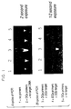
- Figure 1 demonstrates the increased fluorescence of the PCR mixture due to amplification of a specific target DNA in the presence of ethidium bromide. The experiment is described in detail in Example II.
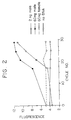
- Figure 2 demonstrates the target specificity of the present detection method and illustrates some of the quantitative aspects of the invention. The details of the experiment are provided at Example IV.
- Figure 3 demonstrates the use of the present invention for genetic screening. The experiment is described in detail at Example V.
- Figures 4A and 4B relate to the quantitative homogeneous assay described in Example VII.
- Figure 4A demonstrates that the increase in fluorescence in the positive samples is greater than two standard deviations away from the average of the negative samples.
- Figure 4B graphically describes a background substraction method for measuring fluorescence.
- the present invention provides improved methods for detecting nucleic acids and is especially suited for use in conjunction with amplification processes.
- the improved methods require neither an oligonucleotide probe nor a labeled amplification primer.
- the methods enable monitoring the accumulation of product while the amplification reaction is in progress.
- the invention also allows target specific quantitation. These methods are suitable for use in automated formats.
- amplified nucleic acids are detected without opening the reaction vessel once the amplification reaction is initiated and without any additional handling or manipulative steps subsequent to the reaction.
- nucleic acid detection methods required a third oligonucleotide reagent as a probe, or a series of manipulative and time consuming steps for target detection, for example, capturing the product on a solid support, which steps are performed after amplification. In some procedures both capture and probe hybridization steps are required.
- the present invention eliminates the need for using a hybridizing probe reagent or a capture procedure for detecting the amplified target.
- the methods of the invention offer speed, simplicity, and decreased opportunity for cross-contamination between samples, particularly between amplified and non-amplified samples.
- the present methods offer means for automated detection, monitoring and quantitation of amplification products during the amplification process and after the amplification reaction is complete.
- the DNA binding agent is an intercalating agent.
- an intercalating agent is an agent or moiety capable of non-covalent insertion between stacked base pairs in the nucleic acid double helix.
- Intercalating agents such as ethidium bromide, fluoresce more intensely when intercalated into double-stranded DNA than when bound to single-stranded DNA, RNA, or in solution.
- Other intercalating agents exhibit a change in the fluorescence spectra when bound to doublestranded DNA. For example, actinomycin D fluoresces red when bound to singlestranded nucleic acids, and green when bound to a double-stranded template.
- Non-intercalating DNA binding agents are also suitable.
- Hoechst 33258 (Searle & Embrey, 1990, Nuc. Acids Res. 18 (13):3753-3762) exhibits altered fluorescence with increasing amount of target.
- Hoechst 33258 is a member of a class of DNA-binding compounds commonly referred to as "groove binders.” This group includes drugs like distamycin, netropsin and others. These compounds recognize and bind the minor groove of duplex DNA.
- a DNA binding agent produces a detectable signal directly or indirectly.
- the signal is detectable directly, such as by fluorescence or absorbance, or indirectly via a substituted label moiety or binding ligand attached to the DNA binding agent.
- a substituted label moiety or binding ligand attached to the DNA binding agent for indirect detection any moiety or ligand that is detectably affected by proximity to double-stranded DNA is suitable.
- the detectable binding agent is present in the amplification reaction during the amplification process.
- the agent produces a detectable signal.
- the agent may be added to the reaction mixture prior to amplification or while the reaction is in progress.
- the agent is included in an amplification buffer comprising appropriate reagents, such as salts and buffering agents. In this manner it is not necessary to separately add the binding agent to the amplification reaction.
- any DNA binding agent is suitable, so long as in the presence of that agent a net increase in the amount of double-stranded DNA present is reflected in a change signal intensity that is detectable directly or indirectly.
- the detectable signal is fluorescence.
- homogeneous detection assay refers to a method for coupled amplification and detection, wherein the process of amplification generates a detectable signal and the need for subsequent sample handling and manipulation to detect the amplified product is minimized or eliminated.
- the present homogeneous assay is suitable for use in conjunction with oligonucleotide probes.
- an oligonucleotide probe specific for detecting a particular target sequence, is included in the amplification reaction in addition to the DNA binding agent of the present invention.
- the probe labeled with a quencher and fluorophore, hybridizes to the amplified target nucleic acid.
- an agent for polymerization capable of 5' to 3' nucleolytic activity, the fluorophore and quencher, when bound to the target, are separated by degradation of the probe by the polymerase.
- the fluorescence of the unbound probe is detectably distinct from the fluorescence of the bound, and subsequently hydrolyzed probe.
- the fluorescence of the DNA binding agent enables detection that amplification has occurred, and the fluorescence of the hybridized probe indicates target specific amplification. So long as amplification is detectable without opening the reaction vessel, or further processing steps once amplification is initiated, the method is within the present definition of a homogeneous assay.
- amplifying which typically refers to an exponential increase in target nucleic acid is being used herein to describe both linear and exponential increases in the numbers of a select target sequence of nucleic acid.
- amplification reaction mixture refers to an aqueous solution comprising the various reagents used to amplify a target nucleic acid. These include enzymes, aqueous buffers, salts, amplification primers, target nucleic acid, and nucleoside triphosphates. Depending upon the context, the mixture can be either a complete or incomplete amplification reaction mixture.
- Amplification of DNA by PCR is disclosed in U.S. Patent Nos. 4,683,195 and 4,683,202.
- Methods for amplifying and detecting nucleic acids by PCR using a thermostable enzyme are disclosed in US. Patent No. 4,965,188.
- PCR amplification of DNA involves repeated cycles of heat-denaturing the DNA, annealing two oligonucleotide primers to sequences that flank the DNA segment to be amplified, and extending the annealed primers with DNA polymerase.
- the primers hybridize to opposite strands of the target sequence and are oriented so that DNA synthesis by the polymerase proceeds across the region between the primers, effectively doubling the amount of the DNA segment.
- each successive cycle essentially doubles the amount of DNA synthesized in the previous cycle. This results in the exponential accumulation of the specific target fragment, at a rate of approximately 2n per cycle, where n is the number of cycles.
- Taq DNA polymerase is preferred although this is not an essential aspect of the invention.
- Taq polymerase a thermostable polymerase
- Methods for the preparation of Taq are disclosed in U.S. Patent No. 4,889,818.
- other thermostable DNA polymerases isolated from other Thermus species or non Thermus species e.g., Thermus thermophilus or Thermotoga maritima
- non-thermostable DNA polymerase such as T4 DNA polymerase, T7 DNA polymerase, E. coli DNA polymerase I, or the Klenow fragment of E. coli, can also be used in PCR.
- primer refers to an oligonucleotide capable of acting as a point of initiation of DNA synthesis when annealed to a nucleic acid template under conditions in which synthesis of a primer extension product is initiated, i.e., in the presence of four different nucleotide triphosphates and a DNA polymerase in an appropriate buffer ("buffer” includes pH, ionic strength, cofactors, etc.) and at a suitable temperature.
- buffer includes pH, ionic strength, cofactors, etc.
- the nucleoside-5'-triphosphates utilized in the extension process are present in total concentration typically ranging from 400 ⁇ M to 4.0 mM during the extension reaction, although preferably the concentration is between 500 ⁇ M and 1.5 mM.
- primers used in the present invention are oligonucleotides, usually deoxyribonucleotides several nucleotides in length, that can be extended in a template-specific manner by the polymerase chain reaction.
- the primer is sufficiently long to prime the synthesis of extension products in the presence of the agent for polymerization and typically contains 10-30 nucleotides, although that exact number is not critical to the successful application of the method. Short primer molecules generally require cooler temperatures to form sufficiently stable hybrid complexes with the template.
- Synthetic oligonucleotides can be prepared using the triester method of Matteucci et al., 1981, J. Am. Chem. Soc. 103 :3185-3191. Alteratively automated synthesis may be preferred, for example, on a Biosearch 8700 DNA Synthesizer using cyanoethyl phosphoramidite chemistry.
- this primer For primer extension to occur, this primer must anneal to the nucleic acid template. Not every nucleotide of the primer must anneal to the template for extension to occur.
- the primer sequence need not reflect the exact sequence of template.
- a non-complementary nucleotide fragment may be attached to the 5' end of the primer with the remainder of the primer sequence being complementary to the template.
- non-complementary bases can be interspersed into the primer, provided that the primer sequence has sufficient complementarily with the template for annealing to occur and allow synthesis of a complementary DNA strand.
- Amplification systems such as PCR require a target nucleic acid in a buffer compatible with the enzymes used to amplify the target.
- the target nucleic acid can be isolated from a variety of biological materials including tissues, body fluids, feces, sputum, saliva, plant cells, bacterial cultures, and the like.
- the nucleic acid in the sample will be a sequence of DNA, most usually genomic DNA.
- the present invention can also be practiced with other nucleic acids, such as messenger RNA, ribosomal RNA, viral RNA, or cloned DNA.
- Suitable nucleic acid samples include single or double-stranded DNA or RNA for use in the present invention.
- the nucleic acid can be amplified merely by making appropriate and well recognized modifications to the method being used.
- nucleic acid sequence To amplify a target nucleic acid sequence in a sample, the sequence must be accessible to the components of the amplification system. In general, this accessibility is ensured by isolating the nucleic acids from a crude biological sample.
- a variety of techniques for extracting nucleic acids from biological samples are known in the art, for example, those described in Maniatis et al., Molecular Cloning: A Laboratory Manual, New York, Cold Spring Harbor Laboratory, 1982; Arrand, Preparation of Nucleic Acid Probes, in pp. 18-30, Nucleic Acid Hybridization: A Practical Approach, Ed Hames and Higgins, IRL Press, 1985); or in PCR Protocols, Chapters 18-20, Innis et al., ed., Academic Press, 1990.
- the PCR process is most usually carried out as an automated process with a thermostable enzyme.
- the reaction mixture is cycled through a denaturing temperature range, a primer annealing temperature range, and an extension temperature range.
- a machine specifically adapted for use with a thermostable enzyme is disclosed more completely in EP No. 236,069 and is commercially available.
- LCR is described in PCT Patent Publication No. WO 89/09835.
- the process involves the use of ligase to join oligonucleotide segments that anneal to the target nucleic acid.
- LCR results in amplification of an original target molecule and can provide millions of copies of product DNA. Consequently, the LCR results in a net increase in double-stranded DNA.
- the present detection methods are applicable to LCR, as well as PCR.
- LCR requires an oligonucleotide probe for detecting the product DNA. When used in conjunction with the disclosed methods for detecting amplification products, a probe step is unnecessary, and the LCR result is immediately detectable.
- Another amplification scheme exploits the use of the replicase from the RNA bacteriophage Q ⁇ .
- a modified recombinant bacteriophage genome with a sequence specific for the targeted sequence is initially hybridized with the nucleic acid to be tested.
- Q ⁇ replicase is added, which, upon recognizing the retained recombinant genome, begins making large numbers of copies.
- the Q ⁇ system does not require primer sequences and there is no heat denaturation step as with the PCR and LCR amplification systems.
- the reaction occurs at one temperature, typically 37°C.
- the preferred template is a substrate for the Q ⁇ replicase, midvariant-1 RNA. A very large increase in the templates is achieved through the use of this system.
- a review of this amplification system can be found in the International Patent Application Pub. No. WO 87/06270 and in Lizardi et al., 1988, Bio/Technology 6 :1197-1202.
- the 3SR system is a variation of an in vitro transcription based amplification system.
- a transcription-based amplification system involves the use of primers that encode a promoter to generate DNA copies of a target strand and the production of RNA copies from the DNA copies with an RNA polymerase. See, e.g., Example 9B of U.S. Patent No. 4,683,202 and EP No. 310,229.
- the 3SR System is a system which uses three enzymes to catty out an isothermal replication of target nucleic acids.
- the system begins with a target of single-stranded RNA to which a T7 RNA DNA primer is bound.
- a cDNA is formed, and RNAseH treatment frees the cDNA from the heteroduplex.
- a second primer is bound to the cDNA and a double stranded cDNA is formed by DNA polymerase (i.e., reverse transcriptase) treatment.
- DNA polymerase i.e., reverse transcriptase
- One or both of the primers encodes a promoter, i.e., the promoter for T7 RNA polymerase, so that the double-stranded cDNA is transcription template for T7 RNA polymerase.
- Transcription competent cDNAs yield antisense RNA copies of the original target.
- the transcripts are then converted by the reverse transcriptase to double standard cDNA containing double-stranded promoters, optionally on both ends in an inverted repeat orientation. These DNAs can yield RNAs, which can reenter the cycle.
- a more complete description of the 3SR system can be found in Guatelli et al., 1990, Proc. Natl. Acad. Sci. USA 87 :1874-1878, and EP No. 329,822.
- the TAS system is also described in Gingeras et al., in Innis et al. eds., 1990, PCR Protocols, Academic Press, San Diego.
- the embodiments of the invention exemplified herein disclose the process for detecting amplified nucleic acids in conjunction with PCR. Accordingly, when used in a PCR-based method, the DNA binding agent is characterized as an agent that will not prevent primer annealing, primer extension along a complementary template, or strand separation of a DNA duplex.
- a sample which contains, or is suspected of containing, a particular oligonucleotide sequence of interest, the "target nucleic acid.”
- the target may be RNA or DNA or an RNA/DNA hybrid.
- the target may be single stranded or double stranded.
- Target preparation will be carried out in a manner appropriate for the particular amplification process to be implemented. For example, in a PCR method where the target nucleic acid is single-stranded DNA, such as mRNA, the target may be first reverse-transcribed into cDNA, prior to amplification.
- RNA targets are well known and described in Maniatis et al., supra. Alteratively, preferred methods for reverse transcription utilize thermoactive DNA polymerases. The present specification teaches that intercalating agents do not prevent DNA polymerase activity. Consequently, the present method provides a homogeneous detection assay for RNA targets as well as DNA targets.
- nested primers are used (Mullis et al., 1986, Cold Spring Harbor Symposium on Quantitative Biology 51 :263). This method may be preferred when the amount of nucleic acid in a sample is extremely limited, for example, where archival, paraffin embedded samples are used.
- nested primers When nested primers are used, the nucleic acid is first amplified with an outer set of primers. This amplification reaction is followed by a second round of amplification cycles using an inner set of primers. Examples VI and VII describe modifications of nested primer methods that provide superior results without necessitating the additional sample handling required by using nested primers in successive rounds of amplification cycles.
- the generation of amplification products can be monitored while the reaction is in progress.
- An apparatus for detecting the signal generated by the binding agent can be used to detect, measure, and quantify the signal before, during, and after amplification.
- the particular type of signal may dictate the choice of detection method.
- fluorescent DNA binding dyes are used to label PCR products. The dyes intercalate, or bind, the double-stranded PCR products, and consequently, the resulting fluorescence increases as the amount of double-stranded DNA increases. The amount of fluorescence can be quantitated by measurement using a spectra-fluorometer with or without opening the PCR vessel. Examples VII and VIII demonstrate this aspect of the invention.
- Example VIII the fluorescence of the positive control sample was well above background measurements. Because signal generation was measured without having to open the reaction tube, this method of detection is readily adaptable to an automated format in which signal is monitored throughout the amplification process.
- Example VIII demonstrates an automated, on-line PCR detection method: a fiber optic lead was used to input excitation light directly to a PCR tube in a heating/cooling block. The same fiber optic was used to return fluorescent emissions back to the spectrafluorometer, where the value was read.
- the amplification reaction is carried out as an automated process.
- a thermocycler currently available uses a heat block capable of holding up to 48 reaction tubes. Consequently, 48 amplification reactions can be carried out simultaneously.
- the present invention permits PCR product detection in all 48 samples, without handling the samples, opening tubes, or interrupting the cycling reaction.
- a suitable optical system moves the excitation light from the source to the reaction tube and measures the emission light from each tube.
- multiple fiber optic leads simultaneously read all PCR tubes undergoing thermocycling.
- only a single fluorometer is needed to read fluorescence from the reaction tubes, as each fiber optic can be read rapidly one at a time, for example, during the time frame of a PCR temperature soak.
- such a detection system is not necessarily limited to a particular thermocycler machine or number of reaction vessels.
- the description of the 48 well thermocycler serves to demonstrate this aspect of the present invention.
- any over plate, tube cap, or lid apparatus that comprises or can be attached to, a fiber optic lead is suitable.
- the reaction tube lids were removed to accommodate the fiber optic.
- use of a reaction vessel that has a clear or translucent cap eliminates the need to insert the cable into the tube. It will be apparent that reaction tubes able accommodate a fiber optic cable, without the cable physically contacting the amplification reaction components, is desirable.
- an optic fiber is not required. The optic fiber is only necessary where a thermocycler and spectrafluorometer are housed independently.
- An analogous detection scheme is suitable in a 96-well microtiter format.
- This type of format is frequently desirable in clinical laboratories for large scale sample screening, for example, for genetic analysis such as screening for sickle-cell anemia or the AIDS virus in blood bank screening procedures.
- the present invention is suitable for this type of analysis and eliminates the need for the numerous washing and extraction procedures that are required with known "in-well” assay procedures such as ELISA type formats or other optical density-based methods (Kolber et al., 1988, J. of Immun. Meth. 108 :255-264; Huschtscha et al., 1989, In Vitro Cell and Dev. Biol. 25 (1):105-108; Voller et al., 1979, The Enzyme Linked Immunosorbent Assay, Dynatech Labs, Alexandria, VA.).
- the present detection methods also allow direct fluorescence measurement using an apparatus similar to ELISA plate reader, but designed to excite and measure fluorescence.
- an apparatus similar to ELISA plate reader for example, the CytoFluorTM 2300 machine manufactured by Millipore is suitable in such a method. It is appropriate to "read" the microtiter plate before and after thermocycling for determining background fluorescence.
- an apparatus providing a continuous determination of fluorescence is useful for monitoring the increase in PCR product during the amplification reaction.
- the size of the amplified product is determined without the use of a probe or size fractionation methods such as HPLC or gel electrophoresis.
- the invention is particularly useful for determining if amplification has occurred and simultaneously distinguishing the amplified target product from, for example, primer-dimer and high molecular weight DNA.
- the detectable binding agent is an intercalating fluorescent dye.
- Ethidium bromide is included in the amplification reaction mixture. Ethidium bromide, like other DNA binding dyes, such as acridines, proflavine, acridine orange, acriflavine, fluorcoumanin, ellipticine, daunomycin, chloroquine, distamycin D, chromomycin, homidium, mithramycin, ruthenium polypyridyls, and anthramycin, exhibit altered fluorescence emissions when bound to double-stranded DNA. In an amplification reaction, a large amount of double-stranded DNA is generated from a starting template.
- ethidium bromide is present in the PCR at a concentration of 0.53 to 1.27 ⁇ M although a concentration of 0.08 ⁇ M (0.03 ⁇ g/ml) to 40.6 ⁇ M (16 ⁇ g/ml) is also suitable; however, an ethidium bromide concentration in the PCR mix in the range of 0.15 ⁇ M (0.06 ⁇ g/ml) to 20.3 ⁇ M (8 g/ml) is preferred. It will be readily apparent to those of ordinary skill in the art how to determine a suitable concentration of alternative DNA binding agents by empirical adjustment.
- a suitable concentration of agent is any amount that provides a signal that is distinguishable when a net increase in doublestranded DNA has occurred in an amplification reaction. Consequently, the preferred concentration of agent included in the amplification reaction mix may vary depending on the amount of double-stranded non-target DNA (i.e., background genomic DNA), the copy number and amount of target, the quantity and fluorescence of the amplification primers and the particular agent utilized. For example, a standard curve using a known amount of target and varying the amount of binding agents may be appropriate.
- the fluorescent dye is added to the PCR mixture before starting temperature cycling. A suitable fluorescent dye does not prevent amplification from occurring. It will be obvious to one of ordinary skill in the art to determine the suitability of any novel dye in the method.
- Example I demonstrates that PCR amplification occurs in the presence of detectable amounts of ethidium bromide.
- the emission spectrum of ethidium bromide peaks at 650 nm for unbound ethidium bromide and at 611 nm when the agent is bound to double-stranded DNA. However, because the emission spectra of bound and unbound ethidium bromide have distinct peaks, with bound ethidium emitting at a lower wavelength, discrimination between bound and unbound ethidium is enhanced by detecting emission at a non-peak wavelength that is less than the peak for bound ethidium.
- the fluorescence emission of the sample was detected at 570 nm.
- the excitation wavelength was set at 500 nm and emission detection was set at 570 nm.
- the detection or excitation wavelength is optimized for practice of the invention.
- a U.V. light box with an excitation wavelength at 300 nm and detection by photography through a red filter is suitable.
- a spectra fluorometer depending on the features of the particular machine utilized, offers the opportunity to set the excitation and emission wavelength, as well as bandwidth. It will be obvious to one of ordinary skill in the art how to determine the wavelength and bandwidth settings for a particular DNA binding agent to be detected.
- each agent has a discrete fluorescence spectrum, a broad range of detection wavelengths are suitable for practicing the invention, as exemplified herein. General guidance is found in, for example, The Merck Index, eds.
- the DNA polymerase is added to the PCR reaction mixture after both the primer and template are added.
- the enzyme and primer are added last or the PCR buffer or template plus buffer are added last. It is generally desirable that at least one component that is essential for polymerization not be present until such time as the primer and template are both present, and the enzyme can bind to and extend the desired primer/template substrate.
- Methods for reducing the effects of cross contamination of amplification reactions require the introduction of unconventional bases into the amplified product and exposing carryover to enzymatic and/or physical chemical treatment which effectively render the product incapable of serving as a template for subsequent amplifications.
- the homogeneous detection assay described herein is suitable in conjunction with sterilization methods. These methods enhance the accuracy and reliability of amplification results by eliminating steps, and thereby minimizing product handling, which reduces carryover.
- the sterilization method provides additional assurance that carryover template is eliminated.
- the DNA binding agent is storage stable and can be included as a component in a PCR reagent buffer.
- a reagent may contain a solution of the DNA binding agent in a kit for detecting nucleic acids by PCR.
- the reagent might contain a DNA binding agent, as well as other PCR buffer components such as Tris-HCl, KCl, and MgCl2, each in appropriate concentrations for carrying out PCR.
- a kit in one embodiment, includes a buffer comprising ethidium bromide at a suitable concentration to provide, in an amplification reaction, a final ethidium bromide concentration in the range of 0.15 ⁇ M to 20.3 ⁇ M.
- the buffer may additionally contain any or all of the following reagents: Tris-HCl, pH 8.0-8.3; KCl, and MgCl2 each in appropriate concentrations for PCR amplification.
- Kits for detecting amplified nucleic acids are also envisioned as including any of the following: an agent for polymerization, dNTPs, appropriate primers, and a positive control template.
- U.S. Patent No. 4,683,202 describes methods for preparing and using primers for PCR which have non-complementary sequences added to the 5' end. These "tails" are useful for engineering particular restriction sites or other purposes, because during PCR, the non-complementary tail sequence is incorporated into the double-stranded PCR product. Particular tail sequences provide binding targets for specific dyes. For example, Hoechst 33258 (Searle and Embrey, 1990, Nuc. Acids Res. 18 :3753-3762) preferentially binds A-T base pairs.
- a PCR primer synthesized with a long A-T rich 5' tail provides a relatively A-T rich PCR product in comparison with genomic DNA. Using Hoechst 33358 the A-T rich PCR product has increased fluorescence relative to genomic DNA and, consequently, is useful for increasing signal strength in the presence of genomic DNA.
- the present invention is suitable for monitoring the amount of two distinct target nucleic acids in one reaction vessel, by including two fluorochromes in the reaction mixture during amplification. It is only necessary that one fluorochrome has sequence specificity and that sequence is included in the tail (i.e., 5, end) of a primer to one of the two target nucleic acids.
- the emission spectra for each fluorochrome is separately determined during and/or after amplification by a spectra fluorometer.
- the invention is particularly useful for quantitative comparisons of two different nucleic acid targets in the same sample and as such may be useful for determining the extent of, for example, an infection or disease if one target is a sequence present in all cells and the other is present in a pathogen.
- Fluorescent DNA binding dyes may each have different emissions and excitation spectra. In a clinical setting, different DNA binding dyes having different DNA sequence specificities are useful for indicating the presence of distinct targets.
- the present invention is suitable in conjunction with methods using an internal standard to determine either the relative amount of a target or accurately quantitate the amount of target present prior to amplification.
- the present invention provides means for determining the intactness of a DNA sample.
- forensic analysis often involve samples containing partially degraded target, such as an archival sample.
- partially degraded target such as an archival sample.
- the ability to monitor the generation of PCR product demonstrated in Example VIII, allows comparison of the amplification profile of a DNA sample with respect to a small PCR product and a large PCR product, in separate reactions. If there is no degradation, monitoring the increase in double-stranded DNA shows that both reactions reach plateau at approximately the same cycle. If there is degradation, the small product would reach plateau sooner than the large fragment due to the presence of more intact target molecules.
- homogeneous detection assay is suitable for a wide variety of applications, including, for example, genetic screening, forensic human identification, pathogen detection, and quantitation, environmental monitoring or tagging and tracing materials with nucleic acid.
- the detectable signal is fluorescence. Fluorescence is suitable for use in qualitative as well as quantitative methods. Qualitative detection can be made simply and rapidly by visual inspection of the reaction tubes by exposure to UV light. This type of rapid, highly sensitive assay is desirable as a rapid screen for the presence of a pathogen or of a particular gene sequence causative of or associated with a disease state.
- the present invention provides means for cutting costs by a plus/minus pre-screen for the presence of any member of a group of targets. Amplifications for many different such targets would allow this if it may be expected that most samples are negative, e.g., environmental monitoring for pathogen detection systems.
- Multiplex PCR is a process for including a number of distinct primer pairs in one amplification reaction (Gibbs et al., 1989, in PCR Technology ed. Erlich, Stockton Press, NY).
- a homogeneous detection assay using multiple primer pairs to assay a wide range of potential targets is then followed by more complex typing procedures only on positive samples. This pre-screen saves the cost of performing the more complex typing procedure on negative samples and/or performing repeated tests on negative samples.
- the present methods for homogeneous detection of target nucleic acids are also suitable for quantitation of a particular target. Whether the methods are automated or manually performed, quantitation can be accomplished by a number of means. For example, a serial dilution of the sample, in parallel with a serial dilution of a known standard, provides a series of templates, which, following PCR and signal detection, are suitable for quantitating the amount of starting material in the known sample. Similarly, because fluorescence can be determined between cycles during the course of a PCR, the exponential phase of PCR and the cycle at which the level of product reaches the plateau phase, can be readily determined.
- the invention is suitable for detecting amplified nucleic acids in the presence of double-stranded genomic DNA. Background levels of DNA as high as 0.5 g or more will not obscure a positive assay result.
- the target nucleic acid can be a cloned segment, a repeat sequence, such as a multi-copy gene or tandem repeat, a single copy gene, or an infectious agent present in a concentration as low as one copy per 70,000 cells.
- the starting template is RNA or DNA because PCR provides a net increase in double-stranded nucleic acid starting from either template nucleic acid.
- the target nucleic acid may be a rare sequence, such as a single copy of AIDS virus DNA in a background of human genomic DNA from more than 70,000 cells.
- procedures for increasing specificity serve to insure that amplification provides a net gain in double-stranded DNA only in response to the presence of the target sequence, and that net gain is detectable in the presence of high background of genomic DNA.
- U.S. Patent No. 4,683,195 demonstrates the use of nested primers to decrease the background in the amplification of single copy genes.
- modified nested amplification procedures are provided.
- the present nested primer methods are vastly improved over prior nested primer procedures for amplifying nucleic acids. These methods provide enhanced specificity and are applicable in any PCR-based amplification scheme. However, in the present disclosure, these procedures are described used in a homogeneous assay.
- a third primer internal to a flanking pair of PCR primers is included in a PCR reaction for amplifying a particular target segment.
- the third primer has an annealing/melting temperature, when it is hybridized to its complementary target strand, that is lower than that of the flanking primer. This property can be imparted to the primer by a shorter length and/or lower G-C content.
- the temperature during the extension phase of each PCR cycle is maintained sufficiently high, i.e. approximately 65°C to prevent the short primer from annealing specifically and initiating amplification.
- the flanking primers anneal sufficiently such that PCR proceeds normally at the high extension temperature.
- the primer flanking the third primer is present at a low concentration.
- the annealing temperature is decreased to approximately 42°C. At this temperature, the third primer "drops in” and proceeds to amplify the target for the remaining 15 or so cycles.
- flanking primer is synthesized with a G-C rich tail. Because the non-complementary primer tail sequence is incorporated into the PCR product after two cycles of amplification, the G-C tails serves to raise the temperature necessary for denaturing the PCR product because of the increased thermostability of G-C pairs versus A-T pairs (Myers et al., 1989, in PCR Technology ed. Erlich, Stockton Press, New York). The resulting difference in the denaturing temperature between the nested and flanking PCR product is then exploited to effectively shut down amplification from the tailed flanking primer.
- the denaturation temperature is lowered, i.e., from 96°C to 86°C, so that the temperature is too low for amplification of the flanking PCR product, but is high enough to allow amplification of the nested PCR product.
- the annealing temperature may also be manipulated, as in the "drop-in" method, to initiate synthesis from the nested primer when desired.
- This example demonstrates the ability of PCR to proceed in the presence of ethidium bromide.
- PCRs Two PCRs were carried out as follows. Each 100 ⁇ l PCR contained: 50 ng human DNA, 10 mM Tris-HCl pH 8, 50 mM KCl, 4 mM MgCl2, 250 ⁇ M each dNTP, 2.5 units Taq polymerase (Perkin-Elmer Cetus Instruments, Norwalk Connecticut), 20 picamole each primer GH26 (SEQ ID NO: 1) and GH27 (SEQ ID NO: 2).
- the human DNA was purified from a human B-cell line. DNA was prepared according to the method described by Maniatis (supra.). An oil overlay was added to each reaction to prevent evaporation.
- the two PCRs were conducted under identical conditions except that 0.51 ⁇ M of ethidium bromide (Sigma) was included in one reaction.
- a thermocycler purchased from Perkin-Elmer Cetus Instruments was programmed with the following cycling parameters; denature at 96°C, hold for 1 minute, anneal at 55°C, hold for 1 minute, extend at 72°C, hold for 1 minute. This profile was repeated for 32 cycles.
- After amplification 5 ⁇ l of each PCR was analyzed by gel electrophoresis using a 3% NuSieve agarose gel (FMC). The gel was stained with ethidium bromide by standard methods and the amount of PCR product made in the two reactions was compared.
- Example II shows that the specificity of PCR is sufficient for fluorescence-based, homogeneous detection of target nucleic acids.
- the experiment described in Example I was expanded upon to determine whether target specific ethidium bromide fluorescence is readily visible using standard PCR conditions. Therefore, five 100 ⁇ l reaction mixtures were prepared containing 10 mM Tris-HCl pH8, 50 mM KCl, 2.5 mM MgCl2 1.27 ⁇ M ethidium bromide, 150 ⁇ M each dNTP, 2.5 units Tag polymerase. Primers and target DNA were included as described below.
- Primer pair GH15 (SEQ ID NO: 3) and GH16 (SEQ ID NO: 4) are specific for amplifying DQ ⁇ : and do not amplify DR ⁇ 1 target DNA.
- DQ ⁇ target DNA was prepared by amplifying the human DQ ⁇ : gene as described in Example I. Following amplification, the DQ ⁇ PCR product was diluted to provide ⁇ 2 x 10⁷ copies (5 pg) of amplified DNA per amplification. The DQ ⁇ product was used as a positive control.
- DR ⁇ 1 target DNA was prepared using primer pair GH46 (SEQ ID NO: 5) and GH50 (SEQ ID NO: 6) to amplify the DR ⁇ 1 gene in a PCR using human genomic DNA.
- the PCR product was diluted to provide ⁇ 2 x 10⁷ copies of DR ⁇ 1 DNA and used in Reaction 4, below, as a negative control.
- the five reaction mixtures contained primers and target as follows: Reaction 1 - No primers + DQ ⁇ : Target Reaction 2 - Primers + DQ ⁇ : target Reaction 3 - Primers + DQ ⁇ : target Reaction 4 - Primers + DR ⁇ 1 target Reaction 5 - Primers + no target Where primers were included 10 pmoles each of GH15 (SEQ ID NO: 3) and GH16 (SEQ ID NO: 4) were added. Reaction mixtures 1 and 2 were not subjected to amplification cycles. Reactions Nos. 3, 4, and 5 were subjected to 20 cycles of amplification. The cycling parameters were 94°C, hold for 1 minute; 45°C, hold for 1 minute; and 72°C, hold for 1 minute.
- reaction tubes were then placed on a UV light box (300 nm) and photographed for two seconds and one-half second exposures.
- the results, shown in Figure 1 demonstrated the increased fluorescence in Reaction No. 3 due to the amplification of the specific target DNA.
- Reaction Nos. 1 and 2 indicated the level of fluorescence present before any amplification occurred.
- Reaction Nos. 4 and 5 demonstrated that fluorescence does not increase visibly in a reaction unless, for the particular primer pair present in the reaction, an appropriate template is also present.
- the different photographic exposures as shown in Figure 1 demonstrate the relative differences in fluorescence.
- PCR mixtures were prepared as follows. Each contained 10 mM TrisHCl, pH 8; 50 mM KCl; 2 mM MgCl2; 2.9 units Taq polymerase; 180 ⁇ m each dNTP; 1.27 ⁇ M ethidium bromide; 15 picamole RH191 (SEQ ID NO: 7); and 15 picamole RH192 (SEQ ID NO: 8).
- the primers RH191 (SEQ ID NO: 7) and RH192 (SEQ ID NO: 8) are derived from primers yl.1 and yl.2, which are described in Kogan et al., 1987, N. Engl. J. Med., 317 :985-990. These primers are specific for a human male-specific sequence that occurs in several thousand copies per human male cell. Fifteen PCR reaction mixtures were set up as follows. Sample DNA was prepared from human blood taken from a male or female as specified for each reaction. DNA was prepared according to Maniatis supra. Reaction Nos. 1-5 contained 2 ng human male DNA Reaction Nos. 6-10 contained no DNA Reaction Nos. 11-15 contained 60 ng human female DNA Reaction Nos.
- 16-20 contained 60 ng human male DNA
- the reactions were all placed in a Perkin-Elmer Cetus Instruments thermocycler programmed to cycle at 94°C for 1 minute; 60°C for 1 minute, for the indicated number of cycles.
- tubes were removed from the thermocycler at various cycles to provide a PCR time course for each template. Specifically, for each set of five tubes, 0, 17, 21, 25, and 29 amplification cycles were performed. PCR product was detected by exposing the tubes to UV-light as described above.
- Tube Nos. 6-15 exhibited no increase in fluorescence.
- Tube Nos. 1 and 2 the 0 and 17 cycle male DNA samples, also had no increase in fluorescence by visual detection. Only the 21, 25, and 29 cycle male DNA reactions fluoresced under UV light, and the amount of fluorescence increased with increasing cycle number.
- the results with Tube Nos. 16-20 were similar, except that an increase in fluorescence was noted by 17 cycles. This is consistent with the presence of more copies of the target DNA sequence present in the sample prior to amplification.
- Example III For quantitative measurement of target specific ethidium bromide fluorescence the reactions of Example III were opened, and the contents transferred to a spectra fluorometer (SPEX Fluorolog-2, purchased from Spex, Edison, NJ) which was used to get a quantitative fluorescence value for each reaction.
- SPEX Fluorolog-2 purchased from Spex, Edison, NJ
- the results shown in Figure 2 not only demonstrate the target specificity of the detection method but illustrate the quantitative aspects of the invention.
- the effect of increased target in the sample is observable by comparing the time course of fluorescence between the 2 ng and 60 ng male templates. The more target DNA in the sample, the sooner the reaction obtains a measurable increase in fluorescence and finally reaches a plateau level of fluorescence. Exactly when the reaction fluorescence begins to increase measurably is effectively a quantitative measure of how much target was present prior to amplification.
- the particular gene to be detected is the ⁇ -globin gene.
- a single base pair change mutates a wild-type ⁇ -globin allele into a sickle cell allele.
- the following primers were used: RH187 (SEQ ID NO: 9), RH188 (SEQ ID NO: 10), and RH189 (SEQ ID NO: 11).
- Primer pair RH187/RH188 SEQ ID NO: 9/SEQ ID NO:10) specifically amplified the wild-type allele.
- Primer pair RH187/RH189 (SEQ ID NO: 9/SEQ ID NO: 11) amplify the sickle cell allele (these primers derive from BGP2, H ⁇ 14A , and H ⁇ 14S described in Wu et al., 1989, PNAS (USA), 86 :27572760.
- PCRs were set up as follows: 10 mM Tris-HCl, pH 8.3; 50 mM KCl; 748 ⁇ M total dNTPs; 2.9 units Taq polymerase; 10 pmoles each primer; 1.5 ⁇ M MgCl2; 1.27 ⁇ M ethidium bromide, and 50 ng of human DNA template as follows: one pair of reactions contained human DNA homozygous for sickle allele (SS); one pair of reactions contained wild-type DNA (AA); and one pair contained heterozygous DNA, i.e., one wild-type and one sickle cell allele (AS).
- SS human DNA homozygous for sickle allele
- AA wild-type DNA
- AS sickle cell allele
- one tube contained primers specific for the sickle globin allele and one tube contained primers for the wild-type sequence.
- the only difference between the primer sets are the 3' nucleotides of one of the primer pairs, which match either the sickle cell or the wild-type target sequence.
- Primer annealing temperature during PCR is set such that amplification will occur only if this 3' nucleotide matches the template.
- the cycling parameters were: 94°C for 60 seconds, 55°C for 60 seconds.
- a detection assay was designed to demonstrate the suitability of the present invention for detecting a rare target sequence in a background of DNA from approximately 70,000 human cells.
- a modified nested primer procedure as briefly described in the "Detailed Description” section as a primer “drop-out” procedure, was designed to enhance PCR specificity.
- This assay was done as follows: into PCR reaction vessels 1 through 8 were aliquoted 50 microliters of solution, each containing 50 mM KCl; 10 mM TrisHCl, pH 8.3; 2.5 mM MgCl2; 600 ⁇ M total dNTPs; 1.25 unit of Taq DNA polymerase (PECI); 1.27 ⁇ M ethidium bromide; 0.5 ⁇ g of human cell-line DNA; the primer pair RH171 (SEQ ID NO: 12) and RH176 (SEQ ID NO: 13), each primer at 0.2 ⁇ M; and the nested primer RH182 (SEQ ID NO: 14) also at 0.2 ⁇ M.
- PECI Taq DNA polymerase
- Primer RH176 (SEQ ID NO: 13) carries a GC-rich, non-homologous (to target sequence), 5' "tail” that raises the denaturation temperature necessary to amplify PCR product made using this primer.
- Reactions 1-4 and 6-8 also contained a target, positive control DNA (purchased from PECI) containing HIV sequences to which RH171 (SEQ ID NO: 12), RH176 (SEQ ID NO: 13), and the 3' portion of RH182 (SEQ ID NO: 14) were homologous. This DNA was diluted such that each reaction containing it had, on average, four copies of the HIV sequence. Because this average number of copies is small, the actual number of copies in a given reaction can vary considerably. Since no HIV DNA target was added to reactions 1 and 5, these reactions served as negative controls.
- This profile was repeated for 2 cycles, during which all three primers efficiently annealed and were extended in amplification such that products were made using either RH171 (SEQ ID NO: 12) and RH176 (SEQ ID NO: 13), or RH171 (SEQ ID NO: 12) and RH182 (SEQ ID NO: 14). Because the use of a third, nested primer, increases product specificity, products made using RH171 (SEQ ID NO: 12) and RH182 (SEQ ID NO: 14) were more likely to be HIV specific. These cycles were followed by denaturation at 86°C, hold for 1 minute, anneal at 52°C, hold for 1 minute.
- reaction 2-4 and 5-8 were shown to contain a product of the expected size (approximately 200 bp) as the predominant band on the gel. Reactions 1 and 5, the negative controls, contained no such product. However, reactions 2-4, which were not given a "hot-start,” could be seen to contain DNA fragments of other than the expected size. These other DNA fragments were also visible in reaction 1, indicating that they are not derived from HIV sequences. These other DNA fragments were not visible in reactions 5-8, indicating that use of the "hot-start" had enhanced the specificity of these reactions.
- Figure 4A graphically demonstrates the significant increase in fluorescence of the two positive samples compared to the two negative samples.
- dotted lines mark the fluorescence values two standard deviations away from the average of the negative samples.
- the PCR-positive samples are both above this line.
- the following experiment demonstrates the suitability of the present method for monitoring a PCR reaction and detecting the net increase in double-stranded DNA due to amplification.
- the apparatus allows on-line detection of PCR product.
- the apparatus was set up as follows: a Spex-Fluorolog-2 fluorometer with a fiber optic accessory (Spex Catalog No. 1950) was set to emit excitation light at 500 nm with a bandwidth of ⁇ 3.4 nm. A GG 435 nm cut off filter used to exclude second order light (Purchased from Melles Grist Inc.). The emission light was detected at 570 nm with a bandwidth of ⁇ 13.6 nm. A OG530 filter (530 nm cut off) was used to remove excitation light.
- Two PCR reactions were set up as described in Example III using primers RH191 (SEQ ID NO: 7) and RH192 (SEQ ID NO: 8).
- One reaction tube contained 60 ng human male DNA, the other contained no target DNA.
- the reactions were set up in 0.5 ml polypropylene tubes; however, the top of the tubes was cut away for attaching the fiber optic cable.
- the fiber optic was glued to the top of the reaction tube with epoxy. Because this apparatus had one fiber optic, only one PCR was run at a time. The emission light was collected through the oil overlay in the tube. A black "shroud" was built around the tube and the reaction was placed in the thermocycler.
- thermocycler was programmed to cycle between 94°C and 50°C for 1 minute each, for 30 cycles, followed by continuous incubation at 25°C.
- the fluorometer and thermocycler were started simultaneously.
- the parameters of the fluorometer were: time based scan with 5 second integration time; the emission signal was ratioed to that of the excitation light to control for changes in source intensity.
- Figure 5A shows the results of the PCR reaction containing no DNA.
- Figure 5A shows that the thermocycler started at 25°C, the fluorescence dropped as the temperature increased to 94°C and fluorescence increased again when the temperature decreased to 50°C. This pattern was repeated for the remaining cycles until the thermocycler again reached 25°C and the fluorescence returned to the approximate starting value.
- Figure 5B demonstrates the fluorescence profile of a PCR reaction containing the appropriate target DNA.
- the fluorescence intensity at 50°C shows a cycle dependent increase reflecting an increase in the amount of double-stranded DNA.
- the thermocycler returned to 25°C, after the 30 cycles were completed, the fluorescence increased to a final value greater than three times the initial fluorescence value at 25°C.
- the on-line method provided a rapid analysis as to the presence or absence of target DNA without the need for probes or further processing steps.
- the continuous detection of fluorescence throughout the amplification provides an amplification profile that reflects the amount of target present at start. If the target DNA is, for example, a human repeat sequence present in millions of copies per cell (e.g., "Alu" sequences; Nelson and Caskey in PCR Technology ed. Erlich, 1989, Stockton Press, NY), this method of quantitation could be used to quickly and simply to measure sub-cellular amounts of DNA, which is at present difficult to do without using radioisotopes.
Abstract
Description
- The present invention provides improved methods for detecting a target nucleic acid in a sample and for monitoring the increase in a double-stranded DNA during amplification of a target nucleic acid in a sample as also a kit for amplifying a target nucleic acid.
- The novel methods for simultaneous nucleic acid amplification and detection enhance the speed and accuracy of prior detection methods and eliminate the need for sample processing following amplification. In a preferred embodiment, the method provides a modification of the polymerase chain reaction and utilizes agents whose fluorescence is enhanced upon binding double-stranded DNA. The methods provided herein have numerous applications, particularly in the fields of molecular biology, medical diagnostics and forensic sciences.
- The disclosed nucleic acid detection methods offer the advantages of speed and simplicity over prior methods for detecting amplified nucleic acids. Nucleic acid detection techniques in general are particularly useful in medical diagnostic assays. For example, U.S. Patent No. 4,358,535 discloses a method for detecting pathogens by spotting a sample (e.g., blood, cells, saliva, etc.) on a filter, lysing the cells and fixing the DNA through chemical denaturation and heating. Then, labeled DNA probes are added and allowed to hybridize with the fixed sample DNA. Hybridization indicates the presence of the pathogen's DNA.
- Nucleic acid detection using oligonucleotide probes has become a standard method for specific target detection. Numerous modifications of the method have been described, including culturing the target cells or organisms in situ on the filter, increasing the amount of target nucleic acid available for detection. Generally, these methods require that the DNA sample is noncovalently bound onto a solid support such as nitrocellulose or nylon and then hybridized to a labeled target-speciflc probe.
- The sensitivity and specificity of nucleic acid detection methods was greatly improved by the invention of the polymerase chain reaction (PCR). PCR is a process for amplifying nucleic acids and involves the use of two oligonucleotide primers, an agent for polymerization, a target nucleic acid template, and successive cycles of denaturation of nucleic acid and annealing and extension of the primers to produce a large number of copies of a particular nucleic acid segment. With this method, segments of single copy genomic DNA can be amplified more than 10 million fold with very high specificity and fidelity. PCR methods are disclosed in U.S. Patent No. 4,683,202.
- Methods for detecting PCR products are particularly described in U.S. Patent No. 4,683,195. Those methods require an oligonucleotide probe capable of hybridizing with the amplified target nucleic acid. EP No. 237,362 also describes a PCR-based detection method termed "reverse dot blot", in which the probe, instead of the amplified DNA, is fixed to the membrane. According to the method, the target, rather than the probe, is labeled for hybridization. These methods require separate steps of amplification, capture, and detection and generally require several hours to complete. In the reverse dot-blot method, storage-stable target-specific reagents are preferred.
- Alterative methods for detecting amplified nucleic acids describe simultaneous amplification and labeling of a target nucleic acid. The methods require that at least one amplification primer is labeled. The amplification primer can be labeled with, for example, a radioisotope for direct detection of the amplified product or labeled with a reagent suitable for capturing the product onto a solid support for subsequent detection.
- Other means of detection include the use of fragment length polymorphism hybridization, allele-specific oligonucleotide (ASO) probes (Saiki et al., 1986, Nature 324:163), or direct sequencing via the dideoxy method using amplified DNA rather than cloned DNA. The fragment length polymorphism method detects insertions and deletions between PCR primers resulting in PCR products of different lengths, detectable by sizing. ASO methods are useful for detecting allelic sequence variations. In an example of ASO hybridization, the amplified DNA is fixed to a nylon filter by, for example, UV irradiation in a series of "dot blots," then allowed to hybridize with an oligonucleotide probe under stringent conditions. The probe may be labeled with, for example, horseradish peroxidase (HRP) and detected by the presence of a blue precipitate following treatment with suitable oxidation reagents.
- Further is known an alterative assay method for detecting amplified nucleic acids. The process employs the 5' to 3' nuclease activity of a nucleic acid polymerase to cleave annealed, labeled oligonucleotides from hybridized duplexes and release labeled oligonucleotide fragments for detection. The method is suitable for detecting PCR products and requires a primer pair and a labeled oligonucleotide probe having a blocked 3'-0H terminus to prevent extension by the polymerase.
- Due to the enormous amplification possible with the PCR process, small levels of DNA carryover from samples with high DNA levels, positive control templates, or from previous amplifications, can result in PCR product even in the absence of purposefully added template DNA. Higuchi and Kwok, 1989, Nature 339:237-238, and Kwok and Orrego, in Innis et al., 1990, PCR Protocols: A Guide to Methods and Applications, Academic Press, Inc., San Diego, CA, describe particular methods and precautions for practicing PCR with a minimum of cross contamination. Because the possibility of introducing contaminating DNA to a sample will be increased as the amount of handling steps required for sample preparation, processing, and analysis is increased, it would be preferable to minimize sample handling, particularly after the amplification reaction is complete.
- A number of agents have been described for labeling nucleic acids, whether probe or target, for facilitating detection of target nucleic acid. Suitable labels may provide signals detectable by fluorescence, radioactivity, colorimetry, X-ray diffraction or absorption, magnetism or enzymatic activity and include, for example, fluorophores, chromophores, radioactive isotopes (particularly ³²P and ¹²⁵I) electron-dense reagents, enzymes, and ligands having specific binding partners.
- Labeling is achieved by a number of means, such as chemical modification of a primer or probe to incorporate a label or the use of polymerizing agents to incorporate a modified nucleoside triphosphate into an extension product. Intercalating agents noncovalently bind the stacked bases of nucleic acids and as a result the fluorescence of the agent either increases or shifts to a different wavelength. For example, U.S. Patent No. 4,582,789 describes several intercalating moieties including psoralens.
- Fluorescent dyes are suitable for detecting nucleic acids. For example, ethidium bromide is an intercalating agent that displays increased fluorescence when bound to double-stranded DNA rather than when in free solution (Sharp et al., 1973, Biochemistry 12:3055). Ethidium bromide can be used to detect both single- and double-stranded nucleic acids, although the affinity of ethidium bromide for singlestranded nucleic acid is relatively low. Ethidium bromide is routinely used to detect nucleic acids following gel electrophoresis. Following size fractionation on an appropriate gel matrix, for example, agarose or acrylamide, the gel is soaked in a dilute solution of ethidium bromide. The DNA is then visualized by examining the gel under UV light (Maniatis et al., 1982 eds., Molecular Cloning: A Laboratory Manual, New York, Cold Spring Harbor Laboratory).
- Alternative fluorescence based methods for detecting DNA have been described. For example, Morrison et al., 1989, Anal. Biochem., 183:231-244 describe a two probe method for detecting target DNA. One probe is labeled with fluorescein, the other probe, complementary to the first, is labeled with a quencher for fluorescein emission. The probes are allowed to anneal with denatured DNA containing the target sequence, and the amount of fluorescence is determined. Fluorescence increases to the extent that the fluorescein probe binds to unlabeled, complementary DNA rather than the complementary, quenching probe.
- Mabuchi et al., 1990, Nucl. Acids Res. 18(24):7461-7462 describe a method for detecting DNA fragments based on the AT content of the nucleic acid segment. Two fluorochromes are used to stain size fractionated DNA in an agarose gel. The selective binding properties of different fluorochromes for AT rich regions are used to distinguish electrophoresed DNA fragments.
- U.S. Patent No. 4,257,774 describes the direct binding of fluorescent intercalators to DNA, e.g., ethidium salts, daunomycin, mepacrine and acridine orange, as well as 4'6-diamidino-α-phenylindole to quantitate the DNA. Fluorescence polarization is used for characterization of nonfluorescent DNA binding compounds which complete with the DNA binding dyes.
- Oser and Valet, 1990, Angew. Chem. Int. Engl. 29(10):1167, describe a nucleic acid detection scheme that requires two oligonucleotide probes complementary to adjacent sites on a target. The probes are labeled differentially with either a salicylate or a DTPA ligand bearing a fluorescence emitter, TbIII. Hybridization of both probes to the target provides steric proximity of the two labels resulting in a measurable increase in TbIII fluorescence. The modified probes are prepared specifically for each target to be detected.
- EP No. 070,685 describes the use of fluorescent labeled polynucleotide probes in polynucleotide hybridization assays. According to the method, probes are prepared by attaching particular absorber-emitter moieties to the 3' and 5' ends of nucleic acid fragments. The fragments are capable of hybridizing to adjacent positions on a target DNA, so that, if both fragments are hybridized, the proximity of the absorber and emitter moieties results in detectable emitter fluorescence.
- According to these methods, the fluorescent dye is introduced to the target DNA after all in vitro nucleic acid polymerization reactions have been completed. The inhibitory effects of intercalating agents on nucleic acid polymerases have been described in numerous publications (for example, Kornberg, 1974, DNA Synthesis, W.H. Freman and Co., San Francisco; Richardson, 1973, J. Mol. Biol. 78:703-714).
- DNA binding dyes are useful as antibiotics because of the inhibitory effects on nucleic acid replication processes that result from the agent binding to the template. EP No. 169,787 describes the use of intercalating agents for blocking replication of influenza or herpes virus. Kornberg (supra) describes a number of DNA binding agents, both intercalators and non-intercalators, and describes how each compound inhibits nucleic acid replication. At page 227, Kornberg specifically describes that ethidium bromide inhibits DNA replication.
- A method for simultaneous amplification and detection of target nucleic acids would provide advantages over prior detection methods. Such a method would minimize the problems of sample contamination inherent in any process involving a series of manipulative steps for discerning a positive or negative test result. By eliminating sample handling and processing steps, a method for simultaneous amplification and detection of target nucleic acids would increase the speed and accuracy of current diagnostic methods. The present invention addresses and solves these needs.
- The present invention provides a method for detecting a target nucleic acid in a sample. The method comprises the steps of: (a) providing an amplification reaction mixture that comprises a sample, a DNA binding agent, wherein said agent is characterized as providing a detectable signal when bound to double-stranded nucleic acid which signal is distinguishable from the signal provided by said agent when it is unbound, and reagents for amplification; (b) determining the amount of signal produced by the mixture of step (a); (c) treating said mixture under conditions for amplifying the target nucleic acid; (d) determining the amount of said signal produced by the mixture of step (c); and (e) determining if amplification has occurred.
- The invention is particularly suitable for practice in PCR amplification methods wherein a net increase in double-stranded DNA results in a change in signal strength or type. In a preferred embodiment, the DNA binding agent is a fluorescent DNA binding dye, such as ethidium bromide, and the signal is fluorescence.
- Figure 1 demonstrates the increased fluorescence of the PCR mixture due to amplification of a specific target DNA in the presence of ethidium bromide. The experiment is described in detail in Example II.
- Figure 2 demonstrates the target specificity of the present detection method and illustrates some of the quantitative aspects of the invention. The details of the experiment are provided at Example IV.
- Figure 3 demonstrates the use of the present invention for genetic screening. The experiment is described in detail at Example V.
- Figures 4A and 4B relate to the quantitative homogeneous assay described in Example VII. Figure 4A demonstrates that the increase in fluorescence in the positive samples is greater than two standard deviations away from the average of the negative samples. Figure 4B graphically describes a background substraction method for measuring fluorescence.
- Figures 5A and 5B demonstrate the results of an on-line, automated homogeneous amplification and detection system, according to the present invention as demonstrated in Example VIII. Figure 5A shows a fluorescence print-out from a PCR containing no target DNA and serves as a negative control. Figure 5B is a fluorescence print out from a PCR containing the appropriate target and demonstrates the continuous monitoring of the PCR and concurrent increase in PCR product.
- The present invention provides improved methods for detecting nucleic acids and is especially suited for use in conjunction with amplification processes. The improved methods require neither an oligonucleotide probe nor a labeled amplification primer. The methods enable monitoring the accumulation of product while the amplification reaction is in progress. The invention also allows target specific quantitation. These methods are suitable for use in automated formats.
- The methods disclosed offer vast improvements over prior methods for detecting amplified nucleic acids. According to the invention, amplified nucleic acids are detected without opening the reaction vessel once the amplification reaction is initiated and without any additional handling or manipulative steps subsequent to the reaction. Prior to the present invention, nucleic acid detection methods required a third oligonucleotide reagent as a probe, or a series of manipulative and time consuming steps for target detection, for example, capturing the product on a solid support, which steps are performed after amplification. In some procedures both capture and probe hybridization steps are required. The present invention eliminates the need for using a hybridizing probe reagent or a capture procedure for detecting the amplified target. In a clinical setting the methods of the invention offer speed, simplicity, and decreased opportunity for cross-contamination between samples, particularly between amplified and non-amplified samples. In addition, the present methods offer means for automated detection, monitoring and quantitation of amplification products during the amplification process and after the amplification reaction is complete.
- The present methods require a detectable agent capable of binding doublestranded DNA. The detectable binding agent may be a fluorescent dye or other chromophore, enzyme, or agent capable of producing a signal, directly or indirectly, when bound to double-stranded DNA. The agent may be also characterized as binding to single-stranded DNA or RNA. It is only necessary that the agent is capable of producing a detectable signal when bound to a double-stranded nucleic acid that is distinguishable from the signal produced when that same agent is in solution or bound to a single-stranded nucleic acid.
- In one embodiment, the DNA binding agent is an intercalating agent. As used herein, an intercalating agent is an agent or moiety capable of non-covalent insertion between stacked base pairs in the nucleic acid double helix. Intercalating agents, such as ethidium bromide, fluoresce more intensely when intercalated into double-stranded DNA than when bound to single-stranded DNA, RNA, or in solution. Other intercalating agents exhibit a change in the fluorescence spectra when bound to doublestranded DNA. For example, actinomycin D fluoresces red when bound to singlestranded nucleic acids, and green when bound to a double-stranded template. Whether the detectable signal increases, decreases or is shifted, as is the case with actinomycin D, any intercalating agent that provides a detectable signal that is distinguishable when the agent is bound to double-stranded DNA or unbound is suitable for practicing the disclosed invention. For example, the interaction between DNA and another photoreactive psoralen, 4-aminomethyle-4-5'8-trimethylpsoralen (AMT) has been described (Johnson et al., 1981, Photochem. & Photobiol., 33:785-791. According to the reference, both the absorption at long wavelengths and fluorescence, decline upon intercalation of AMT into the DNA helix.
- Non-intercalating DNA binding agents are also suitable. For example, Hoechst 33258 (Searle & Embrey, 1990, Nuc. Acids Res. 18(13):3753-3762) exhibits altered fluorescence with increasing amount of target. Hoechst 33258 is a member of a class of DNA-binding compounds commonly referred to as "groove binders." This group includes drugs like distamycin, netropsin and others. These compounds recognize and bind the minor groove of duplex DNA.
- According to the present invention, a DNA binding agent produces a detectable signal directly or indirectly. The signal is detectable directly, such as by fluorescence or absorbance, or indirectly via a substituted label moiety or binding ligand attached to the DNA binding agent. For indirect detection any moiety or ligand that is detectably affected by proximity to double-stranded DNA is suitable.
- According to the invention, the detectable binding agent is present in the amplification reaction during the amplification process. As amplification proceeds, the agent produces a detectable signal. Neither the agent nor the signal prevents amplification from proceeding. Consequently, the agent may be added to the reaction mixture prior to amplification or while the reaction is in progress. For example, the agent is included in an amplification buffer comprising appropriate reagents, such as salts and buffering agents. In this manner it is not necessary to separately add the binding agent to the amplification reaction. For practice of the present invention, any DNA binding agent is suitable, so long as in the presence of that agent a net increase in the amount of double-stranded DNA present is reflected in a change signal intensity that is detectable directly or indirectly. In a preferred embodiment, the detectable signal is fluorescence.
- The term "homogeneous detection assay" is used in the present specification to describe the claimed invention. For ease of understanding, the following definition is provided. Homogeneous detection assay refers to a method for coupled amplification and detection, wherein the process of amplification generates a detectable signal and the need for subsequent sample handling and manipulation to detect the amplified product is minimized or eliminated.
- The present homogeneous assay is suitable for use in conjunction with oligonucleotide probes. For example, in one embodiment the use of an oligonucleotide probe, specific for detecting a particular target sequence, is included in the amplification reaction in addition to the DNA binding agent of the present invention. The probe, labeled with a quencher and fluorophore, hybridizes to the amplified target nucleic acid. In the presence of an agent for polymerization capable of 5' to 3' nucleolytic activity, the fluorophore and quencher, when bound to the target, are separated by degradation of the probe by the polymerase. The fluorescence of the unbound probe is detectably distinct from the fluorescence of the bound, and subsequently hydrolyzed probe. Thus, the fluorescence of the DNA binding agent enables detection that amplification has occurred, and the fluorescence of the hybridized probe indicates target specific amplification. So long as amplification is detectable without opening the reaction vessel, or further processing steps once amplification is initiated, the method is within the present definition of a homogeneous assay.
- The term "amplification reaction system" refers to any in vitro means for multiplying the copies of a target sequence of nucleic acid. Such methods include but are not limited to PCR, DNA ligase chain reaction (LCR), Q RNA replicase and RNA transcription-based amplification systems (TAS and 3SR).
- The term "amplifying" which typically refers to an exponential increase in target nucleic acid is being used herein to describe both linear and exponential increases in the numbers of a select target sequence of nucleic acid.
- The term "amplification reaction mixture" refers to an aqueous solution comprising the various reagents used to amplify a target nucleic acid. These include enzymes, aqueous buffers, salts, amplification primers, target nucleic acid, and nucleoside triphosphates. Depending upon the context, the mixture can be either a complete or incomplete amplification reaction mixture.
- The systems described below are practiced routinely by those of skill in the relevant art. They have been described in detail by others and are summarized below. This invention is not limited to any particular amplification system. As other systems are developed, those systems may benefit by practice of this invention. A recent survey of amplification systems was published in Bio/Technology 8:290-293, April 1990. The following four systems are described below to provide an understanding of the breadth of the present invention.
- Amplification of DNA by PCR is disclosed in U.S. Patent Nos. 4,683,195 and 4,683,202. Methods for amplifying and detecting nucleic acids by PCR using a thermostable enzyme are disclosed in US. Patent No. 4,965,188.
- PCR amplification of DNA involves repeated cycles of heat-denaturing the DNA, annealing two oligonucleotide primers to sequences that flank the DNA segment to be amplified, and extending the annealed primers with DNA polymerase. The primers hybridize to opposite strands of the target sequence and are oriented so that DNA synthesis by the polymerase proceeds across the region between the primers, effectively doubling the amount of the DNA segment. Moreover, because the extension products are also complementary to and capable of binding primers, each successive cycle essentially doubles the amount of DNA synthesized in the previous cycle. This results in the exponential accumulation of the specific target fragment, at a rate of approximately 2n per cycle, where n is the number of cycles.
- In the disclosed embodiment, Taq DNA polymerase is preferred although this is not an essential aspect of the invention. Taq polymerase, a thermostable polymerase, is active at high temperatures. Methods for the preparation of Taq are disclosed in U.S. Patent No. 4,889,818. However, other thermostable DNA polymerases isolated from other Thermus species or non Thermus species (e.g., Thermus thermophilus or Thermotoga maritima), as well as non-thermostable DNA polymerase such as T4 DNA polymerase, T7 DNA polymerase, E. coli DNA polymerase I, or the Klenow fragment of E. coli, can also be used in PCR.
- As used herein, the term "primer" refers to an oligonucleotide capable of acting as a point of initiation of DNA synthesis when annealed to a nucleic acid template under conditions in which synthesis of a primer extension product is initiated, i.e., in the presence of four different nucleotide triphosphates and a DNA polymerase in an appropriate buffer ("buffer" includes pH, ionic strength, cofactors, etc.) and at a suitable temperature.
- The nucleoside-5'-triphosphates utilized in the extension process, typically dATP, dCTP, dGTP, and dTTP, are present in total concentration typically ranging from 400 µM to 4.0 mM during the extension reaction, although preferably the concentration is between 500 µM and 1.5 mM.
- The choice of primers for use in PCR determines the specificity of the amplification reaction. Primers used in the present invention are oligonucleotides, usually deoxyribonucleotides several nucleotides in length, that can be extended in a template-specific manner by the polymerase chain reaction. The primer is sufficiently long to prime the synthesis of extension products in the presence of the agent for polymerization and typically contains 10-30 nucleotides, although that exact number is not critical to the successful application of the method. Short primer molecules generally require cooler temperatures to form sufficiently stable hybrid complexes with the template.
- Synthetic oligonucleotides can be prepared using the triester method of Matteucci et al., 1981, J. Am. Chem. Soc. 103:3185-3191. Alteratively automated synthesis may be preferred, for example, on a Biosearch 8700 DNA Synthesizer using cyanoethyl phosphoramidite chemistry.
- For primer extension to occur, this primer must anneal to the nucleic acid template. Not every nucleotide of the primer must anneal to the template for extension to occur. The primer sequence need not reflect the exact sequence of template. For example, a non-complementary nucleotide fragment may be attached to the 5' end of the primer with the remainder of the primer sequence being complementary to the template. Alteratively, non-complementary bases can be interspersed into the primer, provided that the primer sequence has sufficient complementarily with the template for annealing to occur and allow synthesis of a complementary DNA strand.
- Amplification systems such as PCR require a target nucleic acid in a buffer compatible with the enzymes used to amplify the target. The target nucleic acid can be isolated from a variety of biological materials including tissues, body fluids, feces, sputum, saliva, plant cells, bacterial cultures, and the like.
- In general, the nucleic acid in the sample will be a sequence of DNA, most usually genomic DNA. However, the present invention can also be practiced with other nucleic acids, such as messenger RNA, ribosomal RNA, viral RNA, or cloned DNA. Suitable nucleic acid samples include single or double-stranded DNA or RNA for use in the present invention. Those of skill in the art will recognize that whatever the nature of the nucleic acid, the nucleic acid can be amplified merely by making appropriate and well recognized modifications to the method being used.
- To amplify a target nucleic acid sequence in a sample, the sequence must be accessible to the components of the amplification system. In general, this accessibility is ensured by isolating the nucleic acids from a crude biological sample. A variety of techniques for extracting nucleic acids from biological samples are known in the art, for example, those described in Maniatis et al., Molecular Cloning: A Laboratory Manual, New York, Cold Spring Harbor Laboratory, 1982; Arrand, Preparation of Nucleic Acid Probes, in pp. 18-30, Nucleic Acid Hybridization: A Practical Approach, Ed Hames and Higgins, IRL Press, 1985); or in PCR Protocols, Chapters 18-20, Innis et al., ed., Academic Press, 1990.
- The PCR process is most usually carried out as an automated process with a thermostable enzyme. In this process, the reaction mixture is cycled through a denaturing temperature range, a primer annealing temperature range, and an extension temperature range. A machine specifically adapted for use with a thermostable enzyme is disclosed more completely in EP No. 236,069 and is commercially available.
- LCR is described in PCT Patent Publication No. WO 89/09835. The process involves the use of ligase to join oligonucleotide segments that anneal to the target nucleic acid. LCR results in amplification of an original target molecule and can provide millions of copies of product DNA. Consequently, the LCR results in a net increase in double-stranded DNA. The present detection methods are applicable to LCR, as well as PCR. LCR requires an oligonucleotide probe for detecting the product DNA. When used in conjunction with the disclosed methods for detecting amplification products, a probe step is unnecessary, and the LCR result is immediately detectable.
- Another amplification scheme exploits the use of the replicase from the RNA bacteriophage Qβ. In this amplification scheme, a modified recombinant bacteriophage genome with a sequence specific for the targeted sequence is initially hybridized with the nucleic acid to be tested. Following enrichment of the duplexes formed between the bacteriophage probe and the nucleic acid in a sample, Qβ replicase is added, which, upon recognizing the retained recombinant genome, begins making large numbers of copies.
- The Qβ system does not require primer sequences and there is no heat denaturation step as with the PCR and LCR amplification systems. The reaction occurs at one temperature, typically 37°C. The preferred template is a substrate for the Qβ replicase, midvariant-1 RNA. A very large increase in the templates is achieved through the use of this system. A review of this amplification system can be found in the International Patent Application Pub. No. WO 87/06270 and in Lizardi et al., 1988, Bio/Technology 6:1197-1202.
- The 3SR system is a variation of an in vitro transcription based amplification system. A transcription-based amplification system (TAS) involves the use of primers that encode a promoter to generate DNA copies of a target strand and the production of RNA copies from the DNA copies with an RNA polymerase. See, e.g., Example 9B of U.S. Patent No. 4,683,202 and EP No. 310,229. The 3SR System is a system which uses three enzymes to catty out an isothermal replication of target nucleic acids.
- The system begins with a target of single-stranded RNA to which a T7 RNA DNA primer is bound. By extension of the primer with reverse transcriptase, a cDNA is formed, and RNAseH treatment frees the cDNA from the heteroduplex. A second primer is bound to the cDNA and a double stranded cDNA is formed by DNA polymerase (i.e., reverse transcriptase) treatment. One or both of the primers encodes a promoter, i.e., the promoter for T7 RNA polymerase, so that the double-stranded cDNA is transcription template for T7 RNA polymerase.
- Transcription competent cDNAs yield antisense RNA copies of the original target. The transcripts are then converted by the reverse transcriptase to double standard cDNA containing double-stranded promoters, optionally on both ends in an inverted repeat orientation. These DNAs can yield RNAs, which can reenter the cycle. A more complete description of the 3SR system can be found in Guatelli et al., 1990, Proc. Natl. Acad. Sci. USA 87:1874-1878, and EP No. 329,822. The TAS system is also described in Gingeras et al., in Innis et al. eds., 1990, PCR Protocols, Academic Press, San Diego.
- The embodiments of the invention exemplified herein disclose the process for detecting amplified nucleic acids in conjunction with PCR. Accordingly, when used in a PCR-based method, the DNA binding agent is characterized as an agent that will not prevent primer annealing, primer extension along a complementary template, or strand separation of a DNA duplex.
- In the process described herein, a sample is provided which contains, or is suspected of containing, a particular oligonucleotide sequence of interest, the "target nucleic acid." The target may be RNA or DNA or an RNA/DNA hybrid. The target may be single stranded or double stranded. Target preparation will be carried out in a manner appropriate for the particular amplification process to be implemented. For example, in a PCR method where the target nucleic acid is single-stranded DNA, such as mRNA, the target may be first reverse-transcribed into cDNA, prior to amplification.
- Methods for reverse transcribing RNA into cDNA are well known and described in Maniatis et al., supra. Alteratively, preferred methods for reverse transcription utilize thermoactive DNA polymerases. The present specification teaches that intercalating agents do not prevent DNA polymerase activity. Consequently, the present method provides a homogeneous detection assay for RNA targets as well as DNA targets.
- In another embodiment of the present invention, nested primers are used (Mullis et al., 1986, Cold Spring Harbor Symposium on Quantitative Biology 51:263). This method may be preferred when the amount of nucleic acid in a sample is extremely limited, for example, where archival, paraffin embedded samples are used. When nested primers are used, the nucleic acid is first amplified with an outer set of primers. This amplification reaction is followed by a second round of amplification cycles using an inner set of primers. Examples VI and VII describe modifications of nested primer methods that provide superior results without necessitating the additional sample handling required by using nested primers in successive rounds of amplification cycles.
- According to the present invention, the generation of amplification products can be monitored while the reaction is in progress. An apparatus for detecting the signal generated by the binding agent, can be used to detect, measure, and quantify the signal before, during, and after amplification. Of course, the particular type of signal may dictate the choice of detection method. For example, in a preferred embodiment of the invention, fluorescent DNA binding dyes are used to label PCR products. The dyes intercalate, or bind, the double-stranded PCR products, and consequently, the resulting fluorescence increases as the amount of double-stranded DNA increases. The amount of fluorescence can be quantitated by measurement using a spectra-fluorometer with or without opening the PCR vessel. Examples VII and VIII demonstrate this aspect of the invention.
- In Example VIII the fluorescence of the positive control sample was well above background measurements. Because signal generation was measured without having to open the reaction tube, this method of detection is readily adaptable to an automated format in which signal is monitored throughout the amplification process. Example VIII demonstrates an automated, on-line PCR detection method: a fiber optic lead was used to input excitation light directly to a PCR tube in a heating/cooling block. The same fiber optic was used to return fluorescent emissions back to the spectrafluorometer, where the value was read.
- In preferred methods for PCR, the amplification reaction is carried out as an automated process. A thermocycler currently available uses a heat block capable of holding up to 48 reaction tubes. Consequently, 48 amplification reactions can be carried out simultaneously. The present invention permits PCR product detection in all 48 samples, without handling the samples, opening tubes, or interrupting the cycling reaction. A suitable optical system moves the excitation light from the source to the reaction tube and measures the emission light from each tube. For example, multiple fiber optic leads simultaneously read all PCR tubes undergoing thermocycling. However, only a single fluorometer is needed to read fluorescence from the reaction tubes, as each fiber optic can be read rapidly one at a time, for example, during the time frame of a PCR temperature soak. Alternatively, it will be obvious to one skilled in the art that such a detection system is not necessarily limited to a particular thermocycler machine or number of reaction vessels. However, the description of the 48 well thermocycler serves to demonstrate this aspect of the present invention.
- So long as the reaction wells, or tubes, are light sealed to prevent external light sources from influencing fluorescence detection, any over plate, tube cap, or lid apparatus that comprises or can be attached to, a fiber optic lead is suitable. In the embodiment of the invention at Example VIII, the reaction tube lids were removed to accommodate the fiber optic. However, use of a reaction vessel that has a clear or translucent cap eliminates the need to insert the cable into the tube. It will be apparent that reaction tubes able accommodate a fiber optic cable, without the cable physically contacting the amplification reaction components, is desirable. In a spectrafluorometer capable of heating and cooling a surface, or vessel, an optic fiber is not required. The optic fiber is only necessary where a thermocycler and spectrafluorometer are housed independently.
- An analogous detection scheme is suitable in a 96-well microtiter format. This type of format is frequently desirable in clinical laboratories for large scale sample screening, for example, for genetic analysis such as screening for sickle-cell anemia or the AIDS virus in blood bank screening procedures. The present invention is suitable for this type of analysis and eliminates the need for the numerous washing and extraction procedures that are required with known "in-well" assay procedures such as ELISA type formats or other optical density-based methods (Kolber et al., 1988, J. of Immun. Meth. 108:255-264; Huschtscha et al., 1989, In Vitro Cell and Dev. Biol. 25(1):105-108; Voller et al., 1979, The Enzyme Linked Immunosorbent Assay, Dynatech Labs, Alexandria, VA.).
- The present detection methods also allow direct fluorescence measurement using an apparatus similar to ELISA plate reader, but designed to excite and measure fluorescence. For example, the CytoFluor™ 2300 machine manufactured by Millipore is suitable in such a method. It is appropriate to "read" the microtiter plate before and after thermocycling for determining background fluorescence. Alternatively, an apparatus providing a continuous determination of fluorescence is useful for monitoring the increase in PCR product during the amplification reaction.
- In another embodiment of the invention, following amplification, the size of the amplified product is determined without the use of a probe or size fractionation methods such as HPLC or gel electrophoresis. The invention is particularly useful for determining if amplification has occurred and simultaneously distinguishing the amplified target product from, for example, primer-dimer and high molecular weight DNA.
- In a preferred embodiment of the invention, the detectable binding agent is an intercalating fluorescent dye. The present examples describe that ethidium bromide is included in the amplification reaction mixture. Ethidium bromide, like other DNA binding dyes, such as acridines, proflavine, acridine orange, acriflavine, fluorcoumanin, ellipticine, daunomycin, chloroquine, distamycin D, chromomycin, homidium, mithramycin, ruthenium polypyridyls, and anthramycin, exhibit altered fluorescence emissions when bound to double-stranded DNA. In an amplification reaction, a large amount of double-stranded DNA is generated from a starting template. Consequently, when this occurs in the presence of an intercalating fluorescent dye, a net increase in DNA-dependent fluorescence results. The target specificity of PCR and the use of appropriate positive and negative controls ensure that a detectable increase in fluorescence is due to the presence of the amplified target nucleic acid.
- The present specification incudes several examples demonstrating various aspects of the homogeneous detection assay. The particular embodiments describe that ethidium bromide is present in the PCR at a concentration of 0.53 to 1.27 µM although a concentration of 0.08 µM (0.03 µg/ml) to 40.6 µM (16 µg/ml) is also suitable; however, an ethidium bromide concentration in the PCR mix in the range of 0.15 µM (0.06 µg/ml) to 20.3 µM (8 g/ml) is preferred. It will be readily apparent to those of ordinary skill in the art how to determine a suitable concentration of alternative DNA binding agents by empirical adjustment. A suitable concentration of agent is any amount that provides a signal that is distinguishable when a net increase in doublestranded DNA has occurred in an amplification reaction. Consequently, the preferred concentration of agent included in the amplification reaction mix may vary depending on the amount of double-stranded non-target DNA (i.e., background genomic DNA), the copy number and amount of target, the quantity and fluorescence of the amplification primers and the particular agent utilized. For example, a standard curve using a known amount of target and varying the amount of binding agents may be appropriate. The fluorescent dye is added to the PCR mixture before starting temperature cycling. A suitable fluorescent dye does not prevent amplification from occurring. It will be obvious to one of ordinary skill in the art to determine the suitability of any novel dye in the method. Example I demonstrates that PCR amplification occurs in the presence of detectable amounts of ethidium bromide.
- The emission spectrum of ethidium bromide peaks at 650 nm for unbound ethidium bromide and at 611 nm when the agent is bound to double-stranded DNA. However, because the emission spectra of bound and unbound ethidium bromide have distinct peaks, with bound ethidium emitting at a lower wavelength, discrimination between bound and unbound ethidium is enhanced by detecting emission at a non-peak wavelength that is less than the peak for bound ethidium. In the disclosed embodiment at Example VII, the fluorescence emission of the sample was detected at 570 nm. At Example VIII, the excitation wavelength was set at 500 nm and emission detection was set at 570 nm.
- It is not essential that the detection or excitation wavelength is optimized for practice of the invention. For example, in Example II, a U.V. light box with an excitation wavelength at 300 nm and detection by photography through a red filter is suitable. A spectra fluorometer, depending on the features of the particular machine utilized, offers the opportunity to set the excitation and emission wavelength, as well as bandwidth. It will be obvious to one of ordinary skill in the art how to determine the wavelength and bandwidth settings for a particular DNA binding agent to be detected. Thus, although each agent has a discrete fluorescence spectrum, a broad range of detection wavelengths are suitable for practicing the invention, as exemplified herein. General guidance is found in, for example, The Merck Index, eds. Budavari et al., 1989, Merck Co. Inc. Rahway, NJ, where peak emission wavelengths for particular fluorescent agents bound and unbound to helical DNA are described at each entry. Similarly, the Molecular Probes Inc., Eugene, Oregon, Catalogue, 1990, by Haugland is a suitable reference for describing DNA binding agents useful in the present invention.
- In general, it is preferred but not essential that the DNA polymerase is added to the PCR reaction mixture after both the primer and template are added. Alteratively, for example, the enzyme and primer are added last or the PCR buffer or template plus buffer are added last. It is generally desirable that at least one component that is essential for polymerization not be present until such time as the primer and template are both present, and the enzyme can bind to and extend the desired primer/template substrate.
- Methods for reducing the effects of cross contamination of amplification reactions require the introduction of unconventional bases into the amplified product and exposing carryover to enzymatic and/or physical chemical treatment which effectively render the product incapable of serving as a template for subsequent amplifications. The homogeneous detection assay described herein is suitable in conjunction with sterilization methods. These methods enhance the accuracy and reliability of amplification results by eliminating steps, and thereby minimizing product handling, which reduces carryover. The sterilization method provides additional assurance that carryover template is eliminated.
- Preferably, the DNA binding agent is storage stable and can be included as a component in a PCR reagent buffer. Thus, the invention provides novel reagents suitable for commercialization in a kit format. Such a reagent may contain a solution of the DNA binding agent in a kit for detecting nucleic acids by PCR. Alteratively, the reagent might contain a DNA binding agent, as well as other PCR buffer components such as Tris-HCl, KCl, and MgCl₂, each in appropriate concentrations for carrying out PCR. In one embodiment, a kit includes a buffer comprising ethidium bromide at a suitable concentration to provide, in an amplification reaction, a final ethidium bromide concentration in the range of 0.15 µM to 20.3 µM. The buffer may additionally contain any or all of the following reagents: Tris-HCl, pH 8.0-8.3; KCl, and MgCl₂ each in appropriate concentrations for PCR amplification. Kits for detecting amplified nucleic acids are also envisioned as including any of the following: an agent for polymerization, dNTPs, appropriate primers, and a positive control template.
- U.S. Patent No. 4,683,202 describes methods for preparing and using primers for PCR which have non-complementary sequences added to the 5' end. These "tails" are useful for engineering particular restriction sites or other purposes, because during PCR, the non-complementary tail sequence is incorporated into the double-stranded PCR product. Particular tail sequences provide binding targets for specific dyes. For example, Hoechst 33258 (Searle and Embrey, 1990, Nuc. Acids Res. 18:3753-3762) preferentially binds A-T base pairs. A PCR primer synthesized with a long A-T rich 5' tail provides a relatively A-T rich PCR product in comparison with genomic DNA. Using Hoechst 33358 the A-T rich PCR product has increased fluorescence relative to genomic DNA and, consequently, is useful for increasing signal strength in the presence of genomic DNA.
- Similarly, DAPI forms a fluorescent complex with AT rich DNA (Kapuseinski & Szer, 1979, Nuc. Acids Res., 6(112):3519). In contrast, Actinomycin D forms a fluorescent complex with G-C rich DNA (Jain and Sobell, 1972, J. Mol. Biol. 68:21). Thus, the present invention is suitable for monitoring the amount of two distinct target nucleic acids in one reaction vessel, by including two fluorochromes in the reaction mixture during amplification. It is only necessary that one fluorochrome has sequence specificity and that sequence is included in the tail (i.e., 5, end) of a primer to one of the two target nucleic acids. The emission spectra for each fluorochrome is separately determined during and/or after amplification by a spectra fluorometer. Thus, the invention is particularly useful for quantitative comparisons of two different nucleic acid targets in the same sample and as such may be useful for determining the extent of, for example, an infection or disease if one target is a sequence present in all cells and the other is present in a pathogen.
- Similar agents with distinct binding properties permit multiplex PCR methods for detecting several targets in one sample without ever opening the reaction vessel once the amplification reaction is initiated. Fluorescent DNA binding dyes may each have different emissions and excitation spectra. In a clinical setting, different DNA binding dyes having different DNA sequence specificities are useful for indicating the presence of distinct targets.
- The present invention is suitable in conjunction with methods using an internal standard to determine either the relative amount of a target or accurately quantitate the amount of target present prior to amplification.
- In another embodiment, the present invention provides means for determining the intactness of a DNA sample. For example, forensic analysis often involve samples containing partially degraded target, such as an archival sample. The ability to monitor the generation of PCR product, demonstrated in Example VIII, allows comparison of the amplification profile of a DNA sample with respect to a small PCR product and a large PCR product, in separate reactions. If there is no degradation, monitoring the increase in double-stranded DNA shows that both reactions reach plateau at approximately the same cycle. If there is degradation, the small product would reach plateau sooner than the large fragment due to the presence of more intact target molecules.
- It will be apparent to those of ordinary skill in the art that the homogeneous detection assay provided herein is suitable for a wide variety of applications, including, for example, genetic screening, forensic human identification, pathogen detection, and quantitation, environmental monitoring or tagging and tracing materials with nucleic acid.
- In one embodiment, the detectable signal is fluorescence. Fluorescence is suitable for use in qualitative as well as quantitative methods. Qualitative detection can be made simply and rapidly by visual inspection of the reaction tubes by exposure to UV light. This type of rapid, highly sensitive assay is desirable as a rapid screen for the presence of a pathogen or of a particular gene sequence causative of or associated with a disease state. The present invention provides means for cutting costs by a plus/minus pre-screen for the presence of any member of a group of targets. Amplifications for many different such targets would allow this if it may be expected that most samples are negative, e.g., environmental monitoring for pathogen detection systems. Multiplex PCR is a process for including a number of distinct primer pairs in one amplification reaction (Gibbs et al., 1989, in PCR Technology ed. Erlich, Stockton Press, NY). A homogeneous detection assay using multiple primer pairs to assay a wide range of potential targets is then followed by more complex typing procedures only on positive samples. This pre-screen saves the cost of performing the more complex typing procedure on negative samples and/or performing repeated tests on negative samples.
- The present methods for homogeneous detection of target nucleic acids are also suitable for quantitation of a particular target. Whether the methods are automated or manually performed, quantitation can be accomplished by a number of means. For example, a serial dilution of the sample, in parallel with a serial dilution of a known standard, provides a series of templates, which, following PCR and signal detection, are suitable for quantitating the amount of starting material in the known sample. Similarly, because fluorescence can be determined between cycles during the course of a PCR, the exponential phase of PCR and the cycle at which the level of product reaches the plateau phase, can be readily determined. The more target DNA present at the start of PCR, the sooner the reaction reaches plateau, i.e., the point when the rate of product accumulation begins to diminish. Because the number of cycles needed to reach plateau is directly related to the amount of target present in the sample, monitoring fluorescence while PCR is in progress serves to quantitate small amounts of DNA.
- The invention is suitable for detecting amplified nucleic acids in the presence of double-stranded genomic DNA. Background levels of DNA as high as 0.5 g or more will not obscure a positive assay result. The target nucleic acid can be a cloned segment, a repeat sequence, such as a multi-copy gene or tandem repeat, a single copy gene, or an infectious agent present in a concentration as low as one copy per 70,000 cells. The starting template is RNA or DNA because PCR provides a net increase in double-stranded nucleic acid starting from either template nucleic acid.
- The target nucleic acid may be a rare sequence, such as a single copy of AIDS virus DNA in a background of human genomic DNA from more than 70,000 cells. In such an instance, procedures for increasing specificity serve to insure that amplification provides a net gain in double-stranded DNA only in response to the presence of the target sequence, and that net gain is detectable in the presence of high background of genomic DNA. U.S. Patent No. 4,683,195 demonstrates the use of nested primers to decrease the background in the amplification of single copy genes.
- The procedure for nested amplification in U.S. Patent No. 4,683,195 requires a first primer pair to amplify a target sequence and a second primer pair to amplify a subsegment of the PCR product formed from the first amplification reaction. Following the first PCR, the reaction mixture is diluted 10-fold to reduce the concentration of the first primer pair, and the second primer pair is introduced into the reaction mixture. However, because a particular advantage of the present invention lies in the elimination of steps, the additional steps of stopping an amplification reaction to dilute the sample and adding a second primer pair are not desirable.
- To address this issue, modified nested amplification procedures are provided. The present nested primer methods are vastly improved over prior nested primer procedures for amplifying nucleic acids. These methods provide enhanced specificity and are applicable in any PCR-based amplification scheme. However, in the present disclosure, these procedures are described used in a homogeneous assay.
- In one modified nested primer method, a third primer, internal to a flanking pair of PCR primers is included in a PCR reaction for amplifying a particular target segment. The third primer has an annealing/melting temperature, when it is hybridized to its complementary target strand, that is lower than that of the flanking primer. This property can be imparted to the primer by a shorter length and/or lower G-C content. For the first 15-20 PCR cycles, the temperature during the extension phase of each PCR cycle is maintained sufficiently high, i.e. approximately 65°C to prevent the short primer from annealing specifically and initiating amplification. The flanking primers anneal sufficiently such that PCR proceeds normally at the high extension temperature. However, the primer flanking the third primer is present at a low concentration. In the method, prior to amplification plateau, when the supply of limiting primer is almost exhausted, the annealing temperature is decreased to approximately 42°C. At this temperature, the third primer "drops in" and proceeds to amplify the target for the remaining 15 or so cycles.
- In an alterative method for nested primer amplification, the need for a low concentration of one primer is eliminated. The flanking primer is synthesized with a G-C rich tail. Because the non-complementary primer tail sequence is incorporated into the PCR product after two cycles of amplification, the G-C tails serves to raise the temperature necessary for denaturing the PCR product because of the increased thermostability of G-C pairs versus A-T pairs (Myers et al., 1989, in PCR Technology ed. Erlich, Stockton Press, New York). The resulting difference in the denaturing temperature between the nested and flanking PCR product is then exploited to effectively shut down amplification from the tailed flanking primer. Once PCR product is made from the flanking primers, the denaturation temperature is lowered, i.e., from 96°C to 86°C, so that the temperature is too low for amplification of the flanking PCR product, but is high enough to allow amplification of the nested PCR product. The annealing temperature may also be manipulated, as in the "drop-in" method, to initiate synthesis from the nested primer when desired.
- It would be obvious to one of ordinary skill in the art to empirically determine the appropriate denaturation and annealing temperatures and program a thermocycler accordingly. This "drop-out primer" method provides means for including high concentrations of the flanking primer to maintain PCR efficiency, and allow the amplification initiated by that primer to be terminated during the reaction as desired. This particular method for nested primer amplification is demonstrated in Examples VI and VII.
- The following examples serve to illustrate various aspects of the present invention. The primer sequences used are listed at the end of example VIII.
- This example demonstrates the ability of PCR to proceed in the presence of ethidium bromide.
- Two PCRs were carried out as follows. Each 100 µl PCR contained: 50 ng human DNA, 10 mM Tris-
HCl pH 8, 50 mM KCl, 4 mM MgCl₂, 250 µM each dNTP, 2.5 units Taq polymerase (Perkin-Elmer Cetus Instruments, Norwalk Connecticut), 20 picamole each primer GH26 (SEQ ID NO: 1) and GH27 (SEQ ID NO: 2). The human DNA was purified from a human B-cell line. DNA was prepared according to the method described by Maniatis (supra.). An oil overlay was added to each reaction to prevent evaporation. - The two PCRs were conducted under identical conditions except that 0.51 µM of ethidium bromide (Sigma) was included in one reaction. A thermocycler purchased from Perkin-Elmer Cetus Instruments was programmed with the following cycling parameters; denature at 96°C, hold for 1 minute, anneal at 55°C, hold for 1 minute, extend at 72°C, hold for 1 minute. This profile was repeated for 32 cycles. After
amplification 5 µl of each PCR was analyzed by gel electrophoresis using a 3% NuSieve agarose gel (FMC). The gel was stained with ethidium bromide by standard methods and the amount of PCR product made in the two reactions was compared. The results demonstrated that the amount of amplified DNA produced in the presence of ethidium bromide was indistinguishable from the amount of PCR product made in the absence of the dye. Additionally, the specificity of the PCR, measured by the ability to produce DNA fragments of the expected size, was unchanged. - This example shows that the specificity of PCR is sufficient for fluorescence-based, homogeneous detection of target nucleic acids. The experiment described in Example I was expanded upon to determine whether target specific ethidium bromide fluorescence is readily visible using standard PCR conditions. Therefore, five 100 µl reaction mixtures were prepared containing 10 mM Tris-HCl pH8, 50 mM KCl, 2.5 mM MgCl₂ 1.27 µM ethidium bromide, 150 µM each dNTP, 2.5 units Tag polymerase. Primers and target DNA were included as described below. Primer pair GH15 (SEQ ID NO: 3) and GH16 (SEQ ID NO: 4) are specific for amplifying DQα: and do not amplify DRβ1 target DNA. DQα target DNA was prepared by amplifying the human DQα: gene as described in Example I. Following amplification, the DQα PCR product was diluted to provide ∼2 x 10⁷ copies (5 pg) of amplified DNA per amplification. The DQα product was used as a positive control. DRβ1 target DNA was prepared using primer pair GH46 (SEQ ID NO: 5) and GH50 (SEQ ID NO: 6) to amplify the DRβ1 gene in a PCR using human genomic DNA. The PCR product was diluted to provide ∼2 x 10⁷ copies of DRβ1 DNA and used in
Reaction 4, below, as a negative control. The five reaction mixtures contained primers and target as follows:
Reaction 1 - No primers + DQα: Target
Reaction 2 - Primers + DQα: target
Reaction 3 - Primers + DQα: target
Reaction 4 - Primers + DRβ1 target
Reaction 5 - Primers + no target
Where primers were included 10 pmoles each of GH15 (SEQ ID NO: 3) and GH16 (SEQ ID NO: 4) were added.Reaction mixtures - The reaction tubes were then placed on a UV light box (300 nm) and photographed for two seconds and one-half second exposures. The results, shown in Figure 1, demonstrated the increased fluorescence in Reaction No. 3 due to the amplification of the specific target DNA. Reaction Nos. 1 and 2 indicated the level of fluorescence present before any amplification occurred. Reaction Nos. 4 and 5 demonstrated that fluorescence does not increase visibly in a reaction unless, for the particular primer pair present in the reaction, an appropriate template is also present. The different photographic exposures as shown in Figure 1 demonstrate the relative differences in fluorescence.
- The ability of the present homogeneous assay method for detecting a specific target in the presence of non-target double-stranded DNA was tested. This example demonstrates detection of a specific DNA sequence in a background of genomic DNA. Twenty 100 µl PCR mixtures were prepared as follows. Each contained 10 mM TrisHCl,
pH 8; 50 mM KCl; 2 mM MgCl₂; 2.9 units Taq polymerase; 180 µm each dNTP; 1.27 µM ethidium bromide; 15 picamole RH191 (SEQ ID NO: 7); and 15 picamole RH192 (SEQ ID NO: 8). The primers RH191 (SEQ ID NO: 7) and RH192 (SEQ ID NO: 8) are derived from primers yl.1 and yl.2, which are described in Kogan et al., 1987, N. Engl. J. Med., 317:985-990. These primers are specific for a human male-specific sequence that occurs in several thousand copies per human male cell. Fifteen PCR reaction mixtures were set up as follows. Sample DNA was prepared from human blood taken from a male or female as specified for each reaction. DNA was prepared according to Maniatis supra.
Reaction Nos. 1-5 contained 2 ng human male DNA
Reaction Nos. 6-10 contained no DNA
Reaction Nos. 11-15 contained 60 ng human female DNA
Reaction Nos. 16-20 contained 60 ng human male DNA
The reactions were all placed in a Perkin-Elmer Cetus Instruments thermocycler programmed to cycle at 94°C for 1 minute; 60°C for 1 minute, for the indicated number of cycles. As indicated below, tubes were removed from the thermocycler at various cycles to provide a PCR time course for each template. Specifically, for each set of five tubes, 0, 17, 21, 25, and 29 amplification cycles were performed. PCR product was detected by exposing the tubes to UV-light as described above. - By photography and visual inspection, Tube Nos. 6-15 exhibited no increase in fluorescence. Tube Nos. 1 and 2, the 0 and 17 cycle male DNA samples, also had no increase in fluorescence by visual detection. Only the 21, 25, and 29 cycle male DNA reactions fluoresced under UV light, and the amount of fluorescence increased with increasing cycle number. The results with Tube Nos. 16-20 were similar, except that an increase in fluorescence was noted by 17 cycles. This is consistent with the presence of more copies of the target DNA sequence present in the sample prior to amplification.
- For quantitative measurement of target specific ethidium bromide fluorescence the reactions of Example III were opened, and the contents transferred to a spectra fluorometer (SPEX Fluorolog-2, purchased from Spex, Edison, NJ) which was used to get a quantitative fluorescence value for each reaction. The results shown in Figure 2 not only demonstrate the target specificity of the detection method but illustrate the quantitative aspects of the invention. The effect of increased target in the sample is observable by comparing the time course of fluorescence between the 2 ng and 60 ng male templates. The more target DNA in the sample, the sooner the reaction obtains a measurable increase in fluorescence and finally reaches a plateau level of fluorescence. Exactly when the reaction fluorescence begins to increase measurably is effectively a quantitative measure of how much target was present prior to amplification.
- This example demonstrates the suitability of the homogeneous assay not only for detecting a single copy gene among total human genomic DNA, but for discriminating among two alleles of that single copy gene present in the sample that differ by a single nucleotide. (Methods for allele specific detection are described in detail in European Patent Publication No. 237,362, which is incorporated herein by reference).
- The particular gene to be detected is the β-globin gene. A single base pair change mutates a wild-type β-globin allele into a sickle cell allele. In this example, the following primers were used: RH187 (SEQ ID NO: 9), RH188 (SEQ ID NO: 10), and RH189 (SEQ ID NO: 11). Primer pair RH187/RH188 (SEQ ID NO: 9/SEQ ID NO:10) specifically amplified the wild-type allele. Primer pair RH187/RH189 (SEQ ID NO: 9/SEQ ID NO: 11) amplify the sickle cell allele (these primers derive from BGP2, Hβ14A , and Hβ14S described in Wu et al., 1989, PNAS (USA), 86:27572760. Six PCRs were set up as follows: 10 mM Tris-HCl, pH 8.3; 50 mM KCl; 748 µM total dNTPs; 2.9 units Taq polymerase; 10 pmoles each primer; 1.5 µM MgCl₂; 1.27 µM ethidium bromide, and 50 ng of human DNA template as follows: one pair of reactions contained human DNA homozygous for sickle allele (SS); one pair of reactions contained wild-type DNA (AA); and one pair contained heterozygous DNA, i.e., one wild-type and one sickle cell allele (AS).
- For each pair of reactions, one tube contained primers specific for the sickle globin allele and one tube contained primers for the wild-type sequence. The only difference between the primer sets are the 3' nucleotides of one of the primer pairs, which match either the sickle cell or the wild-type target sequence. Primer annealing temperature during PCR is set such that amplification will occur only if this 3' nucleotide matches the template. The cycling parameters were: 94°C for 60 seconds, 55°C for 60 seconds.
- The reaction was done in triplicate and after 30 cycles of PCR, the tubes were placed on a UV-light source and photographed. The photographs are shown in Figure 3. The results were as follows:
Sickle β-globin Primers Wild-type Primers Homozygous AA - + Heterozygous AS + + Homozygous SS + - A "+" indicates that fluorescence was readily visible under UV light, and when measured on a spectra-fluorometer the "+" tubes had approximately three times greater fluorescence than "-" reactions. The PCR's marked "-" did not change significantly in florescence as a result of the amplification reaction. - Aliquots of each reaction were analyzed by gel electrophoresis. Each "+" reaction exhibited a specific discrete DNA fragment. The "-" reactions had no such DNA fragment by gel analysis.
- A detection assay was designed to demonstrate the suitability of the present invention for detecting a rare target sequence in a background of DNA from approximately 70,000 human cells. A modified nested primer procedure, as briefly described in the "Detailed Description" section as a primer "drop-out" procedure, was designed to enhance PCR specificity. This assay was done as follows: into
PCR reaction vessels 1 through 8 were aliquoted 50 microliters of solution, each containing 50 mM KCl; 10 mM TrisHCl, pH 8.3; 2.5 mM MgCl₂; 600 µM total dNTPs; 1.25 unit of Taq DNA polymerase (PECI); 1.27 µM ethidium bromide; 0.5 µg of human cell-line DNA; the primer pair RH171 (SEQ ID NO: 12) and RH176 (SEQ ID NO: 13), each primer at 0.2 µM; and the nested primer RH182 (SEQ ID NO: 14) also at 0.2 µM. A drop of mineral oil was used to cover the eight solutions in order to prevent evaporation. Primer RH176 (SEQ ID NO: 13) carries a GC-rich, non-homologous (to target sequence), 5' "tail" that raises the denaturation temperature necessary to amplify PCR product made using this primer. - Reactions 1-4 were made with solutions that were at ambient room temperature and included the three primers before temperature cycling was begun. Reactions 5-8 were made with the addition of the three primers postponed until these reactions were equilibrated to a temperature of 72°C before beginning thermocycling. For this reason, reactions 5-8 are referred to as being given a "hot-start." Reactions 2-4 and 6-8 also contained a target, positive control DNA (purchased from PECI) containing HIV sequences to which RH171 (SEQ ID NO: 12), RH176 (SEQ ID NO: 13), and the 3' portion of RH182 (SEQ ID NO: 14) were homologous. This DNA was diluted such that each reaction containing it had, on average, four copies of the HIV sequence. Because this average number of copies is small, the actual number of copies in a given reaction can vary considerably. Since no HIV DNA target was added to
reactions - All eight reactions were subjected to thermocycling as follows: denature at 96°C, hold for 1 minute, anneal at 64°C, hold for 1 minute. This profile was repeated for 29 cycles, during which the flanking primer pair, RH171 (SEQ ID NO: 12) and RH176 (SEQ ID NO: 13), efficiently annealed and were used in amplification, while the nested primer RH182 (SEQ ID NO: 14), which does not efficiently anneal at 64°C, was not used in efficient amplification. This was followed by denaturation at 96°C, hold for 1 minute, annealing at 52°C, hold for 1 minute. This profile was repeated for 2 cycles, during which all three primers efficiently annealed and were extended in amplification such that products were made using either RH171 (SEQ ID NO: 12) and RH176 (SEQ ID NO: 13), or RH171 (SEQ ID NO: 12) and RH182 (SEQ ID NO: 14). Because the use of a third, nested primer, increases product specificity, products made using RH171 (SEQ ID NO: 12) and RH182 (SEQ ID NO: 14) were more likely to be HIV specific. These cycles were followed by denaturation at 86°C, hold for 1 minute, anneal at 52°C, hold for 1 minute. This profile was repeated 18 times, during which, products that included the GC-rich primer RH176 (SEQ ID NO: 13), both HIV specific and non-specific, did not efficiently denature at 86°C and, therefore, did not amplify efficiently, while the amplified HIV sequences made using the nested primer RH182 (SEQ ID NO: 14) and RH171 (SEQ ID NO: 12) did efficiently denature and amplify.
- All eight reactions were analyzed, when completed, by gel electrophoresis. Reactions 2-4 and 5-8 were shown to contain a product of the expected size (approximately 200 bp) as the predominant band on the gel.
Reactions reaction 1, indicating that they are not derived from HIV sequences. These other DNA fragments were not visible in reactions 5-8, indicating that use of the "hot-start" had enhanced the specificity of these reactions. - Eight additional reactions were performed as described above, except that all were given a "hot-start" as described above. The positive control DNA was diluted in these eight reactions, numbered 1 through 8, such that on average, each contains half an HIV target molecule. Since a molecule cannot be divided, this means that some reactions should contain a target molecule and some should not. If this experiment were repeated many times, the fraction of reactions that do contain a target will vary considerably, but should be on average about half. Those that do contain a target molecule are most likely to contain a single target molecule. Upon completion of the reactions, all eight were analyzed by gel electrophoresis. The result was that two of the eight reactions,
numbers - For quantitative detection of the PCR product a spectra-fluorometer Spex fluorolog-2 (Spex, Edison, NJ) was used to quantitate the net increase in fluorescence generated in response to what is expected to be a single HIV target in the presence of genomic DNA. The spectra-fluorometer was used according to manufacture's specifications as described in Example VIII. The PCR reactions described in Example VI, in which positive control DNA was diluted among eight reactions to contain half an HIV target molecule per reaction, were analyzed for their fluorescence. Twenty µl of each completed reaction was added to 100 µl of 10 mM Tris HCl,
pH 8, 0.1 mM EDTA, 1.27 pM ethidium bromide. The fluorescence of these solutions was measured at 570 nm. Figure 4A graphically demonstrates the significant increase in fluorescence of the two positive samples compared to the two negative samples. In the figure, dotted lines mark the fluorescence values two standard deviations away from the average of the negative samples. The PCR-positive samples are both above this line. - The results are also shown in Figure 4B using a background substraction method. The fluorescence up to the second standard deviation below the mean was subtracted from the detected values. Background subtraction is preferably performed by measuring fluorescence in each sample at the beginning of PCR cycling (preferably after the initial denaturation, which reduces the proportion of double-stranded genomic DNA) and subtracting that value from the fluorescence in the sample at the end of PCR cycling.
- The following experiment demonstrates the suitability of the present method for monitoring a PCR reaction and detecting the net increase in double-stranded DNA due to amplification. The apparatus allows on-line detection of PCR product.
- The apparatus was set up as follows: a Spex-Fluorolog-2 fluorometer with a fiber optic accessory (Spex Catalog No. 1950) was set to emit excitation light at 500 nm with a bandwidth of ∼3.4 nm. A GG 435 nm cut off filter used to exclude second order light (Purchased from Melles Grist Inc.). The emission light was detected at 570 nm with a bandwidth of ∼13.6 nm. A OG530 filter (530 nm cut off) was used to remove excitation light.
- Two PCR reactions were set up as described in Example III using primers RH191 (SEQ ID NO: 7) and RH192 (SEQ ID NO: 8). One reaction tube contained 60 ng human male DNA, the other contained no target DNA. The reactions were set up in 0.5 ml polypropylene tubes; however, the top of the tubes was cut away for attaching the fiber optic cable. The fiber optic was glued to the top of the reaction tube with epoxy. Because this apparatus had one fiber optic, only one PCR was run at a time. The emission light was collected through the oil overlay in the tube. A black "shroud" was built around the tube and the reaction was placed in the thermocycler. The thermocycler was programmed to cycle between 94°C and 50°C for 1 minute each, for 30 cycles, followed by continuous incubation at 25°C. The fluorometer and thermocycler were started simultaneously. The parameters of the fluorometer were: time based scan with 5 second integration time; the emission signal was ratioed to that of the excitation light to control for changes in source intensity.
- Figure 5A shows the results of the PCR reaction containing no DNA. Figure 5A shows that the thermocycler started at 25°C, the fluorescence dropped as the temperature increased to 94°C and fluorescence increased again when the temperature decreased to 50°C. This pattern was repeated for the remaining cycles until the thermocycler again reached 25°C and the fluorescence returned to the approximate starting value.
- Figure 5B demonstrates the fluorescence profile of a PCR reaction containing the appropriate target DNA. The fluorescence intensity at 50°C shows a cycle dependent increase reflecting an increase in the amount of double-stranded DNA. When the thermocycler returned to 25°C, after the 30 cycles were completed, the fluorescence increased to a final value greater than three times the initial fluorescence value at 25°C.
- Following amplification, an aliquot of each reaction mixture was analyzed by agarose gel electrophoresis. The gel analysis demonstrated that no PCR product was visible in the lane containing a sample from the negative control. The electrophoresed sample from the positive control PCR showed a clear and unique band at ∼150 base pairs. The predicted size of the PCR product was 154 bp.
- Thus, the on-line method provided a rapid analysis as to the presence or absence of target DNA without the need for probes or further processing steps. In addition, the continuous detection of fluorescence throughout the amplification provides an amplification profile that reflects the amount of target present at start. If the target DNA is, for example, a human repeat sequence present in millions of copies per cell (e.g., "Alu" sequences; Nelson and Caskey in PCR Technology ed. Erlich, 1989, Stockton Press, NY), this method of quantitation could be used to quickly and simply to measure sub-cellular amounts of DNA, which is at present difficult to do without using radioisotopes.


Claims (24)
- A method for detecting a target nucleic acid in a sample, said method comprises the steps of:(a) providing an amplification reaction mixture that comprises said sample, a DNA binding agent, wherein said agent is characterized as providing a detectable signal when bound to double-stranded nucleic acid which signal is distinguishable from the signal provided by said agent when it is unbound, and reagents for amplification;(b) determining the amount of said signal produced by the mixture of step (a);(c) treating said mixture under conditions for amplifying said target nucleic acid;(d) determining the amount of said signal produced by said mixture of step (c); and(e) determining if amplification has occurred.
- Method of claim 1, wherein said DNA binding agent is further characterized as providing an amount of detectable signal when said agent is bound to double-stranded nucleic acids that is greater than the amount of said detectable signal produced when said agent is unbound.
- Method of claim 1 or 2, wherein said DNA binding agent is an intercalating agent.
- Method of claim 3, wherein said intercalating agent is a fluorescent dye.
- Method of claim 4, wherein at step (e) an increase in fluorescence indicates that amplification has occurred.
- Method of claim 5, wherein at steps (b) and (d) the amount of signal produced is determined by exposing said mixture to UV light, and at step (e) comparing the relative amount of signal produced at steps (b) and (d) to determine if amplification has occurred.
- Method of any one of claims 4-6, wherein said fluorescent dye is ethidium bromide.
- Method of any one of claims 4-7, wherein the amount of signal produced is determined using a spectra fluorometer.
- Method of any one of claims 5-8, wherein the amount of target DNA in said sample, prior to amplification, is quantitated by determining the increase in fluorescence before and after amplification.
- Method of any one of claims 1-9, wherein said target nucleic acid is indicative of a genetic or infectious disease.
- A method for monitoring the increase in double-stranded DNA during amplification of a target nucleic acid in a sample, said method comprises the steps of:(a) providing an amplification reaction mixture that comprises said sample and a DNA binding agent, wherein said agent is characterized as providing a detectable signal when bound to double-stranded nucleic acid which signal is distinguishable from the signal provided by said agent when it is unbound;(b) determining the amount of said signal produced by the mixture of step (a);(c) treating said mixture under conditions for amplifying said target nucleic acid; and(d) determining the amount of said signal produced by said mixture during said treating step (c).
- Method of claim 11, wherein at steps (b) and (d), an optic fiber and spectra fluorometer are used to determine the amount of signal produced during said treating step.
- Method of claim 11 or 12, wherein at step (d) the amount of signal is determined continuously throughout amplification reaction.
- Method of any one of claims 11-13, wherein said DNA binding agent is an intercalating agent.
- Method of claim 14, wherein said intercalating agent is a fluorescent dye.
- Method of claim 15, wherein said fluorescent dye is ethidium bromide.
- Method of any one of claims 1-16, wherein said amplification reaction is a polymerase chain reaction (PCR).
- A kit for amplifying a target nucleic acid, that comprises a buffer for an amplification reaction that comprises an intercalating agent.
- Kit of claim 18, wherein said intercalating agent is a fluorescent dye.
- Kit of claim 19, wherein said fluorescent dye is ethidium bromide.
- Kit of any one of claims 18-20, wherein said buffer is a PCR buffer.
- Kit of claim 21, wherein said ethidium bromide is present at a concentration suitable to provide between 0.15 µM and 40.6 µM dye in a PCR reaction.
- Kit of claim 21 or 22, wherein said buffer also comprises Tris-HCl, pH 8.08.3 and KCl, each present in a concentration suitable for amplifying a target nucleic acid in a PCR.
- Kit of any one of claims 18-22 that also comprises a DNA polymerase, MgCl₂, and dNTPs.
Priority Applications (3)
| Application Number | Priority Date | Filing Date | Title |
|---|---|---|---|
| EP98110423A EP0872562B2 (en) | 1991-05-02 | 1992-04-24 | Instrument for monitoring nucleic acid amplification reactions |
| EP02015199A EP1256631A1 (en) | 1991-05-02 | 1992-04-24 | Instrument for monitoring nucleic acid amplification reactions |
| DK98110423.5T DK0872562T4 (en) | 1991-05-02 | 1992-04-24 | Instrument for monitoring nucleic acid amplification reactions |
Applications Claiming Priority (2)
| Application Number | Priority Date | Filing Date | Title |
|---|---|---|---|
| US695201 | 1991-05-02 | ||
| US07/695,201 US5994056A (en) | 1991-05-02 | 1991-05-02 | Homogeneous methods for nucleic acid amplification and detection |
Related Child Applications (2)
| Application Number | Title | Priority Date | Filing Date |
|---|---|---|---|
| EP98110423A Division EP0872562B2 (en) | 1991-05-02 | 1992-04-24 | Instrument for monitoring nucleic acid amplification reactions |
| EP98110423.5 Division-Into | 1998-06-08 |
Publications (3)
| Publication Number | Publication Date |
|---|---|
| EP0512334A2 true EP0512334A2 (en) | 1992-11-11 |
| EP0512334A3 EP0512334A3 (en) | 1993-03-03 |
| EP0512334B1 EP0512334B1 (en) | 1999-09-08 |
Family
ID=24792049
Family Applications (3)
| Application Number | Title | Priority Date | Filing Date |
|---|---|---|---|
| EP98110423A Expired - Lifetime EP0872562B2 (en) | 1991-05-02 | 1992-04-24 | Instrument for monitoring nucleic acid amplification reactions |
| EP92106989A Expired - Lifetime EP0512334B1 (en) | 1991-05-02 | 1992-04-24 | Methods for detecting a target nucleic acid in a sample |
| EP02015199A Withdrawn EP1256631A1 (en) | 1991-05-02 | 1992-04-24 | Instrument for monitoring nucleic acid amplification reactions |
Family Applications Before (1)
| Application Number | Title | Priority Date | Filing Date |
|---|---|---|---|
| EP98110423A Expired - Lifetime EP0872562B2 (en) | 1991-05-02 | 1992-04-24 | Instrument for monitoring nucleic acid amplification reactions |
Family Applications After (1)
| Application Number | Title | Priority Date | Filing Date |
|---|---|---|---|
| EP02015199A Withdrawn EP1256631A1 (en) | 1991-05-02 | 1992-04-24 | Instrument for monitoring nucleic acid amplification reactions |
Country Status (14)
| Country | Link |
|---|---|
| US (3) | US5994056A (en) |
| EP (3) | EP0872562B2 (en) |
| JP (2) | JP3007477B2 (en) |
| AT (2) | ATE223970T1 (en) |
| AU (1) | AU665185B2 (en) |
| BR (1) | BR9201618A (en) |
| CA (2) | CA2067909C (en) |
| DE (3) | DE1256631T1 (en) |
| DK (2) | DK0872562T4 (en) |
| ES (2) | ES2137164T3 (en) |
| IL (1) | IL101696A0 (en) |
| NO (1) | NO311043B1 (en) |
| NZ (1) | NZ242565A (en) |
| ZA (1) | ZA922990B (en) |
Cited By (88)
| Publication number | Priority date | Publication date | Assignee | Title |
|---|---|---|---|---|
| EP0599337A2 (en) * | 1992-11-27 | 1994-06-01 | Canon Kabushiki Kaisha | Method for detection of nucleic acid and probe therefor |
| EP0640828A1 (en) * | 1993-08-27 | 1995-03-01 | F. Hoffmann-La Roche AG | Monitoring multiple reactions simultaneously and analyzing same |
| WO1995006750A1 (en) * | 1993-09-03 | 1995-03-09 | Cellpro, Incorporated | Methods for quantifying the number of cells containing a selected nucleic acid sequence in a heterogenous population of cells |
| EP0643140A1 (en) * | 1993-09-13 | 1995-03-15 | Canon Kabushiki Kaisha | Determination of nucleic acid by PCR, measurement of number of microbial cells, genes, or gene-copies by PCR, and measuring-kit employed for the same |
| EP0678581A1 (en) * | 1994-04-18 | 1995-10-25 | Becton, Dickinson and Company | Fluorescence polarization detection of nucleic acid amplification |
| EP0684239A1 (en) * | 1994-05-26 | 1995-11-29 | Canon Kabushiki Kaisha | A method for detecting a target substance in a sample, utilizing pyrylium compound |
| EP0684316A1 (en) * | 1994-04-29 | 1995-11-29 | Johnson & Johnson Clinical Diagnostics, Inc. | Homogeneous method for assay of double-stranded nucleic acids using fluorescent dyes and kit useful therein |
| EP0699768A1 (en) * | 1994-09-01 | 1996-03-06 | F. Hoffmann-La Roche AG | Methods for in-solution quenching of fluorescently labeled oligonucleotide probes |
| EP0702090A2 (en) | 1994-09-15 | 1996-03-20 | JOHNSON & JOHNSON CLINICAL DIAGNOSTICS, INC. | Coamplification of target nucleic acids using volume exclusion agent, in reaction composition, test kit and test device useful therefor |
| EP0707077A2 (en) | 1994-09-15 | 1996-04-17 | JOHNSON & JOHNSON CLINICAL DIAGNOSTICS, INC. | Methods for capture and selective release of nucleic acids using weakly basic polymer and amplification of same |
| EP0713921A2 (en) * | 1994-11-23 | 1996-05-29 | F. Hoffmann-La Roche AG | Methods for in-solution quenching of fluorescently labeled oligonucleotide probes |
| EP0714986A1 (en) * | 1994-12-01 | 1996-06-05 | Tosoh Corporation | Method of detecting specific nucleic acid sequences |
| EP0726312A2 (en) | 1994-09-13 | 1996-08-14 | JOHNSON & JOHNSON CLINICAL DIAGNOSTICS, INC. | Methods for capture and selective release of nucleic acids |
| US5593867A (en) * | 1994-04-18 | 1997-01-14 | Becton, Dickinson And Company | Fluorerscence polarization detection of nucleic acid amplication |
| US5624798A (en) * | 1992-12-21 | 1997-04-29 | Canon Kabushiki Kaisha | Detection method of nucleic acid by use of fluorescent pyrylium stain in intercalation into nucleic acids |
| EP0776981A2 (en) | 1995-11-29 | 1997-06-04 | F. Hoffmann-La Roche Ag | Oligonucleotide primers for amplifying HCV nucleic acid |
| EP0787806A2 (en) | 1996-01-30 | 1997-08-06 | F. Hoffmann-La Roche Ag | Abo glycosyltransferase sequence polymorphism |
| WO1997044486A1 (en) * | 1996-05-17 | 1997-11-27 | E.I. Du Pont De Nemours And Company | Identification of target bacteria by fluorescence detection of primer directed amplification products |
| US5795748A (en) * | 1996-09-26 | 1998-08-18 | Becton Dickinson And Company | DNA microwell device and method |
| WO1998036096A1 (en) * | 1997-02-14 | 1998-08-20 | E.I. Du Pont De Nemours And Company | Detection of double-stranded dna in a homogeneous solution |
| WO1999001578A1 (en) * | 1997-07-01 | 1999-01-14 | Lorne Park Research, Inc. | Assaying nucleotides in solution using a fluorescent intensity quenching effect |
| US5863736A (en) * | 1997-05-23 | 1999-01-26 | Becton, Dickinson And Company | Method, apparatus and computer program products for determining quantities of nucleic acid sequences in samples |
| WO1999028500A1 (en) * | 1997-11-29 | 1999-06-10 | The Secretary Of State For Defence | Fluorimetric detection system of a nucleic acid |
| US5928907A (en) * | 1994-04-29 | 1999-07-27 | The Perkin-Elmer Corporation., Applied Biosystems Division | System for real time detection of nucleic acid amplification products |
| US5948673A (en) * | 1995-09-12 | 1999-09-07 | Becton Dickinson And Company | Device and method for DNA amplification and assay |
| US6060242A (en) * | 1997-02-27 | 2000-05-09 | Lorne Park Research, Inc. | PNA diagnostic methods |
| US6066458A (en) * | 1998-05-18 | 2000-05-23 | Becton, Dickinson And Company | Methods, apparatus and computer program products for determining quantities of nucleic acid sequences in samples using standard curves and amplification ratio estimates |
| US6124342A (en) * | 1993-12-27 | 2000-09-26 | Canon Kk | Pharmaceutical compositions containing pyrylium compounds, pyrylium salts and process for manufacturing a medicament containing the aforesaid compounds |
| US6171785B1 (en) | 1991-05-02 | 2001-01-09 | Roche Molecular Systems, Inc. | Methods and devices for hemogeneous nucleic acid amplification and detector |
| US6214293B1 (en) * | 1996-05-09 | 2001-04-10 | 3-Dimensional Pharmaceuticals, Inc. | Microplate thermal shift assay apparatus for ligand development and multi-variable protein chemistry optimization |
| US6251591B1 (en) | 1997-02-27 | 2001-06-26 | Lorne Park Research, Inc. | Quantitative method for detecting nucleotide concentration |
| US6255050B1 (en) | 1998-05-22 | 2001-07-03 | Lorne Park Research, Inc. | Dynamic hybridization system |
| US6297008B1 (en) | 1996-10-03 | 2001-10-02 | Canon Kabushiki Kaisha | Process for detecting target nucleic acid, process for quantifying the same, and pyrylium compound for chemiluminescence analysis |
| WO2001092569A2 (en) * | 2000-05-31 | 2001-12-06 | Tepnel Medical Limited | Formulation for polymerase chain reaction and vessel containing same |
| US6348596B1 (en) | 1998-01-23 | 2002-02-19 | Pe Corporation (Ny) | Non-fluorescent asymmetric cyanine dye compounds useful for quenching reporter dyes |
| WO2002014548A1 (en) * | 2000-08-10 | 2002-02-21 | Applied Gene Technologies, Inc. | Compositions and methods for nucleic acids sample processing and amplification |
| US6448047B2 (en) | 1999-07-30 | 2002-09-10 | Applied Gene Technologies, Inc. | Sample processing to release nucleic acids for direct detection |
| WO2002097132A2 (en) * | 2001-05-25 | 2002-12-05 | The Secretary Of State For Defence | Nucleic acid detection method |
| WO2003004596A1 (en) * | 2001-07-06 | 2003-01-16 | Precision System Science Co., Ltd. | Reaction container and reaction device |
| US6569631B1 (en) | 1998-11-12 | 2003-05-27 | 3-Dimensional Pharmaceuticals, Inc. | Microplate thermal shift assay for ligand development using 5-(4″dimethylaminophenyl)-2-(4′-phenyl)oxazole derivative fluorescent dyes |
| US6645733B1 (en) | 1999-06-25 | 2003-11-11 | Ingeneus Corporation | Fluorescent intensity method for assaying binding between proteins or peptides |
| WO2004046331A2 (en) | 2002-11-15 | 2004-06-03 | Idenix (Cayman) Limited | 2’-branched nucleosides and flaviviridae mutation |
| WO2004053155A1 (en) | 2002-12-06 | 2004-06-24 | Roche Diagniostics Gmbh | Multiplex assay detection of pathogenic organisms |
| EP1558756A1 (en) * | 2002-10-23 | 2005-08-03 | Applera Corporation | Methods and composition for detecting targets |
| WO2006049793A3 (en) * | 2004-10-29 | 2006-09-08 | Arkema Inc | Method to rapidly quantify mycrobacteria in industrial fluids |
| EP1798542A1 (en) | 2005-12-19 | 2007-06-20 | Roche Diagnostics GmbH | Analytical method and instrument |
| EP1798650A1 (en) | 2005-12-19 | 2007-06-20 | Roche Diagnostics GmbH | Analytical method and instrument |
| US7262006B1 (en) | 2000-05-01 | 2007-08-28 | Ortho-Clinical Diagnostics, Inc. | Rapid and efficient capture of DNA from sample without using cell lysing reagent |
| US7312054B2 (en) * | 2002-12-11 | 2007-12-25 | Biodynamics S.R.L. | Methods and reaction mixture reagent for increasing the specificity and fidelity of polymerase reactions |
| EP1962085A2 (en) | 2007-02-21 | 2008-08-27 | Roche Diagnostics GmbH | Apparatus for emitting and detecting beams of light |
| DE102007041864A1 (en) | 2007-09-04 | 2009-03-05 | Sirs-Lab Gmbh | Method for the detection of bacteria and fungi |
| EP2036990A1 (en) | 2007-09-11 | 2009-03-18 | Roche Diagnostics GmbH | Diagnostic test for susceptibility to B-Raf kinase inhibitors |
| US7615346B2 (en) | 1999-05-04 | 2009-11-10 | Ortho-Clinical Diagnostics, Inc. | Rapid and efficient capture of DNA from sample without using cell lysing reagent |
| EP2127751A1 (en) | 2008-05-19 | 2009-12-02 | Roche Diagnostics GmbH | Improved cooler / heater arrangement with solid film lubricant |
| EP2147981A1 (en) | 2008-07-25 | 2010-01-27 | Biotype AG | Kit and method for evaluating detection properties in amplification reactions |
| WO2011047329A2 (en) | 2009-10-15 | 2011-04-21 | Life Technologies Corporation | Novel human single nucleotide polymorphisms |
| US7972778B2 (en) | 1997-04-17 | 2011-07-05 | Applied Biosystems, Llc | Method for detecting the presence of a single target nucleic acid in a sample |
| WO2011128096A1 (en) | 2010-04-16 | 2011-10-20 | Roche Diagnostics Gmbh | Polymorphism markers for predicting response to interleukin-6 receptor-inhibiting monoclonal antibody drug treatment |
| WO2011153254A2 (en) | 2010-06-04 | 2011-12-08 | Chronix Biomedical | Prostate cancer associated circulating nucleic acid biomarkers |
| WO2012040403A1 (en) | 2010-09-21 | 2012-03-29 | Life Technologies Corporation | Se33 mutations impacting genotype concordance |
| WO2013030168A1 (en) | 2011-08-31 | 2013-03-07 | F. Hoffmann-La Roche Ag | Method for predicting risk of hypertension associated with anti-angiogenesis therapy |
| WO2013030167A1 (en) | 2011-08-31 | 2013-03-07 | F. Hoffmann-La Roche Ag | Responsiveness to angiogenesis inhibitors |
| WO2013043715A1 (en) | 2011-09-19 | 2013-03-28 | Genentech, Inc. | Combination treatments comprising c-met antagonists and b-raf antagonists |
| WO2013066641A1 (en) | 2011-10-21 | 2013-05-10 | Chronix Biomedical | Colorectal cancer associated circulating nucleic acid biomarkers |
| US8441629B2 (en) | 2009-04-15 | 2013-05-14 | Biocartis Sa | Optical detection system for monitoring rtPCR reaction |
| WO2013076029A1 (en) | 2011-11-23 | 2013-05-30 | F. Hoffmann-La Roche Ag | Responsiveness to angiogenesis inhibitors |
| EP2611917A2 (en) * | 2010-09-03 | 2013-07-10 | Bioneer Corporation | Oligonucleotide marker and method for identifying the same |
| US8512637B2 (en) | 2009-05-06 | 2013-08-20 | Biocartis Sa | Device for cutting a sample carrier |
| WO2013181125A2 (en) | 2012-05-29 | 2013-12-05 | Abbott Laboratories, Inc. | Method of designing primers, method of detecting single nucleotide polymorphisms (snps), method of distinguishing snps, and related primers, detectable oligonucleotides, and kits |
| US8637655B2 (en) | 2009-08-13 | 2014-01-28 | Life Technologies Corporation | Amelogenin SNP on chromosome X |
| US8641971B2 (en) | 2009-04-14 | 2014-02-04 | Biocartis Sa | HIFU induced cavitation with reduced power threshold |
| US8640555B2 (en) | 2009-02-09 | 2014-02-04 | Bioaccel | Performance |
| EP2700723A2 (en) | 2007-08-06 | 2014-02-26 | Orion Genomics, LLC | Novel single nucleotide polymorphisms and combinations of novel and known polymorphisms for determining the allele-specific expression of the IGF2 gene |
| US8721968B2 (en) | 2004-06-07 | 2014-05-13 | Fluidigm Corporation | Optical lens system and method for microfluidic devices |
| WO2014150300A2 (en) | 2013-03-15 | 2014-09-25 | Abbott Molecular Inc. | Method for amplification and assay of rna fusion gene variants, method of distinguishing same and related primers, probes, and kits |
| US9079182B2 (en) | 2009-04-15 | 2015-07-14 | Biocartis Nv | Protection of bioanalytical sample chambers |
| EP2913398A1 (en) | 2007-08-27 | 2015-09-02 | Life Technologies Corporation | Methods and compositions for PCR |
| WO2015150900A2 (en) | 2014-03-31 | 2015-10-08 | Debiopharm International Sa | Fgfr fusions |
| EP2955234A1 (en) | 2009-02-11 | 2015-12-16 | Orion Genomics, LLC | Combinations of polymorphisms for determining allele-specific expression of igf2 |
| WO2016077366A1 (en) | 2014-11-10 | 2016-05-19 | Genentec, Inc. | Therapeutic and diagnostic methods for il-33-mediated disorders |
| US9365902B2 (en) | 2013-03-15 | 2016-06-14 | Abbott Molecular Inc. | Detection of bisulfite converted nucleotide sequences |
| EP3168309A1 (en) | 2015-11-10 | 2017-05-17 | Lifecodexx AG | Detection of foetal chromosomal aneuploidies using dna regions that are differentially methylated between the foetus and the pregnant female |
| WO2017201070A1 (en) | 2016-05-16 | 2017-11-23 | Life Technologies Corporation | Penta e polymorphisms for human identification |
| EP3521454A1 (en) | 2014-05-09 | 2019-08-07 | LifeCodexx AG | Detection of dna that originates from a specific cell-type and related methods |
| WO2019157358A1 (en) | 2018-02-09 | 2019-08-15 | Genentech, Inc. | Therapeutic and diagnostic methods for mast cell-mediated inflammatory diseases |
| US10633707B2 (en) | 2012-04-10 | 2020-04-28 | Vib Vzw | Markers for detecting microsatellite instability in cancer and determining synthetic lethality with inhibition of the DNA base excision repair pathway |
| EP3739064A1 (en) | 2019-05-15 | 2020-11-18 | Biotype GmbH | Comparative analysis of microsatellites by capillary electrophoresis (ce) dna profiles |
| WO2021180858A1 (en) | 2020-03-13 | 2021-09-16 | Medimmune Limited | Therapeutic methods for the treatment of subjects with risk alelles in il33 |
Families Citing this family (466)
| Publication number | Priority date | Publication date | Assignee | Title |
|---|---|---|---|---|
| US5726014A (en) * | 1991-06-27 | 1998-03-10 | Genelabs Technologies, Inc. | Screening assay for the detection of DNA-binding molecules |
| US6372424B1 (en) * | 1995-08-30 | 2002-04-16 | Third Wave Technologies, Inc | Rapid detection and identification of pathogens |
| US6673616B1 (en) * | 1992-12-07 | 2004-01-06 | Third Wave Technologies, Inc. | Methods and compositions for characterizing nucleic acids |
| US5985548A (en) * | 1993-02-04 | 1999-11-16 | E. I. Du Pont De Nemours And Company | Amplification of assay reporters by nucleic acid replication |
| US20050123926A1 (en) * | 1994-01-13 | 2005-06-09 | Enzo Diagnostics, Inc., | In vitro process for producing multiple nucleic acid copies |
| DK0912766T4 (en) * | 1996-06-04 | 2012-04-02 | Univ Utah Res Found | Hybridization monitoring during PCR |
| US20030215857A1 (en) * | 1996-12-20 | 2003-11-20 | Roche Diagnostics Gmbh | Method for the direct, exponential amplification and sequencing of DNA molecules and its application |
| US6605428B2 (en) * | 1996-12-20 | 2003-08-12 | Roche Diagnostics Gmbh | Method for the direct, exponential amplification and sequencing of DNA molecules and its application |
| HU225767B1 (en) | 1997-11-12 | 2007-08-28 | Dimensional Pharmaceuticals 3 | High throughput method for functionally classifying proteins identified using a genomics approach |
| US6893877B2 (en) | 1998-01-12 | 2005-05-17 | Massachusetts Institute Of Technology | Methods for screening substances in a microwell array |
| AU3604199A (en) * | 1998-04-01 | 1999-10-18 | Kyung-Tae Han | Method and device for quantifying dna and rna |
| EP1614475B1 (en) | 1998-05-01 | 2007-05-30 | Gen-Probe Incorporated | Device for agitating the fluid contents of a container |
| GB9819417D0 (en) * | 1998-09-07 | 1998-10-28 | Secr Defence | Reaction method |
| US6703228B1 (en) | 1998-09-25 | 2004-03-09 | Massachusetts Institute Of Technology | Methods and products related to genotyping and DNA analysis |
| EP1001037A3 (en) * | 1998-09-28 | 2003-10-01 | Whitehead Institute For Biomedical Research | Pre-selection and isolation of single nucleotide polymorphisms |
| US6565727B1 (en) * | 1999-01-25 | 2003-05-20 | Nanolytics, Inc. | Actuators for microfluidics without moving parts |
| WO2000056456A1 (en) | 1999-03-19 | 2000-09-28 | Genencor International, Inc. | Multi-through hole testing plate for high throughput screening |
| DE29917313U1 (en) | 1999-10-01 | 2001-02-15 | Mwg Biotech Ag | Device for carrying out chemical or biological reactions |
| US6558509B2 (en) | 1999-11-30 | 2003-05-06 | Applied Materials, Inc. | Dual wafer load lock |
| US20020151040A1 (en) | 2000-02-18 | 2002-10-17 | Matthew O' Keefe | Apparatus and methods for parallel processing of microvolume liquid reactions |
| US20060281112A1 (en) * | 2000-03-24 | 2006-12-14 | Jose Remacle | Design of capture molecules for the detection of amplicons with high sensitivity |
| US7867763B2 (en) | 2004-01-25 | 2011-01-11 | Fluidigm Corporation | Integrated chip carriers with thermocycler interfaces and methods of using the same |
| US8105553B2 (en) | 2004-01-25 | 2012-01-31 | Fluidigm Corporation | Crystal forming devices and systems and methods for using the same |
| ATE497018T1 (en) | 2000-05-19 | 2011-02-15 | Eragen Biosciences Inc | MATERIALS AND METHODS FOR DETECTING NUCLEIC ACIDS |
| WO2002014555A2 (en) | 2000-08-11 | 2002-02-21 | University Of Utah Research Foundation | Single-labeled oligonucleotide probes |
| US7727479B2 (en) * | 2000-09-29 | 2010-06-01 | Applied Biosystems, Llc | Device for the carrying out of chemical or biological reactions |
| ATE422557T1 (en) | 2000-10-14 | 2009-02-15 | Eragen Biosciences Inc | DETECTION SYSTEMS ON SOLID SUPPORTS AND METHODS OF USING NON-STANDARD BASES |
| ZA200303132B (en) * | 2000-10-20 | 2004-09-23 | Expression Diagnostics Inc | Leukocyte expression profiling. |
| US6905827B2 (en) | 2001-06-08 | 2005-06-14 | Expression Diagnostics, Inc. | Methods and compositions for diagnosing or monitoring auto immune and chronic inflammatory diseases |
| US7026121B1 (en) | 2001-06-08 | 2006-04-11 | Expression Diagnostics, Inc. | Methods and compositions for diagnosing and monitoring transplant rejection |
| US7235358B2 (en) | 2001-06-08 | 2007-06-26 | Expression Diagnostics, Inc. | Methods and compositions for diagnosing and monitoring transplant rejection |
| US6825927B2 (en) * | 2001-06-15 | 2004-11-30 | Mj Research, Inc. | Controller for a fluorometer |
| US20040161741A1 (en) * | 2001-06-30 | 2004-08-19 | Elazar Rabani | Novel compositions and processes for analyte detection, quantification and amplification |
| US9777312B2 (en) * | 2001-06-30 | 2017-10-03 | Enzo Life Sciences, Inc. | Dual polarity analysis of nucleic acids |
| US9261460B2 (en) | 2002-03-12 | 2016-02-16 | Enzo Life Sciences, Inc. | Real-time nucleic acid detection processes and compositions |
| US7226732B2 (en) * | 2001-07-16 | 2007-06-05 | Cepheid | Methods, apparatus, and computer programs for verifying the integrity of a probe |
| US20070020622A1 (en) * | 2001-09-14 | 2007-01-25 | Invitrogen Corporation | DNA Polymerases and mutants thereof |
| US20080124717A1 (en) * | 2001-10-12 | 2008-05-29 | Scott Manalis | Method and apparatus for label-free electronic real-time double-stranded nucleic acid detection |
| US8232078B2 (en) | 2001-11-28 | 2012-07-31 | Bio-Rad Laboratories, Inc. | Methods of using improved polymerases |
| AU2002357039A1 (en) * | 2001-11-28 | 2003-06-10 | Mj Bioworks Incorporated | Polymorphism and haplotype scoring by differential amplification of polymorphisms |
| US7198897B2 (en) * | 2001-12-19 | 2007-04-03 | Brandeis University | Late-PCR |
| AUPS076902A0 (en) * | 2002-02-26 | 2002-03-21 | Commonwealth Scientific And Industrial Research Organisation | Novel selective polymerase chain reaction |
| US9353405B2 (en) | 2002-03-12 | 2016-05-31 | Enzo Life Sciences, Inc. | Optimized real time nucleic acid detection processes |
| US7166478B2 (en) * | 2002-03-12 | 2007-01-23 | Enzo Life Sciences, Inc., C/O Enzo Biochem, Inc. | Labeling reagents and labeled targets, target labeling processes and other processes for using same in nucleic acid determinations and analyses |
| US20030219754A1 (en) * | 2002-05-23 | 2003-11-27 | Oleksy Jerome E. | Fluorescence polarization detection of nucleic acids |
| US20030224351A1 (en) * | 2002-06-03 | 2003-12-04 | Overturf Kenneth E. | Detection of infectious hematopoietic necrosis virus |
| US6733976B2 (en) | 2002-07-25 | 2004-05-11 | Idaho Research Foundation | Detection of bacterial kidney disease |
| US8277753B2 (en) | 2002-08-23 | 2012-10-02 | Life Technologies Corporation | Microfluidic transfer pin |
| JP2004085440A (en) * | 2002-08-28 | 2004-03-18 | Yaskawa Electric Corp | Automatic dna amplification analyzer |
| US7329545B2 (en) | 2002-09-24 | 2008-02-12 | Duke University | Methods for sampling a liquid flow |
| US6911132B2 (en) | 2002-09-24 | 2005-06-28 | Duke University | Apparatus for manipulating droplets by electrowetting-based techniques |
| US7074599B2 (en) * | 2002-09-27 | 2006-07-11 | Mayo Foundation For Medical Education And Research | Detection of mecA-containing Staphylococcus spp. |
| GB0223563D0 (en) * | 2002-10-10 | 2002-11-20 | Secr Defence | Detection system |
| EP1565480B1 (en) * | 2002-11-22 | 2018-08-22 | Roche Diagnostics GmbH | Detectable labeled nucleoside analogs and methods of use thereof |
| EP2031070B1 (en) | 2002-12-04 | 2013-07-17 | Life Technologies Corporation | Multiplex amplification of polynucleotides |
| US7718361B2 (en) | 2002-12-06 | 2010-05-18 | Roche Molecular Systems, Inc. | Quantitative test for bacterial pathogens |
| US20040115632A1 (en) * | 2002-12-11 | 2004-06-17 | Mautner Martin Eduardo | Methods and reaction mixture reagent for increasing the 3' end specificity of oligonucleotide priming |
| WO2004074818A2 (en) | 2002-12-20 | 2004-09-02 | Biotrove, Inc. | Assay apparatus and method using microfluidic arrays |
| US8676383B2 (en) * | 2002-12-23 | 2014-03-18 | Applied Biosystems, Llc | Device for carrying out chemical or biological reactions |
| US20070184548A1 (en) * | 2002-12-23 | 2007-08-09 | Lim Hi Tan | Device for carrying out chemical or biological reactions |
| GB0300802D0 (en) | 2003-01-14 | 2003-02-12 | Murray James A H | Method for detecting DNA polymerisation |
| WO2004085670A2 (en) * | 2003-03-24 | 2004-10-07 | Perkinelmer Las, Inc. | Polarization detection |
| WO2004094986A2 (en) | 2003-04-16 | 2004-11-04 | Handylab, Inc. | System and method for electrochemical detection of biological compounds |
| US20070248978A1 (en) * | 2006-04-07 | 2007-10-25 | Expression Diagnostics, Inc. | Steroid responsive nucleic acid expression and prediction of disease activity |
| US7892745B2 (en) | 2003-04-24 | 2011-02-22 | Xdx, Inc. | Methods and compositions for diagnosing and monitoring transplant rejection |
| US7148043B2 (en) * | 2003-05-08 | 2006-12-12 | Bio-Rad Laboratories, Inc. | Systems and methods for fluorescence detection with a movable detection module |
| US7381811B2 (en) | 2003-05-19 | 2008-06-03 | Gen-Probe Incorporated | Compositions and methods for detecting Trichomonas vaginalis in a sample |
| US7570443B2 (en) | 2003-09-19 | 2009-08-04 | Applied Biosystems, Llc | Optical camera alignment |
| US7368560B2 (en) * | 2003-09-23 | 2008-05-06 | Georgia Tech Research Corp. | Intercalation-mediated synthesis of polymers |
| US7767439B2 (en) | 2003-12-10 | 2010-08-03 | Samsung Electronics Co., Ltd. | Real-time PCR monitoring apparatus and method |
| US20050130213A1 (en) * | 2003-12-10 | 2005-06-16 | Tom Morrison | Selective ligation and amplification assay |
| JP4625019B2 (en) * | 2003-12-19 | 2011-02-02 | エフ.ホフマン−ラ ロシュ アーゲー | Reagents and methods for Neisseria gonorrhoeae detection |
| US20050208539A1 (en) * | 2003-12-31 | 2005-09-22 | Vann Charles S | Quantitative amplification and detection of small numbers of target polynucleotides |
| US7360327B2 (en) * | 2004-02-12 | 2008-04-22 | Ralph L. Osgood, Inc. | Material moving pusher/bucket |
| WO2005085475A1 (en) | 2004-03-01 | 2005-09-15 | Applera Corporation | Methods, compositions and kits for use in polynucleotide amplification |
| AU2005222618A1 (en) | 2004-03-12 | 2005-09-29 | Biotrove, Inc. | Nanoliter array loading |
| US20060030037A1 (en) * | 2004-05-28 | 2006-02-09 | Victor Joseph | Thermo-controllable high-density chips for multiplex analyses |
| EP1771250B1 (en) | 2004-06-04 | 2011-01-26 | Abacus Diagnostica OY | Temperature control of reaction vessel, system with reaction vessel and use of system |
| EP1605060A1 (en) * | 2004-06-10 | 2005-12-14 | Biodynamics S.R.L. | Methods and reaction mixture reagent for increasing the specificity and fidelity of polymerase reactions |
| US7745125B2 (en) | 2004-06-28 | 2010-06-29 | Roche Molecular Systems, Inc. | 2′-terminator related pyrophosphorolysis activated polymerization |
| US7776530B2 (en) * | 2004-06-29 | 2010-08-17 | Wallac Oy | Integrated nucleic acid analysis |
| US8124334B2 (en) * | 2004-07-06 | 2012-02-28 | Enzo Biochem, Inc. | Selective detection of oncogenic HPV |
| GB0416944D0 (en) | 2004-07-29 | 2004-09-01 | Lumora Ltd | Method for determining the amount of template nucleic acid present in a sample |
| WO2006029184A2 (en) * | 2004-09-08 | 2006-03-16 | Expression Diagnostics, Inc. | Genes useful for diagnosing and monitoring inflammation related disorders |
| EP2927238B1 (en) * | 2004-10-18 | 2018-01-17 | Brandeis University | Methods for LATE amplification and sequencing of a nucleic acid |
| JP5165376B2 (en) * | 2004-10-18 | 2013-03-21 | ブランデイズ ユニバーシティー | Reagents and methods for improving reproducibility and reducing mispriming in PCR amplification |
| ATE482288T1 (en) | 2004-11-18 | 2010-10-15 | Eppendorf Array Technologies | REAL-TIME PCR OF TARGETS ON A MICROARRAY |
| US7842794B2 (en) | 2004-12-17 | 2010-11-30 | Roche Molecular Systems, Inc. | Reagents and methods for detecting Neisseria gonorrhoeae |
| US8329884B2 (en) | 2004-12-17 | 2012-12-11 | Roche Molecular Systems, Inc. | Reagents and methods for detecting Neisseria gonorrhoeae |
| EP1836319B1 (en) * | 2005-01-06 | 2012-09-19 | Life Technologies Corporation | Polypeptides having nucleic acid binding activity and methods for nucleic acid amplification |
| US7315376B2 (en) * | 2005-01-07 | 2008-01-01 | Advanced Molecular Systems, Llc | Fluorescence detection system |
| WO2006076650A2 (en) * | 2005-01-12 | 2006-07-20 | Applera Corporation | Compositions, methods, and kits for selective amplification of nucleic acids |
| ES2285578T3 (en) | 2005-01-18 | 2007-11-16 | F. Hoffmann-La Roche Ag | VISUALIZATION OF FLUORESCENCE SIGNS USING TELECENTRICITY. |
| US20060166223A1 (en) * | 2005-01-26 | 2006-07-27 | Reed Michael W | DNA purification and analysis on nanoengineered surfaces |
| US20060172324A1 (en) * | 2005-01-28 | 2006-08-03 | Roche Molecular Systems, Inc. | Methods of genotyping using differences in melting temperature |
| PL1859330T3 (en) | 2005-01-28 | 2013-01-31 | Univ Duke | Apparatuses and methods for manipulating droplets on a printed circuit board |
| JP4398886B2 (en) * | 2005-03-07 | 2010-01-13 | ソニー株式会社 | COMMUNICATION TERMINAL DEVICE, COMMUNICATION SYSTEM, COMMUNICATION METHOD, AND PROGRAM |
| US7759469B2 (en) | 2005-03-10 | 2010-07-20 | Roche Diagnostics Operations, Inc. | Labeling reagent |
| CA2871777C (en) | 2005-03-10 | 2015-07-28 | Matthew J. Hayes | System and methods for detecting multiple optical signals |
| EP1700922B1 (en) | 2005-03-10 | 2016-08-24 | Roche Diagnostics GmbH | 3-Substituted 5-Nitroindole derivatives and labeled oligonucleotide probes containing them |
| EP1930730B1 (en) | 2005-03-10 | 2019-08-14 | Gen-Probe Incorporated | Systems and methods to perform assays for detecting or quantifying analytes |
| DE102005015005A1 (en) * | 2005-04-01 | 2006-10-05 | Qiagen Gmbh | Process for treating a sample containing biomolecules |
| US20060275798A1 (en) * | 2005-04-04 | 2006-12-07 | Steichen John C | Detection of organisms using a media sachet and primer directed nucleic acid amplification |
| US20060246493A1 (en) * | 2005-04-04 | 2006-11-02 | Caliper Life Sciences, Inc. | Method and apparatus for use in temperature controlled processing of microfluidic samples |
| US11865172B2 (en) | 2005-04-21 | 2024-01-09 | University Of Florida Research Foundation, Inc. | Materials and methods for respiratory disease control in canines |
| CN107099512A (en) | 2005-04-21 | 2017-08-29 | 佛罗里达大学研究基金公司 | The material and method controlled for respiratory disease in canid |
| US7959929B2 (en) | 2005-04-21 | 2011-06-14 | University Of Florida Research Foundation, Inc. | Materials and methods for respiratory disease control in canines |
| EP1885889A4 (en) * | 2005-05-11 | 2010-01-20 | Expression Diagnostics Inc | Methods of monitoring functional status of transplants using gene panels |
| US7737281B2 (en) * | 2005-05-24 | 2010-06-15 | Enzo Life Sciences, Inc. C/O Enzo Biochem, Inc. | Purine based fluorescent dyes |
| US8357801B2 (en) | 2005-05-24 | 2013-01-22 | Enzo Life Sciences, Inc. | Labeling of target molecules, identification of organelles and other applications, novel compositions, methods and kits |
| US8362250B2 (en) | 2005-05-24 | 2013-01-29 | Enzo Biochem, Inc. | Fluorescent dyes and compounds, methods and kits useful for identifying specific organelles and regions in cells of interest |
| US7569695B2 (en) * | 2005-05-24 | 2009-08-04 | Enzo Life Sciences, Inc. | Dyes for the detection or quantification of desirable target molecules |
| US20060292578A1 (en) | 2005-06-28 | 2006-12-28 | Weidong Zheng | DNA polymerase blends and mutant DNA polymerases |
| US8067208B2 (en) | 2005-06-30 | 2011-11-29 | Roche Molecular Systems, Inc. | Probes and methods for hepatitis C virus typing using multidimensional probe analysis |
| WO2007011867A2 (en) * | 2005-07-15 | 2007-01-25 | Applera Corporation | Fluid processing device and method |
| US7977108B2 (en) * | 2005-07-25 | 2011-07-12 | Roche Molecular Systems, Inc. | Method for detecting a mutation in a repetitive nucleic acid sequence |
| US20070026444A1 (en) * | 2005-07-27 | 2007-02-01 | Allan Heff | Thermal cycling in polymerase chain reactions by thermodynamic methods |
| JP2007046904A (en) | 2005-08-05 | 2007-02-22 | Sanyo Electric Co Ltd | Reaction detector |
| US7618588B2 (en) * | 2005-08-10 | 2009-11-17 | Microfluidic Systems, Inc. | Disposable integrated heater and tube assembly for thermally-driven chemical reactions |
| WO2007024778A2 (en) * | 2005-08-22 | 2007-03-01 | Applera Corporation | Device, system and method for depositing processed immiscible-fluid-discrete-volumes |
| KR101515821B1 (en) * | 2005-09-01 | 2015-04-30 | 오스다이어그나스틱스 피티와이 엘티디. | Methods for the amplification, quantitation and identification of nucleic acids |
| US20070054301A1 (en) * | 2005-09-06 | 2007-03-08 | Gen-Probe Incorporated | Methods, compositions and kits for isothermal amplification of nucleic acids |
| US20070059713A1 (en) * | 2005-09-09 | 2007-03-15 | Lee Jun E | SSB-DNA polymerase fusion proteins |
| US7727473B2 (en) | 2005-10-19 | 2010-06-01 | Progentech Limited | Cassette for sample preparation |
| ES2496315T3 (en) | 2005-10-19 | 2014-09-18 | University Of Florida Research Foundation, Incorporated | Materials for the control of respiratory disease in canines |
| US7754148B2 (en) | 2006-12-27 | 2010-07-13 | Progentech Limited | Instrument for cassette for sample preparation |
| US7785786B2 (en) * | 2006-01-23 | 2010-08-31 | Quest Diagnostics Investments Incorporated | Methods for detecting nucleic acids using multiple signals |
| RU2394915C2 (en) * | 2006-03-24 | 2010-07-20 | Александр Борисович Четверин | Non-contact methods of detecting molecular colonies, sets of reagents and device for realising said methods |
| US8900828B2 (en) * | 2006-05-01 | 2014-12-02 | Cepheid | Methods and apparatus for sequential amplification reactions |
| US8232091B2 (en) * | 2006-05-17 | 2012-07-31 | California Institute Of Technology | Thermal cycling system |
| US20080124726A1 (en) | 2006-05-26 | 2008-05-29 | Althea Technologies, Inc. | Biochemical analysis of partitioned cells |
| US11001881B2 (en) | 2006-08-24 | 2021-05-11 | California Institute Of Technology | Methods for detecting analytes |
| EP2489745B1 (en) * | 2006-06-05 | 2017-01-11 | California Institute Of Technology | Real time micro arrays |
| CN101501251A (en) * | 2006-06-16 | 2009-08-05 | 塞昆纳姆股份有限公司 | Methods and compositions for the amplification, detection and quantification of nucleic acid from a sample |
| WO2008002882A2 (en) * | 2006-06-26 | 2008-01-03 | Blood Cell Storage, Inc. | Device and method for extraction and analysis of nucleic acids from biological samples |
| EP2546364A1 (en) | 2006-07-17 | 2013-01-16 | Brandeis University | Specialized oligonucleotides and their use in nucleic acid amplification and detection |
| EP2069487B1 (en) * | 2006-07-25 | 2014-03-19 | Agilent Technologies, Inc. | Zwitterionic detergents for the storage and use of dna polymerases |
| US8048626B2 (en) * | 2006-07-28 | 2011-11-01 | California Institute Of Technology | Multiplex Q-PCR arrays |
| US11525156B2 (en) | 2006-07-28 | 2022-12-13 | California Institute Of Technology | Multiplex Q-PCR arrays |
| WO2008021431A2 (en) | 2006-08-14 | 2008-02-21 | Xdx, Inc. | Methods and compositions for diagnosing and monitoring the status of transplant rejection and immune disorders |
| EP2054526A2 (en) * | 2006-08-14 | 2009-05-06 | Koninklijke Philips Electronics N.V. | Monitoring of enzymatic processes by using magnetizable or magnetic objects as labels |
| WO2008024293A1 (en) * | 2006-08-24 | 2008-02-28 | E. I. Du Pont De Nemours And Company | Sequences diagnostic for shrimp pathogens |
| CN101611155B (en) * | 2006-08-24 | 2013-05-15 | 纳幕尔杜邦公司 | Diagnostic sequences for shrimp pathogens |
| JP2010501179A (en) | 2006-08-24 | 2010-01-21 | イー・アイ・デュポン・ドウ・ヌムール・アンド・カンパニー | Shrimp pathogen diagnostic sequence |
| US11560588B2 (en) | 2006-08-24 | 2023-01-24 | California Institute Of Technology | Multiplex Q-PCR arrays |
| US7939312B2 (en) * | 2006-08-30 | 2011-05-10 | Dxna Llc | Rapid thermocycler with movable cooling assembly |
| US7709203B2 (en) | 2006-10-20 | 2010-05-04 | E.I. Du Pont De Nemours And Company | Sequences diagnostic for shrimp pathogens |
| US7777877B2 (en) * | 2006-10-20 | 2010-08-17 | California Institute Of Technology | High efficiency coupling optics for pumping and detection of fluorescence |
| WO2008140484A2 (en) | 2006-11-09 | 2008-11-20 | Xdx, Inc. | Methods for diagnosing and monitoring the status of systemic lupus erythematosus |
| EP3118333B1 (en) * | 2006-12-13 | 2019-04-03 | Luminex Corporation | Systems and methods for multiplex analysis of pcr in real time |
| US9938641B2 (en) * | 2006-12-18 | 2018-04-10 | Fluidigm Corporation | Selection of aptamers based on geometry |
| EP2109627A4 (en) | 2007-01-22 | 2014-07-23 | Wafergen Inc | Apparatus for high throughput chemical reactions |
| WO2008116941A1 (en) | 2007-03-26 | 2008-10-02 | Fundación Gaiker | Method and device for detecting genetic material by means of polymerase chain reaction |
| US8652780B2 (en) | 2007-03-26 | 2014-02-18 | Sequenom, Inc. | Restriction endonuclease enhanced polymorphic sequence detection |
| ES2797951T3 (en) | 2007-04-04 | 2020-12-04 | Ande Corp | Integrated nucleic acid analysis |
| US8691164B2 (en) * | 2007-04-20 | 2014-04-08 | Celula, Inc. | Cell sorting system and methods |
| EP2153223A4 (en) * | 2007-05-02 | 2010-05-26 | Febit Holding Gmbh | Computational diagnostic methods for identifying organisms and applications thereof |
| WO2008146754A1 (en) | 2007-05-23 | 2008-12-04 | Trust Co., Ltd. | Container for liquid reaction mixture, reaction-promoting device using the same and method therefor |
| WO2009015390A2 (en) | 2007-07-26 | 2009-01-29 | University Of Chicago | Co-incuating confined microbial communities |
| WO2009021233A2 (en) * | 2007-08-09 | 2009-02-12 | Advanced Liquid Logic, Inc. | Pcb droplet actuator fabrication |
| EP2178641B1 (en) * | 2007-08-09 | 2018-04-11 | Progenity, Inc. | Methods and devices for correlated, multi-parameter single cell measurements and recovery of remnant biological material |
| US7573571B2 (en) * | 2007-10-12 | 2009-08-11 | Los Alamos National Security Llc | Airborne particulate discriminator |
| WO2009072987A1 (en) * | 2007-12-06 | 2009-06-11 | Agency For Science, Technology And Research | Integrated apparatus for conducting and monitoring chemical reactions |
| WO2009117167A1 (en) * | 2008-01-02 | 2009-09-24 | Blood Cell Storage, Inc. | Devices and processes for nucleic acid extraction |
| US20090186344A1 (en) * | 2008-01-23 | 2009-07-23 | Caliper Life Sciences, Inc. | Devices and methods for detecting and quantitating nucleic acids using size separation of amplicons |
| WO2009100933A1 (en) * | 2008-02-15 | 2009-08-20 | Eppendorf Ag | Thermal device |
| WO2009103003A2 (en) * | 2008-02-15 | 2009-08-20 | Bio-Rad Laboratories, Inc. | Scanning fluorescent reader with diffuser system |
| EP2247710A4 (en) | 2008-03-03 | 2016-04-20 | Heatflow Technologies Inc | Heat flow polymerase chain reaction systems and methods |
| AU2009225835B2 (en) * | 2008-03-15 | 2013-02-14 | Hologic, Inc. | Compositions and methods for analysis of nucleic acid molecules during amplification reactions |
| US8153370B2 (en) * | 2008-03-19 | 2012-04-10 | The Board Of Trustees Of The University Of Illinois | RNA from cytology samples to diagnose disease |
| CA2718137A1 (en) | 2008-03-26 | 2009-10-01 | Sequenom, Inc. | Restriction endonuclease enhanced polymorphic sequence detection |
| EP2107125A1 (en) | 2008-03-31 | 2009-10-07 | Eppendorf Array Technologies SA (EAT) | Real-time PCR of targets on a micro-array |
| JP5049404B2 (en) * | 2008-03-31 | 2012-10-17 | エージェンシー フォー サイエンス, テクノロジー アンド リサーチ | Fluid handling and transfer methods using interconnected multi-chamber devices |
| EP2276859B1 (en) * | 2008-04-08 | 2011-12-14 | Roche Diagnostics GmbH | Detection of pcr products in gel electrophoresis |
| US8895240B2 (en) * | 2008-05-19 | 2014-11-25 | E I Du Pont De Nemours And Company | Method, kit and system for collecting and processing samples for DNA and RNA detection |
| US9250249B2 (en) * | 2008-09-08 | 2016-02-02 | Enzo Biochem, Inc. | Autophagy and phospholipidosis pathway assays |
| US9334281B2 (en) * | 2008-09-08 | 2016-05-10 | Enzo Life Sciences, Inc. | Fluorochromes for organelle tracing and multi-color imaging |
| US9156010B2 (en) | 2008-09-23 | 2015-10-13 | Bio-Rad Laboratories, Inc. | Droplet-based assay system |
| US9764322B2 (en) | 2008-09-23 | 2017-09-19 | Bio-Rad Laboratories, Inc. | System for generating droplets with pressure monitoring |
| US8633015B2 (en) | 2008-09-23 | 2014-01-21 | Bio-Rad Laboratories, Inc. | Flow-based thermocycling system with thermoelectric cooler |
| US9399215B2 (en) | 2012-04-13 | 2016-07-26 | Bio-Rad Laboratories, Inc. | Sample holder with a well having a wicking promoter |
| WO2011120024A1 (en) | 2010-03-25 | 2011-09-29 | Quantalife, Inc. | Droplet generation for droplet-based assays |
| US8951939B2 (en) | 2011-07-12 | 2015-02-10 | Bio-Rad Laboratories, Inc. | Digital assays with multiplexed detection of two or more targets in the same optical channel |
| US8709762B2 (en) | 2010-03-02 | 2014-04-29 | Bio-Rad Laboratories, Inc. | System for hot-start amplification via a multiple emulsion |
| US11130128B2 (en) | 2008-09-23 | 2021-09-28 | Bio-Rad Laboratories, Inc. | Detection method for a target nucleic acid |
| US9132394B2 (en) | 2008-09-23 | 2015-09-15 | Bio-Rad Laboratories, Inc. | System for detection of spaced droplets |
| US9492797B2 (en) | 2008-09-23 | 2016-11-15 | Bio-Rad Laboratories, Inc. | System for detection of spaced droplets |
| US9417190B2 (en) | 2008-09-23 | 2016-08-16 | Bio-Rad Laboratories, Inc. | Calibrations and controls for droplet-based assays |
| US10512910B2 (en) | 2008-09-23 | 2019-12-24 | Bio-Rad Laboratories, Inc. | Droplet-based analysis method |
| US8663920B2 (en) | 2011-07-29 | 2014-03-04 | Bio-Rad Laboratories, Inc. | Library characterization by digital assay |
| CA2682439A1 (en) * | 2008-10-17 | 2010-04-17 | F. Hoffmann-La Roche Ag | Cell monitoring and molecular analysis |
| JP2012508015A (en) * | 2008-11-04 | 2012-04-05 | ブラッド・セル・ストレイジ,インコーポレイテッド | Nucleic acid extraction on curved glass surfaces |
| US8367340B2 (en) * | 2008-12-03 | 2013-02-05 | University Of Maryland, Baltimore | Prognostic tools to predict the efficacy of drug treatment targeting chromatin DNA or enzymes acting on DNA |
| WO2010077324A2 (en) | 2008-12-17 | 2010-07-08 | Life Technologies Corporation | Methods, compositions, and kits for detecting allelic variants |
| US20100159452A1 (en) * | 2008-12-22 | 2010-06-24 | Roche Molecular Systems, Inc. | Method For Detecting a Target Nucleic Acid in a Sample |
| EP3889586A1 (en) | 2009-01-08 | 2021-10-06 | IT-IS International Ltd | Optical system for chemical and/or biochemical reactions |
| CA2755207A1 (en) | 2009-03-12 | 2010-09-16 | Brandeis University | Reagents and methods for pcr |
| US9464319B2 (en) | 2009-03-24 | 2016-10-11 | California Institute Of Technology | Multivolume devices, kits and related methods for quantification of nucleic acids and other analytes |
| US9447461B2 (en) | 2009-03-24 | 2016-09-20 | California Institute Of Technology | Analysis devices, kits, and related methods for digital quantification of nucleic acids and other analytes |
| US10196700B2 (en) | 2009-03-24 | 2019-02-05 | University Of Chicago | Multivolume devices, kits and related methods for quantification and detection of nucleic acids and other analytes |
| AU2010229490B2 (en) | 2009-03-24 | 2015-02-12 | University Of Chicago | Slip chip device and methods |
| EP2411539B1 (en) | 2009-03-25 | 2018-10-03 | Life Technologies Corporation | Discriminatory positive/extraction control dna |
| JP5805064B2 (en) | 2009-03-27 | 2015-11-04 | ライフ テクノロジーズ コーポレーション | Methods, compositions, and kits for detecting allelic variants |
| US20100279299A1 (en) * | 2009-04-03 | 2010-11-04 | Helixis, Inc. | Devices and Methods for Heating Biological Samples |
| CA2760439A1 (en) | 2009-04-30 | 2010-11-04 | Good Start Genetics, Inc. | Methods and compositions for evaluating genetic markers |
| WO2010146339A1 (en) | 2009-06-15 | 2010-12-23 | Bg Research Ltd | Nucleic acid detection |
| JP2012529908A (en) | 2009-06-15 | 2012-11-29 | ネットバイオ・インコーポレーテッド | Improved method for quantification of forensic DNA |
| CA2768794C (en) | 2009-07-22 | 2018-09-18 | E. I. Du Pont De Nemours And Company | Sequences and their use for detection and characterization of e. coli o157:h7 |
| GB0912950D0 (en) | 2009-07-24 | 2009-09-02 | Agri Food Biosciences Inst Afb | Virus |
| EP2940153B1 (en) | 2009-09-02 | 2020-05-13 | Bio-Rad Laboratories, Inc. | System for mixing fluids by coalescence of multiple emulsions |
| CN201837588U (en) | 2009-09-09 | 2011-05-18 | 海利克斯公司 | Optical system for multiple reactions |
| US9315860B2 (en) | 2009-10-26 | 2016-04-19 | Genovoxx Gmbh | Conjugates of nucleotides and method for the application thereof |
| EP2494353B1 (en) | 2009-10-30 | 2017-09-27 | Illumina Inc. | An apparatus comprising a plurality of encoded microvessels and a plurality of compartments and a method of reading a plurality of encoded micro vessels |
| US9133343B2 (en) * | 2009-11-30 | 2015-09-15 | Enzo Biochem, Inc. | Dyes and compositions, and processes for using same in analysis of protein aggregation and other applications |
| US8614071B2 (en) | 2009-12-11 | 2013-12-24 | Roche Molecular Systems, Inc. | Preferential amplification of mRNA over DNA using chemically modified primers |
| US20110177512A1 (en) * | 2010-01-19 | 2011-07-21 | Predictive Biosciences, Inc. | Method for assuring amplification of an abnormal nucleic acid in a sample |
| EP2534263B1 (en) | 2010-02-09 | 2020-08-05 | Unitaq Bio | Methods and compositions for universal detection of nucleic acids |
| CN105004596B (en) | 2010-02-23 | 2018-12-21 | 卢米尼克斯公司 | Instrument and method for the sample preparation of integration, reaction and detection |
| EP2539472B1 (en) | 2010-02-26 | 2015-05-20 | Life Technologies Corporation | Fast pcr for str genotyping |
| US8399198B2 (en) | 2010-03-02 | 2013-03-19 | Bio-Rad Laboratories, Inc. | Assays with droplets transformed into capsules |
| EP2545189B1 (en) | 2010-03-08 | 2018-01-10 | Dana-Farber Cancer Institute, Inc. | Full cold-pcr enrichment with reference blocking sequence |
| EP2550351A4 (en) | 2010-03-25 | 2014-07-09 | Quantalife Inc | Detection system for droplet-based assays |
| JP6155419B2 (en) | 2010-03-25 | 2017-07-05 | バイオ−ラッド・ラボラトリーズ・インコーポレーテッド | Droplet transport system for detection |
| US9566583B2 (en) | 2010-04-09 | 2017-02-14 | Life Technologies Corporation | Thermal uniformity for thermal cycler instrumentation using dynamic control |
| WO2011143611A2 (en) | 2010-05-14 | 2011-11-17 | Life Technologies Corporation | Karyotyping assay |
| DK2576577T3 (en) | 2010-05-28 | 2015-04-20 | Life Technologies Corp | Synthesis of 2 ', 3'-dideoxynucleosides for automated DNA synthesis and PYROPHOSPHOROLYSIS ACTIVATED POLYMERIZATION |
| US8722378B2 (en) | 2010-06-18 | 2014-05-13 | Roche Molecular Systems, Inc. | DNA polymerases with increased 3′-mismatch discrimination |
| US8722379B2 (en) | 2010-06-18 | 2014-05-13 | Roche Medical Systems, Inc. | DNA polymerases with increased 3′-mismatch discrimination |
| WO2011157438A1 (en) | 2010-06-18 | 2011-12-22 | Roche Diagnostics Gmbh | Dna polymerases with increasesed 3'-mismatch discrimination |
| WO2011157436A1 (en) | 2010-06-18 | 2011-12-22 | Roche Diagnostics Gmbh | Dna polymerases with increased 3'-mismatch discrimination |
| US8759062B2 (en) | 2010-06-18 | 2014-06-24 | Roche Molecular Systems, Inc. | DNA polymerases with increased 3′- mismatch discrimination |
| CN103003419B (en) | 2010-06-18 | 2015-07-22 | 霍夫曼-拉罗奇有限公司 | DNA polymerases with increased 3'-mismatch discrimination |
| WO2011157434A1 (en) | 2010-06-18 | 2011-12-22 | Roche Diagnostics Gmbh | Dna polymerases with increased 3'-mismatch discrimination |
| CN103025868B (en) | 2010-06-18 | 2015-07-01 | 霍夫曼-拉罗奇有限公司 | DNA polymerases with increased 3'-mismatch discrimination |
| CN103657754B (en) | 2010-07-23 | 2016-01-06 | 贝克曼考尔特公司 | Pipette |
| US9046507B2 (en) | 2010-07-29 | 2015-06-02 | Gen-Probe Incorporated | Method, system and apparatus for incorporating capacitive proximity sensing in an automated fluid transfer procedure |
| JP5887041B2 (en) | 2010-08-02 | 2016-03-16 | ウェイト、ブレント、エル. | Pressurizable cartridge for polymerase chain reaction |
| EP2619329B1 (en) | 2010-09-24 | 2019-05-22 | The Board of Trustees of The Leland Stanford Junior University | Direct capture, amplification and sequencing of target dna using immobilized primers |
| US8563298B2 (en) | 2010-10-22 | 2013-10-22 | T2 Biosystems, Inc. | NMR systems and methods for the rapid detection of analytes |
| US8409807B2 (en) | 2010-10-22 | 2013-04-02 | T2 Biosystems, Inc. | NMR systems and methods for the rapid detection of analytes |
| ES2576927T3 (en) | 2010-10-22 | 2016-07-12 | T2 Biosystems, Inc. | NMR systems and methods for rapid analyte detection |
| CN103429331B (en) | 2010-11-01 | 2016-09-28 | 伯乐生命医学产品有限公司 | For forming the system of emulsion |
| US20150144299A1 (en) | 2010-11-08 | 2015-05-28 | Nelson Nazareth | Apparatus including a vessel cup assembly for heating and cooling low volume biological reaction vessels and methods associated therewith |
| EP2463661B1 (en) | 2010-11-15 | 2014-01-08 | F. Hoffmann-La Roche AG | Instrument and method for the automated thermal treatment of liquid samples |
| WO2012073053A1 (en) | 2010-11-30 | 2012-06-07 | Diagon Kft. | Procedure for nucleic acid-based molecular diagnostic determination of bacterial germ counts and kit for this purpose |
| EP2646576B1 (en) | 2010-12-03 | 2015-10-28 | Brandeis University | Methods and kits for detecting nucleic acid mutants in wild-type populations |
| US9163281B2 (en) | 2010-12-23 | 2015-10-20 | Good Start Genetics, Inc. | Methods for maintaining the integrity and identification of a nucleic acid template in a multiplex sequencing reaction |
| EP2663639B1 (en) | 2011-01-14 | 2017-09-13 | Life Technologies Corporation | Methods for isolation, identification, and quantification of mirnas |
| EP2665833B1 (en) | 2011-01-17 | 2017-04-19 | Life Technologies Corporation | Workflow for detection of ligands using nucleic acids |
| WO2012110061A1 (en) | 2011-02-15 | 2012-08-23 | Roche Diagnostics Gmbh | Dna polymerases with increased 3'-mismatch discrimination |
| WO2012116308A1 (en) | 2011-02-24 | 2012-08-30 | Gen-Probe Incorporated | Systems and methods for distinguishing optical signals of different modulation frequencies in an optical signal detector |
| PT3270141T (en) | 2011-03-08 | 2020-08-28 | Univ Laval | Fluidic centripetal device |
| JP2014509865A (en) | 2011-03-18 | 2014-04-24 | バイオ−ラッド・ラボラトリーズ・インコーポレーテッド | Multiplexed digital assay using a combination of signals |
| ES2656557T3 (en) | 2011-03-31 | 2018-02-27 | Dana-Farber Cancer Institute, Inc. | Method to enrich single-stranded mutant sequences from a mixture of natural and mutant sequences |
| ES2561885T3 (en) | 2011-04-11 | 2016-03-01 | F. Hoffmann-La Roche Ag | Enhanced activity DNA polymerases |
| JP2014512826A (en) | 2011-04-25 | 2014-05-29 | バイオ−ラド ラボラトリーズ インコーポレイテッド | Methods and compositions for nucleic acid analysis |
| DE102012008375A1 (en) | 2011-04-27 | 2012-10-31 | Genovoxx Gmbh | Methods and components for the detection of nucleic acid chains |
| AU2012250619B2 (en) | 2011-05-04 | 2015-11-26 | Luminex Corporation | Apparatus and methods for integrated sample preparation, reaction and detection |
| WO2012154555A1 (en) * | 2011-05-06 | 2012-11-15 | The General Hospital Corporation | Nanocompositions for monitoring polymerase chain reaction (pcr) |
| EP2525211B1 (en) | 2011-05-16 | 2018-01-03 | F. Hoffmann-La Roche AG | Instrument and method for detecting analytes |
| WO2012159063A2 (en) | 2011-05-19 | 2012-11-22 | Blood Cell Strorage, Inc. | Gravity flow fluidic device for nucleic acid extraction |
| EP2801626B1 (en) | 2011-05-24 | 2016-04-06 | ELITechGroup B.V. | Detection of methicillin-resistant staphylococcus aureus |
| US9034581B2 (en) | 2011-05-26 | 2015-05-19 | Roche Molecular Systems, Inc. | Compositions and methods for detection of Staphylococcus aureus |
| CA2835654A1 (en) | 2011-06-01 | 2012-12-06 | Streck, Inc. | Rapid thermocycler system for rapid amplification of nucleic acids and related methods |
| WO2012170907A2 (en) | 2011-06-08 | 2012-12-13 | Life Technologies Corporation | Polymerization of nucleic acids using proteins having low isoelectric points |
| DK2718260T3 (en) | 2011-06-08 | 2018-11-19 | Life Technologies Corp | DESIGN AND DEVELOPMENT OF NEW DETERGENTS FOR USE IN PCR SYSTEMS |
| WO2013013822A1 (en) | 2011-07-28 | 2013-01-31 | Roche Diagnostics Gmbh | Dna polymerases with improved activity |
| US9758837B2 (en) | 2011-08-02 | 2017-09-12 | The United States Of America, As Represented By The Secretary Of Agriculture | Sensitive and rapid method for Candidatus Liberibacter species detection |
| US20140248692A1 (en) | 2011-08-11 | 2014-09-04 | Life Technologies Corporation | Systems and methods for nucleic acid-based identification |
| CA2844692A1 (en) | 2011-08-18 | 2013-02-21 | Nestec S.A. | Compositions and methods for detecting allelic variants |
| JP2014530011A (en) | 2011-09-28 | 2014-11-17 | イー・アイ・デュポン・ドウ・ヌムール・アンド・カンパニーE.I.Du Pont De Nemours And Company | Sequences of STEC bacteria and their use for detection and characterization |
| KR20160100415A (en) | 2011-09-29 | 2016-08-23 | 루미넥스 코포레이션 | Hydrolysis probes |
| US9416153B2 (en) | 2011-10-11 | 2016-08-16 | Enzo Life Sciences, Inc. | Fluorescent dyes |
| US9228233B2 (en) | 2011-10-17 | 2016-01-05 | Good Start Genetics, Inc. | Analysis methods |
| BR112014011035A2 (en) | 2011-11-07 | 2017-06-13 | Beckman Coulter, Inc. | aliquot system and workflow |
| CN103975245A (en) | 2011-11-07 | 2014-08-06 | 贝克曼考尔特公司 | Magnetic damping for specimen transport system |
| CN104105969B (en) | 2011-11-07 | 2016-10-12 | 贝克曼考尔特公司 | Centrifuge system and workflow |
| BR112014011048A2 (en) | 2011-11-07 | 2017-05-02 | Beckman Coulter Inc | robotic arm |
| EP2776844B1 (en) | 2011-11-07 | 2020-09-30 | Beckman Coulter, Inc. | Specimen container detection |
| EP2776848B1 (en) | 2011-11-07 | 2019-12-25 | Beckman Coulter, Inc. | System and method for transporting sample containers |
| JP6224613B2 (en) | 2011-12-08 | 2017-11-01 | エフ.ホフマン−ラ ロシュ アーゲーF. Hoffmann−La Roche Aktiengesellschaft | DNA polymerase with improved activity |
| CA2858264C (en) | 2011-12-08 | 2018-01-02 | F. Hoffmann-La Roche Ag | Dna polymerases with improved activity |
| JP6140182B2 (en) | 2011-12-08 | 2017-05-31 | エフ.ホフマン−ラ ロシュ アーゲーF. Hoffmann−La Roche Aktiengesellschaft | DNA polymerase with improved activity |
| US9115394B2 (en) | 2011-12-22 | 2015-08-25 | Roche Molecular Systems, Inc. | Methods and reagents for reducing non-specific amplification |
| CN104271767B (en) | 2012-02-03 | 2017-08-25 | 加州理工学院 | The coding and decoding of signal in multichannel biochemical measurement |
| US10928321B2 (en) | 2012-03-09 | 2021-02-23 | Ubiquitome Limited | Portable device for detecting molecule(s) |
| US8209130B1 (en) | 2012-04-04 | 2012-06-26 | Good Start Genetics, Inc. | Sequence assembly |
| US10294529B2 (en) | 2012-04-10 | 2019-05-21 | Life Sciences Research Partners Vzw | Microsatellite instability markers in detection of cancer |
| US9562271B2 (en) | 2012-04-20 | 2017-02-07 | T2 Biosystems, Inc. | Compositions and methods for detection of Candida species |
| US9133490B2 (en) | 2012-05-16 | 2015-09-15 | Transgenomic, Inc. | Step-up method for COLD-PCR enrichment |
| WO2013188839A1 (en) | 2012-06-14 | 2013-12-19 | Life Technologies Corporation | Novel compositions, methods and kits for real time polymerase chain reaction (pcr) |
| US9682970B2 (en) | 2012-06-29 | 2017-06-20 | Biotium, Inc. | Fluorescent compounds and uses thereof |
| WO2014018885A2 (en) | 2012-07-27 | 2014-01-30 | Gen-Probe Incorporated | Dual reference calibration method and system for quantifying polynucleotides |
| EP2883039A1 (en) | 2012-08-10 | 2015-06-17 | Streck Inc. | Real-time optical system for polymerase chain reaction |
| US10221442B2 (en) | 2012-08-14 | 2019-03-05 | 10X Genomics, Inc. | Compositions and methods for sample processing |
| US9701998B2 (en) | 2012-12-14 | 2017-07-11 | 10X Genomics, Inc. | Methods and systems for processing polynucleotides |
| US11591637B2 (en) | 2012-08-14 | 2023-02-28 | 10X Genomics, Inc. | Compositions and methods for sample processing |
| MX364957B (en) | 2012-08-14 | 2019-05-15 | 10X Genomics Inc | Microcapsule compositions and methods. |
| US9951386B2 (en) | 2014-06-26 | 2018-04-24 | 10X Genomics, Inc. | Methods and systems for processing polynucleotides |
| US10752949B2 (en) | 2012-08-14 | 2020-08-25 | 10X Genomics, Inc. | Methods and systems for processing polynucleotides |
| US10323279B2 (en) | 2012-08-14 | 2019-06-18 | 10X Genomics, Inc. | Methods and systems for processing polynucleotides |
| US10584381B2 (en) | 2012-08-14 | 2020-03-10 | 10X Genomics, Inc. | Methods and systems for processing polynucleotides |
| US10273541B2 (en) | 2012-08-14 | 2019-04-30 | 10X Genomics, Inc. | Methods and systems for processing polynucleotides |
| US9982313B2 (en) | 2012-08-17 | 2018-05-29 | Roche Molecular Systems, Inc. | Compositions and methods for detection of herpes simplex virus 1 and 2 |
| EP3628746A1 (en) | 2012-11-02 | 2020-04-01 | Life Technologies Corporation | Small rna capture, detection and quantification |
| CA2894694C (en) | 2012-12-14 | 2023-04-25 | 10X Genomics, Inc. | Methods and systems for processing polynucleotides |
| US10533221B2 (en) | 2012-12-14 | 2020-01-14 | 10X Genomics, Inc. | Methods and systems for processing polynucleotides |
| GB2519255B (en) | 2013-02-01 | 2016-01-06 | Univ California | Methods for genome assembly and haplotype phasing |
| US9411930B2 (en) | 2013-02-01 | 2016-08-09 | The Regents Of The University Of California | Methods for genome assembly and haplotype phasing |
| BR112015019159A2 (en) | 2013-02-08 | 2017-07-18 | 10X Genomics Inc | polynucleotide barcode generation |
| WO2014130416A1 (en) | 2013-02-21 | 2014-08-28 | E. I. Du Pont De Nemours And Company | Sequences and their use for detection and characterization of e. coli o157:h7 |
| US20140249037A1 (en) | 2013-03-04 | 2014-09-04 | Fry Laboratories, LLC | Method and kit for characterizing microorganisms |
| EP2971095B1 (en) | 2013-03-12 | 2019-11-20 | Life Technologies Corporation | Universal reporter-based genotyping methods, reaction mixture and kit |
| EP2971159B1 (en) | 2013-03-14 | 2019-05-08 | Molecular Loop Biosolutions, LLC | Methods for analyzing nucleic acids |
| AU2013202805B2 (en) | 2013-03-14 | 2015-07-16 | Gen-Probe Incorporated | System and method for extending the capabilities of a diagnostic analyzer |
| AU2013202788B2 (en) | 2013-03-14 | 2015-10-01 | Gen-Probe Incorporated | Indexing signal detection module |
| EP2989209A1 (en) | 2013-04-23 | 2016-03-02 | Thermo Fisher Scientific OY | Melt curve analysis |
| KR101482624B1 (en) * | 2013-05-16 | 2015-01-19 | 한국과학기술연구원 | Continuous monitoring system and method for target water pollutants |
| WO2014205083A1 (en) | 2013-06-19 | 2014-12-24 | Luminex Corporation | Real-time multiplexed hydrolysis probe assay using spectrally identifiable microspheres |
| AU2014302052B2 (en) | 2013-06-28 | 2018-07-19 | Streck, Inc. | Devices for real-time polymerase chain reaction |
| SG11201600550WA (en) | 2013-07-25 | 2016-02-26 | Dch Molecular Diagnostics Inc | Methods and compositions for detecting bacterial contamination |
| KR102265313B1 (en) | 2013-08-09 | 2021-06-15 | 루미넥스 코포레이션 | Probes for improved melt discrimination and multiplexing in nucleic acid assays |
| US20160186265A1 (en) | 2013-08-15 | 2016-06-30 | Centre Hospitalier Universitarie Vaudois | Methods for Typing HLA Alleles |
| US20160201116A1 (en) | 2013-08-20 | 2016-07-14 | E. I. Du Pont De Nemours And Company | Sequences and their use for detection of salmonella enteritidis and/or salmonella typhimurium |
| US10395758B2 (en) | 2013-08-30 | 2019-08-27 | 10X Genomics, Inc. | Sequencing methods |
| JP6655539B2 (en) | 2013-09-16 | 2020-02-26 | ライフ テクノロジーズ コーポレーション | Apparatus, systems and methods for providing thermocycler thermal uniformity |
| US10851414B2 (en) | 2013-10-18 | 2020-12-01 | Good Start Genetics, Inc. | Methods for determining carrier status |
| EP3539944A1 (en) | 2013-10-25 | 2019-09-18 | Life Technologies Corporation | Novel compounds for use in pcr systems and applications thereof |
| WO2015089243A1 (en) | 2013-12-11 | 2015-06-18 | The Regents For Of The University Of California | Methods for labeling dna fragments to recontruct physical linkage and phase |
| US9824068B2 (en) | 2013-12-16 | 2017-11-21 | 10X Genomics, Inc. | Methods and apparatus for sorting data |
| MX2016010721A (en) | 2014-02-18 | 2016-11-23 | Life Technologies Corp | Apparatuses, systems and methods for providing scalable thermal cyclers and isolating thermoelectric devices. |
| KR102207922B1 (en) | 2014-03-06 | 2021-01-26 | 삼성전자주식회사 | Primer set specific for a vancomycin resistant Enterococcus, composition comprising the same and method for detecting a vancomycin resistant Enterococcus in a sample |
| CN114534806B (en) | 2014-04-10 | 2024-03-29 | 10X基因组学有限公司 | Fluidic devices, systems and methods for packaging and partitioning reagents and uses thereof |
| EP2942401A1 (en) | 2014-05-09 | 2015-11-11 | Lifecodexx AG | Detection of DNA that originates from a specific cell-type |
| EP2942400A1 (en) | 2014-05-09 | 2015-11-11 | Lifecodexx AG | Multiplex detection of DNA that originates from a specific cell-type |
| WO2015175530A1 (en) | 2014-05-12 | 2015-11-19 | Gore Athurva | Methods for detecting aneuploidy |
| CA2950876A1 (en) | 2014-06-10 | 2015-12-17 | Erasmus University Medical Center Rotterdam | Methods for characterizing alternatively or aberrantly spliced mrna isoforms |
| SG11201610691QA (en) | 2014-06-26 | 2017-01-27 | 10X Genomics Inc | Processes and systems for nucleic acid sequence assembly |
| WO2015200893A2 (en) | 2014-06-26 | 2015-12-30 | 10X Genomics, Inc. | Methods of analyzing nucleic acids from individual cells or cell populations |
| WO2016019360A1 (en) | 2014-08-01 | 2016-02-04 | Dovetail Genomics Llc | Tagging nucleic acids for sequence assembly |
| EP3653726B1 (en) | 2014-08-11 | 2021-10-27 | Luminex Corporation | Probes for improved melt discrimination and multiplexing in nucleic acid assays |
| US11408024B2 (en) | 2014-09-10 | 2022-08-09 | Molecular Loop Biosciences, Inc. | Methods for selectively suppressing non-target sequences |
| EP3224595A4 (en) | 2014-09-24 | 2018-06-13 | Good Start Genetics, Inc. | Process control for increased robustness of genetic assays |
| LT3204517T (en) | 2014-10-10 | 2021-11-25 | Rutgers, The State University Of New Jersey | Polymerase chain reaction primers and probes for mycobacterium tuberculosis |
| WO2016061111A1 (en) | 2014-10-13 | 2016-04-21 | Life Technologies Corporation | Methods, kits & compositions for determining gene copy numbers |
| CN107002128A (en) | 2014-10-29 | 2017-08-01 | 10X 基因组学有限公司 | The method and composition being sequenced for target nucleic acid |
| KR102287811B1 (en) | 2014-10-31 | 2021-08-09 | 삼성전자주식회사 | Method of bonding two surfaces and construct therefrom and microfluidic device containing the construct |
| US9975122B2 (en) | 2014-11-05 | 2018-05-22 | 10X Genomics, Inc. | Instrument systems for integrated sample processing |
| US9745618B2 (en) | 2014-11-19 | 2017-08-29 | Roche Molecular Systems, Inc. | Photoblocked probes and methods for sequential detection of nucleic acids |
| US9920381B2 (en) | 2014-12-02 | 2018-03-20 | Roche Molecular Systems, Inc. | Compositions and methods for detecting MECC-containing methicillin-resistant Staphylococcus aureus |
| EP3230467A1 (en) | 2014-12-12 | 2017-10-18 | ELITechGroup B.V. | Methods and compositions for detecting antibiotic resistant bacteria |
| JP2017538418A (en) | 2014-12-12 | 2017-12-28 | エリテックグループ・ベスローテン・フェンノートシャップElitechgroup B.V. | Methods and compositions for detecting antibiotic-resistant bacteria |
| CA3010579A1 (en) | 2015-01-06 | 2016-07-14 | Good Start Genetics, Inc. | Screening for structural variants |
| SG11201705615UA (en) | 2015-01-12 | 2017-08-30 | 10X Genomics Inc | Processes and systems for preparing nucleic acid sequencing libraries and libraries prepared using same |
| CA2968417A1 (en) | 2015-01-13 | 2016-07-21 | 10X Genomics, Inc. | Systems and methods for visualizing structural variation and phasing information |
| CN107208156B (en) | 2015-02-09 | 2021-10-08 | 10X基因组学有限公司 | System and method for determining structural variation and phasing using variation recognition data |
| EP3259696A1 (en) | 2015-02-17 | 2017-12-27 | Dovetail Genomics LLC | Nucleic acid sequence assembly |
| EP3822361A1 (en) | 2015-02-20 | 2021-05-19 | Takara Bio USA, Inc. | Method for rapid accurate dispensing, visualization and analysis of single cells |
| CA2975958A1 (en) | 2015-02-24 | 2016-09-01 | 10X Genomics, Inc. | Methods for targeted nucleic acid sequence coverage |
| US10697000B2 (en) | 2015-02-24 | 2020-06-30 | 10X Genomics, Inc. | Partition processing methods and systems |
| US9708647B2 (en) | 2015-03-23 | 2017-07-18 | Insilixa, Inc. | Multiplexed analysis of nucleic acid hybridization thermodynamics using integrated arrays |
| US11807896B2 (en) | 2015-03-26 | 2023-11-07 | Dovetail Genomics, Llc | Physical linkage preservation in DNA storage |
| US9957393B2 (en) | 2015-03-30 | 2018-05-01 | Enzo Biochem, Inc. | Monoazo dyes with cyclic amine as fluorescence quenchers |
| JP6822974B2 (en) | 2015-05-29 | 2021-01-27 | エフ.ホフマン−ラ ロシュ アーゲーF. Hoffmann−La Roche Aktiengesellschaft | How to inactivate microorganisms with citraconic anhydride |
| US9499861B1 (en) | 2015-09-10 | 2016-11-22 | Insilixa, Inc. | Methods and systems for multiplex quantitative nucleic acid amplification |
| WO2017059049A1 (en) | 2015-09-29 | 2017-04-06 | Life Technologies Corporation | Systems and methods for performing digital pcr |
| CN108368542B (en) | 2015-10-19 | 2022-04-08 | 多弗泰尔基因组学有限责任公司 | Methods for genome assembly, haplotype phasing, and target-independent nucleic acid detection |
| USD799715S1 (en) | 2015-10-23 | 2017-10-10 | Gene POC, Inc. | Fluidic centripetal device |
| US11371094B2 (en) | 2015-11-19 | 2022-06-28 | 10X Genomics, Inc. | Systems and methods for nucleic acid processing using degenerate nucleotides |
| EP4144861A1 (en) | 2015-12-04 | 2023-03-08 | 10X Genomics, Inc. | Methods and compositions for nucleic acid analysis |
| CA3011901A1 (en) | 2016-01-21 | 2017-07-27 | T2 Biosystems, Inc. | Nmr methods and systems for the rapid detection of bacteria |
| CN108779491B (en) | 2016-02-11 | 2021-03-09 | 10X基因组学有限公司 | Systems, methods, and media for de novo assembly of whole genome sequence data |
| JP7441003B2 (en) | 2016-02-23 | 2024-02-29 | ダブテイル ゲノミクス エルエルシー | Generation of phased read sets and haplotype phasing for genome assembly |
| WO2017155858A1 (en) | 2016-03-07 | 2017-09-14 | Insilixa, Inc. | Nucleic acid sequence identification using solid-phase cyclic single base extension |
| US10655190B2 (en) | 2016-03-11 | 2020-05-19 | Roche Molecular Systems, Inc. | Compositions and methods for detection of Zika virus |
| CN109310700A (en) | 2016-04-04 | 2019-02-05 | 希诺皮亚生物科学公司 | Extra Pyramidal Syndrome is treated using trapidil |
| WO2017197338A1 (en) | 2016-05-13 | 2017-11-16 | 10X Genomics, Inc. | Microfluidic systems and methods of use |
| EP3954771A1 (en) | 2016-05-13 | 2022-02-16 | Dovetail Genomics, LLC | Recovering long-range linkage information from preserved samples |
| ES2874259T3 (en) | 2016-05-27 | 2021-11-04 | Roche Diagnostics Gmbh | Compositions and procedures for the detection of Trichomonas vaginalis |
| US10612101B2 (en) | 2016-05-27 | 2020-04-07 | Roche Molecular Systems, Inc. | Compositions and methods for detection of Mycoplasma genitalium |
| EP3472349A1 (en) | 2016-06-16 | 2019-04-24 | Life Technologies Corporation | Novel compositions, methods and kits for microorganism detection |
| EP3472353B1 (en) | 2016-06-16 | 2021-08-04 | Bio-Rad Laboratories, Inc. | Method of detecting salmonella typhimurium |
| WO2017218777A1 (en) | 2016-06-17 | 2017-12-21 | California Institute Of Technology | Nucleic acid reactions and related methods and compositions |
| JP7075394B2 (en) | 2016-07-21 | 2022-05-25 | タカラ バイオ ユーエスエー, インコーポレイテッド | Multi-Z imaging and dispensing using a multi-well device |
| EP3491152B1 (en) | 2016-07-29 | 2022-06-29 | Juno Therapeutics, Inc. | Methods for assessing the presence or absence of replication competent virus |
| US10443095B2 (en) | 2016-08-02 | 2019-10-15 | Roche Molecular Systems, Inc. | Helper oligonucleotide for improved efficiency of amplification and detection/quantitation of nucleic acids |
| WO2018039599A1 (en) | 2016-08-26 | 2018-03-01 | Life Technologies Corporation | Nucleic acid extraction and amplification controls and methods of use thereof |
| US10793923B2 (en) | 2016-11-09 | 2020-10-06 | Roche Molecular Systems, Inc. | Compositions and methods for detection of BK virus |
| US11371090B2 (en) | 2016-12-12 | 2022-06-28 | Dana-Farber Cancer Institute, Inc. | Compositions and methods for molecular barcoding of DNA molecules prior to mutation enrichment and/or mutation detection |
| US10427162B2 (en) | 2016-12-21 | 2019-10-01 | Quandx Inc. | Systems and methods for molecular diagnostics |
| US10815525B2 (en) | 2016-12-22 | 2020-10-27 | 10X Genomics, Inc. | Methods and systems for processing polynucleotides |
| US20190211377A1 (en) | 2016-12-22 | 2019-07-11 | Roche Molecular Systems, Inc. | Cobra probes to detect a marker for epidemic ribotypes of clostridium difficile |
| US10011872B1 (en) | 2016-12-22 | 2018-07-03 | 10X Genomics, Inc. | Methods and systems for processing polynucleotides |
| US10550429B2 (en) | 2016-12-22 | 2020-02-04 | 10X Genomics, Inc. | Methods and systems for processing polynucleotides |
| WO2018140966A1 (en) | 2017-01-30 | 2018-08-02 | 10X Genomics, Inc. | Methods and systems for droplet-based single cell barcoding |
| US10995333B2 (en) | 2017-02-06 | 2021-05-04 | 10X Genomics, Inc. | Systems and methods for nucleic acid preparation |
| WO2018162516A1 (en) | 2017-03-08 | 2018-09-13 | Etablissement Francais Du Sang | Rhd gene allele associated with a weak d phenotype and its uses |
| CN110870018A (en) | 2017-05-19 | 2020-03-06 | 10X基因组学有限公司 | System and method for analyzing a data set |
| US10844372B2 (en) | 2017-05-26 | 2020-11-24 | 10X Genomics, Inc. | Single cell analysis of transposase accessible chromatin |
| EP4230746A3 (en) | 2017-05-26 | 2023-11-01 | 10X Genomics, Inc. | Single cell analysis of transposase accessible chromatin |
| WO2018237327A1 (en) | 2017-06-22 | 2018-12-27 | Triact Therapeutics, Inc. | Methods of treating glioblastoma |
| US10760137B2 (en) | 2017-07-18 | 2020-09-01 | Roche Molecular Systems, Inc. | Compositions and methods for detection of Babesia |
| US11174511B2 (en) | 2017-07-24 | 2021-11-16 | Dana-Farber Cancer Institute, Inc. | Methods and compositions for selecting and amplifying DNA targets in a single reaction mixture |
| WO2019063661A1 (en) | 2017-09-29 | 2019-04-04 | Roche Diagnostics Gmbh | Compositions and methods for detection of trichomonas vaginalis |
| WO2019067991A1 (en) | 2017-09-29 | 2019-04-04 | Triact Therapeutics, Inc. | Iniparib formulations and uses thereof |
| US10837047B2 (en) | 2017-10-04 | 2020-11-17 | 10X Genomics, Inc. | Compositions, methods, and systems for bead formation using improved polymers |
| WO2019084043A1 (en) | 2017-10-26 | 2019-05-02 | 10X Genomics, Inc. | Methods and systems for nuclecic acid preparation and chromatin analysis |
| EP3700672B1 (en) | 2017-10-27 | 2022-12-28 | 10X Genomics, Inc. | Methods for sample preparation and analysis |
| KR20200081476A (en) | 2017-11-13 | 2020-07-07 | 라이프 테크놀로지스 코포레이션 | Compositions, methods and kits for urinary tract microbial detection |
| SG11201913654QA (en) | 2017-11-15 | 2020-01-30 | 10X Genomics Inc | Functionalized gel beads |
| US10829815B2 (en) | 2017-11-17 | 2020-11-10 | 10X Genomics, Inc. | Methods and systems for associating physical and genetic properties of biological particles |
| WO2019108851A1 (en) | 2017-11-30 | 2019-06-06 | 10X Genomics, Inc. | Systems and methods for nucleic acid preparation and analysis |
| JP2021511802A (en) | 2018-01-31 | 2021-05-13 | ジュノー セラピューティクス インコーポレイテッド | Methods and Reagents for Assessing the Presence or Absence of Replicate Virus |
| EP3752832A1 (en) | 2018-02-12 | 2020-12-23 | 10X Genomics, Inc. | Methods characterizing multiple analytes from individual cells or cell populations |
| US11639928B2 (en) | 2018-02-22 | 2023-05-02 | 10X Genomics, Inc. | Methods and systems for characterizing analytes from individual cells or cell populations |
| WO2019180137A1 (en) | 2018-03-21 | 2019-09-26 | F. Hoffmann-La Roche Ag | Dna polymerases for efficient and effective incorporation of methylated-dntps |
| CN112262218A (en) | 2018-04-06 | 2021-01-22 | 10X基因组学有限公司 | System and method for quality control in single cell processing |
| US11932899B2 (en) | 2018-06-07 | 2024-03-19 | 10X Genomics, Inc. | Methods and systems for characterizing nucleic acid molecules |
| US11703427B2 (en) | 2018-06-25 | 2023-07-18 | 10X Genomics, Inc. | Methods and systems for cell and bead processing |
| US20200032335A1 (en) | 2018-07-27 | 2020-01-30 | 10X Genomics, Inc. | Systems and methods for metabolome analysis |
| US20210324461A1 (en) | 2018-08-16 | 2021-10-21 | Life Technologies Corporation | Reagents, mixtures, kits and methods for amplification of nucleic acids |
| JP7308261B2 (en) | 2018-08-30 | 2023-07-13 | ライフ テクノロジーズ コーポレーション | A machine learning system for genotyping PCR assays |
| WO2020047288A1 (en) | 2018-08-31 | 2020-03-05 | Life Technologies Corporation | Image driven quality control for array based pcr |
| WO2020051301A1 (en) | 2018-09-06 | 2020-03-12 | Hygiena, Llc | Sequences and their use for detection and characterization of escherichia coli serotype o157:h7 |
| US11739306B2 (en) | 2018-09-13 | 2023-08-29 | Roche Molecular Systems, Inc. | Mutant DNA polymerase(s) with improved strand displacement ability |
| CN113166802A (en) | 2018-12-03 | 2021-07-23 | 豪夫迈·罗氏有限公司 | Compositions and methods for detecting candida auriculae |
| US11459607B1 (en) | 2018-12-10 | 2022-10-04 | 10X Genomics, Inc. | Systems and methods for processing-nucleic acid molecules from a single cell using sequential co-partitioning and composite barcodes |
| US11845983B1 (en) | 2019-01-09 | 2023-12-19 | 10X Genomics, Inc. | Methods and systems for multiplexing of droplet based assays |
| SG11202108788TA (en) | 2019-02-12 | 2021-09-29 | 10X Genomics Inc | Methods for processing nucleic acid molecules |
| US11851683B1 (en) | 2019-02-12 | 2023-12-26 | 10X Genomics, Inc. | Methods and systems for selective analysis of cellular samples |
| US11467153B2 (en) | 2019-02-12 | 2022-10-11 | 10X Genomics, Inc. | Methods for processing nucleic acid molecules |
| US11655499B1 (en) | 2019-02-25 | 2023-05-23 | 10X Genomics, Inc. | Detection of sequence elements in nucleic acid molecules |
| US11920183B2 (en) | 2019-03-11 | 2024-03-05 | 10X Genomics, Inc. | Systems and methods for processing optically tagged beads |
| EP3937780A4 (en) | 2019-03-14 | 2022-12-07 | InSilixa, Inc. | Methods and systems for time-gated fluorescent-based detection |
| US11634782B2 (en) | 2019-03-20 | 2023-04-25 | Hygiena, Llc | Quantification of microorganisms in samples and methods of determining quantification conditions thereof |
| WO2020205491A1 (en) | 2019-04-05 | 2020-10-08 | Hygiena, Llc | Sequences and their use for detection and characterization of genus cronobacter |
| EP3963102A1 (en) | 2019-05-02 | 2022-03-09 | F. Hoffmann-La Roche AG | Utilization of ditp for preferential/selective amplification of rna versus dna targets based on strand-separation temperature |
| CN113795596A (en) | 2019-05-07 | 2021-12-14 | 豪夫迈·罗氏有限公司 | Compositions and methods for detecting neisseria gonorrhoeae |
| CN114174540A (en) | 2019-07-25 | 2022-03-11 | 豪夫迈·罗氏有限公司 | Compositions and methods for detecting epstein-barr virus (EBV) |
| CN114286866A (en) | 2019-08-27 | 2022-04-05 | 豪夫迈·罗氏有限公司 | Compositions and methods for amplification and detection of hepatitis b viral RNA including HBV RNA transcribed from cccDNA |
| US11441167B2 (en) | 2019-11-20 | 2022-09-13 | Roche Molecular Systems, Inc. | Compositions and methods for rapid identification and phenotypic antimicrobial susceptibility testing of bacteria and fungi |
| JP2023508100A (en) | 2019-12-27 | 2023-02-28 | エフ. ホフマン-ラ ロシュ エージー. | Compositions and methods for detecting methicillin-resistant Staphylococcus aureus |
| EP4085156B1 (en) | 2019-12-31 | 2023-10-25 | F. Hoffmann-La Roche AG | Quantitative pcr screening of inducible prophage from bacterial isolates |
| BR112022016493A2 (en) | 2020-02-18 | 2022-10-25 | Life Technologies Corp | COMPOSITIONS, KITS AND METHODS FOR DETECTION OF VIRAL SEQUENCES |
| WO2021180631A1 (en) | 2020-03-09 | 2021-09-16 | F. Hoffmann-La Roche Ag | Compositions and methods for detecting severe acute respiratory syndrome coronavirus 2 (sars-cov-2), influenza a and influenza b |
| US11851700B1 (en) | 2020-05-13 | 2023-12-26 | 10X Genomics, Inc. | Methods, kits, and compositions for processing extracellular molecules |
| EP4192982A2 (en) | 2020-08-06 | 2023-06-14 | F. Hoffmann-La Roche AG | Compositions and methods for the detection of severe acute respiratory syndrome coronavirus 2 (sars-2), influenza a, and influenza b |
| EP4256089A1 (en) | 2020-12-04 | 2023-10-11 | F. Hoffmann-La Roche AG | Compositions and methods for detection of malaria |
| US20220205020A1 (en) | 2020-12-30 | 2022-06-30 | Roche Molecular Systems, Inc. | Compositions and methods for detection of bacteria and fungi associated with bacterial and candida vaginosis |
| WO2022155588A2 (en) | 2021-01-18 | 2022-07-21 | Life Technologies Corporation | Compositions, kits and methods for direct amplification from crude biological samples |
| WO2022159874A1 (en) | 2021-01-25 | 2022-07-28 | Life Technologies Corporation | Compositions, kits and methods for detection of viral variant sequences |
| CN116806268A (en) | 2021-02-05 | 2023-09-26 | 豪夫迈·罗氏有限公司 | Compositions and methods for detecting human parainfluenza virus 1-4 (HPIV 1-4) |
| US20220290221A1 (en) | 2021-03-15 | 2022-09-15 | Roche Molecular Systems, Inc. | Compositions and methods for detecting severe acute respiratory syndrome coronavirus 2 (sars-cov-2) variants having spike protein mutations |
| EP4314350A1 (en) | 2021-03-23 | 2024-02-07 | Life Technologies Corporation | Compositions, kits, and methods for variant-resistant detection of target viral sequences |
| WO2022233930A1 (en) | 2021-05-06 | 2022-11-10 | F. Hoffmann-La Roche Ag | Compositions and methods for detecting hepatitis delta virus by a dual-target assay |
| WO2023014729A1 (en) | 2021-08-02 | 2023-02-09 | Life Technologies Corporation | Compositions, kits, and methods for detection of nucleic acid sequence loads |
| WO2023069604A1 (en) | 2021-10-20 | 2023-04-27 | Life Technologies Corporation | Compositions, kits, and methods for quantification of nucleic acid sequences using an internal quantitative standard |
| WO2023079032A1 (en) | 2021-11-05 | 2023-05-11 | F. Hoffmann-La Roche Ag | Compositions and methods for detection of malaria |
| WO2023089186A1 (en) | 2021-11-22 | 2023-05-25 | F. Hoffmann-La Roche Ag | Compositions and methods for detecting vana and/or vanb genes associated with multidrug resistance |
| WO2023118399A1 (en) | 2021-12-24 | 2023-06-29 | F. Hoffmann-La Roche Ag | Improved detection of lamp amplification products |
| WO2024006927A1 (en) | 2022-06-29 | 2024-01-04 | Life Technologies Corporation | Compositions, kits, and methods for detecting nucleic acids using intra-channel multiplexing |
| WO2024006924A1 (en) | 2022-06-29 | 2024-01-04 | Life Technologies Corporation | Systems and methods for analyte detection from multiplexed assays |
| WO2024003260A1 (en) | 2022-06-30 | 2024-01-04 | F. Hoffmann-La Roche Ag | Compositions and methods for detecting lymphogranuloma venereum (lgv) serovars of chlamydia trachomatis |
| WO2024042042A1 (en) | 2022-08-24 | 2024-02-29 | F. Hoffmann-La Roche Ag | Compositions and methods for detecting monkeypox virus |
| WO2024043764A1 (en) * | 2022-08-25 | 2024-02-29 | 주식회사 씨젠 | Container position indicating device and assembly-type diagnostic system using same |
| WO2024054925A1 (en) | 2022-09-09 | 2024-03-14 | Life Technologies Corporation | Compositions, kits and methods for detection of viral variant sequences |
Citations (5)
| Publication number | Priority date | Publication date | Assignee | Title |
|---|---|---|---|---|
| SU1231077A1 (en) * | 1984-03-28 | 1986-05-15 | Всесоюзный научно-исследовательский институт прикладной микробиологии | Method of estimating concentration of bacteria in suspension |
| BE1000572A4 (en) * | 1987-05-20 | 1989-02-07 | Block Myron J | Appts. for analysing polynucleotide in liq. sample - using internal reflection cell coated with complementary single strand sequences and selective fluorescent dye |
| WO1990015881A1 (en) * | 1989-06-12 | 1990-12-27 | Cis Bio International | Method for detecting specific nucleic acid sequences and applications of same |
| EP0487218A1 (en) * | 1990-10-31 | 1992-05-27 | Tosoh Corporation | Method for detecting or quantifying target nucleic acid |
| EP0200362B1 (en) * | 1985-03-28 | 1993-01-20 | F. Hoffmann-La Roche Ag | Process for amplifying, detecting, and/or cloning nucleic acid sequences |
Family Cites Families (55)
| Publication number | Priority date | Publication date | Assignee | Title |
|---|---|---|---|---|
| US3856470A (en) | 1973-01-10 | 1974-12-24 | Baxter Laboratories Inc | Rotor apparatus |
| GB1486210A (en) * | 1973-11-14 | 1977-09-21 | Suovaniemi Osmo Antero | Cuvette assembly for use in automatic reading and recording of reaction results |
| FI55093C (en) | 1974-07-05 | 1979-05-10 | Osmo Antero Suovaniemi | FOERFARANDE FOER EXAKT MAETNING AV ABSORPTION AV SMAO VAETSKEMAENGDER SAMT ANORDNING FOER DESS GENOMFOERANDE |
| CH587486A5 (en) | 1974-11-29 | 1977-05-13 | Hoffmann La Roche | |
| US3992631A (en) | 1975-02-27 | 1976-11-16 | International Diagnostic Technology, Inc. | Fluorometric system, method and test article |
| JPS5260708A (en) | 1975-11-11 | 1977-05-19 | Ricoh Kk | Paper feeder employing paperrsizeewise varied cassettes |
| US4119521A (en) * | 1977-04-25 | 1978-10-10 | Stephen Turner | Fluorescent derivatives of activated polysaccharides |
| US4240751A (en) * | 1978-11-09 | 1980-12-23 | Akzona Incorporated | Method and apparatus for specific binding substances |
| US4257774A (en) * | 1979-07-16 | 1981-03-24 | Meloy Laboratories, Inc. | Intercalation inhibition assay for compounds that interact with DNA or RNA |
| CA1190838A (en) * | 1981-07-17 | 1985-07-23 | Cavit Akin | Homogeneous nucleic acid hybridization diagnostics by non-radiative energy transfer |
| US4582789A (en) * | 1984-03-21 | 1986-04-15 | Cetus Corporation | Process for labeling nucleic acids using psoralen derivatives |
| FR2568254B1 (en) | 1984-07-25 | 1988-04-29 | Centre Nat Rech Scient | APPLICATION OF OLIGONUCLEOTIDES LINKED TO AN INTERCALATING AGENT, IN PARTICULAR AS A MEDICAMENT |
| US4563417A (en) | 1984-08-31 | 1986-01-07 | Miles Laboratories, Inc. | Nucleic acid hybridization assay employing antibodies to intercalation complexes |
| US4724215A (en) | 1985-02-27 | 1988-02-09 | Sherwood Medical Company | Automated microbiological testing apparatus and method |
| US5096807A (en) | 1985-03-06 | 1992-03-17 | Murex Corporation | Imaging immunoassay detection system with background compensation and its use |
| JPS61215948A (en) | 1985-03-22 | 1986-09-25 | Fujirebio Inc | Particle flocculation discriminating device |
| US4683202A (en) * | 1985-03-28 | 1987-07-28 | Cetus Corporation | Process for amplifying nucleic acid sequences |
| US5038852A (en) | 1986-02-25 | 1991-08-13 | Cetus Corporation | Apparatus and method for performing automated amplification of nucleic acid sequences and assays using heating and cooling steps |
| US4683195A (en) * | 1986-01-30 | 1987-07-28 | Cetus Corporation | Process for amplifying, detecting, and/or-cloning nucleic acid sequences |
| JPS62105031A (en) | 1985-11-01 | 1987-05-15 | Fujirebio Inc | Decision device for particle flocculation |
| CA1339653C (en) * | 1986-02-25 | 1998-02-03 | Larry J. Johnson | Appartus and method for performing automated amplification of nucleic acid sequences and assays using heating and cooling steps |
| US5242797A (en) | 1986-03-21 | 1993-09-07 | Myron J. Block | Nucleic acid assay method |
| US5374553A (en) | 1986-08-22 | 1994-12-20 | Hoffmann-La Roche Inc. | DNA encoding a thermostable nucleic acid polymerase enzyme from thermotoga maritima |
| US5618711A (en) | 1986-08-22 | 1997-04-08 | Hoffmann-La Roche Inc. | Recombinant expression vectors and purification methods for Thermus thermophilus DNA polymerase |
| US4889818A (en) * | 1986-08-22 | 1989-12-26 | Cetus Corporation | Purified thermostable enzyme |
| US5322770A (en) | 1989-12-22 | 1994-06-21 | Hoffman-Laroche Inc. | Reverse transcription with thermostable DNA polymerases - high temperature reverse transcription |
| US5455170A (en) | 1986-08-22 | 1995-10-03 | Hoffmann-La Roche Inc. | Mutated thermostable nucleic acid polymerase enzyme from Thermus species Z05 |
| EP0266881A3 (en) | 1986-09-30 | 1990-04-04 | Astromed Limited | Method and apparatus for multiple optical assaying |
| US4902624A (en) * | 1987-11-23 | 1990-02-20 | Eastman Kodak Company | Temperature cycling cuvette |
| US5389512A (en) | 1988-10-07 | 1995-02-14 | Hoffman-La Roche Inc. | Method for determining the relative amount of a viral nucleic acid segment in a sample by the polymerase chain reaction |
| US5229297A (en) | 1989-02-03 | 1993-07-20 | Eastman Kodak Company | Containment cuvette for PCR and method of use |
| JPH03122552A (en) | 1989-05-31 | 1991-05-24 | Fujirebio Inc | Photographing apparatus of microplate |
| US5219727A (en) | 1989-08-21 | 1993-06-15 | Hoffmann-Laroche Inc. | Quantitation of nucleic acids using the polymerase chain reaction |
| US5184020A (en) * | 1989-10-26 | 1993-02-02 | Hearst David P | Device and method for photoactivation |
| CA2031912A1 (en) | 1989-12-22 | 1991-06-23 | Robert Fred Pfost | Heated cover device |
| JP3087907B2 (en) | 1990-02-16 | 2000-09-18 | エフ.ホフマン ― ラ ロシュ アーゲー | Improvement of specificity and simplicity of polymerase chain reaction |
| US5049490A (en) * | 1990-02-20 | 1991-09-17 | Eastman Kodak Co. | Quantitative determination of a DNA polymerase and a test kit useful in same |
| JPH03259099A (en) | 1990-03-09 | 1991-11-19 | Teijin Ltd | Detection of hepatitis c virus and relating nucleic acid and oligo-dna set for primer |
| JPH0427399A (en) | 1990-05-23 | 1992-01-30 | Shionogi & Co Ltd | Simultaneous determination of type of epstein-barr virus and dna sequence |
| JPH0484751A (en) | 1990-07-27 | 1992-03-18 | Shimadzu Corp | Gene detecting method |
| JP2943271B2 (en) | 1990-07-27 | 1999-08-30 | 株式会社島津製作所 | Capillary electrophoresis device |
| US5210015A (en) | 1990-08-06 | 1993-05-11 | Hoffman-La Roche Inc. | Homogeneous assay system using the nuclease activity of a nucleic acid polymerase |
| IT1243515B (en) | 1990-09-17 | 1994-06-16 | Fidia Spa | METHOD FOR DETERMINING THE DIFFERENTIAL EXPRESSION OF MESSANGERS RNA FOR AMYLOID PRECURSOR PROTEIN (AMYLOID PRECURSOR PROTEIN, APP) |
| US5269937A (en) | 1990-10-23 | 1993-12-14 | Cetus Corporation | HPLC light scattering detector for biopolymers |
| KR100236506B1 (en) | 1990-11-29 | 2000-01-15 | 퍼킨-엘머시터스인스트루먼츠 | Apparatus for polymerase chain reaction |
| US5795747A (en) | 1991-04-16 | 1998-08-18 | Evotec Biosystems Gmbh | Process for manufacturing new biopolymers |
| US5994056A (en) | 1991-05-02 | 1999-11-30 | Roche Molecular Systems, Inc. | Homogeneous methods for nucleic acid amplification and detection |
| JPH04348258A (en) | 1991-05-27 | 1992-12-03 | Kowa Co | Multi-channel optical measuring device |
| CA2074214C (en) | 1991-07-23 | 2000-02-22 | Will Bloch | Improvements in the in situ pcr |
| DE4234086A1 (en) | 1992-02-05 | 1993-08-12 | Diagen Inst Molekularbio | METHOD FOR DETERMINING NUCLEIC ACID SEQUENCES AMPLIFIED IN VITRO |
| JP3259099B2 (en) | 1992-02-20 | 2002-02-18 | 日本酸素株式会社 | Ultra-high purity nitrogen production apparatus and start-up method thereof |
| JPH0634546A (en) | 1992-07-17 | 1994-02-08 | Tosoh Corp | Fluorescene detector |
| US5355215A (en) | 1992-09-30 | 1994-10-11 | Environmental Research Institute Of Michigan | Method and apparatus for quantitative fluorescence measurements |
| CA2129787A1 (en) | 1993-08-27 | 1995-02-28 | Russell G. Higuchi | Monitoring multiple amplification reactions simultaneously and analyzing same |
| US5415839A (en) | 1993-10-21 | 1995-05-16 | Abbott Laboratories | Apparatus and method for amplifying and detecting target nucleic acids |
-
1991
- 1991-05-02 US US07/695,201 patent/US5994056A/en not_active Expired - Lifetime
-
1992
- 1992-04-24 AU AU15138/92A patent/AU665185B2/en not_active Expired
- 1992-04-24 DK DK98110423.5T patent/DK0872562T4/en active
- 1992-04-24 EP EP98110423A patent/EP0872562B2/en not_active Expired - Lifetime
- 1992-04-24 AT AT98110423T patent/ATE223970T1/en active
- 1992-04-24 ES ES92106989T patent/ES2137164T3/en not_active Expired - Lifetime
- 1992-04-24 ZA ZA922990A patent/ZA922990B/en unknown
- 1992-04-24 DK DK92106989T patent/DK0512334T3/en active
- 1992-04-24 DE DE1256631T patent/DE1256631T1/en active Pending
- 1992-04-24 EP EP92106989A patent/EP0512334B1/en not_active Expired - Lifetime
- 1992-04-24 DE DE69232773T patent/DE69232773T3/en not_active Expired - Lifetime
- 1992-04-24 DE DE69229929T patent/DE69229929T2/en not_active Expired - Lifetime
- 1992-04-24 AT AT92106989T patent/ATE184322T1/en active
- 1992-04-24 ES ES98110423T patent/ES2183256T5/en not_active Expired - Lifetime
- 1992-04-24 EP EP02015199A patent/EP1256631A1/en not_active Withdrawn
- 1992-04-27 IL IL101696A patent/IL101696A0/en not_active IP Right Cessation
- 1992-04-30 NZ NZ24256592A patent/NZ242565A/en not_active IP Right Cessation
- 1992-04-30 NO NO19921731A patent/NO311043B1/en not_active IP Right Cessation
- 1992-04-30 BR BR929201618A patent/BR9201618A/en not_active IP Right Cessation
- 1992-05-01 CA CA002067909A patent/CA2067909C/en not_active Expired - Lifetime
- 1992-05-01 CA CA002218818A patent/CA2218818C/en not_active Expired - Lifetime
- 1992-05-06 JP JP4158454A patent/JP3007477B2/en not_active Expired - Lifetime
-
1995
- 1995-06-06 US US08/470,532 patent/US6171785B1/en not_active Expired - Lifetime
-
1997
- 1997-11-12 US US08/968,208 patent/US6814934B1/en not_active Expired - Lifetime
-
1998
- 1998-02-02 JP JP10021236A patent/JP3136129B2/en not_active Expired - Lifetime
Patent Citations (5)
| Publication number | Priority date | Publication date | Assignee | Title |
|---|---|---|---|---|
| SU1231077A1 (en) * | 1984-03-28 | 1986-05-15 | Всесоюзный научно-исследовательский институт прикладной микробиологии | Method of estimating concentration of bacteria in suspension |
| EP0200362B1 (en) * | 1985-03-28 | 1993-01-20 | F. Hoffmann-La Roche Ag | Process for amplifying, detecting, and/or cloning nucleic acid sequences |
| BE1000572A4 (en) * | 1987-05-20 | 1989-02-07 | Block Myron J | Appts. for analysing polynucleotide in liq. sample - using internal reflection cell coated with complementary single strand sequences and selective fluorescent dye |
| WO1990015881A1 (en) * | 1989-06-12 | 1990-12-27 | Cis Bio International | Method for detecting specific nucleic acid sequences and applications of same |
| EP0487218A1 (en) * | 1990-10-31 | 1992-05-27 | Tosoh Corporation | Method for detecting or quantifying target nucleic acid |
Non-Patent Citations (4)
| Title |
|---|
| BIOTECHNOLOGY vol. 10, no. 4, April 1992, NEW YORK US pages 413 - 417 R.HIGUCHI ET AL. 'Simultaneous amplification and detection of specific DNA sequences' * |
| DATABASE WPIL Week 8651, Derwent Publications Ltd., London, GB; AN 86-338273 & SU-A-1 231 077 (APPL MICROBIOL RES) 15 May 1986 * |
| JOURNAL OF MOLECULAR BIOLOGY vol. 78, no. 4, 25 August 1973, NEW YORK US pages 703 - 714 J.P.RICHARDSON 'Mechanism of ethidium bromide inhibition of RNA polymerase' * |
| NUCLEIC ACIDS RESEARCH vol. 19, no. 1, 11 January 1991, LONDON pages 109 - 116 S.T.ISAACS ET AL. 'Post-PCR sterilization: development and application to an HIV-1 diagnostic assay' * |
Cited By (137)
| Publication number | Priority date | Publication date | Assignee | Title |
|---|---|---|---|---|
| US6171785B1 (en) | 1991-05-02 | 2001-01-09 | Roche Molecular Systems, Inc. | Methods and devices for hemogeneous nucleic acid amplification and detector |
| EP0599337A2 (en) * | 1992-11-27 | 1994-06-01 | Canon Kabushiki Kaisha | Method for detection of nucleic acid and probe therefor |
| EP0599337A3 (en) * | 1992-11-27 | 1995-08-30 | Canon Kk | Method for detection of nucleic acid and probe therefor. |
| US5939256A (en) * | 1992-11-27 | 1999-08-17 | Canon Kabushiki Kaisha | Detection of nucleic acid hybrid variation which interacts with double helix or with second reagent through double helix by charge transfer and probe for hybridizing with target nucleic acid |
| US6022961A (en) * | 1992-12-21 | 2000-02-08 | Canon Kabushiki Kaisha | Fluorescent stain containing pyrylium salt and fluorescent staining method of biological sample |
| US5624798A (en) * | 1992-12-21 | 1997-04-29 | Canon Kabushiki Kaisha | Detection method of nucleic acid by use of fluorescent pyrylium stain in intercalation into nucleic acids |
| EP0640828A1 (en) * | 1993-08-27 | 1995-03-01 | F. Hoffmann-La Roche AG | Monitoring multiple reactions simultaneously and analyzing same |
| CN1090679C (en) * | 1993-08-27 | 2002-09-11 | Pe公司(Ny) | Monitoring multiple amplification reactions simultaneously and analyzing |
| WO1995006750A1 (en) * | 1993-09-03 | 1995-03-09 | Cellpro, Incorporated | Methods for quantifying the number of cells containing a selected nucleic acid sequence in a heterogenous population of cells |
| EP0643140A1 (en) * | 1993-09-13 | 1995-03-15 | Canon Kabushiki Kaisha | Determination of nucleic acid by PCR, measurement of number of microbial cells, genes, or gene-copies by PCR, and measuring-kit employed for the same |
| US5670315A (en) * | 1993-09-13 | 1997-09-23 | Canon Kabushiki Kaisha | Nucleic acid determination employing pyryilium dye |
| US6242477B1 (en) | 1993-12-27 | 2001-06-05 | Canon Kabushiki Kaisha | Pharmaceutical compositions containing pyrylium compounds, pyrylium salts and process for manufacturing a medicament containing the aforesaid compounds |
| US6124342A (en) * | 1993-12-27 | 2000-09-26 | Canon Kk | Pharmaceutical compositions containing pyrylium compounds, pyrylium salts and process for manufacturing a medicament containing the aforesaid compounds |
| US5593867A (en) * | 1994-04-18 | 1997-01-14 | Becton, Dickinson And Company | Fluorerscence polarization detection of nucleic acid amplication |
| EP0678581A1 (en) * | 1994-04-18 | 1995-10-25 | Becton, Dickinson and Company | Fluorescence polarization detection of nucleic acid amplification |
| US5563037A (en) * | 1994-04-29 | 1996-10-08 | Johnson & Johnson Clinical Diagnostics, Inc. | Homogeneous method for assay of double-stranded nucleic acids using fluorescent dyes and kit useful therein |
| EP0684316A1 (en) * | 1994-04-29 | 1995-11-29 | Johnson & Johnson Clinical Diagnostics, Inc. | Homogeneous method for assay of double-stranded nucleic acids using fluorescent dyes and kit useful therein |
| US6015674A (en) * | 1994-04-29 | 2000-01-18 | Perkin-Elmer Corporation Applied Biosystems Division | Apparatus and method for detecting nucleic acid amplification products |
| US5928907A (en) * | 1994-04-29 | 1999-07-27 | The Perkin-Elmer Corporation., Applied Biosystems Division | System for real time detection of nucleic acid amplification products |
| CN1100884C (en) * | 1994-04-29 | 2003-02-05 | 临床诊断系统公司 | Homogenous method for assay of double-stranded nucleic acids using fluorescent dyes and kit useful therein |
| US6156506A (en) * | 1994-05-26 | 2000-12-05 | Canon Kabushiki Kaisha | Method for detecting a target substance in a sample, utilizing pyrylium compound |
| EP0684239A1 (en) * | 1994-05-26 | 1995-11-29 | Canon Kabushiki Kaisha | A method for detecting a target substance in a sample, utilizing pyrylium compound |
| EP0699768A1 (en) * | 1994-09-01 | 1996-03-06 | F. Hoffmann-La Roche AG | Methods for in-solution quenching of fluorescently labeled oligonucleotide probes |
| EP0726312A2 (en) | 1994-09-13 | 1996-08-14 | JOHNSON & JOHNSON CLINICAL DIAGNOSTICS, INC. | Methods for capture and selective release of nucleic acids |
| EP0702090A2 (en) | 1994-09-15 | 1996-03-20 | JOHNSON & JOHNSON CLINICAL DIAGNOSTICS, INC. | Coamplification of target nucleic acids using volume exclusion agent, in reaction composition, test kit and test device useful therefor |
| EP0707077A2 (en) | 1994-09-15 | 1996-04-17 | JOHNSON & JOHNSON CLINICAL DIAGNOSTICS, INC. | Methods for capture and selective release of nucleic acids using weakly basic polymer and amplification of same |
| EP0713921A2 (en) * | 1994-11-23 | 1996-05-29 | F. Hoffmann-La Roche AG | Methods for in-solution quenching of fluorescently labeled oligonucleotide probes |
| AU700885B2 (en) * | 1994-11-23 | 1999-01-14 | F. Hoffmann-La Roche Ag | Methods for in-solution quenching of fluorescently labeled oligonucleotide probes |
| EP0713921A3 (en) * | 1994-11-23 | 1996-09-11 | Hoffmann La Roche | Methods for in-solution quenching of fluorescently labeled oligonucleotide probes |
| EP0714986A1 (en) * | 1994-12-01 | 1996-06-05 | Tosoh Corporation | Method of detecting specific nucleic acid sequences |
| US5948673A (en) * | 1995-09-12 | 1999-09-07 | Becton Dickinson And Company | Device and method for DNA amplification and assay |
| EP0776981A2 (en) | 1995-11-29 | 1997-06-04 | F. Hoffmann-La Roche Ag | Oligonucleotide primers for amplifying HCV nucleic acid |
| EP0787806A2 (en) | 1996-01-30 | 1997-08-06 | F. Hoffmann-La Roche Ag | Abo glycosyltransferase sequence polymorphism |
| US6849458B2 (en) | 1996-05-09 | 2005-02-01 | Michael W. Pantoliano | Microplate thermal shift assay apparatus for ligand development and multi-variable protein chemistry optimization |
| US6214293B1 (en) * | 1996-05-09 | 2001-04-10 | 3-Dimensional Pharmaceuticals, Inc. | Microplate thermal shift assay apparatus for ligand development and multi-variable protein chemistry optimization |
| WO1997044486A1 (en) * | 1996-05-17 | 1997-11-27 | E.I. Du Pont De Nemours And Company | Identification of target bacteria by fluorescence detection of primer directed amplification products |
| US5795748A (en) * | 1996-09-26 | 1998-08-18 | Becton Dickinson And Company | DNA microwell device and method |
| US6297008B1 (en) | 1996-10-03 | 2001-10-02 | Canon Kabushiki Kaisha | Process for detecting target nucleic acid, process for quantifying the same, and pyrylium compound for chemiluminescence analysis |
| WO1998036096A1 (en) * | 1997-02-14 | 1998-08-20 | E.I. Du Pont De Nemours And Company | Detection of double-stranded dna in a homogeneous solution |
| US6251591B1 (en) | 1997-02-27 | 2001-06-26 | Lorne Park Research, Inc. | Quantitative method for detecting nucleotide concentration |
| US6060242A (en) * | 1997-02-27 | 2000-05-09 | Lorne Park Research, Inc. | PNA diagnostic methods |
| US8278071B2 (en) | 1997-04-17 | 2012-10-02 | Applied Biosystems, Llc | Method for detecting the presence of a single target nucleic acid in a sample |
| US8551698B2 (en) | 1997-04-17 | 2013-10-08 | Applied Biosystems, Llc | Method of loading sample into a microfluidic device |
| US9506105B2 (en) | 1997-04-17 | 2016-11-29 | Applied Biosystems, Llc | Device and method for amplifying target nucleic acid |
| US8859204B2 (en) | 1997-04-17 | 2014-10-14 | Applied Biosystems, Llc | Method for detecting the presence of a target nucleic acid sequence in a sample |
| US7972778B2 (en) | 1997-04-17 | 2011-07-05 | Applied Biosystems, Llc | Method for detecting the presence of a single target nucleic acid in a sample |
| US8822183B2 (en) | 1997-04-17 | 2014-09-02 | Applied Biosystems, Llc | Device for amplifying target nucleic acid |
| US8067159B2 (en) | 1997-04-17 | 2011-11-29 | Applied Biosystems, Llc | Methods of detecting amplified product |
| US8563275B2 (en) | 1997-04-17 | 2013-10-22 | Applied Biosystems, Llc | Method and device for detecting the presence of a single target nucleic acid in a sample |
| US8257925B2 (en) | 1997-04-17 | 2012-09-04 | Applied Biosystems, Llc | Method for detecting the presence of a single target nucleic acid in a sample |
| US5863736A (en) * | 1997-05-23 | 1999-01-26 | Becton, Dickinson And Company | Method, apparatus and computer program products for determining quantities of nucleic acid sequences in samples |
| WO1999001578A1 (en) * | 1997-07-01 | 1999-01-14 | Lorne Park Research, Inc. | Assaying nucleotides in solution using a fluorescent intensity quenching effect |
| GB2346972B (en) * | 1997-11-29 | 2002-09-04 | Secr Defence | Fluorimetric detection system of a nucleic acid |
| KR100641595B1 (en) * | 1997-11-29 | 2006-11-06 | 더 세크러터리 오브 스테이트 포 디펜스 | Fluorimetric detection system of a nucleic acid |
| WO1999028500A1 (en) * | 1997-11-29 | 1999-06-10 | The Secretary Of State For Defence | Fluorimetric detection system of a nucleic acid |
| US6833257B2 (en) | 1997-11-29 | 2004-12-21 | The Secretary Of State For Defence | Fluorimetric detection system of a nucleic acid |
| EP1489190A2 (en) * | 1997-11-29 | 2004-12-22 | The Secretary of State for Defence | Fluorimetric detection system of a nucleic acid |
| GB2346972A (en) * | 1997-11-29 | 2000-08-23 | Secr Defence | Fluorimetric detection system of a nucleic acid |
| EP1489190A3 (en) * | 1997-11-29 | 2006-06-07 | The Secretary of State for Defence | Fluorimetric detection system of a nucleic acid |
| US6348596B1 (en) | 1998-01-23 | 2002-02-19 | Pe Corporation (Ny) | Non-fluorescent asymmetric cyanine dye compounds useful for quenching reporter dyes |
| US6066458A (en) * | 1998-05-18 | 2000-05-23 | Becton, Dickinson And Company | Methods, apparatus and computer program products for determining quantities of nucleic acid sequences in samples using standard curves and amplification ratio estimates |
| US6255050B1 (en) | 1998-05-22 | 2001-07-03 | Lorne Park Research, Inc. | Dynamic hybridization system |
| US6569631B1 (en) | 1998-11-12 | 2003-05-27 | 3-Dimensional Pharmaceuticals, Inc. | Microplate thermal shift assay for ligand development using 5-(4″dimethylaminophenyl)-2-(4′-phenyl)oxazole derivative fluorescent dyes |
| US7615346B2 (en) | 1999-05-04 | 2009-11-10 | Ortho-Clinical Diagnostics, Inc. | Rapid and efficient capture of DNA from sample without using cell lysing reagent |
| US6645733B1 (en) | 1999-06-25 | 2003-11-11 | Ingeneus Corporation | Fluorescent intensity method for assaying binding between proteins or peptides |
| US6448047B2 (en) | 1999-07-30 | 2002-09-10 | Applied Gene Technologies, Inc. | Sample processing to release nucleic acids for direct detection |
| US7262006B1 (en) | 2000-05-01 | 2007-08-28 | Ortho-Clinical Diagnostics, Inc. | Rapid and efficient capture of DNA from sample without using cell lysing reagent |
| WO2001092569A3 (en) * | 2000-05-31 | 2002-03-28 | Tepnel Medical Ltd | Formulation for polymerase chain reaction and vessel containing same |
| WO2001092569A2 (en) * | 2000-05-31 | 2001-12-06 | Tepnel Medical Limited | Formulation for polymerase chain reaction and vessel containing same |
| WO2002014548A1 (en) * | 2000-08-10 | 2002-02-21 | Applied Gene Technologies, Inc. | Compositions and methods for nucleic acids sample processing and amplification |
| CN1304596C (en) * | 2001-05-25 | 2007-03-14 | 英国国防部 | Detection system |
| WO2002097132A2 (en) * | 2001-05-25 | 2002-12-05 | The Secretary Of State For Defence | Nucleic acid detection method |
| US7700275B2 (en) | 2001-05-25 | 2010-04-20 | The Secretary Of State Of Defense | Detection system |
| WO2002097132A3 (en) * | 2001-05-25 | 2003-09-12 | Sec Dep Dstl | Nucleic acid detection method |
| WO2003004596A1 (en) * | 2001-07-06 | 2003-01-16 | Precision System Science Co., Ltd. | Reaction container and reaction device |
| EP1558756A1 (en) * | 2002-10-23 | 2005-08-03 | Applera Corporation | Methods and composition for detecting targets |
| EP1558756A4 (en) * | 2002-10-23 | 2006-09-27 | Applera Corp | Methods and composition for detecting targets |
| WO2004046331A2 (en) | 2002-11-15 | 2004-06-03 | Idenix (Cayman) Limited | 2’-branched nucleosides and flaviviridae mutation |
| WO2004053155A1 (en) | 2002-12-06 | 2004-06-24 | Roche Diagniostics Gmbh | Multiplex assay detection of pathogenic organisms |
| US7312054B2 (en) * | 2002-12-11 | 2007-12-25 | Biodynamics S.R.L. | Methods and reaction mixture reagent for increasing the specificity and fidelity of polymerase reactions |
| US9234237B2 (en) | 2004-06-07 | 2016-01-12 | Fluidigm Corporation | Optical lens system and method for microfluidic devices |
| US8926905B2 (en) | 2004-06-07 | 2015-01-06 | Fluidigm Corporation | Optical lens system and method for microfluidic devices |
| US9663821B2 (en) | 2004-06-07 | 2017-05-30 | Fluidigm Corporation | Optical lens system and method for microfluidic devices |
| US10106846B2 (en) | 2004-06-07 | 2018-10-23 | Fluidigm Corporation | Optical lens system and method for microfluidic devices |
| US10745748B2 (en) | 2004-06-07 | 2020-08-18 | Fluidigm Corporation | Optical lens system and method for microfluidic devices |
| US8721968B2 (en) | 2004-06-07 | 2014-05-13 | Fluidigm Corporation | Optical lens system and method for microfluidic devices |
| WO2006049793A3 (en) * | 2004-10-29 | 2006-09-08 | Arkema Inc | Method to rapidly quantify mycrobacteria in industrial fluids |
| EP1798542A1 (en) | 2005-12-19 | 2007-06-20 | Roche Diagnostics GmbH | Analytical method and instrument |
| EP1798650A1 (en) | 2005-12-19 | 2007-06-20 | Roche Diagnostics GmbH | Analytical method and instrument |
| EP1962085A2 (en) | 2007-02-21 | 2008-08-27 | Roche Diagnostics GmbH | Apparatus for emitting and detecting beams of light |
| EP2700723A2 (en) | 2007-08-06 | 2014-02-26 | Orion Genomics, LLC | Novel single nucleotide polymorphisms and combinations of novel and known polymorphisms for determining the allele-specific expression of the IGF2 gene |
| EP2952588A1 (en) | 2007-08-06 | 2015-12-09 | Orion Genomics, LLC | Single nucleotide polymorphism in igf2 for determining the allele-specific expression of the igf2 gene |
| EP2913398A1 (en) | 2007-08-27 | 2015-09-02 | Life Technologies Corporation | Methods and compositions for PCR |
| DE102007041864A1 (en) | 2007-09-04 | 2009-03-05 | Sirs-Lab Gmbh | Method for the detection of bacteria and fungi |
| EP2036990A1 (en) | 2007-09-11 | 2009-03-18 | Roche Diagnostics GmbH | Diagnostic test for susceptibility to B-Raf kinase inhibitors |
| EP2127751A1 (en) | 2008-05-19 | 2009-12-02 | Roche Diagnostics GmbH | Improved cooler / heater arrangement with solid film lubricant |
| EP2147981A1 (en) | 2008-07-25 | 2010-01-27 | Biotype AG | Kit and method for evaluating detection properties in amplification reactions |
| US8640555B2 (en) | 2009-02-09 | 2014-02-04 | Bioaccel | Performance |
| EP2955234A1 (en) | 2009-02-11 | 2015-12-16 | Orion Genomics, LLC | Combinations of polymorphisms for determining allele-specific expression of igf2 |
| US8986612B2 (en) | 2009-04-14 | 2015-03-24 | Biocartis Nv | HIFU induced cavitation with reduced power threshold |
| US8641971B2 (en) | 2009-04-14 | 2014-02-04 | Biocartis Sa | HIFU induced cavitation with reduced power threshold |
| US9097626B2 (en) | 2009-04-14 | 2015-08-04 | Biocartis Nv | HIFU induced cavitation with reduced power threshold |
| US8441629B2 (en) | 2009-04-15 | 2013-05-14 | Biocartis Sa | Optical detection system for monitoring rtPCR reaction |
| US9079182B2 (en) | 2009-04-15 | 2015-07-14 | Biocartis Nv | Protection of bioanalytical sample chambers |
| US8512637B2 (en) | 2009-05-06 | 2013-08-20 | Biocartis Sa | Device for cutting a sample carrier |
| EP2789693A1 (en) | 2009-08-13 | 2014-10-15 | Life Technologies Corporation | Amelogenin SNP on chromosome X |
| US8637655B2 (en) | 2009-08-13 | 2014-01-28 | Life Technologies Corporation | Amelogenin SNP on chromosome X |
| WO2011047329A2 (en) | 2009-10-15 | 2011-04-21 | Life Technologies Corporation | Novel human single nucleotide polymorphisms |
| WO2011128096A1 (en) | 2010-04-16 | 2011-10-20 | Roche Diagnostics Gmbh | Polymorphism markers for predicting response to interleukin-6 receptor-inhibiting monoclonal antibody drug treatment |
| WO2011153254A2 (en) | 2010-06-04 | 2011-12-08 | Chronix Biomedical | Prostate cancer associated circulating nucleic acid biomarkers |
| EP2611917A2 (en) * | 2010-09-03 | 2013-07-10 | Bioneer Corporation | Oligonucleotide marker and method for identifying the same |
| EP2611917A4 (en) * | 2010-09-03 | 2014-03-12 | Bioneer Corp | Oligonucleotide marker and method for identifying the same |
| EP2937423A1 (en) | 2010-09-21 | 2015-10-28 | Life Technologies Corporation | Se33 mutations impacting genotype concordance |
| WO2012040403A1 (en) | 2010-09-21 | 2012-03-29 | Life Technologies Corporation | Se33 mutations impacting genotype concordance |
| WO2013030168A1 (en) | 2011-08-31 | 2013-03-07 | F. Hoffmann-La Roche Ag | Method for predicting risk of hypertension associated with anti-angiogenesis therapy |
| WO2013030167A1 (en) | 2011-08-31 | 2013-03-07 | F. Hoffmann-La Roche Ag | Responsiveness to angiogenesis inhibitors |
| WO2013043715A1 (en) | 2011-09-19 | 2013-03-28 | Genentech, Inc. | Combination treatments comprising c-met antagonists and b-raf antagonists |
| WO2013066641A1 (en) | 2011-10-21 | 2013-05-10 | Chronix Biomedical | Colorectal cancer associated circulating nucleic acid biomarkers |
| WO2013076029A1 (en) | 2011-11-23 | 2013-05-30 | F. Hoffmann-La Roche Ag | Responsiveness to angiogenesis inhibitors |
| US10633707B2 (en) | 2012-04-10 | 2020-04-28 | Vib Vzw | Markers for detecting microsatellite instability in cancer and determining synthetic lethality with inhibition of the DNA base excision repair pathway |
| WO2013181125A2 (en) | 2012-05-29 | 2013-12-05 | Abbott Laboratories, Inc. | Method of designing primers, method of detecting single nucleotide polymorphisms (snps), method of distinguishing snps, and related primers, detectable oligonucleotides, and kits |
| EP3604552A1 (en) | 2012-05-29 | 2020-02-05 | Abbott Molecular Inc. | Method of designing primers, method of detecting single nucleotide polymorphisms (snps), method of distinguishing snps, and related primers, detect able oligonucleotides, and kits |
| WO2014150300A2 (en) | 2013-03-15 | 2014-09-25 | Abbott Molecular Inc. | Method for amplification and assay of rna fusion gene variants, method of distinguishing same and related primers, probes, and kits |
| US9365902B2 (en) | 2013-03-15 | 2016-06-14 | Abbott Molecular Inc. | Detection of bisulfite converted nucleotide sequences |
| WO2015150900A2 (en) | 2014-03-31 | 2015-10-08 | Debiopharm International Sa | Fgfr fusions |
| EP3572093A1 (en) | 2014-03-31 | 2019-11-27 | Debiopharm International SA | Fgfr fusions |
| EP3521454A1 (en) | 2014-05-09 | 2019-08-07 | LifeCodexx AG | Detection of dna that originates from a specific cell-type and related methods |
| WO2016077366A1 (en) | 2014-11-10 | 2016-05-19 | Genentec, Inc. | Therapeutic and diagnostic methods for il-33-mediated disorders |
| WO2017081047A1 (en) | 2015-11-10 | 2017-05-18 | Lifecodexx Ag | Detection of foetal chromosomal aneuploidies using dna regions that are differentially methylated between the foetus and the pregnant female |
| EP3168309A1 (en) | 2015-11-10 | 2017-05-17 | Lifecodexx AG | Detection of foetal chromosomal aneuploidies using dna regions that are differentially methylated between the foetus and the pregnant female |
| WO2017201070A1 (en) | 2016-05-16 | 2017-11-23 | Life Technologies Corporation | Penta e polymorphisms for human identification |
| US10844441B2 (en) | 2016-05-16 | 2020-11-24 | Life Technologies Corporation | Penta e polymorphisms for human identification |
| EP4039826A1 (en) | 2016-05-16 | 2022-08-10 | Life Technologies Corporation | Penta e polymorphisms for human identification |
| US11530455B2 (en) | 2016-05-16 | 2022-12-20 | Life Technologies Corporation | Penta E polymorphisms for human identification |
| WO2019157358A1 (en) | 2018-02-09 | 2019-08-15 | Genentech, Inc. | Therapeutic and diagnostic methods for mast cell-mediated inflammatory diseases |
| EP3739064A1 (en) | 2019-05-15 | 2020-11-18 | Biotype GmbH | Comparative analysis of microsatellites by capillary electrophoresis (ce) dna profiles |
| WO2021180858A1 (en) | 2020-03-13 | 2021-09-16 | Medimmune Limited | Therapeutic methods for the treatment of subjects with risk alelles in il33 |
Also Published As
Similar Documents
| Publication | Publication Date | Title |
|---|---|---|
| EP0872562B1 (en) | Instrument for monitoring nucleic acid amplification reactions | |
| EP0699768B1 (en) | Methods for in-solution quenching of fluorescently labeled oligonucleotide probes | |
| CA2071594C (en) | Methods for nucleic acid amplification | |
| EP1966395B1 (en) | Controls for nucleic acid testing | |
| EP1352091A2 (en) | Compositions and methods enabling a totally internally controlled amplification reaction | |
| US5545528A (en) | Rapid screening method of gene amplification products in polypropylene plates | |
| JP2012529266A (en) | Detection of multiple nucleic acid sequences in reaction cartridges | |
| CA2802548C (en) | Qualitative and quantitative detection of microbial nucleic acids | |
| US10358675B2 (en) | Oligonucleotides for controlling amplification of nucleic acids | |
| EP1713925B9 (en) | Primers and probes for the detection of hiv | |
| WO2001004357A9 (en) | Generic sbe-fret protocol | |
| US6156512A (en) | Allelic ladders for short tandem repeat loci | |
| US7485425B2 (en) | Methods for amplification of nucleic acids using spanning primers | |
| US6440675B1 (en) | Methods for selecting primers |
Legal Events
| Date | Code | Title | Description |
|---|---|---|---|
| PUAI | Public reference made under article 153(3) epc to a published international application that has entered the european phase |
Free format text: ORIGINAL CODE: 0009012 |
|
| 17P | Request for examination filed |
Effective date: 19920429 |
|
| AK | Designated contracting states |
Kind code of ref document: A2 Designated state(s): AT BE CH DE DK ES FR GB IT LI NL SE |
|
| PUAL | Search report despatched |
Free format text: ORIGINAL CODE: 0009013 |
|
| AK | Designated contracting states |
Kind code of ref document: A3 Designated state(s): AT BE CH DE DK ES FR GB IT LI NL SE |
|
| 17Q | First examination report despatched |
Effective date: 19950922 |
|
| GRAG | Despatch of communication of intention to grant |
Free format text: ORIGINAL CODE: EPIDOS AGRA |
|
| GRAG | Despatch of communication of intention to grant |
Free format text: ORIGINAL CODE: EPIDOS AGRA |
|
| GRAG | Despatch of communication of intention to grant |
Free format text: ORIGINAL CODE: EPIDOS AGRA |
|
| GRAH | Despatch of communication of intention to grant a patent |
Free format text: ORIGINAL CODE: EPIDOS IGRA |
|
| GRAH | Despatch of communication of intention to grant a patent |
Free format text: ORIGINAL CODE: EPIDOS IGRA |
|
| GRAA | (expected) grant |
Free format text: ORIGINAL CODE: 0009210 |
|
| AK | Designated contracting states |
Kind code of ref document: B1 Designated state(s): AT BE CH DE DK ES FR GB IT LI NL SE |
|
| REF | Corresponds to: |
Ref document number: 184322 Country of ref document: AT Date of ref document: 19990915 Kind code of ref document: T |
|
| REG | Reference to a national code |
Ref country code: CH Ref legal event code: EP |
|
| REF | Corresponds to: |
Ref document number: 69229929 Country of ref document: DE Date of ref document: 19991014 |
|
| ITF | It: translation for a ep patent filed |
Owner name: BARZANO' E ZANARDO MILANO S.P.A. |
|
| REG | Reference to a national code |
Ref country code: ES Ref legal event code: FG2A Ref document number: 2137164 Country of ref document: ES Kind code of ref document: T3 |
|
| ET | Fr: translation filed | ||
| REG | Reference to a national code |
Ref country code: DK Ref legal event code: T3 |
|
| PLBE | No opposition filed within time limit |
Free format text: ORIGINAL CODE: 0009261 |
|
| STAA | Information on the status of an ep patent application or granted ep patent |
Free format text: STATUS: NO OPPOSITION FILED WITHIN TIME LIMIT |
|
| 26N | No opposition filed | ||
| REG | Reference to a national code |
Ref country code: GB Ref legal event code: IF02 |
|
| PGFP | Annual fee paid to national office [announced via postgrant information from national office to epo] |
Ref country code: DK Payment date: 20110324 Year of fee payment: 20 |
|
| PGFP | Annual fee paid to national office [announced via postgrant information from national office to epo] |
Ref country code: CH Payment date: 20110328 Year of fee payment: 20 Ref country code: FR Payment date: 20110331 Year of fee payment: 20 |
|
| PGFP | Annual fee paid to national office [announced via postgrant information from national office to epo] |
Ref country code: GB Payment date: 20110328 Year of fee payment: 20 Ref country code: DE Payment date: 20110429 Year of fee payment: 20 Ref country code: SE Payment date: 20110406 Year of fee payment: 20 Ref country code: ES Payment date: 20110414 Year of fee payment: 20 |
|
| PGFP | Annual fee paid to national office [announced via postgrant information from national office to epo] |
Ref country code: NL Payment date: 20110420 Year of fee payment: 20 Ref country code: BE Payment date: 20110509 Year of fee payment: 20 Ref country code: AT Payment date: 20110324 Year of fee payment: 20 |
|
| PGFP | Annual fee paid to national office [announced via postgrant information from national office to epo] |
Ref country code: IT Payment date: 20110423 Year of fee payment: 20 |
|
| REG | Reference to a national code |
Ref country code: DE Ref legal event code: R071 Ref document number: 69229929 Country of ref document: DE |
|
| REG | Reference to a national code |
Ref country code: DE Ref legal event code: R071 Ref document number: 69229929 Country of ref document: DE |
|
| BE20 | Be: patent expired |
Owner name: F. *HOFFMANN-LA ROCHE A.G. Effective date: 20120424 |
|
| REG | Reference to a national code |
Ref country code: CH Ref legal event code: PL |
|
| REG | Reference to a national code |
Ref country code: NL Ref legal event code: V4 Effective date: 20120424 |
|
| REG | Reference to a national code |
Ref country code: DK Ref legal event code: EUP |
|
| REG | Reference to a national code |
Ref country code: GB Ref legal event code: PE20 Expiry date: 20120423 |
|
| REG | Reference to a national code |
Ref country code: SE Ref legal event code: EUG |
|
| PG25 | Lapsed in a contracting state [announced via postgrant information from national office to epo] |
Ref country code: DE Free format text: LAPSE BECAUSE OF EXPIRATION OF PROTECTION Effective date: 20120425 |
|
| PG25 | Lapsed in a contracting state [announced via postgrant information from national office to epo] |
Ref country code: GB Free format text: LAPSE BECAUSE OF EXPIRATION OF PROTECTION Effective date: 20120423 |
|
| REG | Reference to a national code |
Ref country code: AT Ref legal event code: MK07 Ref document number: 184322 Country of ref document: AT Kind code of ref document: T Effective date: 20120424 |
|
| REG | Reference to a national code |
Ref country code: ES Ref legal event code: FD2A Effective date: 20130725 |
|
| PG25 | Lapsed in a contracting state [announced via postgrant information from national office to epo] |
Ref country code: ES Free format text: LAPSE BECAUSE OF EXPIRATION OF PROTECTION Effective date: 20120425 |