EP0486559B1 - Nucleic acid fractionation by counter-migration capillary electrophoresis - Google Patents
Nucleic acid fractionation by counter-migration capillary electrophoresis Download PDFInfo
- Publication number
- EP0486559B1 EP0486559B1 EP90912127A EP90912127A EP0486559B1 EP 0486559 B1 EP0486559 B1 EP 0486559B1 EP 90912127 A EP90912127 A EP 90912127A EP 90912127 A EP90912127 A EP 90912127A EP 0486559 B1 EP0486559 B1 EP 0486559B1
- Authority
- EP
- European Patent Office
- Prior art keywords
- fragments
- polymer
- fragment
- nucleic acid
- tube
- Prior art date
- Legal status (The legal status is an assumption and is not a legal conclusion. Google has not performed a legal analysis and makes no representation as to the accuracy of the status listed.)
- Expired - Lifetime
Links
Images


Classifications
-
- C—CHEMISTRY; METALLURGY
- C12—BIOCHEMISTRY; BEER; SPIRITS; WINE; VINEGAR; MICROBIOLOGY; ENZYMOLOGY; MUTATION OR GENETIC ENGINEERING
- C12Q—MEASURING OR TESTING PROCESSES INVOLVING ENZYMES, NUCLEIC ACIDS OR MICROORGANISMS; COMPOSITIONS OR TEST PAPERS THEREFOR; PROCESSES OF PREPARING SUCH COMPOSITIONS; CONDITION-RESPONSIVE CONTROL IN MICROBIOLOGICAL OR ENZYMOLOGICAL PROCESSES
- C12Q1/00—Measuring or testing processes involving enzymes, nucleic acids or microorganisms; Compositions therefor; Processes of preparing such compositions
- C12Q1/68—Measuring or testing processes involving enzymes, nucleic acids or microorganisms; Compositions therefor; Processes of preparing such compositions involving nucleic acids
- C12Q1/6813—Hybridisation assays
- C12Q1/6827—Hybridisation assays for detection of mutation or polymorphism
-
- C—CHEMISTRY; METALLURGY
- C12—BIOCHEMISTRY; BEER; SPIRITS; WINE; VINEGAR; MICROBIOLOGY; ENZYMOLOGY; MUTATION OR GENETIC ENGINEERING
- C12Q—MEASURING OR TESTING PROCESSES INVOLVING ENZYMES, NUCLEIC ACIDS OR MICROORGANISMS; COMPOSITIONS OR TEST PAPERS THEREFOR; PROCESSES OF PREPARING SUCH COMPOSITIONS; CONDITION-RESPONSIVE CONTROL IN MICROBIOLOGICAL OR ENZYMOLOGICAL PROCESSES
- C12Q1/00—Measuring or testing processes involving enzymes, nucleic acids or microorganisms; Compositions therefor; Processes of preparing such compositions
- C12Q1/68—Measuring or testing processes involving enzymes, nucleic acids or microorganisms; Compositions therefor; Processes of preparing such compositions involving nucleic acids
- C12Q1/6813—Hybridisation assays
- C12Q1/6827—Hybridisation assays for detection of mutation or polymorphism
- C12Q1/683—Hybridisation assays for detection of mutation or polymorphism involving restriction enzymes, e.g. restriction fragment length polymorphism [RFLP]
-
- G—PHYSICS
- G01—MEASURING; TESTING
- G01N—INVESTIGATING OR ANALYSING MATERIALS BY DETERMINING THEIR CHEMICAL OR PHYSICAL PROPERTIES
- G01N27/00—Investigating or analysing materials by the use of electric, electrochemical, or magnetic means
- G01N27/26—Investigating or analysing materials by the use of electric, electrochemical, or magnetic means by investigating electrochemical variables; by using electrolysis or electrophoresis
- G01N27/416—Systems
- G01N27/447—Systems using electrophoresis
- G01N27/44704—Details; Accessories
- G01N27/44747—Composition of gel or of carrier mixture
-
- G—PHYSICS
- G01—MEASURING; TESTING
- G01N—INVESTIGATING OR ANALYSING MATERIALS BY DETERMINING THEIR CHEMICAL OR PHYSICAL PROPERTIES
- G01N27/00—Investigating or analysing materials by the use of electric, electrochemical, or magnetic means
- G01N27/26—Investigating or analysing materials by the use of electric, electrochemical, or magnetic means by investigating electrochemical variables; by using electrolysis or electrophoresis
- G01N27/416—Systems
- G01N27/447—Systems using electrophoresis
- G01N27/44756—Apparatus specially adapted therefor
- G01N27/44765—Apparatus specially adapted therefor of the counter-flow type
Definitions
- the present invention relates to electrophoretic separation of nucleic acid fragments by capillary electrophoresis.
- duplex DNA is digested with selected restriction enzyme(s), fractionated according to digest fragment size, then analyzed for fragment positions. This method is widely used in molecular cloning to determine the number and arrangement of restriction sites in a cloning vector, and to confirm insert location and/or orientation in the vector.
- Restriction analysis is an important tool for genetic mapping as well, since it allows relatively long pieces of chromosomal DNA to be mapped, and ultimately sequenced, on the basis of restriction fragment sizes and overlap.
- the discovery of linkages between a number of genetic diseases and restriction fragment length polymorphisms in humans has provided a tool for screening individuals for these genetic diseases.
- Rapid DNA sequencing methods currently in use also rely on the ability to fractionate DNA fragments -- typically single-strand fragments -- on the basis of size.
- Both enzymatic sequencing techniques in which random-termination fragments are generated enzymatically in the presence of dideoxynucleotides (Sanger), and chemical methods in which random-termination fragments are generated chemically (Maxam), rely on fractionation and discrimination of the fragments on the basis of fragment size.
- the fractionation method must be capable of distinguishing fragments which differ from each other by one nucleotide only.
- size fractionation of nucleic acid fragments is valuable for isolating and purifying DNA or RNA fragments.
- molecular cloning it is common to fractionate restriction fragments to obtain selected fragments for vector construction.
- restriction fragments In oligonucleotide synthesis, it is generally desirable to purify fragments having the desired oligonucleotide sequence and subunit number.
- the preferred gel material is agarose, where the concentration of the agarose may vary from about 0.3%, for separating fragments in the 5-60 kilobase size range, up to about 2%, for separating fragments in the 100-3,000 basepair range (Maniatis). Smaller size fragments, typically less than about 1,000 basepairs, are usually separated in polyacrylamide gel.
- the concentration of acrylamide polymer can range from about 3.5%, for separating fragments in the 100-1,000 basepair range, up to about 20%, for achieving separation in the size range 10-100 basepairs.
- DNA fragment separation by capillary electrophoresis has been proposed (Cohen, 1987, 1988, Compton, Kaspar).
- Kaspar capillary electrophoresis
- fragments are separated in a gelled polyacrylamide medium within the tube.
- This approach shares many of the limitations of conventional acrylamide or agarose electrophoresis: the inconvenience of handling a polymerized gel, relatively long run times, and narrow size distribution of fragments which any given concentration of gel is capable of resolving.
- a more specific object of the invention is to provide such a method which gives high resolution, can be completed with short fractionation times, and requires only picogram amounts of fragment sample material.
- Still another object of the invention is to provide such a method which can be carried out under a variety of variable electrophoresis oonditions, some adjustable during an electrophoretic run, to optimize separation between or among selected size fragments.
- a liquid sample of nucleic acid fragments is loaded into one end of a capillary tube filled with a fluid electrolyte solution.
- the inner surface of the tube is negatively charged at the pH of the solution.
- One end of the tube is placed in fluid communication with a cathodic reservoir and the other end of the tube, in communication with an anodic reservoir containing a polymer solution of an uncharged or slightly charged polymer having a molecular weight of at least about 10,000 daltons, which is typically characterized by a viscosity of at least about 0.015 Pa s (15 centipoise) in a 2% solution at room temperature.
- Preferred polymers are hydroxylated polymers having molecular weights of about 50-200 kilodaltons, and are characterized by a viscosity of about 0.2-5 Pa s (200-5,000 centipoise) in 2% solution at room temperature.
- a voltage applied across the reservoirs is effective to draw the polymer solution into and through the tube by electroosmotic flow.
- the fluid flow rate in the tube is greater, in the direction of the cathodic reservoir, than the molecular-weight dependent races of migration of the nucleic acid fragments, in the direction of the anodic reservoir.
- the larger molecular weight nucleic acid fragments move more rapidly toward the cathodic reservoir than smaller fragments, and the total migration distance of all of the fragments relative to the separation medium (the polymer fluid) is substantially greater than the total length of the capillary tube.
- the degree of fractionation of the nucleic acid fragments can be enhanced, according to one aspect of the invention, by selectively adjusting the rate of electroosmotic flow of polymer solution through the tube, to reduce the difference between the flow rate and the migration rate of the selected molecular weight fragments, subject to the constraint that the rate of electroosmotic flow is greater than the upstream migration rate of the slowest fragment to be fractionated.
- the effect of differentially decreasing electroosmotic flow rate or increasing fragment migration in the opposite direction is to increase the effective length over which electrophoretic migration of the fragments can occur.
- the rate of electroosmotic flow of polymer solution through the tube can be adjusted, in one embodiment, by changing the pH of the electrolyte and polymer solutions to control the density of negatively charged groups on the inner wall of the tube. In- creasing or decreasing the negative charge density on the tube wall increase or decrease, respectively, the rate of electroosmotic flow. Alternatively, the rate of migration of larger molecular weight species can be increased by reducing the concentration of polymer in the solution.
- the polymer is a water-soluble hydroxyl polymer, such as dextran, polyvinylalcohol, and water-soluble cellulose compounds, such as methylcellulose and hydroxyethylcellulose.
- the polymer preferably has a molecular weight of at least about 50,000 daltons, or, alternatively, is characterized by a viscosity, in a 2% polymer solution at room temperature, of at least about 0.2 Pa s (200 centipoise).
- the applied voltage may be pulsed at one or more selected frequencies.
- applying a pulsed voltage of a given frequency produces two divergent types of migration rate changes with respect to migration rates in a constant-voltage field.
- the rate of migration is progressively decreased, with respect to the migration time in a constant-voltage field, with increasing fragment size. This behavior is consistent with a model based on inertial effects in which larger fragments are, on average, slower to recover their constant-field velocities.
- the pulse voltage frequency can be set initially to preferentially separate the desired fragments from larger molecular weight species. The frequency is then adjusted to separate the desired fragments from smaller molecular weight species.
- the resolution of fragments in the counter-migration system, and particularly small duplex fragment can be enhanced by binding the fragments to be separated with an intercalating agent, such as ethidium bromide or acridine orange.
- an intercalating agent such as ethidium bromide or acridine orange.
- FIG. 1 is a simplified schematic view of a capillary electrophoresis system 20 suitable for practicing the method of the invention.
- the system includes a capillary tube 22 having a length preferably between about 10-200 cm, typically less than about 100 cm, and an inner diameter of preferably between about 25-200 ⁇ m (microns), typically about 50 ⁇ m.
- the tube is supported in a horizontal position and has downwardly bent end regions.
- the inner surface of the tube has chemical groups which are negatively charged at a pH preferably between about 4-9.
- the surface chemical groups may be an inherent property of the capillary material, such as is the case for a fused silica tube which has surface silane groups.
- the capillary walls may be treated with known derivatization reagents for covalent attachment of negative chemical groups, such as acid groups, to the inner capillary walls, or with known negatively charged surface-coating agents. Methods for derivatizing or coating glass or the like are well known.
- One preferred capillary tube is a fused silica tube having an inner diameter of 50 ⁇ m and available from Polymicro Technologies (Phoenix, AZ).
- the capillary tube may be any tube or channel capable of supporting a column of polymer solution, preferably at a column thickness of 200 ⁇ m or less.
- the tube may take the form of a channel formed in a glass slide or the like, and having negatively charged surface groups.
- An anodic reservoir 26 in the system contains an electrolytic polymer solution 28 (cf. Section B of this description) which is drawn through the tube by electroosmotic flow during electrophoresis, as will be described in Section C.
- the anodic end of the tube, indicated at 22a, is immersed in the polymer solution, as shown, during electrophoresis.
- a sample reservoir 30 in the system contains the nucleic acid fragment mixture which is to be loaded into the anodic end of the tube.
- the sample material is dissolved in the electrolytic solution or in water.
- the sample and anodic reservoirs may be carried on a carousel or the like, for placement at a position in which the lower anodic end of the tube can be immersed in the reservoir fluid.
- the carousel may carry additional reservoirs containing solutions for cleaning and flushing the tube between electrophoretic runs of different polymer solutions, where two or more polymer solutions are employed in a single electrophoretic fractionation method.
- the opposite, cathodic end of the tube, indicated at 22b, is sealed within a cathodic reservoir 32 and is immersed in an cathodic electrolyte solution 34 contained in the reservoir, as shown.
- a second tube 38 in the reservoir is connected to a finely-controlled vacuum system (not shown) for drawing fluid, e.g., washing and cleaning solutions, electrophoresis polymer solution, through the tube and for loading the nucleic acid sample material in reservoir 30 into the tube.
- a high voltage supply 40 in the system is connected to the cathodic and anodic reservoirs as shown, for applying a selected electric potential between the two reservoirs.
- the power supply leads are connected to platinum electrodes 41, 42 in the anodic and cathodic reservoirs, respectively.
- the power supply may be designed for applying a constant voltage (DC) across the electrodes, preferably at a voltage setting of between 5-50 KV.
- the power supply may be designed to apply a selected-frequency, pulsed voltage between the reservoirs. In general, the shorter the capillary tube, the higher the electric field strength that can be applied, and the more rapid the electrophoretic separation.
- the power supply When operated in a pulsed voltage mode, the power supply preferably outputs a square wave pulse at an adjustable frequency of about 50 Hz up to a KHz range, and an RMS voltage output of about 10-30 KV. Higher pulse frequencies, even into the MHz range may be suitable for some applications. Exemplary DC and pulse-voltage power supplies are described in Examples 1 and 8, respectively.
- a detector 44 in the system is positioned adjacent the cathodic end of the tube, for optically monitoring nucleic acid fragments migrating through an optical detection zone 46 in the tube.
- the detector may be designed either for UV absorption detection and/ or for fluorescence emission detection.
- UV absorbance is typically carried out at 240-280 nm, using, for example, a Kratos 783 UV absorbance detector which has been modified by Applied Biosystems (Foster City, CA.), by replacing the flow cell with a capillary holder.
- Fluorescence emission detection is preferably carried out at a selected excitation wavelength which is adjustable between about 240-500 nm, depending on the fluorescent species associated with the nucleic acid fragments, as discussed below.
- One exemplary fluorescence detector is an HP1046A detector available from Hewlett-Packard (Palo Alto, CA), and modified as above for capillary tube detection.
- the detector is connected to an integrator/plotter 45 for recording electrophoretic peaks.
- the capillary tube is thoroughly washed by drawing suitable cleaning and rinsing solutions through the tube by applying a vacuum to reservoir 32, such as detailed in Example 1.
- the tube is then flushed with several volumes of the electrolytic polymer solution and a small volume, typically 1-10 nanoliters of sample material is loaded into the cathodic tube end.
- a voltage is applied between the cathodic and anodic reservoirs until all of the fragment peaks have passed through the detection zone.
- enhanced electrophoretic separation of the nucleic acid fragments is achieved by driving the electrophoretic process with a pulsed voltage at a selected frequency, e.g., 300-1,000 Hz.
- a pulsed voltage is applied until the leading (most downstream) band is just upstream of the detection zone, and then the system is switched to a DC mode, to reduce noise at the detector, until all of the bands have been recorded.
- FIG. 2 shows a fragmentary view of an electrophoretic system 50 which can be operated to the end of the electrophoretic run under a pulsed field.
- the capillary tube 52 in the system has a small-clearance break 54 adjacent and upstream of the detection zone, indicated at 56.
- the tube sections on either side of the break are coupled by porous glass sleeve 58 which allows electrolyte migration into and out of the tube.
- the coupled portion of the tube is sealed within a reservoir 60 filled with a suitable electrolyte solution 62.
- a grounded electrode 64 in the reservoir is connected to the high-voltage side of a pulsed-voltage power supply 66 whose negative side is in communication with a suitable cathodic reservoir.
- the grounded electrode 64 is connected to the high-voltage side of a DC power supply 68 whose negative side is in communication with a suitable anodic reservoir.
- the pulsed-voltage power supply is adjusted to a desired voltage and frequency level, and the DC power supply to a desired voltage level.
- Nucleic fragments in the sample are fractionated under the pulsed field within the portion upstream of break 54. Thereafter, the fragments are carried in a constant-voltage field through the detection zone, where the fragments can be optically detected without pulse-frequency noise effects.
- electrophoresis system can be readily adapted for collecting nucleic acid fragments for preparative electrophoresis applications.
- Sample collection may be accomplished, for example, by providing a series of cathodic reservoirs into which the fragments can be eluted.
- the electrolytic polymer solution used in the method is composed of electrolytes and a polymer which is effective to form a fluid fractionation matrix in the tube.
- the solution has a pH at which the charged surface groups on the inner wall of the tube are at least partially ionized in their deprotonated state.
- the pH of the solution is preferably between 4-9. The pH may be adjusted to achieve a desired degree of tube wall charge density as discussed in Section C below.
- the electrolytes in the solution may include buffer components, typically at a 10 mM buffer concentration, salts, typically at a 5-10 mM concentration, and preferably a heavy metal chelator, such as EDTA.
- the solution may contain a denaturant, such as urea, which functions to minimize interactions among fragments and between fragments and the tube walls.
- a denaturant such as urea
- the solution may also include an intercalating agent, such as ethidium bromide, for a purpose to be described in Section C below.
- the polymer in the Solution is one which is effective, in a fluid, non-gelled state, of differentially retarding nucleic acid fragments on the basis of size, when the fragments are migrating through the matrix under the influence of an electric field.
- the polymers are uncharged, and have molecular weights of at least about 10 kilodaltons, and are characterized by a viscosity of at least about 0.015 Pa s (15 centipoise) in a 2% (weight percent) solution at room temperature.
- uncharged polymer is meant a polymer which does not contain net positive or negative charges at the pH of the polymer solution, or which contains substantially fewer net negative or positive charges than one per polymer subunit.
- Preferred polymers are hydroxylated polymers, such as polyvinyl alcohol,hydroxyl-methacrylate polymers and polysaccharides, such as dextran and water-soluble cellulose derivatives, such as hydroxypropylmethylcellulose (HPMC), hydroxyethylcellulose (HEC), and methylcellulose (MC).
- HPMC hydroxypropylmethylcellulose
- HEC hydroxyethylcellulose
- MC methylcellulose
- hydroxylated polymer means a polymer formed of hydroxyl-containing subunits, such as vinyl alcohol, 2-hydroxyethylene methacrylate, or a monosaccaride.
- Preferred polymers have molecular weights of about 50-200 kilodaltons, and are characterized by a viscosity of about 0.2-5 Pa s (200-5,000 centipoise) in 2% solution at room temperature.
- electrophoretic patterns obtained at various concentrations of hydroxylated polymers indicate that migration rates of nucleic acid fragments are progressively reduced at increasing polymer concentrations. These results indicate that migration rates of nucleic acid through a solution of hydroxylated polymer is determined, at lease in part, by fragment interactions with the hydroxyl groups of the polymers. As indicated above, a comparison of migration rate curves with theoretical modeling curves indicates that the polymer may also effect migration rates by a sieving mechanism.
- the weight percentage of polymer in the solution may vary from about 0.1 to up to 1% or greater, depending on the sizes of fragments to be fractionated and, where a pulsed field is used, the frequency of the applied field, as will be seen in Section D below.
- the fractionation of nucleic acid fragments in the capillary tube occurs by size-dependent migration of the fragments against the bulk electroosmotic flow of polymer solution in the tube.
- This phenomenon which is referred to herein as counter-migration capillary electrophoresis (CMCE) in a polymer solution matrix, is illustrated in Figure 3, which shows an enlarged, fragmentary portion of a capillary electrophoresis tube 70.
- CMCE counter-migration capillary electrophoresis
- the negatively charged groups on the inner tube wall are shielded by positively charged ions in the polymer solution, essentially forming a positively charged shell about the column of fluid in the tube.
- the thickness of the shell of relatively immobilized positive ions at the wall surface is known as the shear distance.
- This outer shell of positive charge and inner bulk phase charge distribution is called an electric double layer, and is characterized by a zeta potential, which is a measure of the potential between the outer shell of positive charges and the bulk medium.
- this column of polymer solution in the medium (which is surrounded by a shell of positive charges) is drawn electro-osmotically in the direction of negative or low potential.
- the rate of electroosmotic flow in the tube is indicated by the arrow e in the figure (arrow e may be thought of as a vector with a magnitude e and a direction along the axis of the tube).
- the rate of electroosmotic flow in the tube is between about 0.0.1 and 0.5 cm/sec.
- Figure 3 shows the rates of migration of three different size duplex nucleic acid fragments F1, F2, and F3 having decreasing sizes, e.g., 1,000, 300, and 50 basepairs, respectively. Because of molecular interactions of the fragments with the polymer molecules, the rates of fragment migration toward the anodic reservoir, indicated at m1, m2, and m3 in the figure, are size dependent, with smaller fragments migrating faster in the anodic direction.
- the net migration rate of the three fragments through the tube in the direction of the cathode is then the vector sum of the electroosmotic flow e and the size dependent migration m, indicated as ⁇ 1, ⁇ 2 and ⁇ 3 in Figure 3. As seen, these net migration rates u i of the particles are size dependent, with the larger fragments migrating more rapidly toward the cathode than smaller fragments.
- Figure 4 shows an electropherogram of DNA restriction fragments containing 1 kb DNA ladder fragments (multiples of 1 kb fragments) and smaller Hin fI fragments.
- the large spike at the left of the electropherogram is the leading edge of the water from the sample loaded into the tube initially.
- the fragment sizes (number of bases) are the bold numbers in the figure. As seen, larger fragments have shorter migration times. It is also noted that under the polymer and electric field conditions employed, the system was able to resolve fragments up to about 3 kbases, and to distinguish, but not baseline resolve, larger fragments. Details of the CMCE conditions are given in Example 1.
- the effective fractionation distance -- that distance which a fragment travels through the polymer matrix -- can be selectively increased, to enhance the fractionation of that fragment, by varying the relative rates of electroosmotic flow and up-stream fragment migration during electrophoresis.
- the rate of electroosmotic flow e and the rate of upstream migration m i of a fragment F i approach a common value, the actual fragment migration rate ⁇ i becomes quite small, providing longer separation times (and effective fractionation lengths) during which the fragment F i can become better resolved, i.e., further separated from the next closest size fragments.
- the migration rates of nucleic acid polymers in the absence of polymer in the capillary tube is shown in Figure 5.
- the electrophoretic conditions are similar to those used in the electrophoretic separation shown in Figure 4, except for the absence of polymer in the capillary medium (Example 2).
- a comparison of the two figures shows first, that the fastest migrating species are the smallest molecular weight fragments, and the slowest migrating species, the largest fragments. Thus, the larger fragments appear to be migrating against the direction of electroosmotic flow faster than smaller fragments, possibly due to their greater total charge.
- the only resolution provided by the system is between broad size classes, again reflecting an apparently gross difference in migration rate according to size.
- Figure 6 is an electropherogram of nucleic acid restriction fragments fractionated in a 0.1 weight percent HPMC polymer.
- the fragments and fractionation conditions used are identical to those employed in the Figure 4 method, except that the polymer concentration in the Figure 6 electrophoresis was about 2.5 fold lower than that in the Figure 4 electrophoresis (Example 3).
- a comparison of the two figures shows that higher molecular weight fragments are better resolved at the lower polymer concentration, whereas fragments in the size range less than about 1 kilobase are better resolved at the higher polymer concentration.
- Figure 7 shows an electropherogram of duplex fragments fractionated by CMCE in a 0.25 weight percent hydroxyethylcellulose (HEC) polymer solution, as detailed in Example 4.
- the polymer gave fractionation characteristics intermediate between those seen with 0.1 and 0.25 weight percent HPMC; specifically, the resolution of high molecular weight species was intermediate that of 0.25% and 0.1% HPMC, which gave best resolution of fragments larger than about 1 kilobase, and intermediate that of 0.1% and 2.5% HPMC, which gave the best resolution of fragments smaller than about 1 kilobase.
- Figure 9 shows an electropherogram of duplex fragments fractionated by 1.5 weight percent of polyvinyl alcohol (PVA, MW about 125,000), a non-polysaccharide hydroxylated polymer. Details of the CMCE method are given in Example 6. Like the system using 1% dextran, 1.5% PVA clearly resolves fragments in the size range below about 1 kb, but shows little resolving ability above this size range. Lower concentrations of PVA may be necessary for improved resolution in the size range above 1 kilobase.
- PVA polyvinyl alcohol
- intercalating agents include ethidium bromide, acridine orange and thiazole orange. These agents have a flat, resonant molecular structure which allows the compounds to intercalate between adjacent bases of nucleic acids (Cantor).
- the agent is preferably included in the polymer solution drawn through the tube, to maintain the intercalating agent in equilibrium between free and DNA-bound forms.
- Figure 10 shows an electropherogram of the above duplex ladder fragments fractionated by CMCE in 0.25 weight percent HPMC in the presence of 10 ⁇ mole ethidium bromide, according to the procedure given in Example 7.
- the intercalating agent enhances the resolution of fragments less than about 500 basepairs in size. This effect may be due to a size-dependent change in the conformation of the fragments when complexed with intercalating agent.
- a similar enhancement of small-fragment resolution was observed for acridine orange, another nonionic intercalating agent.
- Figures 11A-11D show electropherograms of nucleic acid fragment mixtures fractionated by CMCE in the presence (11A and 11C) and absence (11B and 11D) of ethidium bromide.
- the fragment mixtures include the fragment mixture formed by HaeIII digestion of ⁇ X174 phage (11A and 11B), referred to herein as a ⁇ X174/HaeIII mixture, and the same fragment mixture plus fragments produced by PCR (polymerase chain reaction) amplification of an M13 phage sequence fragments, referred to herein as 611 basepair PCR fragments (11C and 11D).
- the ⁇ X174/HaeIII mixture contains 271 and 281 fragments which are resolved only in the presence of ethidium bromide.
- the presence of ethidium bromide enhances the separation efficiency (peak sharpness) and sensitivity (peak height).
- the relative rates of electroosmotic flow and fragment migration rates may be selectively adjusted by:
- the present invention provides a variety of parameters which may be selectively varied to enhance nucleic acid fractionation of selected sizes, either by selectively changing the rate of electroosmotic flow in the direction of the cathodic reservoir, or by differentially changing the upstream migration rates of the fragments being fractionated.
- the rate of electroosmotic flow, relative to counter migration of nucleic acid fragments can be varied by changing the charge density of negatively charged groups on the inner capillary wall.
- the fragment migration rate in an anodic direction can be selectively reduced, for large fragments, by decreasing polymer concentration and/or according to the type of polymer.
- the rates of counter migration can be selectively increased by complexing the fragments with a nonionic intercalating agent.
- Such variations in flow and migrations rates are subject to the constraint that the electroosmotic flow be faster than the upstream fragment migration rate of the smallest fragments which is to fractionated.
- the electrophoretic methods described above were carried out under a constant-voltage field.
- the fractionation of nucleic acid fragments can be enhanced by carrying out the electrophoretic separation under a pulsed-voltage field, at a frequency effective to selectively enhance separation within a given fragment size range.
- the migration rate behavior of nucleic acid fragments in a pulsed field may be governed by two size-related effects.
- the first effect is a resonance effect involving the fragment's rotational modes and the frequency of the electric field.
- Table I shows rotational and stretching resonance frequencies which have been calculated for 100, 1,000, and 10,000 basepair duplex DNA fragments. The rotational resonance frequencies in Hz were calculated on the basis of a prolate ellipsoid model of the duplex molecule (Cantor).
- Table I Model Fragment Size base pairs
- 100 1000 10,000 Prolate Elipsoid (rotational motion) 9.9x10⁴ 1.6x10² 2.2x10 ⁇ 1
- a strong rotational resonance effect predicts that the migration rates of fragments in resonance with the electric field will be preferentially slowed with respect to the migration rate in a time-invariant field. This is because a molecule in rotational resonance with the electric field would be expected to be, on average, least favorably oriented for migration in the direction of the field when the electric field is greatest. Larger molecules, because of their slower rotational times, would be expected to be less perturbed from their field-oriented positions at each voltage-pulse cycle; smaller molecules, with their faster response times, would more quickly reorient in the direction of the field. Thus, if rotational resonance effects are dominant, it should be possible to slow the migration of resonance species during electrophoresis relative to the electroosmotic flow rate and the rate of migration of nonresonance species.
- the second size-dependent effect which may might be expected in a pulsed field is an inertial effect due to the acceleration and deceleration of fragments in a fluid with each voltage pulse.
- This effect is illustrated in Figure 12, which shows hypothetical velocity curves for relatively small (dotted lines) and relatively large (dash-dot lines) nucleic acid fragments, in relation to an applied square-wave voltage having a pulse width and a maximum voltage v max .
- the maximum velocity which the fragments should reach is the terminal or steady-state velocity of the fragments in a constant voltage field of potential v max , i.e., 100% of the constant field migration rate.
- the dashed-line plot in Figure 13 shows the expected effect relationship between migration rate, expressed as a percent of migration rate in a constant-voltage field and fragment molecular weight at a given pulse frequency.
- the plot has been normalized to correct constant voltage levels to rms voltage in a pulsed field.
- the dashed-line plot shows an increasing migration rate, with decreasing particle size, as predicted by the inertial model just discussed. At some small fragment size, where the velocity curves of the fragments tend toward a common upper limit, the migration rate approaches 100% migration in a constant-voltage field. The curve in Figure 13 would therefore be expected to plateau at this point, as indicated by the dashed line.
- Figure 14 is an electropherogram of a mixture of duplex fragments fractionated in a pulsed field at 650 Hz.
- the sample material and fractionation conditions are similar to those used in the fractionation shown in Figure 4, except for the nature of the applied electric field, as detailed in Example 9.
- the migration rates of the same size fragments were measured from the peak times in the two figures and compared, with the results shown in Table 2 below.
- Column A in the table gives the size, in basepairs, of the fragments which are compared, and column B, the difference in migration time to the detection zone, corrected for relative run times of the leading edge of the two runs, between v(constant), the rate in a constant-voltage field, and v(pulse), the rate in a pulsed field.
- the resolution among smaller size fragments can be selectively enhanced by carrying out the electrophoretic separation under a pulsed-voltage field, at a frequency which selectively retards the migration rate of larger fragments, and increases the migration rate of smaller fragments.
- the variables discussed above -- including solution pH, and polymer type and concentration, and field frequency -- may be selectively varied during the electrophoretic run to enhance fragment resolution.
- an electropheretic separation may be carried out under a constant field or low frequency initially, to resolve larger size fragment, then switched to a higher frequency to improve resolution of smaller size fragments.
- the pH or polymer concentration of the polymer solution can be continuously varied during an electrophoretic run, using a standard two-chamber mixing device to produce a continuous solution gradient which is drawn into the capillary tube.
- the fractionation method of the invention finds utility in any of a variety of applications mentioned above requiring size fractionation of single-strand or duplex nucleic acids. These applications include electrophoretic separations for restriction analysis, including analysis of restriction fragment length polymorphisms for genetic screening, confirming vector construction, identifying specific nucleic acid fragments on the basis of size and/or hybridization to nucleic acid probes, and fractionating single-strand fragments for chemical or enzymatic sequencing.
- restriction analysis example it is desired to identify, from a mixture of genomic fragments, a restriction fragment which contains a target sequence of interest.
- the fragment mixture is combined under hybridization conditions with reporter-labelled probe capable of hybridizing with the target sequence.
- the probe preferably includes the complementary target sequence and a covalently bound fluorescent probe which can be readily detected by a fluorescence probe detector.
- the probe may be hybridized with the fragments, for example, by standard denaturation/renaturation conditions involving single-strand species, or bound to the fragments in duplex form by RecA catalyzed triplex formation.
- the sample is fractionated according to present CMCE method.
- the detector is preferably operated in a dual-wavelength mode, in which UV absorption and fluorescence emission detection are carried out concurrently.
- Figure 15 shows an exemplary electropherogram of the fragment mixture in which UV absorption is indicated by solid line, and fluorescence emission, by dotted line.
- the restriction fragment containing the selected target sequence is readily identified by the associated fluorescence signal.
- fragments of interest can be hybridized to a biotinylated probe, selectively isolated by binding to an avidin solid support, then released from the support prior to CMCE fractionation.
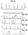
- Figure 16 shows an electropherogram for a mixture of polyA oligonucleotides containing all basepair lengths between 40 and 60 basepairs. The figure shows that all 20 of the different-size oligomers are well resolved. Similar resolution of oligomers in the 19-22 basepair range has been achieved, as detailed in Example 10.
- the oligomers used for sequencing may be prepared by the dideoxy enzyme method (Sanger) or chemical cleavage method (Maxam-Gilbert).
- the oligonucleotide fragments from the four separate reaction mixtures are fractionated, preferably in parallel, and the fragment peaks from each of the four tubes is recorded.
- Figures 17A-17D show oligonucleotide electropherograms from the A, T, G, C -terminated fragments as would be observed for the nucleotide sequence shown at the top in the figure. Automated or semi-automated analysis of the peak positions and construction of the sequence can be performed using standard programed systems.
- the oligonucleotide fractionation may be carried out in a single tube using, for example, the fluorescence labeling method described by Smith.
- Counter-migration capillary electrophoresis was carried out using an ABI Model 270 capillary electrophoresis system.
- the system includes a built-in high-voltage DC power supply capable of voltage settings up to 30 KV.
- the capillary tube used in the system is a fused silica capillary tube 72 cm long with a 50 ⁇ m i.d. and 350 ⁇ m o.d.obtained from Polymicro Technologies (Phoenix, AZ).
- the grounded cathodic reservoir is filled with Tris-borate-EDTA buffer containing 10 mM Tris-borate, pH 8.3, 5 mM NaCl, 0.1 mM EDTA, and 7 M urea (TBE buffer).
- the anodic reservoir contained a polymer solution containing 0.25 weight percent hydroxypropylmethylcellulose (HPMC) in TBE buffer.
- HPMC hydroxypropylmethylcellulose
- the polymer which is characterized by a viscosity of about 4 Pa s (4,000 centipoise) in a 2% by weight solution at room temperature, was obtained by Dow Chemical (Midland, MI).
- a DNA restriction fragment mixture obtained from BRL contained a combination of 1 kb ladder partial restriction fragments (multiples of about 1 kB) and smaller Hin fI digest fragments in the size range between about 50-1,000 basepair.
- the fragment mixture was diluted with water to a final DNA concentration of about 250 ⁇ g/ml.
- About 2 nanoliters of the DNA solution was drawn into the anodic end of the tube by vacuum applied to the cathodic tube end. The tube was then reimmersed in the polymer solution.
- the electrophoretic system was run at a voltage setting of about 20 kV (about 400 V/cm) throughout the run.
- UV detection was with a Kratos 783 UV detector designed for capillary tube detection.
- the detector output signal was integrated and plotted on an HP Model 3396A integrator/plotter.
- the electropherogram obtained is shown in Figure 4. Total run time was about 11 minutes.
- the fragment molecular weights were confirmed by known standards.
- Example 2 The electrophoretic method described in Example 1 was followed, except that the TBE buffer solution used for fragment separation did not contain polymer, and the voltage setting was about 800 V/cm.
- the electropherogram of the fractionated DNA material is shown in Figure 5. In contrast to the electropherograms in Figure 4, the smallest fragments traveled fastest toward the cathode, indicating that the smallest fragments were actually traveling most slowly toward in the upstream direction toward the cathode. There is no significant resolution among the fragments except for broad size ranges, and elution peaks of all of the fragments were closely spaced.
- Example 6 The electrophoretic method described in Example 1 was employed, except that the polymer solution used for fragment separation contained 0.1% weight percent HPMC polymer.
- the electropherogram of the fractionated DNA material is shown in Figure 6. It is apparent from this figure that the lower polymer concentration allowed good peak resolution up to about 10 kb fragment sizes, with some loss of resolution of fragments below the size range of about 500 basepairs.
- Example 2 The electrophoretic method described in Example 1 was employed, except that the TBE polymer solution used for fragment separation contained 0.25 weight percent hydroxyethylcellulose (HEC) polymer.
- HEC hydroxyethylcellulose
- the HEC polymer was obtained from Dow Chemical (Midland, MI), and had a viscosity of about 0.3 Pa s (300 centipoise) in a 2% solution at room temperature.
- the electropherogram of the fractionated DNA material is shown in Figure 7. Interestingly, the electropherogram shows resolution of ladder peaks in the 2,000-8,000 Kbase size region, with less sharp resolution of the smallest peaks, when compared with the 0.25% HMPC separation shown in Figure 4.
- Example 1 The electrophoretic method described in Example 1 was employed, except that the TBE polymer solution used for fragment separation contained 1% dextran polymer.
- the polymer was obtained from Sigma (St. Louis, MO) and had an average molecular weight of about 150,000.
- the electropherogram of the fractionated DNA material is shown in Figure 8. The fragments were less well resolved in this polymer solution than in either the HMPC or HEC polymers.
- Example 1 The electrophoretic method described in Example 1 was employed, except that the TBE polymer solution used for fragment separation contained 1.5 % polyvinyl alcohol (PVA).
- PVA polyvinyl alcohol
- the polymer which is characterized by an average molecular weight of about 125 kilodaltons, and about 88% hydroxylation, was obtained from Scientific Polymer Products (Ontario, New York).
- the electropherogram of the fractionated DNA material is shown in Figure 9.
- the resolution achieved in the fragment size range less than about 1 kbase was about the same as that observed by CMCE 0.1% HMPC ( Figure 6). Fragments of size 1036 bases and greater were not significantly resolved.
- the DNA ladder fragment mixture from Example 1 was mixed with ethidium bromide, to a final 10 ⁇ M ethidium bromide concentration.
- the CMCE polymer solution was a TBE buffer with 0.25% HPMC polymer and 10 ⁇ M ethidium bromide.
- CMCE fractionation was carried out as in Example 1, with the results shown in Figure 10. A comparison of this figure with Figure 4 shows substantially greater resolution of fragments in the size range up to about 1-2 kilobases, and some loss of resolution at higher molecular weights, with respect to CMCE fractionation in the absence of intercalating agent.
- CMCE fractionation was carried out as in Example 1, with aliquots A-D, with the results shown in Figures 11A-11D, respectively.
- the electropherogram in Figure 11A shows a clear resolution of fragments 281 bp and 271 bp, in contrast to the same fragments fractionated in the absence of ethidium bromide ( Figure 11B).
- the electropherogram in Figure 11C shows a clear resolution of fragments 281 bp and 271 bp (in the phiX174 ladder mixture) and 611 and 603 fragments (in the PCR fragment mixture)i, in contrast to the same fragments fractionated in the absence of ethidium bromide (Figure 11D).
- the mixture of partial digest DNA ladder fragments and Hin fI fragments from Example 1 was loaded at the cathodic end of a capillary tube as in Example 1.
- a pulsed voltage was generated using an HP 3314A function generator (for generating a square wave), a Krohn-Hite Model 7500 amplifier, and a Jefferson Electric high-voltage transformer.
- the applied voltage had a peak voltage of about 25 kV, and an RMS voltage of about 12.5 kV, giving an applied RMS voltage of about 170 V/cm, and a pulse frequency of 650 Hz.
- the power supply was operated in a pulsed voltage mode for about 45 minutes, at which point the leading edge of the fractionation mixture was just upstream of the detection zone. The power supply was then switched to a constant voltage mode, at about 12.5V/cm, until the end of the electrophoretic separation.
- the electropherogram obtained by the method is shown in Figure 14.
- the size of the fragments in the fractionated mixture, expressed in basepairs, are given in boldface print.
- the peak positions obtained were compared with those in Figure 4, which gives peak fragment migration times for the same peaks under constant-field electrophoresis conditions.
- the changes in peak migration rates, as a function of size, are discussed above with respect to Table 2. Briefly, fragments larger than about 2,000 basepairs migrated more slowly in a pulsed field, whereas smaller fragments migrated more rapidly with respect to the normalized rates of-migration in a constant-voltage field.
- a mixture of polyA oligonucleotides containing between 40-60 nucleotides was obtained from Pharmacia (Uppsala, Sweden). The oligonucleotide mixture was dissolved in water to 350 ⁇ g/ml. About 2 nanoliters of sample solution was drawn into the anodic end of the tube by vacuum applied to the cathodic tube end, and the material was fractionated in a 0.25% HPMC polymer, at a constant-voltage of about 140 v/cm. Total run time was about 30 minutes. The electropherogram obtained is shown in Figure 16. It is seen that the CMCE fractionation effectively resolved each of the 20 oligomers in the mixture. A mixture of smaller oligonucleotides, in the 19-22 basepair size range were similarly resolved by this method.
Abstract
Description
- The present invention relates to electrophoretic separation of nucleic acid fragments by capillary electrophoresis.
- Cantor, C., et al., "Biophysical Chemistry Parts I, II and III," W.H. Freeman and Co., N.Y. (1980)
Cohen, A.S., et al, Anal Chemistry, 59:1021 (1987). - Cohen, A.S., et al, J Chromatography, 458:323 (1988)
- Compton, S.W., et al., BioTechniques, 6(5)432 (1988).
- Kaspar, T.J., et al, J Chromatography, 458:303 (1988).
- Lauer, H.H., et al., Anal Chem, 58(1):166 (1986).
- Maniatis, , T., et. al., "Molecular Cloning: A Laboratory Manual," Cold Spring Harbor Labs (1982).
- Maxam, A.M., et al., Meth Enzymol, 65:499 (1980).
- Sanger, F., et al., Proc Nat Acad Sci, USA, 74:5363 (1977).
- Smith, L.M., et al., Nature, 321:674 (1986).
- Methods for size fractionating mixtures of single-strand or double-strand (duplex) nucleic acids are crucial to a variety of analytical and preparative techniques in biochemistry. One important example is restriction analysis, in which duplex DNA is digested with selected restriction enzyme(s), fractionated according to digest fragment size, then analyzed for fragment positions. This method is widely used in molecular cloning to determine the number and arrangement of restriction sites in a cloning vector, and to confirm insert location and/or orientation in the vector.
- Restriction analysis is an important tool for genetic mapping as well, since it allows relatively long pieces of chromosomal DNA to be mapped, and ultimately sequenced, on the basis of restriction fragment sizes and overlap. In a related application, the discovery of linkages between a number of genetic diseases and restriction fragment length polymorphisms in humans has provided a tool for screening individuals for these genetic diseases.
- Rapid DNA sequencing methods currently in use also rely on the ability to fractionate DNA fragments -- typically single-strand fragments -- on the basis of size. Both enzymatic sequencing techniques, in which random-termination fragments are generated enzymatically in the presence of dideoxynucleotides (Sanger), and chemical methods in which random-termination fragments are generated chemically (Maxam), rely on fractionation and discrimination of the fragments on the basis of fragment size. In particular, the fractionation method must be capable of distinguishing fragments which differ from each other by one nucleotide only.
- In addition, size fractionation of nucleic acid fragments is valuable for isolating and purifying DNA or RNA fragments. In molecular cloning, it is common to fractionate restriction fragments to obtain selected fragments for vector construction. In oligonucleotide synthesis, it is generally desirable to purify fragments having the desired oligonucleotide sequence and subunit number.
- Heretofore, standard methods available for size-fractionating nucleic acids fragments have used a solid or semisolid gel matrix for electrophoretic fragment separation. In the case of larger molecular weight fragments, typically greater than about 1,000 bases, the preferred gel material is agarose, where the concentration of the agarose may vary from about 0.3%, for separating fragments in the 5-60 kilobase size range, up to about 2%, for separating fragments in the 100-3,000 basepair range (Maniatis). Smaller size fragments, typically less than about 1,000 basepairs, are usually separated in polyacrylamide gel. The concentration of acrylamide polymer can range from about 3.5%, for separating fragments in the 100-1,000 basepair range, up to about 20%, for achieving separation in the size range 10-100 basepairs.
- More recently, DNA fragment separation by capillary electrophoresis (CE) has been proposed (Cohen, 1987, 1988, Compton, Kaspar). In one approach (Kaspar), fragments are separated in a gelled polyacrylamide medium within the tube. This approach shares many of the limitations of conventional acrylamide or agarose electrophoresis: the inconvenience of handling a polymerized gel, relatively long run times, and narrow size distribution of fragments which any given concentration of gel is capable of resolving.
- Alternatively, it has been proposed to carry out fragment separation by CE in a buffer solution, without any separation medium (Cohen, 1987, 1988, and Compton). Generally, this approach has not produced consistent or easily interpretable results.
- It is one general object of the present invention to provide, for size-fractionating nucleic acid fragments, a method which substantially overcomes or reduces the above-noted limitations associated with nucleic acid electrophoretic fractionation methods.
- A more specific object of the invention is to provide such a method which gives high resolution, can be completed with short fractionation times, and requires only picogram amounts of fragment sample material.
- Still another object of the invention is to provide such a method which can be carried out under a variety of variable electrophoresis oonditions, some adjustable during an electrophoretic run, to optimize separation between or among selected size fragments.
- It is yet another object of the invention to provide such a method in which the effective fractionation length of the electrophoretic pathway is substantially longer than the physical length of the capillary tube used for the fractionation, due to counter-migration of nucleic acid fragments in a polymer solution, in one direction, as the solution is drawn through a capillary tube by electroosmotic flow in the opposite direction.
- In practicing the method, a liquid sample of nucleic acid fragments is loaded into one end of a capillary tube filled with a fluid electrolyte solution. The inner surface of the tube is negatively charged at the pH of the solution. One end of the tube is placed in fluid communication with a cathodic reservoir and the other end of the tube, in communication with an anodic reservoir containing a polymer solution of an uncharged or slightly charged polymer having a molecular weight of at least about 10,000 daltons, which is typically characterized by a viscosity of at least about 0.015 Pa s (15 centipoise) in a 2% solution at room temperature. Preferred polymers are hydroxylated polymers having molecular weights of about 50-200 kilodaltons, and are characterized by a viscosity of about 0.2-5 Pa s (200-5,000 centipoise) in 2% solution at room temperature.
- A voltage applied across the reservoirs is effective to draw the polymer solution into and through the tube by electroosmotic flow. The fluid flow rate in the tube is greater, in the direction of the cathodic reservoir, than the molecular-weight dependent races of migration of the nucleic acid fragments, in the direction of the anodic reservoir. As a result, the larger molecular weight nucleic acid fragments move more rapidly toward the cathodic reservoir than smaller fragments, and the total migration distance of all of the fragments relative to the separation medium (the polymer fluid) is substantially greater than the total length of the capillary tube.
- The degree of fractionation of the nucleic acid fragments can be enhanced, according to one aspect of the invention, by selectively adjusting the rate of electroosmotic flow of polymer solution through the tube, to reduce the difference between the flow rate and the migration rate of the selected molecular weight fragments, subject to the constraint that the rate of electroosmotic flow is greater than the upstream migration rate of the slowest fragment to be fractionated. The effect of differentially decreasing electroosmotic flow rate or increasing fragment migration in the opposite direction is to increase the effective length over which electrophoretic migration of the fragments can occur.
- The rate of electroosmotic flow of polymer solution through the tube can be adjusted, in one embodiment, by changing the pH of the electrolyte and polymer solutions to control the density of negatively charged groups on the inner wall of the tube. In- creasing or decreasing the negative charge density on the tube wall increase or decrease, respectively, the rate of electroosmotic flow. Alternatively, the rate of migration of larger molecular weight species can be increased by reducing the concentration of polymer in the solution.
- In a preferred embodiment of the method, the polymer is a water-soluble hydroxyl polymer, such as dextran, polyvinylalcohol, and water-soluble cellulose compounds, such as methylcellulose and hydroxyethylcellulose. The polymer preferably has a molecular weight of at least about 50,000 daltons, or, alternatively, is characterized by a viscosity, in a 2% polymer solution at room temperature, of at least about 0.2 Pa s (200 centipoise).
- Studies on electrophoretic separation of nucleic acid species in various polymer solutions indicate that the rate of migration of nucleic acid species through a solution of hydroxylate polymer depends, at least in part, on interaction of the nucleic acids with the polymer. In particular, the resolution of nucleic acid fragments, especially lower molecular weight species, can be enhanced by fractionation in higher-concentration polymer solutions. A comparison of migration rate curves with theoretical model curves indicates that sieving may also play a role in determining migration rate.
- The applied voltage may be pulsed at one or more selected frequencies. According to one aspect of the invention, it has been discovered that applying a pulsed voltage of a given frequency produces two divergent types of migration rate changes with respect to migration rates in a constant-voltage field. For fragments having relatively large sizes, the rate of migration is progressively decreased, with respect to the migration time in a constant-voltage field, with increasing fragment size. This behavior is consistent with a model based on inertial effects in which larger fragments are, on average, slower to recover their constant-field velocities.
- For fragments having relatively small sizes, on the other hand, the rate of migration is actually increased with respect to the migration time in a constant-voltage field, with decreasing fragment size. This behavior cannot be explained by inertial effects alone. This phenomenon can be exploited in a variety of ways to enhance separation of selected-size nucleic acid fragments. For example, to isolate fragments in a selected size range, the pulse voltage frequency can be set initially to preferentially separate the desired fragments from larger molecular weight species. The frequency is then adjusted to separate the desired fragments from smaller molecular weight species.
- In another aspect of the invention, the resolution of fragments in the counter-migration system, and particularly small duplex fragment, can be enhanced by binding the fragments to be separated with an intercalating agent, such as ethidium bromide or acridine orange.
- These and other objects and features of the invention will become more fully apparent when the following detailed description of the invention is read in conjunction with the accompanying drawings.
-
- Figure 1 is a schematic diagram of a counter-migration capillary electrophoresis system used in practicing the method of the invention;
- Figure 2 is a schematic view of a capillary electrophoresis system designed for operation simultaneously in both a pulsed and constant-voltage mode;
- Figure 3 is an enlarged, fragmentary portion of a capillary electrophoresis tube, illustrating electroosmotic flow (e) in a right-to-left direction, and fragment migration (m₁, m₂, m₃) in a left-to-right direction;
- Figure 4 is an electropherogram of duplex DNA fragments fractionated in accordance with the present invention, in 0.25 weight percent hydropropylmethylcellulose (HPMC) solution;
- Figure 5 is an electropherogram of duplex DNA fragments fractionated in a buffer solution without polymer;
- Figure 6 is an electropherogram of duplex DNA fragments fractionated in accordance with the present invention, in 0.1 weight percent HPMC solution;
- Figure 7 is an electropherogram of duplex DNA fragments fractionated in accordance with the present invention, in a solution containing 0.25 weight percent hydroxyethylcellulose (HEC) polymer solution;
- Figure 8 is an electropherogram of duplex DNA fragments fractionated in accordance with the present invention, in a 1% dextran polymer solution;
- Figure 9 is an electropherogram of duplex DNA fragments fractionated in accordance with the present invention, in a 1.5% polyvinyl alcohol solution;
- Figure 10 is an electropherogram of duplex DNA fragments fractionated in accordance with the present invention, in 0.25 weight percent HPMC solution, in the presence of ethidium bromide;
- Figures 11A-11D are electropherograms of duplex DNA fragments from φX174/HaeIII (A and B) and φX174/HaeIII and 161 basepair PCR fragments (C and D) fractionated by CMCE in the presence (A and C) and absence (B and D) of ethidium bromide;
- Figure 12 shows a pulsed square wave and the corresponding migration rates of relatively large (dashed-dot line) and relatively small (dotted line) nucleic acid fragments during each voltage pulse;
- Figure 13 shows the expected (dotted line) and observed (solid line) relationship between fragment size and migration rate with respect to migration rate in a constant-voltage field;
- Figure 14 is an electropherogram of duplex DNA fragments fractionated in accordance with the present invention, in 0.25 weight percent HPMC solution, at a pulsed field frequency of about 650 Hz;
- Figure 15 shows an exemplary electropherogram of restriction fragments (solid line) containing a fragment with a target sequence which hybridizes to a labeled probe (dotted line);
- Figure 16 is an electropherogram of polydeoxyadenylic acid (polyA) oligonucleotides fractionated in accordance with the invention in 1% weight percent HPMC solution; and
- Figures 17A-17D illustrate model electropherograms obtained in a dideoxy chain termination sequencing method carried out in accordance with the invention.
- Figure 1 is a simplified schematic view of a
capillary electrophoresis system 20 suitable for practicing the method of the invention. The system includes acapillary tube 22 having a length preferably between about 10-200 cm, typically less than about 100 cm, and an inner diameter of preferably between about 25-200 µm (microns), typically about 50 µm. In the embodiment shown, the tube is supported in a horizontal position and has downwardly bent end regions. - The inner surface of the tube has chemical groups which are negatively charged at a pH preferably between about 4-9. The surface chemical groups may be an inherent property of the capillary material, such as is the case for a fused silica tube which has surface silane groups. Alternatively, or in addition, the capillary walls may be treated with known derivatization reagents for covalent attachment of negative chemical groups, such as acid groups, to the inner capillary walls, or with known negatively charged surface-coating agents. Methods for derivatizing or coating glass or the like are well known. One preferred capillary tube is a fused silica tube having an inner diameter of 50 µm and available from Polymicro Technologies (Phoenix, AZ).
- More generally, the capillary tube may be any tube or channel capable of supporting a column of polymer solution, preferably at a column thickness of 200 µm or less. For example, the tube may take the form of a channel formed in a glass slide or the like, and having negatively charged surface groups.
- An
anodic reservoir 26 in the system contains an electrolytic polymer solution 28 (cf. Section B of this description) which is drawn through the tube by electroosmotic flow during electrophoresis, as will be described in Section C. The anodic end of the tube, indicated at 22a, is immersed in the polymer solution, as shown, during electrophoresis. - A
sample reservoir 30 in the system contains the nucleic acid fragment mixture which is to be loaded into the anodic end of the tube. Preferably the sample material is dissolved in the electrolytic solution or in water. The sample and anodic reservoirs may be carried on a carousel or the like, for placement at a position in which the lower anodic end of the tube can be immersed in the reservoir fluid. Although not shown here, the carousel may carry additional reservoirs containing solutions for cleaning and flushing the tube between electrophoretic runs of different polymer solutions, where two or more polymer solutions are employed in a single electrophoretic fractionation method. - The opposite, cathodic end of the tube, indicated at 22b, is sealed within a
cathodic reservoir 32 and is immersed in ancathodic electrolyte solution 34 contained in the reservoir, as shown. Asecond tube 38 in the reservoir is connected to a finely-controlled vacuum system (not shown) for drawing fluid, e.g., washing and cleaning solutions, electrophoresis polymer solution, through the tube and for loading the nucleic acid sample material inreservoir 30 into the tube. - A
high voltage supply 40 in the system is connected to the cathodic and anodic reservoirs as shown, for applying a selected electric potential between the two reservoirs. The power supply leads are connected toplatinum electrodes - Completing the description of the system shown in Figure 1, a
detector 44 in the system is positioned adjacent the cathodic end of the tube, for optically monitoring nucleic acid fragments migrating through anoptical detection zone 46 in the tube. The detector may be designed either for UV absorption detection and/ or for fluorescence emission detection. UV absorbance is typically carried out at 240-280 nm, using, for example, a Kratos 783 UV absorbance detector which has been modified by Applied Biosystems (Foster City, CA.), by replacing the flow cell with a capillary holder. Fluorescence emission detection is preferably carried out at a selected excitation wavelength which is adjustable between about 240-500 nm, depending on the fluorescent species associated with the nucleic acid fragments, as discussed below. One exemplary fluorescence detector is an HP1046A detector available from Hewlett-Packard (Palo Alto, CA), and modified as above for capillary tube detection. The detector is connected to an integrator/plotter 45 for recording electrophoretic peaks. - In operation, the capillary tube is thoroughly washed by drawing suitable cleaning and rinsing solutions through the tube by applying a vacuum to
reservoir 32, such as detailed in Example 1. The tube is then flushed with several volumes of the electrolytic polymer solution and a small volume, typically 1-10 nanoliters of sample material is loaded into the cathodic tube end. A voltage is applied between the cathodic and anodic reservoirs until all of the fragment peaks have passed through the detection zone. - In one embodiment of the method, described in Section C below, enhanced electrophoretic separation of the nucleic acid fragments is achieved by driving the electrophoretic process with a pulsed voltage at a selected frequency, e.g., 300-1,000 Hz. In one preferred method, a pulsed voltage is applied until the leading (most downstream) band is just upstream of the detection zone, and then the system is switched to a DC mode, to reduce noise at the detector, until all of the bands have been recorded.
- Figure 2 shows a fragmentary view of an
electrophoretic system 50 which can be operated to the end of the electrophoretic run under a pulsed field. Thecapillary tube 52 in the system has a small-clearance break 54 adjacent and upstream of the detection zone, indicated at 56. The tube sections on either side of the break are coupled byporous glass sleeve 58 which allows electrolyte migration into and out of the tube. The coupled portion of the tube is sealed within areservoir 60 filled with asuitable electrolyte solution 62. A groundedelectrode 64 in the reservoir is connected to the high-voltage side of a pulsed-voltage power supply 66 whose negative side is in communication with a suitable cathodic reservoir. The groundedelectrode 64 is connected to the high-voltage side of aDC power supply 68 whose negative side is in communication with a suitable anodic reservoir. - In operation, after sample material is loaded into the anodic end of the tube, the pulsed-voltage power supply is adjusted to a desired voltage and frequency level, and the DC power supply to a desired voltage level. Nucleic fragments in the sample are fractionated under the pulsed field within the portion upstream of
break 54. Thereafter, the fragments are carried in a constant-voltage field through the detection zone, where the fragments can be optically detected without pulse-frequency noise effects. - Although not shown here, it will also be appreciated that the electrophoresis system can be readily adapted for collecting nucleic acid fragments for preparative electrophoresis applications. Sample collection may be accomplished, for example, by providing a series of cathodic reservoirs into which the fragments can be eluted.
- The electrolytic polymer solution used in the method is composed of electrolytes and a polymer which is effective to form a fluid fractionation matrix in the tube. In addition, the solution has a pH at which the charged surface groups on the inner wall of the tube are at least partially ionized in their deprotonated state. The pH of the solution is preferably between 4-9. The pH may be adjusted to achieve a desired degree of tube wall charge density as discussed in Section C below.
- The electrolytes in the solution may include buffer components, typically at a 10 mM buffer concentration, salts, typically at a 5-10 mM concentration, and preferably a heavy metal chelator, such as EDTA. In addition, the solution may contain a denaturant, such as urea, which functions to minimize interactions among fragments and between fragments and the tube walls. One preferred polymer solution, described in Examples 1-8, includes as its buffer components 10 mM Tris-borate buffer, pH 8.3, 5 mM NaCl, 0.1 mM EDTA, and 7M urea. The solution may also include an intercalating agent, such as ethidium bromide, for a purpose to be described in Section C below.
- The polymer in the Solution is one which is effective, in a fluid, non-gelled state, of differentially retarding nucleic acid fragments on the basis of size, when the fragments are migrating through the matrix under the influence of an electric field. The polymers are uncharged, and have molecular weights of at least about 10 kilodaltons, and are characterized by a viscosity of at least about 0.015 Pa s (15 centipoise) in a 2% (weight percent) solution at room temperature. By uncharged polymer is meant a polymer which does not contain net positive or negative charges at the pH of the polymer solution, or which contains substantially fewer net negative or positive charges than one per polymer subunit.
- Preferred polymers are hydroxylated polymers, such as polyvinyl alcohol,hydroxyl-methacrylate polymers and polysaccharides, such as dextran and water-soluble cellulose derivatives, such as hydroxypropylmethylcellulose (HPMC), hydroxyethylcellulose (HEC), and methylcellulose (MC). As defined herein, "hydroxylated polymer" means a polymer formed of hydroxyl-containing subunits, such as vinyl alcohol, 2-hydroxyethylene methacrylate, or a monosaccaride. Preferred polymers have molecular weights of about 50-200 kilodaltons, and are characterized by a viscosity of about 0.2-5 Pa s (200-5,000 centipoise) in 2% solution at room temperature.
- As illustrated below, electrophoretic patterns obtained at various concentrations of hydroxylated polymers indicate that migration rates of nucleic acid fragments are progressively reduced at increasing polymer concentrations. These results indicate that migration rates of nucleic acid through a solution of hydroxylated polymer is determined, at lease in part, by fragment interactions with the hydroxyl groups of the polymers. As indicated above, a comparison of migration rate curves with theoretical modeling curves indicates that the polymer may also effect migration rates by a sieving mechanism.
- The weight percentage of polymer in the solution may vary from about 0.1 to up to 1% or greater, depending on the sizes of fragments to be fractionated and, where a pulsed field is used, the frequency of the applied field, as will be seen in Section D below.
- According to an important aspect of the invention, the fractionation of nucleic acid fragments in the capillary tube occurs by size-dependent migration of the fragments against the bulk electroosmotic flow of polymer solution in the tube. This phenomenon, which is referred to herein as counter-migration capillary electrophoresis (CMCE) in a polymer solution matrix, is illustrated in Figure 3, which shows an enlarged, fragmentary portion of a
capillary electrophoresis tube 70. - As seen in the figure, the negatively charged groups on the inner tube wall, indicated by "-" symbols, are shielded by positively charged ions in the polymer solution, essentially forming a positively charged shell about the column of fluid in the tube. The thickness of the shell of relatively immobilized positive ions at the wall surface is known as the shear distance. This outer shell of positive charge and inner bulk phase charge distribution is called an electric double layer, and is characterized by a zeta potential, which is a measure of the potential between the outer shell of positive charges and the bulk medium.
- Under the influence of an electric field, this column of polymer solution in the medium (which is surrounded by a shell of positive charges) is drawn electro-osmotically in the direction of negative or low potential. The rate of electroosmotic flow in the tube is indicated by the arrow e in the figure (arrow e may be thought of as a vector with a magnitude e and a direction along the axis of the tube). The electroosmotic flow rate e in a capillary tube can be described by the equation:

- At the same time the polymer solution in the tube is moving downstream (toward the cathodic reservoir) by electroosmotic flow, the negatively charged nucleic acid fragments are migrating relative to the solution in the opposite direction toward the anodic reservoir. Figure 3 shows the rates of migration of three different size duplex nucleic acid fragments F₁, F₂, and F₃ having decreasing sizes, e.g., 1,000, 300, and 50 basepairs, respectively. Because of molecular interactions of the fragments with the polymer molecules, the rates of fragment migration toward the anodic reservoir, indicated at m₁, m₂, and m₃ in the figure, are size dependent, with smaller fragments migrating faster in the anodic direction.
- The net migration rate of the three fragments through the tube in the direction of the cathode is then the vector sum of the electroosmotic flow e and the size dependent migration m, indicated as µ₁, µ₂ and µ₃ in Figure 3. As seen, these net migration rates ui of the particles are size dependent, with the larger fragments migrating more rapidly toward the cathode than smaller fragments.
- Figure 4 shows an electropherogram of DNA restriction fragments containing 1 kb DNA ladder fragments (multiples of 1 kb fragments) and smaller HinfI fragments. The large spike at the left of the electropherogram is the leading edge of the water from the sample loaded into the tube initially. The fragment sizes (number of bases) are the bold numbers in the figure. As seen, larger fragments have shorter migration times. It is also noted that under the polymer and electric field conditions employed, the system was able to resolve fragments up to about 3 kbases, and to distinguish, but not baseline resolve, larger fragments. Details of the CMCE conditions are given in Example 1.
- It will be appreciated from Figure 3 that the effective fractionation distance -- that distance which a fragment travels through the polymer matrix -- can be selectively increased, to enhance the fractionation of that fragment, by varying the relative rates of electroosmotic flow and up-stream fragment migration during electrophoresis. In particular, as the rate of electroosmotic flow e and the rate of upstream migration mi of a fragment Fi approach a common value, the actual fragment migration rate µi becomes quite small, providing longer separation times (and effective fractionation lengths) during which the fragment Fi can become better resolved, i.e., further separated from the next closest size fragments.
- Experiments conducted in support of the present invention show that the rate of migration of nucleic acid fragments through an uncharged fluid polymer matrix is subject to a number of variables related to fragment size, polymer concentration and type, and, where the electric field is produced by a pulsed voltage, to the pulse frequency of the field. The latter phenomenon will be described in Section D.
- The migration rates of nucleic acid polymers in the absence of polymer in the capillary tube is shown in Figure 5. The electrophoretic conditions are similar to those used in the electrophoretic separation shown in Figure 4, except for the absence of polymer in the capillary medium (Example 2). A comparison of the two figures shows first, that the fastest migrating species are the smallest molecular weight fragments, and the slowest migrating species, the largest fragments. Thus, the larger fragments appear to be migrating against the direction of electroosmotic flow faster than smaller fragments, possibly due to their greater total charge. Secondly, the only resolution provided by the system is between broad size classes, again reflecting an apparently gross difference in migration rate according to size.
- The effect of polymer concentration of the relative rates of migration of nucleic acid fragments is seen from Figure 6, which is an electropherogram of nucleic acid restriction fragments fractionated in a 0.1 weight percent HPMC polymer. The fragments and fractionation conditions used are identical to those employed in the Figure 4 method, except that the polymer concentration in the Figure 6 electrophoresis was about 2.5 fold lower than that in the Figure 4 electrophoresis (Example 3). A comparison of the two figures shows that higher molecular weight fragments are better resolved at the lower polymer concentration, whereas fragments in the size range less than about 1 kilobase are better resolved at the higher polymer concentration.
- Figure 7 shows an electropherogram of duplex fragments fractionated by CMCE in a 0.25 weight percent hydroxyethylcellulose (HEC) polymer solution, as detailed in Example 4. The polymer gave fractionation characteristics intermediate between those seen with 0.1 and 0.25 weight percent HPMC; specifically, the resolution of high molecular weight species was intermediate that of 0.25% and 0.1% HPMC, which gave best resolution of fragments larger than about 1 kilobase, and intermediate that of 0.1% and 2.5% HPMC, which gave the best resolution of fragments smaller than about 1 kilobase.
- Figure 8 shows an electropherogram of duplex fragments fractionated by CMCE in a 1 weight percent dextran (MW=150,000), as described in Example 5. Although the polymer clearly resolves fragments in the size range below about 1 kb, fragments above this range were not resolved. Lower concentrations of dextran may be necessary for improved resolution in the size range above 1 kilobase.
- Figure 9 shows an electropherogram of duplex fragments fractionated by 1.5 weight percent of polyvinyl alcohol (PVA, MW about 125,000), a non-polysaccharide hydroxylated polymer. Details of the CMCE method are given in Example 6. Like the system using 1% dextran, 1.5% PVA clearly resolves fragments in the size range below about 1 kb, but shows little resolving ability above this size range. Lower concentrations of PVA may be necessary for improved resolution in the size range above 1 kilobase.
- Thus it is seen that resolution of selected size fragments can be enhanced according to the polymer used, and the concentration of polymer. That is, the migration rates of nucleic acid fragments through a polymer in the CMCE method is dependent both on type of hydroxylated polymer, and polymer concentration.
- According to another aspect of the invention, it has been discovered that the resolution among smaller size fragments can be selectively enhanced by complexing the fragments, preferably in double-strand form, with an intercalating agent. Exemplary intercalating agents include ethidium bromide, acridine orange and thiazole orange. These agents have a flat, resonant molecular structure which allows the compounds to intercalate between adjacent bases of nucleic acids (Cantor). The agent is preferably included in the polymer solution drawn through the tube, to maintain the intercalating agent in equilibrium between free and DNA-bound forms.
- Figure 10 shows an electropherogram of the above duplex ladder fragments fractionated by CMCE in 0.25 weight percent HPMC in the presence of 10 µmole ethidium bromide, according to the procedure given in Example 7. As seen from a comparison of this figure with Figure 4, the intercalating agent enhances the resolution of fragments less than about 500 basepairs in size. This effect may be due to a size-dependent change in the conformation of the fragments when complexed with intercalating agent. A similar enhancement of small-fragment resolution was observed for acridine orange, another nonionic intercalating agent.
- Figures 11A-11D show electropherograms of nucleic acid fragment mixtures fractionated by CMCE in the presence (11A and 11C) and absence (11B and 11D) of ethidium bromide. The fragment mixtures include the fragment mixture formed by HaeIII digestion of φX174 phage (11A and 11B), referred to herein as a φX174/HaeIII mixture, and the same fragment mixture plus fragments produced by PCR (polymerase chain reaction) amplification of an M13 phage sequence fragments, referred to herein as 611 basepair PCR fragments (11C and 11D). As seen from Figures 11A and 11B, the φX174/HaeIII mixture contains 271 and 281 fragments which are resolved only in the presence of ethidium bromide. With reference to Figures 11C and 11D, the presence of ethidium bromide enhances the separation efficiency (peak sharpness) and sensitivity (peak height).
- In addition to the various factors discussed above, the relative rates of electroosmotic flow and fragment migration rates may be selectively adjusted by:
- a. Changes in the electric field strength. In theory, the electric field E is directly proportional to the magnitude of both the bulk flow rate e and the migration rates of small charged particles through a non-polymer solution, for electric field strengths below 200 v/cm. However, in view of the fact that the order of fragment elution in the presence of polymer (Fig. 4) is opposite from that in the absence of polymer (Fig. 5), the migration of DNA through a polymer solution is evidently more complex than this model. In view of this, size-selective changes in net rates of migration may be achieved by adjusting the electric field strength, particularly in the range above 200 v/cm.
- b. Changes in the charge density of the tube walls. The zeta potentials of the electric double layers of the column of polymer solution in the tube and that of the fragments in the polymer solution can be independently varied, e.g., by treating the surface of the tube to mask charge groups, or by adjusting the pH of the polymer solution to selectively protonate the tube wall charges or nucleic acid fragment charges.
- c. Changes in solution viscosity. Although theory would predict that viscosity changes would effect electroosmotic flow and particle migration rate to the same extent, this prediction would likely not apply to large anionic solutes, such as nucleic acid fragments. Solution viscosity could be adjusted by changing the concentration of polymer (which has been shown to selectively alter fragment migration rates), by addition of polymers or other solute species which do not interact with nucleic acid fragments, and/or by changing the solution temperature.
- Summarizing the above, the present invention provides a variety of parameters which may be selectively varied to enhance nucleic acid fractionation of selected sizes, either by selectively changing the rate of electroosmotic flow in the direction of the cathodic reservoir, or by differentially changing the upstream migration rates of the fragments being fractionated. As discussed above, the rate of electroosmotic flow, relative to counter migration of nucleic acid fragments, can be varied by changing the charge density of negatively charged groups on the inner capillary wall. The fragment migration rate in an anodic direction can be selectively reduced, for large fragments, by decreasing polymer concentration and/or according to the type of polymer. For shorter fragments, the rates of counter migration can be selectively increased by complexing the fragments with a nonionic intercalating agent. Such variations in flow and migrations rates are subject to the constraint that the electroosmotic flow be faster than the upstream fragment migration rate of the smallest fragments which is to fractionated.
- The electrophoretic methods described above were carried out under a constant-voltage field. In accordance with another aspect of the invention, the fractionation of nucleic acid fragments can be enhanced by carrying out the electrophoretic separation under a pulsed-voltage field, at a frequency effective to selectively enhance separation within a given fragment size range.
- In theory, the migration rate behavior of nucleic acid fragments in a pulsed field may be governed by two size-related effects. The first effect is a resonance effect involving the fragment's rotational modes and the frequency of the electric field. Table I below shows rotational and stretching resonance frequencies which have been calculated for 100, 1,000, and 10,000 basepair duplex DNA fragments. The rotational resonance frequencies in Hz were calculated on the basis of a prolate ellipsoid model of the duplex molecule (Cantor).
Table I Model Fragment Size (base pairs) 100 1000 10,000 Prolate Elipsoid (rotational motion) 9.9x10⁴ 1.6x10² 2.2x10⁻¹ Viscoelastic (stretching motion) 3.8x10⁵ 8.1x10³ 1.7x10² - A strong rotational resonance effect predicts that the migration rates of fragments in resonance with the electric field will be preferentially slowed with respect to the migration rate in a time-invariant field. This is because a molecule in rotational resonance with the electric field would be expected to be, on average, least favorably oriented for migration in the direction of the field when the electric field is greatest. Larger molecules, because of their slower rotational times, would be expected to be less perturbed from their field-oriented positions at each voltage-pulse cycle; smaller molecules, with their faster response times, would more quickly reorient in the direction of the field. Thus, if rotational resonance effects are dominant, it should be possible to slow the migration of resonance species during electrophoresis relative to the electroosmotic flow rate and the rate of migration of nonresonance species.
- The second size-dependent effect which may might be expected in a pulsed field is an inertial effect due to the acceleration and deceleration of fragments in a fluid with each voltage pulse. This effect is illustrated in Figure 12, which shows hypothetical velocity curves for relatively small (dotted lines) and relatively large (dash-dot lines) nucleic acid fragments, in relation to an applied square-wave voltage having a pulse width and a maximum voltage vmax. The maximum velocity which the fragments should reach is the terminal or steady-state velocity of the fragments in a constant voltage field of potential vmax, i.e., 100% of the constant field migration rate.
- As seen from the figure, smaller fragments are expected to reach terminal velocity more rapidly than large ones after application of the voltage pulse, but to decelerate to zero voltage substantially at the same rate when the voltage pulse ends. Since the total distance traveled by each fragment during a voltage pulse is just the integral of the velocity curve, the migration rates of larger fragments are expected to be preferentially decreased in a pulse-voltage field. It can also be appreciated that the higher the pulse frequency and shorter pulse duration, the smaller the fragment size which can be preferentially retarded in its migration rate, since the effect of any lag in the velocity curve becomes accentuated at short pulses.
- The dashed-line plot in Figure 13 shows the expected effect relationship between migration rate, expressed as a percent of migration rate in a constant-voltage field and fragment molecular weight at a given pulse frequency. The plot has been normalized to correct constant voltage levels to rms voltage in a pulsed field. The dashed-line plot shows an increasing migration rate, with decreasing particle size, as predicted by the inertial model just discussed. At some small fragment size, where the velocity curves of the fragments tend toward a common upper limit, the migration rate approaches 100% migration in a constant-voltage field. The curve in Figure 13 would therefore be expected to plateau at this point, as indicated by the dashed line.
- Figure 14 is an electropherogram of a mixture of duplex fragments fractionated in a pulsed field at 650 Hz. The sample material and fractionation conditions are similar to those used in the fractionation shown in Figure 4, except for the nature of the applied electric field, as detailed in Example 9. The migration rates of the same size fragments were measured from the peak times in the two figures and compared, with the results shown in Table 2 below. Column A in the table gives the size, in basepairs, of the fragments which are compared, and column B, the difference in migration time to the detection zone, corrected for relative run times of the leading edge of the two runs, between v(constant), the rate in a constant-voltage field, and v(pulse), the rate in a pulsed field. The differences in velocity between corresponding fragments are expressed in cm/min X 10⁻⁴. A positive value in column B means the fragment has a lesser migration rate in a constant-voltage field than in a pulsed field. For fragments above 1 kilobase size, the v(pulse) approaches the v(constant) with decreasing size, as predicted by the Figure 13 plot.
- With smaller fragment sizes, however, the difference between v(constant) and v(pulse) does not plateau at a zero value, as would be expected from the inertial model, but continues to assume progressively more negative values with smaller fragments. The greater migration rate in a pulsed field would indicate that above the 100% crossover point, attractive interactions between the smaller fragments and the polymer are actually reduced, facilitating movement of the fragments through the polymer matrix. In any case, this feature provides greater resolution of smaller fragments above the "crossover" point than can be achieved under similar conditions in a constant voltage field.
Table II A Frag. Size (bp) B v(const.)-v(pulse) 2036 4.0E-04 b 4.1E-04 a 1.5E-04 1018 0.71E-04 396 -3.8E-04 344 -3.9E-04 298 -4.5E-04 220 -5.1E-04 201 -5.2E-04 154 -5.6E-04 134 -5.5E-04 75 -6.9E-04 - It will be appreciated from the foregoing that the resolution among smaller size fragments can be selectively enhanced by carrying out the electrophoretic separation under a pulsed-voltage field, at a frequency which selectively retards the migration rate of larger fragments, and increases the migration rate of smaller fragments.
- In some mixtures of nucleic acid fragments, particularly where it is desired to fractionate species in different size ranges, the variables discussed above -- including solution pH, and polymer type and concentration, and field frequency -- may be selectively varied during the electrophoretic run to enhance fragment resolution. For example, an electropheretic separation may be carried out under a constant field or low frequency initially, to resolve larger size fragment, then switched to a higher frequency to improve resolution of smaller size fragments. As another example, the pH or polymer concentration of the polymer solution can be continuously varied during an electrophoretic run, using a standard two-chamber mixing device to produce a continuous solution gradient which is drawn into the capillary tube.
- The fractionation method of the invention finds utility in any of a variety of applications mentioned above requiring size fractionation of single-strand or duplex nucleic acids. These applications include electrophoretic separations for restriction analysis, including analysis of restriction fragment length polymorphisms for genetic screening, confirming vector construction, identifying specific nucleic acid fragments on the basis of size and/or hybridization to nucleic acid probes, and fractionating single-strand fragments for chemical or enzymatic sequencing.
- The following two applications illustrate how the method may be used for restriction fragment analysis, and as part of an automated sequencing protocol. In the restriction analysis example, it is desired to identify, from a mixture of genomic fragments, a restriction fragment which contains a target sequence of interest. After digesting the genomic mixture with a selected restriction enzyme or enzyme(s), the fragment mixture is combined under hybridization conditions with reporter-labelled probe capable of hybridizing with the target sequence. The probe preferably includes the complementary target sequence and a covalently bound fluorescent probe which can be readily detected by a fluorescence probe detector. The probe may be hybridized with the fragments, for example, by standard denaturation/renaturation conditions involving single-strand species, or bound to the fragments in duplex form by RecA catalyzed triplex formation.
- After binding the probe to the fragments, the sample is fractionated according to present CMCE method. The detector is preferably operated in a dual-wavelength mode, in which UV absorption and fluorescence emission detection are carried out concurrently. Figure 15 shows an exemplary electropherogram of the fragment mixture in which UV absorption is indicated by solid line, and fluorescence emission, by dotted line. The restriction fragment containing the selected target sequence is readily identified by the associated fluorescence signal.
- Alternatively, the fragments of interest can be hybridized to a biotinylated probe, selectively isolated by binding to an avidin solid support, then released from the support prior to CMCE fractionation.
- For DNA sequence analysis, it is necessary to resolve single-strand oligomers which differ from one another by one nucleotide base. The ability of the present method to achieve resolution of this type is illustrated in Figure 16, which shows an electropherogram for a mixture of polyA oligonucleotides containing all basepair lengths between 40 and 60 basepairs. The figure shows that all 20 of the different-size oligomers are well resolved. Similar resolution of oligomers in the 19-22 basepair range has been achieved, as detailed in Example 10.
- The oligomers used for sequencing may be prepared by the dideoxy enzyme method (Sanger) or chemical cleavage method (Maxam-Gilbert). The oligonucleotide fragments from the four separate reaction mixtures are fractionated, preferably in parallel, and the fragment peaks from each of the four tubes is recorded. Figures 17A-17D show oligonucleotide electropherograms from the A, T, G, C -terminated fragments as would be observed for the nucleotide sequence shown at the top in the figure. Automated or semi-automated analysis of the peak positions and construction of the sequence can be performed using standard programed systems.
- Alternatively, the oligonucleotide fractionation may be carried out in a single tube using, for example, the fluorescence labeling method described by Smith.
- The following examples illustrate various separation methods and applications in accordance with the invention, but are in no way intended to limit the scope thereof.
- Counter-migration capillary electrophoresis (CMCE) was carried out using an ABI Model 270 capillary electrophoresis system. The system includes a built-in high-voltage DC power supply capable of voltage settings up to 30 KV. The capillary tube used in the system is a fused
silica capillary tube 72 cm long with a 50 µm i.d. and 350 µm o.d.obtained from Polymicro Technologies (Phoenix, AZ). - The grounded cathodic reservoir is filled with Tris-borate-EDTA buffer containing 10 mM Tris-borate, pH 8.3, 5 mM NaCl, 0.1 mM EDTA, and 7 M urea (TBE buffer). The anodic reservoir contained a polymer solution containing 0.25 weight percent hydroxypropylmethylcellulose (HPMC) in TBE buffer. The polymer, which is characterized by a viscosity of about 4 Pa s (4,000 centipoise) in a 2% by weight solution at room temperature, was obtained by Dow Chemical (Midland, MI).
- The tube was washed with several column volumes of 1M NaOH, then 100 mM NaOH, and finally flushed with several volumes of the polymer solution, by drawing the wash and flush solutions through the tube by a vacuum applied to the cathodic end of the tube. A DNA restriction fragment mixture obtained from BRL (Bethesda, MD) contained a combination of 1 kb ladder partial restriction fragments (multiples of about 1 kB) and smaller HinfI digest fragments in the size range between about 50-1,000 basepair. The fragment mixture was diluted with water to a final DNA concentration of about 250 µg/ml. About 2 nanoliters of the DNA solution was drawn into the anodic end of the tube by vacuum applied to the cathodic tube end. The tube was then reimmersed in the polymer solution.
- The electrophoretic system was run at a voltage setting of about 20 kV (about 400 V/cm) throughout the run. UV detection was with a Kratos 783 UV detector designed for capillary tube detection. The detector output signal was integrated and plotted on an HP Model 3396A integrator/plotter.
- The electropherogram obtained is shown in Figure 4. Total run time was about 11 minutes. The sizes of the fragments in the fractionated mixture, expressed in number of basepairs, are in boldface print. As seen, the method effectively separated fragments in the
range 75 to about 3,000 basepairs, and gave distinct peaks for fragments in the range 3,000 to 6,000 basepairs. The fragment molecular weights were confirmed by known standards. - The electrophoretic method described in Example 1 was followed, except that the TBE buffer solution used for fragment separation did not contain polymer, and the voltage setting was about 800 V/cm. The electropherogram of the fractionated DNA material is shown in Figure 5. In contrast to the electropherograms in Figure 4, the smallest fragments traveled fastest toward the cathode, indicating that the smallest fragments were actually traveling most slowly toward in the upstream direction toward the cathode. There is no significant resolution among the fragments except for broad size ranges, and elution peaks of all of the fragments were closely spaced.
- The electrophoretic method described in Example 1 was employed, except that the polymer solution used for fragment separation contained 0.1% weight percent HPMC polymer. The electropherogram of the fractionated DNA material is shown in Figure 6. It is apparent from this figure that the lower polymer concentration allowed good peak resolution up to about 10 kb fragment sizes, with some loss of resolution of fragments below the size range of about 500 basepairs.
- The electrophoretic method described in Example 1 was employed, except that the TBE polymer solution used for fragment separation contained 0.25 weight percent hydroxyethylcellulose (HEC) polymer. The HEC polymer was obtained from Dow Chemical (Midland, MI), and had a viscosity of about 0.3 Pa s (300 centipoise) in a 2% solution at room temperature. The electropherogram of the fractionated DNA material is shown in Figure 7. Interestingly, the electropherogram shows resolution of ladder peaks in the 2,000-8,000 Kbase size region, with less sharp resolution of the smallest peaks, when compared with the 0.25% HMPC separation shown in Figure 4. Similar results were obtained by CMCE fractionation in 0.1% HEC, although the resolution of both high and low molecular weight species was better at the higher polymer concentration. Polymer solutions of methylcellulose (MC), including 0.25 weight percent MC, gave very good resolution of the smaller-size fragments (less than 500 bp).
- The electrophoretic method described in Example 1 was employed, except that the TBE polymer solution used for fragment separation contained 1% dextran polymer. The polymer was obtained from Sigma (St. Louis, MO) and had an average molecular weight of about 150,000. The electropherogram of the fractionated DNA material is shown in Figure 8. The fragments were less well resolved in this polymer solution than in either the HMPC or HEC polymers.
- The electrophoretic method described in Example 1 was employed, except that the TBE polymer solution used for fragment separation contained 1.5 % polyvinyl alcohol (PVA). The polymer, which is characterized by an average molecular weight of about 125 kilodaltons, and about 88% hydroxylation, was obtained from Scientific Polymer Products (Ontario, New York). The electropherogram of the fractionated DNA material is shown in Figure 9. The resolution achieved in the fragment size range less than about 1 kbase was about the same as that observed by CMCE 0.1% HMPC (Figure 6). Fragments of size 1036 bases and greater were not significantly resolved.
- The DNA ladder fragment mixture from Example 1 was mixed with ethidium bromide, to a final 10 µM ethidium bromide concentration. The CMCE polymer solution was a TBE buffer with 0.25% HPMC polymer and 10 µM ethidium bromide. CMCE fractionation was carried out as in Example 1, with the results shown in Figure 10. A comparison of this figure with Figure 4 shows substantially greater resolution of fragments in the size range up to about 1-2 kilobases, and some loss of resolution at higher molecular weights, with respect to CMCE fractionation in the absence of intercalating agent.
- A similar electrophoretic separation was carried out on the same partial digest fragments prepared in a polymer solution containing 2.6 µM acridine orange. The results were similar to those obtained with ethidium bromide, showing enhanced resolution of lower molecular weight species.
- Aliquots of DNA ladder fragment mixtures containing φX174/HaeIII in the presence (A) and absence (B) of 0.5 µM ethidium bromide, and φX174/HaeIIIZ plus 611 basepair PCR fragments in the presence (C) and absence (D) of 0.5 µM ethidium bromide, were prepared. The CMCE polymer solution was a TBE buffer with 0.5% HEC polymer and 0.5 µM ethidium bromide.
- CMCE fractionation was carried out as in Example 1, with aliquots A-D, with the results shown in Figures 11A-11D, respectively. The electropherogram in Figure 11A shows a clear resolution of
fragments 281 bp and 271 bp, in contrast to the same fragments fractionated in the absence of ethidium bromide (Figure 11B). Similarly, the electropherogram in Figure 11C shows a clear resolution offragments 281 bp and 271 bp (in the phiX174 ladder mixture) and 611 and 603 fragments (in the PCR fragment mixture)i, in contrast to the same fragments fractionated in the absence of ethidium bromide (Figure 11D). - The mixture of partial digest DNA ladder fragments and HinfI fragments from Example 1 was loaded at the cathodic end of a capillary tube as in Example 1. A pulsed voltage was generated using an HP 3314A function generator (for generating a square wave), a Krohn-Hite Model 7500 amplifier, and a Jefferson Electric high-voltage transformer. The applied voltage had a peak voltage of about 25 kV, and an RMS voltage of about 12.5 kV, giving an applied RMS voltage of about 170 V/cm, and a pulse frequency of 650 Hz. The power supply was operated in a pulsed voltage mode for about 45 minutes, at which point the leading edge of the fractionation mixture was just upstream of the detection zone. The power supply was then switched to a constant voltage mode, at about 12.5V/cm, until the end of the electrophoretic separation.
- The electropherogram obtained by the method is shown in Figure 14. The size of the fragments in the fractionated mixture, expressed in basepairs, are given in boldface print. The peak positions obtained were compared with those in Figure 4, which gives peak fragment migration times for the same peaks under constant-field electrophoresis conditions. The changes in peak migration rates, as a function of size, are discussed above with respect to Table 2. Briefly, fragments larger than about 2,000 basepairs migrated more slowly in a pulsed field, whereas smaller fragments migrated more rapidly with respect to the normalized rates of-migration in a constant-voltage field.
- A mixture of polyA oligonucleotides containing between 40-60 nucleotides was obtained from Pharmacia (Uppsala, Sweden). The oligonucleotide mixture was dissolved in water to 350 µg/ml. About 2 nanoliters of sample solution was drawn into the anodic end of the tube by vacuum applied to the cathodic tube end, and the material was fractionated in a 0.25% HPMC polymer, at a constant-voltage of about 140 v/cm. Total run time was about 30 minutes. The electropherogram obtained is shown in Figure 16. It is seen that the CMCE fractionation effectively resolved each of the 20 oligomers in the mixture. A mixture of smaller oligonucleotides, in the 19-22 basepair size range were similarly resolved by this method.
Claims (16)
- A method of fractionating a mixture of different molecular weight nucleic acid fragments, comprising
loading a liquid sample containing the nucleic acid fragments into one end of a microcapillary tube filled with a fluid electrolyte solution, where the inner wall surface of the tube contains negatively charged groups, one end of the tube is placed in fluid communication with an anodic reservoir containing a polymer solution of an uncharged polymer having a molecular weight of at least about 10,000 daltons, and the other tube end is placed in communication with an cathodic reservoir, and
applying a voltage between the anodic and cathodic reservoirs which is effective to draw said polymer solution into and through the tube by electroosmotic flow, at a fluid flow rate in the tube which is greater, in the direction of the cathodic reservoir, than the molecular-weight dependent rates of migration of such nucleic acid fragments, in the direction of the anodic reservoir, such that larger molecular weight nucleic acid fragments move more rapidly toward the cathodic reservoir. - The method of claim 1, for use in enhancing the fractionation of selected molecular weight nucleic acid fragments, which further includes adjusting the rate of electroosmotic flow of polymer solution in the direction of the cathodic reservoir or the rate of fragment migration through the polymer solution in the direction of the anodic reservoir, to reduce the difference between the flow rate and the migration rates of the selected molecular weight fragments, thereby to increase the effective tube length over which electrophoretic migration of the fragments can occur.
- The method of claim 2, wherein said adjusting includes decreasing the concentration of the polymer solution, to increase preferentially the differential fragment migration rates of relatively large polymers through the polymer.
- The method of claim 2, wherein the fragments are double-stranded, and said adjusting includes adding an intercalating agent to the fragments, to increase preferentially the migration rates of smaller molecular weight fragments through the polymer solution.
- The method of claim 4, wherein the intercalating agent is selected from the group consisting of ethidium bromide, acridine orange and thiazole orange.
- The method of claim 2, wherein the voltage applied between said reservoirs is a pulsed voltage, and said adjusting includes selecting a pulsed voltage frequency at which the migration rates of the selected molecular weight fragments are increased with respect to the rates in a constant electrical field of the same field strength.
- The method of claim 6, wherein said adjusting includes applying a pulsed voltage at one selected frequency which enhances separation of the selected size fragments from larger fragments, and applying a pulsed voltage and a second, different selected frequency which enhances separation of the selected size fragments from smaller fragments.
- The method of claim 6, wherein the nucleic acid fragments are in the size range 100 to 2,000 basepairs, and the frequency of the pulsed voltage applied is between about 100-500 Hz.
- The method of claim 1, wherein the polymer is a hydroxylated polymer having a molecular weight of at least about 50,000 daltons.
- The method of claim 9, wherein the hydroxylated polymer is a polysaccharide whose molecular weight is at least about 50,000 daltons.
- The method of claim 10, wherein said polymer is a water-soluble cellulose derivative.
- The method of claim 1, for use in fractionating double-stranded nucleic acid fragments in the size range of about 20 to 5,000 base pairs, which further includes adding an intercalating agent to the fragments prior to said loading.
- The method of claim 1, which further includes detecting the presence of fragment bands at a given position adjacent the cathodic end of the tube, by measuring an optical property of the fragments in the tube.
- The method of claim 13, for use in performing restriction digest analysis of a DNA sample, which further comprises digesting the sample with one or more selected restriction endonucleases.
- The method of claim 13, for use in identifying, in a mixture of DNA fragments, a fragment having a selected target basepair sequence, which further includes hybridizing the fragment with a single-strand nucleic acid probe containing a sequence which is complementary to the target sequence, and detecting the fragment having the target sequence on the basis of its binding to the probe.
- The method of claim 13, for use in determining the sequence of a single-strand nucleic acid fragment, which further includes treating the fragment to generate four sets of random-termination fragments which terminate at one of the nucleic acid bases, and by said detecting, determining the migration rates of each of the fragments in each of the four fragment sets.
Applications Claiming Priority (3)
| Application Number | Priority Date | Filing Date | Title |
|---|---|---|---|
| US07/390,631 US5096554A (en) | 1989-08-07 | 1989-08-07 | Nucleic acid fractionation by counter-migration capillary electrophoresis |
| US390631 | 1989-08-07 | ||
| PCT/US1990/004380 WO1991002244A1 (en) | 1989-08-07 | 1990-08-06 | Nucleic acid fractionation by counter-migration capillary electrophoresis |
Publications (3)
| Publication Number | Publication Date |
|---|---|
| EP0486559A1 EP0486559A1 (en) | 1992-05-27 |
| EP0486559A4 EP0486559A4 (en) | 1993-03-17 |
| EP0486559B1 true EP0486559B1 (en) | 1996-03-20 |
Family
ID=23543283
Family Applications (1)
| Application Number | Title | Priority Date | Filing Date |
|---|---|---|---|
| EP90912127A Expired - Lifetime EP0486559B1 (en) | 1989-08-07 | 1990-08-06 | Nucleic acid fractionation by counter-migration capillary electrophoresis |
Country Status (7)
| Country | Link |
|---|---|
| US (2) | US5096554A (en) |
| EP (1) | EP0486559B1 (en) |
| JP (1) | JPH0797101B2 (en) |
| AT (1) | ATE135606T1 (en) |
| DE (1) | DE69026090T2 (en) |
| DK (1) | DK0486559T3 (en) |
| WO (1) | WO1991002244A1 (en) |
Families Citing this family (70)
| Publication number | Priority date | Publication date | Assignee | Title |
|---|---|---|---|---|
| US5665216A (en) * | 1986-10-21 | 1997-09-09 | Northeastern University | Capillary column for high performance electrophoretic separation and detection of SDS proteins and system and using the same |
| US5234824A (en) * | 1990-11-13 | 1993-08-10 | Specialty Laboratories, Inc. | Rapid purification of DNA |
| US5126021A (en) * | 1991-07-17 | 1992-06-30 | Applied Biosystems Inc. | Low-viscosity polymer solution for capillary electrophoresis |
| US5213669A (en) * | 1992-01-31 | 1993-05-25 | Beckman Instruments, Inc. | Capillary column containing a dynamically cross-linked composition and method of use |
| GB2264496B (en) * | 1992-02-25 | 1995-10-25 | Us Energy | Sizing of fragments from a nucleic acid sequence |
| EP0582256A3 (en) * | 1992-08-06 | 1997-09-17 | Hitachi Ltd | Polynucleotide detecting method and apparatus |
| US5409586A (en) * | 1992-08-26 | 1995-04-25 | Hitachi, Ltd. | Method for analyzing nucleic acid or protein and apparatus therefor |
| DE69316332D1 (en) * | 1992-08-26 | 1998-02-19 | Hitachi Ltd | Capillary electrophoresis |
| WO1994010564A1 (en) * | 1992-11-05 | 1994-05-11 | Evotec Biosystems Gmbh | Process for separating substances from dilute solutions and suspensions |
| ES2055661B1 (en) * | 1993-01-20 | 1995-03-01 | Univ Malaga | DETERMINATION OF THE GENE EXPRESSION BY SPECIFIC CAPTURE OF RNA AND ITS DIRECT QUANTIFICATION BY CAPILLARY ELECTROPHORESIS IN A FREE ZONE. |
| WO1995008640A1 (en) * | 1993-09-23 | 1995-03-30 | E.I. Du Pont De Nemours And Company | An electrophoretic method for the isolation and separation of microorganisms |
| US5391274A (en) * | 1993-10-18 | 1995-02-21 | Beckman Instruments, Inc. | Methods for controlling electroosmotic flow in coated capillary electrophoresis columns |
| US5891313A (en) | 1993-11-23 | 1999-04-06 | The Perkin-Elmer Corp. | Entrapment of nucleic acid sequencing template in sample mixtures by entangled polymer networks |
| US5423966A (en) * | 1994-01-25 | 1995-06-13 | Perkin-Elmer Corporation | On line ion contaminant removal apparatus and method for capillary electrophoresis |
| US6001229A (en) * | 1994-08-01 | 1999-12-14 | Lockheed Martin Energy Systems, Inc. | Apparatus and method for performing microfluidic manipulations for chemical analysis |
| US5573651A (en) * | 1995-04-17 | 1996-11-12 | The Dow Chemical Company | Apparatus and method for flow injection analysis |
| US6329139B1 (en) | 1995-04-25 | 2001-12-11 | Discovery Partners International | Automated sorting system for matrices with memory |
| US6017496A (en) | 1995-06-07 | 2000-01-25 | Irori | Matrices with memories and uses thereof |
| US5534123A (en) * | 1995-07-10 | 1996-07-09 | Molecular Dynamics | Denaturing separation matrix having hydroxyethyl cellulose for nucleic acid electrophoresis |
| US6221654B1 (en) * | 1996-09-25 | 2001-04-24 | California Institute Of Technology | Method and apparatus for analysis and sorting of polynucleotides based on size |
| US6110343A (en) * | 1996-10-04 | 2000-08-29 | Lockheed Martin Energy Research Corporation | Material transport method and apparatus |
| US5964995A (en) * | 1997-04-04 | 1999-10-12 | Caliper Technologies Corp. | Methods and systems for enhanced fluid transport |
| US5948227A (en) | 1997-12-17 | 1999-09-07 | Caliper Technologies Corp. | Methods and systems for performing electrophoretic molecular separations |
| US6537432B1 (en) | 1998-02-24 | 2003-03-25 | Target Discovery, Inc. | Protein separation via multidimensional electrophoresis |
| US7875440B2 (en) * | 1998-05-01 | 2011-01-25 | Arizona Board Of Regents | Method of determining the nucleotide sequence of oligonucleotides and DNA molecules |
| US6780591B2 (en) * | 1998-05-01 | 2004-08-24 | Arizona Board Of Regents | Method of determining the nucleotide sequence of oligonucleotides and DNA molecules |
| WO1999064836A1 (en) | 1998-06-08 | 1999-12-16 | Caliper Technologies Corp. | Microfluidic devices, systems and methods for performing integrated reactions and separations |
| WO2000046594A1 (en) | 1999-02-02 | 2000-08-10 | Caliper Technologies Corp. | Methods, devices and systems for characterizing proteins |
| US6764817B1 (en) * | 1999-04-20 | 2004-07-20 | Target Discovery, Inc. | Methods for conducting metabolic analyses |
| US6818112B2 (en) * | 1999-04-20 | 2004-11-16 | Target Discovery, Inc. | Protein separation via multidimensional electrophoresis |
| US6818395B1 (en) * | 1999-06-28 | 2004-11-16 | California Institute Of Technology | Methods and apparatus for analyzing polynucleotide sequences |
| US7501245B2 (en) * | 1999-06-28 | 2009-03-10 | Helicos Biosciences Corp. | Methods and apparatuses for analyzing polynucleotide sequences |
| EP1368497A4 (en) | 2001-03-12 | 2007-08-15 | California Inst Of Techn | Methods and apparatus for analyzing polynucleotide sequences by asynchronous base extension |
| WO2002102498A1 (en) * | 2001-06-15 | 2002-12-27 | Martin Francis J | Nanopump system |
| JP4657524B2 (en) * | 2001-08-29 | 2011-03-23 | 嘉信 馬場 | Microchip electrophoresis apparatus and electrophoresis method using the same |
| AU2003217749A1 (en) * | 2002-02-26 | 2003-09-09 | Pharmacia Corporation | Sequence detection system calculator |
| US20030196896A1 (en) * | 2002-04-17 | 2003-10-23 | Mcwaid Thomas Harding | Method and apparatus for screening flowable separation media for electrophoresis and related applications |
| US6887668B2 (en) * | 2002-04-19 | 2005-05-03 | Beckman Coulter, Inc. | Nucleic acid separation and detection by electrophoresis with a counter-migrating high-affinity intercalating dye |
| US20050170367A1 (en) * | 2003-06-10 | 2005-08-04 | Quake Stephen R. | Fluorescently labeled nucleoside triphosphates and analogs thereof for sequencing nucleic acids |
| US20050034990A1 (en) * | 2003-08-12 | 2005-02-17 | Crooks Richard M. | System and method for electrokinetic trapping and concentration enrichment of analytes in a microfluidic channel |
| US7169560B2 (en) * | 2003-11-12 | 2007-01-30 | Helicos Biosciences Corporation | Short cycle methods for sequencing polynucleotides |
| US20060172408A1 (en) * | 2003-12-01 | 2006-08-03 | Quake Steven R | Device for immobilizing chemical and biochemical species and methods of using same |
| EP1716254B1 (en) | 2004-02-19 | 2010-04-07 | Helicos Biosciences Corporation | Methods for analyzing polynucleotide sequences |
| US20060046258A1 (en) * | 2004-02-27 | 2006-03-02 | Lapidus Stanley N | Applications of single molecule sequencing |
| US20050239085A1 (en) * | 2004-04-23 | 2005-10-27 | Buzby Philip R | Methods for nucleic acid sequence determination |
| US20050260609A1 (en) * | 2004-05-24 | 2005-11-24 | Lapidus Stanley N | Methods and devices for sequencing nucleic acids |
| ATE507305T1 (en) * | 2004-05-25 | 2011-05-15 | Helicos Biosciences Corp | METHOD FOR NUCLEIC ACID IMMOBILIZATION |
| US20070117103A1 (en) * | 2005-11-22 | 2007-05-24 | Buzby Philip R | Nucleotide analogs |
| US20070117104A1 (en) * | 2005-11-22 | 2007-05-24 | Buzby Philip R | Nucleotide analogs |
| US7476734B2 (en) * | 2005-12-06 | 2009-01-13 | Helicos Biosciences Corporation | Nucleotide analogs |
| US20060024678A1 (en) * | 2004-07-28 | 2006-02-02 | Helicos Biosciences Corporation | Use of single-stranded nucleic acid binding proteins in sequencing |
| US20060118754A1 (en) * | 2004-12-08 | 2006-06-08 | Lapen Daniel C | Stabilizing a polyelectrolyte multilayer |
| US7220549B2 (en) | 2004-12-30 | 2007-05-22 | Helicos Biosciences Corporation | Stabilizing a nucleic acid for nucleic acid sequencing |
| US20060172328A1 (en) * | 2005-01-05 | 2006-08-03 | Buzby Philip R | Methods and compositions for correcting misincorporation in a nucleic acid synthesis reaction |
| US7482120B2 (en) * | 2005-01-28 | 2009-01-27 | Helicos Biosciences Corporation | Methods and compositions for improving fidelity in a nucleic acid synthesis reaction |
| US20060263790A1 (en) * | 2005-05-20 | 2006-11-23 | Timothy Harris | Methods for improving fidelity in a nucleic acid synthesis reaction |
| US7666593B2 (en) | 2005-08-26 | 2010-02-23 | Helicos Biosciences Corporation | Single molecule sequencing of captured nucleic acids |
| US20070117102A1 (en) * | 2005-11-22 | 2007-05-24 | Buzby Philip R | Nucleotide analogs |
| US20070128610A1 (en) * | 2005-12-02 | 2007-06-07 | Buzby Philip R | Sample preparation method and apparatus for nucleic acid sequencing |
| US20090305248A1 (en) * | 2005-12-15 | 2009-12-10 | Lander Eric G | Methods for increasing accuracy of nucleic acid sequencing |
| US7397546B2 (en) * | 2006-03-08 | 2008-07-08 | Helicos Biosciences Corporation | Systems and methods for reducing detected intensity non-uniformity in a laser beam |
| US20080309926A1 (en) * | 2006-03-08 | 2008-12-18 | Aaron Weber | Systems and methods for reducing detected intensity non uniformity in a laser beam |
| DE102009009650B4 (en) | 2009-02-19 | 2013-10-10 | Atotech Deutschland Gmbh | Method and device for producing a plastic layer and their use |
| CA2764678C (en) | 2009-06-04 | 2017-12-12 | Lockheed Martin Corporation | Multiple-sample microfluidic chip for dna analysis |
| CA2814720C (en) | 2010-10-15 | 2016-12-13 | Lockheed Martin Corporation | Micro fluidic optic design |
| US9322054B2 (en) | 2012-02-22 | 2016-04-26 | Lockheed Martin Corporation | Microfluidic cartridge |
| JP6102711B2 (en) * | 2013-12-10 | 2017-03-29 | 株式会社島津製作所 | Electrophoretic separation method |
| US11946900B2 (en) * | 2018-04-09 | 2024-04-02 | Shimadzu Corporation | Electrophoresis flow channel cleaning method and electrophoresis device |
| DE102018009281A1 (en) * | 2018-11-24 | 2020-05-28 | Selectrion GmbH | Method and device for separating ions of the same charge polarity in an electric field |
| CA3120895A1 (en) * | 2018-12-20 | 2020-06-25 | Sergey N. Krylov | Binder selection using capillary electrophoresis |
Family Cites Families (5)
| Publication number | Priority date | Publication date | Assignee | Title |
|---|---|---|---|---|
| SE390766B (en) * | 1972-12-19 | 1977-01-17 | Lkb Produkter Ab | PROCEDURE FOR RIVER ISOTACHOPHORES |
| JPS60138448A (en) * | 1983-12-27 | 1985-07-23 | Shimadzu Corp | Method isotachophoresis analyzing |
| JPS60259945A (en) * | 1984-06-05 | 1985-12-23 | Shimadzu Corp | Leading liquid for tubular type uniform speed electrophoretic analysis |
| US4865707A (en) * | 1986-10-21 | 1989-09-12 | Northeastern University | Capillary gel electrophoresis columns |
| JPS63208763A (en) * | 1987-02-25 | 1988-08-30 | Shimadzu Corp | Base sequence determination device |
-
1989
- 1989-08-07 US US07/390,631 patent/US5096554A/en not_active Expired - Lifetime
-
1990
- 1990-08-06 WO PCT/US1990/004380 patent/WO1991002244A1/en active IP Right Grant
- 1990-08-06 DK DK90912127.9T patent/DK0486559T3/en active
- 1990-08-06 JP JP2511646A patent/JPH0797101B2/en not_active Expired - Fee Related
- 1990-08-06 AT AT90912127T patent/ATE135606T1/en not_active IP Right Cessation
- 1990-08-06 EP EP90912127A patent/EP0486559B1/en not_active Expired - Lifetime
- 1990-08-06 US US07/562,790 patent/US5110424A/en not_active Expired - Lifetime
- 1990-08-06 DE DE69026090T patent/DE69026090T2/en not_active Expired - Fee Related
Also Published As
| Publication number | Publication date |
|---|---|
| JPH0797101B2 (en) | 1995-10-18 |
| DE69026090D1 (en) | 1996-04-25 |
| EP0486559A1 (en) | 1992-05-27 |
| DE69026090T2 (en) | 1996-11-14 |
| US5110424A (en) | 1992-05-05 |
| WO1991002244A1 (en) | 1991-02-21 |
| JPH04507001A (en) | 1992-12-03 |
| US5096554A (en) | 1992-03-17 |
| EP0486559A4 (en) | 1993-03-17 |
| ATE135606T1 (en) | 1996-04-15 |
| DK0486559T3 (en) | 1996-07-22 |
Similar Documents
| Publication | Publication Date | Title |
|---|---|---|
| EP0486559B1 (en) | Nucleic acid fractionation by counter-migration capillary electrophoresis | |
| JP3176373B2 (en) | Capillary electrophoresis molecular weight separation of biomolecules using polymer-containing solutions | |
| Strege et al. | Separation of DNA restriction fragments by capillary electrophoresis using coated fused silica capillaries | |
| WO1991006851A1 (en) | Flow-rate controlled surface charge coating for capillary electrophoresis | |
| CA2153991C (en) | High resolution dna sequencing method using low viscosity medium | |
| Swerdlow et al. | Stability of capillary gels for automated sequencing of DNA | |
| EP1411340A2 (en) | Biochemical purification devices with immobilized capture probes and their uses | |
| JP4342954B2 (en) | Electrophoretic separation detection of nucleic acid using retrograde high affinity intercalation dye | |
| Mitchelson et al. | Capillary electrophoresis of nucleic acids | |
| Jeppesen | [39] Separation and isolation of DNA fragments using linear polyacrylamide gradient gel electrophoresis | |
| Rocheleau et al. | Separation of DNA sequencing fragments at 53 bases/minute by capillary gel electrophoresis | |
| Schwartz et al. | Separation of DNA by capillary electrophoresis | |
| Vilenchik et al. | Monitoring and analysis of antisense DNA by high-performance capillary gel electrophoresis | |
| Ekstrøm et al. | Two-point fluorescence detection and automated fraction collection applied to constant denaturant capillary electrophoresis | |
| Figeys et al. | Pseudo-coulmetric loading in capillary electrophoresis DNA sequencing | |
| JP2001509258A (en) | Electrokinetic sample preparation method | |
| Ye et al. | Capillary electrophoresis as a method for determining dissociation constants of aldohexose isomers | |
| AU671569B2 (en) | Entrapment of nucleic acid sequencing template in sample mixtures by entangled polymer networks | |
| McCord et al. | The application of capillary electrophoresis in the analysis of PCR products used in forensic DNA typing | |
| Bibeau et al. | Separations of DNA fragments with a coated 25‐μm capillary coupled to a 25‐μm high open channel | |
| Gjerde | Douglas T. Gjerde, Christopher P. Hanna, David Hornby | |
| Srinivasan | Development of capillary electrophoretic separation methods for PCR-amplified DNA products | |
| Chiari et al. | Capillary Electrophoresis | |
| Freitag | Capillary Gel Electrophoresis | |
| Kolesar et al. | Capillary Electrophoresis for Quantitative Genetic Analysis |
Legal Events
| Date | Code | Title | Description |
|---|---|---|---|
| PUAI | Public reference made under article 153(3) epc to a published international application that has entered the european phase |
Free format text: ORIGINAL CODE: 0009012 |
|
| 17P | Request for examination filed |
Effective date: 19920302 |
|
| AK | Designated contracting states |
Kind code of ref document: A1 Designated state(s): AT BE CH DE DK ES FR GB IT LI LU NL SE |
|
| A4 | Supplementary search report drawn up and despatched |
Effective date: 19930122 |
|
| AK | Designated contracting states |
Kind code of ref document: A4 Designated state(s): AT BE CH DE DK ES FR GB IT LI LU NL SE |
|
| 17Q | First examination report despatched |
Effective date: 19930908 |
|
| GRAH | Despatch of communication of intention to grant a patent |
Free format text: ORIGINAL CODE: EPIDOS IGRA |
|
| RAP1 | Party data changed (applicant data changed or rights of an application transferred) |
Owner name: THE PERKIN-ELMER CORPORATION |
|
| GRAA | (expected) grant |
Free format text: ORIGINAL CODE: 0009210 |
|
| AK | Designated contracting states |
Kind code of ref document: B1 Designated state(s): AT BE CH DE DK ES FR GB IT LI LU NL SE |
|
| REF | Corresponds to: |
Ref document number: 135606 Country of ref document: AT Date of ref document: 19960415 Kind code of ref document: T |
|
| REG | Reference to a national code |
Ref country code: CH Ref legal event code: NV Representative=s name: KIRKER & CIE SA |
|
| ITF | It: translation for a ep patent filed |
Owner name: DR. ING. A. RACHELI & C. |
|
| REF | Corresponds to: |
Ref document number: 69026090 Country of ref document: DE Date of ref document: 19960425 |
|
| ET | Fr: translation filed | ||
| PG25 | Lapsed in a contracting state [announced via postgrant information from national office to epo] |
Ref country code: ES Free format text: LAPSE BECAUSE OF FAILURE TO SUBMIT A TRANSLATION OF THE DESCRIPTION OR TO PAY THE FEE WITHIN THE PRESCRIBED TIME-LIMIT Effective date: 19960701 |
|
| REG | Reference to a national code |
Ref country code: DK Ref legal event code: T3 |
|
| PLBE | No opposition filed within time limit |
Free format text: ORIGINAL CODE: 0009261 |
|
| STAA | Information on the status of an ep patent application or granted ep patent |
Free format text: STATUS: NO OPPOSITION FILED WITHIN TIME LIMIT |
|
| 26N | No opposition filed | ||
| PGFP | Annual fee paid to national office [announced via postgrant information from national office to epo] |
Ref country code: ES Payment date: 19970818 Year of fee payment: 8 |
|
| REG | Reference to a national code |
Ref country code: GB Ref legal event code: IF02 |
|
| NLS | Nl: assignments of ep-patents |
Owner name: APPLERA CORPORATION |
|
| NLT1 | Nl: modifications of names registered in virtue of documents presented to the patent office pursuant to art. 16 a, paragraph 1 |
Owner name: PE CORPORATION (NY) |
|
| REG | Reference to a national code |
Ref country code: CH Ref legal event code: PUE Owner name: APPLERA CORPORATION Free format text: PE CORPORATION (NY)#850 LINCOLN CENTRE DRIVE#FOSTER CITY CALIFORNIA 94404 (US) -TRANSFER TO- APPLERA CORPORATION#850 LINCOLN CENTRE DRIVE#FOSTER CITY, CA 94404 (US) Ref country code: CH Ref legal event code: PFA Owner name: PE CORPORATION (NY) Free format text: THE PERKIN-ELMER CORPORATION#APPLIED BIOSYSTEMS DIVISION 850 LINCOLN CENTRE DRIVE#FOSTER CITY CALIFORNIA 94404 (US) -TRANSFER TO- PE CORPORATION (NY)#850 LINCOLN CENTRE DRIVE#FOSTER CITY CALIFORNIA 94404 (US) Ref country code: CH Ref legal event code: NV Representative=s name: E. BLUM & CO. PATENTANWAELTE |
|
| PGFP | Annual fee paid to national office [announced via postgrant information from national office to epo] |
Ref country code: NL Payment date: 20040716 Year of fee payment: 15 |
|
| PGFP | Annual fee paid to national office [announced via postgrant information from national office to epo] |
Ref country code: AT Payment date: 20040721 Year of fee payment: 15 |
|
| PGFP | Annual fee paid to national office [announced via postgrant information from national office to epo] |
Ref country code: GB Payment date: 20040728 Year of fee payment: 15 |
|
| REG | Reference to a national code |
Ref country code: FR Ref legal event code: TP |
|
| PGFP | Annual fee paid to national office [announced via postgrant information from national office to epo] |
Ref country code: SE Payment date: 20040819 Year of fee payment: 15 Ref country code: FR Payment date: 20040819 Year of fee payment: 15 |
|
| PGFP | Annual fee paid to national office [announced via postgrant information from national office to epo] |
Ref country code: CH Payment date: 20040823 Year of fee payment: 15 |
|
| PGFP | Annual fee paid to national office [announced via postgrant information from national office to epo] |
Ref country code: DK Payment date: 20040824 Year of fee payment: 15 |
|
| PGFP | Annual fee paid to national office [announced via postgrant information from national office to epo] |
Ref country code: LU Payment date: 20040826 Year of fee payment: 15 |
|
| PGFP | Annual fee paid to national office [announced via postgrant information from national office to epo] |
Ref country code: BE Payment date: 20040917 Year of fee payment: 15 |
|
| PGFP | Annual fee paid to national office [announced via postgrant information from national office to epo] |
Ref country code: DE Payment date: 20040930 Year of fee payment: 15 |
|
| REG | Reference to a national code |
Ref country code: GB Ref legal event code: 732E |
|
| PG25 | Lapsed in a contracting state [announced via postgrant information from national office to epo] |
Ref country code: LU Free format text: LAPSE BECAUSE OF NON-PAYMENT OF DUE FEES Effective date: 20050806 Ref country code: IT Free format text: LAPSE BECAUSE OF NON-PAYMENT OF DUE FEES;WARNING: LAPSES OF ITALIAN PATENTS WITH EFFECTIVE DATE BEFORE 2007 MAY HAVE OCCURRED AT ANY TIME BEFORE 2007. THE CORRECT EFFECTIVE DATE MAY BE DIFFERENT FROM THE ONE RECORDED. Effective date: 20050806 Ref country code: GB Free format text: LAPSE BECAUSE OF NON-PAYMENT OF DUE FEES Effective date: 20050806 Ref country code: AT Free format text: LAPSE BECAUSE OF NON-PAYMENT OF DUE FEES Effective date: 20050806 |
|
| PG25 | Lapsed in a contracting state [announced via postgrant information from national office to epo] |
Ref country code: SE Free format text: LAPSE BECAUSE OF NON-PAYMENT OF DUE FEES Effective date: 20050807 |
|
| PG25 | Lapsed in a contracting state [announced via postgrant information from national office to epo] |
Ref country code: LI Free format text: LAPSE BECAUSE OF NON-PAYMENT OF DUE FEES Effective date: 20050831 Ref country code: DK Free format text: LAPSE BECAUSE OF NON-PAYMENT OF DUE FEES Effective date: 20050831 Ref country code: CH Free format text: LAPSE BECAUSE OF NON-PAYMENT OF DUE FEES Effective date: 20050831 Ref country code: BE Free format text: LAPSE BECAUSE OF NON-PAYMENT OF DUE FEES Effective date: 20050831 |
|
| PG25 | Lapsed in a contracting state [announced via postgrant information from national office to epo] |
Ref country code: NL Free format text: LAPSE BECAUSE OF NON-PAYMENT OF DUE FEES Effective date: 20060301 Ref country code: DE Free format text: LAPSE BECAUSE OF NON-PAYMENT OF DUE FEES Effective date: 20060301 |
|
| REG | Reference to a national code |
Ref country code: CH Ref legal event code: PL |
|
| REG | Reference to a national code |
Ref country code: DK Ref legal event code: EBP |
|
| EUG | Se: european patent has lapsed | ||
| GBPC | Gb: european patent ceased through non-payment of renewal fee |
Effective date: 20050806 |
|
| PG25 | Lapsed in a contracting state [announced via postgrant information from national office to epo] |
Ref country code: FR Free format text: LAPSE BECAUSE OF NON-PAYMENT OF DUE FEES Effective date: 20060428 |
|
| NLV4 | Nl: lapsed or anulled due to non-payment of the annual fee |
Effective date: 20060301 |
|
| REG | Reference to a national code |
Ref country code: FR Ref legal event code: ST Effective date: 20060428 |
|
| BERE | Be: lapsed |
Owner name: THE *PERKIN-ELMER CORP. Effective date: 20050831 |