EP0466222B1 - Ribosome-inactivating proteins, inactive precursor forms thereof, a process for making and a method of using - Google Patents
Ribosome-inactivating proteins, inactive precursor forms thereof, a process for making and a method of using Download PDFInfo
- Publication number
- EP0466222B1 EP0466222B1 EP91201436A EP91201436A EP0466222B1 EP 0466222 B1 EP0466222 B1 EP 0466222B1 EP 91201436 A EP91201436 A EP 91201436A EP 91201436 A EP91201436 A EP 91201436A EP 0466222 B1 EP0466222 B1 EP 0466222B1
- Authority
- EP
- European Patent Office
- Prior art keywords
- prorip
- rip
- protein
- maize
- sequence
- Prior art date
- Legal status (The legal status is an assumption and is not a legal conclusion. Google has not performed a legal analysis and makes no representation as to the accuracy of the status listed.)
- Expired - Lifetime
Links
- 108090000829 Ribosome Inactivating Proteins Proteins 0.000 title claims abstract description 195
- 238000000034 method Methods 0.000 title description 42
- 239000002243 precursor Substances 0.000 title description 10
- 230000008569 process Effects 0.000 title description 3
- 108090000623 proteins and genes Proteins 0.000 claims abstract description 142
- 102000004169 proteins and genes Human genes 0.000 claims abstract description 104
- 240000008042 Zea mays Species 0.000 claims description 104
- 235000016383 Zea mays subsp huehuetenangensis Nutrition 0.000 claims description 102
- 235000002017 Zea mays subsp mays Nutrition 0.000 claims description 102
- 235000009973 maize Nutrition 0.000 claims description 102
- 210000004027 cell Anatomy 0.000 claims description 67
- 230000000415 inactivating effect Effects 0.000 claims description 40
- 210000003705 ribosome Anatomy 0.000 claims description 35
- 210000003527 eukaryotic cell Anatomy 0.000 claims description 10
- 125000003275 alpha amino acid group Chemical group 0.000 claims 3
- 239000004365 Protease Substances 0.000 abstract description 20
- 238000003776 cleavage reaction Methods 0.000 abstract description 19
- 230000007017 scission Effects 0.000 abstract description 19
- 108091005804 Peptidases Proteins 0.000 abstract description 5
- 102000035195 Peptidases Human genes 0.000 abstract description 4
- 102100024365 Arf-GAP domain and FG repeat-containing protein 1 Human genes 0.000 description 131
- 235000018102 proteins Nutrition 0.000 description 97
- 239000012634 fragment Substances 0.000 description 81
- 108020004414 DNA Proteins 0.000 description 58
- 150000001413 amino acids Chemical group 0.000 description 52
- 108090000765 processed proteins & peptides Proteins 0.000 description 50
- 102000004196 processed proteins & peptides Human genes 0.000 description 37
- 229920001184 polypeptide Polymers 0.000 description 35
- 239000002299 complementary DNA Substances 0.000 description 33
- 230000014616 translation Effects 0.000 description 27
- 241001330028 Panicoideae Species 0.000 description 25
- 239000006166 lysate Substances 0.000 description 22
- 238000001243 protein synthesis Methods 0.000 description 18
- 239000013615 primer Substances 0.000 description 17
- 239000013598 vector Substances 0.000 description 17
- 235000001014 amino acid Nutrition 0.000 description 16
- 229940024606 amino acid Drugs 0.000 description 16
- 238000003752 polymerase chain reaction Methods 0.000 description 16
- 108091028043 Nucleic acid sequence Proteins 0.000 description 15
- 108090000526 Papain Proteins 0.000 description 15
- 108010039491 Ricin Proteins 0.000 description 15
- 229940055729 papain Drugs 0.000 description 15
- 235000019834 papain Nutrition 0.000 description 15
- 238000000746 purification Methods 0.000 description 15
- 239000000523 sample Substances 0.000 description 15
- 241000588724 Escherichia coli Species 0.000 description 14
- FAPWRFPIFSIZLT-UHFFFAOYSA-M Sodium chloride Chemical compound [Na+].[Cl-] FAPWRFPIFSIZLT-UHFFFAOYSA-M 0.000 description 14
- 230000000694 effects Effects 0.000 description 14
- 108091034117 Oligonucleotide Proteins 0.000 description 13
- 239000013612 plasmid Substances 0.000 description 13
- 241000196324 Embryophyta Species 0.000 description 12
- LFQSCWFLJHTTHZ-UHFFFAOYSA-N Ethanol Chemical compound CCO LFQSCWFLJHTTHZ-UHFFFAOYSA-N 0.000 description 12
- 239000003112 inhibitor Substances 0.000 description 12
- 230000003389 potentiating effect Effects 0.000 description 12
- 238000010276 construction Methods 0.000 description 11
- 230000002068 genetic effect Effects 0.000 description 11
- 238000003780 insertion Methods 0.000 description 11
- 230000037431 insertion Effects 0.000 description 11
- 238000002415 sodium dodecyl sulfate polyacrylamide gel electrophoresis Methods 0.000 description 11
- 239000000499 gel Substances 0.000 description 10
- 239000000203 mixture Substances 0.000 description 10
- 108091008146 restriction endonucleases Proteins 0.000 description 10
- 108091032973 (ribonucleotides)n+m Proteins 0.000 description 9
- 241000283973 Oryctolagus cuniculus Species 0.000 description 9
- 102100025746 SWI/SNF-related matrix-associated actin-dependent regulator of chromatin subfamily B member 1 Human genes 0.000 description 9
- 239000000463 material Substances 0.000 description 9
- 210000001995 reticulocyte Anatomy 0.000 description 9
- 230000008685 targeting Effects 0.000 description 9
- DHMQDGOQFOQNFH-UHFFFAOYSA-N Glycine Chemical compound NCC(O)=O DHMQDGOQFOQNFH-UHFFFAOYSA-N 0.000 description 8
- DTQVDTLACAAQTR-UHFFFAOYSA-N Trifluoroacetic acid Chemical compound OC(=O)C(F)(F)F DTQVDTLACAAQTR-UHFFFAOYSA-N 0.000 description 8
- 238000004519 manufacturing process Methods 0.000 description 8
- 108020004999 messenger RNA Proteins 0.000 description 8
- 150000007523 nucleic acids Chemical class 0.000 description 8
- 239000002773 nucleotide Substances 0.000 description 8
- 125000003729 nucleotide group Chemical group 0.000 description 8
- 241000894007 species Species 0.000 description 8
- 108700012359 toxins Proteins 0.000 description 8
- 241000209219 Hordeum Species 0.000 description 7
- 235000007340 Hordeum vulgare Nutrition 0.000 description 7
- JLCPHMBAVCMARE-UHFFFAOYSA-N [3-[[3-[[3-[[3-[[3-[[3-[[3-[[3-[[3-[[3-[[3-[[5-(2-amino-6-oxo-1H-purin-9-yl)-3-[[3-[[3-[[3-[[3-[[3-[[5-(2-amino-6-oxo-1H-purin-9-yl)-3-[[5-(2-amino-6-oxo-1H-purin-9-yl)-3-hydroxyoxolan-2-yl]methoxy-hydroxyphosphoryl]oxyoxolan-2-yl]methoxy-hydroxyphosphoryl]oxy-5-(5-methyl-2,4-dioxopyrimidin-1-yl)oxolan-2-yl]methoxy-hydroxyphosphoryl]oxy-5-(6-aminopurin-9-yl)oxolan-2-yl]methoxy-hydroxyphosphoryl]oxy-5-(6-aminopurin-9-yl)oxolan-2-yl]methoxy-hydroxyphosphoryl]oxy-5-(6-aminopurin-9-yl)oxolan-2-yl]methoxy-hydroxyphosphoryl]oxy-5-(6-aminopurin-9-yl)oxolan-2-yl]methoxy-hydroxyphosphoryl]oxyoxolan-2-yl]methoxy-hydroxyphosphoryl]oxy-5-(5-methyl-2,4-dioxopyrimidin-1-yl)oxolan-2-yl]methoxy-hydroxyphosphoryl]oxy-5-(4-amino-2-oxopyrimidin-1-yl)oxolan-2-yl]methoxy-hydroxyphosphoryl]oxy-5-(5-methyl-2,4-dioxopyrimidin-1-yl)oxolan-2-yl]methoxy-hydroxyphosphoryl]oxy-5-(5-methyl-2,4-dioxopyrimidin-1-yl)oxolan-2-yl]methoxy-hydroxyphosphoryl]oxy-5-(6-aminopurin-9-yl)oxolan-2-yl]methoxy-hydroxyphosphoryl]oxy-5-(6-aminopurin-9-yl)oxolan-2-yl]methoxy-hydroxyphosphoryl]oxy-5-(4-amino-2-oxopyrimidin-1-yl)oxolan-2-yl]methoxy-hydroxyphosphoryl]oxy-5-(4-amino-2-oxopyrimidin-1-yl)oxolan-2-yl]methoxy-hydroxyphosphoryl]oxy-5-(4-amino-2-oxopyrimidin-1-yl)oxolan-2-yl]methoxy-hydroxyphosphoryl]oxy-5-(6-aminopurin-9-yl)oxolan-2-yl]methoxy-hydroxyphosphoryl]oxy-5-(4-amino-2-oxopyrimidin-1-yl)oxolan-2-yl]methyl [5-(6-aminopurin-9-yl)-2-(hydroxymethyl)oxolan-3-yl] hydrogen phosphate Polymers Cc1cn(C2CC(OP(O)(=O)OCC3OC(CC3OP(O)(=O)OCC3OC(CC3O)n3cnc4c3nc(N)[nH]c4=O)n3cnc4c3nc(N)[nH]c4=O)C(COP(O)(=O)OC3CC(OC3COP(O)(=O)OC3CC(OC3COP(O)(=O)OC3CC(OC3COP(O)(=O)OC3CC(OC3COP(O)(=O)OC3CC(OC3COP(O)(=O)OC3CC(OC3COP(O)(=O)OC3CC(OC3COP(O)(=O)OC3CC(OC3COP(O)(=O)OC3CC(OC3COP(O)(=O)OC3CC(OC3COP(O)(=O)OC3CC(OC3COP(O)(=O)OC3CC(OC3COP(O)(=O)OC3CC(OC3COP(O)(=O)OC3CC(OC3COP(O)(=O)OC3CC(OC3COP(O)(=O)OC3CC(OC3COP(O)(=O)OC3CC(OC3CO)n3cnc4c(N)ncnc34)n3ccc(N)nc3=O)n3cnc4c(N)ncnc34)n3ccc(N)nc3=O)n3ccc(N)nc3=O)n3ccc(N)nc3=O)n3cnc4c(N)ncnc34)n3cnc4c(N)ncnc34)n3cc(C)c(=O)[nH]c3=O)n3cc(C)c(=O)[nH]c3=O)n3ccc(N)nc3=O)n3cc(C)c(=O)[nH]c3=O)n3cnc4c3nc(N)[nH]c4=O)n3cnc4c(N)ncnc34)n3cnc4c(N)ncnc34)n3cnc4c(N)ncnc34)n3cnc4c(N)ncnc34)O2)c(=O)[nH]c1=O JLCPHMBAVCMARE-UHFFFAOYSA-N 0.000 description 7
- 238000004458 analytical method Methods 0.000 description 7
- 239000003153 chemical reaction reagent Substances 0.000 description 7
- 238000012217 deletion Methods 0.000 description 7
- 230000037430 deletion Effects 0.000 description 7
- 108020004707 nucleic acids Proteins 0.000 description 7
- 102000039446 nucleic acids Human genes 0.000 description 7
- 238000012545 processing Methods 0.000 description 7
- 239000000047 product Substances 0.000 description 7
- 238000007828 protein synthesis assay Methods 0.000 description 7
- 239000011780 sodium chloride Substances 0.000 description 7
- 238000013519 translation Methods 0.000 description 7
- CSCPPACGZOOCGX-UHFFFAOYSA-N Acetone Chemical compound CC(C)=O CSCPPACGZOOCGX-UHFFFAOYSA-N 0.000 description 6
- WEVYAHXRMPXWCK-UHFFFAOYSA-N Acetonitrile Chemical compound CC#N WEVYAHXRMPXWCK-UHFFFAOYSA-N 0.000 description 6
- 102000004190 Enzymes Human genes 0.000 description 6
- 108090000790 Enzymes Proteins 0.000 description 6
- LYCAIKOWRPUZTN-UHFFFAOYSA-N Ethylene glycol Chemical compound OCCO LYCAIKOWRPUZTN-UHFFFAOYSA-N 0.000 description 6
- AGPKZVBTJJNPAG-WHFBIAKZSA-N L-isoleucine Chemical compound CC[C@H](C)[C@H](N)C(O)=O AGPKZVBTJJNPAG-WHFBIAKZSA-N 0.000 description 6
- ISWSIDIOOBJBQZ-UHFFFAOYSA-N Phenol Chemical compound OC1=CC=CC=C1 ISWSIDIOOBJBQZ-UHFFFAOYSA-N 0.000 description 6
- 230000002378 acidificating effect Effects 0.000 description 6
- 238000005119 centrifugation Methods 0.000 description 6
- 238000006243 chemical reaction Methods 0.000 description 6
- 238000005516 engineering process Methods 0.000 description 6
- 229940088598 enzyme Drugs 0.000 description 6
- 239000000284 extract Substances 0.000 description 6
- 239000002596 immunotoxin Substances 0.000 description 6
- 231100000608 immunotoxin Toxicity 0.000 description 6
- 229940051026 immunotoxin Drugs 0.000 description 6
- 230000002637 immunotoxin Effects 0.000 description 6
- 238000000338 in vitro Methods 0.000 description 6
- 238000002955 isolation Methods 0.000 description 6
- 239000003053 toxin Substances 0.000 description 6
- 231100000765 toxin Toxicity 0.000 description 6
- 238000001262 western blot Methods 0.000 description 6
- 108010054576 Deoxyribonuclease EcoRI Proteins 0.000 description 5
- 125000000539 amino acid group Chemical group 0.000 description 5
- 230000003321 amplification Effects 0.000 description 5
- 239000002585 base Substances 0.000 description 5
- 239000000872 buffer Substances 0.000 description 5
- 210000004899 c-terminal region Anatomy 0.000 description 5
- 230000029087 digestion Effects 0.000 description 5
- 108020001507 fusion proteins Proteins 0.000 description 5
- 102000037865 fusion proteins Human genes 0.000 description 5
- 230000002401 inhibitory effect Effects 0.000 description 5
- -1 momordins Proteins 0.000 description 5
- 238000003199 nucleic acid amplification method Methods 0.000 description 5
- 239000008188 pellet Substances 0.000 description 5
- 238000012163 sequencing technique Methods 0.000 description 5
- 239000000243 solution Substances 0.000 description 5
- 238000010561 standard procedure Methods 0.000 description 5
- 238000006467 substitution reaction Methods 0.000 description 5
- XLYOFNOQVPJJNP-UHFFFAOYSA-N water Chemical compound O XLYOFNOQVPJJNP-UHFFFAOYSA-N 0.000 description 5
- MTCFGRXMJLQNBG-REOHCLBHSA-N (2S)-2-Amino-3-hydroxypropansäure Chemical compound OC[C@H](N)C(O)=O MTCFGRXMJLQNBG-REOHCLBHSA-N 0.000 description 4
- 108020004705 Codon Proteins 0.000 description 4
- 241000209205 Coix Species 0.000 description 4
- 102000053602 DNA Human genes 0.000 description 4
- 239000004471 Glycine Substances 0.000 description 4
- KDXKERNSBIXSRK-UHFFFAOYSA-N Lysine Natural products NCCCCC(N)C(O)=O KDXKERNSBIXSRK-UHFFFAOYSA-N 0.000 description 4
- 125000001429 N-terminal alpha-amino-acid group Chemical group 0.000 description 4
- WCUXLLCKKVVCTQ-UHFFFAOYSA-M Potassium chloride Chemical compound [Cl-].[K+] WCUXLLCKKVVCTQ-UHFFFAOYSA-M 0.000 description 4
- 229940123573 Protein synthesis inhibitor Drugs 0.000 description 4
- 108010084592 Saporins Proteins 0.000 description 4
- 240000006394 Sorghum bicolor Species 0.000 description 4
- XSQUKJJJFZCRTK-UHFFFAOYSA-N Urea Chemical compound NC(N)=O XSQUKJJJFZCRTK-UHFFFAOYSA-N 0.000 description 4
- 235000004279 alanine Nutrition 0.000 description 4
- BFNBIHQBYMNNAN-UHFFFAOYSA-N ammonium sulfate Chemical compound N.N.OS(O)(=O)=O BFNBIHQBYMNNAN-UHFFFAOYSA-N 0.000 description 4
- 229910052921 ammonium sulfate Inorganic materials 0.000 description 4
- 235000011130 ammonium sulphate Nutrition 0.000 description 4
- 230000001580 bacterial effect Effects 0.000 description 4
- 230000001413 cellular effect Effects 0.000 description 4
- 230000008859 change Effects 0.000 description 4
- 238000010367 cloning Methods 0.000 description 4
- 238000010790 dilution Methods 0.000 description 4
- 239000012895 dilution Substances 0.000 description 4
- 238000004255 ion exchange chromatography Methods 0.000 description 4
- 230000007246 mechanism Effects 0.000 description 4
- 239000000007 protein synthesis inhibitor Substances 0.000 description 4
- 238000011160 research Methods 0.000 description 4
- 230000009466 transformation Effects 0.000 description 4
- 239000004475 Arginine Substances 0.000 description 3
- 241000894006 Bacteria Species 0.000 description 3
- 102000012410 DNA Ligases Human genes 0.000 description 3
- 108010061982 DNA Ligases Proteins 0.000 description 3
- KCXVZYZYPLLWCC-UHFFFAOYSA-N EDTA Chemical compound OC(=O)CN(CC(O)=O)CCN(CC(O)=O)CC(O)=O KCXVZYZYPLLWCC-UHFFFAOYSA-N 0.000 description 3
- 102100031780 Endonuclease Human genes 0.000 description 3
- WHUUTDBJXJRKMK-UHFFFAOYSA-N Glutamic acid Natural products OC(=O)C(N)CCC(O)=O WHUUTDBJXJRKMK-UHFFFAOYSA-N 0.000 description 3
- QNAYBMKLOCPYGJ-REOHCLBHSA-N L-alanine Chemical compound C[C@H](N)C(O)=O QNAYBMKLOCPYGJ-REOHCLBHSA-N 0.000 description 3
- DCXYFEDJOCDNAF-REOHCLBHSA-N L-asparagine Chemical compound OC(=O)[C@@H](N)CC(N)=O DCXYFEDJOCDNAF-REOHCLBHSA-N 0.000 description 3
- CKLJMWTZIZZHCS-REOHCLBHSA-N L-aspartic acid Chemical compound OC(=O)[C@@H](N)CC(O)=O CKLJMWTZIZZHCS-REOHCLBHSA-N 0.000 description 3
- WHUUTDBJXJRKMK-VKHMYHEASA-N L-glutamic acid Chemical compound OC(=O)[C@@H](N)CCC(O)=O WHUUTDBJXJRKMK-VKHMYHEASA-N 0.000 description 3
- ZDXPYRJPNDTMRX-VKHMYHEASA-N L-glutamine Chemical compound OC(=O)[C@@H](N)CCC(N)=O ZDXPYRJPNDTMRX-VKHMYHEASA-N 0.000 description 3
- HNDVDQJCIGZPNO-YFKPBYRVSA-N L-histidine Chemical compound OC(=O)[C@@H](N)CC1=CN=CN1 HNDVDQJCIGZPNO-YFKPBYRVSA-N 0.000 description 3
- ROHFNLRQFUQHCH-YFKPBYRVSA-N L-leucine Chemical compound CC(C)C[C@H](N)C(O)=O ROHFNLRQFUQHCH-YFKPBYRVSA-N 0.000 description 3
- QIVBCDIJIAJPQS-VIFPVBQESA-N L-tryptophane Chemical compound C1=CC=C2C(C[C@H](N)C(O)=O)=CNC2=C1 QIVBCDIJIAJPQS-VIFPVBQESA-N 0.000 description 3
- ROHFNLRQFUQHCH-UHFFFAOYSA-N Leucine Natural products CC(C)CC(N)C(O)=O ROHFNLRQFUQHCH-UHFFFAOYSA-N 0.000 description 3
- 239000004472 Lysine Substances 0.000 description 3
- 238000012408 PCR amplification Methods 0.000 description 3
- 239000002033 PVDF binder Substances 0.000 description 3
- KWYUFKZDYYNOTN-UHFFFAOYSA-M Potassium hydroxide Chemical compound [OH-].[K+] KWYUFKZDYYNOTN-UHFFFAOYSA-M 0.000 description 3
- 108091034057 RNA (poly(A)) Proteins 0.000 description 3
- VMHLLURERBWHNL-UHFFFAOYSA-M Sodium acetate Chemical compound [Na+].CC([O-])=O VMHLLURERBWHNL-UHFFFAOYSA-M 0.000 description 3
- DBMJMQXJHONAFJ-UHFFFAOYSA-M Sodium laurylsulphate Chemical compound [Na+].CCCCCCCCCCCCOS([O-])(=O)=O DBMJMQXJHONAFJ-UHFFFAOYSA-M 0.000 description 3
- AYFVYJQAPQTCCC-UHFFFAOYSA-N Threonine Natural products CC(O)C(N)C(O)=O AYFVYJQAPQTCCC-UHFFFAOYSA-N 0.000 description 3
- 239000004473 Threonine Substances 0.000 description 3
- 108010021119 Trichosanthin Proteins 0.000 description 3
- QIVBCDIJIAJPQS-UHFFFAOYSA-N Tryptophan Natural products C1=CC=C2C(CC(N)C(O)=O)=CNC2=C1 QIVBCDIJIAJPQS-UHFFFAOYSA-N 0.000 description 3
- 244000075787 Zea mays ssp mexicana Species 0.000 description 3
- 239000002253 acid Substances 0.000 description 3
- 150000007513 acids Chemical class 0.000 description 3
- 238000007792 addition Methods 0.000 description 3
- 239000011543 agarose gel Substances 0.000 description 3
- 238000000137 annealing Methods 0.000 description 3
- 239000008346 aqueous phase Substances 0.000 description 3
- ODKSFYDXXFIFQN-UHFFFAOYSA-N arginine Natural products OC(=O)C(N)CCCNC(N)=N ODKSFYDXXFIFQN-UHFFFAOYSA-N 0.000 description 3
- 238000003556 assay Methods 0.000 description 3
- 230000008901 benefit Effects 0.000 description 3
- 230000015572 biosynthetic process Effects 0.000 description 3
- 238000004113 cell culture Methods 0.000 description 3
- 235000013339 cereals Nutrition 0.000 description 3
- 239000003795 chemical substances by application Substances 0.000 description 3
- 230000000295 complement effect Effects 0.000 description 3
- 231100000433 cytotoxic Toxicity 0.000 description 3
- 230000001472 cytotoxic effect Effects 0.000 description 3
- 238000009396 hybridization Methods 0.000 description 3
- AGPKZVBTJJNPAG-UHFFFAOYSA-N isoleucine Natural products CCC(C)C(N)C(O)=O AGPKZVBTJJNPAG-UHFFFAOYSA-N 0.000 description 3
- 229960000310 isoleucine Drugs 0.000 description 3
- 210000004962 mammalian cell Anatomy 0.000 description 3
- 238000010369 molecular cloning Methods 0.000 description 3
- 229920002981 polyvinylidene fluoride Polymers 0.000 description 3
- 108020003175 receptors Proteins 0.000 description 3
- 102000005962 receptors Human genes 0.000 description 3
- 239000001632 sodium acetate Substances 0.000 description 3
- 235000017281 sodium acetate Nutrition 0.000 description 3
- 239000000126 substance Substances 0.000 description 3
- 231100000331 toxic Toxicity 0.000 description 3
- 230000002588 toxic effect Effects 0.000 description 3
- 238000011144 upstream manufacturing Methods 0.000 description 3
- QKNYBSVHEMOAJP-UHFFFAOYSA-N 2-amino-2-(hydroxymethyl)propane-1,3-diol;hydron;chloride Chemical compound Cl.OCC(N)(CO)CO QKNYBSVHEMOAJP-UHFFFAOYSA-N 0.000 description 2
- CUVGUPIVTLGRGI-UHFFFAOYSA-N 4-(3-phosphonopropyl)piperazine-2-carboxylic acid Chemical compound OC(=O)C1CN(CCCP(O)(O)=O)CCN1 CUVGUPIVTLGRGI-UHFFFAOYSA-N 0.000 description 2
- QFVHZQCOUORWEI-UHFFFAOYSA-N 4-[(4-anilino-5-sulfonaphthalen-1-yl)diazenyl]-5-hydroxynaphthalene-2,7-disulfonic acid Chemical compound C=12C(O)=CC(S(O)(=O)=O)=CC2=CC(S(O)(=O)=O)=CC=1N=NC(C1=CC=CC(=C11)S(O)(=O)=O)=CC=C1NC1=CC=CC=C1 QFVHZQCOUORWEI-UHFFFAOYSA-N 0.000 description 2
- DCXYFEDJOCDNAF-UHFFFAOYSA-N Asparagine Natural products OC(=O)C(N)CC(N)=O DCXYFEDJOCDNAF-UHFFFAOYSA-N 0.000 description 2
- IJGRMHOSHXDMSA-UHFFFAOYSA-N Atomic nitrogen Chemical compound N#N IJGRMHOSHXDMSA-UHFFFAOYSA-N 0.000 description 2
- 241000283707 Capra Species 0.000 description 2
- HEDRZPFGACZZDS-UHFFFAOYSA-N Chloroform Chemical compound ClC(Cl)Cl HEDRZPFGACZZDS-UHFFFAOYSA-N 0.000 description 2
- 244000077995 Coix lacryma jobi Species 0.000 description 2
- 235000007354 Coix lacryma jobi Nutrition 0.000 description 2
- 238000011537 Coomassie blue staining Methods 0.000 description 2
- 230000004544 DNA amplification Effects 0.000 description 2
- 102000016928 DNA-directed DNA polymerase Human genes 0.000 description 2
- 108010014303 DNA-directed DNA polymerase Proteins 0.000 description 2
- 241000238631 Hexapoda Species 0.000 description 2
- 108091092195 Intron Proteins 0.000 description 2
- ODKSFYDXXFIFQN-BYPYZUCNSA-P L-argininium(2+) Chemical compound NC(=[NH2+])NCCC[C@H]([NH3+])C(O)=O ODKSFYDXXFIFQN-BYPYZUCNSA-P 0.000 description 2
- KDXKERNSBIXSRK-YFKPBYRVSA-N L-lysine Chemical compound NCCCC[C@H](N)C(O)=O KDXKERNSBIXSRK-YFKPBYRVSA-N 0.000 description 2
- FFEARJCKVFRZRR-BYPYZUCNSA-N L-methionine Chemical compound CSCC[C@H](N)C(O)=O FFEARJCKVFRZRR-BYPYZUCNSA-N 0.000 description 2
- COLNVLDHVKWLRT-QMMMGPOBSA-N L-phenylalanine Chemical compound OC(=O)[C@@H](N)CC1=CC=CC=C1 COLNVLDHVKWLRT-QMMMGPOBSA-N 0.000 description 2
- OUYCCCASQSFEME-QMMMGPOBSA-N L-tyrosine Chemical compound OC(=O)[C@@H](N)CC1=CC=C(O)C=C1 OUYCCCASQSFEME-QMMMGPOBSA-N 0.000 description 2
- 241000209510 Liliopsida Species 0.000 description 2
- TWRXJAOTZQYOKJ-UHFFFAOYSA-L Magnesium chloride Chemical compound [Mg+2].[Cl-].[Cl-] TWRXJAOTZQYOKJ-UHFFFAOYSA-L 0.000 description 2
- 241001465754 Metazoa Species 0.000 description 2
- DRBBFCLWYRJSJZ-UHFFFAOYSA-N N-phosphocreatine Chemical compound OC(=O)CN(C)C(=N)NP(O)(O)=O DRBBFCLWYRJSJZ-UHFFFAOYSA-N 0.000 description 2
- 108700026244 Open Reading Frames Proteins 0.000 description 2
- 102000007079 Peptide Fragments Human genes 0.000 description 2
- 108010033276 Peptide Fragments Proteins 0.000 description 2
- 241000209504 Poaceae Species 0.000 description 2
- 108010092799 RNA-directed DNA polymerase Proteins 0.000 description 2
- 108020004511 Recombinant DNA Proteins 0.000 description 2
- 240000004808 Saccharomyces cerevisiae Species 0.000 description 2
- 235000014680 Saccharomyces cerevisiae Nutrition 0.000 description 2
- 229920002684 Sepharose Polymers 0.000 description 2
- MTCFGRXMJLQNBG-UHFFFAOYSA-N Serine Natural products OCC(N)C(O)=O MTCFGRXMJLQNBG-UHFFFAOYSA-N 0.000 description 2
- 108010017898 Shiga Toxins Proteins 0.000 description 2
- 108020004682 Single-Stranded DNA Proteins 0.000 description 2
- 235000007230 Sorghum bicolor Nutrition 0.000 description 2
- 235000011684 Sorghum saccharatum Nutrition 0.000 description 2
- 238000002105 Southern blotting Methods 0.000 description 2
- 108091081024 Start codon Proteins 0.000 description 2
- 239000007983 Tris buffer Substances 0.000 description 2
- 108090000631 Trypsin Proteins 0.000 description 2
- 102000004142 Trypsin Human genes 0.000 description 2
- 241000746970 Zea luxurians Species 0.000 description 2
- 235000007244 Zea mays Nutrition 0.000 description 2
- QPMSXSBEVQLBIL-CZRHPSIPSA-N ac1mix0p Chemical compound C1=CC=C2N(C[C@H](C)CN(C)C)C3=CC(OC)=CC=C3SC2=C1.O([C@H]1[C@]2(OC)C=CC34C[C@@H]2[C@](C)(O)CCC)C2=C5[C@]41CCN(C)[C@@H]3CC5=CC=C2O QPMSXSBEVQLBIL-CZRHPSIPSA-N 0.000 description 2
- 235000009582 asparagine Nutrition 0.000 description 2
- 229960001230 asparagine Drugs 0.000 description 2
- 235000003704 aspartic acid Nutrition 0.000 description 2
- OQFSQFPPLPISGP-UHFFFAOYSA-N beta-carboxyaspartic acid Natural products OC(=O)C(N)C(C(O)=O)C(O)=O OQFSQFPPLPISGP-UHFFFAOYSA-N 0.000 description 2
- 230000001588 bifunctional effect Effects 0.000 description 2
- 230000004071 biological effect Effects 0.000 description 2
- 239000004202 carbamide Substances 0.000 description 2
- 125000003178 carboxy group Chemical group [H]OC(*)=O 0.000 description 2
- 108010054847 carboxypeptidase P Proteins 0.000 description 2
- 239000001913 cellulose Substances 0.000 description 2
- 229920002678 cellulose Polymers 0.000 description 2
- ZYWFEOZQIUMEGL-UHFFFAOYSA-N chloroform;3-methylbutan-1-ol;phenol Chemical compound ClC(Cl)Cl.CC(C)CCO.OC1=CC=CC=C1 ZYWFEOZQIUMEGL-UHFFFAOYSA-N 0.000 description 2
- 238000004587 chromatography analysis Methods 0.000 description 2
- 230000009260 cross reactivity Effects 0.000 description 2
- 239000000287 crude extract Substances 0.000 description 2
- 230000034994 death Effects 0.000 description 2
- 238000001035 drying Methods 0.000 description 2
- 230000002255 enzymatic effect Effects 0.000 description 2
- 238000006911 enzymatic reaction Methods 0.000 description 2
- 238000001976 enzyme digestion Methods 0.000 description 2
- 238000000605 extraction Methods 0.000 description 2
- 238000010353 genetic engineering Methods 0.000 description 2
- 235000013922 glutamic acid Nutrition 0.000 description 2
- 239000004220 glutamic acid Substances 0.000 description 2
- ZDXPYRJPNDTMRX-UHFFFAOYSA-N glutamine Natural products OC(=O)C(N)CCC(N)=O ZDXPYRJPNDTMRX-UHFFFAOYSA-N 0.000 description 2
- 238000004128 high performance liquid chromatography Methods 0.000 description 2
- HNDVDQJCIGZPNO-UHFFFAOYSA-N histidine Natural products OC(=O)C(N)CC1=CN=CN1 HNDVDQJCIGZPNO-UHFFFAOYSA-N 0.000 description 2
- 230000006698 induction Effects 0.000 description 2
- 230000005764 inhibitory process Effects 0.000 description 2
- NOESYZHRGYRDHS-UHFFFAOYSA-N insulin Chemical compound N1C(=O)C(NC(=O)C(CCC(N)=O)NC(=O)C(CCC(O)=O)NC(=O)C(C(C)C)NC(=O)C(NC(=O)CN)C(C)CC)CSSCC(C(NC(CO)C(=O)NC(CC(C)C)C(=O)NC(CC=2C=CC(O)=CC=2)C(=O)NC(CCC(N)=O)C(=O)NC(CC(C)C)C(=O)NC(CCC(O)=O)C(=O)NC(CC(N)=O)C(=O)NC(CC=2C=CC(O)=CC=2)C(=O)NC(CSSCC(NC(=O)C(C(C)C)NC(=O)C(CC(C)C)NC(=O)C(CC=2C=CC(O)=CC=2)NC(=O)C(CC(C)C)NC(=O)C(C)NC(=O)C(CCC(O)=O)NC(=O)C(C(C)C)NC(=O)C(CC(C)C)NC(=O)C(CC=2NC=NC=2)NC(=O)C(CO)NC(=O)CNC2=O)C(=O)NCC(=O)NC(CCC(O)=O)C(=O)NC(CCCNC(N)=N)C(=O)NCC(=O)NC(CC=3C=CC=CC=3)C(=O)NC(CC=3C=CC=CC=3)C(=O)NC(CC=3C=CC(O)=CC=3)C(=O)NC(C(C)O)C(=O)N3C(CCC3)C(=O)NC(CCCCN)C(=O)NC(C)C(O)=O)C(=O)NC(CC(N)=O)C(O)=O)=O)NC(=O)C(C(C)CC)NC(=O)C(CO)NC(=O)C(C(C)O)NC(=O)C1CSSCC2NC(=O)C(CC(C)C)NC(=O)C(NC(=O)C(CCC(N)=O)NC(=O)C(CC(N)=O)NC(=O)C(NC(=O)C(N)CC=1C=CC=CC=1)C(C)C)CC1=CN=CN1 NOESYZHRGYRDHS-UHFFFAOYSA-N 0.000 description 2
- 239000003446 ligand Substances 0.000 description 2
- 230000000670 limiting effect Effects 0.000 description 2
- 150000002632 lipids Chemical class 0.000 description 2
- KWGKDLIKAYFUFQ-UHFFFAOYSA-M lithium chloride Chemical compound [Li+].[Cl-] KWGKDLIKAYFUFQ-UHFFFAOYSA-M 0.000 description 2
- 239000003550 marker Substances 0.000 description 2
- 239000002609 medium Substances 0.000 description 2
- 229930182817 methionine Natural products 0.000 description 2
- 244000005700 microbiome Species 0.000 description 2
- 238000012986 modification Methods 0.000 description 2
- 230000004048 modification Effects 0.000 description 2
- 238000001426 native polyacrylamide gel electrophoresis Methods 0.000 description 2
- 230000003647 oxidation Effects 0.000 description 2
- 238000007254 oxidation reaction Methods 0.000 description 2
- 238000004806 packaging method and process Methods 0.000 description 2
- COLNVLDHVKWLRT-UHFFFAOYSA-N phenylalanine Natural products OC(=O)C(N)CC1=CC=CC=C1 COLNVLDHVKWLRT-UHFFFAOYSA-N 0.000 description 2
- YBYRMVIVWMBXKQ-UHFFFAOYSA-N phenylmethanesulfonyl fluoride Chemical compound FS(=O)(=O)CC1=CC=CC=C1 YBYRMVIVWMBXKQ-UHFFFAOYSA-N 0.000 description 2
- 238000004161 plant tissue culture Methods 0.000 description 2
- 108700028325 pokeweed antiviral Proteins 0.000 description 2
- 239000001103 potassium chloride Substances 0.000 description 2
- 235000011164 potassium chloride Nutrition 0.000 description 2
- 239000000843 powder Substances 0.000 description 2
- 230000001376 precipitating effect Effects 0.000 description 2
- 238000002360 preparation method Methods 0.000 description 2
- 230000004952 protein activity Effects 0.000 description 2
- 230000002797 proteolythic effect Effects 0.000 description 2
- 239000011347 resin Substances 0.000 description 2
- 229920005989 resin Polymers 0.000 description 2
- 238000004366 reverse phase liquid chromatography Methods 0.000 description 2
- 239000001488 sodium phosphate Substances 0.000 description 2
- 229910000162 sodium phosphate Inorganic materials 0.000 description 2
- 239000006228 supernatant Substances 0.000 description 2
- 238000003786 synthesis reaction Methods 0.000 description 2
- 238000012360 testing method Methods 0.000 description 2
- 210000001519 tissue Anatomy 0.000 description 2
- 238000012546 transfer Methods 0.000 description 2
- YNJBWRMUSHSURL-UHFFFAOYSA-N trichloroacetic acid Chemical compound OC(=O)C(Cl)(Cl)Cl YNJBWRMUSHSURL-UHFFFAOYSA-N 0.000 description 2
- LENZDBCJOHFCAS-UHFFFAOYSA-N tris Chemical compound OCC(N)(CO)CO LENZDBCJOHFCAS-UHFFFAOYSA-N 0.000 description 2
- RYFMWSXOAZQYPI-UHFFFAOYSA-K trisodium phosphate Chemical compound [Na+].[Na+].[Na+].[O-]P([O-])([O-])=O RYFMWSXOAZQYPI-UHFFFAOYSA-K 0.000 description 2
- 239000012588 trypsin Substances 0.000 description 2
- OUYCCCASQSFEME-UHFFFAOYSA-N tyrosine Natural products OC(=O)C(N)CC1=CC=C(O)C=C1 OUYCCCASQSFEME-UHFFFAOYSA-N 0.000 description 2
- JWDFQMWEFLOOED-UHFFFAOYSA-N (2,5-dioxopyrrolidin-1-yl) 3-(pyridin-2-yldisulfanyl)propanoate Chemical compound O=C1CCC(=O)N1OC(=O)CCSSC1=CC=CC=N1 JWDFQMWEFLOOED-UHFFFAOYSA-N 0.000 description 1
- VILFTWLXLYIEMV-UHFFFAOYSA-N 1,5-difluoro-2,4-dinitrobenzene Chemical compound [O-][N+](=O)C1=CC([N+]([O-])=O)=C(F)C=C1F VILFTWLXLYIEMV-UHFFFAOYSA-N 0.000 description 1
- 108020005096 28S Ribosomal RNA Proteins 0.000 description 1
- QRXMUCSWCMTJGU-UHFFFAOYSA-N 5-bromo-4-chloro-3-indolyl phosphate Chemical compound C1=C(Br)C(Cl)=C2C(OP(O)(=O)O)=CNC2=C1 QRXMUCSWCMTJGU-UHFFFAOYSA-N 0.000 description 1
- 108010066676 Abrin Proteins 0.000 description 1
- HRPVXLWXLXDGHG-UHFFFAOYSA-N Acrylamide Chemical compound NC(=O)C=C HRPVXLWXLXDGHG-UHFFFAOYSA-N 0.000 description 1
- 229930024421 Adenine Natural products 0.000 description 1
- GFFGJBXGBJISGV-UHFFFAOYSA-N Adenine Chemical compound NC1=NC=NC2=C1N=CN2 GFFGJBXGBJISGV-UHFFFAOYSA-N 0.000 description 1
- 102000002260 Alkaline Phosphatase Human genes 0.000 description 1
- 108020004774 Alkaline Phosphatase Proteins 0.000 description 1
- USFZMSVCRYTOJT-UHFFFAOYSA-N Ammonium acetate Chemical compound N.CC(O)=O USFZMSVCRYTOJT-UHFFFAOYSA-N 0.000 description 1
- 239000005695 Ammonium acetate Substances 0.000 description 1
- 244000144725 Amygdalus communis Species 0.000 description 1
- 108010087765 Antipain Proteins 0.000 description 1
- 206010003445 Ascites Diseases 0.000 description 1
- 108010002913 Asialoglycoproteins Proteins 0.000 description 1
- 244000003416 Asparagus officinalis Species 0.000 description 1
- 235000005340 Asparagus officinalis Nutrition 0.000 description 1
- 241001112741 Bacillaceae Species 0.000 description 1
- 244000063299 Bacillus subtilis Species 0.000 description 1
- 235000014469 Bacillus subtilis Nutrition 0.000 description 1
- 235000017166 Bambusa arundinacea Nutrition 0.000 description 1
- 235000017491 Bambusa tulda Nutrition 0.000 description 1
- 108010051479 Bombesin Proteins 0.000 description 1
- 102000013585 Bombesin Human genes 0.000 description 1
- 125000001433 C-terminal amino-acid group Chemical group 0.000 description 1
- 244000025254 Cannabis sativa Species 0.000 description 1
- 235000011305 Capsella bursa pastoris Nutrition 0.000 description 1
- 240000008867 Capsella bursa-pastoris Species 0.000 description 1
- CURLTUGMZLYLDI-UHFFFAOYSA-N Carbon dioxide Chemical compound O=C=O CURLTUGMZLYLDI-UHFFFAOYSA-N 0.000 description 1
- 108090000317 Chymotrypsin Proteins 0.000 description 1
- 108091026890 Coding region Proteins 0.000 description 1
- 102000029816 Collagenase Human genes 0.000 description 1
- 108060005980 Collagenase Proteins 0.000 description 1
- 102000004420 Creatine Kinase Human genes 0.000 description 1
- 108010042126 Creatine kinase Proteins 0.000 description 1
- 241000699802 Cricetulus griseus Species 0.000 description 1
- 244000168525 Croton tiglium Species 0.000 description 1
- 244000241257 Cucumis melo Species 0.000 description 1
- 235000009842 Cucumis melo Nutrition 0.000 description 1
- 241000219104 Cucurbitaceae Species 0.000 description 1
- 108020001019 DNA Primers Proteins 0.000 description 1
- 239000003155 DNA primer Substances 0.000 description 1
- 238000001712 DNA sequencing Methods 0.000 description 1
- 238000002965 ELISA Methods 0.000 description 1
- 108010042407 Endonucleases Proteins 0.000 description 1
- 102000009025 Endorphins Human genes 0.000 description 1
- 108010049140 Endorphins Proteins 0.000 description 1
- 241000588921 Enterobacteriaceae Species 0.000 description 1
- PIICEJLVQHRZGT-UHFFFAOYSA-N Ethylenediamine Chemical compound NCCN PIICEJLVQHRZGT-UHFFFAOYSA-N 0.000 description 1
- 241001585085 Euchlaena Species 0.000 description 1
- 241001485467 Euphorbia serrata Species 0.000 description 1
- 108010074860 Factor Xa Proteins 0.000 description 1
- 108091006027 G proteins Proteins 0.000 description 1
- 102000030782 GTP binding Human genes 0.000 description 1
- 108091000058 GTP-Binding Proteins 0.000 description 1
- 108010010803 Gelatin Proteins 0.000 description 1
- 108700004714 Gelonium multiflorum GEL Proteins 0.000 description 1
- 108700039691 Genetic Promoter Regions Proteins 0.000 description 1
- 102400000321 Glucagon Human genes 0.000 description 1
- 108060003199 Glucagon Proteins 0.000 description 1
- SXRSQZLOMIGNAQ-UHFFFAOYSA-N Glutaraldehyde Chemical compound O=CCCCC=O SXRSQZLOMIGNAQ-UHFFFAOYSA-N 0.000 description 1
- JYPCXBJRLBHWME-IUCAKERBSA-N Gly-Pro-Arg Chemical compound NCC(=O)N1CCC[C@H]1C(=O)N[C@@H](CCCNC(N)=N)C(O)=O JYPCXBJRLBHWME-IUCAKERBSA-N 0.000 description 1
- 108010051696 Growth Hormone Proteins 0.000 description 1
- 241000606768 Haemophilus influenzae Species 0.000 description 1
- 241000393028 Hordeum murinum Species 0.000 description 1
- 244000035744 Hura crepitans Species 0.000 description 1
- VEXZGXHMUGYJMC-UHFFFAOYSA-N Hydrochloric acid Chemical compound Cl VEXZGXHMUGYJMC-UHFFFAOYSA-N 0.000 description 1
- 108060003951 Immunoglobulin Proteins 0.000 description 1
- 102000008394 Immunoglobulin Fragments Human genes 0.000 description 1
- 108010021625 Immunoglobulin Fragments Proteins 0.000 description 1
- 108090001061 Insulin Proteins 0.000 description 1
- 102000004877 Insulin Human genes 0.000 description 1
- 102000015696 Interleukins Human genes 0.000 description 1
- 108010063738 Interleukins Proteins 0.000 description 1
- 241001048891 Jatropha curcas Species 0.000 description 1
- XUJNEKJLAYXESH-REOHCLBHSA-N L-Cysteine Chemical compound SC[C@H](N)C(O)=O XUJNEKJLAYXESH-REOHCLBHSA-N 0.000 description 1
- AYFVYJQAPQTCCC-GBXIJSLDSA-N L-threonine Chemical compound C[C@@H](O)[C@H](N)C(O)=O AYFVYJQAPQTCCC-GBXIJSLDSA-N 0.000 description 1
- KZSNJWFQEVHDMF-BYPYZUCNSA-N L-valine Chemical compound CC(C)[C@H](N)C(O)=O KZSNJWFQEVHDMF-BYPYZUCNSA-N 0.000 description 1
- 108010007622 LDL Lipoproteins Proteins 0.000 description 1
- 102000007330 LDL Lipoproteins Human genes 0.000 description 1
- 101710133652 Lectin-like protein Proteins 0.000 description 1
- 108090001090 Lectins Proteins 0.000 description 1
- 102000004856 Lectins Human genes 0.000 description 1
- GDBQQVLCIARPGH-UHFFFAOYSA-N Leupeptin Natural products CC(C)CC(NC(C)=O)C(=O)NC(CC(C)C)C(=O)NC(C=O)CCCN=C(N)N GDBQQVLCIARPGH-UHFFFAOYSA-N 0.000 description 1
- 108091036060 Linker DNA Proteins 0.000 description 1
- 108010073521 Luteinizing Hormone Proteins 0.000 description 1
- 102000009151 Luteinizing Hormone Human genes 0.000 description 1
- 235000007688 Lycopersicon esculentum Nutrition 0.000 description 1
- 239000000637 Melanocyte-Stimulating Hormone Substances 0.000 description 1
- 108010007013 Melanocyte-Stimulating Hormones Proteins 0.000 description 1
- 241000219470 Mirabilis Species 0.000 description 1
- 241000634445 Morronea parviglumis Species 0.000 description 1
- 102000016943 Muramidase Human genes 0.000 description 1
- 108010014251 Muramidase Proteins 0.000 description 1
- 244000059220 Musa ornata Species 0.000 description 1
- 244000082079 Muscari comosum Species 0.000 description 1
- 235000016549 Muscari comosum Nutrition 0.000 description 1
- 108010062010 N-Acetylmuramoyl-L-alanine Amidase Proteins 0.000 description 1
- 206010028980 Neoplasm Diseases 0.000 description 1
- 244000061176 Nicotiana tabacum Species 0.000 description 1
- 235000002637 Nicotiana tabacum Nutrition 0.000 description 1
- 239000004677 Nylon Substances 0.000 description 1
- 108020005187 Oligonucleotide Probes Proteins 0.000 description 1
- 240000007594 Oryza sativa Species 0.000 description 1
- 235000007164 Oryza sativa Nutrition 0.000 description 1
- 229910019142 PO4 Inorganic materials 0.000 description 1
- 241001327151 Paniceae Species 0.000 description 1
- 241000228143 Penicillium Species 0.000 description 1
- 241000025414 Pentalagus furnessi Species 0.000 description 1
- 241001503978 Petrocoptis Species 0.000 description 1
- 240000007377 Petunia x hybrida Species 0.000 description 1
- 244000082204 Phyllostachys viridis Species 0.000 description 1
- 235000015334 Phyllostachys viridis Nutrition 0.000 description 1
- 108010064851 Plant Proteins Proteins 0.000 description 1
- 101710093543 Probable non-specific lipid-transfer protein Proteins 0.000 description 1
- ONIBWKKTOPOVIA-UHFFFAOYSA-N Proline Natural products OC(=O)C1CCCN1 ONIBWKKTOPOVIA-UHFFFAOYSA-N 0.000 description 1
- 241000589516 Pseudomonas Species 0.000 description 1
- 239000012614 Q-Sepharose Substances 0.000 description 1
- 238000002123 RNA extraction Methods 0.000 description 1
- 239000013614 RNA sample Substances 0.000 description 1
- 102100037486 Reverse transcriptase/ribonuclease H Human genes 0.000 description 1
- 241000630486 Robertus Species 0.000 description 1
- 241000235070 Saccharomyces Species 0.000 description 1
- 241000607142 Salmonella Species 0.000 description 1
- 229920005654 Sephadex Polymers 0.000 description 1
- 239000012507 Sephadex™ Substances 0.000 description 1
- 241000142810 Silene flos-cuculi Species 0.000 description 1
- 240000003768 Solanum lycopersicum Species 0.000 description 1
- 244000061456 Solanum tuberosum Species 0.000 description 1
- 235000002595 Solanum tuberosum Nutrition 0.000 description 1
- 102100038803 Somatotropin Human genes 0.000 description 1
- 241000256251 Spodoptera frugiperda Species 0.000 description 1
- 241000191967 Staphylococcus aureus Species 0.000 description 1
- 241000194017 Streptococcus Species 0.000 description 1
- 108010006785 Taq Polymerase Proteins 0.000 description 1
- 108020005038 Terminator Codon Proteins 0.000 description 1
- 101710097834 Thiol protease Proteins 0.000 description 1
- 108090000190 Thrombin Proteins 0.000 description 1
- 108020004566 Transfer RNA Proteins 0.000 description 1
- 108090000901 Transferrin Proteins 0.000 description 1
- 102000004338 Transferrin Human genes 0.000 description 1
- 102000009618 Transforming Growth Factors Human genes 0.000 description 1
- 108010009583 Transforming Growth Factors Proteins 0.000 description 1
- 241000209138 Tripsacum Species 0.000 description 1
- 244000082267 Tripsacum dactyloides Species 0.000 description 1
- 235000007218 Tripsacum dactyloides Nutrition 0.000 description 1
- 239000007984 Tris EDTA buffer Substances 0.000 description 1
- KZSNJWFQEVHDMF-UHFFFAOYSA-N Valine Natural products CC(C)C(N)C(O)=O KZSNJWFQEVHDMF-UHFFFAOYSA-N 0.000 description 1
- 208000027418 Wounds and injury Diseases 0.000 description 1
- 241000209149 Zea Species 0.000 description 1
- 235000002485 Zea mays ssp. mexicana Nutrition 0.000 description 1
- SWPYNTWPIAZGLT-UHFFFAOYSA-N [amino(ethoxy)phosphanyl]oxyethane Chemical class CCOP(N)OCC SWPYNTWPIAZGLT-UHFFFAOYSA-N 0.000 description 1
- 230000002159 abnormal effect Effects 0.000 description 1
- 150000008043 acidic salts Chemical class 0.000 description 1
- 230000009471 action Effects 0.000 description 1
- 230000004913 activation Effects 0.000 description 1
- 229960000643 adenine Drugs 0.000 description 1
- 125000003295 alanine group Chemical group N[C@@H](C)C(=O)* 0.000 description 1
- 150000001299 aldehydes Chemical class 0.000 description 1
- 239000003513 alkali Substances 0.000 description 1
- 150000001447 alkali salts Chemical class 0.000 description 1
- 230000004075 alteration Effects 0.000 description 1
- 235000019257 ammonium acetate Nutrition 0.000 description 1
- 229940043376 ammonium acetate Drugs 0.000 description 1
- 238000012870 ammonium sulfate precipitation Methods 0.000 description 1
- AVKUERGKIZMTKX-NJBDSQKTSA-N ampicillin Chemical compound C1([C@@H](N)C(=O)N[C@H]2[C@H]3SC([C@@H](N3C2=O)C(O)=O)(C)C)=CC=CC=C1 AVKUERGKIZMTKX-NJBDSQKTSA-N 0.000 description 1
- 229960000723 ampicillin Drugs 0.000 description 1
- 230000003106 anti-papain Effects 0.000 description 1
- 230000000840 anti-viral effect Effects 0.000 description 1
- 239000003429 antifungal agent Substances 0.000 description 1
- 229940121375 antifungal agent Drugs 0.000 description 1
- 230000000890 antigenic effect Effects 0.000 description 1
- SDNYTAYICBFYFH-TUFLPTIASA-N antipain Chemical compound NC(N)=NCCC[C@@H](C=O)NC(=O)[C@H](C(C)C)NC(=O)[C@H](CCCN=C(N)N)NC(=O)N[C@H](C(O)=O)CC1=CC=CC=C1 SDNYTAYICBFYFH-TUFLPTIASA-N 0.000 description 1
- 239000003443 antiviral agent Substances 0.000 description 1
- 239000012223 aqueous fraction Substances 0.000 description 1
- 238000000376 autoradiography Methods 0.000 description 1
- 239000011425 bamboo Substances 0.000 description 1
- 239000012620 biological material Substances 0.000 description 1
- 238000009835 boiling Methods 0.000 description 1
- DNDCVAGJPBKION-DOPDSADYSA-N bombesin Chemical compound C([C@@H](C(=O)N[C@@H](CC(C)C)C(=O)N[C@@H](CCSC)C(N)=O)NC(=O)CNC(=O)[C@@H](NC(=O)[C@H](C)NC(=O)[C@H](CC=1NC2=CC=CC=C2C=1)NC(=O)[C@H](CCC(N)=O)NC(=O)[C@H](CC(N)=O)NC(=O)CNC(=O)[C@H](CC(C)C)NC(=O)[C@H](CCCNC(N)=N)NC(=O)[C@H](CCC(N)=O)NC(=O)[C@H]1NC(=O)CC1)C(C)C)C1=CN=CN1 DNDCVAGJPBKION-DOPDSADYSA-N 0.000 description 1
- 229910021538 borax Inorganic materials 0.000 description 1
- 108010049223 bryodin Proteins 0.000 description 1
- 239000007853 buffer solution Substances 0.000 description 1
- 201000011510 cancer Diseases 0.000 description 1
- 235000014633 carbohydrates Nutrition 0.000 description 1
- 150000001720 carbohydrates Chemical class 0.000 description 1
- 235000011089 carbon dioxide Nutrition 0.000 description 1
- 230000015556 catabolic process Effects 0.000 description 1
- 230000030833 cell death Effects 0.000 description 1
- 230000005859 cell recognition Effects 0.000 description 1
- 238000012512 characterization method Methods 0.000 description 1
- 238000010382 chemical cross-linking Methods 0.000 description 1
- 229960002376 chymotrypsin Drugs 0.000 description 1
- 239000013599 cloning vector Substances 0.000 description 1
- 229960002424 collagenase Drugs 0.000 description 1
- 238000004440 column chromatography Methods 0.000 description 1
- 230000000052 comparative effect Effects 0.000 description 1
- 150000001875 compounds Chemical class 0.000 description 1
- 238000012790 confirmation Methods 0.000 description 1
- 239000000470 constituent Substances 0.000 description 1
- 239000000356 contaminant Substances 0.000 description 1
- 244000240162 corn Species 0.000 description 1
- 235000005822 corn Nutrition 0.000 description 1
- 239000007822 coupling agent Substances 0.000 description 1
- 239000003431 cross linking reagent Substances 0.000 description 1
- 238000002425 crystallisation Methods 0.000 description 1
- 230000008025 crystallization Effects 0.000 description 1
- 238000005520 cutting process Methods 0.000 description 1
- ATDGTVJJHBUTRL-UHFFFAOYSA-N cyanogen bromide Chemical compound BrC#N ATDGTVJJHBUTRL-UHFFFAOYSA-N 0.000 description 1
- 231100000599 cytotoxic agent Toxicity 0.000 description 1
- 231100000135 cytotoxicity Toxicity 0.000 description 1
- 230000003013 cytotoxicity Effects 0.000 description 1
- 239000002619 cytotoxin Substances 0.000 description 1
- 230000006378 damage Effects 0.000 description 1
- 238000006731 degradation reaction Methods 0.000 description 1
- 238000004925 denaturation Methods 0.000 description 1
- 230000036425 denaturation Effects 0.000 description 1
- 238000001212 derivatisation Methods 0.000 description 1
- 238000013461 design Methods 0.000 description 1
- 229930191339 dianthin Natural products 0.000 description 1
- 125000005442 diisocyanate group Chemical group 0.000 description 1
- 238000003113 dilution method Methods 0.000 description 1
- ZLFRJHOBQVVTOJ-UHFFFAOYSA-N dimethyl hexanediimidate Chemical compound COC(=N)CCCCC(=N)OC ZLFRJHOBQVVTOJ-UHFFFAOYSA-N 0.000 description 1
- ZWIBGKZDAWNIFC-UHFFFAOYSA-N disuccinimidyl suberate Chemical compound O=C1CCC(=O)N1OC(=O)CCCCCCC(=O)ON1C(=O)CCC1=O ZWIBGKZDAWNIFC-UHFFFAOYSA-N 0.000 description 1
- VHJLVAABSRFDPM-QWWZWVQMSA-N dithiothreitol Chemical compound SC[C@@H](O)[C@H](O)CS VHJLVAABSRFDPM-QWWZWVQMSA-N 0.000 description 1
- YFWVASUVAGVHAK-NFUJKEFHSA-N dodecandrin Chemical compound C([C@@H](C(=O)N[C@@H](CC(N)=O)C(=O)N[C@H](C(=O)NCC(=O)N[C@@H](CO)C(=O)N[C@H](C(=O)N[C@H](C(=O)N[C@@H]([C@@H](C)CC)C(=O)N[C@@H](CO)C(=O)N[C@@H](CC(N)=O)C(=O)N[C@@H](CC=1C=CC(O)=CC=1)C(=O)N[C@@H](C)C(=O)N[C@@H]([C@@H](C)O)C(=O)N[C@@H](CC=1C=CC=CC=1)C(=O)N[C@@H](CCSC)C(=O)N[C@@H](CC(O)=O)C(=O)N[C@@H](CC(N)=O)C(=O)N[C@@H](CC(C)C)C(=O)N[C@@H](CCCNC(N)=N)C(=O)N[C@@H](CC(N)=O)C(=O)N[C@@H](CCC(O)=O)C(=O)N[C@@H](C)C(=O)N[C@@H](CCCCN)C(=O)N[C@@H](CC(O)=O)C(=O)N1[C@@H](CCC1)C(=O)N[C@@H](CO)C(=O)N[C@@H](CC(C)C)C(O)=O)[C@@H](C)O)[C@@H](C)O)C(C)C)NC(=O)[C@@H](NC(=O)[C@@H](NC(=O)[C@@H](NC(=O)[C@H](CC(N)=O)NC(=O)[C@@H](N)C(C)C)[C@@H](C)O)[C@@H](C)CC)[C@@H](C)CC)C1=CC=C(O)C=C1 YFWVASUVAGVHAK-NFUJKEFHSA-N 0.000 description 1
- 238000001962 electrophoresis Methods 0.000 description 1
- 238000004520 electroporation Methods 0.000 description 1
- 238000010828 elution Methods 0.000 description 1
- 238000012407 engineering method Methods 0.000 description 1
- 238000009585 enzyme analysis Methods 0.000 description 1
- 150000002148 esters Chemical class 0.000 description 1
- 238000012869 ethanol precipitation Methods 0.000 description 1
- 241001233957 eudicotyledons Species 0.000 description 1
- 230000001747 exhibiting effect Effects 0.000 description 1
- 231100000776 exotoxin Toxicity 0.000 description 1
- 239000002095 exotoxin Substances 0.000 description 1
- 238000002474 experimental method Methods 0.000 description 1
- 239000013613 expression plasmid Substances 0.000 description 1
- 239000013604 expression vector Substances 0.000 description 1
- 210000003722 extracellular fluid Anatomy 0.000 description 1
- 239000011536 extraction buffer Substances 0.000 description 1
- 238000000855 fermentation Methods 0.000 description 1
- 230000004151 fermentation Effects 0.000 description 1
- 239000012467 final product Substances 0.000 description 1
- 150000002222 fluorine compounds Chemical class 0.000 description 1
- 230000002538 fungal effect Effects 0.000 description 1
- 230000004927 fusion Effects 0.000 description 1
- 239000007789 gas Substances 0.000 description 1
- 238000001502 gel electrophoresis Methods 0.000 description 1
- 239000008273 gelatin Substances 0.000 description 1
- 229920000159 gelatin Polymers 0.000 description 1
- 235000019322 gelatine Nutrition 0.000 description 1
- 235000011852 gelatine desserts Nutrition 0.000 description 1
- 238000002523 gelfiltration Methods 0.000 description 1
- 230000035784 germination Effects 0.000 description 1
- MASNOZXLGMXCHN-ZLPAWPGGSA-N glucagon Chemical compound C([C@@H](C(=O)N[C@H](C(=O)N[C@@H](CCC(N)=O)C(=O)N[C@@H](CC=1C2=CC=CC=C2NC=1)C(=O)N[C@@H](CC(C)C)C(=O)N[C@@H](CCSC)C(=O)N[C@@H](CC(N)=O)C(=O)N[C@@H]([C@@H](C)O)C(O)=O)C(C)C)NC(=O)[C@H](CC(O)=O)NC(=O)[C@H](CCC(N)=O)NC(=O)[C@H](C)NC(=O)[C@H](CCCNC(N)=N)NC(=O)[C@H](CCCNC(N)=N)NC(=O)[C@H](CO)NC(=O)[C@H](CC(O)=O)NC(=O)[C@H](CC(C)C)NC(=O)[C@H](CC=1C=CC(O)=CC=1)NC(=O)[C@H](CCCCN)NC(=O)[C@H](CO)NC(=O)[C@H](CC=1C=CC(O)=CC=1)NC(=O)[C@H](CC(O)=O)NC(=O)[C@H](CO)NC(=O)[C@@H](NC(=O)[C@H](CC=1C=CC=CC=1)NC(=O)[C@@H](NC(=O)CNC(=O)[C@H](CCC(N)=O)NC(=O)[C@H](CO)NC(=O)[C@@H](N)CC=1NC=NC=1)[C@@H](C)O)[C@@H](C)O)C1=CC=CC=C1 MASNOZXLGMXCHN-ZLPAWPGGSA-N 0.000 description 1
- 229960004666 glucagon Drugs 0.000 description 1
- 229930182470 glycoside Natural products 0.000 description 1
- 150000002338 glycosides Chemical class 0.000 description 1
- JYPCXBJRLBHWME-UHFFFAOYSA-N glycyl-L-prolyl-L-arginine Natural products NCC(=O)N1CCCC1C(=O)NC(CCCN=C(N)N)C(O)=O JYPCXBJRLBHWME-UHFFFAOYSA-N 0.000 description 1
- 108010025801 glycyl-prolyl-arginine Proteins 0.000 description 1
- 239000011544 gradient gel Substances 0.000 description 1
- 239000003102 growth factor Substances 0.000 description 1
- 239000000122 growth hormone Substances 0.000 description 1
- 229960004198 guanidine Drugs 0.000 description 1
- PJJJBBJSCAKJQF-UHFFFAOYSA-N guanidinium chloride Chemical compound [Cl-].NC(N)=[NH2+] PJJJBBJSCAKJQF-UHFFFAOYSA-N 0.000 description 1
- 229940047650 haemophilus influenzae Drugs 0.000 description 1
- 238000003306 harvesting Methods 0.000 description 1
- 230000036541 health Effects 0.000 description 1
- 229940025294 hemin Drugs 0.000 description 1
- BTIJJDXEELBZFS-QDUVMHSLSA-K hemin Chemical compound CC1=C(CCC(O)=O)C(C=C2C(CCC(O)=O)=C(C)\C(N2[Fe](Cl)N23)=C\4)=N\C1=C/C2=C(C)C(C=C)=C3\C=C/1C(C)=C(C=C)C/4=N\1 BTIJJDXEELBZFS-QDUVMHSLSA-K 0.000 description 1
- 238000000265 homogenisation Methods 0.000 description 1
- 230000007446 host cell death Effects 0.000 description 1
- 239000001257 hydrogen Substances 0.000 description 1
- 229910052739 hydrogen Inorganic materials 0.000 description 1
- 230000002209 hydrophobic effect Effects 0.000 description 1
- 150000002463 imidates Chemical class 0.000 description 1
- 102000018358 immunoglobulin Human genes 0.000 description 1
- 238000003364 immunohistochemistry Methods 0.000 description 1
- 239000012535 impurity Substances 0.000 description 1
- 238000001727 in vivo Methods 0.000 description 1
- 230000002779 inactivation Effects 0.000 description 1
- 238000011534 incubation Methods 0.000 description 1
- 239000012678 infectious agent Substances 0.000 description 1
- 208000014674 injury Diseases 0.000 description 1
- 229910001410 inorganic ion Inorganic materials 0.000 description 1
- 239000002198 insoluble material Substances 0.000 description 1
- 229940125396 insulin Drugs 0.000 description 1
- 230000010354 integration Effects 0.000 description 1
- 239000004816 latex Substances 0.000 description 1
- 229920000126 latex Polymers 0.000 description 1
- 239000002523 lectin Substances 0.000 description 1
- 231100001231 less toxic Toxicity 0.000 description 1
- GDBQQVLCIARPGH-ULQDDVLXSA-N leupeptin Chemical compound CC(C)C[C@H](NC(C)=O)C(=O)N[C@@H](CC(C)C)C(=O)N[C@H](C=O)CCCN=C(N)N GDBQQVLCIARPGH-ULQDDVLXSA-N 0.000 description 1
- 108010052968 leupeptin Proteins 0.000 description 1
- 239000007788 liquid Substances 0.000 description 1
- 238000004811 liquid chromatography Methods 0.000 description 1
- 210000004185 liver Anatomy 0.000 description 1
- 229940040129 luteinizing hormone Drugs 0.000 description 1
- 125000003588 lysine group Chemical group [H]N([H])C([H])([H])C([H])([H])C([H])([H])C([H])([H])C([H])(N([H])[H])C(*)=O 0.000 description 1
- 230000002934 lysing effect Effects 0.000 description 1
- 229960000274 lysozyme Drugs 0.000 description 1
- 235000010335 lysozyme Nutrition 0.000 description 1
- 239000004325 lysozyme Substances 0.000 description 1
- 229920002521 macromolecule Polymers 0.000 description 1
- UEGPKNKPLBYCNK-UHFFFAOYSA-L magnesium acetate Chemical compound [Mg+2].CC([O-])=O.CC([O-])=O UEGPKNKPLBYCNK-UHFFFAOYSA-L 0.000 description 1
- 239000011654 magnesium acetate Substances 0.000 description 1
- 235000011285 magnesium acetate Nutrition 0.000 description 1
- 229940069446 magnesium acetate Drugs 0.000 description 1
- 229910001629 magnesium chloride Inorganic materials 0.000 description 1
- 238000012423 maintenance Methods 0.000 description 1
- 230000010534 mechanism of action Effects 0.000 description 1
- 230000001404 mediated effect Effects 0.000 description 1
- MYWUZJCMWCOHBA-VIFPVBQESA-N methamphetamine Chemical compound CN[C@@H](C)CC1=CC=CC=C1 MYWUZJCMWCOHBA-VIFPVBQESA-N 0.000 description 1
- 238000009629 microbiological culture Methods 0.000 description 1
- 238000000520 microinjection Methods 0.000 description 1
- 230000003278 mimic effect Effects 0.000 description 1
- 239000004570 mortar (masonry) Substances 0.000 description 1
- 230000035772 mutation Effects 0.000 description 1
- 230000007935 neutral effect Effects 0.000 description 1
- 229910052757 nitrogen Inorganic materials 0.000 description 1
- 229920001778 nylon Polymers 0.000 description 1
- 239000002751 oligonucleotide probe Substances 0.000 description 1
- 230000003287 optical effect Effects 0.000 description 1
- 210000000056 organ Anatomy 0.000 description 1
- 210000001672 ovary Anatomy 0.000 description 1
- 230000036961 partial effect Effects 0.000 description 1
- 239000000813 peptide hormone Substances 0.000 description 1
- 238000010647 peptide synthesis reaction Methods 0.000 description 1
- 239000012071 phase Substances 0.000 description 1
- NBIIXXVUZAFLBC-UHFFFAOYSA-K phosphate Chemical compound [O-]P([O-])([O-])=O NBIIXXVUZAFLBC-UHFFFAOYSA-K 0.000 description 1
- 239000010452 phosphate Substances 0.000 description 1
- 229940080469 phosphocellulose Drugs 0.000 description 1
- 150000004713 phosphodiesters Chemical class 0.000 description 1
- 235000021118 plant-derived protein Nutrition 0.000 description 1
- 239000004033 plastic Substances 0.000 description 1
- 229920003023 plastic Polymers 0.000 description 1
- 231100000614 poison Toxicity 0.000 description 1
- 239000002574 poison Substances 0.000 description 1
- 238000002264 polyacrylamide gel electrophoresis Methods 0.000 description 1
- 108091033319 polynucleotide Proteins 0.000 description 1
- 102000040430 polynucleotide Human genes 0.000 description 1
- 239000002157 polynucleotide Substances 0.000 description 1
- 230000001323 posttranslational effect Effects 0.000 description 1
- 239000002244 precipitate Substances 0.000 description 1
- 238000001556 precipitation Methods 0.000 description 1
- 125000002924 primary amino group Chemical group [H]N([H])* 0.000 description 1
- 235000019419 proteases Nutrition 0.000 description 1
- 125000006239 protecting group Chemical group 0.000 description 1
- 238000001742 protein purification Methods 0.000 description 1
- 230000017854 proteolysis Effects 0.000 description 1
- 239000012521 purified sample Substances 0.000 description 1
- 238000003127 radioimmunoassay Methods 0.000 description 1
- 239000011535 reaction buffer Substances 0.000 description 1
- 239000011541 reaction mixture Substances 0.000 description 1
- 238000010188 recombinant method Methods 0.000 description 1
- 238000009877 rendering Methods 0.000 description 1
- 230000010076 replication Effects 0.000 description 1
- 230000003362 replicative effect Effects 0.000 description 1
- 230000000717 retained effect Effects 0.000 description 1
- 238000004007 reversed phase HPLC Methods 0.000 description 1
- 235000009566 rice Nutrition 0.000 description 1
- 239000012266 salt solution Substances 0.000 description 1
- 150000003839 salts Chemical class 0.000 description 1
- 239000012898 sample dilution Substances 0.000 description 1
- 229920006395 saturated elastomer Polymers 0.000 description 1
- 239000006152 selective media Substances 0.000 description 1
- 238000002741 site-directed mutagenesis Methods 0.000 description 1
- 239000007974 sodium acetate buffer Substances 0.000 description 1
- 239000012064 sodium phosphate buffer Substances 0.000 description 1
- 235000010339 sodium tetraborate Nutrition 0.000 description 1
- 239000007790 solid phase Substances 0.000 description 1
- 238000010532 solid phase synthesis reaction Methods 0.000 description 1
- 239000002195 soluble material Substances 0.000 description 1
- 238000010186 staining Methods 0.000 description 1
- 239000007858 starting material Substances 0.000 description 1
- 239000011550 stock solution Substances 0.000 description 1
- 239000000758 substrate Substances 0.000 description 1
- 238000000856 sucrose gradient centrifugation Methods 0.000 description 1
- 230000001225 therapeutic effect Effects 0.000 description 1
- 238000002560 therapeutic procedure Methods 0.000 description 1
- 125000003396 thiol group Chemical group [H]S* 0.000 description 1
- CNHYKKNIIGEXAY-UHFFFAOYSA-N thiolan-2-imine Chemical compound N=C1CCCS1 CNHYKKNIIGEXAY-UHFFFAOYSA-N 0.000 description 1
- 229960004072 thrombin Drugs 0.000 description 1
- RUELTTOHQODFPA-UHFFFAOYSA-N toluene 2,6-diisocyanate Chemical compound CC1=C(N=C=O)C=CC=C1N=C=O RUELTTOHQODFPA-UHFFFAOYSA-N 0.000 description 1
- 231100000419 toxicity Toxicity 0.000 description 1
- 230000001988 toxicity Effects 0.000 description 1
- 238000013518 transcription Methods 0.000 description 1
- 230000035897 transcription Effects 0.000 description 1
- 230000005030 transcription termination Effects 0.000 description 1
- 239000012581 transferrin Substances 0.000 description 1
- 230000001131 transforming effect Effects 0.000 description 1
- BSVBQGMMJUBVOD-UHFFFAOYSA-N trisodium borate Chemical compound [Na+].[Na+].[Na+].[O-]B([O-])[O-] BSVBQGMMJUBVOD-UHFFFAOYSA-N 0.000 description 1
- 229960001322 trypsin Drugs 0.000 description 1
- 210000004881 tumor cell Anatomy 0.000 description 1
- 241000701447 unidentified baculovirus Species 0.000 description 1
- 239000004474 valine Substances 0.000 description 1
- 238000003260 vortexing Methods 0.000 description 1
Images
Classifications
-
- C—CHEMISTRY; METALLURGY
- C07—ORGANIC CHEMISTRY
- C07K—PEPTIDES
- C07K14/00—Peptides having more than 20 amino acids; Gastrins; Somatostatins; Melanotropins; Derivatives thereof
- C07K14/415—Peptides having more than 20 amino acids; Gastrins; Somatostatins; Melanotropins; Derivatives thereof from plants
-
- A—HUMAN NECESSITIES
- A61—MEDICAL OR VETERINARY SCIENCE; HYGIENE
- A61P—SPECIFIC THERAPEUTIC ACTIVITY OF CHEMICAL COMPOUNDS OR MEDICINAL PREPARATIONS
- A61P35/00—Antineoplastic agents
Definitions
- Ribosome-inactivating proteins are plant proteins that are capable of catalytically inactivating eukaryotic ribosomes and are consequently extremely potent inhibitors of eukaryotic protein synthesis.
- RIPs have been divided into two classes: type 1 and type 2 RIPs (see Barbieri and Stirpe (1982), Cancer Surveys , 1 :489-520). There is significant amino acid sequence homology between members of both type 1 and type 2 RIPs, and with the bacterial Shiga and Shiga-like toxins which also have the same mechanism of action (see Hovde et al. (1988), Proc. Natl. Acad. Sci. USA , 85 :2568-2572).
- Type 2 RIPs consist of two polypeptides: an RIP (or A-chain) which is covalently attached via a disulfide bond to a lectin-like protein (or B-chain) with an affinity for cell surface carbohydrates.
- the B-chain binds to the cell surface and facilitates subsequent cellular internalization of the RIP A-chain moiety, which results in rapid inactivation of protein synthesis and cell death.
- Type 2 RIPs are therefore extremely potent cytotoxins and animal poisons, the best studied example of which is ricin.
- type 1 RIPs In contrast type 1 RIPs, characterized to date, consist of a single polypeptide chain equivalent in activity to that of A-chain RIPs but lacking a covalently attached B-chain. Consequently, they are scarcely toxic to intact cells but retain their extreme potency against cell-free protein translation systems. Typical concentrations that inhibit cell-free protein translation by 50 percent (IC 50 ) are 0.5 to 10 ng/ml (0.16 to 33 pM).
- IC 50 concentrations that inhibit cell-free protein translation by 50 percent
- type 1 RIPs were a remarkably homogeneous class of basic proteins with Mr values of about 30,000. Type 1 RIPs are found in a great variety of dicot and monocot plants and they may be ubiquitous. They are often abundant proteins in seeds, roots or latex for example.
- both type 1 and type 2 RIPs possess a unique and highly specific N-glycosidase activity which cleaves the glycosidic bond of adenine 4324 of the rat liver ribosomal 28S RNA (see Endo (1988), In: Immunotoxins , (ed.) Frankel).
- eukaryotic cells are generally not used as recombinant host cells.
- the RIP must be constructed so as to be secreted prior to the cell experiencing toxicity (see EP 0 335 476 to Gelfand et al. (Cetus Corp.)). Therefore, prokaryotic host cells are generally used as hosts, notwithstanding disadvantages such as the inability to glycosylate and properly fold heterologously expressed proteins.
- the present invention is directed to a DNA isolate as identified in the claims encoding a homogeneous protein, termed a proRIP, wherein the protein is incapable of substantially inactivating eukaryotic ribosomes, but which can be converted into a protein, termed an ⁇ RIP, that is capable of substantially inactivating eukaryotic ribosomes, said proRIP having a removable, internal peptide linker sequence.
- the present invention is directed to a DNA isolate as identified in the claims encoding a homogeneous protein derived from Panicoideae , termed a proRIP, wherein the proRIP has a removable, internal peptide linker sequence and is incapable of substantially inactivating eukaryotic ribosomes, but which upon removal of the linker can be converted into a protein having ⁇ and ⁇ fragments, termed an ⁇ RIP, that is capable of substantially inactivating eukaryotic ribosomes.
- a proRIP a homogeneous protein derived from Panicoideae
- the proRIP has a removable, internal peptide linker sequence and is incapable of substantially inactivating eukaryotic ribosomes, but which upon removal of the linker can be converted into a protein having ⁇ and ⁇ fragments, termed an ⁇ RIP, that is capable of substantially inactivating eukaryotic ribosomes.
- the present invention is directed to a DNA isolate as identified in the claims encoding a homogeneous protein derived from Panicoideae , termed an ⁇ RIP, that is capable of substantially inactivating eukaryotic ribosomes, wherein the RIP has ⁇ and ⁇ fragments.
- the present invention is directed to a DNA isolate as identified in the claims encoding a homogeneous protein derived from maize, termed a proRIP, wherein the proRIP has a removable, internal peptide linker sequence and is incapable of substantially inactivating eukaryotic ribosomes, but which upon removal of the linker can be converted into a protein having ⁇ and ⁇ fragments, termed an ⁇ RIP, that is capable of substantially inactivating eukaryotic ribosomes, said ⁇ fragment having an amino acid sequence effectively homologous to the following sequence: and said ⁇ fragment having an amino acid sequence effectively homologous to the following sequence:
- the present invention is directed to a method of converting a protein that is incapable of substantially inactivating eukaryotic ribosomes into a protein that is capable of substantially inactivating eukaryotic ribosomes, said method comprising the following steps:
- the present invention is directed to a conjugate comprising a targeting vehicle and a protein derived from Panicoideae encoded by a DNA isolate as identified in the claims, termed a proRIP, wherein the proRIP has a removable, internal peptide linker sequence and is incapable of substantially inactivating eukaryotic ribosomes, but which upon removal of the linker can be converted into a protein having ⁇ and ⁇ fragments, termed an ⁇ RIP, that is capable of substantially inactivating eukaryotic ribosomes.
- the present invention is directed to a conjugate comprising a targeting vehicle and a protein derived from Panicoideae encoded by a DNA isolate as identified in the claims, termed an ⁇ RIP, that is capable of substantially inactivating eukaryotic ribosomes, wherein the ⁇ RIP has ⁇ and ⁇ fragments.
- the present invention is directed to a conjugate comprising a targeting vehicle and a protein derived from maize encoded by a DNA isolate as identified in the claims, termed a proRIP, wherein the proRIP has a removable, internal peptide linker sequence and is incapable of substantially inactivating eukaryotic ribosomes, but which upon removal of the linker can be converted into a protein having ⁇ and ⁇ fragments, termed an ⁇ RIP, that is capable of substantially inactivating eukaryotic ribosomes, said ⁇ fragment having an amino acid sequence effectively homologous to the following sequence: and the ⁇ fragment having an amino acid sequence effectively homologous to the following sequence:
- the present invention is directed to DNA isolate as identified in the claims capable of encoding a protein, termed a proRIP, wherein the proRIP has a removable, internal peptide linker sequence and is incapable of substantially inactivating eukaryotic ribosomes, but which upon removal of the linker can be converted into a protein having ⁇ and ⁇ fragments, termed an ⁇ RIP, that is capable of substantially inactivating eukaryotic ribosomes.
- the present invention is directed to a DNA isolate as identified in the claims encoding a protein, termed an ⁇ RIP, that is capable of substantially inactivating eukaryotic ribosomes, wherein the ⁇ RIP has ⁇ and ⁇ fragments.
- the present invention is directed to a DNA isolate as identified in the claims encoding a protein derived from maize, termed a proRIP, wherein the proRIP has a removable, internal peptide linker sequence and is incapable of substantially inactivating eukaryotic ribosomes, but which upon removal of the linker can be converted into a protein having ⁇ and ⁇ fragments, termed an ⁇ RIP, that is capable of substantially inactivating eukaryotic ribosomes, said ⁇ fragment having an amino acid sequence effectively homologous to the following sequence: and said ⁇ fragment having an amino acid sequence effectively homologous to the following sequence:
- the present invention is directed to a DNA isolate as identified in the claims encoding a fusion protein capable of substantially inactivating eukaryotic ribosomes, said protein having an amino acid sequence effectively homologous to the following sequence:
- the invention is directed to expression vehicles capable of effecting the production of such aforementioned proteins in suitable host cells. It also includes the host cells and cell cultures which result from transformation with these expression vehicles.
- Nucleic acids, amino acids, peptides, protective groups, active groups and so on, when abbreviated, are abbreviated according to the IUPACIUB (Commission on Biological Nomenclature) or the practice in the fields concerned. The following are examples.
- Glycine G Phenylalanine: F Alanine: A Tyrosine: Y Valine: V Threonine: T Leucine: L Cysteine: C Isoleucine: I Methionine: M Serine: S Glutamic acid: E Aspartic acid: D Tryptophan: W Lysine: K Proline: P Arginine: R Asparagine: N Histidine: H Glutamine: Q Unknown X
- proRIP means a precursor protein that contains a leader and linker and that is not capable of inactivating eukaryotic ribosomes.
- leader refers to an N-terminal amino acid sequence of a proRIP that need not present in the mature, fully active form of the ⁇ RIP.
- linker refers to an internal amino acid sequence within a proRIP, whereby the linker is of a length and contains residues effective to render the proRIP incapable of catalytically inhibiting translation of a eukaryotic ribosome.
- RIP means a protein that is capable of inactivating eukaryotic ribosomes.
- ⁇ RIP means a RIP having an ⁇ fragment, which may or may not contain the leader, a ⁇ fragment and being capable of substantially inactivating eukaryotic ribosomes.
- IC 50 means the concentration of a protein necessary to inhibit protein synthesis by 50 percent in a cell-free protein synthesis assay.
- inhibitoring amount refers to the specific ability of RIPs to cause the death or injury of cells against which they are targeted.
- target cells means those cells having ribosomes which the ⁇ RIP of the present invention is capable of inhibiting.
- the target cells may be present in living organisms or they may be preserved or maintained in vitro .
- the cells may be individual or associated to form an organ.
- Exemplary target cells include any eukaryotic cell (e.g., mammalian, insect, fungal and plant cells).
- targeting vehicle means a carrier moiety containing a ligand capable of binding to a receptor of a specific cell or tissue.
- Gene refers to the entire DNA portion involved in the synthesis of a protein.
- a gene embodies the structural or coding portion which begins at the 5' end from a translation start codon and extends to a stop codon at the 3' end. It also contains a promoter region, usually located 5' or upstream to the structural coding portion, which initiates and regulates the expression of a structural gene and a 3' nontranslated region downstream from the translated region.
- “Expression” refers to a two-part process for the transcription and translation of a gene.
- the DNA defining the gene is transcribed into a precursor RNA, which is processed to its mature form, messenger RNA (mRNA).
- mRNA messenger RNA
- Panicoideae is a subfamily of Gramineae (order) and Graminaceae (family).
- the subfamily Panicoideae contains three tribes: Maydeae (e.g., Tripsacum , Coix , Euchlaena and Zea ), Andropogonea (e.g., Sorghum ) and Paniceae .
- Maydeae e.g., Tripsacum , Coix , Euchlaena and Zea
- Andropogonea e.g., Sorghum
- Paniceae for further taxonomic information, see Arber (1934), The Gram ineae , A Study of Cereal, bamboo and Grass , Cambridge University Press, p 410-411.
- the present invention pertains to proteins which are derived from a plant within the subfamily Panicoideae .
- proteins obtained from various plants within the subfamily Panicoideae have shown antigenic cross reactivity (i.e., showing evidence of proRIP in Panicoideae as well as ⁇ and ⁇ fragments of an ⁇ RIP).
- derived from a plant within the subfamily Panicoideae means a protein that is effectively homologous, as defined below, with a proRIP or ⁇ RIP from Panicoideae , regardless of the manner in which the protein is produced.
- proRIP and ⁇ RIP exclusive of irrelevant proteins and contaminants naturally associated therewith in the cellular environment or in extracellular fluids.
- a substantially pure protein will show constant and reproducible characteristics within standard experimental deviations for parameters such as the following: molecular weight, chromatographic behavior, and such other parameters.
- the term is not meant to exclude artificial or synthetic mixtures of a protein with other compounds.
- the term is not meant to exclude the presence of minor impurities which do not interfere with the biological activity of the protein and which may be present, for example, due to incomplete purification.
- Both the proRIP and ⁇ RIP may be purified directly from mature and germinating seeds and developing kernels of plants within the subfamily.
- the purification of the Panicoideae ⁇ RIP and proRIP may be accomplished as follows.
- Seeds or immature kernels of plants within the subfamily Panicoideae may be homogenized to disrupt the individual seeds or kernels. This can be accomplished by any type of commercially available homogenizer.
- Panicoideae proRIP and/or ⁇ RIP may be purified from the homogenization product by any appropriate protein purification technique.
- Exemplary techniques include gel filtration chromatographic techniques, such as conventional liquid chromatography, ion exchange chromatography, high performance liquid chromatography, and reverse phase chromatography.
- the Panicoideae proRIP Upon purification, the Panicoideae proRIP will have insignificant ribosome inactivating ability relative to its corresponding ⁇ RIP.
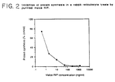
- maize proRIP has an IC 50 of greater than 10 micrograms per milliliter ( ⁇ g/ml) in a cell-free protein synthesis assay.
- the maize ⁇ RIP has an IC 50 of about 1 nanogram per milliliter (ng/ml) in a mammalian cell-free protein translation assay.
- the maize proRIP has a molecular weight of about 34 kD, as determined by SDS-PAGE (see Laemmli (1970), supra ), and will move as a single peak on ion exchange chromatography.
- Homogeneous maize ⁇ RIP will comprise two associated fragments, an ⁇ and ⁇ fragment, having molecular weights of approximately 16.5 kD and 8.5 kD, respectively (as determined by sodium dodecyl sulfate polyacrylamide-gel electrophoresis (SDS-PAGE) (see Laemmli ((1970), Nature , 22 :680-685).
- the homogeneous protein will exhibit two dissociated peaks on reverse phase chromatography, and a single associated peak on ion exchange chromatography.
- Polyclonal anti-sera against each fragment both crossreact with a polypeptide present in maize kernels having a molecular weight of about 34 kD as determined by SDS-PAGE. This demonstrates that the two fragments of the maize ⁇ RIP are in fact derived from a common precursor (i.e., the maize proRIP).
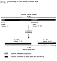
- the maize proRIP amino acid sequence (as set forth in Table 1) contains four sequence subsegments:
- the net charges of these polypeptides are as follows: leader -3; ⁇ , +10; linker, -5; ⁇ , +1. Removal of the leader and linker results in a dramatic change in net charge from the maize proRIP (+3) to maize ⁇ RIP (+11). Additionally, the proRIP isolated from maize has an observed pI of 6.5 which agrees well with the value of 6.1 derived from the deduced amino acid sequence. The pI of the active maize ⁇ RIP is ⁇ 9, compared to the calculated value from the deduced amino acid sequence of 9.06 (i.e., after deletion of the acidic leader, and linker sequences). Experimental data further suggests that the native maize ⁇ RIP has a few amino acids removed from the carboxy-terminal end. Thus, the maize ⁇ RIP has a basic pI, which is consistent with the pI of other RIPs.
- Table 4A shows the first region and the comparative alignment of the N-terminal amino acid sequence of the maize ⁇ RIP ⁇ fragment with the N-terminal sequences of RIPs from other sources.
- the sequences are taken from: barley (see Asano et al. (1986), supra ); ricin A-chain (see Lamb et al. (1985), supra ); dodecandrin (see Ready et al. (1985), Biochem. Biophys, Acta , 791 :314); pokeweed anti-viral protein 2 (AP2) (see Bjorn et al. (1985), Biochim. Biophys.
- Table 4b shows that the other three regions are internally oriented.
- Table 4b specifically shows the alignment of maize ⁇ RIP with regions of homology in the amino acid sequences of other RIPs.
- the sequences are available from the following references: barley (see Asano et al. (1986), supra and Leah et al. (1991), J. Biol. Chem. , 266 :1564-1573); ricin A-chain (see Lamb et al. (1985), supra ); abrin A-chain (see Funatsu et al. (1988), Agric. Biol. Chem . 52 :1095); saporin-6 (see Benatti et al. (1989) Eur. J. Biochem.
- Type I and Type II RIPs have also been purified to homogeniety and these include; momorcharins (see Yeung et al. (1986), Int. J. Peptide Res. , 28 :518-524; tritins (see Roberts and Stewart (1979), Biochem., 18 :2615-2621); rye (see Coleman and Roberts (1982), Biochim. Biophys. Acta , 696 :239-244; agrostins and Hura crepitans (see Stirpe et al. (1983), Biochem. J. , 216 :617-625; Asparagus officianalis (see Stirpe et al. (1983), Biochem. J.
- Proteins from the following other plants have also been shown to possess ribosome inactivating activity: Stellarea holostea , Lychnis flos-cuculi , Hordeum murinum , Aegylops geniculata , Euphorbia serrata , Capsella bursapastoris , Muscari comosum (see Merino et al. (1990), J. Exp Botany , 41 :67-70); and proteins from Croton tiglium and Jatropha curcas (see Stirpe et al. (1976), Biochem J. , 156 :1-6).
- the ⁇ RIP When the internal linker sequence of the proRIP is removed, the ⁇ RIP has significant ribosome inactivating activity. The activity has been found to be significant regardless of whether the leader sequence has been removed (e.g., by recombinant methods). However, the proRIP is most active when the leader sequence is also removed and probably when C-terminus residues are also removed. In nature, it is thought that the linker is cleaved by endogenous proteases released by germinating seeds. Significantly, it has been discovered that the linker may also be cleaved in vitro by certain proteases, e.g., papain, to yield active maize ⁇ RIP from the precursor. While not intended to be bound by theory, it is thought that papain likely mimics the effect of endogenous endoproteinases released during germination.
- proteases e.g., papain
- linker forms an external loop with exposed amino acid residues that are susceptible to proteolysis.
- Support for this suggestion comes from the alignment of the amino acid sequence of the maize proRIP with that of ricin A chain, the three dimensional structure of which is known (see Montfort et al , (1987), J. Biol. Chem. , _ 262 :5398).
- the Glu 177, Arg 180, Asn 209 and Trp 211 of ricin A have been implicated in the active site region of the molecule (see Robertus (1988), In: Immunotoxins , supra ).
- homologous residues of maize ⁇ RIP can be positioned within the three dimensional structure of ricin A chain.
- the superimposed structures indicate that the C-terminal lysine of the ⁇ fragment (at residue 162) is in corresponding alignment with an externally positioned threonine (at residue 156) of the ricin A-chain.
- the N-terminal alanine of the ⁇ fragment (at residue 189) is in corresponding alignment with an externally positioned glycine (at residue 157) of the ricin A-chain.
- the present invention is intended to include the construction of ⁇ RIP and proRIP forms of any RIP.
- RIPs for which a full-length sequence has been determined contain regions with significant homology.
- the N-terminal sequence similarities in an even greater number of RIPs have been compared (set forth in Tables 4a and 4b). It is likely that these regions have particular effect upon the function of the respective RIP.
- An RIP having known amino acid sequences may now be altered into inactive, proRIP forms by the insertion of a linker. Based on the information deduced from the maize system set forth herein, it now becomes possible to engineer inactive forms of any RIP having a three dimensional structure similar to the three dimensional structure of ricin A chain. Cleavage of the linker will result in an ⁇ RIP not heretofore found in nature.
- the art has discussed the methodology for modifying the three dimensional structure of proteins (see, for example, Van Brunt (1986), Biotechnology , 4 :277-283).
- the first step involves selecting plausible sites on the RIP between which the linker may be inserted. One of those sites is the exposed amino acid residues surrounding residue 156 of ricin A-chain or its equivalent in other RIP sequences.
- the present invention is intended to encompass the insertion of a peptide linker in those sequences, provided that the insertion of the linker substantially reduces the ribosome inactivating ability of the RIP.
- substantially reduce is meant that the insertion of a cleavable linker into an active RIP lowers the IC 50 value of the resultant protein by at least 10 fold, preferably 100-fold, and more preferably 1000-fold.
- ricin A-chain has been shown to have sequence homology to many single chain RIPs.
- the RIPs set forth in Tables 4a and 4b are intended for exemplification purposes only. RIPs characterized in the future that meet the above criteria are also considered to be a part of this invention.
- the linker may be of a length, may be of an amino acid sequence, and may be internally positioned so as to substantially reduce the ribosome inactivating activity of the RIP.
- Panicoideae linker(s) is the only known RIP linker found in nature, it is expected that such an amino acid sequence will logically be a primary candidate for insertion into other RIPs.
- the present invention is intended to encompass linkers having effectively homologous sequences to a selected maize linker.
- the factors to be considered in synthetically preparing effectively homologous linkers for ⁇ RIPs generally are the same as set forth above for selecting effectively homologous linkers for a selected maize linker.
- proRIP-encoding gene sequence Any of a variety of procedures may be used to clone proRIP-encoding gene sequence.
- One method for cloning the proRIP gene sequence entails determining the amino acid sequence of the proRIP molecule.
- proRIP or ⁇ RIP protein may be purified (as described above), and analyzed to determine the amino acid sequence of the proRIP or ⁇ RIP. Any method capable of elucidating such a sequence can be employed, however, Edman degradation is preferred. The use of automated sequenators is especially preferred.
- peptides thus prepared may be isolated and purified by procedures well known in the art (see Current Protocols in Molecular Biology (1989), (eds.) Ausebel, et al. , Vol. 1) and Sambrook et al. (1989), Molecular Cloning: A Laboratory Manual ).
- the DNA sequences capable of encoding them are examined in order to clone the gene encoding the proRIP. Because the genetic code is degenerate, more than one codon may be used to encode a particular amino acid.
- proRIP peptide fragments can be obtained by incubating the intact molecule with cyanogen bromide, or with proteases such as papain, chymotrypsin or trypsin.
- oligonucleotide Using the genetic code one or more different oligonucleotides can be identified.
- the probability that a particular oligonucleotide will, in fact, constitute the actual proRIP encoding sequence can be estimated by considering abnormal base pairing relationships and the frequency with which a particular codon is actually used (to encode a particular amino acid) in eukaryotic cells. Using these rules, a single oligonucleotide, or a set of oligonucleotides, that contains a theoretical "most probable" nucleotide sequence capable of encoding the proRIP or ⁇ RIP peptide sequences may be identified.
- the oligonucleotide, or set of oligonucleotides, containing the theoretical "most probable" sequence capable of encoding the proRIP gene fragments may be used to identify the sequence of a complementary oligonucleotide, or set of oligonucleotides, which is capable of hybridizing to the "most probable" sequence, or set of sequences.
- An oligonucleotide containing such a complementary sequence can be employed as a probe to identify and isolate the toxin gene (see Sambrook et al. (1989), supra ).
- the present invention also relates to DNA sequences that encode recombinant proRIP and ⁇ RIP.
- the recombinantly-produced proRIP and ⁇ RIP share the following properties with the proRIP and ⁇ RIP isolated from nature and characterized according to the teachings herein: (1) portions of the amino acid sequence deduced from the nucleotide sequence are equivalent to amino acid sequences obtained directly from nature; (2) the polypeptide is recognized by anti-RIP antibodies; (3) the molecular weight of the proRIP and ⁇ RIP polypeptides encoded corresponds with the naturally occurring proteins; (4) each proRIP protein is convertable to an ⁇ RIP; and (5) each proRIP and ⁇ RIP protein exhibits relatively equivalent ribosome inactivating activity.
- genetic sequences are intended to refer to a nucleic acid molecule (preferably DNA). Genetic sequences which are capable of encoding the toxin may be derived from a variety of sources. These sources include genomic DNA, cDNA, synthetic DNA, and combinations thereof.
- Cells containing the desired sequence may be isolated, and genomic DNA fragmented by one or more restriction enzymes.
- the genomic DNA may or may not include naturally-occurring introns.
- the genomic DNA digested with selected restriction endonucleases yields fragments containing varying numbers of base pairs (bp).
- genomic DNA sequences encoding allelic variant forms of the proRIP gene which may include naturally occurring introns.
- allelic gene may be derived using a hybridization probe made from the DNA or RNA of the proRIP gene as well as its flanking regions. "Flanking regions" are meant to include those DNA sequences 5' and 3' of the proRIP encoding sequences.
- the DNA isolate encoding the proRIP gene may also be obtained from a cDNA library.
- the mRNA may be isolated from a suitable source employing standard techniques of RNA isolation, and the use of oligo-dT cellulose chromatography to enrich for poly-A mRNA.
- a cDNA library is then prepared from the mixture of mRNA using a suitable primer, preferably a nucleic acid sequence which is characteristic of the desired cDNA.
- a single stranded DNA copy of the mRNA is produced using the enzyme reverse transcriptase. From the single stranded cDNA copy of the mRNA, a double-stranded cDNA molecule may be synthesized using either reverse transcriptase or DNA polymerase.
- PCR polymerase chain reaction
- the DNA encoding the proRIP or ⁇ RIP may be chemically synthesized by manual procedures, e.g., the phosphotriester and phosphodiester methods (see Caruthers (1983), In: Methodology of DNA and RNA , (ed.) Weissman); or automated procedures e.g., using diethylphosphoramidites are used as starting materials (see Beaucage et al. (1981), Tetrahedron Letters , 22 :1859-1962).
- the DNA may be constructed by standard techniques of annealing and ligating fragments or by other methods.
- nucleotide sequence of the maize proRIP cDNA and the deduced amino acid sequence of such the corresponding maize proRIP is set forth in Table 1.
- proRIP and ⁇ RIP found in nature.
- an amino acid sequence is effectively homologous to a second amino acid sequence if at least 70 percent, preferably at least 80 percent, and most preferably at least 90 percent of the active portions of the amino acid sequence are identical and retains its intended function.
- an effectively homologous sequence to the ⁇ RIP retains the capacity to interact with and inactivate eukaryotic ribosomes.
- the effectively homologous sequence to the proRIP must retain the capacity to be converted into an ⁇ RIP. That is, the effectively homologous proRIP must have a linker sequence which, when cleaved, will yield a biologically functional ⁇ RIP.
- amino acids within a group may be substituted for other amino acids in that group: (1) glutamic acid and aspartic acid; (2) lysine, arginine and histidine; (3) hydrophobic amino acids such as alanine, valine, leucine and isoleucine; (4) asparagine and glutamine; (5) threonine and serine; (6) phenylalanine, tyrosine and tryptophan; and (7) glycine and alanine.
- linker sequence of the proRIP need not be limited to the sequence set forth in Table 1, above.
- the length of the linker may be modified, provided that (1) the linker is cleavable, and (2) upon cleavage of the linker the resultant protein has an IC 50 value that is at least about 10 times lower than the IC 50 value of the protein containing the linker.
- Primary criteria for selecting an effectively homologous linker include altering the net charge of the ⁇ RIP (e.g., more acidic); creating a conformational shift in the protein or providing steric hindrance to the active site of the protein.
- the maize ⁇ RIP like other RIPs, is basic. However, the maize proRIP has a slightly acidic pI. Thus, it is preferred that any effectively homologous linker selected for the maize proRIP will be acidic.
- the linker should be of a length which, while capable of altering the three-dimensional structure of the protein, when cleaved will permit the protein to retain most of the three dimensional features of the active ⁇ RIP molecule.
- the linker is cleavable it is generally required that the conformation of the proRIP be such that the linker cleavage sites are readily accessible to a selected cleavage agent.
- At least one restriction enzyme site may be engineered into the genetic sequence encoding an RIP, allowing DNA sequences encoding various polypeptide linkers to be inserted into the gene and tested for their ability to create an inactive, yet activatable RIP.
- cleavage will be effected outside of the replicative environment, for example, following harvest of microbial culture.
- the internal linker domain of the proRIP it may be preferable, in some instances, that the internal linker domain of the proRIP be retained, or altered so as to mimic the manner in which a natural, inactive proRIP liberates its active ⁇ and ⁇ fragments.
- Any chemical or enzymatic method which recognizes a specific sequence or structure and causes an appropriate cleavage at a selected site may be utilized for the present invention.
- Exemplary such sequences Pro-Xxx-Gly-Pro (where Xxx is unspecified) which is selectively cleaved by collagenase; Ile-Glu-Gly-Arg which is selectively cleaved by Factor Xa; and Gly-Pro-Arg which is selectively cleaved by thrombin see Nilsson et al. (1988), In: Advances in Gene Technology; Protein Engineering and Production , (ed.) Brew et al. ).
- a chemical or enzymatic method may not be suitable if its cleavage site occurs within the active amino acid sequences of the ⁇ and ⁇ fragments. That is, cleavage within the native amino acid sequence of the ⁇ and ⁇ fragments will generally have a greater likelihood of deleteriously affecting the enzymatic activity of the ⁇ RIP. It is possible to select a specific cleavage sequence of only one amino acid residue so long as that residue does is not accessible in the amino acid sequences of the ⁇ and ⁇ fragments. It is preferred, however, to utilize a specific cleavage sequence which contains two or more amino acid residues, i.e., an extended specific cleavage sequence. This type of sequence takes advantage of the extended active sites of various enzymes. Additionally, by utilizing an extended specific cleavage sequence, it is highly probable that cleavage will only occur at the desired site and not at other sites within the protein.
- an expression vehicle with appropriate promoters may be provided with a DNA sequence coding for enzymes which convert the proRIP to the active form, operating in tandem with the other DNA sequence coding expression of the proRIP.
- protein sequences may be modified by post-translation processing such as becoming associated with other molecules, for example, glycosides, lipids, or such inorganic ions as phosphate.
- the ionization status will also vary depending on the pH of the medium or the pH at which crystallization or precipitation of the isolated form occurs. Further, the presence of air may cause oxidation of labile groups, such as -SH.
- proRIP and ⁇ RIP, and fragments, thereof are all such modifications of a particular primary structure, e.g., both glycosylated and non-glycosylated forms, neutral forms, acidic and basic salts, lipid or other associated peptide forms, side chain alterations due to oxidation or derivatization, and any other such modifications of an amino acid sequence which would be encoded by the same genetic codon sequence.
- Exemplary techniques for nucleotide replacement include the addition, deletion or substitution of various nucleotides, provided that the proper reading frame is maintained.
- Exemplary techniques include using polynucleotide-mediated, site-directed mutagenesis, i.e., using a single strand as a template for extension of the oligonucleotide to produce a strand containing the mutation (see Zoller et al. (1982), Nuc. Acids Res. , 10 :6487-6500; Norris et al. (1983), Nuc. Acids Res. , 11 :5103-5112; Zoller et al. (1984), DNA , 3 :479-488; and Kramer et al. (1982), Nuc.
- the desired DNA fragment may be positioned in a biologically functional vector which may contain appropriate control sequences not present in the selected DNA fragment.
- biologically functional is meant that the vector provides for replication and/or expression in an appropriate host, either by maintenance as an extrachromosomal element or by integration into the host genome. A large number of vectors are available or can be readily prepared, and are well known to skilled artisans.
- vectors containing the appropriate promoters which can be used by the host organism for expression of its own protein, also contain control sequences, ribosome binding sites, and transcription termination sites.
- control sequences ribosome binding sites, and transcription termination sites.
- replicon and control sequences which are derived from species compatible with the host cell are used in connection with these hosts.
- the vectors should desirably have a marker gene that is capable of providing a phenotypical property which allows for identification of host cells containing the vector.
- the DNA fragments encoding the ⁇ fragment and the ⁇ fragment may be inserted into separate vectors, or into the same vector.
- the host cells may be transformed via either cotransformation or targeted transformation techniques.
- Construction of suitable vectors containing the desired coding and control sequences may be produced as follows.
- Restriction endonucleases may be used as a means for inserting the DNA fragments containing the proRIP gene or ⁇ RIP genes into an appropriate expression vehicle.
- Exemplary restriction enzymes include Aat II, Bam HI, Eco RI, Hind III, Nde I, Spe I, Xba I, Sac I, Bgl II, Pst I, Sal I and Pvu II.
- Cleavage is performed by treating the expression vehicle with a restriction enzyme(s).
- a restriction enzyme(s) In general, 10 ⁇ g vector or DNA fragments is used with 10 units of enzyme in 100 ⁇ l of buffer solution. Endonuclease digestion will normally be carried out at temperatures ranging from 37 degrees Centigrade (37°C) to 65°C, at a pH of 7 to 9. Appropriate buffers and substrate amounts for particular restriction enzymes are specified by the manufacturers. Time for the reaction will be from 1 to 18 hours.
- protein may be removed by standard techniques (e.g., extraction with phenol and chloroform).
- the nucleic acid may be then recovered from the aqueous fraction by standard techniques.
- the desired fragment is then purified from the digest. Suitable purification techniques include gel electrophoresis or sucrose gradient centrifugation.
- the vector and foreign DNA fragments may then be ligated with DNA ligase.
- An appropriately buffered medium containing the DNA fragments, DNA ligase, and appropriate cofactors is employed.
- the temperature employed will be between 4°C to 25°C. When DNA segments hydrogen bond, the DNA ligase will be able to introduce a covalent bond between the two segments.
- the time employed for the annealing will vary with the temperature employed, the nature of the salt solution, as well as the nature of the sticky ends or cohesive termini. Generally, the time for ligation may be from 5 to 18 hours (see Sambrook et al. (1989), supra ).
- Suitable host cells include cells derived from unicellular as well as multicellular organisms which are capable of being grown in cultures or by fermentation.
- Prokaryotes include members of the Enterobacteriaceae, such as strains of Escherichia coli , and Salmonella ; Bacillaceae , such as Bacillus subtilis ; Pneumococcus , Streptococcus , and Haemophilus influenzae .
- eukaryotic cells may be employed. As previously stated, eukaryotic cells have not heretofore been used as recombinant host cells for RIPs. By providing inactive forms of RIPs, the present invention provides skilled artisans with the flexibility to use eukaryotic cells as recombinant hosts. By transforming eukaryotic cells with the proRIP gene, the protein may be expressed at high levels without being toxic to the host cell. Since the protein is lacking in bioactivity pending extra-cellular cleavage, the effect is to enhance the biosafety of the procedure. The proRIP may then be selectively chemically or enzymatically converted to the desired ⁇ RIP.
- Exemplary eukaryotic microbes include yeast. Saccharomyces cerevisae , or common baker's yeast, is the most commonly used among eukaryotic microorganisms, although a number of other hosts are commonly available.
- cultures of cells derived from multicellular organisms may also be used as hosts.
- useful host mammalian cell lines are Sp2/0, VERO and HeLa cells, Chinese hamster ovary (CHO) cell lines, and W138, BHK, COS-7 and MDCK cell lines.
- Suitable hosts and expression systems are the baculovirus systems maintained in cultured insect cells, e.g., from Spodoptera frugiperda .
- Suitable plant cells include cells derived from, or seedlings of, tobacco, petunia, tomato, potato, rice, maize and the like.
- the expression vehicle may be inserted into the host cell by any suitable method.
- Conventional technologies for introducing biological material into living cells include electroporation (see Shigekawa and Dower (1988), Biotechniques , 6 :742; Miller, et al. (1988), Proc. Natl. Acad. Sci. USA , 85 :856-860; and Powell, et al (1988), Appl. Environ. Microbiol. , 54 :655-660); direct DNA uptake mechanisms (see Mandel and Higa (1972), J. Mol. Biol. , 53 :159-162; Dityatkin, et al.
- the host cells may be grown for about 48 hours to allow for expression of marker genes.
- the cells are then placed in selective medium and/or screenable media, where untransformed cells are distinguished from transformed cells, either by death or a biochemical property.
- the transformed cells are grown under conditions appropriate to the production of the desired protein, and assayed for expression thereof.
- Exemplary assay techniques include enzyme-linked immunosorbent assay, radioimmunoassay, or fluorescence-activated cell sorter analysis, immunohistochemistry and the like.
- Selected positive cultures are subcloned in order to isolate pure transformed colonies.
- a suitable technique for obtaining subclones is via the limiting dilution method.
- the inactive form of the ⁇ RIP offers the additional advantage, over active RIPs, of not being active until removal of the linker sequence.
- the RIP is not toxic to the majority of mammalian cells it is known that RIP may be made specifically cytotoxic by attachment to a targeting vehicle which is capable of binding to and into target cells.
- Exemplary targeting vehicles include any peptide hormone, growth factor, or other polypeptide cell recognition protein for which a specific receptor exists.
- a few examples include: antibodies and antibody fragments, lectins, insulin, glucagon, endorphins, growth hormone, melanocyte-stimulating hormone, transferrin, bombesin, low density lipoprotein, luteinizing hormone and asialoglycoprotein that bind selectively to target cells (see Immunotoxins (1988), supra ). It is well established that conjugates which contain RIP exhibit maximal cytotoxicity only when the RIP moiety is released from the targeting vehicle.
- ⁇ RIP and proRIP does not contain a reactive sulfhydryl group, it may be necessary to modify the proteins using chemical crosslinking reagents in order to link such proRIP and ⁇ RIP to targeting vehicles.
- Conjugates of a monoclonal antibody and the ⁇ RIP and proRIP may be made using a variety of bifunctional protein coupling agents.
- bifunctional derivatives of imidoesters such as dimethyl adipimidate, active esters such as disuccinimidyl suberate, aldehydes such as glutaraldehyde, bis-azido compounds such as bis(p-diazoniumbenzoyl)ethylenediamine, diisocyanates such as toluene 2,6-diisocyanate, and bis-active fluorine compounds such as 1,5-difluoro-2,4-dinitrobenzene (see, for example, WO 86/05098).
- cytotoxic fusion protein a cytotoxic fusion protein
- chimeric toxins see, for example, Pastan and FitzGerald (1989), The Journal of Biological Chemistry , 264 :15157-15160; and U.S. Patent 4,892,827 to Pastan et al. (The United States of America as represented by the Department of Health and Human Services)).
- modified Pseudomonas exotoxins which comprise a deletion in a receptor binding domain to form a fusion protein capable of rendering the modified toxin less toxic to selected cells.
- ⁇ -TGF human alpha transforming growth factor
- the protein passing straight through the column was collected and applied to a Mono S 10/10 column (Pharmacia LKB Biotechnology, Piscataway, NJ) equilibrated with PS, and eluted with a linear gradient of 0 to 200 mM sodium chloride in PB over 90 min at 2 milliliter/ minute (ml/min).
- the protein can be precipitated with 85 percent ammonium sulfate and dialyzed overnight before applying to the Mono S 10/10 column.
- Fractions containing ribosome inactivating protein activity were pooled and concentrated to 0.5 ml in Centricon-10 devices (Amicon, Danvers, MA), and applied to a Superose 12 column equilibrated in PB (Pharmacia LKB Biotechnology) at a flow-rate of 0.4 ml/min.
- Fractions containing ribosome inactivating protein activity were pooled.
- the ⁇ RIP was usually quite pure as identified by SDS-PAGE (see Laemmli (1970), supra ).
- the inhibitory activity of the maize ⁇ RIP toward mammalian protein synthesis was measured in a rabbit reticulocyte lysate system following the procedures of Pelham and Jackson (see (1976), Eur. J. Biochem. , 67 :247-256).
- a mix of the following reagents was prepared (2.5 milliliter (ml) total volume): 125 microliter ( ⁇ l) 200 mM Tris-HCl, pH 7.6 + 40 mM magnesium acetate + 1.6 M potassium chloride; 12.5 ⁇ l 3 mM hemin hydrochloride in 50 percent ethylene glycol; 1.0 ml untreated rabbit reticulocyte lysate (Promega, Madison, WI); 1.0 ml H 2 O; 62.5 ⁇ l amino acid mix; 125 ⁇ l 20 mM ATP + 4 mM GTP; 125 ⁇ l 200 mM creatine phosphate; 50 ⁇ l 2.5 mg/ml creatine phosphokinase in 50 percent ethylene glycol.
- the amino acid mix contained 50 ⁇ M of each amino acid except glycine (100 ⁇ M), arginine, isoleucine, methionine and tryptophan (10 ⁇ M each) and contained no leucine. All stock solutions were previously adjusted to pH 7.5 prior to addition.
- the ⁇ and ⁇ polypeptide bands were cut from 3 millimeter (mm) SDS-PAGE gels after brief staining with Coomassie blue and were electroeluted using an electroelution device (Bio-Rad, Richmond CA) according to the manufacturer's directions. The polypeptide preparations were then used to immunize rabbits to yield polyclonal anti-sera (prepared by RIBI Immunochem, Montana).
- the polyclonal antisera against the ⁇ and ⁇ fragments were used to identify a common 34 kD precursor polypeptide in crude extracts of maize kernels (maize proRIP).
- the presence of the maize proRIP was monitored during subsequent purification by Western blot analysis as set forth above. All steps of the purification were performed at 4°C, except for HPLC which was performed at room temperature.
- proRIP A purified sample of proRIP was incubated at 0.5 mg/ml with papain, a plant thiol protease, at 0.01 mg/ml in sodium acetate buffer, pH 6 containing 2 mM dithiothreitol. After 1 to 2 hours at room temperature, the 34 kD proRIP was digested to a stable product exhibiting a polypeptide pattern almost identical to that of native, active maize ⁇ RIP. There was a concomitant increase in ribosome inactivating activity in the incubation; the undigested proRIP had no ribosome inactivating activity up to 2 ⁇ g/ml, whereas papain-treated proRIP had an IC 50 of ⁇ 80 ng/ml. In contrast trypsin had no effect on maize proRIP.
- N-Terminal sequence of ⁇ fragment A A D P Q A D T K S X L V K L V V M S/C E G L X F N T V S
- the carboxy-terminal amino acid sequence of the ⁇ maize ⁇ RIP a fragment was determined using sequencing grade carboxypeptidase P from Penicillium japonicum (Boehringer Mannheim, Indianapolis, IN).
- a sample of 16.5 kD polypeptide was purified by reverse-phase HPLC using a Vydac 5 ⁇ C4 4.6 x 30 mm RP column. The column was equilibrated with 0.1 percent trifluoroacetic acid (TFA), and eluted with 0 to 40 percent of 0.1 percent TFA + 80 percent acetonitrile over 8 min, then 40 to 60 percent of 0.1 percent TFA + 80 percent acetonitrile over 20 min. The ⁇ fragment eluted after 21.9 min. and the ⁇ fragment eluted after 23.3 min.
- TFA trifluoroacetic acid
- a lyophilized sample of the ⁇ fragment was dissolved in 20 mM sodium acetate, pH 5.8 + 4 M urea.
- the digestion mix contained the following in 0.1 ml: 1.6 ⁇ g carboxypeptidase P, 66 ⁇ g ⁇ fragment, 0.12 M sodium acetate pH 4.2, 0.8 M urea. After 1, 5, 15, 60, 120 and 480 min, duplicate 8 ⁇ l aliquots from the digestion were added to 8 ⁇ l 0.4 M sodium borate, pH 10.5 and frozen on dry ice.
- Immature kernels from field grown Pioneer hybrid 3737 were harvested, shelled from the cob, and stored at -20°C.
- Ten grams (10 g) of kernels were frozen in liquid nitrogen for several minutes then ground to a powder in a Waring blender.
- the powder was suspended in 20 ml of ice cold TENS buffer (10 mM Tris pH 7.4, 1 mM EDTA, 0.5 percent SDS, 0.3 M NaCl) and extracted immediately with an equal volume of phenol-chloroform-isoamyl alcohol (25:24:1) saturated with TENS buffer.
- the aqueous phase was collected and extracted three more times with fresh phenol mixtures.
- Nucleic acids were precipitated from the aqueous phase by adjusting it to 0.3 M sodium acetate pH 5.5 and adding 2.5 volumes of 100 percent ethanol. Nucleic acids were collected by centrifugation and suspended directly in 1 ml phenol-chloroform-isoamyl alcohol plus 1 ml TENS and extracted by vortexing. The nucleic acid was precipitated from the aqueous phase by ethanol precipitation as above. The precipitate was collected by centrifugation and resuspended in TE buffer (10 mM Tris pH 7.4, 1 mM EDTA). Single strand nucleic acid was precipitated by adjusting the solution to 2M LiCl and incubating for 4 to 12 hours at 4°C. Centrifugation yielded a pellet which consisted of between 2.2 to 2.5 mg of total RNA.
- Poly(A)-containing RNA was enriched from the total RNA sample by using Hybond mAPTM mRNA purification affinity paper (Amersham Corporation, Arlington Heights IL). The supplier's protocol was followed. Typically 5 to 10 ⁇ g of poly(A) enriched RNA were recovered per milligram of total RNA.
- RNA Five micrograms (5 ⁇ g) of poly(A) enriched RNA were converted into double stranded cDNA using a cDNA SynthesisTM kit (Pharmacia LKB Biotechnology).
- the cDNA was ligated into the cloning vector Lambda gt11 (Stratagene Inc., La Jolla CA) following the supplier's instructions.
- Packaging of the ligated vector-insert mixture was done with the Gigapack plus packaging extract (Stratagene, Inc.) again following the supplier's protocol.
- the PicoBlue ImmunodetectionTM kit (Stratagene, Inc.) was used to screen the Lambda gt11 maize kernel cDNA library using rabbit polyclonal antisera raised against the maize proRIP, as described above.
- the nucleotide sequence of the proRIP cDNA was determined by dideoxy chain termination sequencing using the SequenaseTM DNA sequencing kit (United States Biochemical Corp., Cleveland Ohio). The double stranded pUC19-RIP was used as template following the manufacturer's instructions. The first round of sequencing was initiated by the M13/pUC forward sequencing primer (Bethesda Research Labs ). Subsequent primers were derived from the sequenced maize proRIP cDNA. Both strands of the cDNA were fully sequenced at least once.
- the open reading frame encoding the ⁇ RIP protein was confirmed by comparing the cDNA deduced amino acid sequence (set forth in Table 1) to the chemically determined protein sequence data.
- Example 7 Expression of Maize proRIP and Derivatives in Escherichia coli
- R34 represents the intact recombinant proRIP gene which encodes a protein of Mr 33,327 and as expected is not a potent inhibitor of protein synthesis.
- papain treatment it is processed into two associated polypeptides (of approximately 17 + 9 kD) by SDS PhastgelTM analysis) with very potent ribosome inactivating activity.
- N34 represents the native proRIP as isolated from nature.
- the engineered, amplified product was purified from an agarose gel and ligated into the filled-in Hind III site of the expression vector pGEMEX-1 (Promega Corp., Madison, WI) to give plasmid pGR, set forth below.
- Plasmids containing the maize proRIP cDNA were isolated by colony hybridization (see Sambrook et al. , supra ) with a 5' maize proRIP cDNA probe and characterized. Those containing the cDNA probe for the maize proRIP in the correct orientation were tested for expression. Plasmids were transformed into competent E. coli JM109(DE3) (Promega Corp., Madison, WI), transformed cells were grown in 15 ml cultures under ampicillin selection to an optical density at 600 nm of 0.4 to 1.0.
- IPTG Isopropylthio- ⁇ -galactoside
- the protein produced from the maize proRIP cDNA was analyzed by lysing the induced cells in TE containing 1 mg/ml lysozyme 37°C for 15 min. The lysate was fractionated into a crude supernatant and pellet by microcentrifugation. The fractions were analyzed by SDS-PAGE using 20 percent PhastgelsTM reagent (Pharmacia LKB Biotechnology). Coomassie blue staining and Western blot analysis of the gels with anti-maize proRIP sera identified a 34 kD band which was greatly increased upon induction of the cells with IPTG.
- the pellet fraction of an induced culture was dissolved in 6M guanidine HCl and allowed to denature at room temperature for 3 hours. The material was then diluted 200-fold into ice cold TE and incubated at 4°C overnight to allow refolding of the denatured R34. The diluted material was then concentrated by a Centricon 10 device (Amicon). To test whether refolded R34 could undergo the correct proteolytic processing to the fragmented form of the maize proRIP, the material was treated with 10 ⁇ g/ml papain for various times, and samples were analyzed by SDS-Phastgel and Western blot analysis. The R34 material was processed to a stable mixture of two immuno-reactive bands which comigrate with N34 papain-processed material indicating the correct proteolytic processing had occurred.
- the gene 10 coding region of the pGEMEX-1 vector was removed by cutting the maize proRIP gene-containing plasmid (pGR) with Xba I and gel purifying the vector/proRIP DNA away from the gene 10 encoding DNA. Recircularization of pGR, now minus the gene 10 coding region, resulted in a plasmid called pGR1 set forth below.
- the plasmid pGR1 was transformed into JM109(DE3) cells and tested for production of R34 following induction with IPTG. As with pGR, large amounts of R34 were identified in cellular lysates both by Western blot and Coomassie blue staining. Unlike pGR, R34 produced from pGR1 was soluble and fractionated in the supernatant of lysed cells. This soluble material was treated with papain at 10 ⁇ g/ml and the R34 produced from pGR1 was cleaved to products which comigrate with N34, papain-cleaved product. The papain-treated material inhibited translation of reticulocyte lysates at significantly higher dilutions than the untreated material, indicating that the soluble R34 was processed to an active form.
- R34-DL represents the proRIP without the linker.
- the sequences encoding ⁇ and ⁇ were joined directly without intervening linker DNA, i.e., nucleotides A-520 to A-594 are deleted.
- the R34-DL gene encoded a 30.6 kD protein which was a potent inhibitor of protein synthesis.
- Treatment of R34-DL with papain resulted in a 28 kD polypeptide with increased ribosome inactivating activity over the untreated molecule.
- the R34-DL gene directed the synthesis of a polypeptide approximately 30.6 kD, which was recognized by antisera specific for the maize proRIP.
- E.coli lysates containing R34-DL protein were potent inhibitors of protein synthesis in rabbit reticulocyte lysates, in marked contrast to E. coli lysates containing the R34 polypeptide.
- R30-DL nucleotides C-40 to C-84 and A-520 to A-594 inclusive are deleted
- R30-DL encoded a protein (approximately 29.5 kD) which was slightly smaller than R34-DL.
- E.coli lysates containing R30-DL appeared to be even more potent inhibitors of protein synthesis than R34-DL lysates.
- Papain treatment of R30-DL containing lysates further enhanced protein synthesis inhibiting activity. Following this treatment the R30-DL protein underwent a slight decrease in molecular weight representing processing at the ends of the polypeptide..
- a segment of the R30-DL gene was deleted which encodes several acidic residues at the carboxy terminus of the protein.
- the deletion was accomplished using the PCR engineering methods.
- thermocycler Perkin-Elmer Cetus, Norwalk, CT
- a typical run was done with a one minute denaturation step, 2 minute annealing, and a 3 minute extension step. Temperatures used were 94°C, 37°C or 50°C, and 72°C respectively. Following 25 cycles the reaction was held at 72°C for 7 minutes for extension of unfinished products.
- Amplification engineering reactions were done in four separate tubes of 100 ⁇ L each. The tubes were combined following amplification. Normally 100 ng of template was included in each tube. DNA primers were synthesized on a PCR Mate or 380A DNA synthesizer (Applied Biosystems) and were purified on acrylamide gels. Fifty (50) pmol of each primer were included in each reaction. The reaction conditions for the amplification were those recommended by Perkin Elmer Cetus (10 mM Tris-HCl pH8.3, 50 mM KCl, 1.5 mM MgCl2, 0.001 percent gelatin, 200 ⁇ M dNTPs and 2.5 units of Taq DNA polymerase or AmpliTaqTM thermostable DNA polymerase).
- Enzymes used for the engineering were from one of three manufacturers (Pharmacia LKB Biotechnology; Bethesda Research Labs; or New England Biolabs, Beverly, MA). Buffers and protocols used were provided by the manufacturer.
- a modified RIP fragment is amplified from an RIP plasmid template, purified, then used to replace the unmodified region in the RIP gene. All fragment replacements were done in RIP genes already inserted into a pGEMEX expression plasmid.
- a pGEMEX plasmid containing R30-DL, which had the 3' half of the gene removed by Nco I and Stu I digestion followed by gel purification was used as the template for PCR.
- the 3' half of the RIP gene was replaced with the PCR modified fragment described below.
- a 3' PCR primer was synthesized which encoded the 7 amino acid deletion near the carboxy terminus and introduced a new unique Bam HI site.
- the 5' primer directed the deletion of the ⁇ linker and included a Nco I site.
- the sequences of these primers is given in Table 7.
- the primers were used to amplify a modified DNA fragment from a pGEMEX R34-DL template. The amplified fragment was phenol extracted and ethanol precipitated. The insert DNA was cut with Nco I and ligated into the pGEMEX-R30-DL vector.
- the new RIP gene derivative is called RDT and encodes a protein of predicted a 28,233 Daltons and pI ⁇ 9.5.
- the RDT gene encodes a protein with a truncated leader, deleted linker and truncated carboxy terminus.
- the DNA sequence for RDT is shown in Table 8.
- RDT gene expressed in E.coli using the pGEMEX system described above, was purified from bacterial lysates to apparent homogeneity. RDT protein appears to be a more potent inhibitor of protein synthesis than R30-DL. Using the reticulocyte lysate protein synthesis assay, purified RDT has a IC50 value of 1 ng/ml.
- RDT was further engineered to produce another gene called RDT-NP.
- This construction differs from RDT in having two unique restriction sites engineered into the gene. The sites were introduced using PCR methods described in Example 8.
- the PCR primer was designed such that it included the desired change and a unique restriction site in the maize RIP DNA sequence.
- a 99 bp primer which introduced the Not I and Pst I sites at the 3' end of the primer and had to be built back to the unique Nco I site for cloning purposes.
- the primers for the amplification are shown in Table 9.
- RDT-NP allows DNA segments encoding various polypeptide linkers to be inserted into the gene and tested for their ability to create an inactive, yet protease activatable RIP.
- the sequence for RDT-NP is shown in Table 10.
- the RDT-NP polypeptide had a predicted molecular weight of 28,446 Daltons and pI of 9.5.
- Crude lysates of E.coli expressing RDT-NP from a pGEMEX vector are potent inhibitors of eukaryotic protein synthesis.
- ABR Antibody Binding Region
- RDT-A was expressed in E.coli cells using the pGEMEX system.
- the resulting polypeptide had a predicted molecular weight of 35,198 Daltons and pI of 9.2. It was recognized by antisera to both protein A and maize RIP indicating the chimeric nature of the protein. Crude lysates of bacteria expressing RDT-A had potent eukaryotic protein synthesis inhibition activity.
- RDT-A was shown to bind specifically to IgG Sepharose (Pharmacia LKB Biotechnology) following the manufacturer's instructions. Binding was best at pH 7.0. When washed at pH 5.0 the chimeric protein was released in small but detectable quantities from the resin. RDT alone does not bind to the gel.
- the Antibody Binding Domain from Streptococcal Group G protein G was synthesized using oligonucleotides.
- the sequence synthesized was that of the naturally occurring sequence described by Guss et al. ((1986), EMBO Journal , 5 :1567-1575). The only change was the addition of Bam HI and Bgl II sites at the 5' and 3' ends respectively of the synthetic DNA.
- RDT-G-A contains a single ABR-G domain inserted in the correct orientation between the 3' end of RDT and the 5' end of ABR-A.
- a second class contains two properly oriented ABR-G domains.
- the predicted DNA sequences for the genes are shown in Tables 12 and 13.
- the RDT-G-A produced a protein of predicted molecular weight 44,576 Daltons (pI 7.2).
- RDT-G-G-A produced a slightly larger polypeptide predicted to be 53,955 Daltons with a predicted pI of 5.4.
- Crude bacterial lysates of cells expressing RDT-G-A or RDT-G-G-A were potent inhibitors or eukaryotic protein synthesis in the rabbit reticulocyte assay described in Example 1.
- Papain treatment of the lysates further increases activity. Analysis of the papain treated lysates indicates that the intact RDT domain is released from the ABR domains.
- RDT-G-A and RDT-G-G-A bind tightly to IgG Sepharose (Pharmacia LKB Biotechnology). Binding is stable at pH 5.0 Elution was accomplished with 0.5 M ammonium acetate pH 3.5 or by boiling the resin in SDS.
- Zea mays mays were ground to a fine consistency in a mortar and pestle (after removal of the glume if necessary): Zea mays mays , Z.m. mexicana , Z.m. parviglumis , Z. luxurians , Z. mexicana , Z. mexicana , Tripsacum dactyloides , Coix lachryma-jobi , Sorghum bicolor .
- Soluble proteins were extracted from mature dry seed by the following techniques. The proteins were extracted for 2 hours with 50 mM sodium phosphate buffer, pH 7.5 containing 25 ⁇ g/ml leupeptin, 25 ⁇ g/ml antipain, 2 mM EDTA and 4 mM phenylmethane sulfonyl fluoride. Three milliliter extraction buffer per gram seed tissue was used. After centrifugation to remove insoluble material, an aliquot of the extract was analyzed by sodium dodecyl sulfate-polyacrylamide gel electrophoresis using 17-27% gradient gels, then electroblotted onto PVDF paper (Millipore). The blot was probed using rabbit antisera against maize RIP ⁇ and ⁇ fragments (1:2000 dilution each) as primary antibody and goat anti-rabbit IgG antibody as secondary antibody, then developed with NBT/BCIP.
- the total extracted DNA from each sample was subjected to the Southern hybridization technique (see Southern (1975), J.Mol.Biol. , 98 :503-517).
- the DNA fragments were fractionated on the basis of their size by means of electrophoresis on a 0.8% agarose gel.
- the double-stranded DNA fragments were modified into single-stranded DNA fragments in an alkali solution; and then a nylon filter was placed into close contact with the gel to transfer the modified DNA segments onto the filter in the presence of a high salt concentration solution.
- Hybridization was carried out using, as the probe, the cDNA clone of the RIP gene (; the probe fragment is provided from position 2 to 1075 in Table 1).
- the probe was radiolabeled with 32 P and the signals in Southern transfers were visualized by autoradiography.
- the Southern blots were hybridized with the cDNA clone of the RIP gene. A single fragment with strong homology to the clone was observed for each enzyme/species combination except the Coix accession.
- the inbred line of maize has a single major band with two minor bands.
- the other species which, with the exception of Sorghum , are not inbred showed between 2 and 5 minor bands.
- the Coix had either 4 or 5 such bands depending on the enzyme used.
Landscapes
- Chemical & Material Sciences (AREA)
- Health & Medical Sciences (AREA)
- Organic Chemistry (AREA)
- Life Sciences & Earth Sciences (AREA)
- General Health & Medical Sciences (AREA)
- Medicinal Chemistry (AREA)
- Molecular Biology (AREA)
- Proteomics, Peptides & Aminoacids (AREA)
- Genetics & Genomics (AREA)
- Biophysics (AREA)
- Biochemistry (AREA)
- Gastroenterology & Hepatology (AREA)
- Botany (AREA)
- Veterinary Medicine (AREA)
- Public Health (AREA)
- General Chemical & Material Sciences (AREA)
- Animal Behavior & Ethology (AREA)
- Nuclear Medicine, Radiotherapy & Molecular Imaging (AREA)
- Chemical Kinetics & Catalysis (AREA)
- Pharmacology & Pharmacy (AREA)
- Peptides Or Proteins (AREA)
- Preparation Of Compounds By Using Micro-Organisms (AREA)
- Breeding Of Plants And Reproduction By Means Of Culturing (AREA)
- Micro-Organisms Or Cultivation Processes Thereof (AREA)
- Medicines That Contain Protein Lipid Enzymes And Other Medicines (AREA)
Abstract
Description
- Ribosome-inactivating proteins (RIPs) are plant proteins that are capable of catalytically inactivating eukaryotic ribosomes and are consequently extremely potent inhibitors of eukaryotic protein synthesis. RIPs have been divided into two classes:
type 1 and type 2 RIPs (see Barbieri and Stirpe (1982), Cancer Surveys, 1:489-520). There is significant amino acid sequence homology between members of bothtype 1 and type 2 RIPs, and with the bacterial Shiga and Shiga-like toxins which also have the same mechanism of action (see Hovde et al. (1988), Proc. Natl. Acad. Sci. USA, 85:2568-2572). - Type 2 RIPs consist of two polypeptides: an RIP (or A-chain) which is covalently attached via a disulfide bond to a lectin-like protein (or B-chain) with an affinity for cell surface carbohydrates. The B-chain binds to the cell surface and facilitates subsequent cellular internalization of the RIP A-chain moiety, which results in rapid inactivation of protein synthesis and cell death. Type 2 RIPs are therefore extremely potent cytotoxins and animal poisons, the best studied example of which is ricin.
- In
contrast type 1 RIPs, characterized to date, consist of a single polypeptide chain equivalent in activity to that of A-chain RIPs but lacking a covalently attached B-chain. Consequently, they are scarcely toxic to intact cells but retain their extreme potency against cell-free protein translation systems. Typical concentrations that inhibit cell-free protein translation by 50 percent (IC50) are 0.5 to 10 ng/ml (0.16 to 33 pM). Until the discoveries detailed hereinbelow, reportedtype 1 RIPs were a remarkably homogeneous class of basic proteins with Mr values of about 30,000.Type 1 RIPs are found in a great variety of dicot and monocot plants and they may be ubiquitous. They are often abundant proteins in seeds, roots or latex for example. Their in vivo function is unclear but it has been proposed that they may be antiviral agents (see Stevens et al. (1981), Experientia, 37:257-259) or antifungal agents (see Roberts and Seltrennikkoff (1986), Bioscience Reports, 6:19-29). - To date, one article has discussed the presence of an inhibitor of animal cell-free protein synthesis in maize, as well as other cereal crops (see Coleman and Roberts (1982), Biochimica et Biophysica Acta, 696:239-244). The preparation of the maize inhibitor was via ammonium sulfate precipitation and phosphocellulose column chromatography. It is generally believed that the inhibitor isolated from maize was pure. The reported molecular weight of the inhibitor was 23 kiloDaltons (kD), with a reported IC50 of 50 to 100 ng/ml in an ascites cell-free protein synthesis assay.
- Where the effect of RIPs on ribosomes has been examined, both
type 1 and type 2 RIPs possess a unique and highly specific N-glycosidase activity which cleaves the glycosidic bond of adenine 4324 of the rat liver ribosomal 28S RNA (see Endo (1988), In:Immunotoxins, (ed.) Frankel). - Commercial interest in RIPs has primarily focused on their use in construction of therapeutic toxins targeted to specific cells such as tumor cells by attachment of a targeting polypeptide, most frequently a monoclonal antibody (see Immunotoxins (1988), supra). This mimics the binding functionality of the B-chain of type 2 RIPs but replaces their non-specific action with a highly selective ligand. Another recent potential use is in HIV therapy (see U.S. Patent 4,869,903 to Lifson et al., (Genelabs Incorporated and The Regents of the University of California)).
- However, while a maize-derived protein synthesis inhibitor, like protein synthesis inhibitors from other Panicoideae would appear to be useful for construction of cytotoxic conjugates, no artisan to date has reported to have successfully used a Panicoideae RIP. This is somewhat surprising in view of the success achieved with RIPs from non-Panicoideae plants, including cereals such as barley (see Lambert et al. (1988), In:Immunotoxins,, supra). In part, the lack of success to date by skilled artisans in successfully utilizing the maize RIP described by Coleman and Roberts may be attributed to the fact that the protein synthesis inhibitor was relatively uncharacterized.
- There is interest in expressing recombinant RIP in commonly employed host eukaryotic cells, because of the capacity to provide correct post-translational processing. However, as RIPs effectively inhibit protein synthesis in eukaryotic cells, a predictable problem is that heterologous expression of an RIP will result in host cell death. Thus, eukaryotic cells are generally not used as recombinant host cells. Although eukaryotlc cells may be used in certain circumstances, the RIP must be constructed so as to be secreted prior to the cell experiencing toxicity (see EP 0 335 476 to Gelfand et al. (Cetus Corp.)). Therefore, prokaryotic host cells are generally used as hosts, notwithstanding disadvantages such as the inability to glycosylate and properly fold heterologously expressed proteins.
- It is thus an object of the invention to provide a method of preparing inactive forms of RIPs, in which an inactive RIP may be expressed by eukaryotic host cells and then converted to an active form.
- It is yet another object of the invention to provide the DNA sequence of the gene encoding at least one inactive form of RIP, as well as expression vehicles, host cells and cell cultures containing such DNA sequence.
- Other objects and advantages of the present invention will become apparent from the teachings presented hereinafter.
- It is to these objects to which the present invention is directed.
- In a first aspect, the present invention is directed to a DNA isolate as identified in the claims encoding a homogeneous protein, termed a proRIP, wherein the protein is incapable of substantially inactivating eukaryotic ribosomes, but which can be converted into a protein, termed an αβ RIP, that is capable of substantially inactivating eukaryotic ribosomes, said proRIP having a removable, internal peptide linker sequence.
- In a second aspect, the present invention is directed to a DNA isolate as identified in the claims encoding a homogeneous protein derived from Panicoideae, termed a proRIP, wherein the proRIP has a removable, internal peptide linker sequence and is incapable of substantially inactivating eukaryotic ribosomes, but which upon removal of the linker can be converted into a protein having α and β fragments, termed an αβ RIP, that is capable of substantially inactivating eukaryotic ribosomes.
- In a third aspect, the present invention is directed to a DNA isolate as identified in the claims encoding a homogeneous protein derived from Panicoideae, termed an αβ RIP, that is capable of substantially inactivating eukaryotic ribosomes, wherein the RIP has α and β fragments.
- In a fourth aspect, the present invention is directed to a DNA isolate as identified in the claims encoding a homogeneous protein derived from maize, termed a proRIP, wherein the proRIP has a removable, internal peptide linker sequence and is incapable of substantially inactivating eukaryotic ribosomes, but which upon removal of the linker can be converted into a protein having α and β fragments, termed an αβ RIP, that is capable of substantially inactivating eukaryotic ribosomes, said α fragment having an amino acid sequence effectively homologous to the following sequence:and said β fragment having an amino acid sequence effectively homologous to the following sequence:


- In a fifth aspect, the present invention is directed to a method of converting a protein that is incapable of substantially inactivating eukaryotic ribosomes into a protein that is capable of substantially inactivating eukaryotic ribosomes, said method comprising the following steps:
- a) providing a homogeneous protein derived from Panicoideae encoded by a DNA isolate as identified in the claims, termed a proRIP, wherein the proRIP has a removable, internal peptide linker sequence and is incapable of substantially inactivating eukaryotic ribosomes, but which upon removal of the linker can be converted into a protein having α and β fragments, termed an αβ RIP, that is capable of substantially inactivating eukaryotic ribosomes; and
- b) contacting the proRIP with a cleaving agent capable of deleting the linker to form a protein having α and β fragments, termed an αβ RIP, that is capable of substantially inactivating eukaryotic ribosomes.
-
- In a sixth aspect, the present invention is directed to a conjugate comprising a targeting vehicle and a protein derived from Panicoideae encoded by a DNA isolate as identified in the claims, termed a proRIP, wherein the proRIP has a removable, internal peptide linker sequence and is incapable of substantially inactivating eukaryotic ribosomes, but which upon removal of the linker can be converted into a protein having α and β fragments, termed an αβ RIP, that is capable of substantially inactivating eukaryotic ribosomes.
- In a seventh aspect, the present invention is directed to a conjugate comprising a targeting vehicle and a protein derived from Panicoideae encoded by a DNA isolate as identified in the claims, termed an αβ RIP, that is capable of substantially inactivating eukaryotic ribosomes, wherein the αβ RIP has α and β fragments.
- In a eighth aspect, the present invention is directed to a conjugate comprising a targeting vehicle and a protein derived from maize encoded by a DNA isolate as identified in the claims, termed a proRIP, wherein the proRIP has a removable, internal peptide linker sequence and is incapable of substantially inactivating eukaryotic ribosomes, but which upon removal of the linker can be converted into a protein having α and β fragments, termed an αβ RIP, that is capable of substantially inactivating eukaryotic ribosomes, said α fragment having an amino acid sequence effectively homologous to the following sequence:and the β fragment having an amino acid sequence effectively homologous to the following sequence:


- In a ninth aspect, the present invention is directed to DNA isolate as identified in the claims capable of encoding a protein, termed a proRIP, wherein the proRIP has a removable, internal peptide linker sequence and is incapable of substantially inactivating eukaryotic ribosomes, but which upon removal of the linker can be converted into a protein having α and β fragments, termed an αβ RIP, that is capable of substantially inactivating eukaryotic ribosomes.
- In a tenth aspect, the present invention is directed to a DNA isolate as identified in the claims encoding a protein, termed an αβ RIP, that is capable of substantially inactivating eukaryotic ribosomes, wherein the αβ RIP has α and β fragments.
- In an eleventh aspect, the present invention is directed to a DNA isolate as identified in the claims encoding a protein derived from maize, termed a proRIP, wherein the proRIP has a removable, internal peptide linker sequence and is incapable of substantially inactivating eukaryotic ribosomes, but which upon removal of the linker can be converted into a protein having α and β fragments, termed an αβ RIP, that is capable of substantially inactivating eukaryotic ribosomes, said α fragment having an amino acid sequence effectively homologous to the following sequence:and said β fragment having an amino acid sequence effectively homologous to the following sequence:


- In a twelfth aspect, the present invention is directed to a DNA isolate as identified in the claims encoding a fusion protein capable of substantially inactivating eukaryotic ribosomes, said protein having an amino acid sequence effectively homologous to the following sequence:
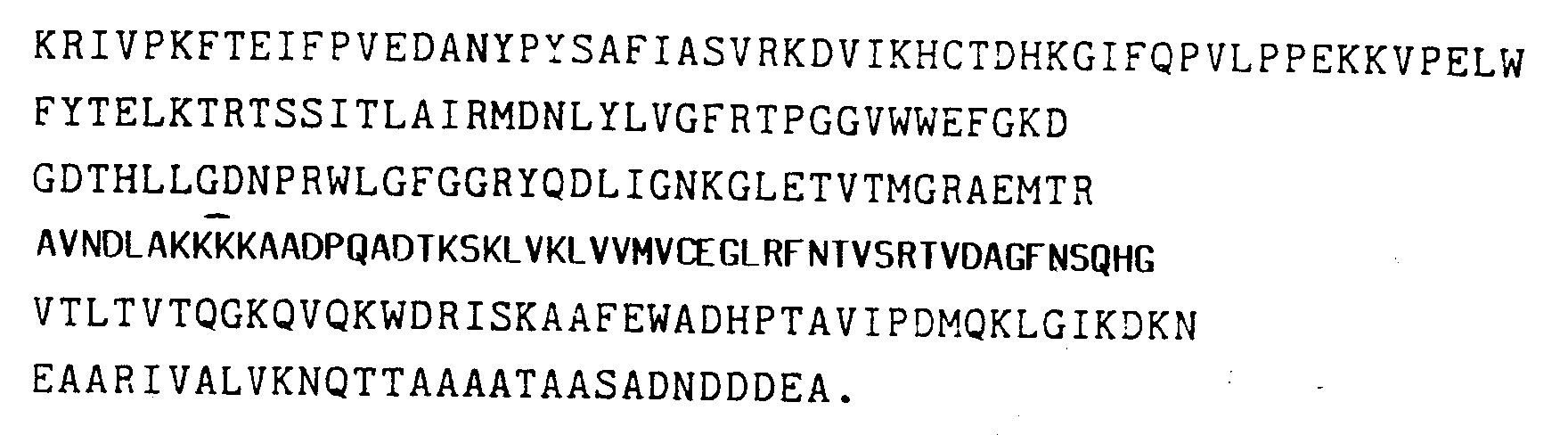
- In other aspects, the invention is directed to expression vehicles capable of effecting the production of such aforementioned proteins in suitable host cells. It also includes the host cells and cell cultures which result from transformation with these expression vehicles.
- A number of aspects of the present invention are further illustrated in the accompanying Drawings, in which:
- Figure 1 shows a schematic representation of the processing of maize proRIP to the active form.
- Figure 2 shows the effect of active maize αβ RIP on mammalian cell-free protein synthesis.
-
- The entire teachings of all references cited herein are incorporated by reference.
- Nucleic acids, amino acids, peptides, protective groups, active groups and so on, when abbreviated, are abbreviated according to the IUPACIUB (Commission on Biological Nomenclature) or the practice in the fields concerned. The following are examples.
- The single letter code for amino acids is set forth below:
Glycine: G Phenylalanine: F Alanine: A Tyrosine: Y Valine: V Threonine: T Leucine: L Cysteine: C Isoleucine: I Methionine: M Serine: S Glutamic acid: E Aspartic acid: D Tryptophan: W Lysine: K Proline: P Arginine: R Asparagine: N Histidine: H Glutamine: Q Unknown X - The term "proRIP" means a precursor protein that contains a leader and linker and that is not capable of inactivating eukaryotic ribosomes.
- The term "leader" refers to an N-terminal amino acid sequence of a proRIP that need not present in the mature, fully active form of the αβ RIP.
- The term "linker" refers to an internal amino acid sequence within a proRIP, whereby the linker is of a length and contains residues effective to render the proRIP incapable of catalytically inhibiting translation of a eukaryotic ribosome.
- The term "RIP" means a protein that is capable of inactivating eukaryotic ribosomes. The term "αβ RIP" means a RIP having an α fragment, which may or may not contain the leader, a β fragment and being capable of substantially inactivating eukaryotic ribosomes.
- The term "IC50" means the concentration of a protein necessary to inhibit protein synthesis by 50 percent in a cell-free protein synthesis assay.
- The term "inhibiting amount" refers to the specific ability of RIPs to cause the death or injury of cells against which they are targeted.
- The term "target cells" means those cells having ribosomes which the αβ RIP of the present invention is capable of inhibiting. The target cells may be present in living organisms or they may be preserved or maintained in vitro. The cells may be individual or associated to form an organ. Exemplary target cells include any eukaryotic cell (e.g., mammalian, insect, fungal and plant cells).
- The term "targeting vehicle" means a carrier moiety containing a ligand capable of binding to a receptor of a specific cell or tissue.
- "Gene" refers to the entire DNA portion involved in the synthesis of a protein. A gene embodies the structural or coding portion which begins at the 5' end from a translation start codon and extends to a stop codon at the 3' end. It also contains a promoter region, usually located 5' or upstream to the structural coding portion, which initiates and regulates the expression of a structural gene and a 3' nontranslated region downstream from the translated region.
- "Expression" refers to a two-part process for the transcription and translation of a gene. The DNA defining the gene is transcribed into a precursor RNA, which is processed to its mature form, messenger RNA (mRNA). During translation, the cell's ribosomes, in conjunction with transfer RNA, translate the RNA "message" into proteins.
- Surprisingly, it has been discovered that studied members of Panicoideae contain αβ RIP and proRIP. Panicoideae is a subfamily of Gramineae (order) and Graminaceae (family). The subfamily Panicoideae contains three tribes: Maydeae (e.g., Tripsacum, Coix, Euchlaena and Zea), Andropogonea (e.g., Sorghum) and Paniceae. For further taxonomic information, see Arber (1934), The Gram ineae, A Study of Cereal, Bamboo and Grass, Cambridge University Press, p 410-411.
- The present invention pertains to proteins which are derived from a plant within the subfamily Panicoideae. As taught herein, proteins obtained from various plants within the subfamily Panicoideae have shown antigenic cross reactivity (i.e., showing evidence of proRIP in Panicoideae as well as α and β fragments of an αβ RIP).
- By "derived" from a plant within the subfamily Panicoideae means a protein that is effectively homologous, as defined below, with a proRIP or αβ RIP from Panicoideae, regardless of the manner in which the protein is produced. Given the present teachings it now becomes possible to prepare generally homogeneous proRIP and αβ RIP exclusive of irrelevant proteins and contaminants naturally associated therewith in the cellular environment or in extracellular fluids. For example, a substantially pure protein will show constant and reproducible characteristics within standard experimental deviations for parameters such as the following: molecular weight, chromatographic behavior, and such other parameters. The term, however, is not meant to exclude artificial or synthetic mixtures of a protein with other compounds. The term is not meant to exclude the presence of minor impurities which do not interfere with the biological activity of the protein and which may be present, for example, due to incomplete purification.
- Both the proRIP and αβ RIP may be purified directly from mature and germinating seeds and developing kernels of plants within the subfamily. Generally, the purification of the Panicoideae αβ RIP and proRIP may be accomplished as follows.
- Seeds or immature kernels of plants within the subfamily Panicoideae may be homogenized to disrupt the individual seeds or kernels. This can be accomplished by any type of commercially available homogenizer.
- The Panicoideae proRIP and/or αβ RIP may be purified from the homogenization product by any appropriate protein purification technique. Exemplary techniques include gel filtration chromatographic techniques, such as conventional liquid chromatography, ion exchange chromatography, high performance liquid chromatography, and reverse phase chromatography.
- Upon purification, the Panicoideae proRIP will have insignificant ribosome inactivating ability relative to its corresponding αβ RIP. For example, maize proRIP has an IC50 of greater than 10 micrograms per milliliter (µg/ml) in a cell-free protein synthesis assay. The maize αβ RIP has an IC50 of about 1 nanogram per milliliter (ng/ml) in a mammalian cell-free protein translation assay.
- The maize proRIP has a molecular weight of about 34 kD, as determined by SDS-PAGE (see Laemmli (1970), supra), and will move as a single peak on ion exchange chromatography. Homogeneous maize αβ RIP will comprise two associated fragments, an α and β fragment, having molecular weights of approximately 16.5 kD and 8.5 kD, respectively (as determined by sodium dodecyl sulfate polyacrylamide-gel electrophoresis (SDS-PAGE) (see Laemmli ((1970), Nature, 22:680-685). The homogeneous protein will exhibit two dissociated peaks on reverse phase chromatography, and a single associated peak on ion exchange chromatography. Polyclonal anti-sera against each fragment both crossreact with a polypeptide present in maize kernels having a molecular weight of about 34 kD as determined by SDS-PAGE. This demonstrates that the two fragments of the maize αβ RIP are in fact derived from a common precursor (i.e., the maize proRIP).
- The maize proRIP amino acid sequence (as set forth in Table 1) contains four sequence subsegments:
- (1) a leader sequence, from
residues 1 to 16; - (2) an α fragment, from residues 17 to 161;
- (3) an internal linker sequence, from residues 162 to 186; and
- (4) a β fragment, from residues 187 to 301.
-
- The net charges of these polypeptides are as follows: leader -3; α, +10; linker, -5; β, +1. Removal of the leader and linker results in a dramatic change in net charge from the maize proRIP (+3) to maize αβ RIP (+11). Additionally, the proRIP isolated from maize has an observed pI of 6.5 which agrees well with the value of 6.1 derived from the deduced amino acid sequence. The pI of the active maize αβ RIP is ≧9, compared to the calculated value from the deduced amino acid sequence of 9.06 (i.e., after deletion of the acidic leader, and linker sequences). Experimental data further suggests that the native maize αβ RIP has a few amino acids removed from the carboxy-terminal end. Thus, the maize αβ RIP has a basic pI, which is consistent with the pI of other RIPs.
- A comparison of the maize proRIP with that of barley, a monocot , as set forth in Table 2 below. The upper sequence shows maize αβ RIP and the lower sequence barley RIP, as taught by Asano et al. (1986), Carlsberg Res. Commun., 51:129. Identical residues are denoted by a solid line, conservative substitutions by a dotted line, and dashes indicate insertions to maximize homology. Residues are numbered on the left.

- As set forth in in Table 2, there is an overall homology of 28 percent (34 percent including conservative substitutions) between the maize αβ RIP and barley RIP. However, the unique nature of the linker region of maize proRIP is clearly shown by the resulting gap that has been introduced in the published barley sequence to maintain homology. A lower, but significant, degree of homology is seen when the maize proRIP sequence is compared to that of ricin A-chain (as set forth in Table 3 below). The upper sequence is maize αβ RIP and the lower sequence is ricin A, as taught by Lamb et al. (1985), Eur. J. Biochem., 148:265. Identical residues are denoted by a solid line and conservative substitutions by a dotted line, dashes indicate insertions to maximize homology. Residues are numbered on the left, the numbering of the ricin sequence corresponds to that of the mature protein.
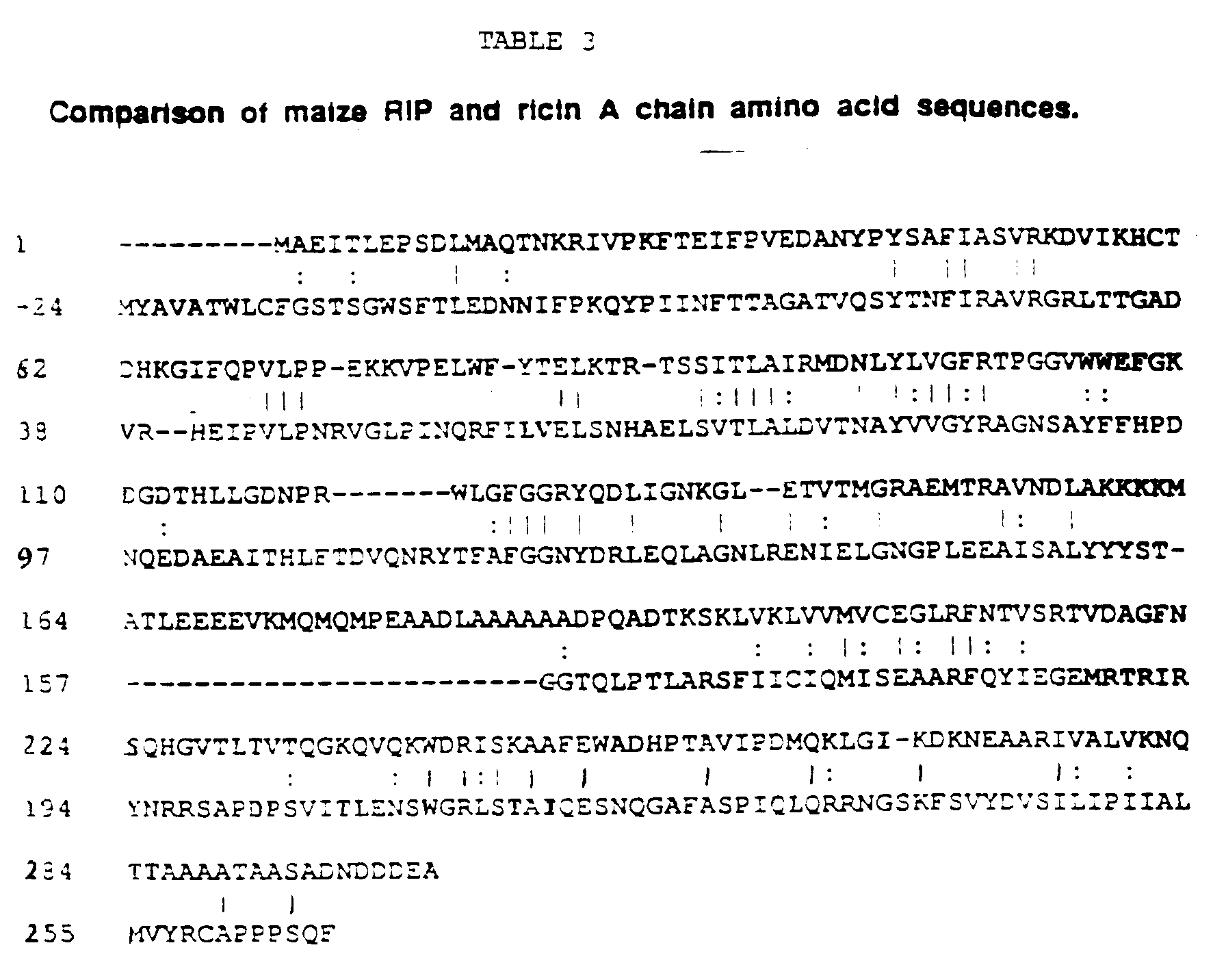
- As set forth in Table 3, a gap was again introduced in the published ricin A sequence to maximize homology corresponding to the linker region of the maize proRIP.
- Further comparison of the maize proRIP sequence with published full-length sequences of other non-Panicoideae RIPs indicate that there are four regions of significant homology between these proteins (as set forth in Tables 4a and 4b below).

- Table 4A shows the first region and the comparative alignment of the N-terminal amino acid sequence of the maize αβ RIP α fragment with the N-terminal sequences of RIPs from other sources. The sequences are taken from: barley (see Asano et al. (1986), supra); ricin A-chain (see Lamb et al. (1985), supra); dodecandrin (see Ready et al. (1985), Biochem. Biophys, Acta, 791:314); pokeweed anti-viral protein 2 (AP2) (see Bjorn et al. (1985), Biochim. Biophys. Acta, 790:154); Shiga-like toxin 1A (SLT-IA) (see Calderwood et al. (1987), Proc. Nat. Acad. Sci. USA, 84:4364); and α-trichosanthin, momordins, bryodin, gelonin, dodeoandrin, pokeweed antiviral protein-2, saporin 5, and saporin 4 (see Montecucchi et al. (1989), Int. J. Peptide Res., 33:263). Positions showing homology in four or more sequences are noted by solid lines (showing identical residues) or dotted lines (showing conservatively substituted residues).
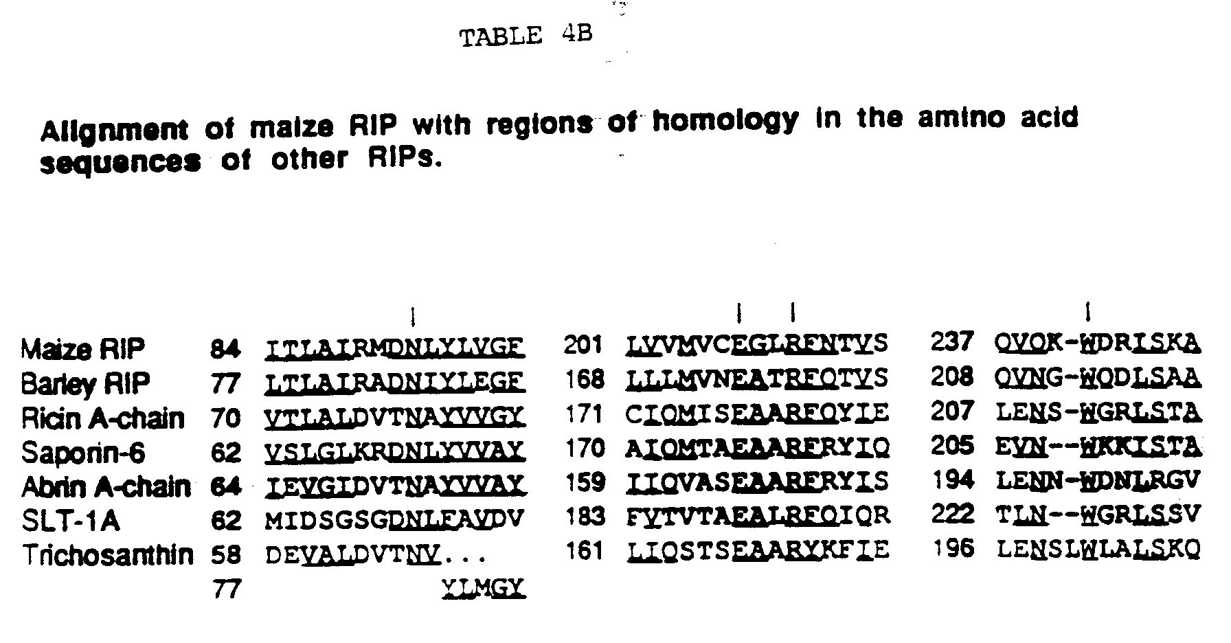
- Table 4b shows that the other three regions are internally oriented. Table 4b specifically shows the alignment of maize αβ RIP with regions of homology in the amino acid sequences of other RIPs. The sequences are available from the following references: barley (see Asano et al. (1986), supra and Leah et al. (1991), J. Biol. Chem., 266:1564-1573); ricin A-chain (see Lamb et al. (1985), supra); abrin A-chain (see Funatsu et al. (1988), Agric. Biol. Chem. 52:1095); saporin-6 (see Benatti et al. (1989) Eur. J. Biochem., 183:465); Shiga-like toxin 1A (SLT-1A) (see Calderwood et al. (1987), supra); and α-trichosanthin (see Xuejun and Jiahuai (1986), Nature, 321:477; Chow et al. (1990), J. Biol. Chem., 265:8670-8674 and Maragonore et al. (1987), J. Biol. Chem., 262:11628-11633). Positions showing identity or conservative substitutions in four or more sequences are underlined, dashes indicate insertions to maximize homology. Vertical lines indicate residues that are conserved in all seven sequences. The starting amino acid of each sequence is indicated (note that trichosanthin contains an insertion sequence at residues 67 to 76).
- The sequences and partial sequences of various additional Type I RIPs are set forth in the following articles: luffin-A (see Islam et al. (1990), Agric. Biol. Chem., 54:2967-2978; mirabilis antiviral protein (see Habuka et al. (1989) J. Bio. Chem., 264:6629-6637; trichokirin, (see Casellas et al. (1988), Eur. J. Biochem., 176:581-588; momordins (see Barbieri et al. (1980), Biochem. J., 186:443-452; dianthins (see Reisbig and Bruland (1983), Arch. Biochem. Biophys., 224:700-706; saporins (see Maras et al., (1990), Biochem. Intl., 21:831-838 and Lappi et al. (1985), Biochem. Biophys. Res Commun., 129:934-942; and momorcochin-S (see Bolognesi et al. (1989), Biochim. Biophys. Acta, 993:287-292.
- Other Type I and Type II RIPs have also been purified to homogeniety and these include; momorcharins (see Yeung et al. (1986), Int. J. Peptide Res., 28:518-524; tritins (see Roberts and Stewart (1979), Biochem., 18:2615-2621); rye (see Coleman and Roberts (1982), Biochim. Biophys. Acta, 696:239-244; agrostins and Hura crepitans (see Stirpe et al. (1983), Biochem. J., 216:617-625; Asparagus officianalis (see Stirpe et al. (1983), Biochem. J., 216:617-625; Cucumis melo (see Ferreras et al. (1989), Biochem Intl., 19:201-207; Cucurbitaceae (see Ng et al., Int. J. Biochem., 21:1353-1358; Petrocoptis (see Ferreras et al., Cell. Molec. Biol., 35:89-95); volkensin-a (see Barbieri et al. (1984), FEBS Lett., 171:277-279; viscumin-a (see Olsnes et al. (1982), J. Biol. Chem., 257:13263-123270; modeccin-a (see Gasperi-Campani (1978), Biochem. J., 174:491-496); Momordia charantia lectin-a (see Lin et al. (1978), Linn. Toxicon., 16:653-660); Phorandendron californicum lectin-a (see Franz et al. (1989), FEBS lett., 248:115-118).
- Proteins from the following other plants have also been shown to possess ribosome inactivating activity: Stellarea holostea, Lychnis flos-cuculi, Hordeum murinum, Aegylops geniculata, Euphorbia serrata, Capsella bursapastoris, Muscari comosum (see Merino et al. (1990), J. Exp Botany, 41:67-70); and proteins from Croton tiglium and Jatropha curcas (see Stirpe et al. (1976), Biochem J., 156:1-6).
- When the internal linker sequence of the proRIP is removed, the αβ RIP has significant ribosome inactivating activity. The activity has been found to be significant regardless of whether the leader sequence has been removed (e.g., by recombinant methods). However, the proRIP is most active when the leader sequence is also removed and probably when C-terminus residues are also removed. In nature, it is thought that the linker is cleaved by endogenous proteases released by germinating seeds. Significantly, it has been discovered that the linker may also be cleaved in vitro by certain proteases, e.g., papain, to yield active maize αβ RIP from the precursor. While not intended to be bound by theory, it is thought that papain likely mimics the effect of endogenous endoproteinases released during germination.
- It appears that, after removal of the internal linker, the two fragments of the processed polypeptide are held together by noncovalent forces. That is, the association of the two polypeptide chains does not depend upon interchain disulfide bonds or the formation of a peptide bond between the fragments.
- Although not intended to be bound by theory, it is believed that the linker forms an external loop with exposed amino acid residues that are susceptible to proteolysis. Support for this suggestion comes from the alignment of the amino acid sequence of the maize proRIP with that of ricin A chain, the three dimensional structure of which is known (see Montfort et al, (1987), J. Biol. Chem., _262:5398). The Glu 177, Arg 180, Asn 209 and Trp 211 of ricin A have been implicated in the active site region of the molecule (see Robertus (1988), In:Immunotoxins, supra).
- Based on this alignment, homologous residues of maize αβ RIP can be positioned within the three dimensional structure of ricin A chain. The superimposed structures indicate that the C-terminal lysine of the α fragment (at residue 162) is in corresponding alignment with an externally positioned threonine (at residue 156) of the ricin A-chain. Also, the N-terminal alanine of the β fragment (at residue 189) is in corresponding alignment with an externally positioned glycine (at residue 157) of the ricin A-chain.
- The present invention is intended to include the construction of αβ RIP and proRIP forms of any RIP. As set forth in Tables 4a and 4b, RIPs for which a full-length sequence has been determined contain regions with significant homology. Similarly the N-terminal sequence similarities in an even greater number of RIPs have been compared (set forth in Tables 4a and 4b). It is likely that these regions have particular effect upon the function of the respective RIP.
- An RIP having known amino acid sequences may now be altered into inactive, proRIP forms by the insertion of a linker. Based on the information deduced from the maize system set forth herein, it now becomes possible to engineer inactive forms of any RIP having a three dimensional structure similar to the three dimensional structure of ricin A chain. Cleavage of the linker will result in an αβ RIP not heretofore found in nature.
- The art has discussed the methodology for modifying the three dimensional structure of proteins (see, for example, Van Brunt (1986), Biotechnology, 4:277-283). The first step involves selecting plausible sites on the RIP between which the linker may be inserted. One of those sites is the exposed amino acid residues surrounding residue 156 of ricin A-chain or its equivalent in other RIP sequences. Thus, the present invention is intended to encompass the insertion of a peptide linker in those sequences, provided that the insertion of the linker substantially reduces the ribosome inactivating ability of the RIP. By "substantially reduce" is meant that the insertion of a cleavable linker into an active RIP lowers the IC50 value of the resultant protein by at least 10 fold, preferably 100-fold, and more preferably 1000-fold.
- As stated previously, ricin A-chain has been shown to have sequence homology to many single chain RIPs. The RIPs set forth in Tables 4a and 4b are intended for exemplification purposes only. RIPs characterized in the future that meet the above criteria are also considered to be a part of this invention.
- Generally, the linker may be of a length, may be of an amino acid sequence, and may be internally positioned so as to substantially reduce the ribosome inactivating activity of the RIP.
- Obviously, since the Panicoideae linker(s) is the only known RIP linker found in nature, it is expected that such an amino acid sequence will logically be a primary candidate for insertion into other RIPs. However the present invention is intended to encompass linkers having effectively homologous sequences to a selected maize linker. The factors to be considered in synthetically preparing effectively homologous linkers for αβ RIPs generally are the same as set forth above for selecting effectively homologous linkers for a selected maize linker.
- Any of a variety of procedures may be used to clone proRIP-encoding gene sequence. One method for cloning the proRIP gene sequence entails determining the amino acid sequence of the proRIP molecule. To accomplish this task proRIP or αβ RIP protein may be purified (as described above), and analyzed to determine the amino acid sequence of the proRIP or αβ RIP. Any method capable of elucidating such a sequence can be employed, however, Edman degradation is preferred. The use of automated sequenators is especially preferred.
- It is possible to synthesize in vitro the proRIP and αβ RIP from their constituent amino acids. A suitable technique includes the solid phase method (see Merrifield (1963), J. Amer. Chem. Soc., 85:2149-2154; and Solid Phase Peptide Synthesis (1969), (eds.) Stewart and Young). Automated synthesizers are also available.
- The peptides thus prepared may be isolated and purified by procedures well known in the art (see Current Protocols in Molecular Biology (1989), (eds.) Ausebel, et al., Vol. 1) and Sambrook et al. (1989), Molecular Cloning: A Laboratory Manual).
- Using the amino acid sequence information, the DNA sequences capable of encoding them are examined in order to clone the gene encoding the proRIP. Because the genetic code is degenerate, more than one codon may be used to encode a particular amino acid.
- Although it is possible to determine the entire amino acid sequence of the proRIP or αβ RIP, it is preferable to determine the sequence of peptide fragments of the molecule, and to use such sequence data to prepare oligonucleotide probes which can be used to isolate the entire proRIP gene sequence. The proRIP peptide fragments can be obtained by incubating the intact molecule with cyanogen bromide, or with proteases such as papain, chymotrypsin or trypsin.
- Using the genetic code one or more different oligonucleotides can be identified. The probability that a particular oligonucleotide will, in fact, constitute the actual proRIP encoding sequence can be estimated by considering abnormal base pairing relationships and the frequency with which a particular codon is actually used (to encode a particular amino acid) in eukaryotic cells. Using these rules, a single oligonucleotide, or a set of oligonucleotides, that contains a theoretical "most probable" nucleotide sequence capable of encoding the proRIP or αβ RIP peptide sequences may be identified.
- The oligonucleotide, or set of oligonucleotides, containing the theoretical "most probable" sequence capable of encoding the proRIP gene fragments may be used to identify the sequence of a complementary oligonucleotide, or set of oligonucleotides, which is capable of hybridizing to the "most probable" sequence, or set of sequences. An oligonucleotide containing such a complementary sequence can be employed as a probe to identify and isolate the toxin gene (see Sambrook et al. (1989), supra ).
- By hybridizing an oligonuleotide having a sequence complementary to the "most probable" gene sequence, one obtains a DNA molecule (or set of DNA molecules), capable of functioning as a probe to identify and isolate the proRIP gene.
- The present invention also relates to DNA sequences that encode recombinant proRIP and αβ RIP. The recombinantly-produced proRIP and αβ RIP share the following properties with the proRIP and αβ RIP isolated from nature and characterized according to the teachings herein: (1) portions of the amino acid sequence deduced from the nucleotide sequence are equivalent to amino acid sequences obtained directly from nature; (2) the polypeptide is recognized by anti-RIP antibodies; (3) the molecular weight of the proRIP and αβ RIP polypeptides encoded corresponds with the naturally occurring proteins; (4) each proRIP protein is convertable to an αβ RIP; and (5) each proRIP and αβ RIP protein exhibits relatively equivalent ribosome inactivating activity.
- The process for genetically engineering the proRIP or αβ RIP according to the invention is facilitated through the cloning of genetic sequences which are capable of encoding the proRIP or αβ RIP, or effectively homologous variants thereof as discussed below, and through the expression of such genetic sequences. As used herein, the term "genetic sequences" is intended to refer to a nucleic acid molecule (preferably DNA). Genetic sequences which are capable of encoding the toxin may be derived from a variety of sources. These sources include genomic DNA, cDNA, synthetic DNA, and combinations thereof.
- Cells containing the desired sequence may be isolated, and genomic DNA fragmented by one or more restriction enzymes. The genomic DNA may or may not include naturally-occurring introns. The genomic DNA digested with selected restriction endonucleases yields fragments containing varying numbers of base pairs (bp).
- Specifically comprehended as part of this invention are genomic DNA sequences encoding allelic variant forms of the proRIP gene which may include naturally occurring introns. The allelic gene may be derived using a hybridization probe made from the DNA or RNA of the proRIP gene as well as its flanking regions. "Flanking regions" are meant to include those DNA sequences 5' and 3' of the proRIP encoding sequences.
- The DNA isolate encoding the proRIP gene may also be obtained from a cDNA library. The mRNA may be isolated from a suitable source employing standard techniques of RNA isolation, and the use of oligo-dT cellulose chromatography to enrich for poly-A mRNA. A cDNA library is then prepared from the mixture of mRNA using a suitable primer, preferably a nucleic acid sequence which is characteristic of the desired cDNA. A single stranded DNA copy of the mRNA is produced using the enzyme reverse transcriptase. From the single stranded cDNA copy of the mRNA, a double-stranded cDNA molecule may be synthesized using either reverse transcriptase or DNA polymerase.
- It is also possible to use primers to amplify the DNA from cells of appropriately prepared seeds and immature kernels by the polymerase chain reaction (PCR). PCR involves exponentially amplifying DNA in vitro using sequence specified oligonucleotides. (see Mullis et al. (1987), Meth. Enz., 155:335-350; Horton et al. (1989), Gene, 77:61; and PCR Technology: Principles and Applications for DNA Amplification, (ed.) Erlich (1989).
- The DNA encoding the proRIP or αβ RIP may be chemically synthesized by manual procedures, e.g., the phosphotriester and phosphodiester methods (see Caruthers (1983), In:Methodology of DNA and RNA, (ed.) Weissman); or automated procedures e.g., using diethylphosphoramidites are used as starting materials (see Beaucage et al. (1981), Tetrahedron Letters, 22:1859-1962). The DNA may be constructed by standard techniques of annealing and ligating fragments or by other methods.
- Thereafter, the desired sequences may be isolated and purified by procedures well known in the art (see Current Protocols in Molecular Biology (1989), supra) and Sambrook et al. (1989), Molecular Cloning: A Laboratory Manual.
- The nucleotide sequence of the maize proRIP cDNA and the deduced amino acid sequence of such the corresponding maize proRIP is set forth in Table 1.
- One need not be confined to the amino acid sequences of proRIP and αβ RIP found in nature. Thus, it is possible to selectively produce both proRIP and αβ RIP via the application of recombinant DNA technology. This in turn allows the production of sufficient quality and quantity of material to create novel forms of the protein unimpeded by the restriction necessarily inherent in the isolation methods involving production and extraction of the protein from natural sources.
- Recombinant procedures make possible the production of effectively homologous proteins possessing part or all of the primary structural conformation and/or one or more of the biological properties of the αβ RIP. For purposes of this invention, an amino acid sequence is effectively homologous to a second amino acid sequence if at least 70 percent, preferably at least 80 percent, and most preferably at least 90 percent of the active portions of the amino acid sequence are identical and retains its intended function. Thus more importantly and critical to the definition, an effectively homologous sequence to the αβ RIP retains the capacity to interact with and inactivate eukaryotic ribosomes. The effectively homologous sequence to the proRIP must retain the capacity to be converted into an αβ RIP. That is, the effectively homologous proRIP must have a linker sequence which, when cleaved, will yield a biologically functional αβ RIP.
- General categories of potentially-equivalent amino acids are set forth below, wherein, amino acids within a group may be substituted for other amino acids in that group: (1) glutamic acid and aspartic acid; (2) lysine, arginine and histidine; (3) hydrophobic amino acids such as alanine, valine, leucine and isoleucine; (4) asparagine and glutamine; (5) threonine and serine; (6) phenylalanine, tyrosine and tryptophan; and (7) glycine and alanine..
- It is envisioned that, compared with changes to the α and β fragments, more significant changes may be made to the proRIP in the leader and linker regions. That is, since the leader and linker sequences are to be cleaved, the length and amino acid residues in their sequences may better be tolerated and considered insignificant, because it will not alter the functionality of the final product.
- Further, the linker sequence of the proRIP need not be limited to the sequence set forth in Table 1, above. For example, the length of the linker may be modified, provided that (1) the linker is cleavable, and (2) upon cleavage of the linker the resultant protein has an IC50 value that is at least about 10 times lower than the IC50 value of the protein containing the linker.
- Primary criteria for selecting an effectively homologous linker include altering the net charge of the αβ RIP (e.g., more acidic); creating a conformational shift in the protein or providing steric hindrance to the active site of the protein.
- As noted previously, the maize αβ RIP, like other RIPs, is basic. However, the maize proRIP has a slightly acidic pI. Thus, it is preferred that any effectively homologous linker selected for the maize proRIP will be acidic.
- The linker should be of a length which, while capable of altering the three-dimensional structure of the protein, when cleaved will permit the protein to retain most of the three dimensional features of the active αβ RIP molecule.
- To ensure that the linker is cleavable it is generally required that the conformation of the proRIP be such that the linker cleavage sites are readily accessible to a selected cleavage agent.
- It is also envisioned that at least one restriction enzyme site may be engineered into the genetic sequence encoding an RIP, allowing DNA sequences encoding various polypeptide linkers to be inserted into the gene and tested for their ability to create an inactive, yet activatable RIP.
- Most commonly, cleavage will be effected outside of the replicative environment, for example, following harvest of microbial culture. Thus, when genetically modifying the proRIP, it may be preferable, in some instances, that the internal linker domain of the proRIP be retained, or altered so as to mimic the manner in which a natural, inactive proRIP liberates its active α and β fragments.
- Any chemical or enzymatic method which recognizes a specific sequence or structure and causes an appropriate cleavage at a selected site may be utilized for the present invention. For example, it may be desirable to design carboxy termini and amino termini of the linker sequences that are subject to cleavage with selected agents. Exemplary such sequences Pro-Xxx-Gly-Pro (where Xxx is unspecified) which is selectively cleaved by collagenase; Ile-Glu-Gly-Arg which is selectively cleaved by Factor Xa; and Gly-Pro-Arg which is selectively cleaved by thrombin (see Nilsson et al. (1988), In: Advances in Gene Technology; Protein Engineering and Production, (ed.) Brew et al.).
- A chemical or enzymatic method may not be suitable if its cleavage site occurs within the active amino acid sequences of the α and β fragments. That is, cleavage within the native amino acid sequence of the α and β fragments will generally have a greater likelihood of deleteriously affecting the enzymatic activity of the αβ RIP. It is possible to select a specific cleavage sequence of only one amino acid residue so long as that residue does is not accessible in the amino acid sequences of the α and β fragments. It is preferred, however, to utilize a specific cleavage sequence which contains two or more amino acid residues, i.e., an extended specific cleavage sequence. This type of sequence takes advantage of the extended active sites of various enzymes. Additionally, by utilizing an extended specific cleavage sequence, it is highly probable that cleavage will only occur at the desired site and not at other sites within the protein.
- The cleavage techniques discussed here are by way of example and are but representative of the many variants which will occur to the skilled artisan in light of the specification.
- In some instances it may prove desirable to effect cleavage of the proRIP within the cell. For example, an expression vehicle with appropriate promoters may be provided with a DNA sequence coding for enzymes which convert the proRIP to the active form, operating in tandem with the other DNA sequence coding expression of the proRIP.
- Further, as is well known, protein sequences may be modified by post-translation processing such as becoming associated with other molecules, for example, glycosides, lipids, or such inorganic ions as phosphate. The ionization status will also vary depending on the pH of the medium or the pH at which crystallization or precipitation of the isolated form occurs. Further, the presence of air may cause oxidation of labile groups, such as -SH. Thus, included within the definition of the proRIP and αβ RIP, and fragments, thereof are all such modifications of a particular primary structure, e.g., both glycosylated and non-glycosylated forms, neutral forms, acidic and basic salts, lipid or other associated peptide forms, side chain alterations due to oxidation or derivatization, and any other such modifications of an amino acid sequence which would be encoded by the same genetic codon sequence.
- Exemplary techniques for nucleotide replacement include the addition, deletion or substitution of various nucleotides, provided that the proper reading frame is maintained. Exemplary techniques include using polynucleotide-mediated, site-directed mutagenesis, i.e., using a single strand as a template for extension of the oligonucleotide to produce a strand containing the mutation (see Zoller et al. (1982), Nuc. Acids Res., 10:6487-6500; Norris et al. (1983), Nuc. Acids Res., 11:5103-5112; Zoller et al. (1984), DNA, 3:479-488; and Kramer et al. (1982), Nuc. Acids Res., 10:6475-6485) and by using PCR, i.e., exponentially amplifying DNA in vitro using sequence specified oligonucleotides to incorporate selected changes (see PCR Technology: Principles and Applications for DNA Amplification, Erlich, (ed.) (1989); and Horton et al., supra).
- By appropriate choice of restriction sites, the desired DNA fragment may be positioned in a biologically functional vector which may contain appropriate control sequences not present in the selected DNA fragment. By "biologically functional" is meant that the vector provides for replication and/or expression in an appropriate host, either by maintenance as an extrachromosomal element or by integration into the host genome. A large number of vectors are available or can be readily prepared, and are well known to skilled artisans.
- In general, vectors containing the appropriate promoters, which can be used by the host organism for expression of its own protein, also contain control sequences, ribosome binding sites, and transcription termination sites. Generally, the replicon and control sequences which are derived from species compatible with the host cell are used in connection with these hosts.
- Finally, the vectors should desirably have a marker gene that is capable of providing a phenotypical property which allows for identification of host cells containing the vector.
- When expressing the αβ RIP, the DNA fragments encoding the α fragment and the β fragment may be inserted into separate vectors, or into the same vector. Specifically, when the α and β fragments are contained in separate vectors, the host cells may be transformed via either cotransformation or targeted transformation techniques.
- Construction of suitable vectors containing the desired coding and control sequences may be produced as follows.
- Restriction endonucleases may be used as a means for inserting the DNA fragments containing the proRIP gene or αβ RIP genes into an appropriate expression vehicle. Exemplary restriction enzymes include Aat II, Bam HI, Eco RI, Hind III, Nde I, Spe I, Xba I, Sac I, Bgl II, Pst I, Sal I and Pvu II.
- Cleavage is performed by treating the expression vehicle with a restriction enzyme(s). In general, 10 µg vector or DNA fragments is used with 10 units of enzyme in 100 µl of buffer solution. Endonuclease digestion will normally be carried out at temperatures ranging from 37 degrees Centigrade (37°C) to 65°C, at a pH of 7 to 9. Appropriate buffers and substrate amounts for particular restriction enzymes are specified by the manufacturers. Time for the reaction will be from 1 to 18 hours.
- After the restriction enzyme digestion is complete, protein may be removed by standard techniques (e.g., extraction with phenol and chloroform). The nucleic acid may be then recovered from the aqueous fraction by standard techniques.
- The desired fragment is then purified from the digest. Suitable purification techniques include gel electrophoresis or sucrose gradient centrifugation. The vector and foreign DNA fragments may then be ligated with DNA ligase.
- An appropriately buffered medium containing the DNA fragments, DNA ligase, and appropriate cofactors is employed. The temperature employed will be between 4°C to 25°C. When DNA segments hydrogen bond, the DNA ligase will be able to introduce a covalent bond between the two segments. The time employed for the annealing will vary with the temperature employed, the nature of the salt solution, as well as the nature of the sticky ends or cohesive termini. Generally, the time for ligation may be from 5 to 18 hours (see Sambrook et al. (1989), supra).
- Thereafter, the vector constructions may be used to transform an appropriate host cell. Suitable host cells include cells derived from unicellular as well as multicellular organisms which are capable of being grown in cultures or by fermentation.
- Various unicellular microorganisms can be used for both cloning and expression. Prokaryotes include members of the Enterobacteriaceae, such as strains of Escherichia coli, and Salmonella; Bacillaceae, such as Bacillus subtilis; Pneumococcus, Streptococcus, and Haemophilus influenzae.
- In addition to prokaryotes, eukaryotic cells may be employed. As previously stated, eukaryotic cells have not heretofore been used as recombinant host cells for RIPs. By providing inactive forms of RIPs, the present invention provides skilled artisans with the flexibility to use eukaryotic cells as recombinant hosts. By transforming eukaryotic cells with the proRIP gene, the protein may be expressed at high levels without being toxic to the host cell. Since the protein is lacking in bioactivity pending extra-cellular cleavage, the effect is to enhance the biosafety of the procedure. The proRIP may then be selectively chemically or enzymatically converted to the desired αβ RIP.
- Exemplary eukaryotic microbes include yeast. Saccharomyces cerevisae, or common baker's yeast, is the most commonly used among eukaryotic microorganisms, although a number of other hosts are commonly available.
- In addition to eukaryotic microbes, cultures of cells derived from multicellular organisms may also be used as hosts. Examples of useful host mammalian cell lines are Sp2/0, VERO and HeLa cells, Chinese hamster ovary (CHO) cell lines, and W138, BHK, COS-7 and MDCK cell lines.
- Other suitable hosts and expression systems are the baculovirus systems maintained in cultured insect cells, e.g., from Spodoptera frugiperda.
- Finally, cells from and portions of higher plants have been found useful as recombinant hosts, and appropriate control sequences are available for expression in these systems. Suitable plant cells include cells derived from, or seedlings of, tobacco, petunia, tomato, potato, rice, maize and the like.
- The expression vehicle may be inserted into the host cell by any suitable method. Conventional technologies for introducing biological material into living cells include electroporation (see Shigekawa and Dower (1988), Biotechniques, 6:742; Miller, et al. (1988), Proc. Natl. Acad. Sci. USA, 85:856-860; and Powell, et al (1988), Appl. Environ. Microbiol., 54:655-660); direct DNA uptake mechanisms (see Mandel and Higa (1972), J. Mol. Biol., 53:159-162; Dityatkin, et al. (1972), Biochimica et Biophysica Acta, 281:319-323; Wigler, et al. (1979), Cell, 16:77; and Uchimiya, et al. (1982), In: Proc. 5th Intl. Cong. Plant Tissue and Cell Culture, A. Fujiwara (ed.), Jap. Assoc. for Plant Tissue Culture, Tokyo, pp. 507-508); fusion mechanisms (see Uchidaz, et al. (1980). In:Introduction of Macromolecules Into Viable Mammalian Cells, Baserga et al. (eds.) Wistar Symposium Series, 1:169-185); infectious agents (see Fraley, et al. (1986), CRC Crit. Rev. Plant Sci., 4:1-46; and Anderson (1984), Science, 226:401-409); microinjection mechanisms (see Crossway, et al. (1986), Mol. Gen. Genet., 202:179-185); and high velocity projectile mechanisms (see EPO 0 405 696 to Miller, Schuchardt, Skokut and Gould, (The Dow Chemical Company). The appropriate procedure may be chosen in accordance with the plant species used.
- Generally after transformation, the host cells may be grown for about 48 hours to allow for expression of marker genes. The cells are then placed in selective medium and/or screenable media, where untransformed cells are distinguished from transformed cells, either by death or a biochemical property.
- The transformed cells are grown under conditions appropriate to the production of the desired protein, and assayed for expression thereof. Exemplary assay techniques include enzyme-linked immunosorbent assay, radioimmunoassay, or fluorescence-activated cell sorter analysis, immunohistochemistry and the like.
- Selected positive cultures are subcloned in order to isolate pure transformed colonies. A suitable technique for obtaining subclones is via the limiting dilution method.
- Essentially all of the uses that the prior art has envisioned for RIPs are intended for the novel αβ RIP and proRIP set forth herein (see Immunotoxins (1988), Frankel (ed.); and U.S. Patent 4,869,903 to Lifson et al. (Genelabs Incorporated and the Regents of the University of California)).
- By providing inactive precursor forms of the αβ RIP, it is now possible to provide protein synthesis inhibitors with uses in practical and improved ways not before possible. The inactive form of the αβ RIP offers the additional advantage, over active RIPs, of not being active until removal of the linker sequence. Although the RIP is not toxic to the majority of mammalian cells it is known that RIP may be made specifically cytotoxic by attachment to a targeting vehicle which is capable of binding to and into target cells.
- Exemplary targeting vehicles include any peptide hormone, growth factor, or other polypeptide cell recognition protein for which a specific receptor exists. A few examples include: antibodies and antibody fragments, lectins, insulin, glucagon, endorphins, growth hormone, melanocyte-stimulating hormone, transferrin, bombesin, low density lipoprotein, luteinizing hormone and asialoglycoprotein that bind selectively to target cells (see Immunotoxins (1988), supra). It is well established that conjugates which contain RIP exhibit maximal cytotoxicity only when the RIP moiety is released from the targeting vehicle.
- Since the αβ RIP and proRIP does not contain a reactive sulfhydryl group, it may be necessary to modify the proteins using chemical crosslinking reagents in order to link such proRIP and αβ RIP to targeting vehicles.
- Conjugates of a monoclonal antibody and the αβ RIP and proRIP may be made using a variety of bifunctional protein coupling agents. General examples of such reagents are N-succinimidyl-3-(2-pyridyldithio)propionate, 2-iminothiolane, bifunctional derivatives of imidoesters such as dimethyl adipimidate, active esters such as disuccinimidyl suberate, aldehydes such as glutaraldehyde, bis-azido compounds such as bis(p-diazoniumbenzoyl)ethylenediamine, diisocyanates such as toluene 2,6-diisocyanate, and bis-active fluorine compounds such as 1,5-difluoro-2,4-dinitrobenzene (see, for example, WO 86/05098).
- In addition, recombinant DNA methodologies may be employed to construct a cytotoxic fusion protein. For a general discussion of chimeric toxins (see, for example, Pastan and FitzGerald (1989), The Journal of Biological Chemistry, 264:15157-15160; and U.S. Patent 4,892,827 to Pastan et al. (The United States of America as represented by the Department of Health and Human Services)). These references teach modified Pseudomonas exotoxins which comprise a deletion in a receptor binding domain to form a fusion protein capable of rendering the modified toxin less toxic to selected cells. These references teach DNA sequences encoding the human alpha transforming growth factor (α-TGF) or human interleukin fused to toxin genes.
- The present invention is illustrated in further detail by the following examples. The examples are for the purposes of illustration only, and are not to be construed as limiting the scope of the present invention. All temperatures not otherwise indicated are Centigrade. All parts and percentages are by weight unless otherwise specifically noted.
- All steps were performed at 4°C, except for high performance liquid chromatography (HPLC) which was performed at room temperature. Five hundred grams (500 g) of finely ground mature maize kernels were extracted for at least 2 hour and up to 24 hours (hr) with 1500 ml, 25 mM sodium phosphate, pH 7.2 (PB) + 50 mM sodium chloride. After the extract was strained through several layers of cheesecloth, the protein precipitating between 55 percent and 85 percent ammonium sulfate was collected and redissolved in PB, then dialyzed overnight against the same buffer. The solution was clarified by centrifugation and applied to a 2.5 x 10 centimeter (cm) DE-52 cellulose column equilibrated with PB. The protein passing straight through the column was collected and applied to a
Mono S 10/10 column (Pharmacia LKB Biotechnology, Piscataway, NJ) equilibrated with PS, and eluted with a linear gradient of 0 to 200 mM sodium chloride in PB over 90 min at 2 milliliter/ minute (ml/min). Alternatively, the protein can be precipitated with 85 percent ammonium sulfate and dialyzed overnight before applying to theMono S 10/10 column. - Fractions containing ribosome inactivating protein activity (as measured by rabbit reticulocyte protein synthesis assay, described above) were pooled and concentrated to 0.5 ml in Centricon-10 devices (Amicon, Danvers, MA), and applied to a Superose 12 column equilibrated in PB (Pharmacia LKB Biotechnology) at a flow-rate of 0.4 ml/min. Fractions containing ribosome inactivating protein activity (as measured by a rabbit reticulocyte protein synthesis assay, described above) (the first major peak) were pooled. At this stage, the αβ RIP was usually quite pure as identified by SDS-PAGE (see Laemmli (1970), supra). If necessary, further purification can be achieved by applying the protein to a Mono S 5/5 column (Pharmacia LKB Biotechnology) equilibrated with PB and eluted at 1 ml/min with 0 to 50 mM sodium chloride in PB over 5 min, then 50 to 200 mM sodium chloride in PB over 25 min.
- Results from a typical purification are presented in Table 5. The effect of purified maize αβ RIP on mammalian protein synthesis is shown in Figure 2.
Purification of maize RIP from mature kernels Step Protein (mg) Total units x 106 Yield (%) Fold Purification IC50 (ng/ml) Crude extract 6816 384 100 1.0 323 85% Ammonium sulfate 1010 115 30 2.0 161 post-DE52 treatment 428 144 38 5.9 54 Mono S 10/10 pool10.2 58 15 102 3.2 Superose 12 pool 1.8 33 8.6 327 0.99 Mono S 5/5 pool 1.32 32.4 8.4 436 0.74 - The inhibitory activity of the maize αβ RIP toward mammalian protein synthesis was measured in a rabbit reticulocyte lysate system following the procedures of Pelham and Jackson (see (1976), Eur. J. Biochem., 67:247-256).
- A mix of the following reagents was prepared (2.5 milliliter (ml) total volume): 125 microliter (µl) 200 mM Tris-HCl, pH 7.6 + 40 mM magnesium acetate + 1.6 M potassium chloride; 12.5 µl 3 mM hemin hydrochloride in 50 percent ethylene glycol; 1.0 ml untreated rabbit reticulocyte lysate (Promega, Madison, WI); 1.0 ml H2O; 62.5 µl amino acid mix; 125
µl 20 mM ATP + 4 mM GTP; 125 µl 200 mM creatine phosphate; 50 µl 2.5 mg/ml creatine phosphokinase in 50 percent ethylene glycol. The amino acid mix contained 50 µM of each amino acid except glycine (100 µM), arginine, isoleucine, methionine and tryptophan (10 µM each) and contained no leucine. All stock solutions were previously adjusted to pH 7.5 prior to addition. - Five microliters (5 µl) of appropriate dilutions of samples to be assayed were placed in the wells of a 96-well plate and 50 µl of the mix added. After 10 minutes, 50 nanoCuries (nCi) 14C-leucine in 10 µl was added to each well. After a further 10 minutes (min.), the reaction was quenched with 10 µl 1.5 M potassium hydroxide and incubated for 45 min. Twenty-five microliters (25 µl) of each sample was then pipetted onto individual 2.1 cm Whatman 3 MM paper disks (Whatman, Clifton, NJ) and after drying for 2 to 3 min, the disks were washed successively by swirling in a flask with 250
ml 10 percent trichloroacetic acid, 250 ml 5 percent trichloroacetic acid (twice), 125 ml ethanol, 250 ml 1:1 ethanol/acetone, and 125 ml acetone. After drying, the filters were placed in vials with 10 ml scintillation cocktail and counted. - The α and β polypeptide bands were cut from 3 millimeter (mm) SDS-PAGE gels after brief staining with Coomassie blue and were electroeluted using an electroelution device (Bio-Rad, Richmond CA) according to the manufacturer's directions. The polypeptide preparations were then used to immunize rabbits to yield polyclonal anti-sera (prepared by RIBI Immunochem, Montana).
- Western blots from Phastgels™ reagent (Pharmacia LKB Biotechnology) were performed by removing the gel from the plastic backing and then electroblotting onto Immobilon paper (Millipore Corporation, Bedford, MA). Blots were developed using the maize αβ RIP primary antiserum at 1:2000 dilution and alkaline phosphatase-conjugated goat anti-rabbit secondary antibody (Bio-Rad), according to the manufacturer's instructions.
- The polyclonal antisera against the α and β fragments were used to identify a common 34 kD precursor polypeptide in crude extracts of maize kernels (maize proRIP). The presence of the maize proRIP was monitored during subsequent purification by Western blot analysis as set forth above. All steps of the purification were performed at 4°C, except for HPLC which was performed at room temperature.
- Two hundred fifty grams (250 g) of immature maize kernels were homogenized in 600
ml 25 mM sodium phosphate, pH 7.2 (PB) + 5 µg/ml antipapain. After the extract was strained through several layers of cheesecloth, the protein precipitating between 45 and 80 percent ammonium sulfate was collected and redissolved in 15 ml PB, then passed over a 2.5 x 15 cm Sephadex G-25 column (Pharmacia LKB) equilibrated in PB. Fractions containing protein were pooled and diluted to ∼60 ml with water. The solution was applied to a Q-Sepharose (fast-flow) column packed in a 10/10 FPLC column (Pharmacia LKB Biotechnology) equilibrated with PB, and eluted with a 0 to 300 mM NaCl gradient at 2 ml/min over 75 min. Fractions containing the 34 kD precursor were pooled and concentrated by aCentriprep 10 device (Amicon) to 1.5 ml. This was diluted four-fold with water and applied to a Mono Q 5/5 column (Pharmacia LKB Biotechnology) equilibrated in PB. The column was eluted with a 0 to 250 mM NaCl gradient over 60 min. Fractions containing the 34 kD polypeptide were pooled, concentrated to 0.5 ml and applied to a Superose 12 column (Pharmacia LKB Biotechnology) equilibrated in PB. The major peak from this column contained the 34 kD maize RIP precursor and appropriate fractions were pooled and stored at -20°C. - SDS-PAGE was performed with a Phastsystem™ reagent (Pharmacia LKB Biotechnology) using 20 percent Phastgels™ reagent and following the manufacturer's instructions. Native PAGE was performed at pH 4.2 as described in the Phastsystem™ reagent application file no. 300, method 1 (Pharmacia LKB Biotechnology).
- SDS-PAGE of highly purified, active maize αβ RIP exhibited two polypeptides an α fragment (16.5 kD) and a β fragment (8.5 kD) under both reducing and non-reducing conditions. A single band was seen in native PAGE analysis of purified, active maize αβ RIP. The minimal Mr value of the associated, native maize αβ RIP was therefore 25 kD.
- By SDS-PAGE, highly purified maize proRIP migrated with a value of 34 kD.
- A purified sample of proRIP was incubated at 0.5 mg/ml with papain, a plant thiol protease, at 0.01 mg/ml in sodium acetate buffer, pH 6 containing 2 mM dithiothreitol. After 1 to 2 hours at room temperature, the 34 kD proRIP was digested to a stable product exhibiting a polypeptide pattern almost identical to that of native, active maize αβ RIP. There was a concomitant increase in ribosome inactivating activity in the incubation; the undigested proRIP had no ribosome inactivating activity up to 2 µg/ml, whereas papain-treated proRIP had an IC50 of <80 ng/ml. In contrast trypsin had no effect on maize proRIP.
- A sample of maize αβ RIP was electrophoresed by the method of Laemmli (1970), supra) in 1.5 mm thick gels and the gel electroblotted onto Immobilon PVDF paper (Millipore) using a Transphor™ apparatus (Pharmacia LKB Biotechnology). The paper was stained briefly with Coomassie blue and the bands corresponding to the 16.5 and 8.5 kD polypeptides were cut out. These were N-terminal sequenced directly from the PVDF paper using an 470A gas phase sequencer (Applied Biosystems, Foster City, CA). The following data was obtained (bracketed residues denote lower confidence assignments):
- N-Terminal sequence of α fragment:
K R I V P K I T E I F P V E D A N Y P V S A F I A [G] V X K D V I - An additional minor species (∼20 percent of the total species) had an N-Terminal sequence of:
A Q T N K[L]I V P K - N-Terminal sequence of β fragment:
A A D P Q A D T K S X L V K L V V M S/C E G L X F N T V S - The carboxy-terminal amino acid sequence of the α maize αβ RIP a fragment was determined using sequencing grade carboxypeptidase P from Penicillium japonicum (Boehringer Mannheim, Indianapolis, IN). A sample of 16.5 kD polypeptide was purified by reverse-phase HPLC using a Vydac 5µ C4 4.6 x 30 mm RP column. The column was equilibrated with 0.1 percent trifluoroacetic acid (TFA), and eluted with 0 to 40 percent of 0.1 percent TFA + 80 percent acetonitrile over 8 min, then 40 to 60 percent of 0.1 percent TFA + 80 percent acetonitrile over 20 min. The β fragment eluted after 21.9 min. and the α fragment eluted after 23.3 min.
- A lyophilized sample of the α fragment was dissolved in 20 mM sodium acetate, pH 5.8 + 4 M urea. The digestion mix contained the following in 0.1 ml: 1.6 µg carboxypeptidase P, 66 µg β fragment, 0.12 M sodium acetate pH 4.2, 0.8 M urea. After 1, 5, 15, 60, 120 and 480 min, duplicate 8 µl aliquots from the digestion were added to 8 µl 0.4 M sodium borate, pH 10.5 and frozen on dry ice.
- Amino acid analysis was performed essentially as described by Jones (1986), In: Methods of Protein Microcharacterization (ed.) J.E. Shively. The following sequence is obtained: NH2-Asp-Leu-Ala-(Lys)n-COOH, where n = 2-4. This was the carboxy terminus of the α polypeptide, therefore this and the N-terminus sequence of the β fragment define the linker region contained in the precursor (see amino acid sequence derived from cDNA in Table 1).
- No N-Terminal sequence data was obtained from a sample of the 34 kD maize proRIP indicating that this polypeptide is N-terminal blocked.
- Immature kernels from field grown Pioneer hybrid 3737 were harvested, shelled from the cob, and stored at -20°C. Ten grams (10 g) of kernels were frozen in liquid nitrogen for several minutes then ground to a powder in a Waring blender. The powder was suspended in 20 ml of ice cold TENS buffer (10 mM Tris pH 7.4, 1 mM EDTA, 0.5 percent SDS, 0.3 M NaCl) and extracted immediately with an equal volume of phenol-chloroform-isoamyl alcohol (25:24:1) saturated with TENS buffer. The aqueous phase was collected and extracted three more times with fresh phenol mixtures.
- Nucleic acids were precipitated from the aqueous phase by adjusting it to 0.3 M sodium acetate pH 5.5 and adding 2.5 volumes of 100 percent ethanol. Nucleic acids were collected by centrifugation and suspended directly in 1 ml phenol-chloroform-isoamyl alcohol plus 1 ml TENS and extracted by vortexing. The nucleic acid was precipitated from the aqueous phase by ethanol precipitation as above. The precipitate was collected by centrifugation and resuspended in TE buffer (10 mM Tris pH 7.4, 1 mM EDTA). Single strand nucleic acid was precipitated by adjusting the solution to 2M LiCl and incubating for 4 to 12 hours at 4°C. Centrifugation yielded a pellet which consisted of between 2.2 to 2.5 mg of total RNA.
- Poly(A)-containing RNA was enriched from the total RNA sample by using Hybond mAP™ mRNA purification affinity paper (Amersham Corporation, Arlington Heights IL). The supplier's protocol was followed. Typically 5 to 10 µg of poly(A) enriched RNA were recovered per milligram of total RNA.
- Five micrograms (5 µg) of poly(A) enriched RNA were converted into double stranded cDNA using a cDNA Synthesis™ kit (Pharmacia LKB Biotechnology). The cDNA was ligated into the cloning vector Lambda gt11 (Stratagene Inc., La Jolla CA) following the supplier's instructions. Packaging of the ligated vector-insert mixture was done with the Gigapack plus packaging extract (Stratagene, Inc.) again following the supplier's protocol.
- The PicoBlue Immunodetection™ kit (Stratagene, Inc.) was used to screen the Lambda gt11 maize kernel cDNA library using rabbit polyclonal antisera raised against the maize proRIP, as described above.
- Positive clones were purified to homogeneity and the cDNA inserts characterized by Eco RI restriction enzyme analysis. One of the largest Eco RI generated cDNA inserts (about 1,100 bp) was ligated into the Eco RI site of plasmid pUC19 (Bethesda Research Labs, Gaithersberg, MD). Clones carrying the proRIP cDNA insert in both orientations were identified by restriction digestion and used for large scale plasmid purification.
- The nucleotide sequence of the proRIP cDNA (set forth in Table 1) was determined by dideoxy chain termination sequencing using the Sequenase™ DNA sequencing kit (United States Biochemical Corp., Cleveland Ohio). The double stranded pUC19-RIP was used as template following the manufacturer's instructions. The first round of sequencing was initiated by the M13/pUC forward sequencing primer (Bethesda Research Labs ). Subsequent primers were derived from the sequenced maize proRIP cDNA. Both strands of the cDNA were fully sequenced at least once.
- The open reading frame encoding the αβ RIP protein was confirmed by comparing the cDNA deduced amino acid sequence (set forth in Table 1) to the chemically determined protein sequence data.
- Various genetic derivatives of maize proRIP may be expressed in E.coli and tested for ribosome inactivating activity. A summary of several constructions and their properties is given below.
- A. R34 (set forth in Table 6 below) represents the intact recombinant proRIP gene which encodes a protein of Mr 33,327 and as expected is not a potent inhibitor of protein synthesis. Upon papain treatment it is processed into two associated polypeptides (of approximately 17 + 9 kD) by SDS Phastgel™ analysis) with very potent ribosome inactivating activity. N34 represents the native proRIP as isolated from nature.
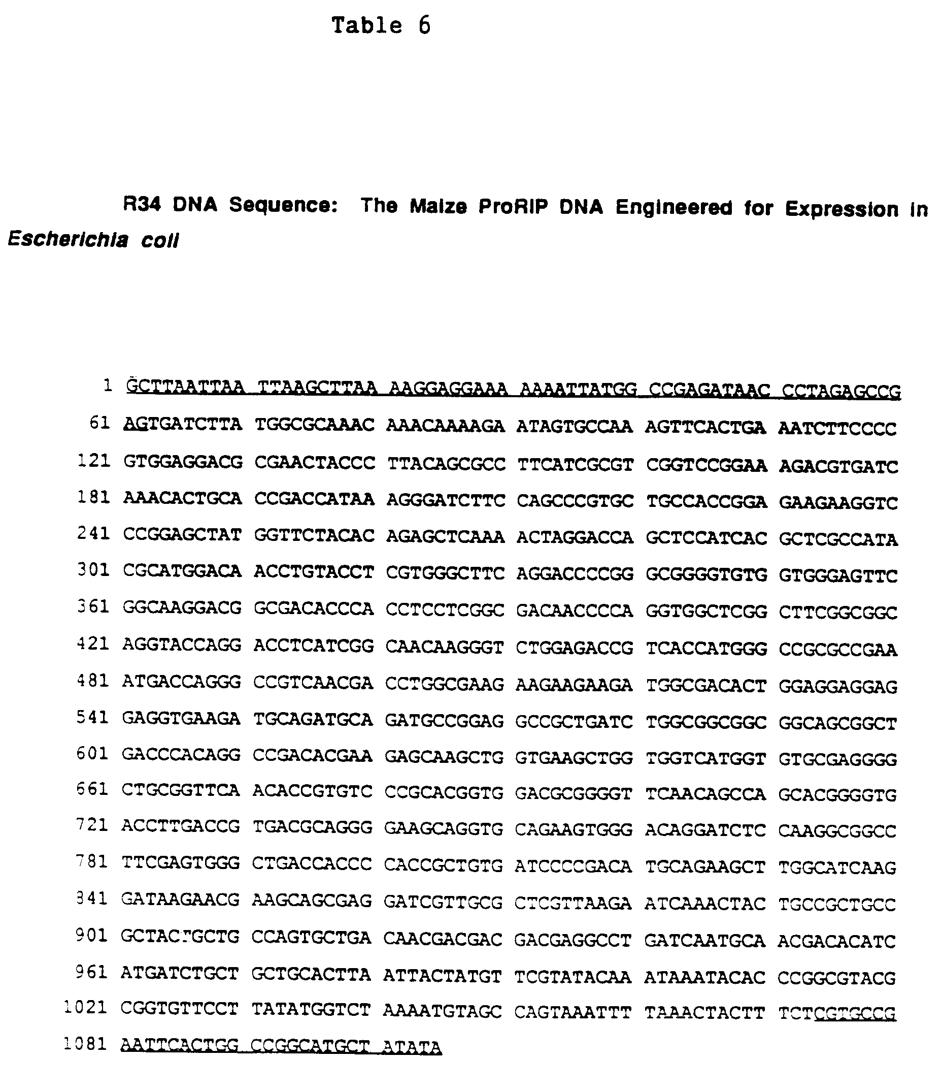
- Expression of the recombinant maize proRIP in E. coli was accomplished by engineering the cDNA via PCR amplification. A 5' primer was synthesized which contained termination codons in all three reading frames to stop translation of vector-encoded proteins upstream of the maize proRIP cDNA. The primer also contained a Shine-Dalgarno sequence several base pairs upstream of an ATG start codon followed by 23 bases which were homologous to the maize proRIP cDNA. The 3' primer spans the 3' cDNA end-pUC19 junction (the primers were shown by the underlined regions set forth in Table 6). PCR amplification of the cDNA in pUC19 using the GeneAmp™ kit (Perkin Elmer-Cetus, Norwalk, CT) yielded a predominant amplification product of approximately 1100 base pairs, as expected.
- The engineered, amplified product, was purified from an agarose gel and ligated into the filled-in Hind III site of the expression vector pGEMEX-1 (Promega Corp., Madison, WI) to give plasmid pGR, set forth below.

- This was transformed into E.coli DH5α (Bethesda Research Labs). Plasmids containing the maize proRIP cDNA were isolated by colony hybridization (see Sambrook et al., supra) with a 5' maize proRIP cDNA probe and characterized. Those containing the cDNA probe for the maize proRIP in the correct orientation were tested for expression. Plasmids were transformed into competent E. coli JM109(DE3) (Promega Corp., Madison, WI), transformed cells were grown in 15 ml cultures under ampicillin selection to an optical density at 600 nm of 0.4 to 1.0. Isopropylthio-β-galactoside (IPTG) was added to 1.3 mM to induce the production of recombinant proRIP and the cultures were grown an additional 4 hours at 37°C. The cells were collected by centrifugation and stored as a pellet at -20°C.
- The protein produced from the maize proRIP cDNA was analyzed by lysing the induced cells in TE containing 1 mg/ml lysozyme 37°C for 15 min. The lysate was fractionated into a crude supernatant and pellet by microcentrifugation. The fractions were analyzed by SDS-PAGE using 20 percent Phastgels™ reagent (Pharmacia LKB Biotechnology). Coomassie blue staining and Western blot analysis of the gels with anti-maize proRIP sera identified a 34 kD band which was greatly increased upon induction of the cells with IPTG. Cells not carrying the plasmid or containing the plasmid with the maize proRIP cDNA in the inverted orientation did not contain this 34 kD immuno-reactive band. The majority of the recombinant maize proRIP was contained in the cellular pellet suggesting the material was insoluble under these conditions.
- To test if R34 could acquire the folding pattern of N34 the pellet fraction of an induced culture was dissolved in 6M guanidine HCl and allowed to denature at room temperature for 3 hours. The material was then diluted 200-fold into ice cold TE and incubated at 4°C overnight to allow refolding of the denatured R34. The diluted material was then concentrated by a
Centricon 10 device (Amicon). To test whether refolded R34 could undergo the correct proteolytic processing to the fragmented form of the maize proRIP, the material was treated with 10 µg/ml papain for various times, and samples were analyzed by SDS-Phastgel and Western blot analysis. The R34 material was processed to a stable mixture of two immuno-reactive bands which comigrate with N34 papain-processed material indicating the correct proteolytic processing had occurred. - In an effort to simplify purification of the R34 polypeptide from induced lysates, the
gene 10 coding region of the pGEMEX-1 vector was removed by cutting the maize proRIP gene-containing plasmid (pGR) with Xba I and gel purifying the vector/proRIP DNA away from thegene 10 encoding DNA. Recircularization of pGR, now minus thegene 10 coding region, resulted in a plasmid called pGR1 set forth below.
- The plasmid pGR1 was transformed into JM109(DE3) cells and tested for production of R34 following induction with IPTG. As with pGR, large amounts of R34 were identified in cellular lysates both by Western blot and Coomassie blue staining. Unlike pGR, R34 produced from pGR1 was soluble and fractionated in the supernatant of lysed cells. This soluble material was treated with papain at 10 µg/ml and the R34 produced from pGR1 was cleaved to products which comigrate with N34, papain-cleaved product. The papain-treated material inhibited translation of reticulocyte lysates at significantly higher dilutions than the untreated material, indicating that the soluble R34 was processed to an active form.
- B. R34-DL represents the proRIP without the linker. The sequences encoding α and β were joined directly without intervening linker DNA, i.e., nucleotides A-520 to A-594 are deleted. The R34-DL gene encoded a 30.6 kD protein which was a potent inhibitor of protein synthesis. Treatment of R34-DL with papain resulted in a 28 kD polypeptide with increased ribosome inactivating activity over the untreated molecule.
- Confirmation that removal of the linker from maize proRIP activated the molecule was obtained independently through genetic engineering. The 75 bp linker encoding region of R34 (A-520 to A-594 inclusive, Table 6) was deleted using PCR amplification. The new construction R34-DL joined directly, in frame, the DNA encoding both the α and β fragments.
- In the pGEMEX-1 system the R34-DL gene directed the synthesis of a polypeptide approximately 30.6 kD, which was recognized by antisera specific for the maize proRIP. At high dilution, E.coli lysates containing R34-DL protein were potent inhibitors of protein synthesis in rabbit reticulocyte lysates, in marked contrast to E. coli lysates containing the R34 polypeptide.
- These genetic deletion data confirm that removal of the linker served to activate the R34 (proRIP) molecule. This experiment also demonstrated that covalent linkage of the α and the β polypeptide fragments resulted in an active αβ RIP. The maize proRIP did not require a break in the polypeptide backbone for enzymatic activity, removal of the linker region was sufficient to confer potent ribosome inactivating activity.
- In addition, when R34-DL lysates were treated with papain a slight decrease in the molecular weight of R34-DL protein is noted (from 30.6 kD to approximately 28 kD). The R34-DL polypeptide remained intact, that is, it was not cleaved to the characteristic maize αβ RIP α and β fragments. Associated with this small change in molecular weight was an increase in protein synthesis inhibition in the E.coli lysates. These data indicated that in bacterial lysates removal of the linker region activated the ribosomal inactivating activity of the protein at least 250-fold, but that additional processing from the ends of the protein increased the activity.
- Another genetic construction was made using PCR technology to remove the leader region from R34-DL. The new construction called R30-DL (nucleotides C-40 to C-84 and A-520 to A-594 inclusive are deleted) encoded a protein (approximately 29.5 kD) which was slightly smaller than R34-DL. E.coli lysates containing R30-DL appeared to be even more potent inhibitors of protein synthesis than R34-DL lysates. Papain treatment of R30-DL containing lysates further enhanced protein synthesis inhibiting activity. Following this treatment the R30-DL protein underwent a slight decrease in molecular weight representing processing at the ends of the polypeptide..
- A segment of the R30-DL gene was deleted which encodes several acidic residues at the carboxy terminus of the protein. The deletion was accomplished using the PCR engineering methods.
- A thermocycler (Perkin-Elmer Cetus, Norwalk, CT) was used for the indicated constructions. A typical run was done with a one minute denaturation step, 2 minute annealing, and a 3 minute extension step. Temperatures used were 94°C, 37°C or 50°C, and 72°C respectively. Following 25 cycles the reaction was held at 72°C for 7 minutes for extension of unfinished products.
- Amplification engineering reactions were done in four separate tubes of 100 µL each. The tubes were combined following amplification. Normally 100 ng of template was included in each tube. DNA primers were synthesized on a PCR Mate or 380A DNA synthesizer (Applied Biosystems) and were purified on acrylamide gels. Fifty (50) pmol of each primer were included in each reaction. The reaction conditions for the amplification were those recommended by Perkin Elmer Cetus (10 mM Tris-HCl pH8.3, 50 mM KCl, 1.5 mM MgCl2, 0.001 percent gelatin, 200 µM dNTPs and 2.5 units of Taq DNA polymerase or AmpliTaq™ thermostable DNA polymerase).
- Several methods of genetic engineering were employed to produce the genetic derivatives described below. The standard methods of DNA purification, restriction enzyme digestion, agarose gel analysis, DNA fragment isolation, ligation and transformation were as described (see Molecular Cloning: A Laboratory Manual, supra and Current Protocols in Molecular Biology, (1987), supra.
- Enzymes used for the engineering were from one of three manufacturers (Pharmacia LKB Biotechnology; Bethesda Research Labs; or New England Biolabs, Beverly, MA). Buffers and protocols used were provided by the manufacturer.
- Using PCR introduced engineering, a modified RIP fragment is amplified from an RIP plasmid template, purified, then used to replace the unmodified region in the RIP gene. All fragment replacements were done in RIP genes already inserted into a pGEMEX expression plasmid.
- A pGEMEX plasmid containing R30-DL, which had the 3' half of the gene removed by Nco I and Stu I digestion followed by gel purification was used as the template for PCR. The 3' half of the RIP gene was replaced with the PCR modified fragment described below.
- A 3' PCR primer was synthesized which encoded the 7 amino acid deletion near the carboxy terminus and introduced a new unique Bam HI site. The 5' primer directed the deletion of the αβ linker and included a Nco I site. The sequences of these primers is given in Table 7. The primers were used to amplify a modified DNA fragment from a pGEMEX R34-DL template. The amplified fragment was phenol extracted and ethanol precipitated. The insert DNA was cut with Nco I and ligated into the pGEMEX-R30-DL vector.

- The new RIP gene derivative is called RDT and encodes a protein of predicted a 28,233 Daltons and pI ∼9.5. The RDT gene encodes a protein with a truncated leader, deleted linker and truncated carboxy terminus.
- The DNA sequence for RDT is shown in Table 8.

- The RDT gene, expressed in E.coli using the pGEMEX system described above, was purified from bacterial lysates to apparent homogeneity. RDT protein appears to be a more potent inhibitor of protein synthesis than R30-DL. Using the reticulocyte lysate protein synthesis assay, purified RDT has a IC50 value of 1 ng/ml.
- RDT was further engineered to produce another gene called RDT-NP. This construction differs from RDT in having two unique restriction sites engineered into the gene. The sites were introduced using PCR methods described in Example 8. The PCR primer was designed such that it included the desired change and a unique restriction site in the maize RIP DNA sequence. A 99 bp primer which introduced the Not I and Pst I sites at the 3' end of the primer and had to be built back to the unique Nco I site for cloning purposes. The primers for the amplification are shown in Table 9.

- The restriction sites (Not I and Pst I) correspond to the site of the alpha/beta linker insertion in the RIP polypeptide. RDT-NP allows DNA segments encoding various polypeptide linkers to be inserted into the gene and tested for their ability to create an inactive, yet protease activatable RIP. The sequence for RDT-NP is shown in Table 10.
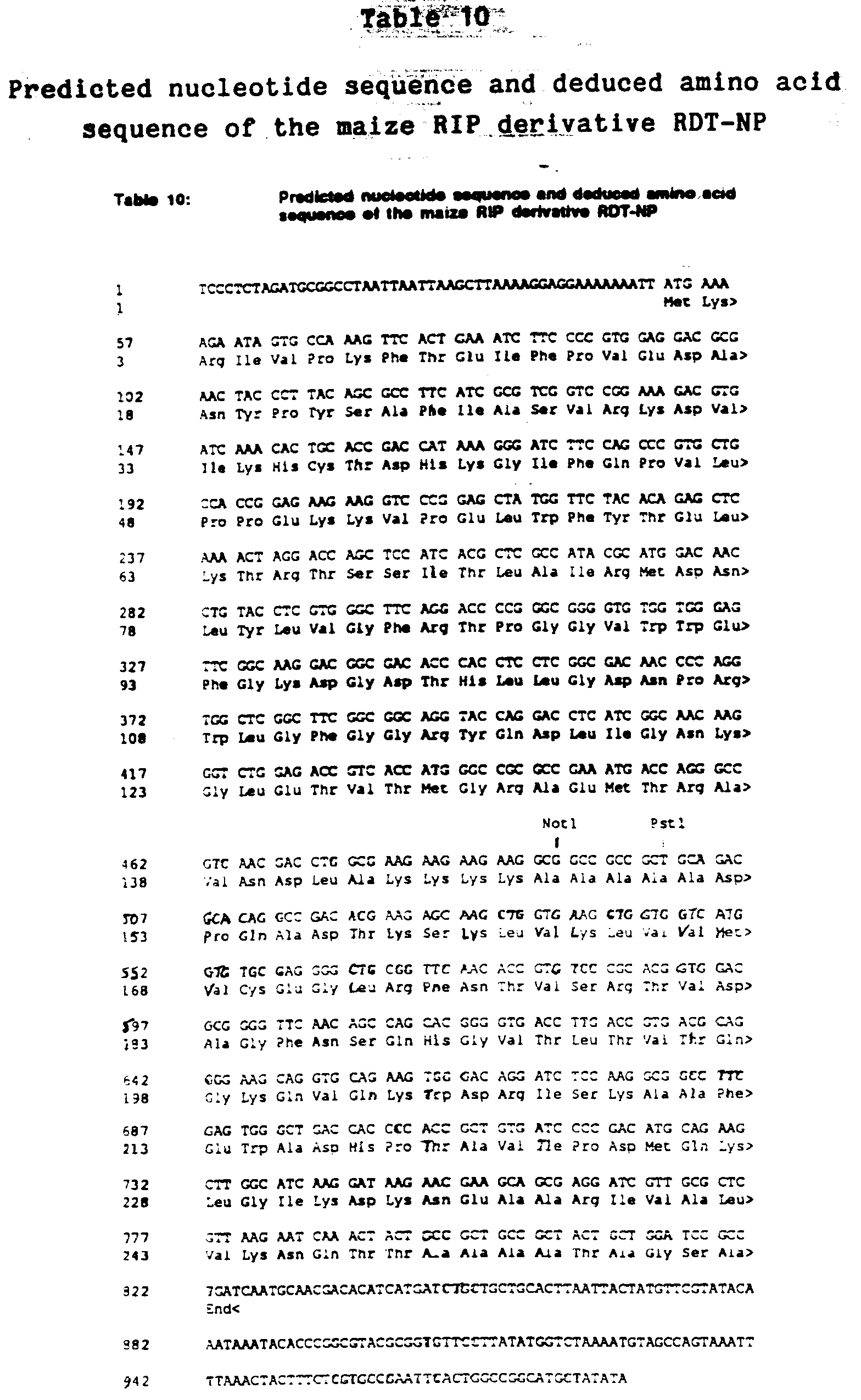
- The RDT-NP polypeptide had a predicted molecular weight of 28,446 Daltons and pI of 9.5. Crude lysates of E.coli expressing RDT-NP from a pGEMEX vector are potent inhibitors of eukaryotic protein synthesis.
- To create an RIP molecule which would bind to immunoglobulin IgG, a single Antibody Binding Region (ABR) domain from the Staphylococcus aureus antibody binding Protein A was subcloned from the plasmid pRIT5 (Pharmacia LKB Biotechnology) using PCR techniques. The antibody binding domain of protein A (ABR-A) was PCR engineered to have a Bam HI site at its 5' end and a Bgl II site at it's 3' end. This allowed insertion of the ABR-A domain into the RDT Bam HI site while retaining the unique Bam HI site. The sequence of RDT-A is shown in Table 11.

- RDT-A was expressed in E.coli cells using the pGEMEX system. The resulting polypeptide had a predicted molecular weight of 35,198 Daltons and pI of 9.2. It was recognized by antisera to both protein A and maize RIP indicating the chimeric nature of the protein. Crude lysates of bacteria expressing RDT-A had potent eukaryotic protein synthesis inhibition activity.
- RDT-A was shown to bind specifically to IgG Sepharose (Pharmacia LKB Biotechnology) following the manufacturer's instructions. Binding was best at pH 7.0. When washed at pH 5.0 the chimeric protein was released in small but detectable quantities from the resin. RDT alone does not bind to the gel.
- To increase the binding ability of the RDT-A to IgG antibodies the Antibody Binding Domain from Streptococcal Group G protein G (ABR-G) was synthesized using oligonucleotides. The sequence synthesized was that of the naturally occurring sequence described by Guss et al. ((1986), EMBO Journal, 5:1567-1575). The only change was the addition of Bam HI and Bgl II sites at the 5' and 3' ends respectively of the synthetic DNA.
- The ABR-G fragment was inserted into the Bam HI site of RDT-A. Two classes of clones have been studied. RDT-G-A contains a single ABR-G domain inserted in the correct orientation between the 3' end of RDT and the 5' end of ABR-A. A second class contains two properly oriented ABR-G domains. The predicted DNA sequences for the genes are shown in Tables 12 and 13.
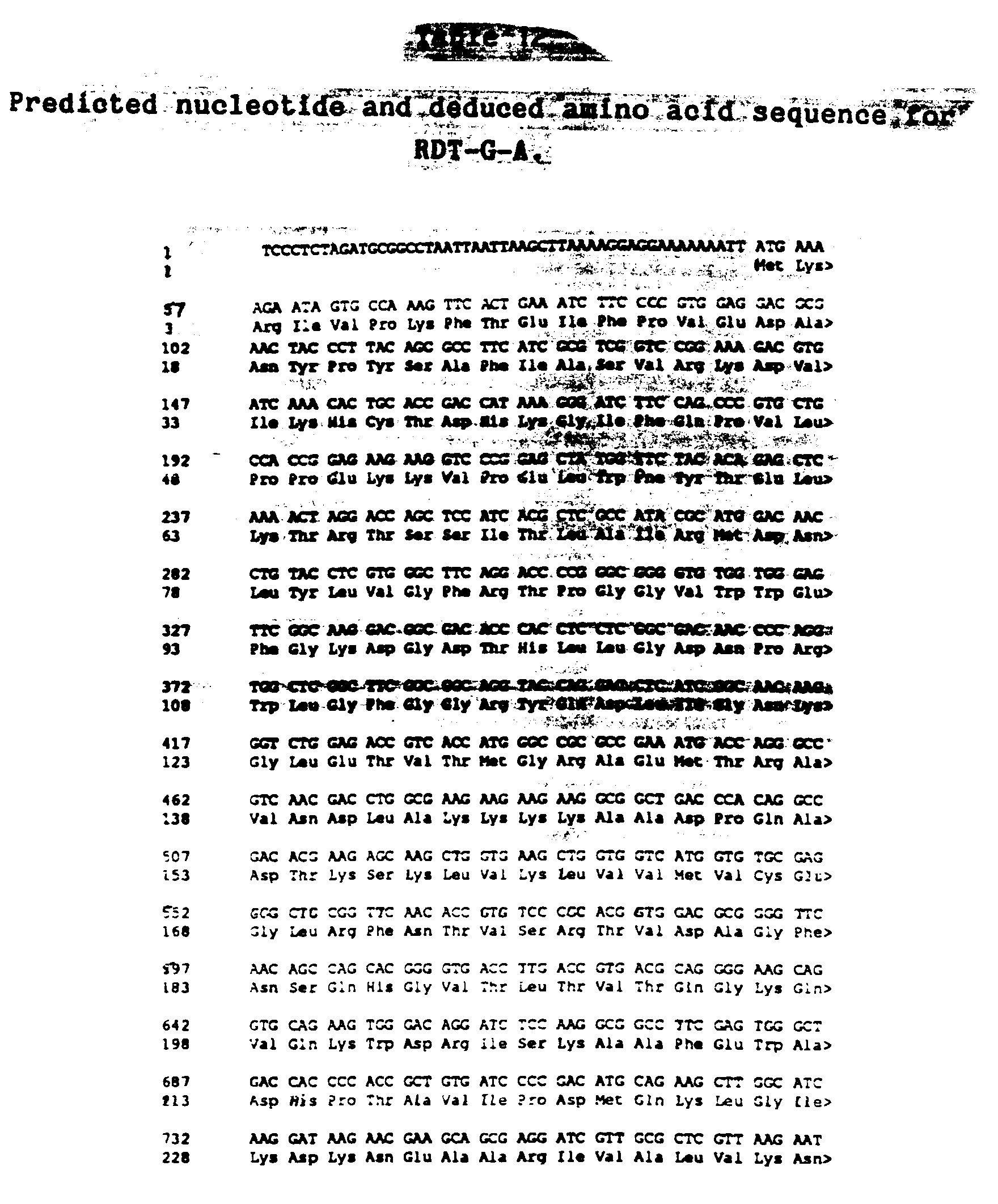
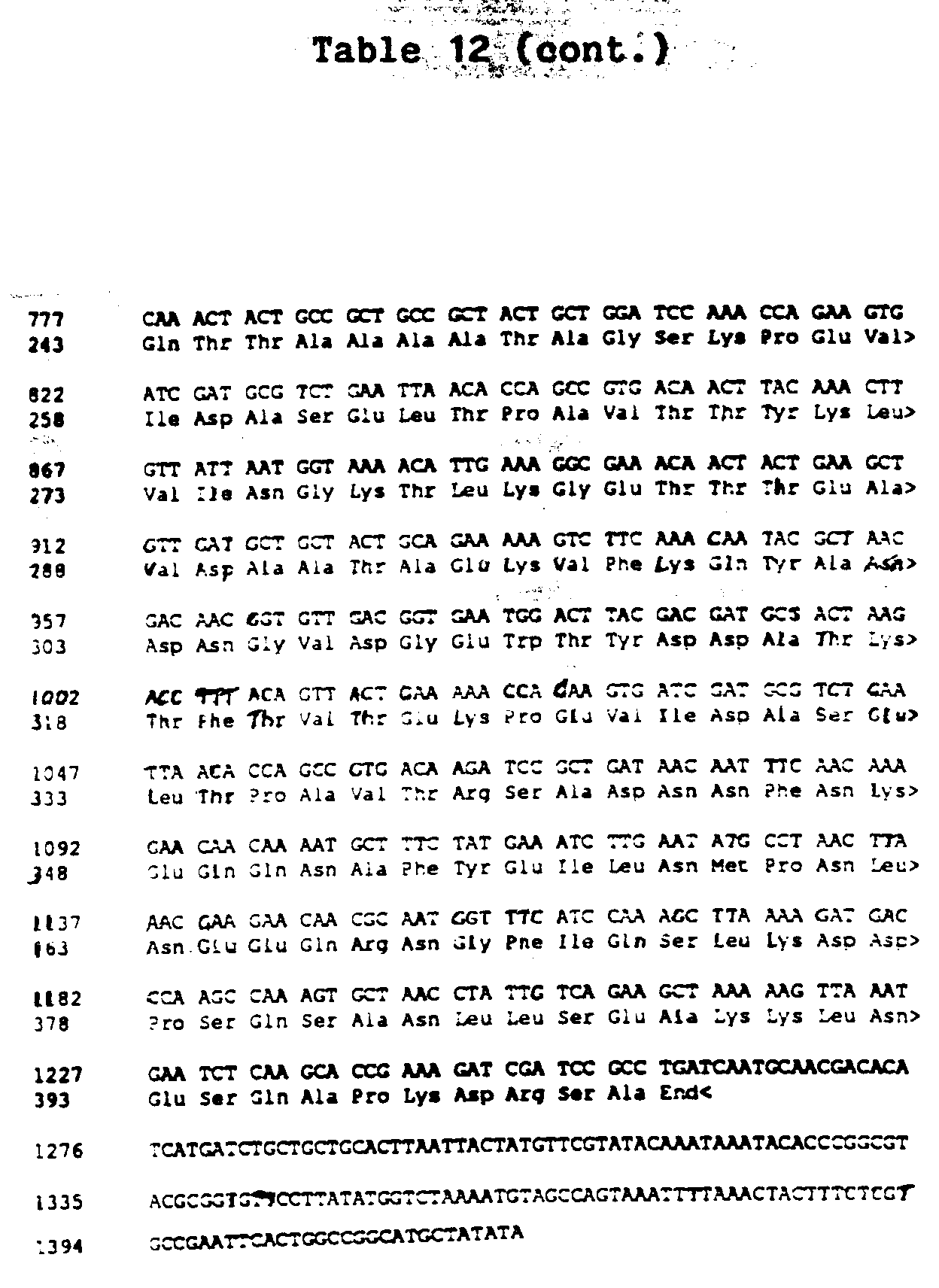

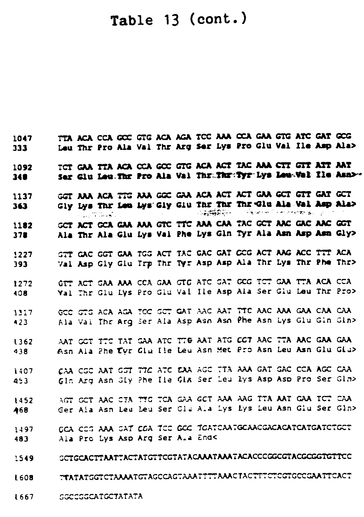
- When these genes were expressed in E.coli using the pGEMEX system the expected chimeric proteins were produced. The RDT-G-A produced a protein of predicted molecular weight 44,576 Daltons (pI 7.2). RDT-G-G-A produced a slightly larger polypeptide predicted to be 53,955 Daltons with a predicted pI of 5.4.
- Crude bacterial lysates of cells expressing RDT-G-A or RDT-G-G-A were potent inhibitors or eukaryotic protein synthesis in the rabbit reticulocyte assay described in Example 1. Papain treatment of the lysates further increases activity. Analysis of the papain treated lysates indicates that the intact RDT domain is released from the ABR domains.
- Both RDT-G-A and RDT-G-G-A bind tightly to IgG Sepharose (Pharmacia LKB Biotechnology). Binding is stable at pH 5.0 Elution was accomplished with 0.5 M ammonium acetate pH 3.5 or by boiling the resin in SDS.
- Seeds from the following species of Panicoideae were ground to a fine consistency in a mortar and pestle (after removal of the glume if necessary): Zea mays mays, Z.m. mexicana, Z.m. parviglumis, Z. luxurians, Z. mexicana, Z. mexicana, Tripsacum dactyloides, Coix lachryma-jobi, Sorghum bicolor.
- Soluble proteins were extracted from mature dry seed by the following techniques. The proteins were extracted for 2 hours with 50 mM sodium phosphate buffer, pH 7.5 containing 25 µg/ml leupeptin, 25 µg/ml antipain, 2 mM EDTA and 4 mM phenylmethane sulfonyl fluoride. Three milliliter extraction buffer per gram seed tissue was used. After centrifugation to remove insoluble material, an aliquot of the extract was analyzed by sodium dodecyl sulfate-polyacrylamide gel electrophoresis using 17-27% gradient gels, then electroblotted onto PVDF paper (Millipore). The blot was probed using rabbit antisera against maize RIP α and β fragments (1:2000 dilution each) as primary antibody and goat anti-rabbit IgG antibody as secondary antibody, then developed with NBT/BCIP.
- Except the Coix extract, all the extracts tested showed cross-reactivity with the maize RIP antisera. A prominent band at ∼34 kD was observed corresponding to proRIP, and bands at ∼16.5 and 8.5 kD were also observed, corresponding to α and β fragments respectively. This indicates that RIP forms equivalent to maize proRIP and the activated αβ form are present in many members of the subfamily Panicoideae.
- Total DNA from the following species was isolated according to the procedures described by Saghai-Maroof et al., PNAS, 81:8014-8018, 1984. The following species of Panicoideae were included: 3 accessions of Zea mays ssp.; parviglumis; Zea luxurians; Zea mays ssp. mexicana; Coix lachryma-jobi; Sorghum bicolor; and Zea mays ssp. mays var. B73.
- Generally, about 8 µg of the extracted DNA from each sample was digested to completion with 20 units of Hind III, Eco RI and Sst I in 20 microliters of a reaction mixture containing the appropriate reaction buffer at 37°C for 2 hours.
- Next, the total extracted DNA from each sample was subjected to the Southern hybridization technique (see Southern (1975), J.Mol.Biol., 98:503-517). The DNA fragments were fractionated on the basis of their size by means of electrophoresis on a 0.8% agarose gel. The double-stranded DNA fragments were modified into single-stranded DNA fragments in an alkali solution; and then a nylon filter was placed into close contact with the gel to transfer the modified DNA segments onto the filter in the presence of a high salt concentration solution.
- Hybridization was carried out using, as the probe, the cDNA clone of the RIP gene (; the probe fragment is provided from position 2 to 1075 in Table 1). The probe was radiolabeled with 32P and the signals in Southern transfers were visualized by autoradiography.
- The Southern blots were hybridized with the cDNA clone of the RIP gene. A single fragment with strong homology to the clone was observed for each enzyme/species combination except the Coix accession. The inbred line of maize has a single major band with two minor bands. The other species which, with the exception of Sorghum, are not inbred showed between 2 and 5 minor bands. The Coix had either 4 or 5 such bands depending on the enzyme used.
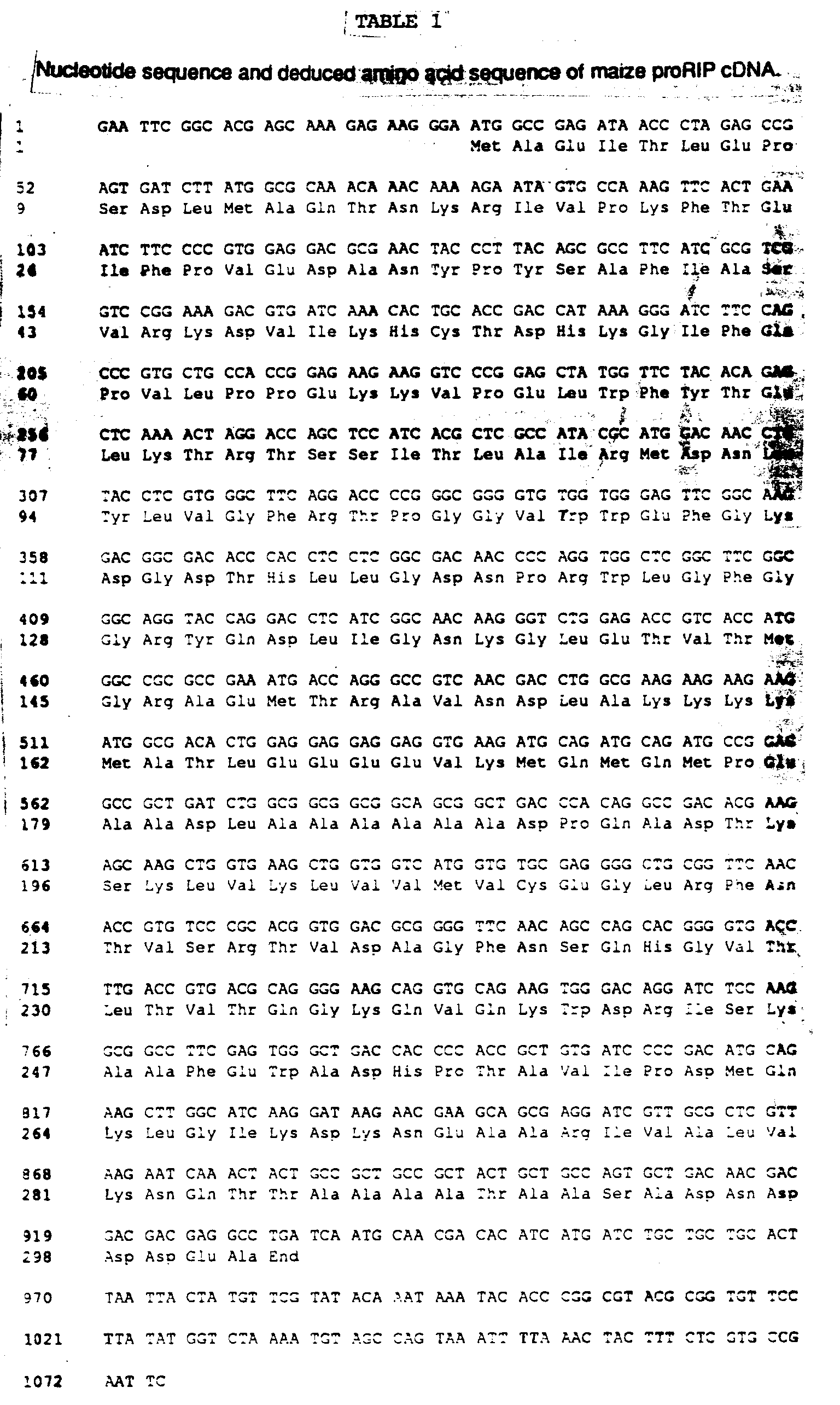
Claims (10)
- A DNA isolate encoding a maize proRIP having a ribosome inactivating protein sequence and a linker sequence wherein the maize proRIP has the following amino acid sequence:

- A DNA isolate encoding a protein capable of substantially inactivating eukaryotic ribosomes, said protein having the following amino acid sequence:

- A biologically functional expression vehicle containing a DNA isolate of claims 1, 2, 7, 8, 9, or 10.
- A host cell transformed with the biologically functional expression vehicle of claim 3.
- The transformed host cell of claim 4, wherein the host cell is a eukaryotic cell.
- The host cell of claim 5, wherein the host cell is maize.
- A DNA isolate encoding a protein capable of substantially inactivating eukaryotic ribosomes, said protein having the following amino acid sequence:

- The DNA isolate of claim 1 designated R34 as shown in Table 6.
- The DNA isolate of claim 2 designated R34-DL as shown in Example 7B.
- The DNA isolate of claim 7 designated as R30-DL as shown in Example 7B.
Applications Claiming Priority (2)
| Application Number | Priority Date | Filing Date | Title |
|---|---|---|---|
| US07/535,636 US5248606A (en) | 1990-06-11 | 1990-06-11 | Dna encoding inactive precursor and active forms of maize ribosome inactivating protein |
| US535636 | 1990-06-11 |
Publications (2)
| Publication Number | Publication Date |
|---|---|
| EP0466222A1 EP0466222A1 (en) | 1992-01-15 |
| EP0466222B1 true EP0466222B1 (en) | 1999-09-08 |
Family
ID=24135102
Family Applications (1)
| Application Number | Title | Priority Date | Filing Date |
|---|---|---|---|
| EP91201436A Expired - Lifetime EP0466222B1 (en) | 1990-06-11 | 1991-06-10 | Ribosome-inactivating proteins, inactive precursor forms thereof, a process for making and a method of using |
Country Status (10)
| Country | Link |
|---|---|
| US (1) | US5248606A (en) |
| EP (1) | EP0466222B1 (en) |
| JP (1) | JPH04279599A (en) |
| CN (1) | CN1062172A (en) |
| AT (1) | ATE184289T1 (en) |
| AU (1) | AU638133B2 (en) |
| BR (1) | BR9102418A (en) |
| CA (1) | CA2044201A1 (en) |
| DE (1) | DE69131589T2 (en) |
| HU (1) | HUT58800A (en) |
Cited By (2)
| Publication number | Priority date | Publication date | Assignee | Title |
|---|---|---|---|---|
| WO2002101036A1 (en) * | 2001-05-11 | 2002-12-19 | Kazunari Taira | Method of forming stable complex of transcription product of dna encoding arbitrary peptide with translation product, nucleic acid construct to be used in the method, complex formed by the method, and screening of functional protein and mrna or dna encoding the protein using the method |
| RU2285045C2 (en) * | 2000-10-14 | 2006-10-10 | Эдванст Технолоджиз (Кембридж) Лимитед | System for eradication of vegetable cells |
Families Citing this family (32)
| Publication number | Priority date | Publication date | Assignee | Title |
|---|---|---|---|---|
| US5672683A (en) * | 1989-09-07 | 1997-09-30 | Alkermes, Inc. | Transferrin neuropharmaceutical agent fusion protein |
| US5635384A (en) * | 1990-06-11 | 1997-06-03 | Dowelanco | Ribosome-inactivating proteins, inactive precursor forms thereof, a process for making and a method of using |
| DE4040954C2 (en) * | 1990-12-20 | 2001-05-17 | Max Planck Gesellschaft | Process for the production of pathogen-resistant plants |
| US6071895A (en) * | 1992-03-11 | 2000-06-06 | Narhex Limited | Polar-substituted hydrocarbons |
| US5888992A (en) * | 1992-03-11 | 1999-03-30 | Narhex Limited | Polar substituted hydrocarbons |
| DE69333270T2 (en) | 1992-03-11 | 2004-08-05 | Narhex Ltd. | AMINE DERIVATIVES OF OXO AND HYDROXY SUBSTITUTED CARBON HYDROGEN |
| JPH07504654A (en) * | 1992-03-11 | 1995-05-25 | ナルヘックス リミテッド | Amine derivatives of oxo- and hydroxy-substituted hydrocarbons |
| GB9304905D0 (en) * | 1992-03-16 | 1993-04-28 | Zeneca Ltd | Processes |
| US5916772A (en) * | 1992-06-16 | 1999-06-29 | Whittier Institute For Diabetes And Endocrinology | Recombinant production of saporin-containing proteins |
| US5332808A (en) * | 1992-09-08 | 1994-07-26 | North Carolina State University | DNA encoding a ribosome inactivating protein |
| KR970001565B1 (en) * | 1993-08-28 | 1997-02-11 | 장기하 | Encoding dna for phytolacca insularis antiviral protein and its expression microorganism |
| EG23907A (en) * | 1994-08-01 | 2007-12-30 | Delta & Pine Land Co | Control of plant gene expression |
| JPH0856655A (en) * | 1994-08-16 | 1996-03-05 | Frontier:Kk | Cell lethal agent and cell death inhibitor |
| DE69734656T2 (en) * | 1996-04-30 | 2006-08-10 | Twinstrand Therapeutics Inc., Burnaby | ANTIVIRAL VARIANT OF RICIN PROTEINS |
| EP0977862B8 (en) | 1997-04-30 | 2009-09-16 | Twinstrand Therapeutics Inc. | Ricin-like toxin variants for treatment of cancer, viral or parasitic infections |
| US7247715B2 (en) | 1997-04-30 | 2007-07-24 | Twinstrand Therapeutics Inc. | Ricin-like toxin variants for treatment of cancer, viral or parasitic infections |
| WO1999003492A1 (en) * | 1997-07-16 | 1999-01-28 | Sophie Chen | Antiviral and antitumor agents |
| WO2000039291A1 (en) * | 1998-12-31 | 2000-07-06 | Rutgers, The State University | Virus-resistant transgenic plants expressing l3 |
| US6531125B1 (en) | 1999-03-02 | 2003-03-11 | Twinstrand Therapeutics Inc. | Antiviral ricin-like proteins |
| CA2386237A1 (en) | 1999-10-04 | 2001-04-12 | Twinstrand Therapeutics Inc. | Improved ricin-like toxins for treatment of cancer |
| AU2006201828B2 (en) * | 2000-10-14 | 2009-07-16 | British American Tobacco (Investments) Limited | Plant cell death system |
| GB0025225D0 (en) * | 2000-10-14 | 2000-11-29 | Cambridge Advanced Tech | Plant cell death system |
| US7629309B2 (en) | 2002-05-29 | 2009-12-08 | Zystor Therapeutics, Inc. | Targeted therapeutic proteins |
| US7560424B2 (en) | 2001-04-30 | 2009-07-14 | Zystor Therapeutics, Inc. | Targeted therapeutic proteins |
| US20030072761A1 (en) | 2001-10-16 | 2003-04-17 | Lebowitz Jonathan | Methods and compositions for targeting proteins across the blood brain barrier |
| CA2553955C (en) | 2004-02-10 | 2012-08-28 | Zystor Therapeutics, Inc. | Acid alpha-glucosidase and fragments thereof |
| ES2629853T3 (en) | 2008-05-07 | 2017-08-16 | Biomarin Pharmaceutical Inc. | Lysosomal direction peptides and uses thereof |
| EP2475376B1 (en) | 2009-06-17 | 2016-03-30 | BioMarin Pharmaceutical Inc. | Formulations for lysosomal enzymes |
| CN102234638B (en) * | 2010-04-27 | 2013-03-27 | 香港中文大学 | Modified corn ribosome inactivating protein precursor and application thereof and protein purification method |
| US10347262B2 (en) | 2017-10-18 | 2019-07-09 | The Nielsen Company (Us), Llc | Systems and methods to improve timestamp transition resolution |
| EP3758686B1 (en) * | 2018-02-26 | 2024-11-06 | R.P. Scherer Technologies, LLC | Pharmaceutical dosage form for an emulsion of simethicone and loperamide |
| CN110455903A (en) * | 2018-05-07 | 2019-11-15 | 天士力生物医药股份有限公司 | A kind of gradient electrophoresis detection method of injection recombinant human urokinase zymogen impurity analysis |
Citations (1)
| Publication number | Priority date | Publication date | Assignee | Title |
|---|---|---|---|---|
| EP0335476A2 (en) * | 1984-02-08 | 1989-10-04 | Cetus Corporation | Recombinant methods for the production of ricin a, ricin b, ricin or diphtheria toxin (dt)a or ab' fragment, suitable hosts and vectors therefor, and conjugates comprising ricin toxin a chain or diphtheria toxin |
Family Cites Families (7)
| Publication number | Priority date | Publication date | Assignee | Title |
|---|---|---|---|---|
| ATE87318T1 (en) * | 1985-11-29 | 1993-04-15 | Consolidated Pharmaceuticals L | RICIN-ANTIBODY CONJUGATES. |
| FR2601679B1 (en) * | 1986-07-15 | 1990-05-25 | Sanofi Sa | IMMUNOTOXINS, PREPARATION METHOD AND PHARMACEUTICAL COMPOSITIONS CONTAINING SAME |
| DE3724323C2 (en) * | 1986-07-29 | 1995-07-20 | Erba Farmitalia | A new inhibitor for protein synthesis |
| US4869903A (en) * | 1987-05-29 | 1989-09-26 | Genelabs Incorporated | Method of selectively inhibiting HIV |
| GB8801877D0 (en) * | 1988-01-28 | 1988-02-24 | Erba Carlo Spa | Nucleotide sequence encoding plant ribosome inactivating protein |
| IT1229216B (en) * | 1989-03-31 | 1991-07-25 | Italfarmaco Spa | PROTEINS INHIBIT RIBOSOMES AND THEIR DERIVATIVES. |
| AU654563B2 (en) * | 1991-07-24 | 1994-11-10 | Imperial Chemical Industries Plc | Proteins |
-
1990
- 1990-06-11 US US07/535,636 patent/US5248606A/en not_active Expired - Fee Related
-
1991
- 1991-06-10 DE DE69131589T patent/DE69131589T2/en not_active Expired - Lifetime
- 1991-06-10 AT AT91201436T patent/ATE184289T1/en not_active IP Right Cessation
- 1991-06-10 CA CA002044201A patent/CA2044201A1/en not_active Abandoned
- 1991-06-10 EP EP91201436A patent/EP0466222B1/en not_active Expired - Lifetime
- 1991-06-11 AU AU78329/91A patent/AU638133B2/en not_active Ceased
- 1991-06-11 HU HU911944A patent/HUT58800A/en unknown
- 1991-06-11 JP JP3139171A patent/JPH04279599A/en active Pending
- 1991-06-11 CN CN91104857A patent/CN1062172A/en active Pending
- 1991-06-11 BR BR919102418A patent/BR9102418A/en unknown
Patent Citations (1)
| Publication number | Priority date | Publication date | Assignee | Title |
|---|---|---|---|---|
| EP0335476A2 (en) * | 1984-02-08 | 1989-10-04 | Cetus Corporation | Recombinant methods for the production of ricin a, ricin b, ricin or diphtheria toxin (dt)a or ab' fragment, suitable hosts and vectors therefor, and conjugates comprising ricin toxin a chain or diphtheria toxin |
Non-Patent Citations (1)
| Title |
|---|
| W.H. Coleman and W.K. Roberts, Biochim. Biophys. Acta 1982, Vol. 696, pages 239-244 * |
Cited By (2)
| Publication number | Priority date | Publication date | Assignee | Title |
|---|---|---|---|---|
| RU2285045C2 (en) * | 2000-10-14 | 2006-10-10 | Эдванст Технолоджиз (Кембридж) Лимитед | System for eradication of vegetable cells |
| WO2002101036A1 (en) * | 2001-05-11 | 2002-12-19 | Kazunari Taira | Method of forming stable complex of transcription product of dna encoding arbitrary peptide with translation product, nucleic acid construct to be used in the method, complex formed by the method, and screening of functional protein and mrna or dna encoding the protein using the method |
Also Published As
| Publication number | Publication date |
|---|---|
| CA2044201A1 (en) | 1991-12-12 |
| HUT58800A (en) | 1992-03-30 |
| EP0466222A1 (en) | 1992-01-15 |
| DE69131589D1 (en) | 1999-10-14 |
| AU638133B2 (en) | 1993-06-17 |
| BR9102418A (en) | 1992-01-14 |
| US5248606A (en) | 1993-09-28 |
| AU7832991A (en) | 1991-12-12 |
| CN1062172A (en) | 1992-06-24 |
| HU911944D0 (en) | 1991-12-30 |
| JPH04279599A (en) | 1992-10-05 |
| ATE184289T1 (en) | 1999-09-15 |
| DE69131589T2 (en) | 2000-05-04 |
Similar Documents
| Publication | Publication Date | Title |
|---|---|---|
| EP0466222A1 (en) | Ribosome-inactivating proteins, inactive precursor forms thereof, a process for making and a method of using | |
| US5635384A (en) | Ribosome-inactivating proteins, inactive precursor forms thereof, a process for making and a method of using | |
| CA1341428C (en) | Synthetic insecticidal crystal protein gene | |
| US8871468B2 (en) | Synthetic genes for plant gums and other hydroxyproline-rich glycoproteins | |
| AU679427B2 (en) | DNA sequences encoding gelonin polypeptide | |
| EP0335476A2 (en) | Recombinant methods for the production of ricin a, ricin b, ricin or diphtheria toxin (dt)a or ab' fragment, suitable hosts and vectors therefor, and conjugates comprising ricin toxin a chain or diphtheria toxin | |
| US5942663A (en) | Biocidal proteins | |
| AU746826B2 (en) | Production of mature proteins in plants | |
| NZ287124A (en) | Hybrid antibody-toxin conjugate and antibodies to insect gut | |
| Lamy et al. | The Aspergillus ribonucleolytic toxins (ribotoxins) | |
| IE911870A1 (en) | New antifungal preparations, process for making such¹preparations and process for obtaining plants with decreased¹susceptibility to fungi | |
| US5824870A (en) | Commercial production of aprotinin in plants | |
| JPH0714845B2 (en) | Method to enhance insect toxin effect | |
| Nolan et al. | Cloning and expression of a gene encoding gelonin, a ribosome-inactivating protein from Gelonium multiflorum | |
| WO1995011977A2 (en) | A NEW RIBOSOME-INACTIVATING PROTEIN ISOLATED FROM THE PLANT $i(BRYONIA DIOICA) | |
| US5629469A (en) | Thiol protease inhibitor | |
| CA2110403A1 (en) | Biocidal proteins | |
| JPH06501396A (en) | Isolation and characterization of a novel protease from Streptomyces lividans | |
| Westby et al. | Preparation and characterization of recombinant proricin containing an alternative protease-sensitive linker sequence | |
| Dore et al. | Mutations dissociating the inhibitory activity of the pokeweed antiviral portein on eukaryote translation and Escherichia coli growth | |
| US5648234A (en) | Expression vector for Phytolacca antiviral protein | |
| Xu et al. | The Low Expression Level of Pokeweed Antiviral Protein (PAP) Gene inEscherichia coliby the InduciblelacPromoter Is Due to Inefficient Transcription and Translation and Not to the Toxicity of the PAP | |
| JPS62232387A (en) | Recombinant ricin toxin | |
| JPS60188070A (en) | Dna coding signal peptide, and dna containing the same | |
| Sinclair | Saporins-Type 1 Ribosome-inactivating proteins from soapwort (saponaria officinalis L.) |
Legal Events
| Date | Code | Title | Description |
|---|---|---|---|
| PUAI | Public reference made under article 153(3) epc to a published international application that has entered the european phase |
Free format text: ORIGINAL CODE: 0009012 |
|
| AK | Designated contracting states |
Kind code of ref document: A1 Designated state(s): AT BE CH DE DK ES FR GB GR IT LI LU NL SE |
|
| K1C3 | Correction of patent application (complete document) published |
Effective date: 19920115 |
|
| 17P | Request for examination filed |
Effective date: 19920714 |
|
| 17Q | First examination report despatched |
Effective date: 19950725 |
|
| GRAG | Despatch of communication of intention to grant |
Free format text: ORIGINAL CODE: EPIDOS AGRA |
|
| RAP1 | Party data changed (applicant data changed or rights of an application transferred) |
Owner name: DOW AGROSCIENCES LLC |
|
| GRAG | Despatch of communication of intention to grant |
Free format text: ORIGINAL CODE: EPIDOS AGRA |
|
| GRAH | Despatch of communication of intention to grant a patent |
Free format text: ORIGINAL CODE: EPIDOS IGRA |
|
| GRAH | Despatch of communication of intention to grant a patent |
Free format text: ORIGINAL CODE: EPIDOS IGRA |
|
| GRAA | (expected) grant |
Free format text: ORIGINAL CODE: 0009210 |
|
| AK | Designated contracting states |
Kind code of ref document: B1 Designated state(s): AT BE CH DE DK ES FR GB GR IT LI LU NL SE |
|
| PG25 | Lapsed in a contracting state [announced via postgrant information from national office to epo] |
Ref country code: SE Free format text: THE PATENT HAS BEEN ANNULLED BY A DECISION OF A NATIONAL AUTHORITY Effective date: 19990908 Ref country code: GR Free format text: LAPSE BECAUSE OF NON-PAYMENT OF DUE FEES Effective date: 19990908 Ref country code: BE Free format text: LAPSE BECAUSE OF FAILURE TO SUBMIT A TRANSLATION OF THE DESCRIPTION OR TO PAY THE FEE WITHIN THE PRESCRIBED TIME-LIMIT Effective date: 19990908 Ref country code: AT Free format text: LAPSE BECAUSE OF FAILURE TO SUBMIT A TRANSLATION OF THE DESCRIPTION OR TO PAY THE FEE WITHIN THE PRESCRIBED TIME-LIMIT Effective date: 19990908 Ref country code: CH Free format text: LAPSE BECAUSE OF FAILURE TO SUBMIT A TRANSLATION OF THE DESCRIPTION OR TO PAY THE FEE WITHIN THE PRESCRIBED TIME-LIMIT Effective date: 19990908 Ref country code: ES Free format text: THE PATENT HAS BEEN ANNULLED BY A DECISION OF A NATIONAL AUTHORITY Effective date: 19990908 Ref country code: NL Free format text: LAPSE BECAUSE OF FAILURE TO SUBMIT A TRANSLATION OF THE DESCRIPTION OR TO PAY THE FEE WITHIN THE PRESCRIBED TIME-LIMIT Effective date: 19990908 Ref country code: LI Free format text: LAPSE BECAUSE OF FAILURE TO SUBMIT A TRANSLATION OF THE DESCRIPTION OR TO PAY THE FEE WITHIN THE PRESCRIBED TIME-LIMIT Effective date: 19990908 |
|
| REF | Corresponds to: |
Ref document number: 184289 Country of ref document: AT Date of ref document: 19990915 Kind code of ref document: T |
|
| REG | Reference to a national code |
Ref country code: CH Ref legal event code: EP |
|
| REF | Corresponds to: |
Ref document number: 69131589 Country of ref document: DE Date of ref document: 19991014 |
|
| PG25 | Lapsed in a contracting state [announced via postgrant information from national office to epo] |
Ref country code: DK Free format text: LAPSE BECAUSE OF FAILURE TO SUBMIT A TRANSLATION OF THE DESCRIPTION OR TO PAY THE FEE WITHIN THE PRESCRIBED TIME-LIMIT Effective date: 19991208 |
|
| ITF | It: translation for a ep patent filed | ||
| ET | Fr: translation filed | ||
| NLV1 | Nl: lapsed or annulled due to failure to fulfill the requirements of art. 29p and 29m of the patents act | ||
| REG | Reference to a national code |
Ref country code: CH Ref legal event code: PL |
|
| PG25 | Lapsed in a contracting state [announced via postgrant information from national office to epo] |
Ref country code: LU Free format text: LAPSE BECAUSE OF NON-PAYMENT OF DUE FEES Effective date: 20000610 |
|
| PLBE | No opposition filed within time limit |
Free format text: ORIGINAL CODE: 0009261 |
|
| STAA | Information on the status of an ep patent application or granted ep patent |
Free format text: STATUS: NO OPPOSITION FILED WITHIN TIME LIMIT |
|
| 26N | No opposition filed | ||
| REG | Reference to a national code |
Ref country code: GB Ref legal event code: IF02 |
|
| PGFP | Annual fee paid to national office [announced via postgrant information from national office to epo] |
Ref country code: FR Payment date: 20100709 Year of fee payment: 20 |
|
| PGFP | Annual fee paid to national office [announced via postgrant information from national office to epo] |
Ref country code: IT Payment date: 20100619 Year of fee payment: 20 |
|
| PGFP | Annual fee paid to national office [announced via postgrant information from national office to epo] |
Ref country code: GB Payment date: 20100609 Year of fee payment: 20 Ref country code: DE Payment date: 20100602 Year of fee payment: 20 |
|
| REG | Reference to a national code |
Ref country code: DE Ref legal event code: R071 Ref document number: 69131589 Country of ref document: DE |
|
| REG | Reference to a national code |
Ref country code: DE Ref legal event code: R071 Ref document number: 69131589 Country of ref document: DE |
|
| REG | Reference to a national code |
Ref country code: GB Ref legal event code: PE20 Expiry date: 20110609 |
|
| PG25 | Lapsed in a contracting state [announced via postgrant information from national office to epo] |
Ref country code: GB Free format text: LAPSE BECAUSE OF EXPIRATION OF PROTECTION Effective date: 20110609 |
|
| PG25 | Lapsed in a contracting state [announced via postgrant information from national office to epo] |
Ref country code: DE Free format text: LAPSE BECAUSE OF EXPIRATION OF PROTECTION Effective date: 20110611 |