EP0426300B1 - Method of producing a reagent containing a narrow distribution of colloidal particles of a selected size and the use thereof - Google Patents
Method of producing a reagent containing a narrow distribution of colloidal particles of a selected size and the use thereof Download PDFInfo
- Publication number
- EP0426300B1 EP0426300B1 EP90310671A EP90310671A EP0426300B1 EP 0426300 B1 EP0426300 B1 EP 0426300B1 EP 90310671 A EP90310671 A EP 90310671A EP 90310671 A EP90310671 A EP 90310671A EP 0426300 B1 EP0426300 B1 EP 0426300B1
- Authority
- EP
- European Patent Office
- Prior art keywords
- agent
- reducing agent
- particles
- gold
- sol
- Prior art date
- Legal status (The legal status is an assumption and is not a legal conclusion. Google has not performed a legal analysis and makes no representation as to the accuracy of the status listed.)
- Expired - Lifetime
Links
- 239000002245 particle Substances 0.000 title claims abstract description 118
- 238000000034 method Methods 0.000 title claims abstract description 77
- 239000003153 chemical reaction reagent Substances 0.000 title abstract description 26
- 238000009826 distribution Methods 0.000 title abstract description 12
- PCHJSUWPFVWCPO-UHFFFAOYSA-N gold Chemical compound [Au] PCHJSUWPFVWCPO-UHFFFAOYSA-N 0.000 claims abstract description 93
- 229910052737 gold Inorganic materials 0.000 claims abstract description 70
- 239000010931 gold Substances 0.000 claims abstract description 70
- 239000003638 chemical reducing agent Substances 0.000 claims abstract description 45
- 229910052751 metal Inorganic materials 0.000 claims abstract description 42
- 239000002184 metal Substances 0.000 claims abstract description 42
- 239000011248 coating agent Substances 0.000 claims abstract description 28
- 238000000576 coating method Methods 0.000 claims abstract description 28
- AVXURJPOCDRRFD-UHFFFAOYSA-N Hydroxylamine Chemical compound ON AVXURJPOCDRRFD-UHFFFAOYSA-N 0.000 claims abstract description 23
- 238000003018 immunoassay Methods 0.000 claims abstract description 17
- 239000003795 chemical substances by application Substances 0.000 claims abstract description 14
- 239000003381 stabilizer Substances 0.000 claims abstract description 14
- 230000012010 growth Effects 0.000 claims abstract description 13
- 235000013336 milk Nutrition 0.000 claims abstract description 13
- 239000008267 milk Substances 0.000 claims abstract description 13
- 210000004080 milk Anatomy 0.000 claims abstract description 13
- 239000001509 sodium citrate Substances 0.000 claims abstract description 10
- 239000000463 material Substances 0.000 claims abstract description 9
- NLJMYIDDQXHKNR-UHFFFAOYSA-K sodium citrate Chemical compound O.O.[Na+].[Na+].[Na+].[O-]C(=O)CC(O)(CC([O-])=O)C([O-])=O NLJMYIDDQXHKNR-UHFFFAOYSA-K 0.000 claims abstract description 8
- 230000000415 inactivating effect Effects 0.000 claims abstract 2
- 239000000243 solution Substances 0.000 claims description 32
- 108091003079 Bovine Serum Albumin Proteins 0.000 claims description 22
- 229940098773 bovine serum albumin Drugs 0.000 claims description 22
- KRKNYBCHXYNGOX-UHFFFAOYSA-K Citrate Chemical group [O-]C(=O)CC(O)(CC([O-])=O)C([O-])=O KRKNYBCHXYNGOX-UHFFFAOYSA-K 0.000 claims description 14
- MUBZPKHOEPUJKR-UHFFFAOYSA-N Oxalic acid Chemical compound OC(=O)C(O)=O MUBZPKHOEPUJKR-UHFFFAOYSA-N 0.000 claims description 13
- BJEPYKJPYRNKOW-UHFFFAOYSA-N malic acid Chemical compound OC(=O)C(O)CC(O)=O BJEPYKJPYRNKOW-UHFFFAOYSA-N 0.000 claims description 12
- 150000003839 salts Chemical class 0.000 claims description 12
- 230000002269 spontaneous effect Effects 0.000 claims description 12
- 239000012131 assay buffer Substances 0.000 claims description 11
- OAKJQQAXSVQMHS-UHFFFAOYSA-N Hydrazine Chemical compound NN OAKJQQAXSVQMHS-UHFFFAOYSA-N 0.000 claims description 10
- KRKNYBCHXYNGOX-UHFFFAOYSA-N citric acid Chemical compound OC(=O)CC(O)(C(O)=O)CC(O)=O KRKNYBCHXYNGOX-UHFFFAOYSA-N 0.000 claims description 9
- 238000004519 manufacturing process Methods 0.000 claims description 9
- 239000012279 sodium borohydride Substances 0.000 claims description 9
- 229910000033 sodium borohydride Inorganic materials 0.000 claims description 9
- MHAJPDPJQMAIIY-UHFFFAOYSA-N Hydrogen peroxide Chemical compound OO MHAJPDPJQMAIIY-UHFFFAOYSA-N 0.000 claims description 8
- 235000013861 fat-free Nutrition 0.000 claims description 8
- CIWBSHSKHKDKBQ-JLAZNSOCSA-N Ascorbic acid Chemical compound OC[C@H](O)[C@H]1OC(=O)C(O)=C1O CIWBSHSKHKDKBQ-JLAZNSOCSA-N 0.000 claims description 7
- WHUUTDBJXJRKMK-VKHMYHEASA-N L-glutamic acid Chemical compound OC(=O)[C@@H](N)CCC(O)=O WHUUTDBJXJRKMK-VKHMYHEASA-N 0.000 claims description 7
- BJEPYKJPYRNKOW-REOHCLBHSA-N (S)-malic acid Chemical compound OC(=O)[C@@H](O)CC(O)=O BJEPYKJPYRNKOW-REOHCLBHSA-N 0.000 claims description 6
- -1 Ba2+ ion Chemical class 0.000 claims description 6
- CKLJMWTZIZZHCS-REOHCLBHSA-N L-aspartic acid Chemical compound OC(=O)[C@@H](N)CC(O)=O CKLJMWTZIZZHCS-REOHCLBHSA-N 0.000 claims description 6
- 239000011668 ascorbic acid Substances 0.000 claims description 6
- 235000010323 ascorbic acid Nutrition 0.000 claims description 6
- 229960005070 ascorbic acid Drugs 0.000 claims description 6
- 230000007159 enucleation Effects 0.000 claims description 6
- 229940049920 malate Drugs 0.000 claims description 6
- 239000001630 malic acid Substances 0.000 claims description 6
- 235000011090 malic acid Nutrition 0.000 claims description 6
- FBPFZTCFMRRESA-KVTDHHQDSA-N D-Mannitol Chemical compound OC[C@@H](O)[C@@H](O)[C@H](O)[C@H](O)CO FBPFZTCFMRRESA-KVTDHHQDSA-N 0.000 claims description 5
- WTDHULULXKLSOZ-UHFFFAOYSA-N Hydroxylamine hydrochloride Chemical compound Cl.ON WTDHULULXKLSOZ-UHFFFAOYSA-N 0.000 claims description 5
- 229930195725 Mannitol Natural products 0.000 claims description 5
- 239000013043 chemical agent Substances 0.000 claims description 5
- PGLTVOMIXTUURA-UHFFFAOYSA-N iodoacetamide Chemical compound NC(=O)CI PGLTVOMIXTUURA-UHFFFAOYSA-N 0.000 claims description 5
- 239000000594 mannitol Substances 0.000 claims description 5
- 235000010355 mannitol Nutrition 0.000 claims description 5
- ZRALSGWEFCBTJO-UHFFFAOYSA-N Guanidine Chemical compound NC(N)=N ZRALSGWEFCBTJO-UHFFFAOYSA-N 0.000 claims description 4
- 239000002981 blocking agent Substances 0.000 claims description 4
- 229930195712 glutamate Natural products 0.000 claims description 4
- 229940049906 glutamate Drugs 0.000 claims description 4
- RWSXRVCMGQZWBV-WDSKDSINSA-N glutathione Chemical compound OC(=O)[C@@H](N)CCC(=O)N[C@@H](CS)C(=O)NCC(O)=O RWSXRVCMGQZWBV-WDSKDSINSA-N 0.000 claims description 4
- JDNTWHVOXJZDSN-UHFFFAOYSA-N iodoacetic acid Chemical compound OC(=O)CI JDNTWHVOXJZDSN-UHFFFAOYSA-N 0.000 claims description 4
- 230000002829 reductive effect Effects 0.000 claims description 4
- 102000009027 Albumins Human genes 0.000 claims description 3
- 108010088751 Albumins Proteins 0.000 claims description 3
- WHUUTDBJXJRKMK-UHFFFAOYSA-N Glutamic acid Natural products OC(=O)C(N)CCC(O)=O WHUUTDBJXJRKMK-UHFFFAOYSA-N 0.000 claims description 3
- GHAZCVNUKKZTLG-UHFFFAOYSA-N N-ethyl-succinimide Natural products CCN1C(=O)CCC1=O GHAZCVNUKKZTLG-UHFFFAOYSA-N 0.000 claims description 3
- HDFGOPSGAURCEO-UHFFFAOYSA-N N-ethylmaleimide Chemical compound CCN1C(=O)C=CC1=O HDFGOPSGAURCEO-UHFFFAOYSA-N 0.000 claims description 3
- XSQUKJJJFZCRTK-UHFFFAOYSA-N Urea Chemical compound NC(N)=O XSQUKJJJFZCRTK-UHFFFAOYSA-N 0.000 claims description 3
- 229940009098 aspartate Drugs 0.000 claims description 3
- 235000003704 aspartic acid Nutrition 0.000 claims description 3
- 229960005261 aspartic acid Drugs 0.000 claims description 3
- OQFSQFPPLPISGP-UHFFFAOYSA-N beta-carboxyaspartic acid Natural products OC(=O)C(N)C(C(O)=O)C(O)=O OQFSQFPPLPISGP-UHFFFAOYSA-N 0.000 claims description 3
- VHJLVAABSRFDPM-QWWZWVQMSA-N dithiothreitol Chemical compound SC[C@@H](O)[C@H](O)CS VHJLVAABSRFDPM-QWWZWVQMSA-N 0.000 claims description 3
- 235000013922 glutamic acid Nutrition 0.000 claims description 3
- 239000004220 glutamic acid Substances 0.000 claims description 3
- 235000006408 oxalic acid Nutrition 0.000 claims description 3
- 125000003396 thiol group Chemical group [H]S* 0.000 claims description 3
- DGVVWUTYPXICAM-UHFFFAOYSA-N β‐Mercaptoethanol Chemical compound OCCS DGVVWUTYPXICAM-UHFFFAOYSA-N 0.000 claims description 3
- 108010024636 Glutathione Proteins 0.000 claims description 2
- XUJNEKJLAYXESH-REOHCLBHSA-N L-Cysteine Chemical compound SC[C@H](N)C(O)=O XUJNEKJLAYXESH-REOHCLBHSA-N 0.000 claims description 2
- CHJJGSNFBQVOTG-UHFFFAOYSA-N N-methyl-guanidine Natural products CNC(N)=N CHJJGSNFBQVOTG-UHFFFAOYSA-N 0.000 claims description 2
- 239000007853 buffer solution Substances 0.000 claims description 2
- 239000004202 carbamide Substances 0.000 claims description 2
- 229910001914 chlorine tetroxide Inorganic materials 0.000 claims description 2
- 235000018417 cysteine Nutrition 0.000 claims description 2
- XUJNEKJLAYXESH-UHFFFAOYSA-N cysteine Natural products SCC(N)C(O)=O XUJNEKJLAYXESH-UHFFFAOYSA-N 0.000 claims description 2
- SWSQBOPZIKWTGO-UHFFFAOYSA-N dimethylaminoamidine Natural products CN(C)C(N)=N SWSQBOPZIKWTGO-UHFFFAOYSA-N 0.000 claims description 2
- VHJLVAABSRFDPM-ZXZARUISSA-N dithioerythritol Chemical compound SC[C@H](O)[C@H](O)CS VHJLVAABSRFDPM-ZXZARUISSA-N 0.000 claims description 2
- 229960003180 glutathione Drugs 0.000 claims description 2
- 150000002429 hydrazines Chemical class 0.000 claims description 2
- BHEPBYXIRTUNPN-UHFFFAOYSA-N hydridophosphorus(.) (triplet) Chemical compound [PH] BHEPBYXIRTUNPN-UHFFFAOYSA-N 0.000 claims description 2
- 150000002443 hydroxylamines Chemical class 0.000 claims description 2
- 238000002156 mixing Methods 0.000 claims description 2
- VLTRZXGMWDSKGL-UHFFFAOYSA-M perchlorate Chemical compound [O-]Cl(=O)(=O)=O VLTRZXGMWDSKGL-UHFFFAOYSA-M 0.000 claims description 2
- SDKPSXWGRWWLKR-UHFFFAOYSA-M sodium;9,10-dioxoanthracene-1-sulfonate Chemical class [Na+].O=C1C2=CC=CC=C2C(=O)C2=C1C=CC=C2S(=O)(=O)[O-] SDKPSXWGRWWLKR-UHFFFAOYSA-M 0.000 claims description 2
- 230000002779 inactivation Effects 0.000 claims 2
- 229940099690 malic acid Drugs 0.000 claims 2
- 230000003196 chaotropic effect Effects 0.000 claims 1
- 229940001468 citrate Drugs 0.000 claims 1
- 229960004106 citric acid Drugs 0.000 claims 1
- 229960002989 glutamic acid Drugs 0.000 claims 1
- 229940039748 oxalate Drugs 0.000 claims 1
- 229940116315 oxalic acid Drugs 0.000 claims 1
- 238000006243 chemical reaction Methods 0.000 abstract description 14
- 230000008569 process Effects 0.000 abstract description 11
- 239000006227 byproduct Substances 0.000 abstract description 3
- 108090000623 proteins and genes Proteins 0.000 description 27
- 235000018102 proteins Nutrition 0.000 description 26
- 102000004169 proteins and genes Human genes 0.000 description 26
- 239000000523 sample Substances 0.000 description 24
- 239000000203 mixture Substances 0.000 description 15
- 238000003556 assay Methods 0.000 description 13
- 239000000872 buffer Substances 0.000 description 13
- 239000000427 antigen Substances 0.000 description 11
- 102000036639 antigens Human genes 0.000 description 11
- 108091007433 antigens Proteins 0.000 description 11
- BWHMMNNQKKPAPP-UHFFFAOYSA-L potassium carbonate Substances [K+].[K+].[O-]C([O-])=O BWHMMNNQKKPAPP-UHFFFAOYSA-L 0.000 description 11
- PXIPVTKHYLBLMZ-UHFFFAOYSA-N Sodium azide Chemical compound [Na+].[N-]=[N+]=[N-] PXIPVTKHYLBLMZ-UHFFFAOYSA-N 0.000 description 10
- FAPWRFPIFSIZLT-UHFFFAOYSA-M Sodium chloride Chemical compound [Na+].[Cl-] FAPWRFPIFSIZLT-UHFFFAOYSA-M 0.000 description 10
- 208000006454 hepatitis Diseases 0.000 description 10
- 239000002253 acid Substances 0.000 description 9
- 230000000694 effects Effects 0.000 description 9
- 238000004626 scanning electron microscopy Methods 0.000 description 9
- 210000002966 serum Anatomy 0.000 description 9
- 238000002835 absorbance Methods 0.000 description 8
- 239000000020 Nitrocellulose Substances 0.000 description 7
- 230000015572 biosynthetic process Effects 0.000 description 7
- 239000003550 marker Substances 0.000 description 7
- 238000000386 microscopy Methods 0.000 description 7
- 229920001220 nitrocellulos Polymers 0.000 description 7
- 230000009467 reduction Effects 0.000 description 7
- 239000000725 suspension Substances 0.000 description 7
- WCUXLLCKKVVCTQ-UHFFFAOYSA-M Potassium chloride Chemical compound [Cl-].[K+] WCUXLLCKKVVCTQ-UHFFFAOYSA-M 0.000 description 6
- 231100000283 hepatitis Toxicity 0.000 description 6
- 238000002372 labelling Methods 0.000 description 6
- 229910000027 potassium carbonate Inorganic materials 0.000 description 6
- 238000002360 preparation method Methods 0.000 description 6
- 238000012360 testing method Methods 0.000 description 6
- XLYOFNOQVPJJNP-UHFFFAOYSA-N water Chemical compound O XLYOFNOQVPJJNP-UHFFFAOYSA-N 0.000 description 6
- 108060003951 Immunoglobulin Proteins 0.000 description 5
- 230000008859 change Effects 0.000 description 5
- 150000001875 compounds Chemical class 0.000 description 5
- 102000018358 immunoglobulin Human genes 0.000 description 5
- TYQCGQRIZGCHNB-JLAZNSOCSA-N l-ascorbic acid Chemical compound OC[C@H](O)[C@H]1OC(O)=C(O)C1=O TYQCGQRIZGCHNB-JLAZNSOCSA-N 0.000 description 5
- 239000004816 latex Substances 0.000 description 5
- 229920000126 latex Polymers 0.000 description 5
- 229920002521 macromolecule Polymers 0.000 description 5
- 239000011780 sodium chloride Substances 0.000 description 5
- 239000000126 substance Substances 0.000 description 5
- 229910052723 transition metal Inorganic materials 0.000 description 5
- 150000003624 transition metals Chemical class 0.000 description 5
- JKMHFZQWWAIEOD-UHFFFAOYSA-N 2-[4-(2-hydroxyethyl)piperazin-1-yl]ethanesulfonic acid Chemical compound OCC[NH+]1CCN(CCS([O-])(=O)=O)CC1 JKMHFZQWWAIEOD-UHFFFAOYSA-N 0.000 description 4
- 238000004220 aggregation Methods 0.000 description 4
- 230000002776 aggregation Effects 0.000 description 4
- 238000001493 electron microscopy Methods 0.000 description 4
- 150000004676 glycans Chemical class 0.000 description 4
- BASFCYQUMIYNBI-UHFFFAOYSA-N platinum Chemical compound [Pt] BASFCYQUMIYNBI-UHFFFAOYSA-N 0.000 description 4
- 229920001223 polyethylene glycol Polymers 0.000 description 4
- 229920001282 polysaccharide Polymers 0.000 description 4
- 239000005017 polysaccharide Substances 0.000 description 4
- 108090000765 processed proteins & peptides Proteins 0.000 description 4
- 102000004196 processed proteins & peptides Human genes 0.000 description 4
- 239000007787 solid Substances 0.000 description 4
- 238000004627 transmission electron microscopy Methods 0.000 description 4
- CSCPPACGZOOCGX-UHFFFAOYSA-N Acetone Chemical compound CC(C)=O CSCPPACGZOOCGX-UHFFFAOYSA-N 0.000 description 3
- 101710132601 Capsid protein Proteins 0.000 description 3
- 239000006173 Good's buffer Substances 0.000 description 3
- 102000004856 Lectins Human genes 0.000 description 3
- 108090001090 Lectins Proteins 0.000 description 3
- 241000283973 Oryctolagus cuniculus Species 0.000 description 3
- OAICVXFJPJFONN-UHFFFAOYSA-N Phosphorus Chemical compound [P] OAICVXFJPJFONN-UHFFFAOYSA-N 0.000 description 3
- HEMHJVSKTPXQMS-UHFFFAOYSA-M Sodium hydroxide Chemical compound [OH-].[Na+] HEMHJVSKTPXQMS-UHFFFAOYSA-M 0.000 description 3
- 239000007983 Tris buffer Substances 0.000 description 3
- 230000027455 binding Effects 0.000 description 3
- 230000005540 biological transmission Effects 0.000 description 3
- 239000000084 colloidal system Substances 0.000 description 3
- 238000009833 condensation Methods 0.000 description 3
- 230000005494 condensation Effects 0.000 description 3
- 238000002425 crystallisation Methods 0.000 description 3
- 230000008025 crystallization Effects 0.000 description 3
- 230000000093 cytochemical effect Effects 0.000 description 3
- 238000001514 detection method Methods 0.000 description 3
- 230000007274 generation of a signal involved in cell-cell signaling Effects 0.000 description 3
- 238000011534 incubation Methods 0.000 description 3
- 239000002523 lectin Substances 0.000 description 3
- 239000003446 ligand Substances 0.000 description 3
- 150000002739 metals Chemical class 0.000 description 3
- 239000002953 phosphate buffered saline Substances 0.000 description 3
- 229920000642 polymer Polymers 0.000 description 3
- 239000001103 potassium chloride Substances 0.000 description 3
- 235000011164 potassium chloride Nutrition 0.000 description 3
- 239000012460 protein solution Substances 0.000 description 3
- 239000000376 reactant Substances 0.000 description 3
- 239000012266 salt solution Substances 0.000 description 3
- 238000001179 sorption measurement Methods 0.000 description 3
- 239000012536 storage buffer Substances 0.000 description 3
- LENZDBCJOHFCAS-UHFFFAOYSA-N tris Chemical compound OCC(N)(CO)CO LENZDBCJOHFCAS-UHFFFAOYSA-N 0.000 description 3
- TUSDEZXZIZRFGC-UHFFFAOYSA-N 1-O-galloyl-3,6-(R)-HHDP-beta-D-glucose Natural products OC1C(O2)COC(=O)C3=CC(O)=C(O)C(O)=C3C3=C(O)C(O)=C(O)C=C3C(=O)OC1C(O)C2OC(=O)C1=CC(O)=C(O)C(O)=C1 TUSDEZXZIZRFGC-UHFFFAOYSA-N 0.000 description 2
- NUFBIAUZAMHTSP-UHFFFAOYSA-N 3-(n-morpholino)-2-hydroxypropanesulfonic acid Chemical compound OS(=O)(=O)CC(O)CN1CCOCC1 NUFBIAUZAMHTSP-UHFFFAOYSA-N 0.000 description 2
- FTOAOBMCPZCFFF-UHFFFAOYSA-N 5,5-diethylbarbituric acid Chemical compound CCC1(CC)C(=O)NC(=O)NC1=O FTOAOBMCPZCFFF-UHFFFAOYSA-N 0.000 description 2
- 239000001263 FEMA 3042 Substances 0.000 description 2
- XEEYBQQBJWHFJM-UHFFFAOYSA-N Iron Chemical compound [Fe] XEEYBQQBJWHFJM-UHFFFAOYSA-N 0.000 description 2
- 206010029719 Nonspecific reaction Diseases 0.000 description 2
- 238000013494 PH determination Methods 0.000 description 2
- 241001494479 Pecora Species 0.000 description 2
- LRBQNJMCXXYXIU-PPKXGCFTSA-N Penta-digallate-beta-D-glucose Natural products OC1=C(O)C(O)=CC(C(=O)OC=2C(=C(O)C=C(C=2)C(=O)OC[C@@H]2[C@H]([C@H](OC(=O)C=3C=C(OC(=O)C=4C=C(O)C(O)=C(O)C=4)C(O)=C(O)C=3)[C@@H](OC(=O)C=3C=C(OC(=O)C=4C=C(O)C(O)=C(O)C=4)C(O)=C(O)C=3)[C@H](OC(=O)C=3C=C(OC(=O)C=4C=C(O)C(O)=C(O)C=4)C(O)=C(O)C=3)O2)OC(=O)C=2C=C(OC(=O)C=3C=C(O)C(O)=C(O)C=3)C(O)=C(O)C=2)O)=C1 LRBQNJMCXXYXIU-PPKXGCFTSA-N 0.000 description 2
- 239000004743 Polypropylene Substances 0.000 description 2
- 238000004458 analytical method Methods 0.000 description 2
- 239000007864 aqueous solution Substances 0.000 description 2
- 230000008901 benefit Effects 0.000 description 2
- 230000000903 blocking effect Effects 0.000 description 2
- 210000004027 cell Anatomy 0.000 description 2
- 230000002596 correlated effect Effects 0.000 description 2
- 238000000502 dialysis Methods 0.000 description 2
- LOKCTEFSRHRXRJ-UHFFFAOYSA-I dipotassium trisodium dihydrogen phosphate hydrogen phosphate dichloride Chemical compound P(=O)(O)(O)[O-].[K+].P(=O)(O)([O-])[O-].[Na+].[Na+].[Cl-].[K+].[Cl-].[Na+] LOKCTEFSRHRXRJ-UHFFFAOYSA-I 0.000 description 2
- 238000005189 flocculation Methods 0.000 description 2
- 230000016615 flocculation Effects 0.000 description 2
- 238000010438 heat treatment Methods 0.000 description 2
- 150000004679 hydroxides Chemical class 0.000 description 2
- 230000001900 immune effect Effects 0.000 description 2
- 230000000937 inactivator Effects 0.000 description 2
- 239000012528 membrane Substances 0.000 description 2
- 150000002736 metal compounds Chemical class 0.000 description 2
- 239000013528 metallic particle Substances 0.000 description 2
- 239000013642 negative control Substances 0.000 description 2
- 238000010899 nucleation Methods 0.000 description 2
- 230000006911 nucleation Effects 0.000 description 2
- 108020004707 nucleic acids Proteins 0.000 description 2
- 102000039446 nucleic acids Human genes 0.000 description 2
- 150000007523 nucleic acids Chemical class 0.000 description 2
- 239000011574 phosphorus Substances 0.000 description 2
- 229910052698 phosphorus Inorganic materials 0.000 description 2
- 229910052697 platinum Inorganic materials 0.000 description 2
- 229920000515 polycarbonate Polymers 0.000 description 2
- 239000004417 polycarbonate Substances 0.000 description 2
- 229920001184 polypeptide Polymers 0.000 description 2
- 229920001155 polypropylene Polymers 0.000 description 2
- 230000002035 prolonged effect Effects 0.000 description 2
- 239000000700 radioactive tracer Substances 0.000 description 2
- 230000000717 retained effect Effects 0.000 description 2
- 238000012552 review Methods 0.000 description 2
- 239000007790 solid phase Substances 0.000 description 2
- 238000010561 standard procedure Methods 0.000 description 2
- 238000003756 stirring Methods 0.000 description 2
- 239000004094 surface-active agent Substances 0.000 description 2
- 229920002258 tannic acid Polymers 0.000 description 2
- LRBQNJMCXXYXIU-NRMVVENXSA-N tannic acid Chemical compound OC1=C(O)C(O)=CC(C(=O)OC=2C(=C(O)C=C(C=2)C(=O)OC[C@@H]2[C@H]([C@H](OC(=O)C=3C=C(OC(=O)C=4C=C(O)C(O)=C(O)C=4)C(O)=C(O)C=3)[C@@H](OC(=O)C=3C=C(OC(=O)C=4C=C(O)C(O)=C(O)C=4)C(O)=C(O)C=3)[C@@H](OC(=O)C=3C=C(OC(=O)C=4C=C(O)C(O)=C(O)C=4)C(O)=C(O)C=3)O2)OC(=O)C=2C=C(OC(=O)C=3C=C(O)C(O)=C(O)C=3)C(O)=C(O)C=2)O)=C1 LRBQNJMCXXYXIU-NRMVVENXSA-N 0.000 description 2
- 229940033123 tannic acid Drugs 0.000 description 2
- 235000015523 tannic acid Nutrition 0.000 description 2
- ZFXYFBGIUFBOJW-UHFFFAOYSA-N theophylline Chemical compound O=C1N(C)C(=O)N(C)C2=C1NC=N2 ZFXYFBGIUFBOJW-UHFFFAOYSA-N 0.000 description 2
- 150000003623 transition metal compounds Chemical class 0.000 description 2
- HRXKRNGNAMMEHJ-UHFFFAOYSA-K trisodium citrate Chemical compound [Na+].[Na+].[Na+].[O-]C(=O)CC(O)(CC([O-])=O)C([O-])=O HRXKRNGNAMMEHJ-UHFFFAOYSA-K 0.000 description 2
- 229940038773 trisodium citrate Drugs 0.000 description 2
- 238000005406 washing Methods 0.000 description 2
- HDTRYLNUVZCQOY-UHFFFAOYSA-N α-D-glucopyranosyl-α-D-glucopyranoside Natural products OC1C(O)C(O)C(CO)OC1OC1C(O)C(O)C(O)C(CO)O1 HDTRYLNUVZCQOY-UHFFFAOYSA-N 0.000 description 1
- TXUICONDJPYNPY-UHFFFAOYSA-N (1,10,13-trimethyl-3-oxo-4,5,6,7,8,9,11,12,14,15,16,17-dodecahydrocyclopenta[a]phenanthren-17-yl) heptanoate Chemical compound C1CC2CC(=O)C=C(C)C2(C)C2C1C1CCC(OC(=O)CCCCCC)C1(C)CC2 TXUICONDJPYNPY-UHFFFAOYSA-N 0.000 description 1
- 241000283707 Capra Species 0.000 description 1
- VYZAMTAEIAYCRO-UHFFFAOYSA-N Chromium Chemical compound [Cr] VYZAMTAEIAYCRO-UHFFFAOYSA-N 0.000 description 1
- RYGMFSIKBFXOCR-UHFFFAOYSA-N Copper Chemical compound [Cu] RYGMFSIKBFXOCR-UHFFFAOYSA-N 0.000 description 1
- LTMHDMANZUZIPE-AMTYYWEZSA-N Digoxin Natural products O([C@H]1[C@H](C)O[C@H](O[C@@H]2C[C@@H]3[C@@](C)([C@@H]4[C@H]([C@]5(O)[C@](C)([C@H](O)C4)[C@H](C4=CC(=O)OC4)CC5)CC3)CC2)C[C@@H]1O)[C@H]1O[C@H](C)[C@@H](O[C@H]2O[C@@H](C)[C@H](O)[C@@H](O)C2)[C@@H](O)C1 LTMHDMANZUZIPE-AMTYYWEZSA-N 0.000 description 1
- 229930091371 Fructose Natural products 0.000 description 1
- RFSUNEUAIZKAJO-ARQDHWQXSA-N Fructose Chemical compound OC[C@H]1O[C@](O)(CO)[C@@H](O)[C@@H]1O RFSUNEUAIZKAJO-ARQDHWQXSA-N 0.000 description 1
- 239000005715 Fructose Substances 0.000 description 1
- 108010010803 Gelatin Proteins 0.000 description 1
- 102000003886 Glycoproteins Human genes 0.000 description 1
- 108090000288 Glycoproteins Proteins 0.000 description 1
- 229910004042 HAuCl4 Inorganic materials 0.000 description 1
- UFHFLCQGNIYNRP-UHFFFAOYSA-N Hydrogen Chemical compound [H][H] UFHFLCQGNIYNRP-UHFFFAOYSA-N 0.000 description 1
- ONIBWKKTOPOVIA-BYPYZUCNSA-N L-Proline Chemical compound OC(=O)[C@@H]1CCCN1 ONIBWKKTOPOVIA-BYPYZUCNSA-N 0.000 description 1
- 102000023108 LH Receptors Human genes 0.000 description 1
- 108010011942 LH Receptors Proteins 0.000 description 1
- PWHULOQIROXLJO-UHFFFAOYSA-N Manganese Chemical compound [Mn] PWHULOQIROXLJO-UHFFFAOYSA-N 0.000 description 1
- 108020004711 Nucleic Acid Probes Proteins 0.000 description 1
- 229920001213 Polysorbate 20 Polymers 0.000 description 1
- BUGBHKTXTAQXES-UHFFFAOYSA-N Selenium Chemical compound [Se] BUGBHKTXTAQXES-UHFFFAOYSA-N 0.000 description 1
- BQCADISMDOOEFD-UHFFFAOYSA-N Silver Chemical compound [Ag] BQCADISMDOOEFD-UHFFFAOYSA-N 0.000 description 1
- 238000002105 Southern blotting Methods 0.000 description 1
- 229920002253 Tannate Polymers 0.000 description 1
- 229910021626 Tin(II) chloride Inorganic materials 0.000 description 1
- RTAQQCXQSZGOHL-UHFFFAOYSA-N Titanium Chemical compound [Ti] RTAQQCXQSZGOHL-UHFFFAOYSA-N 0.000 description 1
- HDTRYLNUVZCQOY-WSWWMNSNSA-N Trehalose Natural products O[C@@H]1[C@@H](O)[C@@H](O)[C@@H](CO)O[C@@H]1O[C@@H]1[C@H](O)[C@@H](O)[C@@H](O)[C@@H](CO)O1 HDTRYLNUVZCQOY-WSWWMNSNSA-N 0.000 description 1
- 241000700605 Viruses Species 0.000 description 1
- SIMFRWFBHUJAER-UHFFFAOYSA-N [Au].NO Chemical compound [Au].NO SIMFRWFBHUJAER-UHFFFAOYSA-N 0.000 description 1
- 238000010521 absorption reaction Methods 0.000 description 1
- 229940022682 acetone Drugs 0.000 description 1
- 230000004520 agglutination Effects 0.000 description 1
- 238000007818 agglutination assay Methods 0.000 description 1
- 150000001299 aldehydes Chemical class 0.000 description 1
- HDTRYLNUVZCQOY-LIZSDCNHSA-N alpha,alpha-trehalose Chemical compound O[C@@H]1[C@@H](O)[C@H](O)[C@@H](CO)O[C@@H]1O[C@@H]1[C@H](O)[C@@H](O)[C@H](O)[C@@H](CO)O1 HDTRYLNUVZCQOY-LIZSDCNHSA-N 0.000 description 1
- 235000001014 amino acid Nutrition 0.000 description 1
- 229940024606 amino acid Drugs 0.000 description 1
- 150000001413 amino acids Chemical class 0.000 description 1
- 150000001450 anions Chemical class 0.000 description 1
- 230000036436 anti-hiv Effects 0.000 description 1
- 238000013459 approach Methods 0.000 description 1
- QVGXLLKOCUKJST-UHFFFAOYSA-N atomic oxygen Chemical compound [O] QVGXLLKOCUKJST-UHFFFAOYSA-N 0.000 description 1
- 229960002319 barbital Drugs 0.000 description 1
- 239000007982 barbital buffer Substances 0.000 description 1
- 230000033228 biological regulation Effects 0.000 description 1
- 238000007664 blowing Methods 0.000 description 1
- 238000009835 boiling Methods 0.000 description 1
- 238000005119 centrifugation Methods 0.000 description 1
- 230000009920 chelation Effects 0.000 description 1
- 238000004587 chromatography analysis Methods 0.000 description 1
- 229910052804 chromium Inorganic materials 0.000 description 1
- 239000011651 chromium Substances 0.000 description 1
- 238000005345 coagulation Methods 0.000 description 1
- 230000015271 coagulation Effects 0.000 description 1
- 238000004737 colorimetric analysis Methods 0.000 description 1
- 230000002860 competitive effect Effects 0.000 description 1
- 238000010276 construction Methods 0.000 description 1
- 239000013068 control sample Substances 0.000 description 1
- 229910052802 copper Inorganic materials 0.000 description 1
- 239000010949 copper Substances 0.000 description 1
- 230000002380 cytological effect Effects 0.000 description 1
- 230000001419 dependent effect Effects 0.000 description 1
- 238000000151 deposition Methods 0.000 description 1
- 229960000633 dextran sulfate Drugs 0.000 description 1
- LTMHDMANZUZIPE-PUGKRICDSA-N digoxin Chemical compound C1[C@H](O)[C@H](O)[C@@H](C)O[C@H]1O[C@@H]1[C@@H](C)O[C@@H](O[C@@H]2[C@H](O[C@@H](O[C@@H]3C[C@@H]4[C@]([C@@H]5[C@H]([C@]6(CC[C@@H]([C@@]6(C)[C@H](O)C5)C=5COC(=O)C=5)O)CC4)(C)CC3)C[C@@H]2O)C)C[C@@H]1O LTMHDMANZUZIPE-PUGKRICDSA-N 0.000 description 1
- 229960005156 digoxin Drugs 0.000 description 1
- LTMHDMANZUZIPE-UHFFFAOYSA-N digoxine Natural products C1C(O)C(O)C(C)OC1OC1C(C)OC(OC2C(OC(OC3CC4C(C5C(C6(CCC(C6(C)C(O)C5)C=5COC(=O)C=5)O)CC4)(C)CC3)CC2O)C)CC1O LTMHDMANZUZIPE-UHFFFAOYSA-N 0.000 description 1
- 239000006185 dispersion Substances 0.000 description 1
- 239000003792 electrolyte Substances 0.000 description 1
- 238000001803 electron scattering Methods 0.000 description 1
- 238000002474 experimental method Methods 0.000 description 1
- 239000012530 fluid Substances 0.000 description 1
- 239000012634 fragment Substances 0.000 description 1
- 229960002737 fructose Drugs 0.000 description 1
- 239000007789 gas Substances 0.000 description 1
- 239000008273 gelatin Substances 0.000 description 1
- 229920000159 gelatin Polymers 0.000 description 1
- 235000019322 gelatine Nutrition 0.000 description 1
- 235000011852 gelatine desserts Nutrition 0.000 description 1
- 238000002523 gelfiltration Methods 0.000 description 1
- 239000011521 glass Substances 0.000 description 1
- FDWREHZXQUYJFJ-UHFFFAOYSA-M gold monochloride Chemical compound [Cl-].[Au+] FDWREHZXQUYJFJ-UHFFFAOYSA-M 0.000 description 1
- 239000008131 herbal destillate Substances 0.000 description 1
- 238000004128 high performance liquid chromatography Methods 0.000 description 1
- 150000004678 hydrides Chemical class 0.000 description 1
- 239000001257 hydrogen Substances 0.000 description 1
- 229910052739 hydrogen Inorganic materials 0.000 description 1
- 238000003365 immunocytochemistry Methods 0.000 description 1
- 229940072221 immunoglobulins Drugs 0.000 description 1
- 230000003993 interaction Effects 0.000 description 1
- 230000002452 interceptive effect Effects 0.000 description 1
- 150000002500 ions Chemical class 0.000 description 1
- 229910052742 iron Inorganic materials 0.000 description 1
- 150000002576 ketones Chemical class 0.000 description 1
- 238000012417 linear regression Methods 0.000 description 1
- 239000007788 liquid Substances 0.000 description 1
- 230000004807 localization Effects 0.000 description 1
- 229910052748 manganese Inorganic materials 0.000 description 1
- 239000011572 manganese Substances 0.000 description 1
- 238000005374 membrane filtration Methods 0.000 description 1
- WSFSSNUMVMOOMR-NJFSPNSNSA-N methanone Chemical compound O=[14CH2] WSFSSNUMVMOOMR-NJFSPNSNSA-N 0.000 description 1
- 238000007814 microscopic assay Methods 0.000 description 1
- 238000012986 modification Methods 0.000 description 1
- 230000004048 modification Effects 0.000 description 1
- 230000009871 nonspecific binding Effects 0.000 description 1
- 239000002853 nucleic acid probe Substances 0.000 description 1
- 230000003287 optical effect Effects 0.000 description 1
- 229920000620 organic polymer Polymers 0.000 description 1
- 239000007800 oxidant agent Substances 0.000 description 1
- 229910052760 oxygen Inorganic materials 0.000 description 1
- 239000001301 oxygen Substances 0.000 description 1
- 238000010979 pH adjustment Methods 0.000 description 1
- 150000002978 peroxides Chemical class 0.000 description 1
- 239000012071 phase Substances 0.000 description 1
- 235000014786 phosphorus Nutrition 0.000 description 1
- 229920000729 poly(L-lysine) polymer Polymers 0.000 description 1
- 108091033319 polynucleotide Proteins 0.000 description 1
- 102000040430 polynucleotide Human genes 0.000 description 1
- 239000002157 polynucleotide Substances 0.000 description 1
- 239000000256 polyoxyethylene sorbitan monolaurate Substances 0.000 description 1
- 235000010486 polyoxyethylene sorbitan monolaurate Nutrition 0.000 description 1
- 239000001267 polyvinylpyrrolidone Substances 0.000 description 1
- 229920000036 polyvinylpyrrolidone Polymers 0.000 description 1
- 235000013855 polyvinylpyrrolidone Nutrition 0.000 description 1
- 239000013641 positive control Substances 0.000 description 1
- 239000000276 potassium ferrocyanide Substances 0.000 description 1
- ZNNZYHKDIALBAK-UHFFFAOYSA-M potassium thiocyanate Chemical compound [K+].[S-]C#N ZNNZYHKDIALBAK-UHFFFAOYSA-M 0.000 description 1
- 229940116357 potassium thiocyanate Drugs 0.000 description 1
- 238000011028 process validation Methods 0.000 description 1
- 239000000047 product Substances 0.000 description 1
- 229960002429 proline Drugs 0.000 description 1
- 230000001681 protective effect Effects 0.000 description 1
- 238000000159 protein binding assay Methods 0.000 description 1
- 239000002516 radical scavenger Substances 0.000 description 1
- 230000002285 radioactive effect Effects 0.000 description 1
- 102000005962 receptors Human genes 0.000 description 1
- 108020003175 receptors Proteins 0.000 description 1
- 238000011946 reduction process Methods 0.000 description 1
- 239000003340 retarding agent Substances 0.000 description 1
- 238000004062 sedimentation Methods 0.000 description 1
- 229910052711 selenium Inorganic materials 0.000 description 1
- 239000011669 selenium Substances 0.000 description 1
- 238000000926 separation method Methods 0.000 description 1
- 229910052709 silver Inorganic materials 0.000 description 1
- 239000004332 silver Substances 0.000 description 1
- 235000011083 sodium citrates Nutrition 0.000 description 1
- RGHFKWPGWBFQLN-UHFFFAOYSA-M sodium;5,5-diethylpyrimidin-3-ide-2,4,6-trione Chemical compound [Na+].CCC1(CC)C([O-])=NC(=O)NC1=O RGHFKWPGWBFQLN-UHFFFAOYSA-M 0.000 description 1
- 238000002798 spectrophotometry method Methods 0.000 description 1
- 230000006641 stabilisation Effects 0.000 description 1
- 238000011105 stabilization Methods 0.000 description 1
- 230000000087 stabilizing effect Effects 0.000 description 1
- 239000001119 stannous chloride Substances 0.000 description 1
- 235000011150 stannous chloride Nutrition 0.000 description 1
- 238000003860 storage Methods 0.000 description 1
- 150000004763 sulfides Chemical class 0.000 description 1
- 239000006228 supernatant Substances 0.000 description 1
- XOGGUFAVLNCTRS-UHFFFAOYSA-N tetrapotassium;iron(2+);hexacyanide Chemical compound [K+].[K+].[K+].[K+].[Fe+2].N#[C-].N#[C-].N#[C-].N#[C-].N#[C-].N#[C-] XOGGUFAVLNCTRS-UHFFFAOYSA-N 0.000 description 1
- 229960000278 theophylline Drugs 0.000 description 1
- 229910052719 titanium Inorganic materials 0.000 description 1
- 239000010936 titanium Substances 0.000 description 1
- 238000012546 transfer Methods 0.000 description 1
- 229910052720 vanadium Inorganic materials 0.000 description 1
- GPPXJZIENCGNKB-UHFFFAOYSA-N vanadium Chemical compound [V]#[V] GPPXJZIENCGNKB-UHFFFAOYSA-N 0.000 description 1
- 230000000007 visual effect Effects 0.000 description 1
- 238000012800 visualization Methods 0.000 description 1
- 238000001262 western blot Methods 0.000 description 1
Images



Classifications
-
- B—PERFORMING OPERATIONS; TRANSPORTING
- B01—PHYSICAL OR CHEMICAL PROCESSES OR APPARATUS IN GENERAL
- B01J—CHEMICAL OR PHYSICAL PROCESSES, e.g. CATALYSIS OR COLLOID CHEMISTRY; THEIR RELEVANT APPARATUS
- B01J13/00—Colloid chemistry, e.g. the production of colloidal materials or their solutions, not otherwise provided for; Making microcapsules or microballoons
- B01J13/0004—Preparation of sols
-
- B—PERFORMING OPERATIONS; TRANSPORTING
- B01—PHYSICAL OR CHEMICAL PROCESSES OR APPARATUS IN GENERAL
- B01J—CHEMICAL OR PHYSICAL PROCESSES, e.g. CATALYSIS OR COLLOID CHEMISTRY; THEIR RELEVANT APPARATUS
- B01J13/00—Colloid chemistry, e.g. the production of colloidal materials or their solutions, not otherwise provided for; Making microcapsules or microballoons
- B01J13/0004—Preparation of sols
- B01J13/0043—Preparation of sols containing elemental metal
-
- G—PHYSICS
- G01—MEASURING; TESTING
- G01N—INVESTIGATING OR ANALYSING MATERIALS BY DETERMINING THEIR CHEMICAL OR PHYSICAL PROPERTIES
- G01N33/00—Investigating or analysing materials by specific methods not covered by groups G01N1/00 - G01N31/00
- G01N33/48—Biological material, e.g. blood, urine; Haemocytometers
- G01N33/50—Chemical analysis of biological material, e.g. blood, urine; Testing involving biospecific ligand binding methods; Immunological testing
- G01N33/53—Immunoassay; Biospecific binding assay; Materials therefor
- G01N33/531—Production of immunochemical test materials
- G01N33/532—Production of labelled immunochemicals
-
- G—PHYSICS
- G01—MEASURING; TESTING
- G01N—INVESTIGATING OR ANALYSING MATERIALS BY DETERMINING THEIR CHEMICAL OR PHYSICAL PROPERTIES
- G01N33/00—Investigating or analysing materials by specific methods not covered by groups G01N1/00 - G01N31/00
- G01N33/48—Biological material, e.g. blood, urine; Haemocytometers
- G01N33/50—Chemical analysis of biological material, e.g. blood, urine; Testing involving biospecific ligand binding methods; Immunological testing
- G01N33/58—Chemical analysis of biological material, e.g. blood, urine; Testing involving biospecific ligand binding methods; Immunological testing involving labelled substances
- G01N33/585—Chemical analysis of biological material, e.g. blood, urine; Testing involving biospecific ligand binding methods; Immunological testing involving labelled substances with a particulate label, e.g. coloured latex
Definitions
- the invention relates to a method for the production of reagents having particles of a selected size and narrow distribution for use in electron microscopy, photonic and light microscopy, and immunoassay techniques.
- Colloidal solutions or sols can be formed by condensation processes.
- the colloid is formed from micromolecular units, the so called “nuclei” which grow into particles of varying sizes depending on the specific conditions employed.
- This condensation process may be regarded as a process of crystallization since the colloid particles have a crystalline structure.
- a reducing agent is added to a metal salt forming metallic particles.
- minute crystallization centers or metallic nuclei are formed.
- the process of nuclei generation determines the number of nuclei or seed particles produced. As with other crystallization processes, the faster the metallic particles are generated,the more nuclei are formed, and therefore the smaller the final particle diameter.
- the nuclei generation process is generally highly dependent on temperature, instantaneous pH of a rapidly changing solution, chelation by the reducing agent, reagent concentration and reagent purity.
- nuclei are generated throughout the reduction process leading to differing growth times and a diverse population of particle diameters.
- the most frequently employed method for preparing gold hydrosols is the reduction of chloroauric acid with a suitable reducing agent.
- reducing agents can be used including formaldehyde, hydrogen peroxide, phosphorus, and substituted ammonias such as hydroxylamine and hydrazine.
- Methods of production of gold sols are generally discussed in: Roth, J. "The Colloidal Gold Marker System for Light and Electron Microscopic Cytochemistry", in Bullock, G.R. and Petrusz, P. (eds.), Techniques in Immunocytochemistry 2, pp. 217-284; Horisberger, M.
- Colloidal Gold A Cytochemical Marker For Light and Fluorescent Microscopy and For Transmission and Scanning Electron Microscopy", SEM, 11:9-31 (1981); and Weiser, H.B., "The Colloidal Elements", in Inorganic Colloid Chemistry, J. Wiley & Sons (1933), pp. 1-107.
- the size of the primary gold particles in a sol will be determined, for a given concentration of reactants, by the velocity with which nuclei form spontaneously and the velocity with which the particles grow from the spontaneously formed nuclei.
- the spontaneous formation of nuclei can be greatly retarded and the growth of particles promoted using specific reducing agents, such as hydroxylamine, hydrazine and their salts.
- specific reducing agents such as hydroxylamine, hydrazine and their salts.
- addition of a dilute aqueous solution of hydroxylamine to a chloroauric acid-potassium carbonate mixture will result in a suspension of relatively large gold particles because hydroxylamine retards the spontaneous formation of nuclei and favors the rapid growth of particles.
- a first reducing agent such as potassium thiocyanate is added to a solution of chloroauric acid to bring about the formation of nuclei.
- a second reducing agent such as hydroxylamine is then added which causes growth on the nuclei already present but suppresses the further formation of nuclei. See, Weiser, page 30, lines 16-23.
- a series of sols with particles of widely varying size can be prepared by the addition of nuclei in varying amounts when adding the reducing agent. Therefore, it is possible to determine conditions for forming sols of like metal content but with varying particle size. Generally, the greater the number of added nuclei, the smaller the particles formed.
- a small amount of nuclei can be added before adding a reducing agent.
- Weiser teaches adding potassium ferrocyanide before reduction. See Weiser page 30, lines 24-40. Weiser teaches making gold nuclei by reduction with phosphorous at page 39, lines 9-16.
- Gold particles ranging in size from 2-140 nm are readily produced by a variety of methods. See Geoghegon, W.D. "Immunoassays at the Microscopic Level: Solid-Phase Colloidal Gold Methods", J. Clin Immunoassay 11(1):11-23 (1988). Using trisodium citrate as a reductant, particle diameter has been found to be a function of the quantity of citrate added. See Frens, G. “Controlled Nucleation for the Regulation of the Particle Size in Monodisperse Gold Suspensions", Nature Physical Sci. 241:20-22 (1973). Particle size may be varied over an approximate size range of 15-140 nm by varying the amount of citrate added.
- the eventual size of the particles is governed by the number of nuclei which form and grow into particles.
- Particles in the 2-20 nm range can be produced using white phosphorus, ascorbic acid, or a mixture of tannic acid and citrate as the reductant.
- Colloidal gold can be used as a particulate marker for the detection and localization of target molecules by various modes of microscopy using both direct and indirect labelling approaches. Under appropriate conditions, colloidal gold will bind macromolecules by non-covalent electrostatic adsorption with little change in the specific activity of the bound macromolecule. Interaction is influenced by ionic concentration, pH conditions in correlation with protein isoelectric points and protein stabilizing levels.
- metal sol particles can be labelled with a variety of macromolecules, including polysaccharides, glycoproteins, proteins, lectins and antibodies.
- metal sol particles this is understood to mean particles of a sol consisting of a metal or transition metal, a metal or transition metal compound or polymer nuclei coated with a metal or transition metal or a metal or transition metal compound.
- the metal sols may be of metals or transition metals or compounds thereof such as oxides, hydroxides, and salts or of polymer nuclei coated with metals or transition metals or compounds thereof. Examples include platinum, gold, silver, iron, copper, selenium, chromium, vanadium, titanium, and manganese.
- a metal sol produced in accordance with the teachings of Applicants' invention may be converted to a suspension of insoluble metal salts, sulphides, oxides, hydroxides or similar compounds.
- insoluble metal salts sulphides, oxides, hydroxides or similar compounds.
- all metals, transition metals or compounds thereof which may be readily demonstrated by means of techniques well known in the art are within the scope of Applicants' invention.
- gold particle labelling techniques see Horisberger, M. "Colloidal Gold: A Cytochemical Marker For Light and Fluorescent Microscopy and For Transmission and Scanning Electron Microscopy", SEM, 11:9-31 (1981). In most cases there is little change in the bioactivity of the adsorbed molecules.
- these probes acquire the specific activity of the adsorbed macromolecule and their stability upon storage is good.
- full stabilization against coagulation by electrolytes is not always observed, especially with larger size markers.
- a number of stabilizing agents including polyvinylpyrrolidone, poly-L-lysine, poly-L-proline, polyethylene glycols (PEG), and Carbowax have been suggested.
- PEG polyethylene glycols
- Carbowax Carbowax
- Bovine serum albumin (BSA) has also been suggested as a protecting protein but is not believed to be a good choice by Horisberger because it binds to numerous physiological anions. See, Horisberger, page 12, lines 58-63.
- non-fat dry milk as a protein-nucleic acid source for blocking non-specific reactions, as an incubation medium, and for subsequent washing to remove unreacted reagents for analysis of proteins (Western blotting) and DNA (Southern blotting) transferred to nitrocellulose for reaction with antibodies or nucleic acid probes is described in Johnson, D.A., Gautsch, J.W. Sportsman, J.R. and Elder, J.H. "Improved Technique Utilizing Non-Fat Dry Milk for Analysis of Proteins and Nucleic Acids Transferred to Nitrocellulose", Gene Anal. Techn. 1:3-8 (1984).
- the use of non-fat dry milk as a replacement for bovine serum albumin or gelatin as reagents used to prevent non-specific binding is disclosed for methods to facilitate the transfer of proteins and nucleic acids to nitrocellulose.
- gold particles are particularly interesting because their electron dense properties allow detection by transmission electron microscopy (TEM), their capability of strong emission of secondary electrons allows visualization by scanning electron microscopy (SEM), their characteristic X-ray signals allow identification of gold markers on cell surfaces.
- gold probes are also useful in fluorescent microscopy by labelling gold particles with fluorescent molecules. Gold particles bound to a cell surface appear as an orange-red coating and are therefore useful in photonic microscopy and in macroscopic observations. The advantages of gold probes are discussed generally in Horisberger, M. "Colloidal Gold: A Cytochemical Marker For Light and Fluorescent Microscopy and For Transmission and Scanning Electron Microscopy", SEM, 11:9-31 (1981); Goodman, S.L. et al. "A Review of the Colloidal Gold Marker System", SEM 11:133-146 (1980). A bibliography of gold probe labelling studies is provided by Goodman, at page 139.
- a protein solution and colloidal gold are pH adjusted to optimize protein adsorption, the minimal protecting amount of protein is determined, the appropriate amount of protein is mixed with the colloidal gold and a secondary stabilizer added.
- the gold probe can then be purified and its concentration adjusted to a predetermined optical density.
- ligands can be bound to gold probes for use in immunoassays.
- the Janssen manual entitled “Colloidal Gold Sols for Macromolecular Labelling" (1985) discloses basic probe construction techniques.
- U.S. Patent No. 4,313,734 discloses a metal sol particle immunoassay. Leuvering teaches the preparation of a gold sol by reducing chloroauric acid with trisodium citrate to produce gold particles with diameters of 45-70 nm. The particles are then labelled with a rabbit anti-HPL serum by adjusting the sol to pH 7.0 with potassium carbonate and then adding a rabbit anti-HPL immunoglobulin solution. The coated gold particles can then be used for determination of HPL by colorimetry or atomic adsorption spectrophotometry.
- Leuvering also discloses the visual detection of hepatitis Bs antigen (HBsAg) by means of a gold particle sheep anti-HBs immunoglobulin conjugate.
- HBsAg hepatitis Bs antigen
- the gold sol and the gold-immunoglobulin conjugate are prepared as before with the exception that a diluted solution of the sheep anti-HBs immunoglobulin solution was used instead of the rabbit anti-HPL immunoglobulin solution.
- Leuvering also discloses the determination of HCG with the aid of a gold particle-anti-HCG conjugate in an agglutination test. The gold sol and gold particle anti-HCG conjugate are prepared using the previously described methods.
- a competitive receptor assay for HCG is also disclosed.
- a gold dispersion consisting of particles having a diameter between 6-15 nm is prepared by adding sodium citrate to a boiling solution of chloroauric acid. A dialyzed HCG solution was added to the gold dispersion. The mixture was stabilized with Carbowax. A sandwich assay for HCG using an insolubilized HCG receptor and the gold particle-anti HCG conjugate is then performed.
- JP-A-63 307 208 is described detailing a method for producing dispensed crystalline fine platinum particles by blowing a reducing gas into an aqueous chloroplatinic acid solution.
- the seeded sol process of the present invention employs a reducing agent from a family of compounds that retard the spontaneous formation of nuclei.
- a reducing agent from a family of compounds that retard the spontaneous formation of nuclei.
- hydroxylamine and gold chloride can be mixed for extended periods of time with no visible reaction, but the addition of nuclei or seeds to the mixture allows gold to deposit on the seeds, producing larger gold particles.
- the final diameter of metal sol particles can be determined almost exclusively by the number of seeds added. The reaction is nearly independent of (1) temperature; (2) small variations in concentration of the reducing agent; (3) pH of reactants; (4) small variations in metal salt concentration. It has been found that using this method, the final metal sol particle diameter is inversely proportional to the cube root of added seed volume.
- the present invention provides a method for producing metallic sol particles of a selected size range which comprises:
- the sol produced according to Applicant's method is then coated with a protein.
- protective proteins it is possible to use antigens, antibodies or polypeptide fragments thereof which are still immunologically active.
- haptens attached to macromolecules e.g. proteins, polysaccharides, lectins or surfactants
- macromolecules e.g. proteins, polysaccharides, lectins or surfactants
- the protein amount and coating pH are determined by a salt flocculation study.
- General procedures for salt flocculation determination of pH and protein coating concentration are summarized in the Janssen manual entitled “Colloidal Gold Sols for Macromolecular Labelling” (1985) which accompanies raw gold sol which may be purchased from that company.
- the required amount of protein is diluted in an appropriate coating buffer which may contain bovine serum albumin (BSA).
- BSA bovine serum albumin
- the BSA acts as a scavenger for the hydroxylamine and its reaction byproducts but does not interfere with the binding of the coating protein to the metal sol particle surface, preventing any loss of activity or stability due to hydroxylamine. Hydroxylamine and its reaction byproducts can also be inactivated by adjusting the pH, a combination of pH and BSA, or use of chemical agents.
- the metal sol particle-protein conjugate is then post-coated with delipidated milk, most preferably using a non-fat dry milk solution to stabilize the reagent.
- non-fat dry milk is an effective post-coater to stabilize the metal sol coated particles.
- the resulting reagent can then be stored in an appropriate storage buffer, preferably containing BSA and a low molecular weight sugar such as mannitol.
- Applicants have found the reagent prepared by this method to be particularly useful in a rapid dot blot assay. In this assay, the antibody is slotted onto nitrocellulose. A mixture containing the sample to be assayed and a gold reagent prepared according to Applicants' method is then filtered through. If the gold is retained, a red or purple dot is visible. This assay can be used to detect the presence of mouse IgG adsorbed onto the gold particles, for example.
- Applicants have also found the reagent to be useful in an immunoassay system developed by Applicants which utilizes scattered total internal reflectance (STIR) of light as a measure of the presence of particular ligands to be determined in an aqueous solution.
- the term "ligand” includes immunoglobulins, whether monoclonal or polyclonal, and their respective binding partners, which may be haptens, antigens, or other analytes.
- Metal sol particles can be used as a label for a solution phase immunologically active component in Applicants' STIR method. It was surprisingly found that the combination of STIR with colloidal gold results in an extremely efficient and sensitive homogenous assay system.
- STIR assays for TSH, T 4 , CEA, AFP, HCG, anti-HIV, HBsAg, HBc, theophylline and digoxin are performed according to this method.
- the method in accordance with the present invention is particularly suitable for the qualitative and/or quantitative determination of an immunochemically reactive component, such as a hapten, antigen, or antibody present in an aqueous test medium, but can also be employed for the histological or cytological determination of such components.
- an immunochemically reactive component such as a hapten, antigen, or antibody present in an aqueous test medium
- the invention similarly relates to the new immunological reagents consisting of an aqueous dispersion of metal sol particles to which either directly or indirectly an immunochemically reactive component has been attached.
- the present invention provides a kit for performing an immunoassay comprising: coated or post-coated sol particles produced by the method of the present invention; and an assay buffer.
- Figure 1 is a graph illustrating the narrow distribution of particles that can be achieved using the method of Applicants' invention. The distribution of particles formed using Applicants' seeded citrate method, a malate reduction method and hydroxylamine seeded particles are shown. The narrowest distribution was achieved with seeded citrate, the next narrowest with seeded hydroxylamine, and the broadest with malate as the reducing agent.
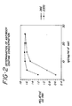
- Figure 2 is a graph which shows the determination of the antibody coating concentration for a sol made in accordance with the teachings of Applicants' invention.
- Figure 3 is a bar graph which illustrates the effect of adding a chaotrophic agent to the assay buffer.
- the signal was significantly improved by adding the agent when Figure 3A (without the addition of chaotrophic agent) is compared with Figure 3B (with the chaotrophic agent added), showing that serum induced aggregation rates were reduced when these agents are used.
- Applicants' novel colloidal seeded metal sol process produces a reagent which can be utilized in microscopic and immunoassay techniques.
- Applicants first prepare nuclei or seed using a strong reducing agent such as sodium borohydride, hydrogen peroxide, ascorbic acid, phosphorus, sodium citrate or malic acid, in the presence of a stabilizing agent such as citrate, malate, citric acid, malic acid, oxalate, oxalic acid, aspartate, aspartic acid, glutamate, or glutamic acid.
- a strong reducing agent such as sodium borohydride, hydrogen peroxide, ascorbic acid, phosphorus, sodium citrate or malic acid
- a stabilizing agent such as citrate, malate, citric acid, malic acid, oxalate, oxalic acid, aspartate, aspartic acid, glutamate, or glutamic acid.
- the reducing and stabilizing agent are selected so that they will not appreciably affect the subsequent growth of
- Applicants' method for producing nuclei comprises adding a suitable stabilizing agent to a metal salt solution and then adding a suitable reducing agent.
- a suitable stabilizing agent Preferably, a gold salt solution is used and most preferably a chloro-auro-tetrahydrate solution.
- the seed produced according to Applicants' method are very stable over time as shown in the following Table 1.
- the nuclei produced are then added to a metal-containing solution, preferably a metal salt solution, containing a second reducing agent which retards spontaneous enucleation and allows metal sol particles to form.
- Hydroxylamine is the most preferable reducing agent for this step.
- the reaction is nearly independent of the concentration of the reducing agent, as shown in the following Table 2. Effect of Hydroxylamine Concentration on Particle Size and Particle Distribution Hydroxylamine (50 mg/ml) Particle Size (nm) Particle Peak Width 0.4 ml 63.3 47.87 0.2 ml 63.3 46.89 0.08 ml 66.4 47.97
- reducing agents which retard spontaneous enucleation include substituted hydroxylamine, hydrazine, and substituted hydrazine. This step can also be performed by using separate reducing and enucleation retarding agents.
- the reducing agent is then inactivated. This can be achieved by pH adjustment, a combination of pH and bovine serum albumin, or by using a chemical agent.
- Ketones, aldehydes, amino acids or various combinations of chemical agents e.g., ascorbic acid, ascorbic acid and acetone, ascorbic acid and fructose, are effective inactivators.
- Other useful chemical inactivators include reducing agents such as a hydride or hydrogen and oxidizing agents such as oxygen or a peroxide.
- a reducing agent is added before a significant amount of spontaneous enucleation occurs.
- Reducing agents which are useful in this embodiment include citrate, malate, malic acid, tannate, tannic acid, hydrogen peroxide, oxalate, glutamate, stannous chloride, and sodium hydroxide.
- citrate, malate, malic acid, tannate, tannic acid hydrogen peroxide, oxalate, glutamate, stannous chloride, and sodium hydroxide.
- sodium citrate is added to a chloroauric acid solution, followed immediately by the addition of sodium borohydride prepared seed. The solution turns from a faintly blue to a dark red within less than a minute. Neither prolonged heating or the addition of extra citrate or seed produces any substantial change in the solution after that period. As shown in Figure 1, this method produces particles of a selected size and narrow distribution.
- the metal sol particles formed are then coated with a selected material.
- the particles can be coated with a proteinaceous material such as an antibody or an antigen, or conjugates thereof, for example. They may also be coated with polysaccharides, lectins, peptides, conjugated peptides or surfactants, as appropriate for the use of the reagent.
- the coating antibody When the coating antibody is pretreated with a reducing agent and capped with a sulfhydryl blocking agent, it was surprisingly found that the resulting derivatized antibody, when coated on gold sol, exhibits an improved signal generation.
- Useful reducing agents include dithiothreitol, dithioerythritol, glutathione, cysteine, ⁇ -mercaptoethanol, sodium borohydride, borohydride salts.
- Useful blocking agents include iodoacetamide, iodoacetic acid, iodoacetate salts and N-ethyl maleimide.
- the coated particles are then post-coated by coating the particles with delipidated milk, most preferably non-fat dry milk which Applicants surprisingly found to be a very effective post-coater.
- the post-coated particles are then stored in a buffer solution containing albumin, most preferably bovine serum albumin, and a low molecular weight sugar, most preferably, mannitol.
- the same particle size sol can be prepared at a bench top scale of 4.5 ml and proportionally scaled up to 40,000 ml according to the teachings of Applicants' method.
- the same procedure will yield sol with the same particle size for 1x fold sol up to 10x sol.
- the effect of gold concentration on particle size is shown in the following Table 3. Effect of Gold Concentration on Particle Size Volume of 2% Au Added (ml) Particle Size (nm) Change in Avg. Particle Size (nm) 0.8 (-20%) 58.7 -3.9 0.9 (-10%) 60.6 -2 1 62.6 0 1.1 (+10%) 65.6 3 1.2 (+20%) 66.4 3.8
- a kit for performing an immunoassay comprising a reagent made in accordance with Applicants' method and an assay buffer containing a chaotrophic agent it was surprisingly found that serum induced aggregation can be eliminated or minimized when a chaotrophic agent is added to the assay buffer.
- the chaotrophic agent can be comprised of a combination of an ion selected from the group consisting of NO 3 - , ClO 4 - , I - , SCN - , Na + , Cs + , Li + , Mg 2+ , Ca 2 - and Ba 2+ , most preferably NaI.
- a rapid, simple and qualitative dot blot assay is also provided to determine the presence and/or immunological activity on coated metal sol particles.
- the dot blot assay is a membrane filtration type assay used to qualitatively detect the presence of IgG, TSH or HCG, for example, adsorbed onto the gold particles.
- a gold reagent made in accordance with Applicants' method appropriately diluted in a buffer containing Tween 20 is filtered through a nitrocellulose membrane onto which goat anti-mouse IgG is immobilized.
- Mouse IgG coated gold particles will be retained on the filter as a reddish dot (within seconds) whereas unreacted materials will be readily washed away.
- this method can be used as an immunoassay tool to detect the presence of HCG or TSH in serum samples.
- the assay involves the immobilization of a mouse anti-HCG or anti-TSH to the nitrocellulose filter paper. A mixture containing sample and anti-TSH or HCG gold reagent in an appropriate buffer is then filtered through the nitrocellulose membrane. TSH and HCG can be readily detected in Lyphocheck Immunoassay Control Serum Levels I within 15 seconds.
- a DuPont sedimentation of field flow fractionator (SF3) was used to determine gold sol size distributions.
- This instrument resembles an HPLC instrument with a thin rotating channel, mounted in a centrifuge, replacing the column.
- a UV detector quantitates the particles by absorption and feeds data to a computer/controller for size computation.
- the principle of separation is based on the fact that large dense particles are pushed closer to the wall of the chromatography channel than small particles when a centripetal force is applied.
- This instrument was calibrated with gold particles against electron microscopy and found to be far superior to light scatter correlation instruments for gold sols of this size. The resolution of the instrument is approximately one nanometer.
- citrate seeded sol The standard procedure for obtaining citrate seeded sol is as follows. To 450 ml of MilliQ water heated to 95°C was added 5 ml of 1% HAuCl4 with constant stirring. When the temperature was re-equilibrated to 95°C, 2 ml of 1% Na-Citrate was added followed immediately by 0.15 ml of NaBH4 seed. The reaction was then allowed to proceed for another 15 minutes at 95°C. The solution turned first from a faint blue rapidly to dark red within 60 seconds. Neither prolonged heating or the addition of extra citrate or seed produced any substantially change in the solution after that period.
- the gold sol pH is adjusted to pH 6.5 with 0.2 M potassium carbonate.
- Bovine serum albumin is added to a final concentration of 2 mg/ml of sol and stirred for 30 minutes.
- the gold sol pH is adjusted to the desired coating pH or to pH 7.0 if the coating pH is above 7.0.
- BSA is added to a final concentration of 2 mg/ml of sol and stirred for 30 minutes.
- the protein was diluted in an appropriate Good's buffer to 250 mg/ml and 100 ⁇ l was added to each glass or polypropylene tube.
- Sigma Chemical Company's publication concerning buffers sold by that company lists a number of Good's buffers and their compositions which would be useful in Applicants' invention.
- the gold sol was titrated to pH 6.0 to 9.0 in 1.0 pH increments with fresh .02 M potassium carbonate removing 1 ml at each point and adding each aliquot to the protein solution. The mixtures were allowed to stand for 30 minutes at room temperature. 100 ml of a 10% sodium chloride solution was added and allowed to stand for 10 minutes at room temperature.
- the absorbance at 580 nm was read using a spectrophotometer and recorded for each sample.
- the results, shown in Figure 1, are plotted using absorbance as the Y axis and pH as the X axis.
- the coating pH is that at which the absorbance readings level off and become parallel to the X axis. This reading will be the same as the control sample in which 100 ⁇ l at buffer is added instead of the 10% sodium chloride.
- the coating protein is diluted to 25, 50, 100, 150, 200 and 250 mg/ml in an appropriate buffer determined in part A and 100 ml added to each tube.
- the gold sol is titrated to the pH determined in A, above, with 0.2 M potassium carbonate. 1.0 ml of pH adjusted gold sol is added to each tube and allowed to stand for 30 minutes at room temperature. 100 ml of 10% sodium chloride is added to each tube and allowed to stand 10 minutes at room temperature. Using water as a blank, the absorbance is recorded at 580 nm for each sample. The absorbance versus mg protein/ml of sol is then plotted. The coating level is that just before the absorbance levels off. See Figure 1.
- the gold sol is titrated to the coating pH with 0.2 M potassium carbonate.
- the required amount of protein is diluted to 1/20th the sol volume in 10 mM of the appropriate coating buffer.
- the protein is diluted in the appropriate coating buffer containing 0.002% BSA.
- the protein solution is added to the raw sol and allowed to stand for a minimum of 60 minutes at room temperature.
- the sol is then post-coated with 1/10th sol volume of a 0.1% non-fat dry milk in water solution, which has been 0.45 ⁇ m filtered for 30 minutes (final milk concentration is 0.01%).
- the coated sol is washed by centrifuging and removing the supernatant.
- the sol is resuspended in a 10 mM Good's buffer 0.5% BSA (RIA grade), 300 mM mannitol, 0.01% sodium azide, pH 7.0 (storage buffer). The washing step is performed three times. After the third centrifugation, storage buffer is added to 1/10th initial sol volume (i.e., 100 ml for each 1000 ml of raw sol). The absorbance is read at 540 nm. The volume is adjusted such that the final absorbance is 10 OD units. The solution is then filtered through a 0.2 ⁇ m low protein-binding filter and stored.
- BSA RIA grade
- storage buffer is added to 1/10th initial sol volume (i.e., 100 ml for each 1000 ml of raw sol).
- the absorbance is read at 540 nm.
- the volume is adjusted such that the final absorbance is 10 OD units.
- the solution is then filtered through a 0.2 ⁇ m low protein-binding filter and stored.
- Polycarbonate cuvettes were coated with mouse monoclonal anti-hepatitis surface antigen antibodies by incubating 200 microliters of a 100 microgram per ml solution of antibody in 0.01 M phosphate buffered saline, pH 7.4, overnight at room temperature followed by three aspirate fill steps with 300 microliters of 0.05 M Hepes/Tris, pH 8.3 buffer.
- the cuvettes were then overcoated with 300 microliters of 0.05 M Hepes/Tris, ph 8.3, buffer containing 1% bovine serum albumin for 60 minutes at room temperature, washed twice with 300 microliters of overcoating solution, incubated 15 minutes at room temperature with 300 microliters of 3% trehalose in 0.05 M Hepes/Tris, pH 8.3 buffer, aspirated, dried in room air and stored at room temperature in a desiccator below 20% relative humidity. The cuvettes were then mounted in applicant's STIR instrument.
- the pipette then dispensed 54 microliters of buffer (containing 2.0 M potassium chloride, 2% bovine serum albumin, 50 micrograms per ml of normal mouse IgG and 0.05% sodium azide dissolved in 0.05 M sodium barbital buffer at pH 8.5) and 180 microliters of 105 nm diameter 0.1% monoclonal anti-hepatitis surface antigen coated gold colloid suspension (in 10 mM Hepes buffer, pH 7.0, containing 0.05% sodium azide, 300 mM mannitol, and 0.5% bovine serum albumin) at a rate sufficient to mix the fluids in the cuvette. The light scattered by each cuvette was then recorded for the next ten minutes.
- buffer containing 2.0 M potassium chloride, 2% bovine serum albumin, 50 micrograms per ml of normal mouse IgG and 0.05% sodium azide dissolved in 0.05 M sodium barbital buffer at pH 8.5
- Polycarbonate cuvettes were coated as in Example 8, with recombinant hepatitis core antigen using a 5 microgram per ml coating solution. The cuvettes were then dried, stored and mounted in the STIR instrument, which was enclosed and equipped with 37° air circulation.
- Gold particles of 77.7 nm diameter were prepared by adding seed particles to a gold salt-hydroxylamine mixture as described above and coated by the standard method with anti-TSH monoclonal antibody.
- 36 microliters of Cone Biotech commercial standard plus 128 microliters of assay buffer plus 80 microliters coated gold sol were incubated for 5 minutes at 37°C in a rotating parabolic mirror automatic STIR instrument. After incubation a 150 microliter aliquot was delivered to an anti-TSH monoclonal coated STIR cuvette and read for 10 minutes.
- the assay buffer contained 0.05 M Barbital, 1 M KCl, 1% BSA, .05% NaN 3 and 100 micrograms per ml of polymerized mouse IgG, adjusted to pH 8.5.
- the polymerized mouse IgG was found to be the optimum agent to eliminate anti-mouse activity caused nonspecific reactions in STIR assays.
- the integral of the fifth order fit of the measured scattered light, the STIR signal was as follows: Duplicate Signal of TSH Standards Zero STD 0.5 ⁇ IU/ml 2 ⁇ IU/ml 5 ⁇ IU/ml 20 ⁇ IU/ml 0.419 3.624 13.44 34.04 140.2 0.321 3.714 12.80 33.37 143.8
- the antibody is reacted with an appropriate reducing agent, e.g., DTT, ⁇ -mercaptoethanol, at a concentration of 0.01 to 0.1 M for at least 5 minutes at a temperature of 2°C to 40°C.
- an appropriate sulfhydryl blocking agent is added, e.g., iodoacetamide, N-ethyl-maleimide, at a concentration of 0.1 M to 1.0 M for at least 1 minute.
- the material is then desalted by dialysis or gel filtration.
- mouse anti-hCG monoclonal antibody (clone number 16a) at 17.6 mg/ml in PBS with 0.1% sodium azide is added 15.4 mg solid dithiothreitol (0.1 M) at 22°C. The mixture is incubated for 30 min. After incubation, 185 mg solid iodoacetamide (1.0 M) is added and allowed to react for 5 minutes. The antibody is separated from the reactants by dialysis versus 5 mM MOPSO, pH 7.0 and coated in the usual fashion.
- a small volume of sodium borohydride seed nuclei, prepared as described in Example 2, are added to a dilute suspension of polymer latex particles to allow attachment of the seed nuclei to the latex particles' surface.
- 2% chloroauric acid and hydroxylamine hydrochloride, 50 mg/ml, are then added to the mixture stirred continuously for 5 minutes at room temperature.
- This procedure produces polymeric latex particles coated with metallic gold capable of being coated as described in the foregoing examples. These particles exhibit settling rates intermediate between solid gold particles and uncoated latex particles.
- An advantage of this method is that slow settling, inexpensive very large particles (e.g., microns in diameter) can be prepared without the rapid settling problems of large solid gold particles.
Landscapes
- Health & Medical Sciences (AREA)
- Chemical & Material Sciences (AREA)
- Life Sciences & Earth Sciences (AREA)
- Immunology (AREA)
- Engineering & Computer Science (AREA)
- Urology & Nephrology (AREA)
- Hematology (AREA)
- Organic Chemistry (AREA)
- Biomedical Technology (AREA)
- Molecular Biology (AREA)
- Analytical Chemistry (AREA)
- Biochemistry (AREA)
- Microbiology (AREA)
- Biotechnology (AREA)
- Dispersion Chemistry (AREA)
- Food Science & Technology (AREA)
- Medicinal Chemistry (AREA)
- Physics & Mathematics (AREA)
- Chemical Kinetics & Catalysis (AREA)
- Cell Biology (AREA)
- General Health & Medical Sciences (AREA)
- General Physics & Mathematics (AREA)
- Pathology (AREA)
- Manufacture Of Metal Powder And Suspensions Thereof (AREA)
- Medicines Containing Antibodies Or Antigens For Use As Internal Diagnostic Agents (AREA)
- Measuring Or Testing Involving Enzymes Or Micro-Organisms (AREA)
- Peptides Or Proteins (AREA)
- Investigating Or Analysing Biological Materials (AREA)
- Colloid Chemistry (AREA)
- Medicines That Contain Protein Lipid Enzymes And Other Medicines (AREA)
Abstract
Description
- said stabilising agent is citrate, malate, citric acid, malic acid, oxalate, oxalic acid, aspartate, aspartic acid, glutamate or glutamic acid;
- said first reducing agent and said stabilising agent are selected so that in step (b) they will not generate spontaneous nuclei and will not retard growth of nuclei in the subsequent particle growth step; and
- said second reducing agent retards spontaneous enucleation.
| Hydroxylamine Seeded Sol Process Validation (90 ml reaction volume, 2x gold sol concentration) | ||||
| Seed Lot # | Average Particle Size (nm) | Date | ||
| Seed Volume (ul) | 2 | 3 | 4 | |
| 100 | 55 | 48 , 47 | 10/22/87 | |
| 56 | 56 | 52 , 61 | 11/16/87 | |
| 55 | 55 | 52 , 52 | 12/08/87 | |
| 57 | 58 | 54 | 03/21/88 | |
| 60 | 65 | 57 , 55 | 10/22/87 | |
| 66 | 67 | 61 , 60 | 11/16/87 | |
| 64 | 68 | 61 , 60 | 12/08/87 | |
| 66 | 70 | 64 | 03/21/88 | |
| 20 | 92 | 79 , 78 | 10/22/87 | |
| 102 | 96 | 86 , 87 | 11/16/87 | |
| 93 | 96 | 86 , 89 | 12/08/87 | |
| 94 | 99 | 90 | 03/21/88 |
| Effect of Hydroxylamine Concentration on Particle Size and Particle Distribution | ||
| Hydroxylamine (50 mg/ml) | Particle Size (nm) | Particle Peak Width |
| 0.4 ml | 63.3 | 47.87 |
| 0.2 ml | 63.3 | 46.89 |
| 0.08 ml | 66.4 | 47.97 |
| Effect of Gold Concentration on Particle Size | ||
| Volume of 2% Au Added (ml) | Particle Size (nm) | Change in Avg. Particle Size (nm) |
| 0.8 (-20%) | 58.7 | -3.9 |
| 0.9 (-10%) | 60.6 | -2 |
| 1 | 62.6 | 0 |
| 1.1 (+10%) | 65.6 | 3 |
| 1.2 (+20%) | 66.4 | 3.8 |
| EFFECT OF REACTOR VOLUME, SOL CONCENTRATION AND SEED VOLUME ON THE GOLD SOL AVERAGE PARTICLE SIZE | |||
| Particle Size (nm) | Reactor Volume (ml) | BOL Concentration | |
| 09/28/87 | 68.8 | 90 | 1x |
| 66.4 | 90 | 1x | |
| 76.7 | 90 | 1x | |
| 69.7 | 450 | 1x | |
| 92.9 | 90 | 1x | |
| 114.0 | 90 | | |
| 10/01/87 | 75.7 | 90 | 1x |
| 75.7 | 450 | | |
| 10/02/87 | 74.5 | 450 | 1x |
| 67.5 | 450 | 1x | |
| 68.5 | 450 | | |
| 10/05/87 | 68.5 | 4.5 | 8x |
| 68.5 | 4.5 | | |
| 10/06/87 | 70.5 | 4.5 | 8x |
| 69.7 | 4.5 | 10x | |
| 81.8 | 4.5 | 10x | |
| 62.3 | 4.5 | | |
| 10/13/87 | 74.3 | 450 | 1x |
| 67.5 | 450 | 1x | |
| 68.6 | 450 | 1x | |
| 11/01/87 | 72.0 | 450 | 1x |
| 11/10/87 | 76.0 | 4050 | 1x |
| 11/13/87 | 72.0 | 450 | |
| 12/09/87 | 71.0 | 900 | |
| 12/21/87 | 75.0 | 450 | |
| 12/22/87 | 75.0 | 1800 | 1x |
| 01/11/88 | 73.0 | 450 | 1x |
| 73.0 | 900 | 1x | |
| 01/12/88 | 75.0 | 450 | 1x |
| 01/28/88 | 69.7 | 40000 | 1x |
| Unless specified otherwise, the seed volume used was proportionally scaled to give 70 nm particles, .02 ml for 90 ml 1x sol. |
| HEPATITIS SURFACE ANTIGEN CONCENTRATION | MEAN SIGNAL |
| 0 | 1.7358 |
| 0.1 ng/ml | 2.2376 |
| 0.2 ng/ml | 2.9421 |
| 0.4 ng/ml | 3.99235 |
| 0.6 ng/ml | 5.0442 |
| 0.8 ng/ml | 6.72185 |
| 1.0 ng/ml | 7.0187 |
| 1.5 ng/ml | 9.31175 |
| 2.0 ng/ml | 10.7365 |
| 2.5 ng/ml | 14.0444 |
| 5.0 ng/ml | 24.9279 |
| 10.0 ng/ml | 47.4585 |
| Sample Type | Number of Replicates | Mean | Test Outcome |
| | 4 | 0.9524 | S.D.=0.51 |
| | 4 | 60.2099 | S.D.=6.5 |
| Negative sample 1 | 2 | 2.1671 | - |
| Positive sample 1 | 2 | 10.483 | + |
| | 2 | 41.058 | + |
| Positive sample 3 | 2 | 33.494 | + |
| | 2 | 2.6043 | + |
| Positive sample 5 | 2 | 74.2235 | + |
| Duplicate Signal of TSH Standards | ||||
| Zero STD | 0.5µIU/ml | 2µIU/ml | 5µIU/ml | 20µIU/ml |
| 0.419 | 3.624 | 13.44 | 34.04 | 140.2 |
| 0.321 | 3.714 | 12.80 | 33.37 | 143.8 |
| Sample | Net Signal | Signal 0 µIU/ | Signal | 100 µIU/ml |
| standard reagent | 395.32 | 3.68 | 399 | |
| reduced/blocked reagent | 550.50 | 5.50 | 556 |
Claims (21)
- A method for producing metallic sol particles of a selected size range which comprises:wherein:(a) reducing a solution containing a metal and a stabilising agent with a first reducing agent to form metallic nuclei; and(b) mixing the metallic nuclei formed in step (a) with a solution containing said metal and a second reducing agent to form metallic sol particles,said stabilising agent is citrate, malate, citric acid, malic acid, oxalate, oxalic acid, aspartate, aspartic acid, glutamate or glutamic acid; and said stabilizing agent aresaid first reducing agent selected so that in step (b) they will not generate nuclei and will not retard growth of nuclei in the subsequent particle growth step; andsaid second reducing agent retards spontaneous enucleation.
- The method of claim 1, wherein said first reducing agent is a borohydride salt, hydrogen peroxide, ascorbic acid, phosphorous, sodium citrate or malic acid.
- The method of claim 2, wherein said first reducing agent is a borohydride salt, preferably sodium borohydride.
- The method of any one of claims 1 to 3, wherein said second reducing agent is hydroxylamine, hydrazine, substituted hydroxylamine or substituted hydrazine.
- The method of claim 4, wherein said second reducing agent is hydroxylamine or hydroxylamine hydrochloride.
- The method of any one of claims 1 to 5, wherein the stabilising agent is sodium citrate.
- The method of any one of claims 1 to 6, wherein step (a) comprises adding said stabilising agent to said metal-containing solution and then adding said first reducing agent to the stabilised metal-containing solution.
- The method of any one of claims 1 to 7, wherein between steps (a) and (b), the metallic nuclei are absorbed onto a polymeric surface.
- The method of any one of claims 1 to 8, wherein step (b) is carried out to produce sol particles having a diameter from 10 to 200 nm.
- The method of any one of claims 1 to 9, further including the step of inactivating any reactive species present after the completion of step (b).
- The method of claim 10, wherein the inactivation step is carried out by adjusting the pH of the sol.
- The method of claim 10 or claim 11, wherein the inactivation step is carried out by adding a chemical agent.
- The method of claim 12, wherein the chemical agent is a proteinaceous agent, such as albumin, preferably bovine serum albumin.
- The method of any one of claims 10 to 13, further including the step of coating the inactivated sol particles with a selected, preferably proteinaceous, material.
- The method of claim 14, wherein the selected material is an antibody.
- The method of claim 15 wherein the antibody has been reduced using a third reducing agent, such as dithiothreitol, dithioerythritol, glutathione, cysteine, β-mercaptoethanol, or a borohydride salt, and capped with a sulfhydryl blocking agent, such as iodoacetamide, iodoacetic acid, an iodoacetate salt or N-ethylmaleimide.
- The method of any one of claims 14 to 16, further including the step of post-coating sol particles, preferably with a delipidated milk such as non-fat dry milk.
- The method of any one of claims 14 to 17, further including the step of adding said coated or post-coated sol particles to a buffer solution comprising an albumin, preferably bovine serum albumin, and a low molecular weight sugar, preferably mannitol.
- The method of any one of claims 1 to 18, wherein the metal-containing solutions contain gold, preferably in the form of a gold salt such as chloro-auro-tetrahydrate.
- A kit for performing an immunoassay comprising: coated or post-coated sol particles produced by the method of any one of claims 14 to 18; and an assay buffer.
- The kit of claim 20, wherein said assay buffer contains a chaotropic agent, which is preferably an NO2 -, ClO4 - I-, SCN-, Na+, Cs+, Li+, Mg2+, Ca2+ or Ba2+ ion, guanidine, urea, TCA or TFA, and is more preferably NaI.
Applications Claiming Priority (2)
| Application Number | Priority Date | Filing Date | Title |
|---|---|---|---|
| US41518489A | 1989-09-29 | 1989-09-29 | |
| US415184 | 1999-10-07 |
Publications (2)
| Publication Number | Publication Date |
|---|---|
| EP0426300A1 EP0426300A1 (en) | 1991-05-08 |
| EP0426300B1 true EP0426300B1 (en) | 1998-06-03 |
Family
ID=23644702
Family Applications (1)
| Application Number | Title | Priority Date | Filing Date |
|---|---|---|---|
| EP90310671A Expired - Lifetime EP0426300B1 (en) | 1989-09-29 | 1990-09-28 | Method of producing a reagent containing a narrow distribution of colloidal particles of a selected size and the use thereof |
Country Status (8)
| Country | Link |
|---|---|
| EP (1) | EP0426300B1 (en) |
| JP (1) | JPH03206959A (en) |
| AT (1) | ATE166975T1 (en) |
| CA (1) | CA2026409C (en) |
| DE (1) | DE69032363T2 (en) |
| DK (1) | DK0426300T3 (en) |
| ES (1) | ES2117628T3 (en) |
| GR (1) | GR1000722B (en) |
Cited By (5)
| Publication number | Priority date | Publication date | Assignee | Title |
|---|---|---|---|---|
| WO2001039873A1 (en) * | 1999-12-03 | 2001-06-07 | Surromed, Inc. | Hydroxylamine seeding of colloidal metal nanoparticles |
| CN102539773A (en) * | 2011-12-31 | 2012-07-04 | 上海凯创生物技术有限公司 | KET (Ketamine) colloid gold detection kit and preparation method thereof |
| EP2835402A1 (en) | 2013-08-09 | 2015-02-11 | Leibniz-Institut für Neue Materialien gemeinnützige GmbH | Formation of surface modified metal colloids |
| DE102013108664A1 (en) | 2013-08-09 | 2015-02-12 | Leibniz-Institut Für Neue Materialien Gemeinnützige Gmbh | Surface-modified metal colloids and their preparation |
| CN105436516A (en) * | 2015-12-03 | 2016-03-30 | 南昌大学 | Preparing method of multi-branch colloidal gold nano particles with controllable grain diameter and high light absorbing intensity |
Families Citing this family (21)
| Publication number | Priority date | Publication date | Assignee | Title |
|---|---|---|---|---|
| TW239881B (en) * | 1992-12-22 | 1995-02-01 | Sienna Biotech Inc | |
| US6200820B1 (en) | 1992-12-22 | 2001-03-13 | Sienna Biotech, Inc. | Light scatter-based immunoassay |
| US5851777A (en) * | 1996-02-05 | 1998-12-22 | Dade Behring Inc. | Homogeneous sol-sol assay |
| EP1073902A2 (en) * | 1998-04-20 | 2001-02-07 | University Of North Carolina At Chapel Hill | Nanometer particles containing a reactive monolayer |
| IL126776A (en) * | 1998-10-27 | 2001-04-30 | Technion Res & Dev Foundation | Method for gold depositions |
| US7364920B2 (en) | 1999-10-27 | 2008-04-29 | Technion Research And Development Foundation Ltd. | Method for gold deposition |
| US7928149B2 (en) | 2003-08-28 | 2011-04-19 | Tama-Tlo, Ltd. | Precious metal colloid, precious metal fine-particle, composition, and method for producing precious metal fine-particle |
| WO2008103824A1 (en) * | 2007-02-23 | 2008-08-28 | Chinese Academy Of Inspection And Quarantine (Caiq) | Sensitivity-enhanced dot-antibody linked immunogold assay for virus detection |
| GB0724870D0 (en) | 2007-12-21 | 2008-01-30 | Univ Lincoln The | Preparation of metal colloids |
| JP5035622B2 (en) * | 2008-01-11 | 2012-09-26 | 積水化学工業株式会社 | Colored latex |
| ES2347034B1 (en) * | 2009-04-21 | 2011-09-08 | Centro De Investigacion Biomedica En Red En Bioingenieria. Biomateriales Y Nanomedicina | PROCEDURE FOR OBTAINING METAL NANOPARTICLES. |
| WO2016002743A1 (en) | 2014-07-01 | 2016-01-07 | 新日鉄住金化学株式会社 | Marker, immunoassay method, immunoassay reagent, method for assaying analyte, kit for assaying analyte, and test strip for lateral flow chromatography |
| US11366112B2 (en) | 2014-07-01 | 2022-06-21 | Nippon Steel Chemical & Material Co., Ltd. | Resin-metal composite, marker, immunoassay method, immunoassay reagent, method for measuring analyte, analyte measurement kit, and lateral-flow chromatographic test strip |
| CN105067807B (en) * | 2015-04-30 | 2018-01-09 | 珠海丽珠试剂股份有限公司 | A kind of preparation method of Colloidal Gold Used in Immunoassay |
| CN110542761B (en) | 2015-07-11 | 2023-08-29 | 日铁化学材料株式会社 | Resin-platinum complex and use thereof |
| KR102640613B1 (en) | 2016-12-28 | 2024-02-26 | 닛테츠 케미컬 앤드 머티리얼 가부시키가이샤 | Metal-resin composite and its uses |
| CN111229321A (en) * | 2020-01-16 | 2020-06-05 | 吉林大学 | Glutathione-protected platinum alloy nanocluster with strong catalytic function, preparation method and application thereof |
| CN112094699B (en) * | 2020-09-27 | 2023-03-28 | 广西壮族自治区林业科学研究院 | Method for extracting camphor tree hydrosol |
| CN115575623B (en) * | 2022-12-06 | 2023-03-28 | 深圳市卓润生物科技有限公司 | Colloidal gold and preparation method and application thereof |
| CN116217991B (en) * | 2023-03-09 | 2024-02-27 | 四川大学 | Preparation method and application of circularly polarized light-emitting film |
| CN116930482B (en) * | 2023-07-31 | 2025-05-09 | 山东康华生物医疗科技股份有限公司 | Colloidal gold preparation method for helicobacter pylori antigen detection test strip and test strip preparation method |
Family Cites Families (4)
| Publication number | Priority date | Publication date | Assignee | Title |
|---|---|---|---|---|
| NL7807532A (en) | 1978-07-13 | 1980-01-15 | Akzo Nv | METAL IMMUNO TEST. |
| JPS63307208A (en) * | 1987-06-08 | 1988-12-14 | Chiyoda Chem Eng & Constr Co Ltd | Manufacturing method of precious metal fine powder |
| US4859612A (en) * | 1987-10-07 | 1989-08-22 | Hygeia Sciences, Inc. | Metal sol capture immunoassay procedure, kit for use therewith and captured metal containing composite |
| NL8702769A (en) * | 1987-11-19 | 1989-06-16 | Holland Biotechnology | METHOD FOR DETERMINING A TEST SAMPLE OF COMPONENTS OF THE REACTION BETWEEN A SPECIFIC BINDING PROTEIN AND THE ACCOMPANYING BINDABLE SUBSTANCE USING AT LEAST ONE BRANDED COMPONENT, METHOD FOR PREPARING THE MARKED COMPONENT, AND TESTING COMPONENT |
-
1990
- 1990-09-27 CA CA002026409A patent/CA2026409C/en not_active Expired - Lifetime
- 1990-09-28 DK DK90310671T patent/DK0426300T3/en active
- 1990-09-28 GR GR900100722A patent/GR1000722B/en not_active IP Right Cessation
- 1990-09-28 DE DE69032363T patent/DE69032363T2/en not_active Expired - Lifetime
- 1990-09-28 EP EP90310671A patent/EP0426300B1/en not_active Expired - Lifetime
- 1990-09-28 ES ES90310671T patent/ES2117628T3/en not_active Expired - Lifetime
- 1990-09-28 AT AT90310671T patent/ATE166975T1/en not_active IP Right Cessation
- 1990-09-29 JP JP2262922A patent/JPH03206959A/en active Pending
Non-Patent Citations (1)
| Title |
|---|
| Weiser, H.B., "The Colloidal Elements" in Inorganic Colloid Chemistry, J. Wiley & Sons (1933) pages 1-107. * |
Cited By (6)
| Publication number | Priority date | Publication date | Assignee | Title |
|---|---|---|---|---|
| WO2001039873A1 (en) * | 1999-12-03 | 2001-06-07 | Surromed, Inc. | Hydroxylamine seeding of colloidal metal nanoparticles |
| CN102539773A (en) * | 2011-12-31 | 2012-07-04 | 上海凯创生物技术有限公司 | KET (Ketamine) colloid gold detection kit and preparation method thereof |
| CN102539773B (en) * | 2011-12-31 | 2014-07-16 | 上海凯创生物技术有限公司 | KET (Ketamine) colloid gold detection kit and preparation method thereof |
| EP2835402A1 (en) | 2013-08-09 | 2015-02-11 | Leibniz-Institut für Neue Materialien gemeinnützige GmbH | Formation of surface modified metal colloids |
| DE102013108664A1 (en) | 2013-08-09 | 2015-02-12 | Leibniz-Institut Für Neue Materialien Gemeinnützige Gmbh | Surface-modified metal colloids and their preparation |
| CN105436516A (en) * | 2015-12-03 | 2016-03-30 | 南昌大学 | Preparing method of multi-branch colloidal gold nano particles with controllable grain diameter and high light absorbing intensity |
Also Published As
| Publication number | Publication date |
|---|---|
| ATE166975T1 (en) | 1998-06-15 |
| ES2117628T3 (en) | 1998-08-16 |
| DE69032363D1 (en) | 1998-07-09 |
| EP0426300A1 (en) | 1991-05-08 |
| CA2026409A1 (en) | 1991-03-30 |
| GR900100722A (en) | 1992-01-20 |
| CA2026409C (en) | 2004-11-09 |
| GR1000722B (en) | 1992-11-23 |
| DK0426300T3 (en) | 1999-02-01 |
| JPH03206959A (en) | 1991-09-10 |
| DE69032363T2 (en) | 1998-10-22 |
Similar Documents
| Publication | Publication Date | Title |
|---|---|---|
| CA2026409C (en) | Method of producing a reagent containing a narrow distribution of colloidal particles of a selected size and the use thereof | |
| US5571726A (en) | Kit containing glutaraldehyde coated colloidal metal particles of a preselected size | |
| RU2025732C1 (en) | Method of analyte immunoassay in aqueous sample | |
| US5294369A (en) | Ligand gold bonding | |
| US5773307A (en) | Method and device for determining an analyte in a sample | |
| AU614109B2 (en) | Test method and reagent kit therefor | |
| US7517700B2 (en) | Core-shell particles | |
| US5650333A (en) | Immunoassay method and kit using superaggregated complex | |
| US5175112A (en) | Submicron particles, preparation and utilization in immunodiagnosis | |
| CA2105515A1 (en) | Visual immunoassay method for the detection of ligands, based on the use of opaque plastic supports | |
| JPH09504094A (en) | Method for assaying immunological substances using magnetic latex particles and non-magnetic particles | |
| Kapmeyer et al. | Automated nephelometric immunoassays with novel shell/core particles | |
| EP0679892B1 (en) | Microparticle immunoassay reagents, sensitive and specific immunoassay reagents and immunoassay methods using these reagents | |
| AU650632B2 (en) | Method of producing a metal sol reagent containing colloidal metal particles of a preselected size | |
| Price et al. | Light scattering immunoassay | |
| CA2108439A1 (en) | Enhanced sensitivity agglutination assays using multivalent ligands | |
| JP2714964B2 (en) | A method for determining in a test sample one or more components of a reaction between a specifically binding protein and a corresponding bindable substance using at least one labeled component, comprising: Preparation method and test kit for measurement of immune component | |
| EP0064275B1 (en) | Immunochemical reagent | |
| US5641689A (en) | Stable aqueous carbon sol composition for determining analyte | |
| US5196349A (en) | Immunoassays for thyroid hormones using thyroglobulin | |
| Quesada et al. | A simple kinetic model of antigen-antibody reactions in particle-enhanced light scattering immunoassays | |
| Lucocq | Particulate markers for immunoelectron microscopy | |
| JP3220546B2 (en) | Method for measuring antigen or antibody in the presence of immune complex | |
| JPH10253629A (en) | Immunoassay reagent manufacturing method | |
| HK1001234A (en) | Test method and reagent kit therefor |
Legal Events
| Date | Code | Title | Description |
|---|---|---|---|
| PUAI | Public reference made under article 153(3) epc to a published international application that has entered the european phase |
Free format text: ORIGINAL CODE: 0009012 |
|
| AK | Designated contracting states |
Kind code of ref document: A1 Designated state(s): AT BE CH DE DK ES FR GB IT LI LU NL SE |
|
| 17P | Request for examination filed |
Effective date: 19911014 |
|
| 17Q | First examination report despatched |
Effective date: 19931105 |
|
| GRAG | Despatch of communication of intention to grant |
Free format text: ORIGINAL CODE: EPIDOS AGRA |
|
| GRAG | Despatch of communication of intention to grant |
Free format text: ORIGINAL CODE: EPIDOS AGRA |
|
| GRAH | Despatch of communication of intention to grant a patent |
Free format text: ORIGINAL CODE: EPIDOS IGRA |
|
| GRAH | Despatch of communication of intention to grant a patent |
Free format text: ORIGINAL CODE: EPIDOS IGRA |
|
| GRAA | (expected) grant |
Free format text: ORIGINAL CODE: 0009210 |
|
| ITF | It: translation for a ep patent filed | ||
| AK | Designated contracting states |
Kind code of ref document: B1 Designated state(s): AT BE CH DE DK ES FR GB IT LI LU NL SE |
|
| REF | Corresponds to: |
Ref document number: 166975 Country of ref document: AT Date of ref document: 19980615 Kind code of ref document: T |
|
| REG | Reference to a national code |
Ref country code: CH Ref legal event code: EP |
|
| REG | Reference to a national code |
Ref country code: CH Ref legal event code: NV Representative=s name: E. BLUM & CO. PATENTANWAELTE |
|
| REF | Corresponds to: |
Ref document number: 69032363 Country of ref document: DE Date of ref document: 19980709 |
|
| REG | Reference to a national code |
Ref country code: ES Ref legal event code: FG2A Ref document number: 2117628 Country of ref document: ES Kind code of ref document: T3 |
|
| ET | Fr: translation filed | ||
| REG | Reference to a national code |
Ref country code: DK Ref legal event code: T3 |
|
| PLBE | No opposition filed within time limit |
Free format text: ORIGINAL CODE: 0009261 |
|
| STAA | Information on the status of an ep patent application or granted ep patent |
Free format text: STATUS: NO OPPOSITION FILED WITHIN TIME LIMIT |
|
| 26N | No opposition filed | ||
| REG | Reference to a national code |
Ref country code: GB Ref legal event code: IF02 |
|
| REG | Reference to a national code |
Ref country code: CH Ref legal event code: PFA Owner name: ORTHO DIAGNOSTIC SYSTEMS INC. Free format text: ORTHO DIAGNOSTIC SYSTEMS INC.#U.S. ROUTE NO. 202#RARITAN, NEW JERSEY 08869-0602 (US) -TRANSFER TO- ORTHO DIAGNOSTIC SYSTEMS INC.#U.S. ROUTE NO. 202#RARITAN, NEW JERSEY 08869-0602 (US) |
|
| PGFP | Annual fee paid to national office [announced via postgrant information from national office to epo] |
Ref country code: DK Payment date: 20090914 Year of fee payment: 20 |
|
| PGFP | Annual fee paid to national office [announced via postgrant information from national office to epo] |
Ref country code: SE Payment date: 20090910 Year of fee payment: 20 Ref country code: AT Payment date: 20090911 Year of fee payment: 20 Ref country code: NL Payment date: 20090903 Year of fee payment: 20 Ref country code: LU Payment date: 20090929 Year of fee payment: 20 Ref country code: GB Payment date: 20090923 Year of fee payment: 20 |
|
| PGFP | Annual fee paid to national office [announced via postgrant information from national office to epo] |
Ref country code: DE Payment date: 20090923 Year of fee payment: 20 Ref country code: ES Payment date: 20091006 Year of fee payment: 20 Ref country code: CH Payment date: 20091006 Year of fee payment: 20 |
|
| PGFP | Annual fee paid to national office [announced via postgrant information from national office to epo] |
Ref country code: BE Payment date: 20090917 Year of fee payment: 20 |
|
| PGFP | Annual fee paid to national office [announced via postgrant information from national office to epo] |
Ref country code: FR Payment date: 20091012 Year of fee payment: 20 Ref country code: IT Payment date: 20090917 Year of fee payment: 20 |
|
| BE20 | Be: patent expired |
Owner name: *ORTHO DIAGNOSTIC SYSTEMS INC. Effective date: 20100928 |
|
| REG | Reference to a national code |
Ref country code: NL Ref legal event code: V4 Effective date: 20100928 |
|
| REG | Reference to a national code |
Ref country code: CH Ref legal event code: PL |
|
| REG | Reference to a national code |
Ref country code: DK Ref legal event code: EUP |
|
| REG | Reference to a national code |
Ref country code: GB Ref legal event code: PE20 Expiry date: 20100927 |
|
| EUG | Se: european patent has lapsed | ||
| PG25 | Lapsed in a contracting state [announced via postgrant information from national office to epo] |
Ref country code: GB Free format text: LAPSE BECAUSE OF EXPIRATION OF PROTECTION Effective date: 20100927 |
|
| PG25 | Lapsed in a contracting state [announced via postgrant information from national office to epo] |
Ref country code: NL Free format text: LAPSE BECAUSE OF EXPIRATION OF PROTECTION Effective date: 20100928 |
|
| PG25 | Lapsed in a contracting state [announced via postgrant information from national office to epo] |
Ref country code: DE Free format text: LAPSE BECAUSE OF EXPIRATION OF PROTECTION Effective date: 20100928 |
|
| REG | Reference to a national code |
Ref country code: ES Ref legal event code: FD2A Effective date: 20140826 |
|
| PG25 | Lapsed in a contracting state [announced via postgrant information from national office to epo] |
Ref country code: ES Free format text: LAPSE BECAUSE OF EXPIRATION OF PROTECTION Effective date: 20100929 |