EP0359745B1 - Growth factor receptor - Google Patents
Growth factor receptor Download PDFInfo
- Publication number
- EP0359745B1 EP0359745B1 EP88902901A EP88902901A EP0359745B1 EP 0359745 B1 EP0359745 B1 EP 0359745B1 EP 88902901 A EP88902901 A EP 88902901A EP 88902901 A EP88902901 A EP 88902901A EP 0359745 B1 EP0359745 B1 EP 0359745B1
- Authority
- EP
- European Patent Office
- Prior art keywords
- grp
- spantide
- cells
- bombesin
- antagonist
- Prior art date
- Legal status (The legal status is an assumption and is not a legal conclusion. Google has not performed a legal analysis and makes no representation as to the accuracy of the status listed.)
- Expired - Lifetime
Links
- 102000009465 Growth Factor Receptors Human genes 0.000 title 1
- 108010009202 Growth Factor Receptors Proteins 0.000 title 1
- 239000005557 antagonist Substances 0.000 claims abstract description 47
- 230000004663 cell proliferation Effects 0.000 claims abstract description 9
- 238000000034 method Methods 0.000 claims description 20
- 150000001875 compounds Chemical class 0.000 claims description 12
- 238000011282 treatment Methods 0.000 claims description 9
- 238000003745 diagnosis Methods 0.000 claims description 7
- 241001465754 Metazoa Species 0.000 claims description 4
- 238000000338 in vitro Methods 0.000 claims description 4
- 238000002560 therapeutic procedure Methods 0.000 claims description 4
- 125000000180 D-prolyl group Chemical group N1[C@@H](C(=O)*)CCC1 0.000 claims description 3
- 125000000174 L-prolyl group Chemical group [H]N1C([H])([H])C([H])([H])C([H])([H])[C@@]1([H])C(*)=O 0.000 claims description 3
- 239000003814 drug Substances 0.000 claims description 3
- 238000004519 manufacturing process Methods 0.000 claims description 3
- 239000003085 diluting agent Substances 0.000 claims description 2
- 239000003937 drug carrier Substances 0.000 claims description 2
- 125000002698 D-tryptophano group Chemical group C(=O)(O)[C@@H](CC1=CNC2=CC=CC=C12)N* 0.000 claims 2
- XVOCEQLNJQGCQG-UNAVROQCSA-N (2s)-2-[[(2s)-2-[[(2s)-1-[(2s)-6-amino-2-[[(2s)-1-[(2s)-2-amino-5-(diaminomethylideneamino)pentanoyl]pyrrolidine-2-carbonyl]amino]hexanoyl]pyrrolidine-2-carbonyl]amino]-3-phenylpropanoyl]amino]-n-[(2s)-1-[[(2s)-1-[[(2s)-1-[[(2s)-1-[[(2s)-1-amino-4-methyl- Chemical compound C([C@@H](C(=O)N[C@@H](CC=1C2=CC=CC=C2NC=1)C(=O)N[C@@H](CC(C)C)C(=O)N[C@@H](CC(C)C)C(N)=O)NC(=O)[C@H](CC=1C2=CC=CC=C2NC=1)NC(=O)[C@H](CCC(N)=O)NC(=O)[C@H](CC=1C=CC=CC=1)NC(=O)[C@H]1N(CCC1)C(=O)[C@H](CCCCN)NC(=O)[C@H]1N(CCC1)C(=O)[C@@H](N)CCCN=C(N)N)C1=CC=CC=C1 XVOCEQLNJQGCQG-UNAVROQCSA-N 0.000 claims 1
- 239000008194 pharmaceutical composition Substances 0.000 claims 1
- 108090000765 processed proteins & peptides Proteins 0.000 abstract description 38
- 102000004196 processed proteins & peptides Human genes 0.000 abstract description 28
- DNDCVAGJPBKION-DOPDSADYSA-N bombesin Chemical compound C([C@@H](C(=O)N[C@@H](CC(C)C)C(=O)N[C@@H](CCSC)C(N)=O)NC(=O)CNC(=O)[C@@H](NC(=O)[C@H](C)NC(=O)[C@H](CC=1NC2=CC=CC=C2C=1)NC(=O)[C@H](CCC(N)=O)NC(=O)[C@H](CC(N)=O)NC(=O)CNC(=O)[C@H](CC(C)C)NC(=O)[C@H](CCCNC(N)=N)NC(=O)[C@H](CCC(N)=O)NC(=O)[C@H]1NC(=O)CC1)C(C)C)C1=CN=CN1 DNDCVAGJPBKION-DOPDSADYSA-N 0.000 abstract description 27
- 108010051479 Bombesin Proteins 0.000 abstract description 24
- 229920001184 polypeptide Polymers 0.000 abstract description 6
- 102000013585 Bombesin Human genes 0.000 abstract 2
- ONIBWKKTOPOVIA-SCSAIBSYSA-N D-Proline Chemical compound OC(=O)[C@H]1CCCN1 ONIBWKKTOPOVIA-SCSAIBSYSA-N 0.000 abstract 1
- COLNVLDHVKWLRT-MRVPVSSYSA-N D-phenylalanine Chemical compound OC(=O)[C@H](N)CC1=CC=CC=C1 COLNVLDHVKWLRT-MRVPVSSYSA-N 0.000 abstract 1
- -1 [D-Pro<2>]-spantide Chemical compound 0.000 abstract 1
- 210000004027 cell Anatomy 0.000 description 75
- 102000004862 Gastrin releasing peptide Human genes 0.000 description 71
- 108090001053 Gastrin releasing peptide Proteins 0.000 description 71
- PUBCCFNQJQKCNC-XKNFJVFFSA-N gastrin-releasingpeptide Chemical compound C([C@@H](C(=O)N[C@@H](CC(C)C)C(=O)N[C@@H](CCSC)C(N)=O)NC(=O)CNC(=O)[C@@H](NC(=O)[C@H](C)NC(=O)[C@H](CC=1C2=CC=CC=C2NC=1)NC(=O)[C@H](CC=1N=CNC=1)NC(=O)[C@H](CC(N)=O)NC(=O)CNC(=O)[C@H](CCCN=C(N)N)NC(=O)[C@H]1N(CCC1)C(=O)[C@H](CC=1C=CC(O)=CC=1)NC(=O)[C@H](CCSC)NC(=O)[C@H](CCCCN)NC(=O)[C@@H](NC(=O)[C@H](CC(C)C)NC(=O)[C@@H](NC(=O)[C@@H](NC(=O)CNC(=O)CNC(=O)CNC(=O)[C@H](C)NC(=O)[C@H]1N(CCC1)C(=O)[C@H](CC(C)C)NC(=O)[C@H]1N(CCC1)C(=O)[C@@H](N)C(C)C)[C@@H](C)O)C(C)C)[C@@H](C)O)C(C)C)C1=CNC=N1 PUBCCFNQJQKCNC-XKNFJVFFSA-N 0.000 description 69
- GFMHGFPPCYBUCK-HBUFSDLISA-N dnc012382 Chemical compound C([C@@H](C(=O)N[C@@H](CC=1C2=CC=CC=C2NC=1)C(=O)N[C@@H](CC(C)C)C(=O)N[C@@H](CCCC)C(O)=O)NC(=O)[C@H](CC=1C2=CC=CC=C2NC=1)NC(=O)[C@H](CC(N)=O)NC(=O)[C@H](CC=1C=CC=CC=1)NC(=O)[C@H]1N(CC(Cl)C1Cl)C(=O)[C@H](C)NC(=O)[C@H]1N(CCC1C=1C=NC=CC=1)C(=O)[C@H](CCCCN)NC(=O)C=1C=NC=CC=1)C1=CC=CC=C1 GFMHGFPPCYBUCK-HBUFSDLISA-N 0.000 description 60
- 108700024670 spantide Proteins 0.000 description 60
- 102000004169 proteins and genes Human genes 0.000 description 29
- 108090000623 proteins and genes Proteins 0.000 description 29
- 230000027455 binding Effects 0.000 description 26
- QLHLYJHNOCILIT-UHFFFAOYSA-N 4-o-(2,5-dioxopyrrolidin-1-yl) 1-o-[2-[4-(2,5-dioxopyrrolidin-1-yl)oxy-4-oxobutanoyl]oxyethyl] butanedioate Chemical compound O=C1CCC(=O)N1OC(=O)CCC(=O)OCCOC(=O)CCC(=O)ON1C(=O)CCC1=O QLHLYJHNOCILIT-UHFFFAOYSA-N 0.000 description 22
- 102100036519 Gastrin-releasing peptide Human genes 0.000 description 22
- 230000000694 effects Effects 0.000 description 22
- 108020003175 receptors Proteins 0.000 description 18
- 102000005962 receptors Human genes 0.000 description 18
- 230000006820 DNA synthesis Effects 0.000 description 13
- NOESYZHRGYRDHS-UHFFFAOYSA-N insulin Chemical compound N1C(=O)C(NC(=O)C(CCC(N)=O)NC(=O)C(CCC(O)=O)NC(=O)C(C(C)C)NC(=O)C(NC(=O)CN)C(C)CC)CSSCC(C(NC(CO)C(=O)NC(CC(C)C)C(=O)NC(CC=2C=CC(O)=CC=2)C(=O)NC(CCC(N)=O)C(=O)NC(CC(C)C)C(=O)NC(CCC(O)=O)C(=O)NC(CC(N)=O)C(=O)NC(CC=2C=CC(O)=CC=2)C(=O)NC(CSSCC(NC(=O)C(C(C)C)NC(=O)C(CC(C)C)NC(=O)C(CC=2C=CC(O)=CC=2)NC(=O)C(CC(C)C)NC(=O)C(C)NC(=O)C(CCC(O)=O)NC(=O)C(C(C)C)NC(=O)C(CC(C)C)NC(=O)C(CC=2NC=NC=2)NC(=O)C(CO)NC(=O)CNC2=O)C(=O)NCC(=O)NC(CCC(O)=O)C(=O)NC(CCCNC(N)=N)C(=O)NCC(=O)NC(CC=3C=CC=CC=3)C(=O)NC(CC=3C=CC=CC=3)C(=O)NC(CC=3C=CC(O)=CC=3)C(=O)NC(C(C)O)C(=O)N3C(CCC3)C(=O)NC(CCCCN)C(=O)NC(C)C(O)=O)C(=O)NC(CC(N)=O)C(O)=O)=O)NC(=O)C(C(C)CC)NC(=O)C(CO)NC(=O)C(C(C)O)NC(=O)C1CSSCC2NC(=O)C(CC(C)C)NC(=O)C(NC(=O)C(CCC(N)=O)NC(=O)C(CC(N)=O)NC(=O)C(NC(=O)C(N)CC=1C=CC=CC=1)C(C)C)CC1=CN=CN1 NOESYZHRGYRDHS-UHFFFAOYSA-N 0.000 description 12
- 239000002609 medium Substances 0.000 description 12
- 230000005764 inhibitory process Effects 0.000 description 11
- 238000007792 addition Methods 0.000 description 10
- 239000000463 material Substances 0.000 description 10
- 102000010780 Platelet-Derived Growth Factor Human genes 0.000 description 9
- 108010038512 Platelet-Derived Growth Factor Proteins 0.000 description 9
- 206010041067 Small cell lung cancer Diseases 0.000 description 9
- 238000004132 cross linking Methods 0.000 description 9
- 239000003431 cross linking reagent Substances 0.000 description 9
- LOKCTEFSRHRXRJ-UHFFFAOYSA-I dipotassium trisodium dihydrogen phosphate hydrogen phosphate dichloride Chemical compound P(=O)(O)(O)[O-].[K+].P(=O)(O)([O-])[O-].[Na+].[Na+].[Cl-].[K+].[Cl-].[Na+] LOKCTEFSRHRXRJ-UHFFFAOYSA-I 0.000 description 9
- 239000000499 gel Substances 0.000 description 9
- 238000002372 labelling Methods 0.000 description 9
- 239000002953 phosphate buffered saline Substances 0.000 description 9
- 230000003389 potentiating effect Effects 0.000 description 9
- 238000002415 sodium dodecyl sulfate polyacrylamide gel electrophoresis Methods 0.000 description 9
- VBEQCZHXXJYVRD-GACYYNSASA-N uroanthelone Chemical compound C([C@@H](C(=O)N[C@H](C(=O)N[C@@H](CS)C(=O)N[C@@H](CC(N)=O)C(=O)N[C@@H](CS)C(=O)N[C@H](C(=O)N[C@@H]([C@@H](C)CC)C(=O)NCC(=O)N[C@@H](CC=1C=CC(O)=CC=1)C(=O)N[C@@H](CO)C(=O)NCC(=O)N[C@@H](CC(O)=O)C(=O)N[C@@H](CCCNC(N)=N)C(=O)N[C@@H](CS)C(=O)N[C@@H](CCC(N)=O)C(=O)N[C@@H]([C@@H](C)O)C(=O)N[C@@H](CCCNC(N)=N)C(=O)N[C@@H](CC(O)=O)C(=O)N[C@@H](CC(C)C)C(=O)N[C@@H](CCCNC(N)=N)C(=O)N[C@@H](CC=1C2=CC=CC=C2NC=1)C(=O)N[C@@H](CC=1C2=CC=CC=C2NC=1)C(=O)N[C@@H](CCC(O)=O)C(=O)N[C@@H](CC(C)C)C(=O)N[C@@H](CCCNC(N)=N)C(O)=O)C(C)C)[C@@H](C)O)NC(=O)[C@H](CO)NC(=O)[C@H](CC(O)=O)NC(=O)[C@H](CC(C)C)NC(=O)[C@H](CO)NC(=O)[C@H](CCC(O)=O)NC(=O)[C@@H](NC(=O)[C@H](CC=1NC=NC=1)NC(=O)[C@H](CCSC)NC(=O)[C@H](CS)NC(=O)[C@@H](NC(=O)CNC(=O)CNC(=O)[C@H](CC(N)=O)NC(=O)[C@H](CC(C)C)NC(=O)[C@H](CS)NC(=O)[C@H](CC=1C=CC(O)=CC=1)NC(=O)CNC(=O)[C@H](CC(O)=O)NC(=O)[C@H](CC=1C=CC(O)=CC=1)NC(=O)[C@H](CO)NC(=O)[C@H](CO)NC(=O)[C@H]1N(CCC1)C(=O)[C@H](CS)NC(=O)CNC(=O)[C@H]1N(CCC1)C(=O)[C@H](CC=1C=CC(O)=CC=1)NC(=O)[C@H](CO)NC(=O)[C@@H](N)CC(N)=O)C(C)C)[C@@H](C)CC)C1=CC=C(O)C=C1 VBEQCZHXXJYVRD-GACYYNSASA-N 0.000 description 9
- FXYPGCIGRDZWNR-UHFFFAOYSA-N (2,5-dioxopyrrolidin-1-yl) 3-[[3-(2,5-dioxopyrrolidin-1-yl)oxy-3-oxopropyl]disulfanyl]propanoate Chemical compound O=C1CCC(=O)N1OC(=O)CCSSCCC(=O)ON1C(=O)CCC1=O FXYPGCIGRDZWNR-UHFFFAOYSA-N 0.000 description 8
- 108091003079 Bovine Serum Albumin Proteins 0.000 description 8
- 102400001368 Epidermal growth factor Human genes 0.000 description 8
- 101800003838 Epidermal growth factor Proteins 0.000 description 8
- ZWIBGKZDAWNIFC-UHFFFAOYSA-N disuccinimidyl suberate Chemical compound O=C1CCC(=O)N1OC(=O)CCCCCCC(=O)ON1C(=O)CCC1=O ZWIBGKZDAWNIFC-UHFFFAOYSA-N 0.000 description 8
- 229940116977 epidermal growth factor Drugs 0.000 description 8
- 239000006144 Dulbecco’s modified Eagle's medium Substances 0.000 description 7
- 229940098773 bovine serum albumin Drugs 0.000 description 7
- 230000012010 growth Effects 0.000 description 7
- 239000003446 ligand Substances 0.000 description 7
- 102000004877 Insulin Human genes 0.000 description 6
- 108090001061 Insulin Proteins 0.000 description 6
- 239000002790 bombesin antagonist Substances 0.000 description 6
- 229940125396 insulin Drugs 0.000 description 6
- 239000012679 serum free medium Substances 0.000 description 6
- SAPCBHPQLCZCDV-QVBDNUAOSA-N (2s)-2-[[(2s)-1-[(2s)-6-amino-2-[[(2r)-1-[(2r)-2-amino-5-(diaminomethylideneamino)pentanoyl]pyrrolidine-2-carbonyl]amino]hexanoyl]pyrrolidine-2-carbonyl]amino]-n-[(2s)-5-amino-1-[[(2r)-1-[[(2s)-1-[[(2r)-1-[[(2s)-1-[[(2s)-1-amino-4-methyl-1-oxopentan-2-yl] Chemical compound C([C@@H](C(=O)N[C@H](CC=1C2=CC=CC=C2NC=1)C(=O)N[C@@H](CC(C)C)C(=O)N[C@@H](CC(C)C)C(N)=O)NC(=O)[C@@H](CC=1C2=CC=CC=C2NC=1)NC(=O)[C@H](CCC(N)=O)NC(=O)[C@H](CCC(N)=O)NC(=O)[C@H]1N(CCC1)C(=O)[C@H](CCCCN)NC(=O)[C@@H]1N(CCC1)C(=O)[C@H](N)CCCN=C(N)N)C1=CC=CC=C1 SAPCBHPQLCZCDV-QVBDNUAOSA-N 0.000 description 5
- 229940123804 Bombesin antagonist Drugs 0.000 description 5
- 239000012980 RPMI-1640 medium Substances 0.000 description 5
- 125000003275 alpha amino acid group Chemical group 0.000 description 5
- 238000002474 experimental method Methods 0.000 description 5
- 239000003102 growth factor Substances 0.000 description 5
- 230000036515 potency Effects 0.000 description 5
- 230000009870 specific binding Effects 0.000 description 5
- QDZOEBFLNHCSSF-PFFBOGFISA-N (2S)-2-[[(2R)-2-[[(2S)-1-[(2S)-6-amino-2-[[(2S)-1-[(2R)-2-amino-5-carbamimidamidopentanoyl]pyrrolidine-2-carbonyl]amino]hexanoyl]pyrrolidine-2-carbonyl]amino]-3-(1H-indol-3-yl)propanoyl]amino]-N-[(2R)-1-[[(2S)-1-[[(2R)-1-[[(2S)-1-[[(2S)-1-amino-4-methyl-1-oxopentan-2-yl]amino]-4-methyl-1-oxopentan-2-yl]amino]-3-(1H-indol-3-yl)-1-oxopropan-2-yl]amino]-1-oxo-3-phenylpropan-2-yl]amino]-3-(1H-indol-3-yl)-1-oxopropan-2-yl]pentanediamide Chemical compound C([C@@H](C(=O)N[C@H](CC=1C2=CC=CC=C2NC=1)C(=O)N[C@@H](CC(C)C)C(=O)N[C@@H](CC(C)C)C(N)=O)NC(=O)[C@@H](CC=1C2=CC=CC=C2NC=1)NC(=O)[C@H](CCC(N)=O)NC(=O)[C@@H](CC=1C2=CC=CC=C2NC=1)NC(=O)[C@H]1N(CCC1)C(=O)[C@H](CCCCN)NC(=O)[C@H]1N(CCC1)C(=O)[C@H](N)CCCNC(N)=N)C1=CC=CC=C1 QDZOEBFLNHCSSF-PFFBOGFISA-N 0.000 description 4
- YPFNACALNKVZNK-MFNIMNRCSA-N (2s)-2-[(2-aminoacetyl)amino]-n-[(2s)-1-[[(2s)-1-[[(2s)-1-[[(2s,3r)-1-[[2-[[(2s)-1-[[(2s)-1-[[(2s)-1-amino-4-methylsulfanyl-1-oxobutan-2-yl]amino]-1-oxo-3-phenylpropan-2-yl]amino]-3-(1h-imidazol-5-yl)-1-oxopropan-2-yl]amino]-2-oxoethyl]amino]-3-hydroxy-1- Chemical compound C([C@@H](C(=O)N[C@@H](CCSC)C(N)=O)NC(=O)[C@H](CC=1NC=NC=1)NC(=O)CNC(=O)[C@@H](NC(=O)[C@H](C)NC(=O)[C@H](CC=1C2=CC=CC=C2NC=1)NC(=O)[C@H](CC(C)C)NC(=O)[C@H](CC(N)=O)NC(=O)CN)[C@@H](C)O)C1=CC=CC=C1 YPFNACALNKVZNK-MFNIMNRCSA-N 0.000 description 4
- OHCNRADJYUSTIV-FPNHNIPFSA-N (2s)-n-[(2s)-1-[[(2s)-1-[[(2s)-1-[[2-[[(2s)-1-[[(2s)-1-[[(2s)-1-amino-4-methylsulfanyl-1-oxobutan-2-yl]amino]-1-oxo-3-phenylpropan-2-yl]amino]-3-(1h-imidazol-5-yl)-1-oxopropan-2-yl]amino]-2-oxoethyl]amino]-3-methyl-1-oxobutan-2-yl]amino]-1-oxopropan-2-yl] Chemical compound C([C@@H](C(=O)N[C@@H](CCSC)C(N)=O)NC(=O)[C@H](CC=1NC=NC=1)NC(=O)CNC(=O)[C@@H](NC(=O)[C@H](C)NC(=O)[C@H](CC=1C2=CC=CC=C2NC=1)NC(=O)[C@H](CCC(N)=O)NC(=O)[C@H]1NC(=O)CC1)C(C)C)C1=CC=CC=C1 OHCNRADJYUSTIV-FPNHNIPFSA-N 0.000 description 4
- 102100038819 Neuromedin-B Human genes 0.000 description 4
- 101800001639 Neuromedin-B Proteins 0.000 description 4
- FAPWRFPIFSIZLT-UHFFFAOYSA-M Sodium chloride Chemical compound [Na+].[Cl-] FAPWRFPIFSIZLT-UHFFFAOYSA-M 0.000 description 4
- 101800003906 Substance P Proteins 0.000 description 4
- 102400000096 Substance P Human genes 0.000 description 4
- GXBMIBRIOWHPDT-UHFFFAOYSA-N Vasopressin Natural products N1C(=O)C(CC=2C=C(O)C=CC=2)NC(=O)C(N)CSSCC(C(=O)N2C(CCC2)C(=O)NC(CCCN=C(N)N)C(=O)NCC(N)=O)NC(=O)C(CC(N)=O)NC(=O)C(CCC(N)=O)NC(=O)C1CC1=CC=CC=C1 GXBMIBRIOWHPDT-UHFFFAOYSA-N 0.000 description 4
- 108010004977 Vasopressins Proteins 0.000 description 4
- 102000002852 Vasopressins Human genes 0.000 description 4
- KBZOIRJILGZLEJ-LGYYRGKSSA-N argipressin Chemical compound C([C@H]1C(=O)N[C@@H](CCC(N)=O)C(=O)N[C@@H](CC(N)=O)C(=O)N[C@@H](CSSC[C@@H](C(N[C@@H](CC=2C=CC(O)=CC=2)C(=O)N1)=O)N)C(=O)N1[C@@H](CCC1)C(=O)N[C@@H](CCCN=C(N)N)C(=O)NCC(N)=O)C1=CC=CC=C1 KBZOIRJILGZLEJ-LGYYRGKSSA-N 0.000 description 4
- 230000010261 cell growth Effects 0.000 description 4
- 230000001419 dependent effect Effects 0.000 description 4
- 108010061043 litorin Proteins 0.000 description 4
- OHCNRADJYUSTIV-UHFFFAOYSA-N litorin Natural products C=1N=CNC=1CC(NC(=O)CNC(=O)C(NC(=O)C(C)NC(=O)C(CC=1C2=CC=CC=C2NC=1)NC(=O)C(CCC(N)=O)NC(=O)C1NC(=O)CC1)C(C)C)C(=O)NC(C(=O)NC(CCSC)C(N)=O)CC1=CC=CC=C1 OHCNRADJYUSTIV-UHFFFAOYSA-N 0.000 description 4
- 230000002441 reversible effect Effects 0.000 description 4
- 229960003726 vasopressin Drugs 0.000 description 4
- HEAUFJZALFKPBA-JPQUDPSNSA-N (3s)-3-[[(2s,3r)-2-[[(2s)-6-amino-2-[[(2s)-2-amino-3-(1h-imidazol-5-yl)propanoyl]amino]hexanoyl]amino]-3-hydroxybutanoyl]amino]-4-[[(2s)-1-[[(2s)-1-[[(2s)-1-[[2-[[(2s)-1-[[(2s)-1-amino-4-methylsulfanyl-1-oxobutan-2-yl]amino]-4-methyl-1-oxopentan-2-yl]amin Chemical compound C([C@@H](C(=O)N[C@H](C(=O)NCC(=O)N[C@@H](CC(C)C)C(=O)N[C@@H](CCSC)C(N)=O)C(C)C)NC(=O)[C@H](CO)NC(=O)[C@H](CC(O)=O)NC(=O)[C@@H](NC(=O)[C@H](CCCCN)NC(=O)[C@@H](N)CC=1NC=NC=1)[C@@H](C)O)C1=CC=CC=C1 HEAUFJZALFKPBA-JPQUDPSNSA-N 0.000 description 3
- QTBSBXVTEAMEQO-UHFFFAOYSA-N Acetic acid Chemical compound CC(O)=O QTBSBXVTEAMEQO-UHFFFAOYSA-N 0.000 description 3
- 108010073466 Bombesin Receptors Proteins 0.000 description 3
- PEDCQBHIVMGVHV-UHFFFAOYSA-N Glycerine Chemical compound OCC(O)CO PEDCQBHIVMGVHV-UHFFFAOYSA-N 0.000 description 3
- 101800000399 Neurokinin A Proteins 0.000 description 3
- 102400000097 Neurokinin A Human genes 0.000 description 3
- HEMHJVSKTPXQMS-UHFFFAOYSA-M Sodium hydroxide Chemical compound [OH-].[Na+] HEMHJVSKTPXQMS-UHFFFAOYSA-M 0.000 description 3
- DBMJMQXJHONAFJ-UHFFFAOYSA-M Sodium laurylsulphate Chemical compound [Na+].CCCCCCCCCCCCOS([O-])(=O)=O DBMJMQXJHONAFJ-UHFFFAOYSA-M 0.000 description 3
- 108010056088 Somatostatin Proteins 0.000 description 3
- 102000005157 Somatostatin Human genes 0.000 description 3
- IQFYYKKMVGJFEH-XLPZGREQSA-N Thymidine Chemical compound O=C1NC(=O)C(C)=CN1[C@@H]1O[C@H](CO)[C@@H](O)C1 IQFYYKKMVGJFEH-XLPZGREQSA-N 0.000 description 3
- 238000003556 assay Methods 0.000 description 3
- 238000000211 autoradiogram Methods 0.000 description 3
- IQFYYKKMVGJFEH-UHFFFAOYSA-N beta-L-thymidine Natural products O=C1NC(=O)C(C)=CN1C1OC(CO)C(O)C1 IQFYYKKMVGJFEH-UHFFFAOYSA-N 0.000 description 3
- 230000001588 bifunctional effect Effects 0.000 description 3
- 239000012634 fragment Substances 0.000 description 3
- 108010091651 gastrin releasing peptide (1-16) Proteins 0.000 description 3
- 238000010348 incorporation Methods 0.000 description 3
- 230000002401 inhibitory effect Effects 0.000 description 3
- 230000003834 intracellular effect Effects 0.000 description 3
- 238000001155 isoelectric focusing Methods 0.000 description 3
- CWWARWOPSKGELM-SARDKLJWSA-N methyl (2s)-2-[[(2s)-2-[[2-[[(2s)-2-[[(2s)-2-[[(2s)-5-amino-2-[[(2s)-5-amino-2-[[(2s)-1-[(2s)-6-amino-2-[[(2s)-1-[(2s)-2-amino-5-(diaminomethylideneamino)pentanoyl]pyrrolidine-2-carbonyl]amino]hexanoyl]pyrrolidine-2-carbonyl]amino]-5-oxopentanoyl]amino]-5 Chemical compound C([C@@H](C(=O)NCC(=O)N[C@@H](CC(C)C)C(=O)N[C@@H](CCSC)C(=O)OC)NC(=O)[C@H](CC=1C=CC=CC=1)NC(=O)[C@H](CCC(N)=O)NC(=O)[C@H](CCC(N)=O)NC(=O)[C@H]1N(CCC1)C(=O)[C@H](CCCCN)NC(=O)[C@H]1N(CCC1)C(=O)[C@@H](N)CCCN=C(N)N)C1=CC=CC=C1 CWWARWOPSKGELM-SARDKLJWSA-N 0.000 description 3
- 239000003226 mitogen Substances 0.000 description 3
- BQJRUJTZSGYBEZ-YVQNUNKESA-N phorbol 12,13-dibutanoate Chemical compound C([C@]1(O)C(=O)C(C)=C[C@H]1[C@@]1(O)[C@H](C)[C@H]2OC(=O)CCC)C(CO)=C[C@H]1[C@H]1[C@]2(OC(=O)CCC)C1(C)C BQJRUJTZSGYBEZ-YVQNUNKESA-N 0.000 description 3
- 229920002401 polyacrylamide Polymers 0.000 description 3
- NHXLMOGPVYXJNR-ATOGVRKGSA-N somatostatin Chemical compound C([C@H]1C(=O)N[C@H](C(N[C@@H](CO)C(=O)N[C@@H](CSSC[C@@H](C(=O)N[C@@H](CCCCN)C(=O)N[C@@H](CC(N)=O)C(=O)N[C@@H](CC=2C=CC=CC=2)C(=O)N[C@@H](CC=2C=CC=CC=2)C(=O)N[C@@H](CC=2C3=CC=CC=C3NC=2)C(=O)N[C@@H](CCCCN)C(=O)N[C@H](C(=O)N1)[C@@H](C)O)NC(=O)CNC(=O)[C@H](C)N)C(O)=O)=O)[C@H](O)C)C1=CC=CC=C1 NHXLMOGPVYXJNR-ATOGVRKGSA-N 0.000 description 3
- 229960000553 somatostatin Drugs 0.000 description 3
- 238000000539 two dimensional gel electrophoresis Methods 0.000 description 3
- DGVVWUTYPXICAM-UHFFFAOYSA-N β‐Mercaptoethanol Chemical compound OCCS DGVVWUTYPXICAM-UHFFFAOYSA-N 0.000 description 3
- VOXZDWNPVJITMN-ZBRFXRBCSA-N 17β-estradiol Chemical compound OC1=CC=C2[C@H]3CC[C@](C)([C@H](CC4)O)[C@@H]4[C@@H]3CCC2=C1 VOXZDWNPVJITMN-ZBRFXRBCSA-N 0.000 description 2
- IAZDPXIOMUYVGZ-UHFFFAOYSA-N Dimethylsulphoxide Chemical compound CS(C)=O IAZDPXIOMUYVGZ-UHFFFAOYSA-N 0.000 description 2
- TWRXJAOTZQYOKJ-UHFFFAOYSA-L Magnesium chloride Chemical compound [Mg+2].[Cl-].[Cl-] TWRXJAOTZQYOKJ-UHFFFAOYSA-L 0.000 description 2
- 108010052285 Membrane Proteins Proteins 0.000 description 2
- 206010028980 Neoplasm Diseases 0.000 description 2
- 108090000189 Neuropeptides Proteins 0.000 description 2
- 102400001103 Neurotensin Human genes 0.000 description 2
- 101800001814 Neurotensin Proteins 0.000 description 2
- CDBYLPFSWZWCQE-UHFFFAOYSA-L Sodium Carbonate Chemical compound [Na+].[Na+].[O-]C([O-])=O CDBYLPFSWZWCQE-UHFFFAOYSA-L 0.000 description 2
- 102400000368 Surface protein Human genes 0.000 description 2
- 230000003305 autocrine Effects 0.000 description 2
- 239000000872 buffer Substances 0.000 description 2
- 230000015556 catabolic process Effects 0.000 description 2
- 230000001413 cellular effect Effects 0.000 description 2
- 238000006243 chemical reaction Methods 0.000 description 2
- 238000006731 degradation reaction Methods 0.000 description 2
- 229960001760 dimethyl sulfoxide Drugs 0.000 description 2
- 231100000673 dose–response relationship Toxicity 0.000 description 2
- 238000001962 electrophoresis Methods 0.000 description 2
- 125000001495 ethyl group Chemical group [H]C([H])([H])C([H])([H])* 0.000 description 2
- JYGXADMDTFJGBT-VWUMJDOOSA-N hydrocortisone Chemical compound O=C1CC[C@]2(C)[C@H]3[C@@H](O)C[C@](C)([C@@](CC4)(O)C(=O)CO)[C@@H]4[C@@H]3CCC2=C1 JYGXADMDTFJGBT-VWUMJDOOSA-N 0.000 description 2
- 239000003112 inhibitor Substances 0.000 description 2
- 230000001404 mediated effect Effects 0.000 description 2
- PCJGZPGTCUMMOT-ISULXFBGSA-N neurotensin Chemical compound C([C@@H](C(=O)N[C@@H]([C@@H](C)CC)C(=O)N[C@@H](CC(C)C)C(O)=O)NC(=O)[C@H]1N(CCC1)C(=O)[C@H](CCCN=C(N)N)NC(=O)[C@H](CCCN=C(N)N)NC(=O)[C@H]1N(CCC1)C(=O)[C@H](CCCCN)NC(=O)[C@H](CC(N)=O)NC(=O)[C@H](CCC(O)=O)NC(=O)[C@H](CC=1C=CC(O)=CC=1)NC(=O)[C@H](CC(C)C)NC(=O)[C@H]1NC(=O)CC1)C1=CC=C(O)C=C1 PCJGZPGTCUMMOT-ISULXFBGSA-N 0.000 description 2
- 230000009871 nonspecific binding Effects 0.000 description 2
- 229920003023 plastic Polymers 0.000 description 2
- 239000004033 plastic Substances 0.000 description 2
- 230000002285 radioactive effect Effects 0.000 description 2
- 230000004044 response Effects 0.000 description 2
- 239000012723 sample buffer Substances 0.000 description 2
- 238000009738 saturating Methods 0.000 description 2
- 239000011780 sodium chloride Substances 0.000 description 2
- 235000019333 sodium laurylsulphate Nutrition 0.000 description 2
- 239000003890 substance P antagonist Substances 0.000 description 2
- 125000001174 sulfone group Chemical group 0.000 description 2
- IFIFCDMFVPNPSB-QEJDUTAOSA-N (2S)-2-[[2-[[(2S)-2-[[(2S)-1-[(2S)-2-[[(2S)-2-amino-4-methylsulfanylbutanoyl]amino]-3-(4-hydroxyphenyl)propanoyl]pyrrolidine-2-carbonyl]amino]-5-carbamimidamidopentanoyl]amino]acetyl]amino]-N-[(2S)-1-[[(2S)-1-[[(2S)-1-[[(2S)-1-[[2-[[(2S)-1-[[(2S)-1-[[(2S)-1-amino-4-methylsulfanyl-1-oxobutan-2-yl]amino]-4-methyl-1-oxopentan-2-yl]amino]-3-(1H-imidazol-5-yl)-1-oxopropan-2-yl]amino]-2-oxoethyl]amino]-3-methyl-1-oxobutan-2-yl]amino]-1-oxopropan-2-yl]amino]-3-(1H-indol-3-yl)-1-oxopropan-2-yl]amino]-3-(1H-imidazol-5-yl)-1-oxopropan-2-yl]butanediamide Chemical compound C([C@H](NC(=O)[C@@H](N)CCSC)C(=O)N1[C@@H](CCC1)C(=O)N[C@@H](CCCNC(N)=N)C(=O)NCC(=O)N[C@@H](CC(N)=O)C(=O)N[C@@H](CC=1NC=NC=1)C(=O)N[C@@H](CC=1C2=CC=CC=C2NC=1)C(=O)N[C@@H](C)C(=O)N[C@@H](C(C)C)C(=O)NCC(=O)N[C@@H](CC=1NC=NC=1)C(=O)N[C@@H](CC(C)C)C(=O)N[C@@H](CCSC)C(N)=O)C1=CC=C(O)C=C1 IFIFCDMFVPNPSB-QEJDUTAOSA-N 0.000 description 1
- JKMHFZQWWAIEOD-UHFFFAOYSA-N 2-[4-(2-hydroxyethyl)piperazin-1-yl]ethanesulfonic acid Chemical compound OCC[NH+]1CCN(CCS([O-])(=O)=O)CC1 JKMHFZQWWAIEOD-UHFFFAOYSA-N 0.000 description 1
- QKNYBSVHEMOAJP-UHFFFAOYSA-N 2-amino-2-(hydroxymethyl)propane-1,3-diol;hydron;chloride Chemical compound Cl.OCC(N)(CO)CO QKNYBSVHEMOAJP-UHFFFAOYSA-N 0.000 description 1
- 238000010600 3H thymidine incorporation assay Methods 0.000 description 1
- HRPVXLWXLXDGHG-UHFFFAOYSA-N Acrylamide Chemical compound NC(=O)C=C HRPVXLWXLXDGHG-UHFFFAOYSA-N 0.000 description 1
- 108010039627 Aprotinin Proteins 0.000 description 1
- 210000003771 C cell Anatomy 0.000 description 1
- UXVMQQNJUSDDNG-UHFFFAOYSA-L Calcium chloride Chemical compound [Cl-].[Cl-].[Ca+2] UXVMQQNJUSDDNG-UHFFFAOYSA-L 0.000 description 1
- 125000003941 D-tryptophan group Chemical group [H]C1=C([H])C([H])=C2C(C([C@@](N([H])[H])(C(=O)[*])[H])([H])[H])=C([H])N([H])C2=C1[H] 0.000 description 1
- KCXVZYZYPLLWCC-UHFFFAOYSA-N EDTA Chemical compound OC(=O)CN(CC(O)=O)CCN(CC(O)=O)CC(O)=O KCXVZYZYPLLWCC-UHFFFAOYSA-N 0.000 description 1
- 239000007836 KH2PO4 Substances 0.000 description 1
- 108010023244 Lactoperoxidase Proteins 0.000 description 1
- 102000045576 Lactoperoxidases Human genes 0.000 description 1
- 102000003797 Neuropeptides Human genes 0.000 description 1
- 102000003923 Protein Kinase C Human genes 0.000 description 1
- 108090000315 Protein Kinase C Proteins 0.000 description 1
- 230000010799 Receptor Interactions Effects 0.000 description 1
- 239000012722 SDS sample buffer Substances 0.000 description 1
- 102000004338 Transferrin Human genes 0.000 description 1
- 108090000901 Transferrin Proteins 0.000 description 1
- XSQUKJJJFZCRTK-UHFFFAOYSA-N Urea Chemical compound NC(N)=O XSQUKJJJFZCRTK-UHFFFAOYSA-N 0.000 description 1
- 235000010724 Wisteria floribunda Nutrition 0.000 description 1
- 239000000556 agonist Substances 0.000 description 1
- 238000004458 analytical method Methods 0.000 description 1
- 229960004405 aprotinin Drugs 0.000 description 1
- 239000012298 atmosphere Substances 0.000 description 1
- 238000000376 autoradiography Methods 0.000 description 1
- 239000011324 bead Substances 0.000 description 1
- 238000010364 biochemical engineering Methods 0.000 description 1
- 230000008512 biological response Effects 0.000 description 1
- 230000005540 biological transmission Effects 0.000 description 1
- 230000001275 ca(2+)-mobilization Effects 0.000 description 1
- 239000001110 calcium chloride Substances 0.000 description 1
- 235000011148 calcium chloride Nutrition 0.000 description 1
- 229910001628 calcium chloride Inorganic materials 0.000 description 1
- 239000004202 carbamide Substances 0.000 description 1
- 239000000969 carrier Substances 0.000 description 1
- 238000004113 cell culture Methods 0.000 description 1
- 230000036755 cellular response Effects 0.000 description 1
- 238000012512 characterization method Methods 0.000 description 1
- 238000010382 chemical cross-linking Methods 0.000 description 1
- 239000003153 chemical reaction reagent Substances 0.000 description 1
- 239000003795 chemical substances by application Substances 0.000 description 1
- 230000002860 competitive effect Effects 0.000 description 1
- 239000011243 crosslinked material Substances 0.000 description 1
- 230000001086 cytosolic effect Effects 0.000 description 1
- 239000007857 degradation product Substances 0.000 description 1
- 238000000326 densiometry Methods 0.000 description 1
- 229940042399 direct acting antivirals protease inhibitors Drugs 0.000 description 1
- 208000037265 diseases, disorders, signs and symptoms Diseases 0.000 description 1
- BNIILDVGGAEEIG-UHFFFAOYSA-L disodium hydrogen phosphate Chemical compound [Na+].[Na+].OP([O-])([O-])=O BNIILDVGGAEEIG-UHFFFAOYSA-L 0.000 description 1
- 229910000397 disodium phosphate Inorganic materials 0.000 description 1
- 235000019800 disodium phosphate Nutrition 0.000 description 1
- BVTBRVFYZUCAKH-UHFFFAOYSA-L disodium selenite Chemical compound [Na+].[Na+].[O-][Se]([O-])=O BVTBRVFYZUCAKH-UHFFFAOYSA-L 0.000 description 1
- 208000035475 disorder Diseases 0.000 description 1
- 239000008151 electrolyte solution Substances 0.000 description 1
- 238000011067 equilibration Methods 0.000 description 1
- 150000002148 esters Chemical class 0.000 description 1
- 229960005309 estradiol Drugs 0.000 description 1
- DEFVIWRASFVYLL-UHFFFAOYSA-N ethylene glycol bis(2-aminoethyl)tetraacetic acid Chemical compound OC(=O)CN(CC(O)=O)CCOCCOCCN(CC(O)=O)CC(O)=O DEFVIWRASFVYLL-UHFFFAOYSA-N 0.000 description 1
- 230000005284 excitation Effects 0.000 description 1
- 239000012091 fetal bovine serum Substances 0.000 description 1
- 210000002950 fibroblast Anatomy 0.000 description 1
- 239000012847 fine chemical Substances 0.000 description 1
- YFHXZQPUBCBNIP-UHFFFAOYSA-N fura-2 Chemical compound CC1=CC=C(N(CC(O)=O)CC(O)=O)C(OCCOC=2C(=CC=3OC(=CC=3C=2)C=2OC(=CN=2)C(O)=O)N(CC(O)=O)CC(O)=O)=C1 YFHXZQPUBCBNIP-UHFFFAOYSA-N 0.000 description 1
- 230000002496 gastric effect Effects 0.000 description 1
- 108010048151 gastrin releasing peptide (14-27) Proteins 0.000 description 1
- 238000001502 gel electrophoresis Methods 0.000 description 1
- 239000001963 growth medium Substances 0.000 description 1
- 229960000890 hydrocortisone Drugs 0.000 description 1
- 238000011534 incubation Methods 0.000 description 1
- ZPNFWUPYTFPOJU-LPYSRVMUSA-N iniprol Chemical compound C([C@H]1C(=O)NCC(=O)NCC(=O)N[C@H]2CSSC[C@H]3C(=O)N[C@@H](CCCCN)C(=O)N[C@@H](C)C(=O)N[C@@H](CCCNC(N)=N)C(=O)N[C@H](C(N[C@H](C(=O)N[C@@H](CCCNC(N)=N)C(=O)N[C@@H](CC=4C=CC(O)=CC=4)C(=O)N[C@@H](CC=4C=CC=CC=4)C(=O)N[C@@H](CC=4C=CC(O)=CC=4)C(=O)N[C@@H](CC(N)=O)C(=O)N[C@@H](C)C(=O)N[C@@H](CCCCN)C(=O)N[C@@H](C)C(=O)NCC(=O)N[C@@H](CC(C)C)C(=O)N[C@@H](CSSC[C@H](NC(=O)[C@H](CC(O)=O)NC(=O)[C@H](CCC(O)=O)NC(=O)[C@H](C)NC(=O)[C@H](CO)NC(=O)[C@H](CCCCN)NC(=O)[C@H](CC=4C=CC=CC=4)NC(=O)[C@H](CC(N)=O)NC(=O)[C@H](CC(N)=O)NC(=O)[C@H](CCCNC(N)=N)NC(=O)[C@H](CCCCN)NC(=O)[C@H](C)NC(=O)[C@H](CCCNC(N)=N)NC2=O)C(=O)N[C@@H](CCSC)C(=O)N[C@@H](CCCNC(N)=N)C(=O)N[C@@H]([C@@H](C)O)C(=O)N[C@@H](CSSC[C@H](NC(=O)[C@H](CC=2C=CC=CC=2)NC(=O)[C@H](CC(O)=O)NC(=O)[C@H]2N(CCC2)C(=O)[C@@H](N)CCCNC(N)=N)C(=O)N[C@@H](CC(C)C)C(=O)N[C@@H](CCC(O)=O)C(=O)N2[C@@H](CCC2)C(=O)N2[C@@H](CCC2)C(=O)N[C@@H](CC=2C=CC(O)=CC=2)C(=O)N[C@@H]([C@@H](C)O)C(=O)NCC(=O)N2[C@@H](CCC2)C(=O)N3)C(=O)NCC(=O)NCC(=O)N[C@@H](C)C(O)=O)C(=O)N[C@@H](CCC(N)=O)C(=O)N[C@H](C(=O)N[C@@H](CC=2C=CC=CC=2)C(=O)N[C@H](C(=O)N1)C(C)C)[C@@H](C)O)[C@@H](C)CC)=O)[C@@H](C)CC)C1=CC=C(O)C=C1 ZPNFWUPYTFPOJU-LPYSRVMUSA-N 0.000 description 1
- 238000002955 isolation Methods 0.000 description 1
- 229940057428 lactoperoxidase Drugs 0.000 description 1
- 238000004020 luminiscence type Methods 0.000 description 1
- 229910001629 magnesium chloride Inorganic materials 0.000 description 1
- 210000001161 mammalian embryo Anatomy 0.000 description 1
- 230000010534 mechanism of action Effects 0.000 description 1
- 108020004084 membrane receptors Proteins 0.000 description 1
- 102000006240 membrane receptors Human genes 0.000 description 1
- 230000002297 mitogenic effect Effects 0.000 description 1
- 239000000203 mixture Substances 0.000 description 1
- 230000004048 modification Effects 0.000 description 1
- 238000012986 modification Methods 0.000 description 1
- 229910000402 monopotassium phosphate Inorganic materials 0.000 description 1
- 235000019796 monopotassium phosphate Nutrition 0.000 description 1
- 230000010807 negative regulation of binding Effects 0.000 description 1
- 230000017066 negative regulation of growth Effects 0.000 description 1
- 230000037361 pathway Effects 0.000 description 1
- 229950000964 pepstatin Drugs 0.000 description 1
- 108010091212 pepstatin Proteins 0.000 description 1
- FAXGPCHRFPCXOO-LXTPJMTPSA-N pepstatin A Chemical compound OC(=O)C[C@H](O)[C@H](CC(C)C)NC(=O)[C@H](C)NC(=O)C[C@H](O)[C@H](CC(C)C)NC(=O)[C@H](C(C)C)NC(=O)[C@H](C(C)C)NC(=O)CC(C)C FAXGPCHRFPCXOO-LXTPJMTPSA-N 0.000 description 1
- 239000000137 peptide hydrolase inhibitor Substances 0.000 description 1
- YBYRMVIVWMBXKQ-UHFFFAOYSA-N phenylmethanesulfonyl fluoride Chemical compound FS(=O)(=O)CC1=CC=CC=C1 YBYRMVIVWMBXKQ-UHFFFAOYSA-N 0.000 description 1
- GNSKLFRGEWLPPA-UHFFFAOYSA-M potassium dihydrogen phosphate Chemical compound [K+].OP(O)([O-])=O GNSKLFRGEWLPPA-UHFFFAOYSA-M 0.000 description 1
- 230000008569 process Effects 0.000 description 1
- 239000000047 product Substances 0.000 description 1
- 230000017854 proteolysis Effects 0.000 description 1
- 238000000163 radioactive labelling Methods 0.000 description 1
- 230000009467 reduction Effects 0.000 description 1
- 239000000523 sample Substances 0.000 description 1
- 238000013391 scatchard analysis Methods 0.000 description 1
- 210000002966 serum Anatomy 0.000 description 1
- 208000000587 small cell lung carcinoma Diseases 0.000 description 1
- 239000011734 sodium Substances 0.000 description 1
- 235000017550 sodium carbonate Nutrition 0.000 description 1
- 229910000029 sodium carbonate Inorganic materials 0.000 description 1
- 239000011781 sodium selenite Substances 0.000 description 1
- 235000015921 sodium selenite Nutrition 0.000 description 1
- 229960001471 sodium selenite Drugs 0.000 description 1
- 239000002904 solvent Substances 0.000 description 1
- 241000894007 species Species 0.000 description 1
- 230000000638 stimulation Effects 0.000 description 1
- ADNPLDHMAVUMIW-CUZNLEPHSA-N substance P Chemical compound C([C@@H](C(=O)NCC(=O)N[C@@H](CC(C)C)C(=O)N[C@@H](CCSC)C(N)=O)NC(=O)[C@H](CC=1C=CC=CC=1)NC(=O)[C@H](CCC(N)=O)NC(=O)[C@H](CCC(N)=O)NC(=O)[C@H]1N(CCC1)C(=O)[C@H](CCCCN)NC(=O)[C@H]1N(CCC1)C(=O)[C@@H](N)CCCN=C(N)N)C1=CC=CC=C1 ADNPLDHMAVUMIW-CUZNLEPHSA-N 0.000 description 1
- 239000012581 transferrin Substances 0.000 description 1
- 238000005406 washing Methods 0.000 description 1
Images


Classifications
-
- C—CHEMISTRY; METALLURGY
- C07—ORGANIC CHEMISTRY
- C07K—PEPTIDES
- C07K14/00—Peptides having more than 20 amino acids; Gastrins; Somatostatins; Melanotropins; Derivatives thereof
- C07K14/435—Peptides having more than 20 amino acids; Gastrins; Somatostatins; Melanotropins; Derivatives thereof from animals; from humans
- C07K14/705—Receptors; Cell surface antigens; Cell surface determinants
- C07K14/71—Receptors; Cell surface antigens; Cell surface determinants for growth factors; for growth regulators
-
- C—CHEMISTRY; METALLURGY
- C07—ORGANIC CHEMISTRY
- C07K—PEPTIDES
- C07K14/00—Peptides having more than 20 amino acids; Gastrins; Somatostatins; Melanotropins; Derivatives thereof
- C07K14/435—Peptides having more than 20 amino acids; Gastrins; Somatostatins; Melanotropins; Derivatives thereof from animals; from humans
- C07K14/575—Hormones
-
- A—HUMAN NECESSITIES
- A61—MEDICAL OR VETERINARY SCIENCE; HYGIENE
- A61K—PREPARATIONS FOR MEDICAL, DENTAL OR TOILETRY PURPOSES
- A61K38/00—Medicinal preparations containing peptides
Definitions
- THIS INVENTION relates to uses of antagonists and and novel antagonists to bombesin receptors.
- the amphibian tetradecapeptide bombesin (6) and mammalian peptides structurally related to bombesin, which include gastric releasing peptide (GRP) and the neuromedins (7 to 12) are growth factors which are believed to be implicated in the control of cell proliferation.
- Bombesin-like peptides are present in high concentrations in small cell lung carcinoma (18 to 21) where they could act as autocrine growth factors (22).
- the bombesin group of peptides interact in the cell with receptors but while a certain amount is known about the chemistry of the bombesin group of peptides, very little is known about the chemistry of the receptors of the bombesin-like peptides.
- a detailed study of the receptors is clearly of importance.
- bombesin family of peptides do have structural differences from one another but also have a common 7-amino acid sequence and we believe that the receptor we have identified is capable of acting as receptor to various members of the bombesin family, regardless of the species of origin of the bombesin-like peptide.
- Substance P is an 11-mer neuropeptide, of interest in studies in pain transmission, which has the formula: Arg-Pro-Lys-Pro-Gln-Gln-Phe-Phe-Gly-Leu-Met-NH2
- Substance P There is a commercially available structural variant of Substance P, known as [D-Arg1, D-Pro2, D- Trp 7,9 , Leu11]
- Substance P and hence of the formula: D-Arg-D-Pro-Lys-Pro-Gln-Gln-D-Trp-Phe-D-Trp-Leu-Leu-NH2 which we call Antagonist A and which is also known as [D-Pro2]spantide, is, able to act as a bombesin antagonist.
- Antagonist D which is also known as [D-Phe5]spantide and which has the formula: D-Arg-Pro-Lys-Pro-D-Phe-Gln-D-Trp-Phe-D-Trp-Leu-Leu-NH2 is even more potent as a bombesin antagonist than Antagonist A and hence is also of great interest for the modification of cell growth influenced by the presence of bombesin-like peptides.
- Antagonist D indicates to us the importance of structural variation at amino acid positions 2 and 5 in compounds of this type to produce improved bombesin antagonists and the present invention includes new compounds of the formula (I) D-Arg-X-Lys-Pro-Y-Gln-D-Trp-Phe-D-Trp-Leu-Leu-NH 2(I) wherein X is D-Pro or Pro and Y is D-Trp and also relates to their use in a method of treatment of the human or animal body by therapy or in a method of diagnosis carried out on the human or animal body.
- the Compounds of formula I of the invention may be formulated with pharmaceutically acceptable carriers or diluents e.g. conventional parenteral carriers so that the Compounds I can be administered parenterally where they are of interest for use in a method of treatment of the human or animal body for diagnosis or therapy, and more specifically in the diagnosis or therapy of cancers where uncontrolled cell growth is associated with disorders of proteins of the bombesin family.
- pharmaceutically acceptable carriers or diluents e.g. conventional parenteral carriers so that the Compounds I can be administered parenterally where they are of interest for use in a method of treatment of the human or animal body for diagnosis or therapy, and more specifically in the diagnosis or therapy of cancers where uncontrolled cell growth is associated with disorders of proteins of the bombesin family.
- the Compounds I are also of interest for use in the production of a medicament for the treatment of uncontrolled cell proliferation.
- the Compounds I of the invention and Antagonist D are also of interest for use in the in vitro diagnosis of uncontrolled cell proliferation by a method which includes the step of bringing the antagonist or antibody into contact with a body sample from a host suspected of suffering from uncontrolled cell proliferation.
- the method is particularly useful in the diagnosis of cancers by histology or serum assay.
- the isolation and molecular characterisation of the receptors requires a procedure for their identification and we have developed a system involving the use of certain bifunctional cross-linking reagents which have already been used to identify membrane receptors in other systems (23 to 28) .
- 125I-GRP covalently 125I labelled gastrin releasing peptide
- This surface protein when isolated from the Swiss 3T3 cells, displays the characteristics of a specific receptor for the peptides of the bombesin family. This protein was not present in other cell lines which do not exhibit receptor properties for the bombesin-like peptides.
- the polypeptide receptor can be isolated by a process which comprises incubating a culture of Swiss 3T3 cells in a culture medium including 125I labelled gastrin releasing peptide (125I-GRP), further incubating the 125I-GRP treated Swiss 3T3 cells in the presence of a bifunctional crosslinking reagent and solubilising the resulting 125I-GRP/ crosslinking reagent/polypeptide conjugate to release the polypeptide from the cell surface.
- 125I-GRP 125I labelled gastrin releasing peptide
- Swiss 3T3 cells are widely available for experimental use and are available from the American Type Culture Collection in Rockville, Maryland, U.S.A., under the Deposit No. ATCC-CCL92.
- GRP the 14-27 amino acid fragment of GRP and [D-Arg1,D-Pro2,D-Trp 7,9 ,Leu11] substance P were obtained from Bachem Fine Chemicals (Saffrom Walden, U.K.) ad the 1-16 fragment of GRP and neuromedin B were from Peninsula Laboratories (San Carlos, CA). Highly purified platelet-derived growth factor (PDGF) was obtained from Bioprocessing.
- PDGF platelet-derived growth factor
- Ethylene glycol bis (succinimidylsuccinate) (EGS), disuccinimidyl suberate (DSS), dithio- bis (succinimidylpropionate) (DSP) ad bis [2-(succinimidooxycarbomyloxy)ethyl] sulphone (BSCOES) were purchased from Pierce Chemical Co.
- the labelled peptide was separated from unreacted Na 125I as described (8).
- 125I-GRP exhibited mitogenic activity within a similar concentration range to that observed with the unlabelled peptide. All other reagents used were of the highest grade available.
- the cells were washed three times at 15°C with phosphate buffered saline (PBS) ad then incubated for 15 min at 15°C In 1 ml of binding medium, pH 7.4 In the presence of the appropriate cross-linking agent at the concentration indicated.
- PBS phosphate buffered saline
- the cross-linking agents (EGS, DSS, DSP and BSCOES) were dissolved in dimethyl sulphoxide immediately prior to use and were added to medium to give a final concentration of dimethylsulphoxide of 1-2%.
- the cultures were rapidly rinsed twice with PBS at 4°C and solubilized in 0.1ml of 2 x sample buffer 0.2 M Tris-HCl, pH 6.8, 10% (w/v) glycerol, 6% sodium dodecyl sulphate (SDS) (w/v) 4% ⁇ -mercaptoethanol (v/v) and 2 mM ethylenediaminetetraacetic acid. Samples were immediately heated at 100°C for 3-5 min and analysed by either one or two-dimensional gel electrophoresis.
- Slab gel electrophoresis was performed using 7.5% acrylamide in the separating gel ad 5% in the stacking gel, ad 0.1% SDS (31). After electrophoresis gels were stained, destained and dried down onto paper for autoradiography with Fuji X-ray film (Fuji Photo Film Co. Ltd. Japan). Dried gels were exposed to film for 4-8 days. Two-dimensional gel electrophoresis was performed as described by O'Farrell (32) using isoelectric focusing in the 1st dimension and SDS-polyacrylamide gel electrophoresis (SDS-PAGE) (8% polyacrylamide) in the second dimension.
- SDS-PAGE SDS-polyacrylamide gel electrophoresis
- the samples prepared for isoelectric focusing containing 1.4% LKB ampholytes, pH 5-7, plus 0.6% LKD ampholytes, pH 3.5-10, 6M urea, and 2% Nonidet P-40.
- the Mr 75000-85000 band from autoradiograms was scanned using a Joyce-Loebl double beam densitometer and the areas under specific peaks were measured with a Hewlett-Packard digitizer.
- Mr 75000-85000 protein appeared as a multicomponent spot migrating with an isoelectic point of 6.4 to 6.9 when cross-linked cultures of Swiss 3T3 cells were analysed by two-dimensional gel electrophoresis using isoelectric focusing in the first dimension and SDS-PAGE in the second dimension (results not shown).
- Mr 75000-85000 band was not obtained when the cross-linking reaction was carried out either in the absence of the cross-linking agent, in plastic dishes without cells, or using other cell lines including Rat-1, whole mouse embryo fibroblasts ad Balbc/3T3, which neither exhibit significant specific 125I-GRP binding, nor respond mitogenically to bombesin-related peptides (13).
- the affinity labelled band was a degradation product of a higher molecular weight protein was tested by extracting cultures after the cross-linking reaction in the presence of the protease inhibitors aprotinin (100 »g/ml), pepstatin (4»g/ml), phenylmethylsulphonylfluoride (2 mM) and ethyleneglycol- bis -( ⁇ -aminoethyl ether)N,N'-tetra-acetic acid (4 mM). These treatments had no significant effect on the level of the Mr 75000-85000 protein and did not result in the appearance of any higher molecular weight proteins. In another experiment performed at 4°C to prevent ligand internalization and degradation identical results were obtained to those shown in fig.
- the Mr 75000-85000 protein was neither a intracellular component associated with internalized 125I-GRP or a product of peptide degradation, nor a fragment arising from proteolysis of a larger molecule.
- treatment with 0.6 M 2-mercaptoethanol did not result in the appearance of additional bands of lower molecular weight suggesting that the Mr 75000-85000 protein consists of a single polypeptide chain.
- the Mr 75000-85000 band was a surface component of Swiss 3T3 cells closely related to the receptor for peptides of the bombesin family.
- Mr 75000-85000 protein is a major component of the receptor for peptides of the bombesin family in Swiss 3T3 cells was further strengthened by measuring the level of the protein as a function of the concentration of the radioiodinated ligand.
- Fig. 2B shows that the cross-linking of 125I-GRP to the Mr 75000-85000 band increased in a saturable manner with increasing concentration of the labelled peptide.
- a double-reciprocal plot of these data (not shown) produced a straight line and gave a value for the Kd of 1 nM which compares very favourably with the Kd (0.5 x 10 ⁇ 9M) obtained from Scatchard analysis of binding curves (13).
- GRP gastrin releasing peptide
- BSA bovine serum albumin
- PBS phosphate-buffered saline
- EGF spidermal growth factor.
- PDGF platelet-derived growth factor
- EGS ethyleneglycol bis (succinimidylsuccinate); DSS, disuccinimidyl suberate; DSP, dithio- bis -(succinimidylpropionate);
- BSCOES bis [2-(succimidooxycarbomyloxy)ethyl]sulphone
- SDS-PAGE sodium dodecyl sulphate-polyacrylamide gel electrophoresis.
- [DPhe5]spantide is a potent inhibitor of GRP-mediated mitogenesis
- Substance P has a slight amino acid sequence homology with bombesin (Table 2) and neither inhibits the binding of GRP to Swiss 3T3 cells nor stimulates DNA synthesis.
- (DPro2]spantide (Antagonist A, Table 2) which was synthesized as a substance P antagonist was found to be a bombesin antagonist in pancreatic acinar cells and to block the growth-promoting effects of bombesin in Swiss 3T3 cells.
- substance P antagonists at 50 »m (Table 2) for their ability to inhibit mitogenesis stimulated by GRP (the mammalian homologue of bombesin in Swiss 3T3 cells.
- [DPhe5]spantide (Antagonist D) was clearly the most potent GRP antagonist. In contrast, peptides, B, C, E, F, G, H, J and K were less potent than either A or D. Spantide (B) had no antagonist activity even at 100 uM. None of the peptides stimulated DNA synthesis when tested at 20 »m with insulin at 1 »g/ml i.e. none exhibited any agonist activity.
- [DPhe5]spantide binds competitively to the GRP receptor
- [DPhe5]spantide inhibits the early events elicited by GRP
- Antagonists A and D can inhibit the growth of SCLC cells in a specific and reversible fashion.
- SCLC is known to secrete bombesin-like peptides which have been suggested to act as autocrine growth factors. Thus it is plausible but as yet unproven that an antagonist to bombesin/GRP will inhibit SCLC growth, so we have now tested the effects of [DPro2]spantide and [DPhe5]spantide on SCLC in vitro.
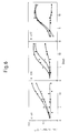
- Figure 6 shows that the rate of growth of the SCLC lines H69, H128 and H417 in serum-free medium was abolished by addition of [DPro2]spantide at 150 »m, a concentration that reversibly inhibits GRP-induced mitogenesis in Swiss 3T3 cells.
- the inhibition of growth by the antagonist in SCLC cell lines was reversed by washing the cells and resuspending them in serum-free medium.
- Figure 1 A. Time-dependence of 125I-GRP binding to Swiss 3T3 cells at 15°C. Cultures were washed and then incubated it 15°C in 1 ml binding medium containing 125I-GRP (2 nM). After various times the cells were rapidly washed four times with cold (4°C) PBS supplemented with 1 mg/ml bovine serum albumin (BSA) and then incubated with 1 ml 0.2 M acetic acid, 0.5 M NaCl at 4°C for 6 min to remove cell-surface associated ligand. This medium was then removed for counting and the remaining intracellular cell-associated radioactivity extracted with 1 ml 0.1 M NaOH containing 2% Na2CO3 and 1% SDS.
- BSA bovine serum albumin
- Open squares denote surface bound ligand and closed squares intracellular radioactivity.
- B 125I-GRP affinity labelling of an Mr 75000-85000 cellular protein using dissuccinimidyl cross-linking agents. Confluent cultures of Swiss 3T3 cells were washed and then incubated with 125I-GRP (5 nM) in the presence (+) or absence (-) of 0.9 »M unlabelled GRP for 2.5 h at 15°C. The cells were then rinsed to remove free ligand and treated with either EGS, DSS, DSP or BSCOES at concentrations of 6 mM, 2 mM, 2 mM and 4 mM respectively.
- the cells were solubilized in SDS sample buffer and electrophoresed on a 7.5% polyacrylamide gel.
- the arrow indicates the position of the Mr 75000-85000 protein. All other experimental procedures used in this and similar experiments were as described in Materials and Methods. A broad and intensely radioactive band migrating at low molecular weight was observed in all our gels. This material was not obtained in the presence of excess unlabelled GRP and therefore is most likely to represent the unreacted peptide.
- C Concentration-dependence of the effect of EGS on the affinity-labelling of the Mr 75000-85000 protein. Confluent cultures of Swiss 3T3 cells were incubated with 1 nM 125I-GRP at 15°C and then treated with various concentrations of EGS as indicated. Samples were prepared for SDS PAGE as described in Materials and Methods. The arrow indicates the position of the Mr 75000-85000 protein. In this and other experiments the efficiency of cross-linking using EGS ranged from 5 to 10% of cell-surface associated radioactivity.
- Figure 2 A. Effect of varying concentrations of unlabelled GRP on the 125I-GRP binding to intact Swiss 3T3 cells ( ⁇ ) and the affinity labelling of the Mr 75000-85000 protein ( ⁇ ).
- Confluent cultures of 3T3 cells were incubated with 5 nM 125I-GRP in the presence of varying concentrations of non-radioactive ligand at 15°C for 2.5 h. After this time some cells were washed four times with cold (4°C) PBS supplemented with 1 mg/ml BSA, solubilized and total cell associated radioactivity was then determined in a gamma counter. Parallel cultures were rinsed with PBS at 15°C and then treated with EGS (6mM) to cross-link bound 125I-GRP.
- the level of the Mr 75000-85000 protein expressed in arbitrary units is shown as a function of the concentration of the labelled ligand. Inset: autoradiogram of the gel used for scanning; the Mr 75000-85000 and 160,000 bands are indicated by arrows. All other experimental details were as described in Materials and Methods.
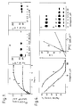
- FIG. 3 Inhibition of GRP-induced DNA synthesis by [DPro2]spantide (A) and [DPhe5]spantide (D).
- A [DPro2]spantide
- D [DPhe5]spantide
- FIG. 3 Left: Confluent and quiescent cultures of Swiss 3T3 cells in 35 mm plastic dishes were washed twice with DMEM then incubated at 37°C in 2 ml of a 1:1 mixture of DMEM/Waymouth medium containing [3H]thymidine at 1 »Ci/ml (1 »m), insulin at 1 »g/ml and increasing concentrations of GRP in the absence ( ⁇ ) or presence of antagonist A at 20 »m (o) or antagonist D at 20 »m ( ⁇ ). After 40 hours DNA synthesis was estimated by [3H]thymidine incorporation into acid-precipitable material.
- FIG. 4 Effects of [DPro2]spantide (A) and [DPhe5]spantide (D) on the binding of 125I-labelled GRP ([125I]GRP) to Swiss 3T3 cells.
- Each point represents the mean of 3 determinations.
- Center Effect of antagonist D on the affinity of binding sites in Swiss 3T3 cells for [125I]GRP. Confluent and quiescent cells were washed twice with DMEM then incubated for 30 minutes at 37°C in 1 ml of binding medium containing various concentrations of [125I]GRP in the absence ( ⁇ ) or presence ( ⁇ ) of D at 10 »m. Specific binding (B) is expressed in pmol/106 cells and is shown in a double-reciprocal plot. Each point represents the mean of duplicate determinations.
- Right Effects of antagonists A and D on the affinity labelling of the bombesin receptor-associated Mr 75000-85000 protein.
- Confluent cultures of Swiss 3T3 cells were washed twice with DMEM and incubated at 15°C in 1 ml of binding medium (pH 7.0) (15) containing 0.5 nM [125I]GRP and various concentrations of the antagonists. After 2 hours, the cultures were washed twice with binding medium then incubated in 1 ml containing 6 mM ethylene glycol bis (succinimidylsuccinate) (EGS) at pH 7.4 for 15 minutes at 15°C. The cultures were then washed twice with cold phosphate-buffered saline and solubilised in 0.1 ml of 2x sample buffer, then immediately heated at 100°C for 5 minutes and electrophoresed on a 10% polyacrylamide gel.
- binding medium pH 7.0
- EGS ethylene glycol bis (succinimidylsuccinate)
- FIG. 1 Effects of [DPro2]spantide (A) and [DPhe5]spantide (D) on the early cellular responses stimulated by GRP.
- Left Effects of antagonists on [Ca2+] i .
- Quiescent Swiss 3T3 cells grown on Cytodex 2 beads were washed twice with DMEM and incubated for 10 minutes with fura-2 tetracetoxymethyl ester at 1 »m, then washed three times and suspended in 2 ml of electrolyte solution (6) in the fluorimeter at 37°C and stirred. Fluorescence was recorded continuously in a Perkins-Elmer LS5 luminescence spectrometer with an excitation wavelength of 335 nM and emission wavelength of 510 nM.
- Cells were washed twice with RPMI 1640 medium then incubated in the serum-free medium in the absence ( ⁇ ) or presence ( ⁇ , ⁇ ) of [DPro2]spantide at 150 »m. After 4 days they were again washed twice with RPMI 1640 medium then resuspended at a density of 5 x 10⁴ cells per ml in the absence ( ⁇ , ⁇ ) or presence ( ⁇ ) of [DPro2]spantide at 150 »m (Day 0). Cell number was determined at intervals over 14 days in a Coulter Counter after disaggregation of cell clumps by syringing through 19G and 21G needles. Each point represents the mean of 3 determinations.
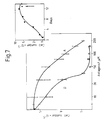
- FIG. 7 Inhibition of SCLC growth in vitro by [DPro2]spantide and [DPhe5]spantide is concentration-dependent. Cultures of H69 cells were washed twice with RPMI 1640 medium then incubated in serum-free medium (as in Figure 6) in the absence ( ⁇ ) or presence of various concentrations of [DPro2]spantide (o,A) and [DPhe5]spantide ( ⁇ ,D). Cell number was determined in a Coulter Counter after disaggregation of cell clumps by syringing through 19G and 21 G needles. Samples were incubated for 13 days, when the controls (inset) had achieved 10-fold increase in number, indicated by the arrow.
- GRP 3T3 cells were incubated with 125I-GRP (0.5 nM) at 15°C for 2.5 h in either the absence (-) or presence of the following GRP-related peptides at the concentrations indicated: GRP (360 nM), the 14-27 amino acid fragment of GRP (GRP (14-127)) (30 nM), bombesin (30 nM), litorin (92 nM), neuromedin B (220 nM), the bombesin antagonist [D-Arg1,D-Pro2,D-Trp 7,9 ,Leu11] substance P (100 »M) and the 1-16 amino acid fragment of GRP (GRP (1-16)) (3230 nM).
Landscapes
- Health & Medical Sciences (AREA)
- Chemical & Material Sciences (AREA)
- Life Sciences & Earth Sciences (AREA)
- Organic Chemistry (AREA)
- Genetics & Genomics (AREA)
- Proteomics, Peptides & Aminoacids (AREA)
- Zoology (AREA)
- Biochemistry (AREA)
- Biophysics (AREA)
- General Health & Medical Sciences (AREA)
- Toxicology (AREA)
- Medicinal Chemistry (AREA)
- Molecular Biology (AREA)
- Gastroenterology & Hepatology (AREA)
- Endocrinology (AREA)
- Cell Biology (AREA)
- Immunology (AREA)
- Peptides Or Proteins (AREA)
- Preparation Of Compounds By Using Micro-Organisms (AREA)
- Micro-Organisms Or Cultivation Processes Thereof (AREA)
- Medicines That Contain Protein Lipid Enzymes And Other Medicines (AREA)
- Medicines Containing Antibodies Or Antigens For Use As Internal Diagnostic Agents (AREA)
Abstract
Description
- THIS INVENTION relates to uses of antagonists and and novel antagonists to bombesin receptors.
- The amphibian tetradecapeptide bombesin (6) and mammalian peptides structurally related to bombesin, which include gastric releasing peptide (GRP) and the neuromedins (7 to 12) are growth factors which are believed to be implicated in the control of cell proliferation. Bombesin-like peptides are present in high concentrations in small cell lung carcinoma (18 to 21) where they could act as autocrine growth factors (22). The bombesin group of peptides interact in the cell with receptors but while a certain amount is known about the chemistry of the bombesin group of peptides, very little is known about the chemistry of the receptors of the bombesin-like peptides. In view of the involvement of the bombesin-like peptides in cell growth and the implications on cell growth of the presence or absence of bombesin/receptor interactions, a detailed study of the receptors is clearly of importance.
- We have now developed methods that have enabled us to identify certain receptors to certain peptides of the bombesin family which enables them to be characterised as new compounds and it is with such receptors and, additionally, to antagonists and antibodies to such receptors, that the present invention is directed. The bombesin family of peptides do have structural differences from one another but also have a common 7-amino acid sequence and we believe that the receptor we have identified is capable of acting as receptor to various members of the bombesin family, regardless of the species of origin of the bombesin-like peptide.
- So-called Substance P is an 11-mer neuropeptide, of interest in studies in pain transmission, which has the formula:
Arg-Pro-Lys-Pro-Gln-Gln-Phe-Phe-Gly-Leu-Met-NH₂
There is a commercially available structural variant of Substance P, known as [D-Arg¹, D-Pro², D- Trp7,9, Leu¹¹] Substance P and hence of the formula:
D-Arg-D-Pro-Lys-Pro-Gln-Gln-D-Trp-Phe-D-Trp-Leu-Leu-NH₂
which we call Antagonist A and which is also known as [D-Pro²]spantide, is, able to act as a bombesin antagonist. - We have found that a further commercially available structural variant of Substance P, which we call Antagonist D, which is also known as [D-Phe⁵]spantide and which has the formula:
D-Arg-Pro-Lys-Pro-D-Phe-Gln-D-Trp-Phe-D-Trp-Leu-Leu-NH₂
is even more potent as a bombesin antagonist than Antagonist A and hence is also of great interest for the modification of cell growth influenced by the presence of bombesin-like peptides. - Our studies of Antagonist D indicate to us the importance of structural variation at
amino acid positions
D-Arg-X-Lys-Pro-Y-Gln-D-Trp-Phe-D-Trp-Leu-Leu-NH2(I)
wherein X is D-Pro or Pro and Y is D-Trp and also relates to their use in a method of treatment of the human or animal body by therapy or in a method of diagnosis carried out on the human or animal body. - The Compounds of formula I of the invention may be formulated with pharmaceutically acceptable carriers or diluents e.g. conventional parenteral carriers so that the Compounds I can be administered parenterally where they are of interest for use in a method of treatment of the human or animal body for diagnosis or therapy, and more specifically in the diagnosis or therapy of cancers where uncontrolled cell growth is associated with disorders of proteins of the bombesin family. The Compounds I are also of interest for use in the production of a medicament for the treatment of uncontrolled cell proliferation.
- The Compounds I of the invention and Antagonist D are also of interest for use in the in vitro diagnosis of uncontrolled cell proliferation by a method which includes the step of bringing the antagonist or antibody into contact with a body sample from a host suspected of suffering from uncontrolled cell proliferation. The method is particularly useful in the diagnosis of cancers by histology or serum assay.
- The isolation and molecular characterisation of the receptors requires a procedure for their identification and we have developed a system involving the use of certain bifunctional cross-linking reagents which have already been used to identify membrane receptors in other systems (23 to 28) . We have used the cross-linking agent ethylene glycol bis(succinimidyl succinate) to link covalently ¹²⁵I labelled gastrin releasing peptide (¹²⁵I-GRP) to a surface protein in Swiss 3T3 cells. This surface protein, when isolated from the Swiss 3T3 cells, displays the characteristics of a specific receptor for the peptides of the bombesin family. This protein was not present in other cell lines which do not exhibit receptor properties for the bombesin-like peptides.
- More specifically, the polypeptide receptor can be isolated by a process which comprises incubating a culture of Swiss 3T3 cells in a culture medium including ¹²⁵I labelled gastrin releasing peptide (¹²⁵I-GRP), further incubating the ¹²⁵I-GRP treated Swiss 3T3 cells in the presence of a bifunctional crosslinking reagent and solubilising the resulting ¹²⁵I-GRP/ crosslinking reagent/polypeptide conjugate to release the polypeptide from the cell surface.
- Swiss 3T3 cells are widely available for experimental use and are available from the American Type Culture Collection in Rockville, Maryland, U.S.A., under the Deposit No. ATCC-CCL92.
- The following Examples are given to illustrate present invention.
- Bombesin and litorin were obtained from Sigma. GRP, the 14-27 amino acid fragment of GRP and [D-Arg¹,D-Pro²,D-Trp7,9,Leu¹¹] substance P were obtained from Bachem Fine Chemicals (Saffrom Walden, U.K.) ad the 1-16 fragment of GRP and neuromedin B were from Peninsula Laboratories (San Carlos, CA). Highly purified platelet-derived growth factor (PDGF) was obtained from Bioprocessing. Ethylene glycol bis (succinimidylsuccinate) (EGS), disuccinimidyl suberate (DSS), dithio-bis (succinimidylpropionate) (DSP) ad bis [2-(succinimidooxycarbomyloxy)ethyl] sulphone (BSCOES) were purchased from Pierce Chemical Co. ¹²⁵I-GRP (2000 Ci/mmol; 1Ci = 37 GBq) was obtained from the Radiochemical Centre (Amersham, U.K.) or was prepared by radiolabelling GRP with ¹²⁵I using the soluble lactoperoxidase method (29,30). The labelled peptide was separated from unreacted Na ¹²⁵I as described (8). ¹²⁵I-GRP exhibited mitogenic activity within a similar concentration range to that observed with the unlabelled peptide. All other reagents used were of the highest grade available.
- Confluent and quiescent cultures of Swiss 3T3 cells were incubated at 15°C in 1ml of medium consisting of 0.14 M NaCl, 5 mM KCl, 0.01 M Na₂HPO₄, 1.8 mM KH₂PO₄, 1.8 mM CaCl₂, 1 mM MgCl₂, 25 mM 4-(2-hydroxyethyl)-1-piperazine ethane sulphonic acid (binding medium) pH 7.0, supplemented with 0.1% BSA and the appropriate concentration of ¹²⁵I-GRP in the presence or absence of a 500-fold excess of unlabelled GRP. After 2.5 h the cells were washed three times at 15°C with phosphate buffered saline (PBS) ad then incubated for 15 min at 15°C In 1 ml of binding medium, pH 7.4 In the presence of the appropriate cross-linking agent at the concentration indicated. The cross-linking agents (EGS, DSS, DSP and BSCOES) were dissolved in dimethyl sulphoxide immediately prior to use and were added to medium to give a final concentration of dimethylsulphoxide of 1-2%. The cultures were rapidly rinsed twice with PBS at 4°C and solubilized in 0.1ml of 2 x sample buffer 0.2 M Tris-HCl, pH 6.8, 10% (w/v) glycerol, 6% sodium dodecyl sulphate (SDS) (w/v) 4% β-mercaptoethanol (v/v) and 2 mM ethylenediaminetetraacetic acid. Samples were immediately heated at 100°C for 3-5 min and analysed by either one or two-dimensional gel electrophoresis.
- Slab gel electrophoresis was performed using 7.5% acrylamide in the separating
gel ad 5% in the stacking gel, ad 0.1% SDS (31). After electrophoresis gels were stained, destained and dried down onto paper for autoradiography with Fuji X-ray film (Fuji Photo Film Co. Ltd. Japan). Dried gels were exposed to film for 4-8 days. Two-dimensional gel electrophoresis was performed as described by O'Farrell (32) using isoelectric focusing in the 1st dimension and SDS-polyacrylamide gel electrophoresis (SDS-PAGE) (8% polyacrylamide) in the second dimension. The samples prepared for isoelectric focusing containing 1.4% LKB ampholytes, pH 5-7, plus 0.6% LKD ampholytes, pH 3.5-10, 6M urea, and 2% Nonidet P-40. The Mr 75000-85000 band from autoradiograms was scanned using a Joyce-Loebl double beam densitometer and the areas under specific peaks were measured with a Hewlett-Packard digitizer. - Cell culture procedure (33), assays of DNA synthesis by ³H-thymidine incorporation (34) ad ¹²⁵I-GRP binding to intact cells (13) were performed as described previously.
- When cultures of confluent and quiescent Swiss 3T3 cells were incubated with ¹²⁵I-GRP at 15°C cell-associated radioactivity reached a maximum after 2.5 h (Fig. 1A) and was considerably enhanced compared with surface binding at either 37°C or 4°C (unpublished observations). Analysis by SDS-PAGE of cells which were incubated with 5nM ¹²⁵I-GRP at 15°C for 2.5 h, and treated with the homobifunctional cross-linking agents EGS, DSS, DSP or BSCOES revealed the presence of a single major band of Mr 75000-85000 (Fig. 1B). Identical results were obtained using either commercially available ¹²⁵I-GRP or GRP radiolabelled with ¹²⁵I in our laboratory. In the presence of 500-fold excess unlabelled GRP(+) this band was completely abolished. EGS displayed the greatest efficiency of cross-linking and this agent was therefore used in subsequent experiments. The effect of EGS on the level of the Mr 75000-85000 protein was concentration-dependent; the half-maximal effect of EGS was obtained at a concentration of 2 mM (Fig. 1C). The rank order of cross-linking efficiency (EGS>DSS>DSP>BSCOES) may be related to the chain length of the arms of these bifunctional molecules. Thus BSCOES which has the shortest chain length was virtually ineffective (Fig. 1B). The Mr 75000-85000 protein appeared as a multicomponent spot migrating with an isoelectic point of 6.4 to 6.9 when cross-linked cultures of Swiss 3T3 cells were analysed by two-dimensional gel electrophoresis using isoelectric focusing in the first dimension and SDS-PAGE in the second dimension (results not shown).
- The Mr 75000-85000 band was not obtained when the cross-linking reaction was carried out either in the absence of the cross-linking agent, in plastic dishes without cells, or using other cell lines including Rat-1, whole mouse embryo fibroblasts ad Balbc/3T3, which neither exhibit significant specific ¹²⁵I-GRP binding, nor respond mitogenically to bombesin-related peptides (13). The possibility that the affinity labelled band was a degradation product of a higher molecular weight protein was tested by extracting cultures after the cross-linking reaction in the presence of the protease inhibitors aprotinin (100 »g/ml), pepstatin (4»g/ml), phenylmethylsulphonylfluoride (2 mM) and ethyleneglycol-bis-(β-aminoethyl ether)N,N'-tetra-acetic acid (4 mM). These treatments had no significant effect on the level of the Mr 75000-85000 protein and did not result in the appearance of any higher molecular weight proteins. In another experiment performed at 4°C to prevent ligand internalization and degradation identical results were obtained to those shown in fig. 1 (results not shown). Thus, the Mr 75000-85000 protein was neither a intracellular component associated with internalized ¹²⁵I-GRP or a product of peptide degradation, nor a fragment arising from proteolysis of a larger molecule. In addition, treatment with 0.6 M 2-mercaptoethanol did not result in the appearance of additional bands of lower molecular weight suggesting that the Mr 75000-85000 protein consists of a single polypeptide chain. These important controls strongly suggested that the Mr 75000-85000 band was a surface component of Swiss 3T3 cells closely related to the receptor for peptides of the bombesin family.
- The above conclusion was further substantiated by cross-linking ¹²⁵I-GRP to cultures incubated in the presence of different concentrations of unlabelled peptide. The decrease in the level of the Mr 75000-85000 band with increasing concentrations of unlabelled GRP (Fig. 2A, open symbols) closely paralleled the ability of GRP to Inhibit the binding of an identical concentration of ¹²⁵I-GRP in a parallel set of cultures (Fig. 2A, closed symbols). The cross-linking of ¹²⁵I-GRP to Swiss 3T3 cells was also markedly inhibited by other peptides structurally related to GRP including bombesin, neuromedin B, litorin and the bombesin antagonist [D-Arg¹,D-Pro²,D-Trp7,9,Leu¹¹] substance P (13,17,35,36). The amino-terminal fragment of GRP (GRP(1-16)) which neither inhibits ¹²⁵I-GRP binding, nor stimulates DNA synthesis (13) caused no reduction in the level of the Mr 75000-85000 protein (Table 1). To ascertain the specificity with which ¹²⁵I-GRP recognizes the Mr 75000-85000 protein, Swiss 3T3 cells were incubated with ¹²⁵I-GRP In the presence of a variety of other mitogens for these cells. As shown In Table 1, the level of the protein obtained by treatment with EGS was not substantially affected by saturating concentrations of PDGF, epidermal growth factor (EGF), vasopressin, insulin,
ad phorbol 12,13-dibutyrate (1,37,38). In addition the neuropeptides substance P, substance K and somatostatin also bad no effect on affinity labelling of the Mr 75000-85000 band (Table 1). This result is in accord with the finding that the binding of ¹²⁵I-GRP to Intact 3T3 cells is also not inhibited by these mitogens (13). - The conclusion that the Mr 75000-85000 protein is a major component of the receptor for peptides of the bombesin family in Swiss 3T3 cells was further strengthened by measuring the level of the protein as a function of the concentration of the radioiodinated ligand. Fig. 2B shows that the cross-linking of ¹²⁵I-GRP to the Mr 75000-85000 band increased in a saturable manner with increasing concentration of the labelled peptide. A double-reciprocal plot of these data (not shown) produced a straight line and gave a value for the Kd of 1 nM which compares very favourably with the Kd (0.5 x 10⁻⁹M) obtained from Scatchard analysis of binding curves (13). Furthermore, the dependence of affinity labelling of the Mr 75000-85000 protein on ¹²⁵I-GRP concentration closely parallels the ability of the peptide to stimulate a variety of early biological responses (16,17) and DNA synthesis (13) In quiescent Swiss 3T3 cells. At high concentrations of ¹²⁵I-GRP a higher molecular weight band of approximately Mr 160,000 was observed (Fig. 2B). This band represented 4% of the total cross-linked material and was almost undetectable either at lower levels of ¹²⁵I-GRP or at 4°C.
- Footnote¹ The abbreviations used are: GRP, gastrin releasing peptide; BSA, bovine serum albumin; PBS, phosphate-buffered saline; EGF, spidermal growth factor. PDGF, platelet-derived growth factor; EGS, ethyleneglycolbis (succinimidylsuccinate); DSS, disuccinimidyl suberate; DSP, dithio-bis-(succinimidylpropionate); BSCOES, bis[2-(succimidooxycarbomyloxy)ethyl]sulphone; SDS-PAGE, sodium dodecyl sulphate-polyacrylamide gel electrophoresis.
- Substance P has a slight amino acid sequence homology with bombesin (Table 2) and neither inhibits the binding of GRP to Swiss 3T3 cells nor stimulates DNA synthesis. However, (DPro²]spantide (Antagonist A, Table 2) which was synthesized as a substance P antagonist was found to be a bombesin antagonist in pancreatic acinar cells and to block the growth-promoting effects of bombesin in Swiss 3T3 cells. In order to identify a more potent antagonist of bombesin-like peptides, we have tested ten substance P antagonists at 50 »m (Table 2) for their ability to inhibit mitogenesis stimulated by GRP (the mammalian homologue of bombesin in Swiss 3T3 cells. [DPhe⁵]spantide (Antagonist D) was clearly the most potent GRP antagonist. In contrast, peptides, B, C, E, F, G, H, J and K were less potent than either A or D. Spantide (B) had no antagonist activity even at 100 uM. None of the peptides stimulated DNA synthesis when tested at 20 »m with insulin at 1 »g/ml i.e. none exhibited any agonist activity.
- Following the identification of [DPhe⁵]spantide as the most promising GRP antagonist, we compared the potency of [DPhe⁵]spantide with that of [DPro²]spantide. Figure 3 shows that [DPhe⁵]spantide at 20 »m markedly increased the concentration of GRP required to produce half-maximal stimulation of DNA synthesis whereas addition of [DPro²]spantide also at 20 »m had only a slight effect. Inhibition of DNA synthesis by [DPhe⁵]spantide was completely reversed by high concentrations of GRP, indicating that its inhibitory effect was competitive and reversible. The dose response curves for the two antagonists in the presence of GRP 3.6 nM are shown in Figure 3 (right) . Half-maximal inhibition of DNA synthesis was obtained with 22 »m [DPhe⁵]spantide and 118 »m [DPro²]spantide. Thus, [DPhe⁵]spantide is 5.4-fold more potent than [DPro²]spantide in inhibiting DNA synthesis induced by GRP.
- The following Examples demonstrate that Antagonist D inhibits all of the events initiated by Bombesin/GRP in a specific manner.
- The preceding results demonstrate that [DPhe⁵]spantide is a potent inhibitor of GRP-induced DNA synthesis which exhibits specificity against other mitogens. To elucidate its mechanism of action, we examined the effect of [DPhe⁵]spantide on the specific binding of [¹²⁵I]GRP to Swiss 3T3 cells. Figure 4 (left) shows that both [DPro²]spantide and [DPhe⁵]spantide caused a concentration-dependent inhibition in the specific binding of [¹²⁵I]GRP (1 nM). Half-maximal inhibition of binding was achieved with 2.3 »m of [DPhe⁵]spantide and 14 »m of [DPro²]spantide, a 6.1-fold difference in potency. This is consistent with the relative potencies of the two antagonists in inhibiting DNA synthesis induced by GRP.
- The binding of different concentrations of [¹²⁵I]GRP was measured in the absence and presence of 10 »m [DPhe⁵ ]spantide. A double reciprocal plot of these data (Figure 4, center) shows that [DPhe⁵]spantide markedly reduces the affinity of the receptors of [¹²⁵I]GRP, although the number of binding sites is unchanged. This is consistent with results previously obtained with [DPro²[spantide and strongly suggests that these peptides bind competitively to the GRP receptor.
- To further substantiate these findings, we investigated the effects of the two antagonists on the affinity-labelling of the recently described Mr 75000-85000 protein which is a putative bombesin receptor (Figure 4, right). They were both able to differentially inhibit the Mr 75000-85000 protein obtained by cross-linking [¹²⁵I]GRP to Swiss 3T3 cells with EGS. Half-maximal inhibition (obtained by scanning densitometry of the autoradiographs) was achieved with [DPhe⁵]spantide at 5.5 »m and [DPro²]spantide at 20 »m, again demonstrating the superiority of [DPhe⁵]spantide.
- One of the earliest events stimulated by addition of bombesin or GRP to quiescent Swiss 3T3 cells is an increase in cytosolic Ca²⁺ concentration ([Ca²⁺]i). Figure 5 (left) shows that the rise in [Ca²⁺]i caused by the addition of GRP (1 nM) to quiescent Swiss 3T3 cells was prevented by the addition of [DPro²]spantide at 20 »m but not at 5 »m. In contrast [DPhe⁵]spantide was effective at 5 »m, demonstrating that [DPhe⁵]spantide is at least 4-fold more potent than [DPro²]spantide in this assay. These effects were specific and reversible since [DPhe⁵] spantide at 5 »m did not prevent a response to PDGF and because the effects of the antagonist in preventing Ca²⁺ mobilization in response to 5 nM GRP was reversed by addition of GRP at 50 nM.
- Inhibition of [¹²⁵I]EGF binding by GRP, which is mediated by the protein kinase C pathway, was reversed in a concentration-dependent fashion by [DPro²]spantide and [DPhe⁵]spantide (Figure 5, right). Half-maximal reversal of inhibition was obtained with [DPhe⁵]spantide at 8.7 »m and [DPro²]spantide at 30 »m. These findings further substantiate the conclusion that [DPhe⁵]spantide is a potent GRP antagonist.
- In addition to the demonstration of inhibition of the cellular effects of Bombesin/GRP in mouse 3T3 cells we have now shown that Antagonists A and D can inhibit the growth of SCLC cells in a specific and reversible fashion.
- SCLC is known to secrete bombesin-like peptides which have been suggested to act as autocrine growth factors. Thus it is plausible but as yet unproven that an antagonist to bombesin/GRP will inhibit SCLC growth, so we have now tested the effects of [DPro²]spantide and [DPhe⁵]spantide on SCLC in vitro.
- Figure 6 shows that the rate of growth of the SCLC lines H69, H128 and H417 in serum-free medium was abolished by addition of [DPro²]spantide at 150 »m, a concentration that reversibly inhibits GRP-induced mitogenesis in Swiss 3T3 cells. The inhibition of growth by the antagonist in SCLC cell lines was reversed by washing the cells and resuspending them in serum-free medium.
- The effects of [DPro²]spantide and [DPhe⁵]spantide on H69 cells are compared in Figure 7. The cells achieve 10-fold increase in number in about 12 days in serum-free medium (inset). Both antagonists inhibited growth in a dose-dependent manner; half-maximal effect was seen at 24 »m and [DPhe⁵]spantide and at 82 »m with [DPro²]spantide. The difference in potency is of the same magnitude as that demonstrated in Swiss 3T3 cells and supports the contention that bombesin-like peptides (or vasopressin) may be important growth factors for SCLC. These results show the more than 5-fold potency of Antagonist D over Antagonist A.
- Figure 1: A. Time-dependence of ¹²⁵I-GRP binding to Swiss 3T3 cells at 15°C. Cultures were washed and then incubated it 15°C in 1 ml binding medium containing ¹²⁵I-GRP (2 nM). After various times the cells were rapidly washed four times with cold (4°C) PBS supplemented with 1 mg/ml bovine serum albumin (BSA) and then incubated with 1 ml 0.2 M acetic acid, 0.5 M NaCl at 4°C for 6 min to remove cell-surface associated ligand. This medium was then removed for counting and the remaining intracellular cell-associated radioactivity extracted with 1 ml 0.1 M NaOH containing 2% Na₂CO₃ and 1% SDS. Open squares denote surface bound ligand and closed squares intracellular radioactivity. B. ¹²⁵I-GRP affinity labelling of an Mr 75000-85000 cellular protein using dissuccinimidyl cross-linking agents. Confluent cultures of Swiss 3T3 cells were washed and then incubated with ¹²⁵I-GRP (5 nM) in the presence (+) or absence (-) of 0.9 »M unlabelled GRP for 2.5 h at 15°C. The cells were then rinsed to remove free ligand and treated with either EGS, DSS, DSP or BSCOES at concentrations of 6 mM, 2 mM, 2 mM and 4 mM respectively. The cells were solubilized in SDS sample buffer and electrophoresed on a 7.5% polyacrylamide gel. The arrow indicates the position of the Mr 75000-85000 protein. All other experimental procedures used in this and similar experiments were as described in Materials and Methods. A broad and intensely radioactive band migrating at low molecular weight was observed in all our gels. This material was not obtained in the presence of excess unlabelled GRP and therefore is most likely to represent the unreacted peptide. C. Concentration-dependence of the effect of EGS on the affinity-labelling of the Mr 75000-85000 protein. Confluent cultures of Swiss 3T3 cells were incubated with 1 nM ¹²⁵I-GRP at 15°C and then treated with various concentrations of EGS as indicated. Samples were prepared for SDS PAGE as described in Materials and Methods. The arrow indicates the position of the Mr 75000-85000 protein. In this and other experiments the efficiency of cross-linking using EGS ranged from 5 to 10% of cell-surface associated radioactivity.
- Figure 2: A. Effect of varying concentrations of unlabelled GRP on the ¹²⁵I-GRP binding to intact Swiss 3T3 cells (●) and the affinity labelling of the Mr 75000-85000 protein (○). Confluent cultures of 3T3 cells were incubated with 5 nM ¹²⁵I-GRP in the presence of varying concentrations of non-radioactive ligand at 15°C for 2.5 h. After this time some cells were washed four times with cold (4°C) PBS supplemented with 1 mg/ml BSA, solubilized and total cell associated radioactivity was then determined in a gamma counter. Parallel cultures were rinsed with PBS at 15°C and then treated with EGS (6mM) to cross-link bound ¹²⁵I-GRP. The cells were then solubilized and samples were analyzed by SDS-PAGE. After electrophoresis, gels were dried, exposed to film and scanned as described in Materials and Methods. The areas under the individual peaks for the Mr 75000-85000 protein are shown as a function of the concentration of the unlabelled peptide and are expressed as a percentage of the control. The inset shows the region of the autoradiogram used for the scanning. B. Concentration-dependence of ¹²⁵I-GRP affinity labelling of the Mr 75000-85000 protein. Confluent cultures of Swiss 3T3 cells were incubated with different concentrations of ¹²⁵I-GRP at 15°C for 2.5h, and treated with EGS (6 mM). The level of the Mr 75000-85000 protein expressed in arbitrary units is shown as a function of the concentration of the labelled ligand. Inset: autoradiogram of the gel used for scanning; the Mr 75000-85000 and 160,000 bands are indicated by arrows. All other experimental details were as described in Materials and Methods.
- Figure 3. Inhibition of GRP-induced DNA synthesis by [DPro²]spantide (A) and [DPhe⁵]spantide (D). Left: Confluent and quiescent cultures of Swiss 3T3 cells in 35 mm plastic dishes were washed twice with DMEM then incubated at 37°C in 2 ml of a 1:1 mixture of DMEM/Waymouth medium containing [³H]thymidine at 1 »Ci/ml (1 »m), insulin at 1 »g/ml and increasing concentrations of GRP in the absence (■) or presence of antagonist A at 20 »m (o) or antagonist D at 20 »m (△). After 40 hours DNA synthesis was estimated by [³H]thymidine incorporation into acid-precipitable material. Values are expressed as a percentage of [³H]thymidine-incorporation obtained with a saturating level of GRP (36 nM) in the absence of antagonist (100% = 8.4 x 10⁵ cpm per dish). Each point represents the mean of duplicate determinations. Right: Cultures of Swiss 3T3 cells were washed and incubated as above, except that the concentration of GRP was fixed at 3.6 nM with various concentrations of antagonists A (o) and D (△). Values are expressed as a percentage of [³H]thymidine incorporation obtained in the absence of antagonists (100% = 8.6 x 10⁵ cpm per dish). Each point represents the mean of 4 determinations.
- Figure 4. Effects of [DPro²]spantide (A) and [DPhe⁵]spantide (D) on the binding of ¹²⁵I-labelled GRP ([¹²⁵I]GRP) to Swiss 3T3 cells. Left: Inhibition of specific [¹²⁵I]GRP binding by antagonists A and D. Confluent and quiescent cells were washed twice with DMEM then incubated at 37°C in 1 ml of binding medium (3) containing [¹²⁵I]GRP at 1 nM and various concentrations of antagonist A (o) and D (△). Cell-associated [¹²⁵I]GRP binding was measured after 30 minutes. Values are expressed as percentage of the specific binding obtained in the absence of antagonists. Non-specific binding was determined by the addition of 300-fold excess of unlabelled GRP. Each point represents the mean of 3 determinations. Center: Effect of antagonist D on the affinity of binding sites in Swiss 3T3 cells for [¹²⁵I]GRP. Confluent and quiescent cells were washed twice with DMEM then incubated for 30 minutes at 37°C in 1 ml of binding medium containing various concentrations of [¹²⁵I]GRP in the absence (●) or presence (△) of D at 10 »m. Specific binding (B) is expressed in pmol/10⁶ cells and is shown in a double-reciprocal plot. Each point represents the mean of duplicate determinations. Right: Effects of antagonists A and D on the affinity labelling of the bombesin receptor-associated Mr 75000-85000 protein. Confluent cultures of Swiss 3T3 cells were washed twice with DMEM and incubated at 15°C in 1 ml of binding medium (pH 7.0) (15) containing 0.5 nM [¹²⁵I]GRP and various concentrations of the antagonists. After 2 hours, the cultures were washed twice with binding medium then incubated in 1 ml containing 6 mM ethylene glycol bis (succinimidylsuccinate) (EGS) at pH 7.4 for 15 minutes at 15°C. The cultures were then washed twice with cold phosphate-buffered saline and solubilised in 0.1 ml of 2x sample buffer, then immediately heated at 100°C for 5 minutes and electrophoresed on a 10% polyacrylamide gel.
- Figure 5. Effects of [DPro²]spantide (A) and [DPhe⁵]spantide (D) on the early cellular responses stimulated by GRP. Left: Effects of antagonists on [Ca²⁺]i. Quiescent Swiss 3T3 cells grown on
Cytodex 2 beads were washed twice with DMEM and incubated for 10 minutes with fura-2 tetracetoxymethyl ester at 1 »m, then washed three times and suspended in 2 ml of electrolyte solution (6) in the fluorimeter at 37°C and stirred. Fluorescence was recorded continuously in a Perkins-Elmer LS5 luminescence spectrometer with an excitation wavelength of 335 nM and emission wavelength of 510 nM. After a period of equilibration, the following additions were made: solvent, S; antagonist A at 5 »m, A, 5 and 20 »m, A, 20; antagonist D at 5»m, D, 5. After 3 minutes in each case, GRP was added at 1 nM, G or at 50 nM, G, 50; PDGF at 1 nM, P. Right: Antagonists A and D reverse the inhibition of [¹²⁵I]EGF binding induced by GRP. Confluent and quiescent cultures of Swiss 3T3 cells were washed twice with DMEM then incubated for 1 hour at 37°C in 1 ml of binding medium (9) containing [¹²⁵I]EGF at 0.2 nM and GRP at 3.6 nM alone (■) or in the presence of various concentrations of A (o) or D (△). Values are expressed as percentages of the specific binding obtained with [¹²⁵I]EGF alone at 0.2 nM. The non-specific binding was obtained by the addition of 500-fold excess of unlabelled EGF. Each point represents the mean of 6 determinations. - Figure 6. [DPro²]spantide reversibly inhibits the growth of SCLC cell lines. Stock cultures of cell lines H69, H128 and H417 were maintained in RPMI 1640 medium with 10% fetal bovine serum (heat inactivated) in a humidified atmosphere of 10% CO₂:90% air at 37°C. They were passaged every 7 days. Identical growth was obtained in the serum-free medium of RPMI 1640 supplemented with HITES (29) (hydrocortisone, 10 nM; insulin, 5 »g/ml; transferrin, 100 »g/ml; 17β-estradiol, 10 nM; sodium selenite, 30 nM) and 0.25% bovine serum albumin. Cells were washed twice with RPMI 1640 medium then incubated in the serum-free medium in the absence (●) or presence (□,■) of [DPro²]spantide at 150 »m. After 4 days they were again washed twice with RPMI 1640 medium then resuspended at a density of 5 x 10⁴ cells per ml in the absence (●,□) or presence (■) of [DPro²]spantide at 150 »m (Day 0). Cell number was determined at intervals over 14 days in a Coulter Counter after disaggregation of cell clumps by syringing through 19G and 21G needles. Each point represents the mean of 3 determinations.
- Figure 7. Inhibition of SCLC growth in vitro by [DPro²]spantide and [DPhe⁵]spantide is concentration-dependent. Cultures of H69 cells were washed twice with RPMI 1640 medium then incubated in serum-free medium (as in Figure 6) in the absence (●) or presence of various concentrations of [DPro²]spantide (o,A) and [DPhe⁵]spantide (△,D). Cell number was determined in a Coulter Counter after disaggregation of cell clumps by syringing through 19G and 21 G needles. Samples were incubated for 13 days, when the controls (inset) had achieved 10-fold increase in number, indicated by the arrow. Each point represents the mean (± standard deviation) of 5 determinations.
Table 1 Specificity of ¹²⁵I-GRP affinity labelling of the Mr 75000-85000 protein Addition Mr 75000-85000 protein % of control - 100 GRP 0.8 GRP (14-27) 0.4 Bombesin 0.5 Litorin 2 Neuromedin B 10 [D-Arg¹,D-Pro²,D-Trp7,9,Leu¹¹] Substance P 9.7 GRP (1-16) 93 PDGF 99 EGF 94 Vasopressin 94 Insulin 90 Phorbol 12,13-dibutyrate99 Substance P 91 Substance K 94 Somatostatin 97 Neurotensin 93 - Confluent cultures of Swiss 3T3 cells were incubated with ¹²⁵I-GRP (0.5 nM) at 15°C for 2.5 h in either the absence (-) or presence of the following GRP-related peptides at the concentrations indicated: GRP (360 nM), the 14-27 amino acid fragment of GRP (GRP (14-127)) (30 nM), bombesin (30 nM), litorin (92 nM), neuromedin B (220 nM), the bombesin antagonist [D-Arg¹,D-Pro²,D-Trp7,9,Leu¹¹] substance P (100 »M) and the 1-16 amino acid fragment of GRP (GRP (1-16)) (3230 nM). Parallel cultures were incubated with the same concentration of ¹²⁵I-GRP in the presence of the following unrelated factors: PDGF (5 nM), EGF (83 nM), vasopressin (1000 nM), insulin (1 »g/ml),
phorbol 12,13 dibutyrate (2000 nM), substance P (740 nM), substance K (880 nM), somatostatin (610 nM) and neurotensin (600 nM). After the incubation period cultures were treated with 6 mM EGS as described in Materials and Methods. Each value is expressed as a percentage of the level of the Mr 75000-85000 protein obtained with no additons (-). Other experimental details are as described in Materials and Methods.
-
- 1. Rozengurt, E., Legg, A. and Pettican, P. (1979) Proc. Natl. Acad. Sci. USA. 76, 1284-1287.
- 2. Rozengurt, E. and Sinnett-Smith, J. (1983) Proc. Natl. Acad. Sci. USA. 80, 2936-2940.
- 3. Nilsson, J., von Euler, A.M. and Dalsgaard, C.-J. (1985) Nature 315, 61-63.
- 4. Payan, D.G. (1985) Biochem. Biophys. Res. Commun. 130, 104-109.
- 5. Singh, P., Walker, P., Townsend, C.M. Jr and Thompson, J.C (1986) Cancer Res. 46, 1612-1616.
- 6. Anastasi, A., Erpsamer, V. and Bucci, M. (1971) Experientia 27, 166-167.
- 7. McDonald, T.J., Jornvall, H., Nilsson, G., Vagne, M., Ghatei, M., Bloom, S.R. and Mutt, V. (1979) Biochem. Biophys. Res. Commun. 90, 227-233.
- 8. Wharton, J., Polak, J.M., Bloom, S.R., Ghatei, M.A., Solicia, E., Brown, M.R. and Pearse, A.E.G. (1978) Nature 273, 769-770.
- 9. Moody, T.W. and Pert, C.B. (1979) Biochem. Biophys. Res. Commun. 90, 7-14.
- 10. Minamino, N., Kargawa, K. and Matsuo, H. (1983) Biochem. Biophys. Res. commun. 114, 541-548.
- 11. Minamino, N., Kangawa, K. and Matsuo, H. (1964) Biochem. Biophys. Res. Commun. 119, 14-20.
- 12. Minamino, N., Sudoh, T., Kangawa, K. and Matsuo, H. (1985) Biochem. Biophys. Res. Commun. 130, 685-691.
- 13. Zachary, I. and Rozengurt, E. (1985) Proc. Natl. Acad. Sci. USA. 82, 7616-7620.
- 14. Brown, K.D., Blay, J., Irvine, R.F., Heslop, J.P. and Berridge, M.J. (1984) Biochem. Biophys. Res. Commun. 123, 377-384.
- 15. Wakelam, M.J.O, Davies, S.A., Houslay, M.D., McKay, I., Marshall, C.J. and Hall, A. (1986) Nature 323, 173-176.
- 16. Mondoza, S.A., Schneider, J.A., Lopez-Rivas, A., Sinnett-Smith, J.W. and Rozengurt, E. (1986) J. Cell Biol. 102, 2223-2233.
- 17. Zachary, I., Sinnett-Smith, J.W. and Rozengurt, E. (1986) J. Cell Biol. 102, 2211-2222.
- 18. Moody, T.W., Pert, C.B., Gazdar, A.F., Carney, D.N. and Minna, J.D. (1981) Science 214, 1246-1248.
- 19. Wood, S.M., Wood, J.R., Ghatei, M.A., Lee, Y.C., O'Shaughnessy, D. and Bloom, S.R. (1981) J. Clin. Endocrin. Metab. 53, 1310-1312.
- 20. Erisman, M.D., Linnoila, R.I., Hernandez, O., Di Augustime, R.P. and Lazarus, L.H. (1982) Proc. Natl. Acad. Sci. USA. 79, 2379-2383.
- 21. Roth, K.A., Evans, C.J., Weber, E., Barchas, J.D., Bostwick, D.G. and Bersch, K.G. (1983) Cancer Res. 43, 5411-5415.
- 22. Cuttita, F., Carney, D.N., Mulshine, J., Moody, T.W., Fedorko, J., Fischler, A. and Minna, J.D. (1985) Nature 316, 823-826.
- 23. Pilch, P.F. and Czech, M.P. (1979) J. Biol. Chem. 254, 3375-3381.
- 24. Massaque, J., Guilletta, B.J. and Czech, M.P.C. (1981) J. Biol. Chem. 256, 2122-2125.
- 25. Glenn, K., Bowen-Pope, D.F. and Ross, R. (1982) J. Biol. Chem. 257, 5172-5176.
- 26. Wood, C.L. and O'Dorisio, M.S. (1985) J. Biol. Chem. 260, 1243-1247.
- 27. Brenner, M.B., Trowbridge, I.S. and Strominger, J.L. (1985)
Cell 40, 183-190. - 28. Sorensen, P., Farber, N.M. and Krystal, G. (1986) J. Biol. Chem. 261, 9094-9097.
- 29. Rozengurt, E., Brown, K.D. and Pettican, P. (1981) J. Biol. Chem. 256, 716-722.
- 30. Rozengurt, E., Collins, M., Brown, K.D. and Pettican, P. (1982) J. Biol. Chem. 257, 3680-3686.
- 31. Laemmli, U.K. (1970) Nature 227, 680-685.
- 32. O'Farrel, P.H. (1975) J. Biol. Chem. 250, 4007-4021.
- 33. Todaro, G.J. and Green, H. (1963) J. Cell Biol. 17, 299-313.
- 34. Dicker, P. and Rozengurt, E. (1980) Nature 287, 607-612.
- 35. Jensen, R.T., Jones, S.W., Folkers, K. and Gardner, J.D. (1984) Nature 309, 61-63.
- 36. Zachary, I. and Rozengurt, E. (1986) Biochem. Biophys. Res. Commun. 137, 135-141.
- 37. Rozengurt, E., Stroobant, P., Waterfield, M.D., Deuel, T.F. and Keehan, M. (1983) Cell 34, 265-272.
- 38. Dicker, P. and Rozengurt, E. (1978) Nature 276, 723-726.
- 39. Wray, W., Boulikas, T., Wray, V.P. and Hancock, R. (1981) Analy. Biochem. 118, 197-203.
Claims (6)
- The use of [D-Arg¹, D-Phe⁵, D-Trp7,9 ,Leu¹¹] substance P (Antagonist D)for the production of a medicament for the treatment of uncontrolled cell proliferation.
- The use of a compound of formula (I)
D-Arg-X-Lys-Pro-Y-Gln-D-Trp-Phe-D-Trp-Leu-Leu-NH₂ (I)
wherein X is D-Pro or Pro and Y is D-Trp, for the production of a medicament for the treatment of uncontrolled cell proliferation. - A compound of the general formula (I)
D-Arg-X-Lys-Pro-Y-Gln-D-Trp-Phe-D-Trp-Leu-Leu-NH₂ (I)
wherein X is D-Pro or Pro and Y is D-Trp. - A pharmaceutical composition comprising a compound according to claim 3 together with a pharmaceutically acceptable carrier or diluent.
- A compound according to claim 3 for use in a method for the treatment of the human or animal body by therapy or in a method of diagnosis.
- A method for the in vitro diagnosis of uncontrolled cell proliferation which includes the step of bringing into contact with one another a body sample from a host suspected of suffering from uncontrolled cell proliferation and Antagonist D as defined in claim 1 or a compound as defined in claim 3.
Applications Claiming Priority (5)
| Application Number | Priority Date | Filing Date | Title |
|---|---|---|---|
| GB8707607 | 1987-03-31 | ||
| GB878707607A GB8707607D0 (en) | 1987-03-31 | 1987-03-31 | Growth factor receptors |
| GB878727638A GB8727638D0 (en) | 1987-11-25 | 1987-11-25 | Improvements relating to growth factor receptors |
| GB8727638 | 1987-11-25 | ||
| PCT/GB1988/000255 WO1988007551A2 (en) | 1987-03-31 | 1988-03-31 | Growth factor receptor |
Publications (2)
| Publication Number | Publication Date |
|---|---|
| EP0359745A1 EP0359745A1 (en) | 1990-03-28 |
| EP0359745B1 true EP0359745B1 (en) | 1994-08-24 |
Family
ID=26292080
Family Applications (1)
| Application Number | Title | Priority Date | Filing Date |
|---|---|---|---|
| EP88902901A Expired - Lifetime EP0359745B1 (en) | 1987-03-31 | 1988-03-31 | Growth factor receptor |
Country Status (5)
| Country | Link |
|---|---|
| EP (1) | EP0359745B1 (en) |
| JP (1) | JP2728476B2 (en) |
| AT (1) | ATE110390T1 (en) |
| DE (1) | DE3851226T2 (en) |
| WO (1) | WO1988007551A2 (en) |
Families Citing this family (8)
| Publication number | Priority date | Publication date | Assignee | Title |
|---|---|---|---|---|
| US5434132A (en) * | 1988-03-21 | 1995-07-18 | Imperial Cancer Research Technology, Ltd. | Neuropeptide antagonists |
| GB8806644D0 (en) * | 1988-03-21 | 1988-04-20 | Imp Cancer Res Tech | Neuropeptide antagonists |
| US5215963A (en) * | 1989-10-24 | 1993-06-01 | Berlex Laboratories, Inc. | Solubilization and purification of the active gastrin releasing peptide receptor |
| DE3940209A1 (en) * | 1989-12-05 | 1991-06-06 | Boehringer Mannheim Gmbh | METHOD FOR DETECTING A SMALL-CELL BRONCHIAL CARCINOMA |
| HU209312B (en) * | 1992-02-11 | 1994-04-28 | Richter Gedeon Vegyeszet | Process for producing short peptide and pseudopeptide amides inhibiting the proliferation of the cells of small cellular and epithelial lung cancer, as well as pharmaceutical compositions comprising same as active ingredient |
| US6602856B1 (en) * | 1995-01-17 | 2003-08-05 | J. Mark Quillan | Antagonists of alpha-melanocyte stimulating hormone and methods based thereon |
| GB0011100D0 (en) * | 2000-05-08 | 2000-06-28 | Matrix Therapeutics Limited | Small peptide amides inhibiting the proliferation of cancer cells |
| WO2002102835A2 (en) * | 2000-11-21 | 2002-12-27 | New England Medical Center | Novel antimicrobial compounds |
Family Cites Families (1)
| Publication number | Priority date | Publication date | Assignee | Title |
|---|---|---|---|---|
| US4481139A (en) * | 1983-04-13 | 1984-11-06 | Board Of Regents, The University Of Texas System | Peptide antagonists of substance P |
-
1988
- 1988-03-31 DE DE3851226T patent/DE3851226T2/en not_active Expired - Fee Related
- 1988-03-31 EP EP88902901A patent/EP0359745B1/en not_active Expired - Lifetime
- 1988-03-31 JP JP63502924A patent/JP2728476B2/en not_active Expired - Fee Related
- 1988-03-31 AT AT88902901T patent/ATE110390T1/en active
- 1988-03-31 WO PCT/GB1988/000255 patent/WO1988007551A2/en not_active Ceased
Non-Patent Citations (4)
| Title |
|---|
| Acta Chemica Scandinavica, volume B84, 1986, K Folkers et al.: "Design and synthsis of antagonists of substance P", pages 295-302 see table 1,nos.1, 4,10; table 2, nos 1,3,6; table 7, no.1 * |
| Layton et al.: 1987, J. Cell Biochem. Suppl, vol 340, pp. 285-302 * |
| Nature, volume 316, 29 august 1985, F.Cuttitta et al.: "Bombesin-like peptides can function as autocrine growth factors in human small-cell lung cancer", pages 823-826 see page 826, colume 1, lines 6-16 citedin the application * |
| The Journal of Biological Chemistry: volume 262, no. 9, 25 march 1987, The Amercian Society of Biological Chemists, Inc., (US), I. Zachary et al.: "Identification of a receptor for peptides of the bombesin family in Swiss 3T3 cells by affinity cross-linking", pages 3947-3950 see whole document * |
Also Published As
| Publication number | Publication date |
|---|---|
| WO1988007551A2 (en) | 1988-10-06 |
| JPH02503195A (en) | 1990-10-04 |
| WO1988007551A3 (en) | 1990-02-22 |
| ATE110390T1 (en) | 1994-09-15 |
| DE3851226D1 (en) | 1994-09-29 |
| JP2728476B2 (en) | 1998-03-18 |
| EP0359745A1 (en) | 1990-03-28 |
| DE3851226T2 (en) | 1994-12-08 |
Similar Documents
| Publication | Publication Date | Title |
|---|---|---|
| Zachary et al. | Identification of a receptor for peptides of the bombesin family in Swiss 3T3 cells by affinity cross-linking. | |
| Woll et al. | [D-Arg1, D-Phe5, D-Trp7, 9, Leu11] substance P, a potent bombesin antagonist in murine Swiss 3T3 cells, inhibits the growth of human small cell lung cancer cells in vitro. | |
| Zachary et al. | High-affinity receptors for peptides of the bombesin family in Swiss 3T3 cells. | |
| Graf et al. | A pentapeptide from the laminin B1 chain mediates cell adhesion and binds to 67000 laminin receptor | |
| Pober et al. | Two distinct monokines, interleukin 1 and tumor necrosis factor, each independently induce biosynthesis and transient expression of the same antigen on the surface of cultured human vascular endothelial cells. | |
| Guo et al. | Characterization of insulinlike growth factor I receptors in human colon cancer | |
| Dubeykovskaya et al. | Secreted trefoil factor 2 activates the CXCR4 receptor in epithelial and lymphocytic cancer cell lines | |
| US5888965A (en) | Method of stimulating bone marrow regeneration | |
| AU685765B2 (en) | (Eck) receptor ligands | |
| Horrigan et al. | Characterization of an associated microfibril protein through recombinant DNA techniques. | |
| Chen et al. | Overexpression of epidermal growth factor and insulin-like growth factor-I receptors and autocrine stimulation in human esophageal carcinoma cells | |
| Schweigerer et al. | Basic fibroblast growth factor: expression in cultured cells derived from corneal endothelium and lens epithelium | |
| Layton et al. | Effects of bombesin antagonists on the growth of small cell lung cancer cells in vitro | |
| Meyers et al. | Sorcin (V19), a soluble acidic calcium-binding protein overproduced in multidrug-resistant cells: identification of the protein by anti-sorcin antibody | |
| Ishioka et al. | Inhibitory effects of prostaglandin A2 on c-myc expression and cell cycle progression in human leukemia cell line HL-60 | |
| WO1988007551A2 (en) | Growth factor receptor | |
| AU616881B2 (en) | Inhibitors of angiogenin | |
| Weiss-Messer et al. | Prolactin and MA-10 Leydig cell steroidogenesis: biphasic effects of prolactin and signal transduction | |
| US5441935A (en) | Growth factor receptors | |
| EP1466619A2 (en) | Method for modulating cell apoptosis | |
| CA1341530C (en) | Inhibitors of angiogenin | |
| NZ241368A (en) | Fibroblast growth factor receptor blocking peptides and their use in diagnostic methods and test kits | |
| Matsuo et al. | Growth factor production by human thyroid carcinoma cells: abundant expression of a platelet-derived growth factor-B-like protein by a human papillary carcinoma cell line | |
| Moody et al. | Characterization of receptors for bombesin/gastrin-releasing peptide in human and murine cells | |
| JP2818679B2 (en) | Neuropeptide antagonist |
Legal Events
| Date | Code | Title | Description |
|---|---|---|---|
| PUAI | Public reference made under article 153(3) epc to a published international application that has entered the european phase |
Free format text: ORIGINAL CODE: 0009012 |
|
| 17P | Request for examination filed |
Effective date: 19890927 |
|
| AK | Designated contracting states |
Kind code of ref document: A1 Designated state(s): AT BE CH DE FR GB IT LI LU NL SE |
|
| D17D | Deferred search report published (deleted) | ||
| RAP1 | Party data changed (applicant data changed or rights of an application transferred) |
Owner name: IMPERIAL CANCER RESEARCH TECHNOLOGY LTD |
|
| 17Q | First examination report despatched |
Effective date: 19910724 |
|
| GRAA | (expected) grant |
Free format text: ORIGINAL CODE: 0009210 |
|
| AK | Designated contracting states |
Kind code of ref document: B1 Designated state(s): AT BE CH DE FR GB IT LI LU NL SE |
|
| PG25 | Lapsed in a contracting state [announced via postgrant information from national office to epo] |
Ref country code: NL Effective date: 19940824 Ref country code: AT Effective date: 19940824 |
|
| REF | Corresponds to: |
Ref document number: 110390 Country of ref document: AT Date of ref document: 19940915 Kind code of ref document: T |
|
| REF | Corresponds to: |
Ref document number: 3851226 Country of ref document: DE Date of ref document: 19940929 |
|
| ITF | It: translation for a ep patent filed | ||
| ET | Fr: translation filed | ||
| PG25 | Lapsed in a contracting state [announced via postgrant information from national office to epo] |
Ref country code: SE Effective date: 19941124 |
|
| NLV1 | Nl: lapsed or annulled due to failure to fulfill the requirements of art. 29p and 29m of the patents act | ||
| PLBE | No opposition filed within time limit |
Free format text: ORIGINAL CODE: 0009261 |
|
| STAA | Information on the status of an ep patent application or granted ep patent |
Free format text: STATUS: NO OPPOSITION FILED WITHIN TIME LIMIT |
|
| 26N | No opposition filed | ||
| REG | Reference to a national code |
Ref country code: GB Ref legal event code: IF02 |
|
| PGFP | Annual fee paid to national office [announced via postgrant information from national office to epo] |
Ref country code: GB Payment date: 20030326 Year of fee payment: 16 |
|
| PGFP | Annual fee paid to national office [announced via postgrant information from national office to epo] |
Ref country code: LU Payment date: 20030328 Year of fee payment: 16 |
|
| PGFP | Annual fee paid to national office [announced via postgrant information from national office to epo] |
Ref country code: CH Payment date: 20030331 Year of fee payment: 16 |
|
| PGFP | Annual fee paid to national office [announced via postgrant information from national office to epo] |
Ref country code: DE Payment date: 20030409 Year of fee payment: 16 |
|
| REG | Reference to a national code |
Ref country code: GB Ref legal event code: 732E |
|
| REG | Reference to a national code |
Ref country code: CH Ref legal event code: PUE Owner name: CANCER RESEARCH TECHNOLOGY LIMITED Free format text: IMPERIAL CANCER RESEARCH TECHNOLOGY LIMITED#SARDINIA STREET#LONDON WC2A 3NL (GB) -TRANSFER TO- CANCER RESEARCH TECHNOLOGY LIMITED#SARDINIA HOUSE, SARDINIA STREET#LONDON WC2A 3NL (GB) |
|
| REG | Reference to a national code |
Ref country code: FR Ref legal event code: TP |
|
| PGFP | Annual fee paid to national office [announced via postgrant information from national office to epo] |
Ref country code: FR Payment date: 20040309 Year of fee payment: 17 |
|
| PG25 | Lapsed in a contracting state [announced via postgrant information from national office to epo] |
Ref country code: LU Free format text: LAPSE BECAUSE OF NON-PAYMENT OF DUE FEES Effective date: 20040331 Ref country code: LI Free format text: LAPSE BECAUSE OF NON-PAYMENT OF DUE FEES Effective date: 20040331 Ref country code: GB Free format text: LAPSE BECAUSE OF NON-PAYMENT OF DUE FEES Effective date: 20040331 Ref country code: CH Free format text: LAPSE BECAUSE OF NON-PAYMENT OF DUE FEES Effective date: 20040331 |
|
| PGFP | Annual fee paid to national office [announced via postgrant information from national office to epo] |
Ref country code: BE Payment date: 20040519 Year of fee payment: 17 |
|
| PG25 | Lapsed in a contracting state [announced via postgrant information from national office to epo] |
Ref country code: DE Free format text: LAPSE BECAUSE OF NON-PAYMENT OF DUE FEES Effective date: 20041001 |
|
| REG | Reference to a national code |
Ref country code: CH Ref legal event code: PL |
|
| GBPC | Gb: european patent ceased through non-payment of renewal fee | ||
| PG25 | Lapsed in a contracting state [announced via postgrant information from national office to epo] |
Ref country code: IT Free format text: LAPSE BECAUSE OF NON-PAYMENT OF DUE FEES;WARNING: LAPSES OF ITALIAN PATENTS WITH EFFECTIVE DATE BEFORE 2007 MAY HAVE OCCURRED AT ANY TIME BEFORE 2007. THE CORRECT EFFECTIVE DATE MAY BE DIFFERENT FROM THE ONE RECORDED. Effective date: 20050331 Ref country code: BE Free format text: LAPSE BECAUSE OF NON-PAYMENT OF DUE FEES Effective date: 20050331 |
|
| BERE | Be: lapsed |
Owner name: *CANCER RESEARCH TECHNOLOGY LTD Effective date: 20050331 |
|
| PG25 | Lapsed in a contracting state [announced via postgrant information from national office to epo] |
Ref country code: FR Free format text: LAPSE BECAUSE OF NON-PAYMENT OF DUE FEES Effective date: 20051130 |
|
| REG | Reference to a national code |
Ref country code: FR Ref legal event code: ST Effective date: 20051130 |
|
| BERE | Be: lapsed |
Owner name: *CANCER RESEARCH TECHNOLOGY LTD Effective date: 20050331 |