EP0357856A1 - DNA coding for human ventricular myosin light chain 2 - Google Patents
DNA coding for human ventricular myosin light chain 2 Download PDFInfo
- Publication number
- EP0357856A1 EP0357856A1 EP88312271A EP88312271A EP0357856A1 EP 0357856 A1 EP0357856 A1 EP 0357856A1 EP 88312271 A EP88312271 A EP 88312271A EP 88312271 A EP88312271 A EP 88312271A EP 0357856 A1 EP0357856 A1 EP 0357856A1
- Authority
- EP
- European Patent Office
- Prior art keywords
- protein
- dna segment
- light chain
- myosin light
- hvmlc2
- Prior art date
- Legal status (The legal status is an assumption and is not a legal conclusion. Google has not performed a legal analysis and makes no representation as to the accuracy of the status listed.)
- Withdrawn
Links
- 102100026925 Myosin regulatory light chain 2, ventricular/cardiac muscle isoform Human genes 0.000 title claims abstract description 22
- 101000629029 Homo sapiens Myosin regulatory light chain 2, ventricular/cardiac muscle isoform Proteins 0.000 title claims abstract description 20
- 108090000623 proteins and genes Proteins 0.000 claims abstract description 88
- 102000004169 proteins and genes Human genes 0.000 claims abstract description 86
- 239000012634 fragment Substances 0.000 claims abstract description 30
- 239000013612 plasmid Substances 0.000 claims abstract description 26
- 239000013600 plasmid vector Substances 0.000 claims abstract description 9
- 238000012258 culturing Methods 0.000 claims abstract description 4
- 238000000034 method Methods 0.000 claims description 29
- 125000003275 alpha amino acid group Chemical group 0.000 claims description 28
- 239000002773 nucleotide Substances 0.000 claims description 16
- 125000003729 nucleotide group Chemical group 0.000 claims description 16
- 239000003814 drug Substances 0.000 claims description 15
- 239000003795 chemical substances by application Substances 0.000 claims description 12
- 238000002372 labelling Methods 0.000 claims description 8
- 208000019622 heart disease Diseases 0.000 claims description 6
- 210000005003 heart tissue Anatomy 0.000 claims description 6
- 229940124597 therapeutic agent Drugs 0.000 claims description 6
- 238000011321 prophylaxis Methods 0.000 claims description 5
- 238000011282 treatment Methods 0.000 claims description 5
- 239000003085 diluting agent Substances 0.000 claims description 4
- 239000000546 pharmaceutical excipient Substances 0.000 claims description 4
- 239000012857 radioactive material Substances 0.000 claims description 4
- 108091008146 restriction endonucleases Proteins 0.000 claims description 4
- 241000588724 Escherichia coli Species 0.000 claims description 3
- 230000009466 transformation Effects 0.000 claims description 2
- 238000004519 manufacturing process Methods 0.000 claims 2
- 230000001580 bacterial effect Effects 0.000 claims 1
- 244000005700 microbiome Species 0.000 abstract description 2
- 108020004414 DNA Proteins 0.000 description 54
- 239000002299 complementary DNA Substances 0.000 description 24
- 108020004999 messenger RNA Proteins 0.000 description 19
- 108090000765 processed proteins & peptides Proteins 0.000 description 18
- 102000004196 processed proteins & peptides Human genes 0.000 description 18
- 229920001184 polypeptide Polymers 0.000 description 15
- 108091032973 (ribonucleotides)n+m Proteins 0.000 description 14
- FAPWRFPIFSIZLT-UHFFFAOYSA-M Sodium chloride Chemical compound [Na+].[Cl-] FAPWRFPIFSIZLT-UHFFFAOYSA-M 0.000 description 14
- 108010067385 Myosin Light Chains Proteins 0.000 description 12
- 102000016349 Myosin Light Chains Human genes 0.000 description 12
- 108020005187 Oligonucleotide Probes Proteins 0.000 description 10
- 239000002751 oligonucleotide probe Substances 0.000 description 10
- 239000000523 sample Substances 0.000 description 10
- 210000001519 tissue Anatomy 0.000 description 8
- 108091028043 Nucleic acid sequence Proteins 0.000 description 7
- 108091034117 Oligonucleotide Proteins 0.000 description 7
- 239000000463 material Substances 0.000 description 7
- 239000011780 sodium chloride Substances 0.000 description 7
- 239000000243 solution Substances 0.000 description 7
- 241000283973 Oryctolagus cuniculus Species 0.000 description 6
- HEMHJVSKTPXQMS-UHFFFAOYSA-M Sodium hydroxide Chemical compound [OH-].[Na+] HEMHJVSKTPXQMS-UHFFFAOYSA-M 0.000 description 6
- 230000000747 cardiac effect Effects 0.000 description 6
- 210000004027 cell Anatomy 0.000 description 6
- 238000010367 cloning Methods 0.000 description 6
- 238000002405 diagnostic procedure Methods 0.000 description 6
- 210000004165 myocardium Anatomy 0.000 description 6
- 108010051609 Cardiac Myosins Proteins 0.000 description 5
- 102000013602 Cardiac Myosins Human genes 0.000 description 5
- KCXVZYZYPLLWCC-UHFFFAOYSA-N EDTA Chemical compound OC(=O)CN(CC(O)=O)CCN(CC(O)=O)CC(O)=O KCXVZYZYPLLWCC-UHFFFAOYSA-N 0.000 description 5
- AIYUHDOJVYHVIT-UHFFFAOYSA-M caesium chloride Chemical compound [Cl-].[Cs+] AIYUHDOJVYHVIT-UHFFFAOYSA-M 0.000 description 5
- 239000003153 chemical reaction reagent Substances 0.000 description 5
- 238000009396 hybridization Methods 0.000 description 5
- 239000012160 loading buffer Substances 0.000 description 5
- 239000008188 pellet Substances 0.000 description 5
- 238000002360 preparation method Methods 0.000 description 5
- 239000013598 vector Substances 0.000 description 5
- 230000002861 ventricular Effects 0.000 description 5
- 102000004190 Enzymes Human genes 0.000 description 4
- 108090000790 Enzymes Proteins 0.000 description 4
- LFQSCWFLJHTTHZ-UHFFFAOYSA-N Ethanol Chemical compound CCO LFQSCWFLJHTTHZ-UHFFFAOYSA-N 0.000 description 4
- ZRALSGWEFCBTJO-UHFFFAOYSA-N Guanidine Chemical compound NC(N)=N ZRALSGWEFCBTJO-UHFFFAOYSA-N 0.000 description 4
- 239000000020 Nitrocellulose Substances 0.000 description 4
- VMHLLURERBWHNL-UHFFFAOYSA-M Sodium acetate Chemical compound [Na+].CC([O-])=O VMHLLURERBWHNL-UHFFFAOYSA-M 0.000 description 4
- ROOXNKNUYICQNP-UHFFFAOYSA-N ammonium persulfate Chemical compound [NH4+].[NH4+].[O-]S(=O)(=O)OOS([O-])(=O)=O ROOXNKNUYICQNP-UHFFFAOYSA-N 0.000 description 4
- 238000004458 analytical method Methods 0.000 description 4
- 230000000295 complement effect Effects 0.000 description 4
- 229940088598 enzyme Drugs 0.000 description 4
- 239000000203 mixture Substances 0.000 description 4
- 229920001220 nitrocellulos Polymers 0.000 description 4
- 238000012216 screening Methods 0.000 description 4
- 239000001632 sodium acetate Substances 0.000 description 4
- 235000017281 sodium acetate Nutrition 0.000 description 4
- 241001465754 Metazoa Species 0.000 description 3
- 150000001413 amino acids Chemical class 0.000 description 3
- 239000012472 biological sample Substances 0.000 description 3
- 239000000872 buffer Substances 0.000 description 3
- 239000007788 liquid Substances 0.000 description 3
- 238000011160 research Methods 0.000 description 3
- FQENQNTWSFEDLI-UHFFFAOYSA-J sodium diphosphate Chemical compound [Na+].[Na+].[Na+].[Na+].[O-]P([O-])(=O)OP([O-])([O-])=O FQENQNTWSFEDLI-UHFFFAOYSA-J 0.000 description 3
- 229940048086 sodium pyrophosphate Drugs 0.000 description 3
- 241000894007 species Species 0.000 description 3
- 239000006228 supernatant Substances 0.000 description 3
- 235000019818 tetrasodium diphosphate Nutrition 0.000 description 3
- 239000001577 tetrasodium phosphonato phosphate Substances 0.000 description 3
- GVJHHUAWPYXKBD-UHFFFAOYSA-N (±)-α-Tocopherol Chemical compound OC1=C(C)C(C)=C2OC(CCCC(C)CCCC(C)CCCC(C)C)(C)CCC2=C1C GVJHHUAWPYXKBD-UHFFFAOYSA-N 0.000 description 2
- QKNYBSVHEMOAJP-UHFFFAOYSA-N 2-amino-2-(hydroxymethyl)propane-1,3-diol;hydron;chloride Chemical compound Cl.OCC(N)(CO)CO QKNYBSVHEMOAJP-UHFFFAOYSA-N 0.000 description 2
- VHUUQVKOLVNVRT-UHFFFAOYSA-N Ammonium hydroxide Chemical compound [NH4+].[OH-] VHUUQVKOLVNVRT-UHFFFAOYSA-N 0.000 description 2
- IJGRMHOSHXDMSA-UHFFFAOYSA-N Atomic nitrogen Chemical compound N#N IJGRMHOSHXDMSA-UHFFFAOYSA-N 0.000 description 2
- 230000007023 DNA restriction-modification system Effects 0.000 description 2
- 101000635878 Homo sapiens Myosin light chain 3 Proteins 0.000 description 2
- 208000029578 Muscle disease Diseases 0.000 description 2
- 102000003505 Myosin Human genes 0.000 description 2
- 108060008487 Myosin Proteins 0.000 description 2
- CHJJGSNFBQVOTG-UHFFFAOYSA-N N-methyl-guanidine Natural products CNC(N)=N CHJJGSNFBQVOTG-UHFFFAOYSA-N 0.000 description 2
- 238000002105 Southern blotting Methods 0.000 description 2
- 239000007983 Tris buffer Substances 0.000 description 2
- JLCPHMBAVCMARE-UHFFFAOYSA-N [3-[[3-[[3-[[3-[[3-[[3-[[3-[[3-[[3-[[3-[[3-[[5-(2-amino-6-oxo-1H-purin-9-yl)-3-[[3-[[3-[[3-[[3-[[3-[[5-(2-amino-6-oxo-1H-purin-9-yl)-3-[[5-(2-amino-6-oxo-1H-purin-9-yl)-3-hydroxyoxolan-2-yl]methoxy-hydroxyphosphoryl]oxyoxolan-2-yl]methoxy-hydroxyphosphoryl]oxy-5-(5-methyl-2,4-dioxopyrimidin-1-yl)oxolan-2-yl]methoxy-hydroxyphosphoryl]oxy-5-(6-aminopurin-9-yl)oxolan-2-yl]methoxy-hydroxyphosphoryl]oxy-5-(6-aminopurin-9-yl)oxolan-2-yl]methoxy-hydroxyphosphoryl]oxy-5-(6-aminopurin-9-yl)oxolan-2-yl]methoxy-hydroxyphosphoryl]oxy-5-(6-aminopurin-9-yl)oxolan-2-yl]methoxy-hydroxyphosphoryl]oxyoxolan-2-yl]methoxy-hydroxyphosphoryl]oxy-5-(5-methyl-2,4-dioxopyrimidin-1-yl)oxolan-2-yl]methoxy-hydroxyphosphoryl]oxy-5-(4-amino-2-oxopyrimidin-1-yl)oxolan-2-yl]methoxy-hydroxyphosphoryl]oxy-5-(5-methyl-2,4-dioxopyrimidin-1-yl)oxolan-2-yl]methoxy-hydroxyphosphoryl]oxy-5-(5-methyl-2,4-dioxopyrimidin-1-yl)oxolan-2-yl]methoxy-hydroxyphosphoryl]oxy-5-(6-aminopurin-9-yl)oxolan-2-yl]methoxy-hydroxyphosphoryl]oxy-5-(6-aminopurin-9-yl)oxolan-2-yl]methoxy-hydroxyphosphoryl]oxy-5-(4-amino-2-oxopyrimidin-1-yl)oxolan-2-yl]methoxy-hydroxyphosphoryl]oxy-5-(4-amino-2-oxopyrimidin-1-yl)oxolan-2-yl]methoxy-hydroxyphosphoryl]oxy-5-(4-amino-2-oxopyrimidin-1-yl)oxolan-2-yl]methoxy-hydroxyphosphoryl]oxy-5-(6-aminopurin-9-yl)oxolan-2-yl]methoxy-hydroxyphosphoryl]oxy-5-(4-amino-2-oxopyrimidin-1-yl)oxolan-2-yl]methyl [5-(6-aminopurin-9-yl)-2-(hydroxymethyl)oxolan-3-yl] hydrogen phosphate Polymers Cc1cn(C2CC(OP(O)(=O)OCC3OC(CC3OP(O)(=O)OCC3OC(CC3O)n3cnc4c3nc(N)[nH]c4=O)n3cnc4c3nc(N)[nH]c4=O)C(COP(O)(=O)OC3CC(OC3COP(O)(=O)OC3CC(OC3COP(O)(=O)OC3CC(OC3COP(O)(=O)OC3CC(OC3COP(O)(=O)OC3CC(OC3COP(O)(=O)OC3CC(OC3COP(O)(=O)OC3CC(OC3COP(O)(=O)OC3CC(OC3COP(O)(=O)OC3CC(OC3COP(O)(=O)OC3CC(OC3COP(O)(=O)OC3CC(OC3COP(O)(=O)OC3CC(OC3COP(O)(=O)OC3CC(OC3COP(O)(=O)OC3CC(OC3COP(O)(=O)OC3CC(OC3COP(O)(=O)OC3CC(OC3COP(O)(=O)OC3CC(OC3CO)n3cnc4c(N)ncnc34)n3ccc(N)nc3=O)n3cnc4c(N)ncnc34)n3ccc(N)nc3=O)n3ccc(N)nc3=O)n3ccc(N)nc3=O)n3cnc4c(N)ncnc34)n3cnc4c(N)ncnc34)n3cc(C)c(=O)[nH]c3=O)n3cc(C)c(=O)[nH]c3=O)n3ccc(N)nc3=O)n3cc(C)c(=O)[nH]c3=O)n3cnc4c3nc(N)[nH]c4=O)n3cnc4c(N)ncnc34)n3cnc4c(N)ncnc34)n3cnc4c(N)ncnc34)n3cnc4c(N)ncnc34)O2)c(=O)[nH]c1=O JLCPHMBAVCMARE-UHFFFAOYSA-N 0.000 description 2
- 235000011114 ammonium hydroxide Nutrition 0.000 description 2
- 229910001870 ammonium persulfate Inorganic materials 0.000 description 2
- 238000013459 approach Methods 0.000 description 2
- 230000001746 atrial effect Effects 0.000 description 2
- KGBXLFKZBHKPEV-UHFFFAOYSA-N boric acid Chemical compound OB(O)O KGBXLFKZBHKPEV-UHFFFAOYSA-N 0.000 description 2
- 239000004327 boric acid Substances 0.000 description 2
- 238000006243 chemical reaction Methods 0.000 description 2
- 238000007796 conventional method Methods 0.000 description 2
- SWSQBOPZIKWTGO-UHFFFAOYSA-N dimethylaminoamidine Natural products CN(C)C(N)=N SWSQBOPZIKWTGO-UHFFFAOYSA-N 0.000 description 2
- GNBHRKFJIUUOQI-UHFFFAOYSA-N fluorescein Chemical compound O1C(=O)C2=CC=CC=C2C21C1=CC=C(O)C=C1OC1=CC(O)=CC=C21 GNBHRKFJIUUOQI-UHFFFAOYSA-N 0.000 description 2
- 238000000338 in vitro Methods 0.000 description 2
- 238000001727 in vivo Methods 0.000 description 2
- 238000003780 insertion Methods 0.000 description 2
- 230000037431 insertion Effects 0.000 description 2
- 238000002955 isolation Methods 0.000 description 2
- 108010065781 myosin light chain 2 Proteins 0.000 description 2
- 238000007899 nucleic acid hybridization Methods 0.000 description 2
- 238000010839 reverse transcription Methods 0.000 description 2
- 239000001509 sodium citrate Substances 0.000 description 2
- NLJMYIDDQXHKNR-UHFFFAOYSA-K sodium citrate Chemical compound O.O.[Na+].[Na+].[Na+].[O-]C(=O)CC(O)(CC([O-])=O)C([O-])=O NLJMYIDDQXHKNR-UHFFFAOYSA-K 0.000 description 2
- 238000002560 therapeutic procedure Methods 0.000 description 2
- LENZDBCJOHFCAS-UHFFFAOYSA-N tris Chemical compound OCC(N)(CO)CO LENZDBCJOHFCAS-UHFFFAOYSA-N 0.000 description 2
- DGVVWUTYPXICAM-UHFFFAOYSA-N β‐Mercaptoethanol Chemical compound OCCS DGVVWUTYPXICAM-UHFFFAOYSA-N 0.000 description 2
- KMEMIMRPZGDOMG-UHFFFAOYSA-N 2-cyanoethoxyphosphonamidous acid Chemical compound NP(O)OCCC#N KMEMIMRPZGDOMG-UHFFFAOYSA-N 0.000 description 1
- CJIJXIFQYOPWTF-UHFFFAOYSA-N 7-hydroxycoumarin Natural products O1C(=O)C=CC2=CC(O)=CC=C21 CJIJXIFQYOPWTF-UHFFFAOYSA-N 0.000 description 1
- ZCYVEMRRCGMTRW-UHFFFAOYSA-N 7553-56-2 Chemical compound [I] ZCYVEMRRCGMTRW-UHFFFAOYSA-N 0.000 description 1
- 102000012440 Acetylcholinesterase Human genes 0.000 description 1
- 108010022752 Acetylcholinesterase Proteins 0.000 description 1
- 102000002260 Alkaline Phosphatase Human genes 0.000 description 1
- 108020004774 Alkaline Phosphatase Proteins 0.000 description 1
- USFZMSVCRYTOJT-UHFFFAOYSA-N Ammonium acetate Chemical compound N.CC(O)=O USFZMSVCRYTOJT-UHFFFAOYSA-N 0.000 description 1
- 241000972773 Aulopiformes Species 0.000 description 1
- 235000014469 Bacillus subtilis Nutrition 0.000 description 1
- 102100026189 Beta-galactosidase Human genes 0.000 description 1
- 241000283690 Bos taurus Species 0.000 description 1
- CURLTUGMZLYLDI-UHFFFAOYSA-N Carbon dioxide Chemical compound O=C=O CURLTUGMZLYLDI-UHFFFAOYSA-N 0.000 description 1
- 102100035882 Catalase Human genes 0.000 description 1
- 108010053835 Catalase Proteins 0.000 description 1
- 108091026890 Coding region Proteins 0.000 description 1
- 108020004705 Codon Proteins 0.000 description 1
- 206010056370 Congestive cardiomyopathy Diseases 0.000 description 1
- 102000012410 DNA Ligases Human genes 0.000 description 1
- 108010061982 DNA Ligases Proteins 0.000 description 1
- 102000004594 DNA Polymerase I Human genes 0.000 description 1
- 108010017826 DNA Polymerase I Proteins 0.000 description 1
- 108010008286 DNA nucleotidylexotransferase Proteins 0.000 description 1
- 102100029764 DNA-directed DNA/RNA polymerase mu Human genes 0.000 description 1
- XPDXVDYUQZHFPV-UHFFFAOYSA-N Dansyl Chloride Chemical compound C1=CC=C2C(N(C)C)=CC=CC2=C1S(Cl)(=O)=O XPDXVDYUQZHFPV-UHFFFAOYSA-N 0.000 description 1
- 238000002965 ELISA Methods 0.000 description 1
- 241000196324 Embryophyta Species 0.000 description 1
- 241001646716 Escherichia coli K-12 Species 0.000 description 1
- 229940123457 Free radical scavenger Drugs 0.000 description 1
- -1 H2O saturated butanol Chemical class 0.000 description 1
- 108010001336 Horseradish Peroxidase Proteins 0.000 description 1
- 102100034343 Integrase Human genes 0.000 description 1
- 239000006391 Luria-Bertani Medium Substances 0.000 description 1
- ITWQLSZTLBKWJM-YUMQZZPRSA-N Lys-Gly-Ala Chemical compound OC(=O)[C@H](C)NC(=O)CNC(=O)[C@@H](N)CCCCN ITWQLSZTLBKWJM-YUMQZZPRSA-N 0.000 description 1
- 229910003177 MnII Inorganic materials 0.000 description 1
- 102000005604 Myosin Heavy Chains Human genes 0.000 description 1
- 108010084498 Myosin Heavy Chains Proteins 0.000 description 1
- 102100030971 Myosin light chain 3 Human genes 0.000 description 1
- KWYHDKDOAIKMQN-UHFFFAOYSA-N N,N,N',N'-tetramethylethylenediamine Chemical compound CN(C)CCN(C)C KWYHDKDOAIKMQN-UHFFFAOYSA-N 0.000 description 1
- 238000000636 Northern blotting Methods 0.000 description 1
- 241001494479 Pecora Species 0.000 description 1
- 108091000080 Phosphotransferase Proteins 0.000 description 1
- 108010004729 Phycoerythrin Proteins 0.000 description 1
- 108091034057 RNA (poly(A)) Proteins 0.000 description 1
- 108010092799 RNA-directed DNA polymerase Proteins 0.000 description 1
- 102000006382 Ribonucleases Human genes 0.000 description 1
- 108010083644 Ribonucleases Proteins 0.000 description 1
- 240000004808 Saccharomyces cerevisiae Species 0.000 description 1
- 235000014680 Saccharomyces cerevisiae Nutrition 0.000 description 1
- 108091081024 Start codon Proteins 0.000 description 1
- 241000187398 Streptomyces lividans Species 0.000 description 1
- 102000019197 Superoxide Dismutase Human genes 0.000 description 1
- 108010012715 Superoxide dismutase Proteins 0.000 description 1
- 239000004809 Teflon Substances 0.000 description 1
- 229920006362 Teflon® Polymers 0.000 description 1
- 108091036066 Three prime untranslated region Proteins 0.000 description 1
- YZCKVEUIGOORGS-NJFSPNSNSA-N Tritium Chemical compound [3H] YZCKVEUIGOORGS-NJFSPNSNSA-N 0.000 description 1
- XSQUKJJJFZCRTK-UHFFFAOYSA-N Urea Chemical compound NC(N)=O XSQUKJJJFZCRTK-UHFFFAOYSA-N 0.000 description 1
- 229930003427 Vitamin E Natural products 0.000 description 1
- 229940022698 acetylcholinesterase Drugs 0.000 description 1
- AVKUERGKIZMTKX-NJBDSQKTSA-N ampicillin Chemical compound C1([C@@H](N)C(=O)N[C@H]2[C@H]3SC([C@@H](N3C2=O)C(O)=O)(C)C)=CC=CC=C1 AVKUERGKIZMTKX-NJBDSQKTSA-N 0.000 description 1
- 229960000723 ampicillin Drugs 0.000 description 1
- 230000003321 amplification Effects 0.000 description 1
- 238000003556 assay Methods 0.000 description 1
- 238000000376 autoradiography Methods 0.000 description 1
- 108010005774 beta-Galactosidase Proteins 0.000 description 1
- 230000000740 bleeding effect Effects 0.000 description 1
- UDSAIICHUKSCKT-UHFFFAOYSA-N bromophenol blue Chemical compound C1=C(Br)C(O)=C(Br)C=C1C1(C=2C=C(Br)C(O)=C(Br)C=2)C2=CC=CC=C2S(=O)(=O)O1 UDSAIICHUKSCKT-UHFFFAOYSA-N 0.000 description 1
- 229940041514 candida albicans extract Drugs 0.000 description 1
- 239000004202 carbamide Substances 0.000 description 1
- 235000011089 carbon dioxide Nutrition 0.000 description 1
- 238000003163 cell fusion method Methods 0.000 description 1
- 229920002678 cellulose Polymers 0.000 description 1
- 239000001913 cellulose Substances 0.000 description 1
- 238000005119 centrifugation Methods 0.000 description 1
- 238000012512 characterization method Methods 0.000 description 1
- 238000004587 chromatography analysis Methods 0.000 description 1
- 238000004440 column chromatography Methods 0.000 description 1
- 238000012790 confirmation Methods 0.000 description 1
- 239000000470 constituent Substances 0.000 description 1
- 238000010276 construction Methods 0.000 description 1
- 238000001816 cooling Methods 0.000 description 1
- 210000004351 coronary vessel Anatomy 0.000 description 1
- 230000009260 cross reactivity Effects 0.000 description 1
- 238000012217 deletion Methods 0.000 description 1
- 230000037430 deletion Effects 0.000 description 1
- 238000013461 design Methods 0.000 description 1
- 230000004069 differentiation Effects 0.000 description 1
- 230000029087 digestion Effects 0.000 description 1
- 201000011304 dilated cardiomyopathy 1A Diseases 0.000 description 1
- 238000001962 electrophoresis Methods 0.000 description 1
- 239000003480 eluent Substances 0.000 description 1
- 239000012149 elution buffer Substances 0.000 description 1
- 230000002255 enzymatic effect Effects 0.000 description 1
- ZMMJGEGLRURXTF-UHFFFAOYSA-N ethidium bromide Chemical compound [Br-].C12=CC(N)=CC=C2C2=CC=C(N)C=C2[N+](CC)=C1C1=CC=CC=C1 ZMMJGEGLRURXTF-UHFFFAOYSA-N 0.000 description 1
- 229960005542 ethidium bromide Drugs 0.000 description 1
- 239000013604 expression vector Substances 0.000 description 1
- MHMNJMPURVTYEJ-UHFFFAOYSA-N fluorescein-5-isothiocyanate Chemical compound O1C(=O)C2=CC(N=C=S)=CC=C2C21C1=CC=C(O)C=C1OC1=CC(O)=CC=C21 MHMNJMPURVTYEJ-UHFFFAOYSA-N 0.000 description 1
- WIGCFUFOHFEKBI-UHFFFAOYSA-N gamma-tocopherol Natural products CC(C)CCCC(C)CCCC(C)CCCC1CCC2C(C)C(O)C(C)C(C)C2O1 WIGCFUFOHFEKBI-UHFFFAOYSA-N 0.000 description 1
- 238000001502 gel electrophoresis Methods 0.000 description 1
- 239000011521 glass Substances 0.000 description 1
- ZJYYHGLJYGJLLN-UHFFFAOYSA-N guanidinium thiocyanate Chemical compound SC#N.NC(N)=N ZJYYHGLJYGJLLN-UHFFFAOYSA-N 0.000 description 1
- 230000001744 histochemical effect Effects 0.000 description 1
- 230000001900 immune effect Effects 0.000 description 1
- 230000002757 inflammatory effect Effects 0.000 description 1
- 229910052740 iodine Inorganic materials 0.000 description 1
- 239000011630 iodine Substances 0.000 description 1
- 208000028867 ischemia Diseases 0.000 description 1
- HWYHZTIRURJOHG-UHFFFAOYSA-N luminol Chemical compound O=C1NNC(=O)C2=C1C(N)=CC=C2 HWYHZTIRURJOHG-UHFFFAOYSA-N 0.000 description 1
- 210000004962 mammalian cell Anatomy 0.000 description 1
- 239000002609 medium Substances 0.000 description 1
- 238000002156 mixing Methods 0.000 description 1
- 210000003205 muscle Anatomy 0.000 description 1
- 230000035772 mutation Effects 0.000 description 1
- 208000010125 myocardial infarction Diseases 0.000 description 1
- ZTLGJPIZUOVDMT-UHFFFAOYSA-N n,n-dichlorotriazin-4-amine Chemical compound ClN(Cl)C1=CC=NN=N1 ZTLGJPIZUOVDMT-UHFFFAOYSA-N 0.000 description 1
- 239000013642 negative control Substances 0.000 description 1
- 229910052757 nitrogen Inorganic materials 0.000 description 1
- 238000003199 nucleic acid amplification method Methods 0.000 description 1
- 102000020233 phosphotransferase Human genes 0.000 description 1
- 125000002924 primary amino group Chemical group [H]N([H])* 0.000 description 1
- 125000001500 prolyl group Chemical group [H]N1C([H])(C(=O)[*])C([H])([H])C([H])([H])C1([H])[H] 0.000 description 1
- NLJUOWMZPAVXHU-UHFFFAOYSA-N prop-2-enamide;urea Chemical compound NC(N)=O.NC(=O)C=C NLJUOWMZPAVXHU-UHFFFAOYSA-N 0.000 description 1
- 238000000746 purification Methods 0.000 description 1
- 239000002516 radical scavenger Substances 0.000 description 1
- 230000002285 radioactive effect Effects 0.000 description 1
- 239000011535 reaction buffer Substances 0.000 description 1
- PYWVYCXTNDRMGF-UHFFFAOYSA-N rhodamine B Chemical compound [Cl-].C=12C=CC(=[N+](CC)CC)C=C2OC2=CC(N(CC)CC)=CC=C2C=1C1=CC=CC=C1C(O)=O PYWVYCXTNDRMGF-UHFFFAOYSA-N 0.000 description 1
- 239000012146 running buffer Substances 0.000 description 1
- 235000019515 salmon Nutrition 0.000 description 1
- 239000012723 sample buffer Substances 0.000 description 1
- 238000000926 separation method Methods 0.000 description 1
- 210000002966 serum Anatomy 0.000 description 1
- 210000002027 skeletal muscle Anatomy 0.000 description 1
- 238000010532 solid phase synthesis reaction Methods 0.000 description 1
- 238000006467 substitution reaction Methods 0.000 description 1
- 239000013589 supplement Substances 0.000 description 1
- 238000003786 synthesis reaction Methods 0.000 description 1
- 238000012360 testing method Methods 0.000 description 1
- 150000003573 thiols Chemical group 0.000 description 1
- 210000001541 thymus gland Anatomy 0.000 description 1
- 238000013519 translation Methods 0.000 description 1
- 229910052722 tritium Inorganic materials 0.000 description 1
- 239000012137 tryptone Substances 0.000 description 1
- ORHBXUUXSCNDEV-UHFFFAOYSA-N umbelliferone Chemical compound C1=CC(=O)OC2=CC(O)=CC=C21 ORHBXUUXSCNDEV-UHFFFAOYSA-N 0.000 description 1
- HFTAFOQKODTIJY-UHFFFAOYSA-N umbelliferone Natural products Cc1cc2C=CC(=O)Oc2cc1OCC=CC(C)(C)O HFTAFOQKODTIJY-UHFFFAOYSA-N 0.000 description 1
- 229940046009 vitamin E Drugs 0.000 description 1
- 235000019165 vitamin E Nutrition 0.000 description 1
- 239000011709 vitamin E Substances 0.000 description 1
- 238000003260 vortexing Methods 0.000 description 1
- 210000005253 yeast cell Anatomy 0.000 description 1
- 239000012138 yeast extract Substances 0.000 description 1
Images

Classifications
-
- C—CHEMISTRY; METALLURGY
- C07—ORGANIC CHEMISTRY
- C07K—PEPTIDES
- C07K14/00—Peptides having more than 20 amino acids; Gastrins; Somatostatins; Melanotropins; Derivatives thereof
- C07K14/435—Peptides having more than 20 amino acids; Gastrins; Somatostatins; Melanotropins; Derivatives thereof from animals; from humans
- C07K14/46—Peptides having more than 20 amino acids; Gastrins; Somatostatins; Melanotropins; Derivatives thereof from animals; from humans from vertebrates
- C07K14/47—Peptides having more than 20 amino acids; Gastrins; Somatostatins; Melanotropins; Derivatives thereof from animals; from humans from vertebrates from mammals
- C07K14/4701—Peptides having more than 20 amino acids; Gastrins; Somatostatins; Melanotropins; Derivatives thereof from animals; from humans from vertebrates from mammals not used
- C07K14/4716—Muscle proteins, e.g. myosin, actin
-
- A—HUMAN NECESSITIES
- A61—MEDICAL OR VETERINARY SCIENCE; HYGIENE
- A61P—SPECIFIC THERAPEUTIC ACTIVITY OF CHEMICAL COMPOUNDS OR MEDICINAL PREPARATIONS
- A61P9/00—Drugs for disorders of the cardiovascular system
-
- A—HUMAN NECESSITIES
- A61—MEDICAL OR VETERINARY SCIENCE; HYGIENE
- A61K—PREPARATIONS FOR MEDICAL, DENTAL OR TOILETRY PURPOSES
- A61K38/00—Medicinal preparations containing peptides
Definitions
- the invention relates to a DNA segment having a sequence which codes for human ventricular myosin light chain 2 (HVMLC2) protein or a fragment of the DNA segment.
- the DNA segment of the invention can be used to produce human ventricular myosin light chain 2 protein or part of the protein by culturing, a transformant microorganism which includes a recombinant plasmid comprising a plasmid vector into which the DNA segment of the invention or a fragment of the DNA segment has been inserted.
- the DNA segment of the invention also permits selection of oligonucleotide fragments and polypeptides which are unique to human ventricular myosin light chain 2 protein or to myosin light chain proteins from other species and/or tissues.
- Myosin is the major protein constituent of cardiac muscle and it consists of two myosin heavy chains and four myosin light chains. Two classes of light chains have been characterized in cardiac myosin: cardiac myosin light chain 1 with a molecular weight of 27,000 daltons and myosin light chain 2 with a molecular weight of 20,000 daltons.
- the light chain of myosin is released from cardiac muscle following myocardial infarction and may remain elevated in serum for several days (Khaw et al, Circulation 58 p.1130, 1978 and Trahern et al. Am. J. Cardiol 41 p. 641, 1978).
- Katus et al. JACC, Vol. 2 (3) p. 487) recently reported a correlation between the presence of myosin light chains and signs of ischemia in the electrocardiogram and extent of coronary artery narrowing.
- An elevated plasma light chain concentration has also been observed in patients with congestive cardiomyopathy associated with an inflammatory infiltrate (Haver, E., J. Mol. & Cell Cardiol. 17 (Supplement 2) p.53, 1985).
- Polyclonal or monoclonal antibodies specific for cardiac myosin light chains can be used for various types of therapy.
- such antibodies can be conjugated to a radioisotope to image damaged cardiac muscle tissue or to a therapeutic agent to deliver the agent to damaged cardiac muscle tissue.
- the present inventor using a cDNA cloning method has identified and sequenced a DNA segment having a sequence which codes for the HVMLC2 protein.
- the HVMLC2 protein has hitherto not been completely sequenced at the protein level using conventional methods due to the blockage of the N-terminal end of the protein.
- HVMLC2 protein is readily degradable.
- the HVMLC2 protein has also hitherto not been sequenced by DNA cloning methods due to difficulties in isolating mRNA from cardiac tissue since it is readily degraded by RNase, and difficulties in obtaining sufficient quantities of human material for characterization.
- the mRNA that codes for HVMLC2 is presnt in extremely small quantities and it is contaminated with much larger amounts of other mRNA's that act similarly to it in the cloning methodology and which are not related to HVMLC2. Therefore, at each stage of the cloning method, a complex mixture is encountered, only a very small part of which is related to the HVMLC2 protein.
- the present invention therefore provides a means and method of screening complex mixtures of mRNA in order to select the mRNA that codes for HVMLC2 protein.
- the present invention further provides a DNA segment having a sequence which codes for HVMLC2 protein, or a fragment of the DNA segment.
- the DNA segment has a sequence which codes for HVMLC2 protein of the amino acid sequence as shown in Table I.
- the DNA segment has a sequence which codes for HVMLC2 protein having the nucleotide base sequence as shown in Table I.
- the DNA segment of the invention has the nucleotide sequence as shown in Table II.
- the invention also contemplates a DNA segment or a fragment of the DNA segment hybridizing to a DNA segment or a fragment of DNA segment of the invention from whatever source obtained including natural, synthetic or semisynthetic sources, that is related by mutations, including nucleotide substitutions, nucleotide deletions, nucleotide insertions and inversions of nucleotide stretches to the DNA segment or arrangment of the DNA segment according to the invention and having a sequence which codes for HVMLC2 protein.
- the invention further contemplates a recombinant plasmid adapted for transformation of a host comprising a plasmid vector into which a DNA segment having a sequence which codes for HVMLC2 protein, or a fragment of the DNA segment, has been inserted.
- Preferred recombinant plasmids are those comprising a plasmid vector into which a DNA segment having a sequence which codes for a HVMLC2 protein of the amino acid sequence as shown in Table I, or of the nucleotide sequence as shown in Table I, or a fragment of such a DNA segment, has been inserted.
- the recombinant plasmid of the invention can be introduced into a host cell which can then be cultured to produce HVMLC2 protein or a part of the protein.
- the invention also provides a process for producing HVMLC2 protein or a part of the protein comprising culturing a transformant host cell including a recombinant plasmid comprising a plasmid vector into which a DNA segment having a sequence which codes for HVMLC2 protein or a fragment of the DNA segment has been inserted, in a suitable medium until HVMLC2 protein or a part of the protein is formed and thereafter isolating the HVMLC2 protein or a part of the protein.
- the invention further contemplates a polyclonal or monoclonal antibody directed at HVMLC2 protein or a part of the protein prepared in accordance with the methods of the invention, methods for preparing the polyclonal or monoclonal antibody and diagnostic methods for cardiac muscle diseases using the polyclonal or monoclonal antibody.
- the invention further contemplates a medicament for the prophylaxis and treatment of heart disease which comprises a polyclonal or monoclonal antibody directed at HVMLC2 protein prepared in accordance with the methods of the invention and one or more of a therapeutic agent and a pharmaceutically acceptable excipient, diluent and auxiliary.
- the invention still further contemplates an agent for detecting damaged cardiac tissue which comprises a polyclonal or monoclonal antibody directed at HVMLC2 protein or part of the protein prepared in accordance with the methods of the invention and a labelling agent.
- the present invention permits, for the first time, the precise identification of a polypeptide sequence or a part of a polypeptide sequence, or a nucleotide sequence or a part of a nucleotide sequence, which is unique to HVMLC2.
- polypeptide or nucleotide sequences of HVMLC2 with their detailed conformations deduced on energetic and thermodynamic groups, enable the separation with unprecedented specificity of HVMLC2 by immunologic (e.g. based on poly- or monoclonal antibody against a polypeptide or a part of a polypeptide, used alone or in concert), by nucleotide hybridization techniques (e.g. using DNA or RNA complementary strands with/without signal amplifications), by receptor-based or receptor-like assays, or by other variants of these well-known methods.
- immunologic e.g. based on poly- or monoclonal antibody against a polypeptide or a part of a polypeptide, used alone or in concert
- the HVMLC2 protein encoded in the DNA segment of the invention is composed of the 165 amino acids shown in Table I.
- Table I A comparison of the deduced amino acid sequence of the HVMLC2 protein encoded in the DNA segment of the embodiment of the invention shown in Table I with the known partial amino acid sequence determined from chymotryptic peptides of human cardiac myosin light chain 2 by Klotz et al, (Circulation Research, 50 , p. 201, 1982) indicates that the amino acid sequences are similar (Table III).
- the deduced amino acid sequence of the HVMLC2 protein encoded in the DNA of the embodiment of the invention is also similar to the amino acid sequence of rabbit cardiac light chain LC2 (Collins et al, supra ) and the deduced amino acid sequence of rat cardiac light chain LC2 (Kumar et al, supra ) (Table IV).
- the similarities between the rabbit and rat and the human cardiac myosin light chain sequences indicate that the rabbit and rat sequences as well as antibodies directed against the rabbit and rat light chain 2 proteins, may be useful as standards in diagnostic methods for detecting HVMLC2 protein or DNA or mRNA sequences of HVMLC2.
- the conformation of a protein may be deduced only after the complete amino acid sequence of the protein is known. Accordingly, the confirmation of the HVMLC2 protein may be deduced from the amino acid sequence identified in the embodiment of the invention.
- Figure 2 shows the conformation of the HVMLC2 protein deduced from the amino acid sequence shown in Table I. As shown in Figure 2, ⁇ -helical structures are found at the proline residues of the amino acid sequence shown in Table I and there are polypeptides located at exposed sites in the HVMLC2 protein.
- HVMLC2 protein identified in the embodiment of the present invention A comparison of the amino acid sequence of HVMLC2 protein identified in the embodiment of the present invention with the amino acid sequences of myosin light chain proteins described in the literature, as well as the conformations of these proteins, indicates that there are sequences which are unique to HVMLC2. It is the unique sequences of the HVMLC2 protein encoded in the DNA segment of the present invention and the specific conformation of the protein which permits specific differentiation of ventricular myosin light chain 2 protein from myosin light chain proteins from other species and/or tissues.
- a comparison of the deduced amino acid sequence identified in the embodiment of the present invention with the deduced amino acid sequence of rat cardiac light chain LC2 indicates that the following polypeptides are unique to HVMLC2.
- Ala-Gly-Gly-Ala-Asn amino acids 10 to 14 in Table I
- Asp-Pro-Glu-Gly-Lys-Gly-Val-Leu-Lys-Ala-Asp-Tyr-Val-Arg (amino acids 106 to 119 in Table I)
- the polypeptides are located at exposed sites and are particularly useful in differentiating HVMLC2. ( Figure 2).
- the polypeptides unique to HVMLC2 can be synthetically synthesized using solid phase synthesis (Eberle, A.N.J. Chem. Soc. Perkin. Trans, 1, p.361, 1986).
- nucleotide positions from 1 to 102 represent the oligo dG tailed linker used in the cloning, and those from 103 to 146 represent the noncoding area.
- the ATG sequence at positions 147 to 149 is the start codon.
- the sequence between positions 150 and 647 is the portion that encodes the HVMLC2 protein.
- the sequence at positions 648 to 650 is the stop codon and the sequence after position 650 is the 3′-noncoding region.
- the DNA segment of the present invention can be prepared, for example, by the method as described below.
- RNA was separated by the guanidine thiocyanate/cesium chloride method from human ventricle.
- Poly A messenger RNA was isolated from the total RNA by oligo dT column chromatography.
- a human ventricular cDNA library was prepared by using the messenger RNA as a template.
- the cDNA library is deposited at the ATCC under the deposit number ATCC 67789.
- the cDNA library was then screened using a probe consisting of a DNA that encodes a portion of the amino acid sequence of HVMLC2 which was partially elucidated by Klotz et al, supra .
- a DNA segment containing a DNA sequence coding for HVMLC2 protein was contained in a resulting clone (pCD-HVLC2).
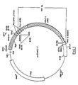
- the restriction map of pCD-HVLC2 is set forth in Figure 1.
- the plasmid pCD-HVLC2 is deposited at the ATCC under the deposit number ATCC 67791.
- the DNA segment of the present invention having a sequence which codes for HVMLC2 protein or a fragment of the DNA segment can be incorporated in a known manner in a plasmid vector which ensures good expression of the HVMLC2 protein or a part of the protein.
- plasmid vectors which can be used are pBR322, pBR325, pUC7, pUC8, pUC18 and pUC19, or other generally accessible plasmids.
- a number of unique restriction sequences for restriction enzymes are incorporated in the DNA segment identified in Table II and these provide access to nucleotide sequences which code for polypeptides that are unique to HVMLC2. It is possible to make use of the following recognition sites for restriction enzymes to prepare fragments of the DNA segment of the embodiment of the invention: AsuI (G ⁇ GNCC, positions 147 to 180, 182 to 254, 255 to 366, 367 to 422, and 423 to 645) Fnu4HI (GC ⁇ NGC, positions 147 to 306, 307 to 561, and 562 to 645) DdeI (C ⁇ TNAG, positions 147 to 154, 155 to 291, 292 to 427, and 428 to 645) MnII (CCTC, positions 147 to 237, 238 to 357, 358 to 389, 390 to 429, 430 to 527, 528 to 540, 541 to 543 and 544 to 645) ScrFI (CCNGG, positions 147 to 175, 176 to 224,
- DNA fragments having sequences unique to HVMLC2 can also be constructed by chemical synthesis and enzymatic ligation reactions carried out in a manner known per se.
- the HVMLC2 protein or a part of the protein can be obtained by expression in a suitable host cell in a manner known per se.
- suitable host cells are procaryotic organisms, preferably E, coli, B. subtilis, S. lividans and mammalian and yeast cells, preferably Saccharomyces cerevisiae.
- the DNA segment of the present invention allows those skilled in the art to prepare antibodies against HVMLC2 protein or a part of the protein which can be used in in vivo and in vitro analytical methods.
- Antibody against HVMLC2 protein or a part of the protein can be prepared by conventional procedures by injecting HVMLC2 protein or a part of the protein obtained according to the process of the invention, or synthetically synthesized HVMLC2 protein or a part of the protein, into a suitable host animal; then bleeding the animal and concentrating the antiserum.
- Monoclonal antibodies specific for HVMLC2 protein or a part of the protein can also be prepared using the known cell fusion method.
- Polyclonal or monoclonal antibodies against the HVMLC2 protein or a part of the protein can be used as medicaments, in particular as medicaments for the prophylaxis and treatment of heart disease.
- the polyclonal or monoclonal antibodies may be administered alone, in mixtures, in combination with one or more pharmaceutically acceptable excipients, diluents and auxiliaries or they may be conjugated to a therapeutic agent using known methods. Suitable therapeutic agents are enzymes or free radical scavengers, for example superoxide dismutase, catalase and vitamin E.
- the polyclonal or monoclonal antibodies against the HVMLC2 protein may also be conjugated to a labelling agent and used to detect damaged cardiac tissue.
- the labelling agent may be various enzymes, fluorescent materials, luminescent materials and radioactive materials.
- suitable enzymes include horseradish peroxidase, alkaline phosphatase, or ⁇ -galactosidase, acetylcholinesterase
- suitable fluorescent materials include umbelliferone, fluorescein, fluorescein isothiocyanate, rhodamine, dichlorotriazinylamine fluorescein, dansyl chloride or phycoerythrin
- examples of luminescent materials include luminol
- suitable radioactive materials include radioactive iodine I125, I131 or tritium.
- the enzymes, fluorescent materials, luminescent materials and radioactive materials can be conjugated to the polyclonal or monoclonal antibodies using conventional methods known in the art.
- the DNA segment of the present invention also allows those skilled in the art to select DNA fragments from the DNA sequence shown in Table I, and complementary mRNA fragments which are unique to HVMLC2/.
- the invention would also permit one skilled in the art to select oligonucleotide sequences from the known sequences of myosin light chains from atrial, thymus or skeletal muscle tissue which are unique to the myosin light chains of those tissues.
- polypeptides unique to myosin light chain proteins of specific tissues can be deduced from the unique DNA sequences.
- the selected DNA and mRNA oligonucleotide fragments can be used as reagents to detect the presence or absence of a complementary oligonucleotide in a biological sample.
- oligonucleotides identified from the DNA segment of the present invention can be used as probes in diagnostic tests for heart disease to detect the presence or absence of complementary oligonucleotides, such as DNA or mRNA nucleotide fragments, in a biological sample.
- Antibodies against the selected polypeptides can be prepared using known methods and these antibodies can be used in in vivo or in vitro analytical methods or as medicaments.
- oligonucleotide fragments or polypeptides useful as specific reagents, or in preparing specific reagents to detect HVMLC2 in a biological sample or to use as medicaments is facilitated by knowing the conformation of the HVMLC2 protein.
- polypeptides located in the exposed sites of the HVMLC2 protein, as shown in Figure 2 would be particularly useful in preparing antibodies to be used in analytical methods or as medicaments.
- RNA pellet transparent was dissolved in 500 ⁇ l of autoclaved H2O, immediately transferred into 100% ETOH + 10% of 3M sodium acetate, pH 5.2, and left on ice for 16 hours at -20°C to precipitate. The amount of RNA was estimated by reading O.D.260 - 330 ⁇ g. Several preparations were performed.
- oligo dT cellulose 100 mg oligo dT cellulose (Pharmacia) was resuspended in loading buffer (20 mM TrisHCl pH 7.6, 0.5M NaCl, 1mM EDTA, 0.1% SDS). The mixture was poured into a column (BIORAD, 2 ml disposable poly-prep chromatography column) and was washed with 2ml sterile H2O, 2 ml 0.1M NaOH + 5 mM EDTA, 5 ml sterile H2O). The pH of the eluent was 6.8. The column was washed with 5 column volumes of loading buffer. 500 ⁇ g of total RNA (combined preparations) were used. O.D.260 was checked to confirm the amount.
- RNA was dissolved in 500 ⁇ l H2O (autoclaved) and heated up to 65°C for 5 minutes. An equal amount of 2 X loading buffer was added. The sample was cooled down to room temperature and applied on the column. The flow through was collected and heated again to 65°C and cooled down to room temperature. The sample was reapplied on the column. The column was washed with 10 column volumes of loading buffer and 4 column volumes of 0.1 M NaCl loading buffer. Poly A+ was eluted with 3 column volumes of elution buffer (10mM Tris-HCl pH 7.5, 1 mM EDTA, 0.05% SDS). The Poly A+ fraction was immediately precipitated with 100% ETOH + 10% of 3 M sodium acetate pH 5.2 and stored in -20°C. The amount of Poly A+ RNA was estimated by reading O.D.260 - 20 ⁇ g. The yield was approximately 4.2%.
- the mRNA was tested using oligo p(dT)12 ⁇ 18-primed reverse transcription to determine the size range of the mRNA population and its ability to serve as a template for reverse transcription.
- Poly A+mRNA (10 ⁇ g) was hybridized to oligo (dT) tailed pCDV-1 expression vector and was reverse transcribed using AMV reverse transcriptase.
- the cDNA was synthesized directly onto the vector, thus requiring no insertion steps.
- An oligo (dC)-tail (7 dC residues per 3′end) was added to the 3′-terminus of both the cDNA and the vector using terminal transferase. The dC-tail was removed from the 3′ end of the vector using Hind III digestion.
- the oligo (dC)-tail Hind III cleaved cDNA:RNA vector was then circularized using oligo (dG)-tailed linked DNA (pL-1, 524 bp, 1/10 scale reaction).
- the RNA strand in the cDNA:RNA plasmid was replaced with DNA through nick translation using RNaseH, DNA polymerase I and DNA ligase (1/10 scale).
- the circular cDNA vector was then transformed into E. coli k12 strain MC1061 (1/10 scale) to create a cDNA library. Once the conditions were determined the procedure was increased 10 fold to obtain the cDNA library.
- the cDNA library was stored at -70°C in 1.5 ml aliquots which were quick frozen in liquid nitrogen.
- the cDNA:RNA plasmid transformed into E coli K12 strain MC1061 is deposited at ATCC under the deposit number ATCC 67789.
- the synthetic oligonucleotide probe shown in Table V was derived from the partial amino acid sequence determined from chymotryptic peptides of human cardiac light chain 2 (HVMLC2) protein published by Klotz et al (Cir. Res. 50 , p. 201, 1982). The probe was derived from the amino terminal end of the HVMLC2 protein corresponding to positions 27 - 32 of HVMLC2. The detailed amino acid sequence of HCMLC2 was not known.
- the gel was run for approximately 3 hours at a constant current of 20mA, using 0.089M Tris-borate, 0.089M boric acid and 0.002M EDTA as a running buffer.
- the gel was stained with Ethidium Bromide (0.2 mg/ml in H2O) for 15 minutes. Under UV light, the band corresponding to the size of the 17 mer was cut out, pulverized and resuspended in 8 ml of 0.5 M NH4OAc, left at 37°C for 16 hours and then, spun down. The supernatant was extracted with H2O saturated butanol, and the oligomer was precipitated with 5 vol. of 100% ETOH + 10% 3 M sodium acetate pH 5.2.
- the oligomer was put on dry ice for 1 hour. The pellet was collected by centrifugation in a microfuge for 15 minutes and washed with 70% ETOH. The final pellet containing the purified oligomer was dried using a speed - vac and stored at 4°C. The purified oligomer was dissolved in 50 ⁇ l H2O and the concentration was determined by A260 nm (2.5 ⁇ g/ ⁇ l). Approximately 200 ng of oligomer was used for 5′ end labelling.
- Nitrocellulose filters were placed on the culture plate and then put onto wet Whatman 3MM paper presoaked with 0.5m NaOH, 1M NaCl for 10 minutes, and dry blotted on Whatman 3MM paper. The filter was then transferred onto Whatman paper presoaked with 1M Tris, pH 7.4, 2.5 M NaCl for 10 minutes and dry blotted on Whatman's 3MM paper and the procedure was repeated again with 1M Tris, pH 7.4, 2.5M NaCl. The filters were air dried, baked in a vacuum oven at 80°C for 2 hours and wet in 3X SCC (20xSCC - 175.3g NaCl 88.2g sodium citrate).
- the filter was put in prehybridization buffer (6X SSC, 1X Denhardt's solution, 0.5% SDS, 100 ⁇ g/ml sheared and denatured salmon sperm DNA, 0.05% sodium pyrophosphate) and incubated for at least 2 hours at 37°C.
- the prehybridization buffer was removed and replaced with hybridization buffer (6X SSC, 1X Denhardt's 20 ⁇ g/ml tRNA, 0.05% sodium pyrophosphate) and 5′ end labelled oligonucleotide probe.
- the purified synthetic oligonucleotide probe (example 5) was labelled by mixing 2 ⁇ l + 17-mer (0.2 ⁇ g), 27 ⁇ l H2O, 10 ⁇ l 5x forward reaction buffer (5′ DNA Terminus Labelling System from BRL) 9 ⁇ l 32ATP SA 5000 Ci/mM, and 2 ⁇ l T-4 Kinase (10U/ml) and incubating for one hour at 37°C. Hybridization was carried out at 37°C for 16 hours. The nitrocellulose filters after the hybridization were washed in 6X SCC + 0.05% sodium pyrophosphate. The filters were washed twice at 37°C for 1/2 hour each and once at 47°C for 10-15 minutes. The filters were briefly air dried. Autoradiography was performed using an x-ray film (Kodak X-Omat RP) for 16 hours at -70°C.
- Kodak X-Omat RP x-ray film
- Figure 1 shows a schematic illustration of the restriction map of pCD-HVLC2.
- the plasmid pCD-HVLC2 is deposited at ATCC under the deposit number ATCC 67791.
- the HVMLC2 cDNA was sequenced using the dideoxy chain determination method. (Biggin, M. et al. (1983) Proc. Natl. Acad. Sci. U.S.A. 80 , p. 3963-3965). The amino acid sequence deduced from the cDNA was compared with the known partial amino acid sequence of human cardiac myosin light chain 2 as described in the literature (Klotz, Circulation Research, 50 , p. 201, 1982).
Landscapes
- Health & Medical Sciences (AREA)
- Chemical & Material Sciences (AREA)
- Life Sciences & Earth Sciences (AREA)
- Organic Chemistry (AREA)
- General Health & Medical Sciences (AREA)
- Medicinal Chemistry (AREA)
- Engineering & Computer Science (AREA)
- Heart & Thoracic Surgery (AREA)
- Biochemistry (AREA)
- Genetics & Genomics (AREA)
- Gastroenterology & Hepatology (AREA)
- Molecular Biology (AREA)
- Proteomics, Peptides & Aminoacids (AREA)
- Zoology (AREA)
- Toxicology (AREA)
- Bioinformatics & Cheminformatics (AREA)
- Cardiology (AREA)
- Biophysics (AREA)
- Chemical Kinetics & Catalysis (AREA)
- General Chemical & Material Sciences (AREA)
- Nuclear Medicine, Radiotherapy & Molecular Imaging (AREA)
- Pharmacology & Pharmacy (AREA)
- Animal Behavior & Ethology (AREA)
- Public Health (AREA)
- Veterinary Medicine (AREA)
- Preparation Of Compounds By Using Micro-Organisms (AREA)
- Peptides Or Proteins (AREA)
- Micro-Organisms Or Cultivation Processes Thereof (AREA)
- Medicines Containing Antibodies Or Antigens For Use As Internal Diagnostic Agents (AREA)
Abstract
The DNA segment is provided having a sequence which codes for human ventricular myosin light chain 2 (HVMLC2) protein, or a fragment of the DNA segment. The DNA segment or a fragment of the DNA segment of the invention can be used to produce HVMLC2 protein or a part of the protein by culturing a transformant microorganism which includes a recombinant plasmid comprising a plasmid vector into which the DNA segment or a fragment of the DNA segment of the invention has been inserted.
Description
- The invention relates to a DNA segment having a sequence which codes for human ventricular myosin light chain 2 (HVMLC2) protein or a fragment of the DNA segment. The DNA segment of the invention can be used to produce human ventricular
myosin light chain 2 protein or part of the protein by culturing, a transformant microorganism which includes a recombinant plasmid comprising a plasmid vector into which the DNA segment of the invention or a fragment of the DNA segment has been inserted. The DNA segment of the invention also permits selection of oligonucleotide fragments and polypeptides which are unique to human ventricularmyosin light chain 2 protein or to myosin light chain proteins from other species and/or tissues. - Myosin is the major protein constituent of cardiac muscle and it consists of two myosin heavy chains and four myosin light chains. Two classes of light chains have been characterized in cardiac myosin: cardiac myosin light chain 1 with a molecular weight of 27,000 daltons and
myosin light chain 2 with a molecular weight of 20,000 daltons. - The light chain of myosin is released from cardiac muscle following myocardial infarction and may remain elevated in serum for several days (Khaw et al, Circulation 58 p.1130, 1978 and Trahern et al. Am. J. Cardiol 41 p. 641, 1978). Katus et al. (JACC, Vol. 2 (3) p. 487) recently reported a correlation between the presence of myosin light chains and signs of ischemia in the electrocardiogram and extent of coronary artery narrowing. An elevated plasma light chain concentration has also been observed in patients with congestive cardiomyopathy associated with an inflammatory infiltrate (Haver, E., J. Mol. & Cell Cardiol. 17 (Supplement 2) p.53, 1985).
- There are a number of reports describing diagnostic methods for muscle diseases utilizing polyclonal or monoclonal antibodies directed at myosin light chains (Trahern et al. supra; Khaw et al. Supra; Nagai et al. Biochem. Biophys. Res. Commun. 86 p.683, 1979; and Katus et al. Am. J. Cardiol. 54 p.964, 1984, Katus et al. Mol. Immuno. 19 p.451, 1982, and Masahito et al. European Patent Application D205 177 A2). The diagnostic methods however suffer from a number of disadvantages such as cross-reactivity between cardiac myosin light chains and myosin light chains from other tissues or species.
- Polyclonal or monoclonal antibodies specific for cardiac myosin light chains can be used for various types of therapy. For example, such antibodies can be conjugated to a radioisotope to image damaged cardiac muscle tissue or to a therapeutic agent to deliver the agent to damaged cardiac muscle tissue.
- The identification of the DNA sequences that code for human myosin light chains would facilitate the preparation of highly specific monclonal and polyclonal antibodies which would be useful in diagnostic methods and for various types of therapy.
- Weeds and Pope (Nature, 234, p. 85. 1981) examined thiol sequences of cardiac light chains from bovine and sheep hearts. The amino acid sequence of myosin light chains from normal and hypertrophied human hearts was partially elucidated by Klotz et al. (Circulation Research 50, p. 201, 1982). Klotz et al. sequenced peptides isolated from human cardiac
myosin light chain 2 and found that their sequences corresponded to 70% of the whole molecule. Collins et al. (Bioscience Reports 6, p.655, 1986) disclosed the amino acid sequence for rabbit ventricular myosin light chain 2 (LC2). - A few studies have been directed at characterizing the nucleotide sequences of cardiac myosin light chains. The present inventor has described a DNA segment having a sequence which codes for human ventricular myosin light chain 1 protein (U.S .Patent Application Serial No. 07/160,892 filed February 26, 1988). Kumar et al. (J. Biol. Chem. 261 p.2866, 1986) has described the nucleotide sequence of a recombinant plasmid containing cDNA corresponding to rat heart
myosin light chain 2. - The present inventor using a cDNA cloning method has identified and sequenced a DNA segment having a sequence which codes for the HVMLC2 protein. The HVMLC2 protein has hitherto not been completely sequenced at the protein level using conventional methods due to the blockage of the N-terminal end of the protein. In addition, at the protein level HVMLC2 protein is readily degradable. The HVMLC2 protein has also hitherto not been sequenced by DNA cloning methods due to difficulties in isolating mRNA from cardiac tissue since it is readily degraded by RNase, and difficulties in obtaining sufficient quantities of human material for characterization. Furthermore, the mRNA that codes for HVMLC2 is presnt in extremely small quantities and it is contaminated with much larger amounts of other mRNA's that act similarly to it in the cloning methodology and which are not related to HVMLC2. Therefore, at each stage of the cloning method, a complex mixture is encountered, only a very small part of which is related to the HVMLC2 protein.
- The present invention therefore provides a means and method of screening complex mixtures of mRNA in order to select the mRNA that codes for HVMLC2 protein. The present invention further provides a DNA segment having a sequence which codes for HVMLC2 protein, or a fragment of the DNA segment.
- In accordance with one embodiment of the invention, the DNA segment has a sequence which codes for HVMLC2 protein of the amino acid sequence as shown in Table I. In accordance with a second embodiment of the invention the DNA segment has a sequence which codes for HVMLC2 protein having the nucleotide base sequence as shown in Table I. In accordance with a third embodiment of the invention the DNA segment of the invention has the nucleotide sequence as shown in Table II.
- The invention also contemplates a DNA segment or a fragment of the DNA segment hybridizing to a DNA segment or a fragment of DNA segment of the invention from whatever source obtained including natural, synthetic or semisynthetic sources, that is related by mutations, including nucleotide substitutions, nucleotide deletions, nucleotide insertions and inversions of nucleotide stretches to the DNA segment or arrangment of the DNA segment according to the invention and having a sequence which codes for HVMLC2 protein.
- The invention further contemplates a recombinant plasmid adapted for transformation of a host comprising a plasmid vector into which a DNA segment having a sequence which codes for HVMLC2 protein, or a fragment of the DNA segment, has been inserted. Preferred recombinant plasmids are those comprising a plasmid vector into which a DNA segment having a sequence which codes for a HVMLC2 protein of the amino acid sequence as shown in Table I, or of the nucleotide sequence as shown in Table I, or a fragment of such a DNA segment, has been inserted. The recombinant plasmid of the invention can be introduced into a host cell which can then be cultured to produce HVMLC2 protein or a part of the protein.
- The invention also provides a process for producing HVMLC2 protein or a part of the protein comprising culturing a transformant host cell including a recombinant plasmid comprising a plasmid vector into which a DNA segment having a sequence which codes for HVMLC2 protein or a fragment of the DNA segment has been inserted, in a suitable medium until HVMLC2 protein or a part of the protein is formed and thereafter isolating the HVMLC2 protein or a part of the protein.
- The invention further contemplates a polyclonal or monoclonal antibody directed at HVMLC2 protein or a part of the protein prepared in accordance with the methods of the invention, methods for preparing the polyclonal or monoclonal antibody and diagnostic methods for cardiac muscle diseases using the polyclonal or monoclonal antibody. The invention further contemplates a medicament for the prophylaxis and treatment of heart disease which comprises a polyclonal or monoclonal antibody directed at HVMLC2 protein prepared in accordance with the methods of the invention and one or more of a therapeutic agent and a pharmaceutically acceptable excipient, diluent and auxiliary. The invention still further contemplates an agent for detecting damaged cardiac tissue which comprises a polyclonal or monoclonal antibody directed at HVMLC2 protein or part of the protein prepared in accordance with the methods of the invention and a labelling agent.
- The present invention permits, for the first time, the precise identification of a polypeptide sequence or a part of a polypeptide sequence, or a nucleotide sequence or a part of a nucleotide sequence, which is unique to HVMLC2. To those skilled in the art, such polypeptide or nucleotide sequences of HVMLC2, with their detailed conformations deduced on energetic and thermodynamic groups, enable the separation with unprecedented specificity of HVMLC2 by immunologic (e.g. based on poly- or monoclonal antibody against a polypeptide or a part of a polypeptide, used alone or in concert), by nucleotide hybridization techniques (e.g. using DNA or RNA complementary strands with/without signal amplifications), by receptor-based or receptor-like assays, or by other variants of these well-known methods.
- The invention will be better understood with reference to the drawings in which:
- Figure 1 is a schematic illustration of a restriction map of a plasmid containing the DNA segment of one embodiment of the invention.
- Figure 2 shows the conformation of HVMLC2 constructed from the deduced amino acid sequence of the DNA segment according to one embodiment of the invention.
- In accordance with one embodiment of the invention, the HVMLC2 protein encoded in the DNA segment of the invention is composed of the 165 amino acids shown in Table I. A comparison of the deduced amino acid sequence of the HVMLC2 protein encoded in the DNA segment of the embodiment of the invention shown in Table I with the known partial amino acid sequence determined from chymotryptic peptides of human cardiac
myosin light chain 2 by Klotz et al, (Circulation Research, 50, p. 201, 1982) indicates that the amino acid sequences are similar (Table III). The deduced amino acid sequence of the HVMLC2 protein encoded in the DNA of the embodiment of the invention is also similar to the amino acid sequence of rabbit cardiac light chain LC2 (Collins et al, supra) and the deduced amino acid sequence of rat cardiac light chain LC2 (Kumar et al, supra) (Table IV). The similarities between the rabbit and rat and the human cardiac myosin light chain sequences indicate that the rabbit and rat sequences as well as antibodies directed against the rabbit andrat light chain 2 proteins, may be useful as standards in diagnostic methods for detecting HVMLC2 protein or DNA or mRNA sequences of HVMLC2. - The conformation of a protein may be deduced only after the complete amino acid sequence of the protein is known. Accordingly, the confirmation of the HVMLC2 protein may be deduced from the amino acid sequence identified in the embodiment of the invention. Figure 2 shows the conformation of the HVMLC2 protein deduced from the amino acid sequence shown in Table I. As shown in Figure 2, α-helical structures are found at the proline residues of the amino acid sequence shown in Table I and there are polypeptides located at exposed sites in the HVMLC2 protein.
- A comparison of the amino acid sequence of HVMLC2 protein identified in the embodiment of the present invention with the amino acid sequences of myosin light chain proteins described in the literature, as well as the conformations of these proteins, indicates that there are sequences which are unique to HVMLC2. It is the unique sequences of the HVMLC2 protein encoded in the DNA segment of the present invention and the specific conformation of the protein which permits specific differentiation of ventricular
myosin light chain 2 protein from myosin light chain proteins from other species and/or tissues. - A comparison of the deduced amino acid sequence identified in the embodiment of the present invention with the deduced amino acid sequence of rat cardiac light chain LC2 (Kumar et al, supra), indicates that the following polypeptides are unique to HVMLC2.
Ala-Gly-Gly-Ala-Asn (amino acids 10 to 14 in Table I)
Asp-Pro-Glu-Gly-Lys-Gly-Val-Leu-Lys-Ala-Asp-Tyr-Val-Arg (amino acids 106 to 119 in Table I)
The polypeptides are located at exposed sites and are particularly useful in differentiating HVMLC2. (Figure 2). The polypeptides unique to HVMLC2 can be synthetically synthesized using solid phase synthesis (Eberle, A.N.J. Chem. Soc. Perkin. Trans, 1, p.361, 1986). - In the nucleotide base sequence shown in Table II, the nucleotide positions from 1 to 102 represent the oligo dG tailed linker used in the cloning, and those from 103 to 146 represent the noncoding area. The ATG sequence at positions 147 to 149 is the start codon. The sequence between positions 150 and 647 is the portion that encodes the HVMLC2 protein. The sequence at positions 648 to 650 is the stop codon and the sequence after position 650 is the 3′-noncoding region.
- The DNA segment of the present invention can be prepared, for example, by the method as described below.
- The total RNA was separated by the guanidine thiocyanate/cesium chloride method from human ventricle. Poly A messenger RNA was isolated from the total RNA by oligo dT column chromatography. A human ventricular cDNA library was prepared by using the messenger RNA as a template. The cDNA library is deposited at the ATCC under the deposit number ATCC 67789. The cDNA library was then screened using a probe consisting of a DNA that encodes a portion of the amino acid sequence of HVMLC2 which was partially elucidated by Klotz et al, supra. A DNA segment containing a DNA sequence coding for HVMLC2 protein was contained in a resulting clone (pCD-HVLC2). The restriction map of pCD-HVLC2 is set forth in Figure 1. The plasmid pCD-HVLC2 is deposited at the ATCC under the deposit number ATCC 67791.
- The DNA segment of the present invention having a sequence which codes for HVMLC2 protein or a fragment of the DNA segment can be incorporated in a known manner in a plasmid vector which ensures good expression of the HVMLC2 protein or a part of the protein. Examples of plasmid vectors which can be used are pBR322, pBR325, pUC7, pUC8, pUC18 and pUC19, or other generally accessible plasmids.
- A number of unique restriction sequences for restriction enzymes are incorporated in the DNA segment identified in Table II and these provide access to nucleotide sequences which code for polypeptides that are unique to HVMLC2. It is possible to make use of the following recognition sites for restriction enzymes to prepare fragments of the DNA segment of the embodiment of the invention:
AsuI (G↓GNCC, positions 147 to 180, 182 to 254, 255 to 366, 367 to 422, and 423 to 645)
Fnu4HI (GC↓NGC, positions 147 to 306, 307 to 561, and 562 to 645)
DdeI (C↓TNAG, positions 147 to 154, 155 to 291, 292 to 427, and 428 to 645)
MnII (CCTC, positions 147 to 237, 238 to 357, 358 to 389, 390 to 429, 430 to 527, 528 to 540, 541 to 543 and 544 to 645)
ScrFI (CCNGG, positions 147 to 175, 176 to 224, 225 to 363, 364 to 605, and 606 to 645) - DNA fragments having sequences unique to HVMLC2 can also be constructed by chemical synthesis and enzymatic ligation reactions carried out in a manner known per se.
- The HVMLC2 protein or a part of the protein can be obtained by expression in a suitable host cell in a manner known per se. Suitable host cells are procaryotic organisms, preferably E, coli, B. subtilis, S. lividans and mammalian and yeast cells, preferably Saccharomyces cerevisiae.
- The DNA segment of the present invention allows those skilled in the art to prepare antibodies against HVMLC2 protein or a part of the protein which can be used in in vivo and in vitro analytical methods. Antibody against HVMLC2 protein or a part of the protein can be prepared by conventional procedures by injecting HVMLC2 protein or a part of the protein obtained according to the process of the invention, or synthetically synthesized HVMLC2 protein or a part of the protein, into a suitable host animal; then bleeding the animal and concentrating the antiserum. Monoclonal antibodies specific for HVMLC2 protein or a part of the protein can also be prepared using the known cell fusion method. As to details relating to the preparation of monoclonal antibodies reference can be made to Goding, J.W., Monoclonal Antibodies: Principles and Practice, 2nd Ed., Academic Press, London, 1986. It is possible according to the invention to use the antibodies against HVMLC2 protein or a part of the protein, particularly a part having a unique amino acid sequence to HVMLC2, as a reagent for detecting HVMLC2, for example in an ELISA, radioimmunoassy, or histochemical tests.
- Polyclonal or monoclonal antibodies against the HVMLC2 protein or a part of the protein can be used as medicaments, in particular as medicaments for the prophylaxis and treatment of heart disease. The polyclonal or monoclonal antibodies may be administered alone, in mixtures, in combination with one or more pharmaceutically acceptable excipients, diluents and auxiliaries or they may be conjugated to a therapeutic agent using known methods. Suitable therapeutic agents are enzymes or free radical scavengers, for example superoxide dismutase, catalase and vitamin E. The polyclonal or monoclonal antibodies against the HVMLC2 protein may also be conjugated to a labelling agent and used to detect damaged cardiac tissue. The labelling agent may be various enzymes, fluorescent materials, luminescent materials and radioactive materials. Examples of suitable enzymes include horseradish peroxidase, alkaline phosphatase, or β-galactosidase, acetylcholinesterase; examples of suitable fluorescent materials include umbelliferone, fluorescein, fluorescein isothiocyanate, rhodamine, dichlorotriazinylamine fluorescein, dansyl chloride or phycoerythrin; examples of luminescent materials include luminol; and examples of suitable radioactive materials include radioactive iodine I¹²⁵, I¹³¹ or tritium. The enzymes, fluorescent materials, luminescent materials and radioactive materials can be conjugated to the polyclonal or monoclonal antibodies using conventional methods known in the art.
- The DNA segment of the present invention also allows those skilled in the art to select DNA fragments from the DNA sequence shown in Table I, and complementary mRNA fragments which are unique to HVMLC2/.The invention would also permit one skilled in the art to select oligonucleotide sequences from the known sequences of myosin light chains from atrial, thymus or skeletal muscle tissue which are unique to the myosin light chains of those tissues. In addition polypeptides unique to myosin light chain proteins of specific tissues can be deduced from the unique DNA sequences. The selected DNA and mRNA oligonucleotide fragments can be used as reagents to detect the presence or absence of a complementary oligonucleotide in a biological sample. In particular oligonucleotides identified from the DNA segment of the present invention can be used as probes in diagnostic tests for heart disease to detect the presence or absence of complementary oligonucleotides, such as DNA or mRNA nucleotide fragments, in a biological sample. Antibodies against the selected polypeptides can be prepared using known methods and these antibodies can be used in in vivo or in vitro analytical methods or as medicaments.
- The selection of oligonucleotide fragments or polypeptides useful as specific reagents, or in preparing specific reagents to detect HVMLC2 in a biological sample or to use as medicaments, is facilitated by knowing the conformation of the HVMLC2 protein. For example, polypeptides located in the exposed sites of the HVMLC2 protein, as shown in Figure 2, would be particularly useful in preparing antibodies to be used in analytical methods or as medicaments.
- The following examples below illustrate the invention.
- 2g of human ventricle frozen in liquid N₂ was pulverized and homogenized in 20ml GTC solution (4M guanidine thiocynate, 25mM sodium citrate, 0.1M 2-mercaptoethanol, 0.5% sarkozyl) with a polytron (Brinkman Inst., PST 20 probe) at 5.5 rpm for 10 seconds, then homogenized in a teflon - glass homogenizer 2X low speed, the spun down in COREX tubes at 7,000 rpm for 10 minutes. Eight grams of cesium chloride were added to the supernatant and dissolved by vortexing. The solution was applied on 1.2 ml of cesium chloride solution (5.7 M cesium chloride, 25mM sodium acetate) and centrifuged at 35,000 rpm using a SW 50.1 rotor (6 tubes) for 16 hours at 20°C.
- The GTC solution was removed, the sample was rinsed with fresh GTC solution and the cesium chloride was poured out. The tubes were cut 1.5 cm from the bottom. The RNA pellet (transparent) was dissolved in 500 µl of autoclaved H₂O, immediately transferred into 100% ETOH + 10% of 3M sodium acetate, pH 5.2, and left on ice for 16 hours at -20°C to precipitate. The amount of RNA was estimated by reading O.D.₂₆₀ - 330 µg. Several preparations were performed.
- 100 mg oligo dT cellulose (Pharmacia) was resuspended in loading buffer (20 mM TrisHCl pH 7.6, 0.5M NaCl, 1mM EDTA, 0.1% SDS). The mixture was poured into a column (BIORAD, 2 ml disposable poly-prep chromatography column) and was washed with 2ml sterile H₂O, 2 ml 0.1M NaOH + 5 mM EDTA, 5 ml sterile H₂O). The pH of the eluent was 6.8. The column was washed with 5 column volumes of loading buffer. 500 µg of total RNA (combined preparations) were used. O.D.₂₆₀ was checked to confirm the amount. RNA was dissolved in 500 µl H₂O (autoclaved) and heated up to 65°C for 5 minutes. An equal amount of 2 X loading buffer was added. The sample was cooled down to room temperature and applied on the column. The flow through was collected and heated again to 65°C and cooled down to room temperature. The sample was reapplied on the column. The column was washed with 10 column volumes of loading buffer and 4 column volumes of 0.1 M NaCl loading buffer. Poly A⁺ was eluted with 3 column volumes of elution buffer (10mM Tris-HCl pH 7.5, 1 mM EDTA, 0.05% SDS). The Poly A⁺ fraction was immediately precipitated with 100% ETOH + 10% of 3 M sodium acetate pH 5.2 and stored in -20°C. The amount of Poly A⁺ RNA was estimated by reading O.D.₂₆₀ - 20 µg. The yield was approximately 4.2%.
- The Okayama - Berg procedure (Mol. & Cell. Biol. 2, p. 161,. 1982) as set out in the cDNA cloning Manual by Pharmacia was used to make the cDNA library. All reagents were bought from Pharmacia. The method used to make the cDNA library is briefly described below.
- The mRNA was tested using oligo p(dT)₁₂₋₁₈-primed reverse transcription to determine the size range of the mRNA population and its ability to serve as a template for reverse transcription. Poly A⁺mRNA (10 µg) was hybridized to oligo (dT) tailed pCDV-1 expression vector and was reverse transcribed using AMV reverse transcriptase. The cDNA was synthesized directly onto the vector, thus requiring no insertion steps. An oligo (dC)-tail (7 dC residues per 3′end) was added to the 3′-terminus of both the cDNA and the vector using terminal transferase. The dC-tail was removed from the 3′ end of the vector using Hind III digestion. The oligo (dC)-tail Hind III cleaved cDNA:RNA vector was then circularized using oligo (dG)-tailed linked DNA (pL-1, 524 bp, 1/10 scale reaction). The RNA strand in the cDNA:RNA plasmid was replaced with DNA through nick translation using RNaseH, DNA polymerase I and DNA ligase (1/10 scale). The circular cDNA vector was then transformed into E. coli k12 strain MC1061 (1/10 scale) to create a cDNA library. Once the conditions were determined the procedure was increased 10 fold to obtain the cDNA library.
- The cDNA library was stored at -70°C in 1.5 ml aliquots which were quick frozen in liquid nitrogen. The cDNA:RNA plasmid transformed into E coli K12 strain MC1061 is deposited at ATCC under the deposit number ATCC 67789.
- The synthetic oligonucleotide probe shown in Table V was derived from the partial amino acid sequence determined from chymotryptic peptides of human cardiac light chain 2 (HVMLC2) protein published by Klotz et al (Cir. Res. 50, p. 201, 1982). The probe was derived from the amino terminal end of the HVMLC2 protein corresponding to positions 27 - 32 of HVMLC2. The detailed amino acid sequence of HCMLC2 was not known. However, the same amino acid sequence for the synthetic oligonucleotide probe (Gln-GLu-Phe-Lys-Glu-Ala) is found in rabbit cardiac muscle light chain LC2 (Collins et al, supra) and rat cardiac light chain LC2 (Kumar et al, supra). This suggested that the amino acid sequence for the synthetic probe is well conserved in the genes of different muscles and different animals. Therefore, this probe was used to screen the human ventricular cDNA library. The synthetic oligonucleotide probe was synthesized by an automatic DNA-synthesizer (Pharmacia Gene Assembler, using cyanoethyl phosphoramidite).
Table V 17-mer probe for HVCMLC-2 32 combinations A A T A A CAG GAG TTC AAG GAG GC- Gln Glu Phe Lys Gly Ala - Cassettes containing the synthetic oligomer were spun down in Eppendorf tubes, dried on speed-vac and soaked in 25% NH₄OH (1.2 ml). The Eppendorf tubes were sealed with parafilm and soaked in 25% NH₄OH to minimize the leakage and left for 16 hours at 50°C. After cooling down on ice, the cassettes were transferred into new Eppendorf tubes and spun down to get all the liquid out. Both supernatants were combined and divided into 4 tubes and dried using the speed vac. The pellets were dissolved in 60 µl of H₂O. 30 µl DNA sample buffer (0.4% bromophenol blue, 25% ficol, 100 mM EDTA) was added, the solution was left for 5 minutes at 65°C, and then run on 20% acrylamide-urea gel (26.6 ml of 45% acrylamide-bis; 1.5% of 43% acrylamide-bis; 25.2g of Urea; 10X TBE (6.0 ml 0.89 M Tris-borate, 0.89 M boric acid, 0.02M - EDTA); 6.0 ml of H₂O; 15 mg of APS (ammonium persulfate); and, 40 µl TEMED).
- The gel was run for approximately 3 hours at a constant current of 20mA, using 0.089M Tris-borate, 0.089M boric acid and 0.002M EDTA as a running buffer. The gel was stained with Ethidium Bromide (0.2 mg/ml in H₂O) for 15 minutes. Under UV light, the band corresponding to the size of the 17 mer was cut out, pulverized and resuspended in 8 ml of 0.5 M NH₄OAc, left at 37°C for 16 hours and then, spun down. The supernatant was extracted with H₂O saturated butanol, and the oligomer was precipitated with 5 vol. of 100% ETOH + 10% 3 M sodium acetate pH 5.2. The oligomer was put on dry ice for 1 hour. The pellet was collected by centrifugation in a microfuge for 15 minutes and washed with 70% ETOH. The final pellet containing the purified oligomer was dried using a speed - vac and stored at 4°C. The purified oligomer was dissolved in 50 µl H₂O and the concentration was determined by A₂₆₀ nm (2.5 µg/µl). Approximately 200 ng of oligomer was used for 5′ end labelling.
- An aliquot of the cDNA library was thawed on ice, centrifuged at 1200 rpm for 2 minutes, the pellet was dissolved in 1 ml LB (Luria Bertani medium - 10g Tryptone, 5 g yeast extract, 5 g sodium chloride, pH 7.5) and put into 10ml LB + ampicillin (50 µg/ml). The culture was grown for 6 hours at 37°C, titred and plated the next day. The E. coli transformants were grown on 40 small plates with approximately 5000 colonies per plate.
- Nitrocellulose filters were placed on the culture plate and then put onto wet Whatman 3MM paper presoaked with 0.5m NaOH, 1M NaCl for 10 minutes, and dry blotted on Whatman 3MM paper. The filter was then transferred onto Whatman paper presoaked with 1M Tris, pH 7.4, 2.5 M NaCl for 10 minutes and dry blotted on Whatman's 3MM paper and the procedure was repeated again with 1M Tris, pH 7.4, 2.5M NaCl. The filters were air dried, baked in a vacuum oven at 80°C for 2 hours and wet in 3X SCC (20xSCC - 175.3g NaCl 88.2g sodium citrate). The filter was put in prehybridization buffer (6X SSC, 1X Denhardt's solution, 0.5% SDS, 100 µg/ml sheared and denatured salmon sperm DNA, 0.05% sodium pyrophosphate) and incubated for at least 2 hours at 37°C. The prehybridization buffer was removed and replaced with hybridization buffer (6X SSC, 1X Denhardt's 20 µg/ml tRNA, 0.05% sodium pyrophosphate) and 5′ end labelled oligonucleotide probe. The purified synthetic oligonucleotide probe (example 5) was labelled by mixing 2 µl + 17-mer (0.2 µg), 27 µl H₂O, 10 µl 5x forward reaction buffer (5′ DNA Terminus Labelling System from BRL) 9 µl ³²ATP SA 5000 Ci/mM, and 2 µl T-4 Kinase (10U/ml) and incubating for one hour at 37°C. Hybridization was carried out at 37°C for 16 hours. The nitrocellulose filters after the hybridization were washed in 6X SCC + 0.05% sodium pyrophosphate. The filters were washed twice at 37°C for 1/2 hour each and once at 47°C for 10-15 minutes. The filters were briefly air dried. Autoradiography was performed using an x-ray film (Kodak X-Omat RP) for 16 hours at -70°C.
- Positives were obtained in primary screening, of which two proved to be true in secondary screening. One pCD-HVLC2 clone contained the complete 3′ untranslated and coding regions. Figure 1 shows a schematic illustration of the restriction map of pCD-HVLC2. The plasmid pCD-HVLC2 is deposited at ATCC under the deposit number ATCC 67791.
- Northern blot analysis was performed with a BamHI restriction fragment from plasmid pCD-HVLC2 according to the procedure of "B.D. Hames & S.J. Higgins, "Nucleic Acid Hybridization", A Practical Approach, IRL Press, 1985. Human atrial total mRNA as a negative control and human ventricular total mRNA were separated by electrophoresis, transferred to a nitrocellulose filter and hybridization was carried out with ³²P labelled BamHI restriction fragments from plasmid pCD-HVLC2. A transcript of approximately 1500 bp which corresponds to the size of HVMLC2 mRNA was detected with the human ventricular mRNA.
- Southern blot analysis of DNA restriction fragments from the plasmid pCD-HVLC2 was performed according to the procedure of B.D. Hames & S.J. Higgins, "Nucleic Acid Hybridization A Practical Approach", IRL Press, 1985. SalI, Bam HI and Pst I restriction fragments from the plasmid pCD-HVLC2 were separated by gel electrophoresis and then transferred to a nitrocellulose filter. The immobilized DNA fragments were then probed with the labelled 17-mer oligonucleotide probe (Example 6). The Southern Blot analysis of the DNA restriction fragments from the plasmid pCD-HVLC2 probed with the 17 mer oligonucleotide probe demonstrated that the insert contained HVMLC2 specific sequences.
- The HVMLC2 cDNA was sequenced using the dideoxy chain determination method. (Biggin, M. et al. (1983) Proc. Natl. Acad. Sci. U.S.A. 80, p. 3963-3965). The amino acid sequence deduced from the cDNA was compared with the known partial amino acid sequence of human cardiac
myosin light chain 2 as described in the literature (Klotz, Circulation Research, 50, p. 201, 1982).


Claims (22)
1. A DNA segment having a sequence which codes for human ventricular myosin light chain 2 protein or a fragment of the DNA segment.
2. A DNA segment having a sequence which codes for human ventricular myosin light chain 2 protein of the amino acid sequence as shown in Table I or a fragment of the DNA segment.
3. A DNA segment having a sequence which codes for human ventricular myosin light chain 2 protein of the nucleotide sequence as shown in Table I or a fragment of the DNA segment.
4. The DNA segment as claimed in claim 1, which has the nucleotide sequence as shown in Table II.
5. A recombinant plasmid adapted for transformation of a host cell comprising a plasmid vector into which a DNA segment or a fragment of a DNA segment as claimed in any one of claims 1 to 4 has been inserted.
6. The recombinant plasmid as claimed in claim 5, wherein the plasmid vector comprises E. coli plasmid pBR322.
7. The recombinant plasmid as claimed in claims 5 or 6, which includes Pst I restriction endonuclease sites.
8. The recombinant plasmid as claimed in claims 5 or 6, which includes Bam HI restriction endonuclease sites.
9. The recombinant plasmid as shown in Figure 1.
10. A transformant host plasmid as claimed in any one of claims 5 to 9.
11. The transformed host cell as claimed in claim 10, wherein the host cell is a bacterial cell.
12. The transformant host cell as claimed in claims 10 or 11 wherein the host cell is E. coli.
13. A process for producing human ventricular myosin light chain 2 protein or a part of the protein comprising culturing a transformant host cell as claimed in claims 10, 11 or 12, in a suitable medium until human ventricular myosin light chain 2 protein or part of the protein is formed and isolating the human ventricular myosin light chain 2 protein or part of the protein.
14. A polyclonal antibody directed against human ventricular myosin light chain 2 protein or a part of the protein, which is prepared in a manner known per se using the human ventricular myosin light chain 2 protein or a part of the protein prepared according to the process as claimed in claim 13 or synthetically synthesized HVMLC2 protein or a part of the protein.
15. A monoclonal antibody directed against human ventricular myosin light chain 2 protein or a part of the protein, which is prepared in a manner known per se using the human ventricular myosin light chain 2 protein or a part of the protein prepared according to the process as claimed in claim 13 or synthetically synthesized HVMLC2 protein or a part of the protein.
16. A medicament for the prophylaxis and treatment of heart disease which comprises a polyclonal antibody as claimed in claim 14 and one or more of a therapeutic agent and a pharmaceutically acceptable excipient, diluent and auxiliary.
17. A medicament for the prophylaxis and treatment of heart disease which comprises a monoclonal antibody as claimed in claim 15 and one or more of a therapeutic agent and a pharmaceutically acceptable excipient, diluent and auxiliary.
18. An agent for detecting damaged cardiac tissue which comprises a polyclonal antibody as claimed in claim 14 and a labelling agent.
19. An agent for detecting damaged cardiac tissue which comprises a monoclonal antibody as claimed in claim 15 and a labelling agent.
20. The agent as claimed in claims 18 or 19, wherein the labelling agent is a radioactive material.
21. A process for producing a DNA segment according to any one of claims 1 to 4, a recombinant plasmid according to any one of claims 5 to 9, or a transformed host according to any one of claims 10 to 12.
22. Use of polyclonal antibody according to claim 14 or a monoclonal antibody according to claim 15 in the manufacture of a medicament for the prophylaxis and treatment of heart disease or in the manufacture of an agent for detecting damaged cardiac tissue.
Applications Claiming Priority (2)
| Application Number | Priority Date | Filing Date | Title |
|---|---|---|---|
| US24167288A | 1988-09-08 | 1988-09-08 | |
| US241672 | 1988-09-08 |
Publications (1)
| Publication Number | Publication Date |
|---|---|
| EP0357856A1 true EP0357856A1 (en) | 1990-03-14 |
Family
ID=22911699
Family Applications (1)
| Application Number | Title | Priority Date | Filing Date |
|---|---|---|---|
| EP88312271A Withdrawn EP0357856A1 (en) | 1988-09-08 | 1988-12-23 | DNA coding for human ventricular myosin light chain 2 |
Country Status (3)
| Country | Link |
|---|---|
| EP (1) | EP0357856A1 (en) |
| JP (1) | JPH0276584A (en) |
| AU (1) | AU4120289A (en) |
Cited By (3)
| Publication number | Priority date | Publication date | Assignee | Title |
|---|---|---|---|---|
| WO2000064489A3 (en) * | 1999-04-28 | 2001-04-12 | Max Delbrueck Centrum | Diagnostic marker for the human heart |
| WO2002024889A3 (en) * | 2000-09-12 | 2003-09-04 | Us Gov Health & Human Serv | Optimized cardiac contraction through differential phosphorylation of myosin |
| WO2002010755A3 (en) * | 2000-07-28 | 2003-10-09 | Marlene Lydia Rose | Organ transplant rejection and associated conditions |
Citations (1)
| Publication number | Priority date | Publication date | Assignee | Title |
|---|---|---|---|---|
| EP0205177A2 (en) * | 1985-06-13 | 1986-12-17 | Yamasa Shoyu Kabushiki Kaisha | Method of assaying myosin light chain |
-
1988
- 1988-12-23 EP EP88312271A patent/EP0357856A1/en not_active Withdrawn
-
1989
- 1989-02-27 JP JP1046347A patent/JPH0276584A/en active Pending
- 1989-09-08 AU AU41202/89A patent/AU4120289A/en not_active Abandoned
Patent Citations (1)
| Publication number | Priority date | Publication date | Assignee | Title |
|---|---|---|---|---|
| EP0205177A2 (en) * | 1985-06-13 | 1986-12-17 | Yamasa Shoyu Kabushiki Kaisha | Method of assaying myosin light chain |
Non-Patent Citations (8)
| Title |
|---|
| BIOCHEMICAL AND BIOPHYSICAL RESEARCH COMMUNICATIONS, vol. 86, no. 3, 14th February 1979, pages 683-688, Academic Press, Inc.; R. NAGAI et al.: "Radioimmunoassay of cardiac myosin light chain II in the serum following experimental myocardial infarction" * |
| BIOLOGICAL ABSTRACTS, no. 32088807, 1987; C.C. KUMAR et al.: "Cloning and characterization of smooth muscle myosin light chain-2 from chicken and human tissues", & Biophysical Journal (US), 1987, vol. 51, no. 2 Part 2 p 120A * |
| CHEMICAL ABSTRACTS, vol. 103, no. 7, August 1985, page 216, abstract no. 49971b, Columbus, Ohio, US; T. SHIMIZU et al.: "Analysis of the metal-induced conformational change in myosin with a monoclonal antibody to light chain two", & J. MOL. BIOL. 1985, 183(2), 271-82 * |
| CYTOGENET. CELL GENET, vol. 40, 1985, page 574; L. BALAZS et al.: "Chromosomal mapping and characterization of human DNA sequences homologous to cardfac myosin light chain gene" * |
| JOURNAL OF MOLECULAR AND CELLULAR CARDIOLOGY, vol. 14, suppl. 3, 1982, pages 139-146; E. HABER et al.: "Detection and quantification of myocardial cell death: Application of monoclonal antibodies specific for cardiac myosin" * |
| NUCLEIC ACIDS RESEARCH, vol. 16, no. 10, 1988, page 4722, IRL Press Ltd, Oxford, GB; S.A. HENDERSON et al.: "Nucleotide sequence of full length cDNAs encoding rat cardiac myosin light chain-2" * |
| THE JOURNAL OF BIOLOGICAL CHEMISTRY, vol. 257, no. 18, 25th September 1982, pages 11078-11086; L.I. GARFINKEL et al.: "Cloning and characterization of cDNA sequences corresponding to myosin light chains 1,2, and 3, Troponin-C, Troponin-T, alpha-Tropomyosin, and alpha-Actin" * |
| THE JOURNAL OF BIOLOGICAL CHEMISTRY, vol. 261, no. 6, 25th February 1986, pages 2866-2872, The American Society of Biological Chemists, Inc., US; C. CHANDRA KUMART et al.: "Heart myosin light chain 2 gene. Nucleotide sequence of full length cDNA and expression in normal and hypertensive rat" * |
Cited By (7)
| Publication number | Priority date | Publication date | Assignee | Title |
|---|---|---|---|---|
| WO2000064489A3 (en) * | 1999-04-28 | 2001-04-12 | Max Delbrueck Centrum | Diagnostic marker for the human heart |
| WO2002010755A3 (en) * | 2000-07-28 | 2003-10-09 | Marlene Lydia Rose | Organ transplant rejection and associated conditions |
| AU2001275713B2 (en) * | 2000-07-28 | 2006-08-03 | Imperial Innovations Ltd | Organ transplant rejection and associated conditions |
| US7192716B2 (en) | 2000-07-28 | 2007-03-20 | Marlene Lydia Rose | Organ transplant rejection and associated conditions |
| WO2002024889A3 (en) * | 2000-09-12 | 2003-09-04 | Us Gov Health & Human Serv | Optimized cardiac contraction through differential phosphorylation of myosin |
| US7375185B2 (en) | 2000-09-12 | 2008-05-20 | The United States Of America As Represented By The Department Of Health And Human Services | Cardiac myosin light chain kinase polypeptide, encoding nucleic acid, and methods of use |
| US7824682B2 (en) | 2000-09-12 | 2010-11-02 | The United States Of America As Represented By The Department Of Health And Human Services | Cardiac myosin light chain kinase-specific antibodies and methods of detecting |
Also Published As
| Publication number | Publication date |
|---|---|
| AU4120289A (en) | 1990-03-15 |
| JPH0276584A (en) | 1990-03-15 |
Similar Documents
| Publication | Publication Date | Title |
|---|---|---|
| Slamon et al. | Studies of the human c-myb gene and its product in human acute leukemias | |
| EP0521586B1 (en) | recombinant FSH | |
| Ernst et al. | Cloning and sequencing of complementary DNAs encoding the alpha-subunit of translational initiation factor eIF-2. Characterization of the protein and its messenger RNA. | |
| Jaye et al. | Human endothelial cell growth factor: cloning, nucleotide sequence, and chromosome localization | |
| DK175807B1 (en) | Process for producing human growth factor from endothelial cells | |
| US5258288A (en) | Vector containing DNA encoding mature human protein S | |
| Lee-Huang | Cloning and expression of human erythropoietin cDNA in Escherichia coli. | |
| AU594014B2 (en) | Recombinant DNA molecules | |
| Horrigan et al. | Characterization of an associated microfibril protein through recombinant DNA techniques. | |
| CA1340787C (en) | Cdnas coding for members of the carcinoembryonic antigen family | |
| JPS61501627A (en) | DNA encoding human erythropoietin | |
| JPH09510869A (en) | Hematopoietic maturation factor | |
| AU691868B2 (en) | Ileal bile acid transporter compositions and methods | |
| US6555667B1 (en) | Hypoxia-regulated genes | |
| CA2183961A1 (en) | Fibroblast growth factor-10 | |
| Dörner et al. | Structure of human ferritin light subunit messenger RNA: comparison with heavy subunit message and functional implications. | |
| Chang et al. | Human myeloperoxidase gene: molecular cloning and expression in leukemic cells | |
| EP0578909A1 (en) | Human prohibitin and DNA coding for the same | |
| US5026839A (en) | DNA encoding a basic fibroblast growth factor | |
| WO1990006950A1 (en) | Recombinant dna molecules, hosts, processes and human somatomedin carrier protein-like polypeptides | |
| Funderburgh et al. | Sequence, molecular properties, and chromosomal mapping of mouse lumican. | |
| Mehrabian et al. | Human apolipoprotein B: identification of cDNA clones and characterization of mRNA | |
| US5844082A (en) | Human acute phase response factor | |
| US5156957A (en) | Follicle stimulating hormone | |
| EP0226181B1 (en) | Human placenta angiogenic factor capable of stimulating capillary endothelial cell protease synthesis, DNA synthesis and migration |
Legal Events
| Date | Code | Title | Description |
|---|---|---|---|
| PUAI | Public reference made under article 153(3) epc to a published international application that has entered the european phase |
Free format text: ORIGINAL CODE: 0009012 |
|
| AK | Designated contracting states |
Kind code of ref document: A1 Designated state(s): AT BE CH DE ES FR GB GR IT LI LU NL SE |
|
| 17P | Request for examination filed |
Effective date: 19901012 |
|
| STAA | Information on the status of an ep patent application or granted ep patent |
Free format text: STATUS: THE APPLICATION IS DEEMED TO BE WITHDRAWN |
|
| 18D | Application deemed to be withdrawn |
Effective date: 19920703 |