EP0353822B1 - Immunoenzymatic single-plate elisa method with competitive inhibitition for detecting antisporozoite antibodies of plasmodium falciparum - Google Patents
Immunoenzymatic single-plate elisa method with competitive inhibitition for detecting antisporozoite antibodies of plasmodium falciparum Download PDFInfo
- Publication number
- EP0353822B1 EP0353822B1 EP89201983A EP89201983A EP0353822B1 EP 0353822 B1 EP0353822 B1 EP 0353822B1 EP 89201983 A EP89201983 A EP 89201983A EP 89201983 A EP89201983 A EP 89201983A EP 0353822 B1 EP0353822 B1 EP 0353822B1
- Authority
- EP
- European Patent Office
- Prior art keywords
- plate
- tbs
- buffered saline
- sample
- tris buffered
- Prior art date
- Legal status (The legal status is an assumption and is not a legal conclusion. Google has not performed a legal analysis and makes no representation as to the accuracy of the status listed.)
- Expired - Lifetime
Links
- 238000000034 method Methods 0.000 title claims abstract description 63
- 241000223960 Plasmodium falciparum Species 0.000 title claims abstract description 16
- 230000002860 competitive effect Effects 0.000 title claims abstract description 7
- 238000002965 ELISA Methods 0.000 claims abstract description 30
- KQPKMEYBZUPZGK-UHFFFAOYSA-N 4-[(4-azido-2-nitroanilino)methyl]-5-(hydroxymethyl)-2-methylpyridin-3-ol Chemical compound CC1=NC=C(CO)C(CNC=2C(=CC(=CC=2)N=[N+]=[N-])[N+]([O-])=O)=C1O KQPKMEYBZUPZGK-UHFFFAOYSA-N 0.000 claims abstract description 29
- 101001111984 Homo sapiens N-acylneuraminate-9-phosphatase Proteins 0.000 claims abstract description 29
- 102100023906 N-acylneuraminate-9-phosphatase Human genes 0.000 claims abstract description 29
- 210000004369 blood Anatomy 0.000 claims abstract description 22
- 239000008280 blood Substances 0.000 claims abstract description 22
- 108090000765 processed proteins & peptides Proteins 0.000 claims abstract description 18
- 241000255925 Diptera Species 0.000 claims abstract description 17
- 239000000284 extract Substances 0.000 claims abstract description 16
- 108010008038 Synthetic Vaccines Proteins 0.000 claims abstract description 15
- 239000003112 inhibitor Substances 0.000 claims abstract description 9
- 239000003656 tris buffered saline Substances 0.000 claims description 69
- 239000000523 sample Substances 0.000 claims description 46
- BECPQYXYKAMYBN-UHFFFAOYSA-N casein, tech. Chemical compound NCCCCC(C(O)=O)N=C(O)C(CC(O)=O)N=C(O)C(CCC(O)=N)N=C(O)C(CC(C)C)N=C(O)C(CCC(O)=O)N=C(O)C(CC(O)=O)N=C(O)C(CCC(O)=O)N=C(O)C(C(C)O)N=C(O)C(CCC(O)=N)N=C(O)C(CCC(O)=N)N=C(O)C(CCC(O)=N)N=C(O)C(CCC(O)=O)N=C(O)C(CCC(O)=O)N=C(O)C(COP(O)(O)=O)N=C(O)C(CCC(O)=N)N=C(O)C(N)CC1=CC=CC=C1 BECPQYXYKAMYBN-UHFFFAOYSA-N 0.000 claims description 25
- 239000005018 casein Substances 0.000 claims description 24
- 235000021240 caseins Nutrition 0.000 claims description 24
- FAPWRFPIFSIZLT-UHFFFAOYSA-M Sodium chloride Chemical compound [Na+].[Cl-] FAPWRFPIFSIZLT-UHFFFAOYSA-M 0.000 claims description 23
- 210000002966 serum Anatomy 0.000 claims description 20
- 239000000758 substrate Substances 0.000 claims description 20
- 239000003599 detergent Substances 0.000 claims description 18
- 238000002835 absorbance Methods 0.000 claims description 15
- 102000004190 Enzymes Human genes 0.000 claims description 12
- 108090000790 Enzymes Proteins 0.000 claims description 12
- 238000005406 washing Methods 0.000 claims description 10
- 229920001223 polyethylene glycol Polymers 0.000 claims description 6
- 239000000243 solution Substances 0.000 claims description 6
- 238000009007 Diagnostic Kit Methods 0.000 claims description 4
- 229940124158 Protease/peptidase inhibitor Drugs 0.000 claims description 4
- 239000000137 peptide hydrolase inhibitor Substances 0.000 claims description 4
- DCXYFEDJOCDNAF-UHFFFAOYSA-N Asparagine Natural products OC(=O)C(N)CC(N)=O DCXYFEDJOCDNAF-UHFFFAOYSA-N 0.000 claims description 2
- QNAYBMKLOCPYGJ-REOHCLBHSA-N L-alanine Chemical compound C[C@H](N)C(O)=O QNAYBMKLOCPYGJ-REOHCLBHSA-N 0.000 claims description 2
- DCXYFEDJOCDNAF-REOHCLBHSA-N L-asparagine Chemical compound OC(=O)[C@@H](N)CC(N)=O DCXYFEDJOCDNAF-REOHCLBHSA-N 0.000 claims description 2
- ONIBWKKTOPOVIA-UHFFFAOYSA-N Proline Natural products OC(=O)C1CCCN1 ONIBWKKTOPOVIA-UHFFFAOYSA-N 0.000 claims description 2
- 235000004279 alanine Nutrition 0.000 claims description 2
- 235000009582 asparagine Nutrition 0.000 claims description 2
- 229960001230 asparagine Drugs 0.000 claims description 2
- 239000011248 coating agent Substances 0.000 claims description 2
- 238000000576 coating method Methods 0.000 claims description 2
- 239000012470 diluted sample Substances 0.000 claims description 2
- 210000003046 sporozoite Anatomy 0.000 claims description 2
- ISAVYTVYFVQUDY-UHFFFAOYSA-N 4-tert-Octylphenol Chemical compound CC(C)(C)CC(C)(C)C1=CC=C(O)C=C1 ISAVYTVYFVQUDY-UHFFFAOYSA-N 0.000 claims 2
- XZIIFPSPUDAGJM-UHFFFAOYSA-N 6-chloro-2-n,2-n-diethylpyrimidine-2,4-diamine Chemical compound CCN(CC)C1=NC(N)=CC(Cl)=N1 XZIIFPSPUDAGJM-UHFFFAOYSA-N 0.000 claims 1
- 239000002202 Polyethylene glycol Substances 0.000 claims 1
- 229940035044 sorbitan monolaurate Drugs 0.000 claims 1
- 201000004792 malaria Diseases 0.000 abstract description 10
- 230000006957 competitive inhibition Effects 0.000 abstract description 9
- 230000035945 sensitivity Effects 0.000 abstract description 8
- 239000000427 antigen Substances 0.000 abstract description 5
- 102000036639 antigens Human genes 0.000 abstract description 5
- 108091007433 antigens Proteins 0.000 abstract description 5
- 230000015572 biosynthetic process Effects 0.000 abstract description 3
- 229920004890 Triton X-100 Polymers 0.000 description 37
- 239000000203 mixture Substances 0.000 description 28
- 239000000872 buffer Substances 0.000 description 23
- OHDRQQURAXLVGJ-HLVWOLMTSA-N azane;(2e)-3-ethyl-2-[(e)-(3-ethyl-6-sulfo-1,3-benzothiazol-2-ylidene)hydrazinylidene]-1,3-benzothiazole-6-sulfonic acid Chemical compound [NH4+].[NH4+].S/1C2=CC(S([O-])(=O)=O)=CC=C2N(CC)C\1=N/N=C1/SC2=CC(S([O-])(=O)=O)=CC=C2N1CC OHDRQQURAXLVGJ-HLVWOLMTSA-N 0.000 description 19
- 102000003992 Peroxidases Human genes 0.000 description 18
- 239000002953 phosphate buffered saline Substances 0.000 description 18
- 108040007629 peroxidase activity proteins Proteins 0.000 description 16
- 230000002255 enzymatic effect Effects 0.000 description 14
- 230000005764 inhibitory process Effects 0.000 description 12
- XZKIHKMTEMTJQX-UHFFFAOYSA-N 4-Nitrophenyl Phosphate Chemical compound OP(O)(=O)OC1=CC=C([N+]([O-])=O)C=C1 XZKIHKMTEMTJQX-UHFFFAOYSA-N 0.000 description 11
- 235000018102 proteins Nutrition 0.000 description 10
- 102000004169 proteins and genes Human genes 0.000 description 10
- 108090000623 proteins and genes Proteins 0.000 description 10
- 210000002381 plasma Anatomy 0.000 description 9
- 238000003556 assay Methods 0.000 description 7
- 102000002260 Alkaline Phosphatase Human genes 0.000 description 6
- 108020004774 Alkaline Phosphatase Proteins 0.000 description 6
- 229920002584 Polyethylene Glycol 6000 Polymers 0.000 description 5
- 239000013504 Triton X-100 Substances 0.000 description 5
- 238000006911 enzymatic reaction Methods 0.000 description 5
- 229940093429 polyethylene glycol 6000 Drugs 0.000 description 5
- 208000015181 infectious disease Diseases 0.000 description 4
- YBYRMVIVWMBXKQ-UHFFFAOYSA-N phenylmethanesulfonyl fluoride Chemical compound FS(=O)(=O)CC1=CC=CC=C1 YBYRMVIVWMBXKQ-UHFFFAOYSA-N 0.000 description 4
- 239000000126 substance Substances 0.000 description 4
- HTTJABKRGRZYRN-UHFFFAOYSA-N Heparin Chemical compound OC1C(NC(=O)C)C(O)OC(COS(O)(=O)=O)C1OC1C(OS(O)(=O)=O)C(O)C(OC2C(C(OS(O)(=O)=O)C(OC3C(C(O)C(O)C(O3)C(O)=O)OS(O)(=O)=O)C(CO)O2)NS(O)(=O)=O)C(C(O)=O)O1 HTTJABKRGRZYRN-UHFFFAOYSA-N 0.000 description 3
- 241000224016 Plasmodium Species 0.000 description 3
- 238000010790 dilution Methods 0.000 description 3
- 239000012895 dilution Substances 0.000 description 3
- 238000011156 evaluation Methods 0.000 description 3
- 229960002897 heparin Drugs 0.000 description 3
- 229920000669 heparin Polymers 0.000 description 3
- 244000045947 parasite Species 0.000 description 3
- 230000035484 reaction time Effects 0.000 description 3
- 239000011780 sodium chloride Substances 0.000 description 3
- 238000001179 sorption measurement Methods 0.000 description 3
- QRXMUCSWCMTJGU-UHFFFAOYSA-L (5-bromo-4-chloro-1h-indol-3-yl) phosphate Chemical compound C1=C(Br)C(Cl)=C2C(OP([O-])(=O)[O-])=CNC2=C1 QRXMUCSWCMTJGU-UHFFFAOYSA-L 0.000 description 2
- QKNYBSVHEMOAJP-UHFFFAOYSA-N 2-amino-2-(hydroxymethyl)propane-1,3-diol;hydron;chloride Chemical compound Cl.OCC(N)(CO)CO QKNYBSVHEMOAJP-UHFFFAOYSA-N 0.000 description 2
- VYPSYNLAJGMNEJ-UHFFFAOYSA-N Silicium dioxide Chemical compound O=[Si]=O VYPSYNLAJGMNEJ-UHFFFAOYSA-N 0.000 description 2
- 210000001015 abdomen Anatomy 0.000 description 2
- 239000008351 acetate buffer Substances 0.000 description 2
- 230000004913 activation Effects 0.000 description 2
- 230000000903 blocking effect Effects 0.000 description 2
- 239000003153 chemical reaction reagent Substances 0.000 description 2
- 239000000306 component Substances 0.000 description 2
- 238000002405 diagnostic procedure Methods 0.000 description 2
- 238000011534 incubation Methods 0.000 description 2
- 238000005259 measurement Methods 0.000 description 2
- 229920001184 polypeptide Polymers 0.000 description 2
- 102000004196 processed proteins & peptides Human genes 0.000 description 2
- 239000000741 silica gel Substances 0.000 description 2
- 229910002027 silica gel Inorganic materials 0.000 description 2
- 238000012360 testing method Methods 0.000 description 2
- 230000000007 visual effect Effects 0.000 description 2
- UAIUNKRWKOVEES-UHFFFAOYSA-N 3,3',5,5'-tetramethylbenzidine Chemical compound CC1=C(N)C(C)=CC(C=2C=C(C)C(N)=C(C)C=2)=C1 UAIUNKRWKOVEES-UHFFFAOYSA-N 0.000 description 1
- CPOBTYJRKAJERX-UHFFFAOYSA-N 3-ethyl-n-[(3-ethyl-1,3-benzothiazol-2-ylidene)amino]-1,3-benzothiazol-2-imine Chemical compound S1C2=CC=CC=C2N(CC)C1=NN=C1SC2=CC=CC=C2N1CC CPOBTYJRKAJERX-UHFFFAOYSA-N 0.000 description 1
- YRNWIFYIFSBPAU-UHFFFAOYSA-N 4-[4-(dimethylamino)phenyl]-n,n-dimethylaniline Chemical compound C1=CC(N(C)C)=CC=C1C1=CC=C(N(C)C)C=C1 YRNWIFYIFSBPAU-UHFFFAOYSA-N 0.000 description 1
- 241000256186 Anopheles <genus> Species 0.000 description 1
- 241000256113 Culicidae Species 0.000 description 1
- 238000012286 ELISA Assay Methods 0.000 description 1
- 108060003951 Immunoglobulin Proteins 0.000 description 1
- 241000272168 Laridae Species 0.000 description 1
- 208000030852 Parasitic disease Diseases 0.000 description 1
- 206010035500 Plasmodium falciparum infection Diseases 0.000 description 1
- 241000223821 Plasmodium malariae Species 0.000 description 1
- 241001505293 Plasmodium ovale Species 0.000 description 1
- 241000223810 Plasmodium vivax Species 0.000 description 1
- 229920001213 Polysorbate 20 Polymers 0.000 description 1
- 101001059240 Saccharomyces cerevisiae (strain ATCC 204508 / S288c) Site-specific recombinase Flp Proteins 0.000 description 1
- 229920002684 Sepharose Polymers 0.000 description 1
- 241000251539 Vertebrata <Metazoa> Species 0.000 description 1
- WMDDNKROYKCDJC-UHFFFAOYSA-N [4-[3-oxo-1-(4-phosphonooxyphenyl)-2-benzofuran-1-yl]phenyl] dihydrogen phosphate Chemical compound C1=CC(OP(O)(=O)O)=CC=C1C1(C=2C=CC(OP(O)(O)=O)=CC=2)C2=CC=CC=C2C(=O)O1 WMDDNKROYKCDJC-UHFFFAOYSA-N 0.000 description 1
- 230000003213 activating effect Effects 0.000 description 1
- 230000000078 anti-malarial effect Effects 0.000 description 1
- JXLHNMVSKXFWAO-UHFFFAOYSA-N azane;7-fluoro-2,1,3-benzoxadiazole-4-sulfonic acid Chemical compound N.OS(=O)(=O)C1=CC=C(F)C2=NON=C12 JXLHNMVSKXFWAO-UHFFFAOYSA-N 0.000 description 1
- 239000012503 blood component Substances 0.000 description 1
- 239000007853 buffer solution Substances 0.000 description 1
- 244000309464 bull Species 0.000 description 1
- 230000015556 catabolic process Effects 0.000 description 1
- 210000003850 cellular structure Anatomy 0.000 description 1
- 238000006243 chemical reaction Methods 0.000 description 1
- 238000006731 degradation reaction Methods 0.000 description 1
- 230000001419 dependent effect Effects 0.000 description 1
- 230000006866 deterioration Effects 0.000 description 1
- 238000003745 diagnosis Methods 0.000 description 1
- 238000007865 diluting Methods 0.000 description 1
- LOKCTEFSRHRXRJ-UHFFFAOYSA-I dipotassium trisodium dihydrogen phosphate hydrogen phosphate dichloride Chemical compound P(=O)(O)(O)[O-].[K+].P(=O)(O)([O-])[O-].[Na+].[Na+].[Cl-].[K+].[Cl-].[Na+] LOKCTEFSRHRXRJ-UHFFFAOYSA-I 0.000 description 1
- 229940079593 drug Drugs 0.000 description 1
- 239000003814 drug Substances 0.000 description 1
- 238000001035 drying Methods 0.000 description 1
- 230000008030 elimination Effects 0.000 description 1
- 238000003379 elimination reaction Methods 0.000 description 1
- 238000011841 epidemiological investigation Methods 0.000 description 1
- 230000002949 hemolytic effect Effects 0.000 description 1
- 230000002163 immunogen Effects 0.000 description 1
- 102000018358 immunoglobulin Human genes 0.000 description 1
- 238000010324 immunological assay Methods 0.000 description 1
- 230000001939 inductive effect Effects 0.000 description 1
- 238000002955 isolation Methods 0.000 description 1
- 238000004519 manufacturing process Methods 0.000 description 1
- 238000002156 mixing Methods 0.000 description 1
- 230000003287 optical effect Effects 0.000 description 1
- 230000036281 parasite infection Effects 0.000 description 1
- 229940118768 plasmodium malariae Drugs 0.000 description 1
- 238000006068 polycondensation reaction Methods 0.000 description 1
- 239000000256 polyoxyethylene sorbitan monolaurate Substances 0.000 description 1
- 235000010486 polyoxyethylene sorbitan monolaurate Nutrition 0.000 description 1
- 239000013641 positive control Substances 0.000 description 1
- 239000002244 precipitate Substances 0.000 description 1
- 238000004321 preservation Methods 0.000 description 1
- 239000000047 product Substances 0.000 description 1
- 230000002285 radioactive effect Effects 0.000 description 1
- 229920005989 resin Polymers 0.000 description 1
- 239000011347 resin Substances 0.000 description 1
- 239000011369 resultant mixture Substances 0.000 description 1
- 239000001488 sodium phosphate Substances 0.000 description 1
- 229910000162 sodium phosphate Inorganic materials 0.000 description 1
- 239000007787 solid Substances 0.000 description 1
- 241000894007 species Species 0.000 description 1
- RYFMWSXOAZQYPI-UHFFFAOYSA-K trisodium phosphate Chemical compound [Na+].[Na+].[Na+].[O-]P([O-])([O-])=O RYFMWSXOAZQYPI-UHFFFAOYSA-K 0.000 description 1
Classifications
-
- G—PHYSICS
- G01—MEASURING; TESTING
- G01N—INVESTIGATING OR ANALYSING MATERIALS BY DETERMINING THEIR CHEMICAL OR PHYSICAL PROPERTIES
- G01N33/00—Investigating or analysing materials by specific methods not covered by groups G01N1/00 - G01N31/00
- G01N33/48—Biological material, e.g. blood, urine; Haemocytometers
- G01N33/50—Chemical analysis of biological material, e.g. blood, urine; Testing involving biospecific ligand binding methods; Immunological testing
- G01N33/53—Immunoassay; Biospecific binding assay; Materials therefor
- G01N33/569—Immunoassay; Biospecific binding assay; Materials therefor for microorganisms, e.g. protozoa, bacteria, viruses
- G01N33/56905—Protozoa
-
- Y—GENERAL TAGGING OF NEW TECHNOLOGICAL DEVELOPMENTS; GENERAL TAGGING OF CROSS-SECTIONAL TECHNOLOGIES SPANNING OVER SEVERAL SECTIONS OF THE IPC; TECHNICAL SUBJECTS COVERED BY FORMER USPC CROSS-REFERENCE ART COLLECTIONS [XRACs] AND DIGESTS
- Y02—TECHNOLOGIES OR APPLICATIONS FOR MITIGATION OR ADAPTATION AGAINST CLIMATE CHANGE
- Y02A—TECHNOLOGIES FOR ADAPTATION TO CLIMATE CHANGE
- Y02A50/00—TECHNOLOGIES FOR ADAPTATION TO CLIMATE CHANGE in human health protection, e.g. against extreme weather
- Y02A50/30—Against vector-borne diseases, e.g. mosquito-borne, fly-borne, tick-borne or waterborne diseases whose impact is exacerbated by climate change
-
- Y—GENERAL TAGGING OF NEW TECHNOLOGICAL DEVELOPMENTS; GENERAL TAGGING OF CROSS-SECTIONAL TECHNOLOGIES SPANNING OVER SEVERAL SECTIONS OF THE IPC; TECHNICAL SUBJECTS COVERED BY FORMER USPC CROSS-REFERENCE ART COLLECTIONS [XRACs] AND DIGESTS
- Y10—TECHNICAL SUBJECTS COVERED BY FORMER USPC
- Y10S—TECHNICAL SUBJECTS COVERED BY FORMER USPC CROSS-REFERENCE ART COLLECTIONS [XRACs] AND DIGESTS
- Y10S435/00—Chemistry: molecular biology and microbiology
- Y10S435/962—Prevention or removal of interfering materials or reactants or other treatment to enhance results, e.g. determining or preventing nonspecific binding
-
- Y—GENERAL TAGGING OF NEW TECHNOLOGICAL DEVELOPMENTS; GENERAL TAGGING OF CROSS-SECTIONAL TECHNOLOGIES SPANNING OVER SEVERAL SECTIONS OF THE IPC; TECHNICAL SUBJECTS COVERED BY FORMER USPC CROSS-REFERENCE ART COLLECTIONS [XRACs] AND DIGESTS
- Y10—TECHNICAL SUBJECTS COVERED BY FORMER USPC
- Y10S—TECHNICAL SUBJECTS COVERED BY FORMER USPC CROSS-REFERENCE ART COLLECTIONS [XRACs] AND DIGESTS
- Y10S435/00—Chemistry: molecular biology and microbiology
- Y10S435/967—Standards, controls, materials, e.g. validation studies, buffer systems
Definitions
- This invention relates to an immunoenzymatic method for use in diagnosing infections induced by malarial parasites in man.
- the invention relates to an immunoenzymatic single-plate ELISA method with competitive inhibition for detecting antisporozoite antibodies of Plasmodium falciparum in human blood and in mosquito extract.
- Malaria one of the most serious parasite infections of man, is caused by a protozoon of the Plasmodium genus which develops, following a multi-stage cycle, partly in the host vertebrate (man) and partly in the mosquito vector.
- the infection originates in man by the anopheles mosquito injecting the sporozoite form of Plasmodium.
- Plasmodium ovale Plasmodium malariae
- Plasmodium vivax Plasmodium falciparum .
- An essential element in conducting an antimalaria campaign is to obtain information on the incidence of the infection before, during and at the end of such a campaign.
- This latter specifically describes an ELISA which comprises adsorbing the synthetic antigen (NANP)40 in plate wells, blocking the residual binding sites of the plate with an inert protein and adding the serum under examination to the wells.
- NANP synthetic antigen
- the method then continues by adding to the plate wells, in succession, an anti-human immunoglobulin antibody bound to a detector enzyme and a specific colourless substrate for the enzyme, which if the serum is positive results in a coloured product which can be evaluated spectrophotometrically.
- the method is carried out using two plates, of which one is treated as described and the other is treated only with the inert protein (blocking protein).
- the ELISA is applicable to serum samples and not to whole blood samples or mosquito extract.
- GB-A-2 199 140 describes an ELISA method, indicated as P-E/SPA, for determining antisporozoite antibodies of P. falciparum using a protein able to specifically bind the antisporozoite antibodies of any antibody-synthetic antigen-enzyme complex which may be present in the sample, where said protein is bound to an insoluble solid support such as Sepharose.
- P-E/SPA an ELISA method, indicated as P-E/SPA
- EP-A-0 341 767 published on 15/11/89 relates to an immunoenzymatic method (P-E/PEG) which operates in the homogeneous phase with a synthetic antigen-enzyme conjugate and an inert substance able to quantitatively precipitate the tagged P. falciparum antisporozoite antibody/synthetic antigen/enzyme complex.
- P-E/PEG immunoenzymatic method
- this method is very sensitive and specific, it is operationally very laborious and is therefore not completely suitable for epidemiological studies.
- WO-A-84/02472 describes an ELISA for detecting Plasmodium falciparum , comprising the use of one microtitre plate coated with an immunogenic polypeptide of P.falciparum only. It uses Triton X-100 (Registered Trademark) to solubilize cellular structures and the proteinic components of the malarial parasites in a process for preparing polypeptide fractions capable of inducing the formation of malarial antibodies.
- Triton X-100 (Registered Trademark) to solubilize cellular structures and the proteinic components of the malarial parasites in a process for preparing polypeptide fractions capable of inducing the formation of malarial antibodies.
- the present invention proposes to provide an immunoenzymatic method for determining antisporozoite antibodies of P. falciparum in a sample of human blood and in the vector mosquito which is free or substantially free of the aforesaid drawbacks.
- This object is attained according to the present invention by an immunoenzymatic ELISA method with competitive inhibition which uses a single plate pretreated with only the synthetic antigen (NANP)20 using, as the total competitive inhibitor for the formation of the complex between the synthetic antigen adsorbed on the plate and the antibody contained in the sample, the synthetic peptide (NANP)20 in a weight ratio of at least 20:1 with respect to the adsorbed antigen.
- one object of the present invention is to provide an immunoenzymatic single-plate ELISA method with competitive inhibition for detecting antisporozoite antibodies of P. falciparum in human blood samples and in mosquito extract.
- a further object of the invention is to provide a diagnostic kit for detecting antisporozoite antibodies of P. falciparum in a human blood sample or mosquito extract by the method of the present invention comprising a plate pretreated with only the antigen (NANP)20 and a tris buffered saline solution containing 0.5 %(w/v) casein and 0.1 % (v/v) non-ionic detergent.
- the method of the invention comprises:
- stage a) of the method of the present invention the microtitre plate is pretreated by generally known methods.
- NANP peptide
- TBS buffer 0.15M NaCl, pH 7.8
- PBS buffer 0.01M sodium phosphate, 0.15M NaCl, pH 7.8
- the preferred plates for the purposes of the present invention are DYNATECH plates (DYNATECH, Alexandria, Va, USA) of medium adsorption capacity, which enable the aspecific background caused by plate adsorption of the blood components and the enzymatic conjugate to be reduced.
- the plates are then kept in a humid chamber at ambient temperature (20-25°C) for the time required for the (NANP)20 peptide to adsorb onto the well walls.
- the plate is then washed with TBS or PBS buffer to remove the excess peptide, and the plate if not used immediately is then dried in an oven at 37°C for about 1 hour and stored in a closed container at 4°C.
- a plate pretreated with only the synthetic peptide has the advantages of uniformity and increase in storage stability.
- stage c) of the method of the invention the pretreated plate is activated by a mixture of buffer, protein and detergent.
- the components of said mixture are chosen such that the resultant mixture is able to reduce the aspecific background as much as possible and to give the immunoenzymatic assay high sensitivity, but is unable to separate during the execution of the method the (NANP)20 antigen adsorbed on the plate.
- the mixture used during the plate activation, the dilution of the sample and enzymatic conjugate and the washing operations consists of TBS buffer, casein (0.5 w/v) and Triton X-100® (0.1% v/v).
- polyethyleneglycol-6000 can be added to said mixture to a concentration of 2% (w/v).
- the plate is activated by adding to each well one aliquot of said preferred mixture and operating at a temperature of between 20 and 37°C for a time of at least 30 minutes. (stage d)
- stages e)-h) of the method according to the invention the samples under examination are analyzed by total competitive inhibition with (NANP)20 using the same activated plate as described in stages c) and d).
- the samples used can be serum, plasma, whole blood (as such or extract on paper) including totally hemolyzed, or mosquito extract on paper.
- the samples are suitably diluted with the TBS/casein/TX-100® mixture, possibly with the addition of PEG-6000, and are then introduced into all the wells with the exception of those reserved for the blank substrate.
- human serum and plasma samples are diluted 1:100 or 1:200 while whole blood samples are diluted 1:50 or 1:100.
- the mosquito extract consisting of the mosquito abdomen squeezed out and dried on filter paper, is extracted under cold conditions (4°C) with 0.5-0.6 ml of the TBS/casein/TX-100® mixture containing a protease inhibitor such as phenylmethylsulphonylfluoride (PMSF) and then analyzed.
- a protease inhibitor such as phenylmethylsulphonylfluoride (PMSF)
- Said inhibitor can be introduced into the inhibition wells simultaneously with the sample under examination, or preferably before the sample. (stage f)
- the quantity of said peptide used is such that its weight ratio to the (NANP)20 synthetic peptide adsorbed on the plate is at least 20:1.
- the plate is kept at a temperature of between 20 and 37°C for a time of between 30 and 60 minutes. (stage g)
- the plate is then washed with the TBS/C/TX-100® mixture to which PEG-6000 has been possibly added.
- stage h Generally three or four washes are carried out, each of about 3 minutes.
- stage i) of the method of the invention the enzymatic antibody/anti-human-immunoglobulin/enzyme conjugate suitably diluted with the TBS/casein/TX-100® mixture is introduced into the wells containing the sample under examination.
- Enzymes suitable for the purposes of the present invention can be chosen from those generally used in an immunoenzymatic diagnostic assay. Examples of such enzymes are peroxidase, alkaline phosphatase and glucosoxidase.
- the peroxidase enzyme is preferred for the purposes of the present invention as it increases the specific sensitivity of the assay in terns both of the specific absorbance and the "visibility" of the result as a developed colour.
- a commercially available peroxidase conjugate diluted 1:3000 in the TBS/casein/TX-100® or TBS/casein/TX-100® PEG-6000 mixture is used.
- stage j The plates treated in this manner are kept at a temperature of between 20 and 37°C for a time of between 30 and 60 minutes.
- stage j The plates are then washed firstly with the buffer mixture and then with the buffer alone to remove the residual casein. (stages k) and l))
- a colourless enzymatic substrate specific for the enzyme used is added to each well, it being generally chosen from ABTS® (2,2'-azino-di-(3-ethyl-benzothiazoline) sulphonate.
- ABTS® 2,2'-azino-di-(3-ethyl-benzothiazoline) sulphonate.
- TMB (3,3',5,5'-tetramethylbenzidine), NPP (nitrophenylphosphate), phenolphthaleinphosphate or 5-BCIP (5-bromo-4-chloro-3-indolylphosphate).
- the substrate can be ABTS® or TMB.
- the substrate ABTS® is used because it allows easier evaluation of the colour difference (green-blue) developed between the uninhibited sample and the same sample inhibited by (NANP)20 in the neighbouring wells.
- said substrate can be dissolved in 0.1M pH 5.0 acetate buffer containing H2O2 (1.3 mM) at a concentration of 1.1 mg/ml.
- the criterion used to classify a sample as positive or negative when using the peroxidase conjugate and the substrate ABTS is the following:
- the single-plate ELISA method with competitive inhibition has considerable advantages compared with the state of the art (see Examples 7 and 9), and in particular compared with the double-plate ELISA method.
- the sensitivity and specificity of the method according to the invention are attributable to the TBS/C/TX-100® mixture and the (NANP)20 peptide bath as the antigen adsorbed on the plate and as the competitive inhibitor.
- the method is applicable not only to serum and plasma samples but also to whole blood samples and mosquito extract without aspecific background problems.
- a considerable advantage of the method according to the invention is its simple execution and the fast visual evaluation of results.
- Diagnostic kits containing all the reagents required for implementing the method of the invention are well suited to determining antisporozoite antibodies of P. falciparum in human blood samples and mosquito extract.
- a diagnostic kit can contain a plate pretreated with the (NANP)20 synthetic antigen alone, the synthetic peptide as competitive inhibitor. and possibly the buffer/protein/detergent mixture. the enzymatic conjugate and the specific substrate.
- kits of this or of a similar type allows P. falciparum antisporozoite antibodies to be determined at the lowest possible cost.
- the buffer mixtures TBS/C/TX-100® [Tris-HCl 20 mM, NaCl 0.15 M, pH 7.8/Casein (0.5% w/v)/Triton X-100® (0.1% v/v)] and PBS/C/TK-100® [phosphate buffered saline pH 7.8/casein (0.5% w/v)/X-100 (0.1% v/v)] were tested.
- the plate was kept at ambient temperature (20-25°C) overnight in a humid chamber and then washed twice with PBS buffer.
- the plate was then oven-dried at 37°C for 1 hour and stored in a closed container at 4°C ( pretreated plate ). Before use, the plate was activated by introducing 200 ⁇ l of TBS/C/TX-100® buffer mixture into one half of the wells and 200 ⁇ l of PBS/C/TX-100® buffer mixture into the remaining half.
- the plate was kept at ambient temperature (20-25°C) for about 30 minutes.
- a sample of negative serum from a healthy donor and 10 serum samples from an endemic malaria zone were each diluted 1:200 in the TBS/C/TX-100® and in the PBS/C/TX-100® buffer mixtures. 4 x 100 ⁇ l aliquots of each sample diluted as described were added to 4 vertically neighbouring wells, to the last two of which (inhibition wells) 5 ⁇ l of TBS/C/TX-100® or PBS/C/TX-100® containing 2 ⁇ g of (NANP)20 had been previously added. The plate was kept at ambient temperature (20-25°C) for 1 hour and then washed (4 times for 2-3 minutes) with 200 ⁇ l of TBS/C/TX-100® or PBS/C/TX-100® buffer mixture as appropriate.
- the plate was kept at ambient temperature for 1 hour after which one half of the wells were washed with 200 ⁇ l (4 times for 2-3 minutes) with TBS/C/TX-100® and the other half with 200 ⁇ l of PBS/C/TX-100.®
- the enzymatic reaction was conducted at ambient temperature for 45 minutes. After this time the results were evaluated by determining the absorbance with an ELISA reader at 405 nm.
- the specific value for the sample ( ⁇ A) was given by: Absorbance without inhibition (A-inhib) - Absorbance with inhibition (A+inhib).
- Example 1 In order to identify the optimum incubation time for the activation of a pretreated plate, the procedure of Example 1 was followed incubating one and the same plate pretreated with TBS/C/TX-100 30 and 60 minutes before introducing the blood samples to be analyzed.
- a negative serum and 6 African sera (1, 3, 4, 5, 6, 8) originating from an endemic malarial zone diluted 1:200 in the TBS/C/TX-100 mixture were used.
- the peroxidase conjugate and ABTS enzymatic substrate were used, with an enzymatic reaction time of 30 minutes.
- Example 2 The procedure of Example 1 was followed but using the TBS/C/TX-100 buffer mixture and 19 heparin plasmas from healthy donors diluted 1:200 with said mixture. Table 3 gives the results obtained.
- Example 1 The procedure of Example 1 was followed but using two Dynatech M129 A plates, one negative serum and twenty sera originating from an endemic malaria zone diluted 1:200 in the TBS/C/TX-100 buffer mixture.
- the peroxidase (Bio-Rad) conjugate and substrate ABTS® were then used for one plate and the antibody/anti-human-immunoglobulin/alkaline phosphatase (SCLAVO S.p.A.) conjugate diluted 1:800 in TBS/C/TX-100 and the enzymatic substrate NPP (nitrophenyl phosphate) were used for the other plate.
- Example 1 The procedure of Example 1 was followed, but using double the quantity of the sera examined in Example 4 and of a negative heparin plasma (dilution 1:100 in TBS/C/TX-100), peroxidase and ABTS®.
- Example 1 A plate treated as in Example 1 was used, together with the TBS/C/TK-100® buffer mixture, the peroxidase (Bio-Rad) conjugate and the enzymatic TS for 30 minutes.
- Example 1 The procedure of Example 1 was followed using 39 samples of whole blood of African origin dried on Whatman No. 1® paper and kept at ambient temperature for about 15 days in the presence of silica gel.
- the samples (10 ⁇ l of whole blood) were extracted with 1 ml of TBS/C/TX-100® buffer at ambient temperature for 1 hour and then assayed as described in Example 1.
- the TBS/C/TX-100 buffer mixture, the peroxidase conjugate, the ABTS® enzymatic substrate and an enzymatic reaction time of 30 minutes were used.
- Example 7 The sera assayed in Example 7 were analyzed both by the P-E/PEG method described in EP-A-0 341 767. using the peroxidase conjugate and the TMB (tetramethylbenzidine) enzymatic substrate, and by the P-E/SPA method described in GB-A-2 199 140 using the peroxidase enzyme and the ABTS enzymatic substrate.
- column 6 shows the spectrophotometric results obtained by the P-E/PEG method expressed as ⁇ A
- column 7 shows the results obtained by the P-E/SPA method where + indicates positive sera, - indicates negative sera and ? indicates uncertainty
- columns 2 and 3 show the ( ⁇ A) results obtained by the "single-plate ELISA with inhibition” using the peroxidase conjugate with ABTS® and the conjugate with alkaline phosphatase and NPP respectively
- the columns 4 and 5 show the results ( ⁇ A) obtained by the "double-plate ELISA” using casein, peroxidase and ABTS® (HRP/ABTS®) and, respectively, BSA, alkaline phosphatase and NPP (ALP/NPP).
- Sample 17 which is positive by the single-plate method, is uncertain by the P-E/PEG and P-E/SPA methods in that the high aspecific value and the low specific value ( ⁇ A) make for poor reliability in the result.
- TABLE 9 Sample: Single-plate ELISA with inhibition Double-plate ELISA P-E/PEG P-E/SPA (Sera) (HRP/ABTS®) (ALP/NPP) (HRP/ABTS®) (ALP/NPP) (HRP/TMB) (HRP/ABTS®) Negative -.053 -.053 -.196 -.020 -.014 - Positive .464 N.D .321 .304 N.D + 1 .603 .425 .487 -.040 .722 + 2 .035 -.013 N.D -.040 .063 - 3 .980 .777 .670 .269 .425 + 4 .0
Landscapes
- Health & Medical Sciences (AREA)
- Life Sciences & Earth Sciences (AREA)
- Immunology (AREA)
- Engineering & Computer Science (AREA)
- Urology & Nephrology (AREA)
- Hematology (AREA)
- Biomedical Technology (AREA)
- Chemical & Material Sciences (AREA)
- Molecular Biology (AREA)
- Medicinal Chemistry (AREA)
- Physics & Mathematics (AREA)
- Cell Biology (AREA)
- Tropical Medicine & Parasitology (AREA)
- Biotechnology (AREA)
- Food Science & Technology (AREA)
- Virology (AREA)
- Microbiology (AREA)
- Analytical Chemistry (AREA)
- Biochemistry (AREA)
- General Health & Medical Sciences (AREA)
- General Physics & Mathematics (AREA)
- Pathology (AREA)
- Measuring Or Testing Involving Enzymes Or Micro-Organisms (AREA)
- Peptides Or Proteins (AREA)
- Preparation Of Compounds By Using Micro-Organisms (AREA)
Abstract
Description
- This invention relates to an immunoenzymatic method for use in diagnosing infections induced by malarial parasites in man.
- More particularly, the invention relates to an immunoenzymatic single-plate ELISA method with competitive inhibition for detecting antisporozoite antibodies of Plasmodium falciparum in human blood and in mosquito extract.
- Malaria, one of the most serious parasite infections of man, is caused by a protozoon of the Plasmodium genus which develops, following a multi-stage cycle, partly in the host vertebrate (man) and partly in the mosquito vector.
- The infection originates in man by the anopheles mosquito injecting the sporozoite form of Plasmodium.
- The main Plasmodium species which induce malaria in man are:
Plasmodium ovale, Plasmodium malariae, Plasmodium vivax and Plasmodium falciparum. - This latter, in particular, represents the most widespread species, and is also that which causes most of the morbidity and mortality associated with said infection.
- Malaria is currently in considerable upsurge, particularly in the tropical zones of Asia, Africa and America, because of the appearance and uncontrolled spread of drug-resistant parasites and deterioration in control systems with consequent increase in the cost of elimination programmes.
- An essential element in conducting an antimalaria campaign is to obtain information on the incidence of the infection before, during and at the end of such a campaign.
- This therefore requires the availability of a diagnostic method which is suitable for large-scale study while at the same time having a high degree of sensitivity. There is also a much felt need in this field for a method for detecting antisporozoite antibodies in the mosquito extract sample, ie that taken from the abdomen by squeezing out and absorbing onto paper. This would in fact enable the spread of the vector mosquito to be easily followed, thus simplifying epidemiological studies on the vector itself.
- Methods are known in this field for diagnosing malaria in man by microscopic examination of blood samples or by immunological assays based on fluorescence measurements (IFA) [Nardin, E,H, et al. (1979), Bull, WHO, 57 (Suppl. 1), 211-217] and radioactivity measurements (IRMA) [Zavala, F. et al. (1985) Science, 228 1436].
- These known methods are however hardly suitable for epidemiological investigations in which hundreds of thousands of samples are examined, mainly because of the time required to carry out the test and the use of poorly stable or very harmful substances such as fluorescent substances and radioactive isotopes.
- Diagnostic methods have therefore been proposed in this field which exclude the use of such substances, they being essentially based on the use of enzymes for catalyzing a colorimetric reaction (EIA).
- Of these methods the most commonly used for determining malaria in man is the ELISA (Enzyme Linked Immunosorbent Assay) such as reported by Zavala F. et al. [(1985) Fed. Proc. 44, 980] and by Del Giudice G. et al. [(1987) J. Clin. Microbiol. 25, 91-96].
- This latter specifically describes an ELISA which comprises adsorbing the synthetic antigen (NANP)₄₀ in plate wells, blocking the residual binding sites of the plate with an inert protein and adding the serum under examination to the wells.
- The method then continues by adding to the plate wells, in succession, an anti-human immunoglobulin antibody bound to a detector enzyme and a specific colourless substrate for the enzyme, which if the serum is positive results in a coloured product which can be evaluated spectrophotometrically. The method is carried out using two plates, of which one is treated as described and the other is treated only with the inert protein (blocking protein). Although this method overcomes many of the problems of the known art it has drawbacks deriving from the need to work with two plates.
- In this respect, in this assay the specific value of a sample, defined by the absorbance difference between two plates, is strongly dependent on the uniformity and reproducibility of the supports and consequently on the adsorption of the synthetic antigen and the protein.
- This can result in a certain number of "false negatives" whereas any differences between the microtitre plate wells used for one and the same sample can generate "false positives". In addition the use of plates pretreated with an inert protein a considerable time prior to use involves stability and preservation problems due to possible degradation of the protein itself. Again, the ELISA is applicable to serum samples and not to whole blood samples or mosquito extract.
- Other immunoenzymatic assays have therefore been proposed in this field for determining antisporozoite antibodies of P. falciparum based essentially on the use of special supports or special reagents or special operating conditions.
- For example, GB-A-2 199 140 describes an ELISA method, indicated as P-E/SPA, for determining antisporozoite antibodies of P. falciparum using a protein able to specifically bind the antisporozoite antibodies of any antibody-synthetic antigen-enzyme complex which may be present in the sample, where said protein is bound to an insoluble solid support such as Sepharose. However, this method suffers from drawbacks deriving from the use of expensive resins.
- EP-A-0 341 767 published on 15/11/89 relates to an immunoenzymatic method (P-E/PEG) which operates in the homogeneous phase with a synthetic antigen-enzyme conjugate and an inert substance able to quantitatively precipitate the tagged P. falciparum antisporozoite antibody/synthetic antigen/enzyme complex. Although this method is very sensitive and specific, it is operationally very laborious and is therefore not completely suitable for epidemiological studies.
- WO-A-84/02472 describes an ELISA for detecting Plasmodium falciparum, comprising the use of one microtitre plate coated with an immunogenic polypeptide of P.falciparum only. It uses Triton X-100 (Registered Trademark) to solubilize cellular structures and the proteinic components of the malarial parasites in a process for preparing polypeptide fractions capable of inducing the formation of malarial antibodies.
- Consequently the present invention proposes to provide an immunoenzymatic method for determining antisporozoite antibodies of P. falciparum in a sample of human blood and in the vector mosquito which is free or substantially free of the aforesaid drawbacks. This object is attained according to the present invention by an immunoenzymatic ELISA method with competitive inhibition which uses a single plate pretreated with only the synthetic antigen (NANP)₂₀ using, as the total competitive inhibitor for the formation of the complex between the synthetic antigen adsorbed on the plate and the antibody contained in the sample, the synthetic peptide (NANP)₂₀ in a weight ratio of at least 20:1 with respect to the adsorbed antigen. With this method, aspecific background problems are advantageously reduced even in the case of totally hemolyzed whole blood samples, and in addition the positive or negative result of the sample under examination can be seen after just a few minutes of final enzymatic reaction, with the avoidance of "false positives" or "false negatives" due to non-uniformity of the plates. Consequently one object of the present invention is to provide an immunoenzymatic single-plate ELISA method with competitive inhibition for detecting antisporozoite antibodies of P. falciparum in human blood samples and in mosquito extract.
- A further object of the invention is to provide a diagnostic kit for detecting antisporozoite antibodies of P. falciparum in a human blood sample or mosquito extract by the method of the present invention comprising a plate pretreated with only the antigen (NANP)₂₀ and a tris buffered saline solution containing 0.5 %(w/v) casein and 0.1 % (v/v) non-ionic detergent. Further objects of the invention will be apparent on reading the description and examples given hereinafter.
- Specifically, the method of the invention comprises:
- (a) coating the wells or a plate with the synthetic antigen (NANP)₂₀, wherein N is Asparagine, A is Alanine and P is Proline, by incubating the plate overnight in a humid chamber, at a temperature of from 20°C to 25°C, with 0,1 ml/well of 1 µg/ml (NANP)₂₀ solution in the TRIS buffered saline; (TBS)
- (b) washing the plate with TRIS buffered saline solution (TBS) to remove the loosely bound synthetic antigen;
- (c) adding to each well of the plate TRIS buffered saline solution (TBS) containing 0.5% (w/v) casein and 0.1% (v/v) of a non-ionic detergent,
- (d) incubating the plate at a temperature of from 20°C and 37°C for at least 30 min;
- (e) adding an aliquot of sample selected from whole blood, serum, plasma and a mosquito extract extracted with a TRIS buffered saline (TBS) containing a protease inhibitor, said sample having been diluted with Tris buffered saline solution containing 0.5% (w/v) casein and 0.1% (v/v) non-ionic detergent, to each of four adjacent wells of the plate of step (c);
- (f) introducing into two of said wells, either before, or simultaneously with, the diluted sample, the synthetic peptide (NANP)₂₀ in a minimum weight ratio of 20:1 with respect to the adsorbed synthetic antigen;
- (g) incubating said plate at a temperature of from 20°C to 37°C for a time of from 30 min to 60 min;
- (h) washing the plate with a TRIS buffered saline solution (TBS) containing 0.5% (w/v) casein and 0.1% (v/v) non-ionic detergent,
- (i) adding to each well of the plate an antibody/anti-human immunoglobin/enzyme conjugate diluted in a TRIS buffered saline solution (TBS) containing 0.5% (w/v) casein and 0.1% (v/v) non-ionic detergent,
- (j) incubating the plate at room temperature for a time of from 30 min to 60 min;
- (k) washing the plate with a TRIS buffered saline solution (TBS) containing 0.5% (w/v) casein and 0.1% (v/v) non-ionic detergent,
- (l) washing the plate with a TRIS buffered saline solution (TBS);
- (m) adding to each well a colourless substrate solution specific for the enzyme of the conjugate used in step (i);
- (n) incubating the plate for a time of from 10 min to 60 min, and
- (o) detecting the specific value of each sample by the difference in absorbance between the uninhibited and the fully inhibited sample.
- In stage a) of the method of the present invention the microtitre plate is pretreated by generally known methods.
- In practice, the peptide (NANP)₂₀, synthesized as described in EP-A-0209643, i .e. by polycondensation of (NANP) and isolation of the fraction having the molecular weight which corresponds to (NANP)₂₀, is dissolved in TBS buffer (0.02M Tris-HCl, 0.15M NaCl, pH 7.8) or PBS buffer (0.01M sodium phosphate, 0.15M NaCl, pH 7.8) to a final concentration of 1 µg/ml. One aliquot of said solution is then introduced into all the wells of a microtitre plate chosen from those available commercially.
- The preferred plates for the purposes of the present invention are DYNATECH plates (DYNATECH, Alexandria, Va, USA) of medium adsorption capacity, which enable the aspecific background caused by plate adsorption of the blood components and the enzymatic conjugate to be reduced.
- The plates are then kept in a humid chamber at ambient temperature (20-25°C) for the time required for the (NANP)₂₀ peptide to adsorb onto the well walls. The plate is then washed with TBS or PBS buffer to remove the excess peptide, and the plate if not used immediately is then dried in an oven at 37°C for about 1 hour and stored in a closed container at 4°C.
- Compared with a plate treated with the peptide and the inert protein, a plate pretreated with only the synthetic peptide has the advantages of uniformity and increase in storage stability.
- In stage c) of the method of the invention the pretreated plate is activated by a mixture of buffer, protein and detergent.
- According to the present invention the components of said mixture are chosen such that the resultant mixture is able to reduce the aspecific background as much as possible and to give the immunoenzymatic assay high sensitivity, but is unable to separate during the execution of the method the (NANP)₂₀ antigen adsorbed on the plate.
- According to the present invention it has been found that a mixture formed from TBS buffer, casein and a non-ionic detergent chosen from Triton X-100®, Tween-20®, NONIDET P-40® and Bry 35® possesses these characteristics.
- Preferably the mixture used during the plate activation, the dilution of the sample and enzymatic conjugate and the washing operations consists of TBS buffer, casein (0.5 w/v) and Triton X-100® (0.1% v/v).
- From comparison tests it was found that the detergent Triton X-100® (TX100) although hemolyzing whole blood samples does not give any aspecific background problem.
- According to one embodiment of the method of the present invention, polyethyleneglycol-6000 (PEG-6000) can be added to said mixture to a concentration of 2% (w/v).
- This advantageously enables the operating time to be reduced by about 1 hour. The plate is activated by adding to each well one aliquot of said preferred mixture and operating at a temperature of between 20 and 37°C for a time of at least 30 minutes. (stage d)
- In stages e)-h) of the method according to the invention the samples under examination are analyzed by total competitive inhibition with (NANP)₂₀ using the same activated plate as described in stages c) and d).
- According to the method of the invention the samples used can be serum, plasma, whole blood (as such or extract on paper) including totally hemolyzed, or mosquito extract on paper. The samples are suitably diluted with the TBS/casein/TX-100® mixture, possibly with the addition of PEG-6000, and are then introduced into all the wells with the exception of those reserved for the blank substrate. Typically, human serum and plasma samples are diluted 1:100 or 1:200 while whole blood samples are diluted 1:50 or 1:100.
- The mosquito extract, consisting of the mosquito abdomen squeezed out and dried on filter paper, is extracted under cold conditions (4°C) with 0.5-0.6 ml of the TBS/casein/TX-100® mixture containing a protease inhibitor such as phenylmethylsulphonylfluoride (PMSF) and then analyzed.
- In practice, excluding the first vertical column of the plate reserved for the blank substrate, four aliquots of one and the same sample are introduced into four neighbouring wells in a vertical column, the last two of which are reserved for inhibition by the peptide (NANP)₂₀.
- Said inhibitor can be introduced into the inhibition wells simultaneously with the sample under examination, or preferably before the sample. (stage f)
- According to the invention the quantity of said peptide used is such that its weight ratio to the (NANP)₂₀ synthetic peptide adsorbed on the plate is at least 20:1.
- The plate is kept at a temperature of between 20 and 37°C for a time of between 30 and 60 minutes. (stage g)
- The plate is then washed with the TBS/C/TX-100® mixture to which PEG-6000 has been possibly added.
- Generally three or four washes are carried out, each of about 3 minutes. (stage h)
- In stage i) of the method of the invention the enzymatic antibody/anti-human-immunoglobulin/enzyme conjugate suitably diluted with the TBS/casein/TX-100® mixture is introduced into the wells containing the sample under examination.
- Enzymes suitable for the purposes of the present invention can be chosen from those generally used in an immunoenzymatic diagnostic assay. Examples of such enzymes are peroxidase, alkaline phosphatase and glucosoxidase.
- The peroxidase enzyme is preferred for the purposes of the present invention as it increases the specific sensitivity of the assay in terns both of the specific absorbance and the "visibility" of the result as a developed colour. According to the invention, a commercially available peroxidase conjugate diluted 1:3000 in the TBS/casein/TX-100® or TBS/casein/TX-100® PEG-6000 mixture is used.
- The plates treated in this manner are kept at a temperature of between 20 and 37°C for a time of between 30 and 60 minutes. (stage j) The plates are then washed firstly with the buffer mixture and then with the buffer alone to remove the residual casein. (stages k) and l))
- In these stages, a colourless enzymatic substrate specific for the enzyme used is added to each well, it being generally chosen from ABTS® (2,2'-azino-di-(3-ethyl-benzothiazoline) sulphonate. TMB (3,3',5,5'-tetramethylbenzidine), NPP (nitrophenylphosphate), phenolphthaleinphosphate or 5-BCIP (5-bromo-4-chloro-3-indolylphosphate).
- In particular if using the peroxidase conjugate the substrate can be ABTS® or TMB. Preferably according to the invention the substrate ABTS® is used because it allows easier evaluation of the colour difference (green-blue) developed between the uninhibited sample and the same sample inhibited by (NANP)₂₀ in the neighbouring wells. In practice said substrate can be dissolved in 0.1M pH 5.0 acetate buffer containing H₂O₂ (1.3 mM) at a concentration of 1.1 mg/ml.
- One aliquot of said solution is then introduced into each well and the treated plate is kept at ambient temperature for about 30 minutes. After this time it is possible to make a preliminary evaluation of the assay by observing the colour developed in the neighbouring wells for one and the same sample. Any colour difference between the uninhibited sample and the same sample inhibited by the (NANP)₂₀ peptide is an indication of positiveness, whereas no colour difference is a negative indication. For a quantitative determination the plate is read in an ELISA reader to measure the absorbance (A) for each well at an optical length which is specific for each type of enzymatic substrate. The specific value (ΔA) is given by:

- According to the method of the invention the criterion used to classify a sample as positive or negative when using the peroxidase conjugate and the substrate ABTS is the following:
- a sample is negative if the specific value ΔA at 30 minutes is less than 0.050 whatever the % inhibition calculated as
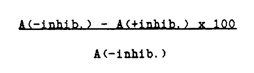
- a sample is positive if the specific value ΔA is greater than 0.050 and the % inhibition which corresponds to it is greater than 50.
- As a general practical criterion, for use side by side with that based on the ΔA values, it is useful to note the colour difference between wells with and without inhibitor as time progresses, even after several hours.
- According to the invention the single-plate ELISA method with competitive inhibition has considerable advantages compared with the state of the art (see Examples 7 and 9), and in particular compared with the double-plate ELISA method.
- These include high sensitivity, specificity and reproducibility of results, speed and ease of execution and the cost-effectiveness of the method.
- The sensitivity and specificity of the method according to the invention are attributable to the TBS/C/TX-100® mixture and the (NANP)₂₀ peptide bath as the antigen adsorbed on the plate and as the competitive inhibitor. The method is applicable not only to serum and plasma samples but also to whole blood samples and mosquito extract without aspecific background problems.
- Moreover, the use of a single plate to determine the specific absorbance for one and the same sample obviates all the problems due to the possible lack of uniformity between different plates.
- A considerable advantage of the method according to the invention is its simple execution and the fast visual evaluation of results.
- Diagnostic kits containing all the reagents required for implementing the method of the invention are well suited to determining antisporozoite antibodies of P. falciparum in human blood samples and mosquito extract.
- For example a diagnostic kit can contain a plate pretreated with the (NANP)₂₀ synthetic antigen alone, the synthetic peptide as competitive inhibitor. and possibly the buffer/protein/detergent mixture. the enzymatic conjugate and the specific substrate.
- A kit of this or of a similar type allows P. falciparum antisporozoite antibodies to be determined at the lowest possible cost.
- The experimental examples given hereinafter illustrate the invention.
- Examples 7 and 9 are comparisons with known immunoenzymatic methods.
- In order to identify the best buffer system for activating the pretreated plate, for diluting the sample and the enzymatic conjugate and for washing purposes, the buffer mixtures TBS/C/TX-100® [Tris-HCl 20 mM, NaCl 0.15 M, pH 7.8/Casein (0.5% w/v)/Triton X-100® (0.1% v/v)] and PBS/C/TK-100® [phosphate buffered saline pH 7.8/casein (0.5% w/v)/X-100 (0.1% v/v)] were tested.
- 100 µl of PBS buffer pH 7.8 containing 1 µg/ml of (NANP)₂₀ were added to each well of a 96-well plate flat-bottomed Dynatech M 129 A plate (Dynatech, Alexandria, Va, USA).
- The plate was kept at ambient temperature (20-25°C) overnight in a humid chamber and then washed twice with PBS buffer.
- The plate was then oven-dried at 37°C for 1 hour and stored in a closed container at 4°C (pretreated plate). Before use, the plate was activated by introducing 200 µl of TBS/C/TX-100® buffer mixture into one half of the wells and 200 µl of PBS/C/TX-100® buffer mixture into the remaining half.
- The plate was kept at ambient temperature (20-25°C) for about 30 minutes.
- A sample of negative serum from a healthy donor and 10 serum samples from an endemic malaria zone were each diluted 1:200 in the TBS/C/TX-100® and in the PBS/C/TX-100® buffer mixtures. 4 x 100 µl aliquots of each sample diluted as described were added to 4 vertically neighbouring wells, to the last two of which (inhibition wells) 5 µl of TBS/C/TX-100® or PBS/C/TX-100® containing 2 µg of (NANP)₂₀ had been previously added. The plate was kept at ambient temperature (20-25°C) for 1 hour and then washed (4 times for 2-3 minutes) with 200 µl of TBS/C/TX-100® or PBS/C/TX-100® buffer mixture as appropriate.
- The procedure was then continued by adding to each well, with the exception of the first vertical column reserved to the blank substrate, 100 µl of the enzymatic antibody/anti-human-immunoglobulin/peroxidase (Bio Rad) conjugate diluted 1:3000 in TBS/C/TX-100® or in PBS/C/TX-100® as appropriate.
- The plate was kept at ambient temperature for 1 hour after which one half of the wells were washed with 200 µl (4 times for 2-3 minutes) with TBS/C/TX-100® and the other half with 200 µl of PBS/C/TX-100.®
- All the wells were then washed 1-2 times with TBS or PBS to eliminate the casein residue.
- 100 µl of 0.1 M pH 5.0 acetate buffer containing 1.1 mg.ml of ABTS® and 1.3 mM of H₂O₂ were then added to each well.
- The enzymatic reaction was conducted at ambient temperature for 45 minutes. After this time the results were evaluated by determining the absorbance with an ELISA reader at 405 nm. The specific value for the sample (ΔA) was given by:

The results, given in Table 1 below, show that the TBS/C/TX-100® buffer mixture was clearly to be preferred to the PBS/C/TX-100® mixture, both because of the better specific values (ΔA) of the positive samples and because of the low aspecific background values (A+inhib column).TABLE 1 Sample: TBS/C/TX-100 PBS/C/TX-1000 Serum A-inhib A+inhib ΔA A-inhib A+inhib ΔA Negative .091 .088 .003 2.393 2.294 .099 1 1.056 .096 .960 1.187 .304 .883 2 .176 .092 .080 1.204 1.053 .151 3 1.586 .145 1.441 2.380 1.487 .893 4 .063 .021 .042 .192 .147 .045 5 .836 .117 .719 1.736 1.148 .588 6 .770 .103 .667 1.419 .803 .616 7 .293 .073 .220 .596 .365 .231 8 1.474 .268 1.206 1.883 .719 1.164 9 .470 .039 .431 .843 .342 .501 10 2.537 .217 2.320 2.575 .470 2.105 - In order to identify the optimum incubation time for the activation of a pretreated plate, the procedure of Example 1 was followed incubating one and the same plate pretreated with TBS/C/TX-100 30 and 60 minutes before introducing the blood samples to be analyzed.
- A negative serum and 6 African sera (1, 3, 4, 5, 6, 8) originating from an endemic malarial zone diluted 1:200 in the TBS/C/TX-100 mixture were used.
- The peroxidase conjugate and ABTS enzymatic substrate were used, with an enzymatic reaction time of 30 minutes.
- Absorbance was determined with an ELISA reader at 405 nm. The results, given in Table 2 below, show that the specific values (ΔA) and aspecific values (A+inhib) were practically equal for the two incubation times.
TABLE 2 Sample: t = 30 minutes t = 60 minutes Serum (1:200) A-inhib A+inhib ΔA A-inhib A+inhib ΔA Negative .177 .204 -.027 .206 .195 .011 1 .789 .098 .691 .634 .072 .562 3 1.332 .184 1.148 1.238 .177 1.061 4 .051 .033 .018 .048 .031 .017 5 .663 .157 .506 .705 .145 .560 6 .546 .117 .429 .605 .117 .488 8 1.113 .126 .987 1.188 .117 1.071 - The procedure of Example 1 was followed but using the TBS/C/TX-100 buffer mixture and 19 heparin plasmas from healthy donors diluted 1:200 with said mixture. Table 3 gives the results obtained.
- The low values of ΔA (between -0.026 and +0.047) together with the low aspecific background values (A+inhib) confirm the high specificity of the single-plate ELISA assay with competitive inhibition.
TABLE 3 Sample A-inhib A+inhib ΔA heparin plasma 1 .074 .067 .007 2 .207 .168 .039 3 .125 .078 .047 4 .155 .150 .005 5 .158 .163 -.005 6 .104 .101 .003 7 .093 .095 -.002 8 .077 .084 -.011 9 .124 .150 -.026 10 .115 .100 .015 11 .136 .111 .025 12 .124 .104 .020 13 .030 .037 -.007 14 .075 .066 .009 15 .040 .031 .009 16 .115 .119 -.004 17 .105 .120 -.015 18 .220 .204 .016 19 .085 .111 -.026 - The procedure of Example 1 was followed but using two Dynatech M129 A plates, one negative serum and twenty sera originating from an endemic malaria zone diluted 1:200 in the TBS/C/TX-100 buffer mixture.
- The peroxidase (Bio-Rad) conjugate and substrate ABTS® were then used for one plate and the antibody/anti-human-immunoglobulin/alkaline phosphatase (SCLAVO S.p.A.) conjugate diluted 1:800 in TBS/C/TX-100 and the enzymatic substrate NPP (nitrophenyl phosphate) were used for the other plate.
- Absorbance was determined for the two plates in an ELISA reader at 405 nm after 30 and 60 minutes.
- The results, given in Table 4, show specific absorbance values (ΔA) for the positive samples which are clearly greater for the peroxidase than for the alkaline phosphatase.
TABLE 4 Sample ΔA (30 minutes) ΔA (60 minutes) Serum (dilute) ABTS® NPP ABTS® NPP Negative -.053 -.053 -.051 -.095 1 .603 .425 1.105 .833 2 .035 -.013 .009 -.029 3 .980 .777 1.678 1.440 4 .046 .019 .076 .033 5 .586 .390 1.027 .782 6 .515 .335 .911 .663 7 .185 .072 .334 .124 8 .925 .639 1.663 1.190 9 .322 .239 .582 .486 10 1.535 1.055 2.555 1.880 11 .126 .067 .218 .118 12 .174 .101 .320 .207 13 .198 .146 .405 .314 15 .025 -.037 .060 -.065 16 -.001 -.007 .011 -.006 17 .176 .136 .376 .284 18 .166 .104 .343 .240 19 .293 .101 .597 .219 20 .540 .433 1.074 .840 21 .290 .191 .593 .403 - The procedure of Example 1 was followed, but using double the quantity of the sera examined in Example 4 and of a negative heparin plasma (dilution 1:100 in TBS/C/TX-100), peroxidase and ABTS®.
- The results, given in Table 5, show that the specific values (ΔA) at 30 minutes for the positive samples are greater than those given in Table 4 (second column) for the same samples, and at the same time show an absence of aspecific background problems (low A+inhib values).
TABLE 5 Sample: A-inhib. A+inhib. ΔA Serum and plasma (dilution 1:100) Negative .095 .107 -.012 1 1.002 .116 .886 3 1.641 .162 1.479 4 .076 .037 .039 5 .737 .081 .656 6 .914 .077 .837 7 .302 .088 .214 8 1.614 .170 1.444 9 .453 .050 .403 10 2.139 .085 2.054 11 .290 .100 .180 12 .265 .036 .229 13 .459 .061 .398 15 .081 .045 .036 16 .042 .025 .017 17 .430 .118 .312 18 .357 .070 .287 19 .446 .086 .360 20 1.105 .162 .943 21 .555 .096 .459 Negative plasma .100 .107 -.007 - A plate treated as in Example 1 was used, together with the TBS/C/TK-100® buffer mixture, the peroxidase (Bio-Rad) conjugate and the enzymatic TS for 30 minutes.
- The procedure of Example 1 was followed using 39 samples of whole blood of African origin dried on Whatman No. 1® paper and kept at ambient temperature for about 15 days in the presence of silica gel.
- The samples (10 µl of whole blood) were extracted with 1 ml of TBS/C/TX-100® buffer at ambient temperature for 1 hour and then assayed as described in Example 1.
- In addition an examination was made of a negative whole blood sample, a negative serum and a positive control obtained by mixing the positive serum No. 8 (Table 2) with negative whole blood, all absorbed on Whatman® paper and then extracted after some days as heretofore described.


- 20 sera originating from an endemic malaria zone, one negative serum and one positive serum were assayed both by the single-plate ELISA method and by the classical double-plate ELISA method, using for this latter one plate pretreated with (NANP)²⁰ and the other pretreated with PBS alone.
- The TBS/C/TX-100 buffer mixture, the peroxidase conjugate, the ABTS® enzymatic substrate and an enzymatic reaction time of 30 minutes were used.
- The results given in Table 7 show that the double-plate method when compared with the single-plate method gives higher aspecific inhibition values (PBS column compared with A+inhib column) and lower specific values (ΔA), with the result that some samples (Nos. 4, 7, 11, 12, 13, 17 and 18) show "false negatives" by the double-plate method.
TABLE 7 Sample: SINGLE-PLATE DOUBLE-PLATE Sera (dil. 1:200) A-inhib A+inhib ΔA A-inhib PBS ΔA Negative .270 .285 -.015 .270 .466 -.196 Positive .675 .211 .464 .675 .354 .321 1 .867 .158 .709 .867 .380 .487 3 1.358 .345 1.013 1.358 .688 .670 4 .111 .071 .040 .111 .132 -.021 5 .740 .220 .520 .740 .356 .384 6 .564 .181 .383 .564 .355 .209 7 .400 .205 .195 .400 .448 -.048 8 1.097 .248 .849 1.097 .304 .793 9 .528 .157 .371 .528 .393 .135 10 1.892 .158 1.734 1.892 .279 1.613 11 .248 .141 .107 .248 .303 -.055 12 .214 .101 .113 .214 .224 -.010 13 .341 .134 .207 .341 .314 -.027 15 .211 .178 .033 .211 .351 -.140 16 .076 .066 .010 .076 .155 -.079 17 .460 .269 .191 .460 .521 -.061 18 .363 .152 .211 .363 .310 .053 19 .451 .177 .274 .451 .284 .167 20 1.057 .416 .641 1.057 .722 .335 21 .679 .312 .367 .679 .575 .104 KX .604 .164 .440 .604 .297 .307 - Three extracts from mosquitos of an African endemic malaria zone obtained by squeezing-out onto Whatman No. 1 filter paper, drying and storing in the presence of silica gel, were extracted together with 0.6 ml of TBS/C/TX-100 containing the protease inhibitor PMSF (phenylmethylsulphonylfluoride) at 4°C for 60 minutes.
- Four 0.1 ml aliquots of extract were then assayed operating as described in Example 1.
- The spectrophotometric results, determined after 30 and 60 minutes and given in Table 8, agree perfectly with the visual observation of colour difference in positive diagnosis of the sample examined. This confirms the high sensitivity and specificity of the single-plate method with inhibition.
TABLE 8 Enzymatic reaction time A-inhib A+inhib ΔA 30 minutes .153 .008 .145 60 minutes .338 .038 .300 - The sera assayed in Example 7 were analyzed both by the P-E/PEG method described in EP-A-0 341 767. using the peroxidase conjugate and the TMB (tetramethylbenzidine) enzymatic substrate, and by the P-E/SPA method described in GB-A-2 199 140 using the peroxidase enzyme and the ABTS enzymatic substrate.
- In Table 9:
column 6 shows the spectrophotometric results obtained by the P-E/PEG method expressed as ΔA;
column 7 shows the results obtained by the P-E/SPA method where + indicates positive sera, - indicates negative sera and ? indicates uncertainty;
columns 2 and 3 show the ( Δ A) results obtained by the "single-plate ELISA with inhibition" using the peroxidase conjugate with ABTS® and the conjugate with alkaline phosphatase and NPP respectively;
the columns 4 and 5 show the results (ΔA) obtained by the "double-plate ELISA" using casein, peroxidase and ABTS® (HRP/ABTS®) and, respectively, BSA, alkaline phosphatase and NPP (ALP/NPP). - A comparison of the various methods shows the greater sensitivity and specificity of the single-plate ELISA method with competitive inhibition using peroxidase and ABTS®.
- Sample 17, which is positive by the single-plate method, is uncertain by the P-E/PEG and P-E/SPA methods in that the high aspecific value and the low specific value (ΔA) make for poor reliability in the result.
TABLE 9 Sample: Single-plate ELISA with inhibition Double-plate ELISA P-E/PEG P-E/SPA (Sera) (HRP/ABTS®) (ALP/NPP) (HRP/ABTS®) (ALP/NPP) (HRP/TMB) (HRP/ABTS®) Negative -.053 -.053 -.196 -.020 -.014 - Positive .464 N.D .321 .304 N.D + 1 .603 .425 .487 -.040 .722 + 2 .035 -.013 N.D -.040 .063 - 3 .980 .777 .670 .269 .425 + 4 .046 .019 -.021 .084 .258 - 5 .586 .390 .384 .159 .416 + 6 .515 .335 .209 .884 .765 + 7 .195 .072 -.048 -.024 .584 + 8 .925 .639 .793 1.048 1.441 + 9 .322 .239 .135 .119 .435 + 10 1.535 1.058 1.613 .336 1.706 + 11 .126 .067 -.055 .013 .239 + 12 .174 .101 -.010 .005 .367 + 13 .198 .146 -.027 .126 .304 + 15 .025 -.037 -.140 .011 .093 - 16 -.001 .007 -.079 -.045 -.083 - 17 .176 .136 .061 -.221 ? ? 18 .166 .104 .053 N.D .822 + 19 .293 .101 .167 N.D .402 + 20 .546 .433 .335 N.D .792 + 21 .290 .191 .104 N.D .442 + KX .440 N.D .307 .097 .513 +
Claims (4)
- A competitive single-plate ELISA method for detecting antibodies to Plasmodium falciparum sporozoites in a human blood sample, comprising the steps of:(a) coating the wells of a plate with the synthetic antigen (NANP)₂₀, wherein N is Asparagine, A is Alanine and P is Proline, by incubating the plate overnight in a humid chamber, at a temperature of from 20°C to 25°C, with 0,1 ml/well of 1 µg/ml (NANP)₂₀ solution in TRIS buffered saline; (TBS)(b) washing the plate with TRIS buffered saline solution (TBS) to remove the loosely bound synthetic antigen;(c) adding to each well of the plate TRIS buffered saline solution (TBS) containing 0.5% (w/v) casein and 0.1% (v/v) of a non-ionic detergent,(d) incubating the plate at a temperature of from 20°C and 37°C for at least 30 min;(e) adding an aliquot of sample selected from whole blood, serum plasma and a mosquito extract extracted with TRIS buffered saline (TBS) containing a protease inhibitor, said sample having been diluted with Tris buffered saline solution containing 0.5% (w/v) casein and 0.1% (v/v) non-ionic detergent, to each of four adjacent wells of the plate of step (c);(f) introducing into two of said wells, either before, or simultaneously with, the diluted sample, the synthetic peptide (NANP)₂₀ in a minimum weight ratio of 20:1 with respect to the adsorbed synthetic antigen;(g) incubating said plate at a temperature of from 20°C to 37°C for a time of from 30 min to 60 min;(h) washing the plate with a TRIS buffered saline solution (TBS) containing 0.5% (w/v) casein and 0.1% (v/v) non-ionic detergent,(i) adding to each well of the plate an antibody/anti-human immunoglobin/enzyme conjugate diluted in a TRIS buffered saline solution (TBS) containing 0.5% (w/v) casein and 0.1% (v/v) non-ionic detergent,(j) incubating the plate at room temperature for a time of from 30 min to 60 min;(k) washing the plate with a TRIS buffered saline solution (TBS) containing 0.5% (w/v) casein and 0.1% (v/v) non-ionic detergent,(l) washing the plate with a TRIS buffered saline solution (TBS);(m) adding to each well a colourless substrate solution specific for the enzyme of the conjugate used in step (i);(n) incubating the plate for a time of from 10 min to 60 min, and(o) detecting the specific value of each sample by the difference in absorbance between the uninhibited and the fully inhibited sample.
- Single-plate ELISA method according to Claim 1 , wherein the non-ionic detergent is selected from
polyoxyethyleneglycol (9) p-t-octylphenol;
polyoxyethyleneglycol sorbitanmonolaurate, and
polyoxyethyleneglycol (9,10) p-t-octylphenol. - Single-plate ELISA method according to Claim 1, wherein the TRIS buffered saline solution (TBS) containing 0.5% (w/v) casein and 0.1% (v/v) of a non-ionic detergent, additionally contains a polyethyleneglycol having a molecularweight of 6000.
- A diagnostic kit for detecting antisporozoite antibodies of Plasmodium falciparum in a human blood sample or mosquito extract by the ELISA method claimed in claim 1, comprising a plate pretreated with the synthetic antigen (NANP)₂₀, the competitive inhibitor in the form of the synthetic peptide (NANP)₂₀, and a tris buffered saline solution containing 0.5% (w/v) casein and 0.1% (v/v) non-ionic detergent
Priority Applications (1)
| Application Number | Priority Date | Filing Date | Title |
|---|---|---|---|
| AT89201983T ATE93970T1 (en) | 1988-08-05 | 1989-07-28 | SINGLE PLATE IMMUNOENZYMATIC ELISA TEST WITH COMPETITIVE INHIBITION FOR THE DETECTION OF ANTISPOROZOITE ANTIBODIES OF PLASMODIUM FALCIPARUM. |
Applications Claiming Priority (2)
| Application Number | Priority Date | Filing Date | Title |
|---|---|---|---|
| IT8821665A IT1226712B (en) | 1988-08-05 | 1988-08-05 | IMMUNOENZYMATIC METHOD ELISA SINGLE PLATE WITH COMPETITIVE INHIBITION FOR THE DETECTION OF PLASMODIUM FALCIPARUM ANTI-SHORT ANTIBODIES. |
| IT2166588 | 1988-08-05 |
Publications (2)
| Publication Number | Publication Date |
|---|---|
| EP0353822A1 EP0353822A1 (en) | 1990-02-07 |
| EP0353822B1 true EP0353822B1 (en) | 1993-09-01 |
Family
ID=11185058
Family Applications (1)
| Application Number | Title | Priority Date | Filing Date |
|---|---|---|---|
| EP89201983A Expired - Lifetime EP0353822B1 (en) | 1988-08-05 | 1989-07-28 | Immunoenzymatic single-plate elisa method with competitive inhibitition for detecting antisporozoite antibodies of plasmodium falciparum |
Country Status (6)
| Country | Link |
|---|---|
| US (1) | US5374530A (en) |
| EP (1) | EP0353822B1 (en) |
| AT (1) | ATE93970T1 (en) |
| DE (1) | DE68908814T2 (en) |
| ES (1) | ES2058477T3 (en) |
| IT (1) | IT1226712B (en) |
Families Citing this family (22)
| Publication number | Priority date | Publication date | Assignee | Title |
|---|---|---|---|---|
| US5124141A (en) * | 1990-06-14 | 1992-06-23 | Flow Incorporated | Method for diagnosing malaria |
| US6908770B1 (en) | 1998-07-16 | 2005-06-21 | Board Of Regents, The University Of Texas System | Fluid based analysis of multiple analytes by a sensor array |
| AU3494600A (en) * | 1999-02-19 | 2000-09-04 | Medical Analysis Systems, Inc. | Rapid assay for arthropod-borne disease vectors and pathogens |
| US7022517B1 (en) | 1999-07-16 | 2006-04-04 | Board Of Regents, The University Of Texas System | Method and apparatus for the delivery of samples to a chemical sensor array |
| AU773679B2 (en) | 1999-07-16 | 2004-06-03 | Board Of Regents, The University Of Texas System | Method and apparatus for the delivery of samples to a chemical sensor array |
| CA2401782A1 (en) | 2000-01-31 | 2001-08-02 | John T. Mcdevitt | Portable sensor array system |
| US8257967B2 (en) | 2002-04-26 | 2012-09-04 | Board Of Regents, The University Of Texas System | Method and system for the detection of cardiac risk factors |
| US6977079B2 (en) * | 2003-01-17 | 2005-12-20 | Regents Of The University Of Minnesota | Avian pneumovirus vaccine |
| WO2005059551A2 (en) | 2003-12-11 | 2005-06-30 | Board Of Regents, The University Of Texas System | Method and system for the analysis of saliva using a sensor array |
| US8105849B2 (en) | 2004-02-27 | 2012-01-31 | Board Of Regents, The University Of Texas System | Integration of fluids and reagents into self-contained cartridges containing sensor elements |
| US8101431B2 (en) | 2004-02-27 | 2012-01-24 | Board Of Regents, The University Of Texas System | Integration of fluids and reagents into self-contained cartridges containing sensor elements and reagent delivery systems |
| NL1026271C2 (en) * | 2004-05-26 | 2005-11-30 | Keygene Nv | Method and device for isolating nucleic acid from material containing nucleic acid. |
| US20080268426A1 (en) * | 2004-06-18 | 2008-10-30 | Regents Of The University Of Minnesota | Identifying virally infected and vaccinated organisms |
| TW200604526A (en) * | 2004-06-18 | 2006-02-01 | Unviersity Of Minnesota | Identifying virally infected and vaccinated organisms |
| KR101035111B1 (en) * | 2004-06-30 | 2011-05-19 | 주식회사 엘지생명과학 | Methods of immunological measurement of malaria plasmodium falciparum and measuring means used therein |
| WO2007053186A2 (en) | 2005-05-31 | 2007-05-10 | Labnow, Inc. | Methods and compositions related to determination and use of white blood cell counts |
| PT2369001T (en) | 2005-06-24 | 2016-10-25 | Univ Minnesota | Prrs viruses, infectious clones, mutants thereof, and methods of use |
| US8257938B2 (en) * | 2009-02-27 | 2012-09-04 | Medical Diagnostic Laboratories, Llc | Hemolysin and its protein fragments in sero-detection of Anaplasma phagocytophilum |
| US9835621B2 (en) | 2011-07-14 | 2017-12-05 | University Of Kentucky Research Foundation | Process for detection of alzheimer's disease from a serum sample |
| KR101353930B1 (en) | 2012-02-20 | 2014-01-27 | 주식회사 나노엔텍 | A Novel Method for Detecting an Antigen and Apparatus Using It |
| US20160281128A1 (en) * | 2013-11-08 | 2016-09-29 | Veterinary Diagnostics Institute, Inc. | Method and apparatus for detecting vector-borne diseases in mammals |
| KR20170108203A (en) | 2016-03-16 | 2017-09-27 | 주식회사 피플바이오 | Method for Detecting Aggregate Form of Aggregate-Forming Polypeptides |
Family Cites Families (4)
| Publication number | Priority date | Publication date | Assignee | Title |
|---|---|---|---|---|
| WO1984002472A1 (en) * | 1982-12-27 | 1984-07-05 | Pasteur Institut | Polypeptidic fractions inducing protection antibodies against parasites of paludism, and immunizing compositions containing them |
| DE3327642A1 (en) * | 1983-07-30 | 1985-02-14 | Boehringer Mannheim Gmbh, 6800 Mannheim | METHOD FOR DETERMINING A PARTNER OF AN IMMUNE REACTION AND REAGENT FOR CARRYING OUT THIS METHOD |
| SE454465B (en) * | 1986-04-15 | 1988-05-02 | Biopool Ab | NON COMPETITIVE FAST-PHASE IMMUNAL ANALYSIS WITH COMPENSATION FOR UNSPECIFIC ADSORPTION |
| IT1198233B (en) * | 1986-12-23 | 1988-12-21 | Eniricerche Spa | METHOD FOR THE DETERMINATION OF ANTI-SPORTS ANTIBODIES OF P. FALCIPARUM IN HUMAN BLOOD |
-
1988
- 1988-08-05 IT IT8821665A patent/IT1226712B/en active
-
1989
- 1989-07-28 ES ES89201983T patent/ES2058477T3/en not_active Expired - Lifetime
- 1989-07-28 DE DE89201983T patent/DE68908814T2/en not_active Expired - Fee Related
- 1989-07-28 AT AT89201983T patent/ATE93970T1/en not_active IP Right Cessation
- 1989-07-28 EP EP89201983A patent/EP0353822B1/en not_active Expired - Lifetime
-
1992
- 1992-05-13 US US07/882,289 patent/US5374530A/en not_active Expired - Fee Related
Also Published As
| Publication number | Publication date |
|---|---|
| IT8821665A0 (en) | 1988-08-05 |
| EP0353822A1 (en) | 1990-02-07 |
| ES2058477T3 (en) | 1994-11-01 |
| ATE93970T1 (en) | 1993-09-15 |
| DE68908814T2 (en) | 1994-01-27 |
| DE68908814D1 (en) | 1993-10-07 |
| IT1226712B (en) | 1991-02-05 |
| US5374530A (en) | 1994-12-20 |
Similar Documents
| Publication | Publication Date | Title |
|---|---|---|
| US5374530A (en) | Immunoenzymatic single-plate ELISA method with competitive inhibition for detecting antisporozoite antibodies of plasmodium falciparum | |
| EP0615129B1 (en) | Methods for selectively detecting perinuclear anti-neutrophil cytoplasmic antibody of ulcerative colitis or primary sclerosing cholangitis | |
| Newbold et al. | Plasmodium falciparum: the human agglutinating antibody response to the infected red cell surface is predominantly variant specific | |
| Grauballe et al. | Optimized enzyme‐linked immunosorbent assay for detection of human and bovine rotavirus in stools: Comparison with electron‐microscopy, immunoelectro‐osmophoresis, and fluorescent antibody techniques | |
| Decary et al. | An investigation of nonhemolytic transfusion reactions | |
| EP1825262B1 (en) | Delta-9-tetrahydrocannabinol detection method | |
| EP0802411B1 (en) | Measurement method and kit for hemoglobin A1c | |
| US4888276A (en) | Method and composition for the diagnosis of Lyme disease | |
| GB2268935A (en) | Antigen extracted from endothelial cells or from monocytes | |
| EP0173295A1 (en) | Assay for simultaneous detection of antigen and antibody in given serum | |
| Malan et al. | Serological diagnosis of Trypanosoma cruzi: evaluation of three enzyme immunoassays and an indirect immunofluorescent assay | |
| Lillie et al. | Artifactual immunofluorescent labelling in yeast, demonstrated by affinity purification of antibody | |
| US8110663B2 (en) | Monoclonal antibodies for cartillage oligomeric matrix protein | |
| EP0341767B1 (en) | Immunoenzymatic method in homogeneous phase for the determination of anti-plasmodium falciparum-sporozoite antibodies in human blood | |
| Jean-Pierre et al. | Serological diagnosis of toxoplasmosis: evaluation of the commercial test recomLine Toxoplasma IgG immunoblot (Mikrogen) based on recombinant antigens | |
| CA1295551C (en) | Immunoassay for antibodies to htlv-iii | |
| Maeda et al. | An enzyme-linked immunosorbent assay for thyroxine in dried blood spotted on filter paper | |
| EP0662611A2 (en) | Method for assaying glycoconjugates and reagent thereof | |
| Schlaf et al. | A novel enzyme-linked immunosorbent assay for determination of synaptophysin as compared with other quantification procedures | |
| US5149623A (en) | Rapid, easy, and economical screening test for antibodies to human immunodeficiency virus | |
| Crowther et al. | Evaluation of the use of chromogenic and fluorogenic substrates in solid-phase enzyme linked immunosorbent assays (ELISA) | |
| WO1995002186A1 (en) | New diagnostic assay for detection of syphilis | |
| WO1997024141A1 (en) | Monoclonal antibodies and immuno-capture method for quantitation and speciation of malaria parasites | |
| Khusmith et al. | Two-site sandwich ELISA for detection of Plasmodium vivax blood stage antigens using monoclonal and polyclonal antibodies | |
| EP0324008A4 (en) | A color-coded solid phase analysis method |
Legal Events
| Date | Code | Title | Description |
|---|---|---|---|
| PUAI | Public reference made under article 153(3) epc to a published international application that has entered the european phase |
Free format text: ORIGINAL CODE: 0009012 |
|
| AK | Designated contracting states |
Kind code of ref document: A1 Designated state(s): AT BE CH DE ES FR GB GR LI LU NL SE |
|
| 17P | Request for examination filed |
Effective date: 19900613 |
|
| 17Q | First examination report despatched |
Effective date: 19920214 |
|
| GRAA | (expected) grant |
Free format text: ORIGINAL CODE: 0009210 |
|
| AK | Designated contracting states |
Kind code of ref document: B1 Designated state(s): AT BE CH DE ES FR GB GR LI LU NL SE |
|
| REF | Corresponds to: |
Ref document number: 93970 Country of ref document: AT Date of ref document: 19930915 Kind code of ref document: T |
|
| REF | Corresponds to: |
Ref document number: 68908814 Country of ref document: DE Date of ref document: 19931007 |
|
| ET | Fr: translation filed | ||
| REG | Reference to a national code |
Ref country code: GR Ref legal event code: FG4A Free format text: 3009216 |
|
| PGFP | Annual fee paid to national office [announced via postgrant information from national office to epo] |
Ref country code: GR Payment date: 19940617 Year of fee payment: 6 |
|
| PGFP | Annual fee paid to national office [announced via postgrant information from national office to epo] |
Ref country code: ES Payment date: 19940620 Year of fee payment: 6 |
|
| PLBE | No opposition filed within time limit |
Free format text: ORIGINAL CODE: 0009261 |
|
| STAA | Information on the status of an ep patent application or granted ep patent |
Free format text: STATUS: NO OPPOSITION FILED WITHIN TIME LIMIT |
|
| PGFP | Annual fee paid to national office [announced via postgrant information from national office to epo] |
Ref country code: AT Payment date: 19940711 Year of fee payment: 6 |
|
| PGFP | Annual fee paid to national office [announced via postgrant information from national office to epo] |
Ref country code: BE Payment date: 19940713 Year of fee payment: 6 |
|
| PGFP | Annual fee paid to national office [announced via postgrant information from national office to epo] |
Ref country code: GB Payment date: 19940715 Year of fee payment: 6 |
|
| PGFP | Annual fee paid to national office [announced via postgrant information from national office to epo] |
Ref country code: FR Payment date: 19940721 Year of fee payment: 6 |
|
| PGFP | Annual fee paid to national office [announced via postgrant information from national office to epo] |
Ref country code: DE Payment date: 19940728 Year of fee payment: 6 |
|
| PGFP | Annual fee paid to national office [announced via postgrant information from national office to epo] |
Ref country code: SE Payment date: 19940731 Year of fee payment: 6 Ref country code: NL Payment date: 19940731 Year of fee payment: 6 Ref country code: LU Payment date: 19940731 Year of fee payment: 6 |
|
| 26N | No opposition filed | ||
| EPTA | Lu: last paid annual fee | ||
| PGFP | Annual fee paid to national office [announced via postgrant information from national office to epo] |
Ref country code: CH Payment date: 19941024 Year of fee payment: 6 |
|
| REG | Reference to a national code |
Ref country code: ES Ref legal event code: FG2A Ref document number: 2058477 Country of ref document: ES Kind code of ref document: T3 |
|
| EAL | Se: european patent in force in sweden |
Ref document number: 89201983.7 |
|
| PG25 | Lapsed in a contracting state [announced via postgrant information from national office to epo] |
Ref country code: LU Free format text: LAPSE BECAUSE OF NON-PAYMENT OF DUE FEES Effective date: 19950728 Ref country code: GB Effective date: 19950728 Ref country code: AT Effective date: 19950728 |
|
| PG25 | Lapsed in a contracting state [announced via postgrant information from national office to epo] |
Ref country code: SE Effective date: 19950729 Ref country code: ES Free format text: LAPSE BECAUSE OF NON-PAYMENT OF DUE FEES Effective date: 19950729 |
|
| PG25 | Lapsed in a contracting state [announced via postgrant information from national office to epo] |
Ref country code: LI Effective date: 19950731 Ref country code: CH Effective date: 19950731 Ref country code: BE Effective date: 19950731 |
|
| BERE | Be: lapsed |
Owner name: ENIRICERCHE S.P.A. Effective date: 19950731 |
|
| PG25 | Lapsed in a contracting state [announced via postgrant information from national office to epo] |
Ref country code: GR Free format text: THE PATENT HAS BEEN ANNULLED BY A DECISION OF A NATIONAL AUTHORITY Effective date: 19960131 |
|
| PG25 | Lapsed in a contracting state [announced via postgrant information from national office to epo] |
Ref country code: NL Effective date: 19960201 |
|
| REG | Reference to a national code |
Ref country code: CH Ref legal event code: PL |
|
| GBPC | Gb: european patent ceased through non-payment of renewal fee |
Effective date: 19950728 |
|
| REG | Reference to a national code |
Ref country code: GR Ref legal event code: MM2A Free format text: 3009216 |
|
| NLV4 | Nl: lapsed or anulled due to non-payment of the annual fee |
Effective date: 19960201 |
|
| PG25 | Lapsed in a contracting state [announced via postgrant information from national office to epo] |
Ref country code: DE Effective date: 19960402 |
|
| EUG | Se: european patent has lapsed |
Ref document number: 89201983.7 |
|
| PG25 | Lapsed in a contracting state [announced via postgrant information from national office to epo] |
Ref country code: FR Effective date: 19960430 |
|
| REG | Reference to a national code |
Ref country code: FR Ref legal event code: ST |
|
| REG | Reference to a national code |
Ref country code: FR Ref legal event code: ST |
|
| REG | Reference to a national code |
Ref country code: FR Ref legal event code: ST |
|
| REG | Reference to a national code |
Ref country code: ES Ref legal event code: FD2A Effective date: 20020603 |