EP0345546B1 - DNA coding for enzyme capable of acylating the 4''-position of macrolide antibiotics - Google Patents
DNA coding for enzyme capable of acylating the 4''-position of macrolide antibiotics Download PDFInfo
- Publication number
- EP0345546B1 EP0345546B1 EP89109425A EP89109425A EP0345546B1 EP 0345546 B1 EP0345546 B1 EP 0345546B1 EP 89109425 A EP89109425 A EP 89109425A EP 89109425 A EP89109425 A EP 89109425A EP 0345546 B1 EP0345546 B1 EP 0345546B1
- Authority
- EP
- European Patent Office
- Prior art keywords
- dna
- plasmid
- microorganism
- gene
- fragment
- Prior art date
- Legal status (The legal status is an assumption and is not a legal conclusion. Google has not performed a legal analysis and makes no representation as to the accuracy of the status listed.)
- Expired - Lifetime
Links
Images









Classifications
-
- C—CHEMISTRY; METALLURGY
- C07—ORGANIC CHEMISTRY
- C07H—SUGARS; DERIVATIVES THEREOF; NUCLEOSIDES; NUCLEOTIDES; NUCLEIC ACIDS
- C07H17/00—Compounds containing heterocyclic radicals directly attached to hetero atoms of saccharide radicals
- C07H17/04—Heterocyclic radicals containing only oxygen as ring hetero atoms
- C07H17/08—Hetero rings containing eight or more ring members, e.g. erythromycins
-
- C—CHEMISTRY; METALLURGY
- C12—BIOCHEMISTRY; BEER; SPIRITS; WINE; VINEGAR; MICROBIOLOGY; ENZYMOLOGY; MUTATION OR GENETIC ENGINEERING
- C12N—MICROORGANISMS OR ENZYMES; COMPOSITIONS THEREOF; PROPAGATING, PRESERVING, OR MAINTAINING MICROORGANISMS; MUTATION OR GENETIC ENGINEERING; CULTURE MEDIA
- C12N9/00—Enzymes; Proenzymes; Compositions thereof; Processes for preparing, activating, inhibiting, separating or purifying enzymes
- C12N9/10—Transferases (2.)
- C12N9/1025—Acyltransferases (2.3)
- C12N9/1029—Acyltransferases (2.3) transferring groups other than amino-acyl groups (2.3.1)
-
- C—CHEMISTRY; METALLURGY
- C12—BIOCHEMISTRY; BEER; SPIRITS; WINE; VINEGAR; MICROBIOLOGY; ENZYMOLOGY; MUTATION OR GENETIC ENGINEERING
- C12P—FERMENTATION OR ENZYME-USING PROCESSES TO SYNTHESISE A DESIRED CHEMICAL COMPOUND OR COMPOSITION OR TO SEPARATE OPTICAL ISOMERS FROM A RACEMIC MIXTURE
- C12P19/00—Preparation of compounds containing saccharide radicals
- C12P19/44—Preparation of O-glycosides, e.g. glucosides
- C12P19/60—Preparation of O-glycosides, e.g. glucosides having an oxygen of the saccharide radical directly bound to a non-saccharide heterocyclic ring or a condensed ring system containing a non-saccharide heterocyclic ring, e.g. coumermycin, novobiocin
- C12P19/62—Preparation of O-glycosides, e.g. glucosides having an oxygen of the saccharide radical directly bound to a non-saccharide heterocyclic ring or a condensed ring system containing a non-saccharide heterocyclic ring, e.g. coumermycin, novobiocin the hetero ring having eight or more ring members and only oxygen as ring hetero atoms, e.g. erythromycin, spiramycin, nystatin
Definitions
- This invention relates to a DNA fragment containing a gene (named "acyB”) which gives a host microorganism the enzyme activity of acylating the 4"-position of a macrolide antibiotic, a recombinant DNA plasmid containing this DNA fragment and a microorganism transformed with this recombinant plasmid.
- acyB a DNA fragment containing a gene which gives a host microorganism the enzyme activity of acylating the 4"-position of a macrolide antibiotic, a recombinant DNA plasmid containing this DNA fragment and a microorganism transformed with this recombinant plasmid.
- Macrolide antibiotics having 14-membered and 16-membered rings are useful antibiotics which are widely used as pharmaceuticals for man and animals (for domestic animals and fish culture).
- S. Omura et al. Journal of Antibiotics, vol. 28, pages 401-433 (1980)] and R. Okamoto et al. [Journal of Antibiotics, vol. 27, pages 542-544 (1979)] reported that the antimicrobial activity of a 16-membered ring macrolide antibiotic is increased by acylation of the OH group at its 4"-position.
- the present invention provides an industrially very useful method in which a microorganism strain capable of producing a 4"-acylation product of a macrolide antibiotic is obtained by a genetic engineering technique from a microorganism strain which inherently cannot directly produce the 4"-acylation product.
- a macrolide antibiotic having -OH at the 3- and/or 4 ⁇ -position is added as a substrate to cells, a culture or a cell extract of, for example, Streptomyces thermotolerans (ATCC 11416), Streptomyces hygroscopicus (ATCC 21582) or Streptomyces kitasatoensis (IFO 13686).
- the macrolide antibiotic-producing microorganism differs from the microorganism having acylating activity. This is economically disadvantageous in industrial production.
- acylate biologically a macrolide antibiotic of the type having OH at the 3-position and/or 4"-position, a two-step operation is necessary in which the macrolide-producing microorganism strain is cultured and the macrolide antibiotic is isolated, and then the macrolide antibiotic substance is transformed with a microorganism having an acylating enzyme.
- EP-A-0 354 641 discloses a method for increasing the amount of 4"-O-isovalerylacylase enzyme in an organism that comprises (1) transforming said organism with a recombinant DNA vector that codes for expression of the car E gene product; and (2) culturing said organism transformed in step (1) under conditions that allow for gene expression.
- the present inventors made extensive investigations in order to isolate the DNA of an enzyme capable of acylating the 3- and/or 4"-position of a macrolide from a microorganism containing it, and consequently succeeded in isolating the 4"-position acylating enzyme gene acyB from a certain microorganism belonging to the genus Streptomyces. This has led to the accomplishment of the present invention.
- a purified and isolated DNA fragment derived from Streptomyces thermotolerans having a size of about 3.1 kb and bounded by the Sau 3AI and SphI restriction endonuclease sites depicted in Fig. 1, said fragment comprising DNA sequences encoding for an acy B1 gene and acy B2 gene and containing a DNA portion coding for the following amino acid sequence and a DNA portion coding for the following amino acid sequence
- DNA restriction fragments obtained by digesting the DNA fragment with restriction endonucleases denote DNA fragments of various sizes digested with restriction endonuleases to lengths necessary for expression of acylating enzyme activity.
- the DNA fragment containing gene acyB and DNA restriction fragments in accordance with this invention are useful for commercial production of macrolide antibiotic derivatives such as 4"-acylated tylosin. They are particularly useful in that they make it possible to utilize the recombinant DNA technique commercially for microorganisms of the genus Streptomyces and other antibiotic-producing microorganisms.
- Microorganism strains belonging to the genus Streptomyces as sources of the DNA fragment containing gene acyB are strains of Streptomyces thermotolerans.
- thermotolerans is deposited, for example, at The American Type Culture Collection under the deposit No. ATCC 11416 and is easily available.
- This microorganism is aerobically cultured at 28 °C as described in detail in Example 1 given hereinafter, and the cells in the logarithmic growth period were obtained.
- the cells were treated in accordance with the method of Hopwood et al. (Genetic Manipulation of Streptomyces: A Laboratory Manual, The John Innes Foundation, Norwich, England, 1985) to prepare a genome DNA containing acyB gene.
- the resulting genome DNA containing acyB gene is ligated with a suitable vector plasmid such as pIJ702, pIJ943, pIJ350 or pUC18.
- the vector plasmid pIJ702 can be isolated from a microorganism containing it by phenol extraction and density gradient ultracentrifugation using cesium chloride.
- the resulting plasmid pIJ702 is digested with restriction endonuclease BalII.
- DNA fragment obtained by partially digesting the above genome DNA with restriction endonuclease Sau3AI is inserted into the resulting digested plasmid.
- Streptomyces lividans TK24 is transformed with the resulting plasmid containing part of the genome DNA of Streptomyces thermotolerans ATCC 11416 to obtain transformants which have acquired tylosin acylating ability (for details, see Example 3 given hereinafter).
- plasmid pAOY-17 containing a DNA inserted fragment having a size of about 6 kb can be obtained.
- plasmid pAO207 can be obtained in which an about 13 kb restriction endonuclease Sau3AI digested fragment derived from Streptomyces thermotolerans is inserted into the restriction endonuclease BglII-cleaved site of plasmid pIJ943 (see Example 4 given hereinafter).
- Plasmid pIJ702 is available from The American Type Culture Collection under deposit No. ATCC 35287. Plasmid pIJ943 is disclosed by Hopwood et al. [see Genetic Manipulation of Streptomyces: A Laboratory Manual, pages 314-315, 1985, The John Innes Foundation], and may be obtained under Streptomyces lividans TK24/pIJ943 from The John Innes Streptomyces Culture Collection, The John Innes Institute, Colney Lane, Norwich NR4 7UH, England. Streptomyces lividans TK24, its host organism, may likewise be obtained from The John Innes Streptomyces Culture Collection. Maps showing the restriction endonuclease cleavage sites and functions of plasmid pAOY-17 and pAO207 are shown in Figures 2 and 3 of the accompanying drawings.
- the acyB gene of the DNA fragment inserted into plasmids pAOY-17 and pAO207 can be further restricted.
- the inserted DNA fragment is digested with a suitable restriction endonuclease, such as BglII, SacI and PstI to form a smaller DNA fragment, and the smaller DNA fragment is inserted into a suitable plasmid for the genus Streptomyces.
- Streptomyces lividans TK24 is transformed with the plasmid having inserted thereinto the smaller DNA fragment, and the ability of the transformant to acylate the 4 ⁇ -position of tylosin to determine whether the inserted DNA fragment contains the acyB gene (see Example 7 below for details).
- the acyB gene in the DNA fragment inserted in the plasmids pAOY-17 and pAO207 can be restricted as shown in Figure 1.
- This DNA fragment is contained in the common DNA region present in the plasmid pAOY-17 and pAO207.
- a 13 kb PvuII-cleaved fragment is labelled with dCTP, and subjected to Southern hybridization with the genome DNA of Streptomyces thermotolerans ATCC 11416 or Streptomyces lividans TK24.
- the Southern hybridization is disclosed by Southern in Journal of Molecular Biology, vol. 98, page 503, 1975. This has led to the determination that the DNA fragments inserted into pAOY-17 and pAO207 are derived from S. thermotolerans ATCC 11416.
- the above plasmids pAOY-17 and pAO207 contain a Streptomyces replicon, a restriction fragment which imparts enzyme activity of acylating the 4 ⁇ -position of tylosin, and as a selection marker, a thiostrepton-resistant gene.
- the two plasmids can be utilized as useful starting substances for many plasmids of the invention containing acyB gene.
- Proliferation of plasmids derived from Streptomyces is more rapid and efficient than that of plasmids derived from Escherichia coli. It is advantageous therefore to introduce a DNA restriction fragment containing the acyB gene into a plasmid from Escherichia coli such as pBR322 and pUC18, and replicate the fragment in Escherichia coli.
- a DNA restriction fragment containing acyB gene into plasmid pUC18 by the procedures described in Examples 5 and 6 given hereinbelow, plasmids pUY-17, pY-17 ⁇ Eco, pUS81, pUS82, pS82 ⁇ Pst, or pS82 ⁇ Sac can be prepared.
- Plasmid pUC18 A commercial product is conveniently used as plasmid pUC18 (such as the one produced by Pharmacia). Maps showing the restriction endonuclease cleavage sites and functions of the above plasmids are shown in Figures 4 and 9 of the accompanying drawings.
- pUY-17 has both a replicon of Escherichia coli and a replicon of genus Streptomyces, and can transform Streptomyces lividans to impart the ability to acylate the 4 ⁇ -position of tylosin.
- the above plasmids except the pS82 ⁇ Pst completely retain the acyB gene. But in plasmid pS82 ⁇ Pst, part of the acyB-gene is deleted. Restriction fragments containing the acyB gene can be cut out from the above plasmids completely retaining the acyB gene by using restriction endonucleases such as SphI and SacI.
- the resulting restriction fragments can be inserted into such plasmids as pIJ702, pIJ943 and pIJ350 [Kieser et al.: Molecular and General Genetics, vol. 185, pages 223-238 (1982)].
- the recombinant plasmids can transform Streptomyces lividans TK24 and impart to it the ability to acylate the 4 ⁇ -position of a macrolide antibiotic.
- the DNA fragments containing the acyB gene which are used for constructing plasmids in this invention may usually be modified in order to facilitate ligation.
- the DNA fragment containing the acyB gene may be bound to a synthetic linker.
- a site specific for the subsequent ligation reaction can be constructed conveniently.
- vector plasmids that can be used to introduce the DNA fragment containing the acyB gene of this invention into host microorganisms, but these specific examples are not limitative.
- vector plasmids containing Streptomyces replicons or Escherichia coli replicons which are known to be useful equivalently in various host ranges may also be utilized. Part of the above plasmids may be utilized as a vector so long as the function of the replicon is not destroyed.
- the DNA base sequence of the acyB gene can be determined by the dideoxy chain terminal method [F. Sanger et al.: Proc. Nat. Acad. Sci., vol. 74, pages 5463-5467 (1977)).
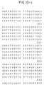
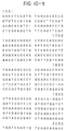
- a base sequence composed of 2749 bases beginning with G of the sequence GATC at the Sau3AI, one end of the acyB gene-containing fragment on plasmid pS82 ⁇ Sac can be determined as shown in Figure 10.
- the translation region of the acyB gene is determined, and the amino acid sequence of an enzyme capable of acylating the 4 ⁇ -position of a macrolide antibiotic encoded by the translation region of the acyB gene can be anticipated.
- two open reading frames going in two opposite directions from the base sequence shown in Figure 10, namely going outwardly from the central part of the sequence, are discovered.
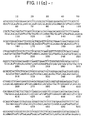
- the amino acid sequence of the upstream reading frame is shown in Figure 11-(a) as acyB1
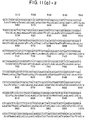
- the amino acid sequence of the downstream reading frame is shown in Figure 11-(b) as acyB2.
- the DNA restriction fragment containing the acyB enzyme gene capable of acylating the 4 ⁇ -position of a macrolide antibiotic has a wide range of utility.
- the DNA fragment of this invention can be inserted into a vector plasmid, and introduced into a microorganism of the genus Streptomyces and a related microorganism.
- the restricting transformant is cultured, and by using the cultured cells, or the culture medium, or a treated product thereof, an enzyme capable of acylating the 4 ⁇ -position of a macrolide antibiotic, or a material containing the enzyme, a 4 ⁇ -acylated macrolide antibiotic can be produced.
- An enzyme capable of acylating the 4 ⁇ -position of a macrolide antibiotic, which is expressed by using the acyB gene of this invention is especially useful for acylating the 4 ⁇ -position of tylosin. It has the ability to acylate the 4 ⁇ -positions of other macrolide antibiotics such as spiramycin, angoramycin, leucomycin and deltamycin.
- a plasmid having the acyB gene of the invention inserted thereinto is introduced into a microorganism having the ability to produce a macrolide antibiotic, a 4 ⁇ -acylated macrolide antibiotic can be directly produced. It is also possible to introduce such a recombinant plasmid into microorganisms which do not produce macrolide antibiotics. If the transformants are cultured in a medium containing a macrolide antibiotic, a 4 ⁇ -acylated macrolide antibiotic can be produced.
- the 4 ⁇ -acylating enzyme produced by the transformants can of course be used as a catalyst for an enzyme reaction between the macrolide antibiotic and an acylating agent.
- a plasmid having inserted thereinto the acyB gene can be introduced into suitable host microorganisms according to the vector plasmid.
- the host microorganism may be those having the ability to produce macrolide antibiotics or those which do not produce macrolide antibiotics.
- Examples of the host organisms that can be used in this invention include Streptomyces kitasatoensis, S. narbonensis var josamyceticus, S. hygroscopicus, S. platensis, S. albireticuli, S. cinerochromogenes, S. djalitis, S. macrosporeus, S. tendae, S. deltae, S. fradiae, S. eurythermus, S. ambofaciens, S. kasugaensis, S. erythreus and S. kanamyceticus.
- macrolide antibiotic-producing microorganisms substantially incapable of producing an enzyme capable of acylating the 4 ⁇ -position of a macrolide antibiotic such as S. eurythermus ATCC 14975 (a microorganism producing angoramycin), and S. fradiae ATCC 19609 (a microorganism producing tylosin) are suitable, particularly the latter.
- the transformation of these host organisms with the recombinant plasmids containing the acyB gene can be performed by methods known per se for example the method described in Genetic Manipulation of Streptomyces: A Laboratory Manual, 1985, The John Innes Institute.
- a transformant having the ability to produce a macrolide antibiotic may be acylated at the 4 ⁇ -position by culturing it in a medium free from a macrolide antibiotic.
- a 4 ⁇ -acylated macrolide antibiotic can also be produced by culturing the transformant and reacting the macrolide antibiotic with an acylating agent, such as acetyl coenzyme A, propionyl coenzyme A, n-butyryl coenzyme A, isovaleryl coenzyme A, phenylacetyl coenzyme A, or a biosynthesis precursor for donating an acyl group, such as leucine, in the presence of the cultured cells or a treated product thereof such as a cell-free extract obtained by disrupting the cultured cells by ultrasonic treatment.
- an acylating agent such as acetyl coenzyme A, propionyl coenzyme A, n-butyryl coenzyme A, isovaleryl coenzyme A, phenylacetyl coenzyme A, or a biosynthesis precursor for donating an acyl group, such as leucine, in the presence of the cultured cells or a treated
- macrolide antibiotics in which the 4 ⁇ -position is acylated with an alkanoyl group having 1 to 5 carbon atoms, such as acetyl, propionyl, n-butyryl or isovaleryl group, for example 4 ⁇ -acetyltylosin, 4 ⁇ -propionyltylosin, 4 ⁇ -n-butyryltylosin, 4 ⁇ -isovaleryltylosin, 4 ⁇ -acetylangoramycin, 4 ⁇ -propionylangoramycin, 4 ⁇ -n-butyrylangoramycin, 4 ⁇ -isovalerylangoramycin, 4 ⁇ -acetylspiramycin, 4 ⁇ -propionylspiramycin, 4 ⁇ -n-butyrylspiramycin, and 4 ⁇ -isovalerylspiramycin.
- an alkanoyl group having 1 to 5 carbon atoms such as acetyl, propionyl, n-
- a host microorganism for example, a microorganism of the genus Streptomyces, transformed with a plasmid bearing the DNA fragment containing acyB gene of this invention by many methods using some different media.
- Preferred carbon sources to be added to the culture medium include, for example, molasses, glucose, starch, oils and fats and glycerol.
- Suitable nitrogen sources include, for example, soybean meal, amino acid mixtures, dry yeasts and peptone.
- Nutrient inorganic salts may also be added to the medium. They include ordinary salts capable of releasing potassium, sodium, magnesium, calcium, phosphate, chlorine, sulfate ions, etc. Trace components such as vitamins may also be added as required. These trace components can be usually fed as impurities incidental to the addition of the other components.
- a macrolide antibiotic to be acylated may also be added as required.
- the transformed microorganism of the genus Streptomyces can be cultured in a medium at a broad pH range of from about 5 to 9 at a temperature of about 20 to 40 o C under aerobic conditions. At this time, a condition necessary for maintaining the stability of the plasmid may be introduced. For example, a drug such as thiostrepton may be added as a selective pressure.
- plasmid pIJ702 Twenty micrograms of plasmid pIJ702 was dissolved in 19 microliters of TE buffer, and 10 microliters of 10 x BglII buffer [NaCl (1 M)/Tris-HCl (100 mM, pH 7.4)/MgCl 2 /dithiothreitol (100 mM)/bovine serum albumin (1 mg/ml)] and 80 microliters of water were added.
- 5 micrograms of restriction endonuclease BglII (a product of Nippon Gene; 5 to 15 units/microliter) was added, and well mixed. The mixture was incubated at 37 o C for 15 hours.
- the reaction product was treated with a phenol/chloroform solution, and by using 0.1 volume of a 3 M sodium acetate solution and 3 volumes of ethanol, the digested plasmid was precipitated. The precipitates were washed with 70 % ethanol and ethanol. Plasmid pIJ702 digested with restriction endonuclease BglII was dissolved in TE buffer so that its final concentration became 0.1 microgram/microliter.
- the restriction endonuclease Sau3AI was obtained from New England Biolabs, Tozer Road Beverly Massachusetts, 01915-9990, U. S. A. After phenol/chloroform treatment, ethanol precipitation was carried out.
- the partially digested genome DNA was dissolved in 100 microliters of TE buffer, and then electrophoresed for 15 hours at a voltage of 3 V/cm using a 0.8 % agarose gel. After the electrophoresis, DNA was stained with ethidium bromide. On a transilluminator, a 5-10 kb fraction and a 10-20 kb fraction were cut out and put in a DNA cell (made by Daiichi Chemical Co., Ltd.), and DNA was electrically eluted from the gel. DNA fragments (about 50 micrograms) were obtained from the two fractions. After precipitation with ethanol, they were respectively dissolved in TE buffer to a final concentration of 0.5 microgram/microliter.
- the P medium was prepared by mixing 10.3 g of sucrose, 0.025 g of K 2 SO 4 , 0.202 g of MgCl 2 .6H 2 O and 0.2 ml of a trace metal solution containing 40 mg of ZnCl 2 , 200 mg of FeCl 3 .6H 2 O, 10 mg of CuCl 2 .2H 2 O, 10 mg of MnCl 2 .4H 2 O, 10 mg of Na 2 B 4 O 7 .10 H 2 O and 10 mg of (NH 4 ) 6 Mo 7 O 24 .4H 2 O, and adding 80 ml of deionized water to form 80 ml of a mixed solution, sterilizing the solution at 120 o C for 15 minutes, and then adding 1 ml of 0.5 % KH 2 PO 4 , 10 ml of CaCl 2 .2H 2 O, and 10 ml of TES [2-(tris-(hydroxymethyl(methyl)amino)methanesulfonic acid] buffer (0.
- the protoplasts transformed in the P medium were washed and re-suspended in 1 ml of P medium.
- 0.1 ml of the suspension was inoculated on a R 2 YE agar plate, and cultured at 28 o C for 16 hours. After the culturing, a soft agar medium containing thiopeptin was superimposed so that the final concentration of thiopeptin would become 50 micrograms/ml.
- the R 2 YE plate had been prepared by dissolving 10.3 g of sucrose, 0.025 g of K 2 SO 4 , 1.012 g of MgCl 2 .6H 2 O, 1 g of glucose, 0.01 g of Casamino acid (a product of Difco) and 2.2 g of bactoagar in 80 ml of distilled water, sterilizing the solution at 120 o C for 15 minutes, then adding 0.2 ml of each of the above sterilized trace metal solution, 1 ml of 0.5 % KH 2 PO 4 , 8 ml of 3.68 % CaCl 2 .2H 2 O, 1.5 ml of 20 % L-proline, 10 ml of TES buffer (0.25 M, pH 7.4), 0.5 ml of 1N NaOH and 5 ml of 10 % yeast extract, putting 20 ml of the mixture in a plastic petri dish having a diameter of 9 cm, and then drying it in a clean bench for about 2 hours.
- transformants obtained were inoculated in an agar medium composed of 1 % of glucose, 0.5 % of yeast extract, 1 % of malt extract, 0.02 % of L-leucine, 10 % of TES buffer (0.25 M, pH 7.2), 1.5 % of bactoagar, and 50 micrograms/ml of thiopeptin, and cultured at 28 o C for 1 week.
- a soft agar medium containing tylosin was overlayed so that the final concentration of tylosin would become 1000 micrograms/ml, and the culturing was continued for 24 hours.
- the center of each growing colony was punched out with a cork borer having a diameter of about 6 mm, and the agar piece was placed on a TLC plate (LK6DF produced by Watman Co.) and air dried.
- the plate was developed with a developing solvent composed of 150 ml of ethyl acetate, 3 ml of diethylamine, 1.5 ml of methanol and 3 ml of water, and acylated tylosin was detected under ultraviolet irradiation at 253.7 nm.
- transformant No. Y-17 was found to produce acylated tylosin.
- the acylated tylosin produced by Y-17 was identified as 4 ⁇ -isovaleryl tylosin from the results of analysis by TLC, HPLC, UV spectrum, NMR spectrum and antimicrobial spectrum.
- Plasmid pAOY-17 was isolated from the transformant Streptomyces lividans #Y-17 substantially in accordance with the method described in Example 2. About 6 kb foreign DNA was seen to be inserted in this plasmid.
- Figure 2 shows maps showing its restriction endonuclease cleavege sites and function. Only those restriction endonuclease sites which were significant are shown in Figure 2, and the others were omitted.
- pIJ943 was used as a vector plasmid, and the 10-20 kb fraction described in Example 3 was used as a genome DNA digestion fragment.
- the transformant Streptomyces lividans TK24/pAO207 having the ability to acylate tylosin was isolated. This transformant is deposited at Fermentation Research Institute, Agency of Industrial Science and Technology, Ministry of International Trade and Industry, 1-3, Higashi, 1-chome, Tsukuba-shi, Ibaraki-ken, Japan under FERM BP-1880.
- Plasmid pAO207 was isolated from this transformant in accordance with the method described in Example 2. Maps showing its restriction endonuclease cleavage sites and function are shown in Figure 3. Plasmid pAO207 contained an about 13 kb DNA fragments inserted thereinto.
- Plasmid pAOY-17 obtained by the method described in Example 3 was digested with restriction endonuclease BglII to prepare 0.6 microgram of a digestion fragment.
- E. coli plasmid pUC18 (a product of Toyobo) was completely digested with restriction endonuclease BamHI, and then treated with alkaline phosphatase derived from bovine intestines (a product of Boehringer Manheim Co.) to give about 0.2 microgram of a digestion fragment. The two fragments were mixed, and 2 microliters of a 10 x ligation buffer and 1 microliter of T 4 -ligase were added. Water was added to adjust the total amount of the mixture to 20 microliters. The mixture was reacted at 15 o C for 16 hours. The ligated DNA was used to transform E. coli K-12 JM103 by the method shown below.
- E. coli K-12 JM103 having the ability to undergo transformation used in this experiment was one produced by Pharmacia Co.
- a single colony was isolated from it, inoculated in 50 ml of an L-broth (prepared by adding distilled water to 10 g of bactotryptone, 10 g of sodium chloride and 5 g of bactoyeast extract to make 1 liter of a solution), and cultured at 37 o C for 5 hours.
- the optical density at 600 nm of the culture broth was about 0.5.
- the culture broth was centrifuged (3,000 rpm, 5 minutes). The cells were collected and suspended in 2 ml of 50 mM CaCl 2 .
- the suspension was cooled over an ice water bath for 30 minutes and then centrifuged (3,000 rpm, 5 minutes, 4 o C).
- the cells were collected and re-suspended in 5 ml of 50 mM CaCl 2 .
- Ten microliters of the ligated DNA was added to 100 microliters of the suspension.
- the mixture was incubated for 30 minutes over an ice water bath, and then given a thermal shock at 42 o C for 30 minutes.
- One milliliter of L-broth was added, and the mixture incubated at 37 o C for 1 hour.
- the BamHI site on the plasmid pUC18 is contained in a polylinker forming part of the DNA sequence coding for the lacZ ⁇ -fragment.
- the expression of the lacZ ⁇ -fragment in E. coli mutant JM103 revives the beta-galactosidase activity of this E. coli strain.
- Insertion of DNA in a restriction endonuclease cleavage site of the polylinker existing in pUC18 destructs the lacZ ⁇ -fragment gene and causes a loss of the complementary ability of plasmid pUC18 with respect to JM103.
- ⁇ -Galactosidase hydrolyzes a colorless compound X-Gal (5-bromo-4-chloro-3-indolyl- ⁇ -D-galactopyranoside) to form a blue product, and therefore permits easy distinction between a transformant containing plasmid pUC18 and a transformant containing plasmid pUC18 having DNA inserted thereinto in the transformed cells.
- the transformed strain described above is inoculated on a plate of L-agar (containing 15 g of agar per liter of L-broth) containing 100 micrograms/ml of ampicillin, 40 micrograms/ml of X-Gal and 40 micrograms/ml of IPTG (isopropyl- ⁇ -D-thiogalactopyranoside), and cultured at 37 o C for 15 hours.
- IPTG has the function of inducing the expression of the lac gene on plasmid pUC18.
- a colony containing the plasmid having DNA inserted thereinto as was the case with plasmid pUY-17 was white. Two white colonies which showed resistance to ampicillin were separated, and the plasmids were isolated. The two colonies contained the same plasmid pUY-17. Maps showing the restriction endonuclease cleavage sites and function of plasmid pUY-17 are shown in Figure 4.
- plasmid pUY-17 contains a replicon of the genus Streptomyces, it can impart the ability to transform Streptomyces lividans TK24 and acylate the 4 ⁇ -position of tylosin.
- the plasmid pUY-17 was digested with restriction endonuclease EcoRI and then by using agarose gel electrophoresis and a DNA cell, about 9 kb fragment was obtained. About 0.1 microgram of the fragment was dissolved in 10 microliters of TE buffer, and 2 microliters of 10 x ligation buffer and 7 microliters of water were added. Furthermore, 1 microliter of T 4 -ligase was added. Ligation was carried out at 15 o C for 16 hours. Using the recombinant plasmid, E. coli K-12 JM103 was transformed.
- Plasmid pY-17 ⁇ Eco was isolated from the transformant. This plasmid does not have a replicon of the genus Streptomyces and cannot transform Streptomyces lividans TK24. Maps showing the restriction endonuclease cleavage sites and function of the plasmid pY-17 ⁇ Eco are shown in Figure 5.
- pAO207 obtained by the method described in Example 4 was completely digested with restriction endonuclease SphI, and by using agarose gel electrophoresis and a DNA cell, about 1 microgram of a 8.8 kb DNA fragment was obtained.
- This fragment contained a DNA fragment which contained part of the tyrosinase gene and all of the acyB gene of plasmid pIJ943.
- This DNA fragment was ligated with the restriction endonuclease SphI digestion product of pUS18 by the method described in Example 5.
- E. Coli K-12 JM103 was transformed with the resulting recombinant plasmid, and plasmids pUS81 and pUS82 were isolated from ampicillin-resistant white transformants.
- Figures 6 and 7 give maps showing the restriction endonuclease cleavage sites and functions of plasmids pUS81 and pUS82.
- the direction of the inserted DNA in plasmid pUS81 was reverse to that in pUS82.
- plasmid pUS82 was digested with restriction endonuclease PstI, and agarose gel electrophoresis and a DNA cell, about 0.1 microgram of a 5.2 kb fragment was obtained. This fragment was dissolved in TE buffer, and ligated by using a T 4 ligase. Then, by using the recombinant plasmid, E. coli K-12 JM103 was transformed by the method described in Example 5. About 20 colonies which were white and ampicillin-resistant were separated. Plasmids were isolated from these transformants. All plasmids obtained were pS82 ⁇ Pst.
- Figure 8-(a) shows a map of the restriction endonuclease cleavage sites and function of plasmid pS82 ⁇ Pst. Part of the acyB gene in the plasmid pS82 ⁇ Pst was deleted.
- Plasmid pS82 ⁇ Sac completely retained the acyB gene.
- Plasmids pAOY-17 and pAO207 were prepared by the methods described in Examples 3 and 4 in order to restrict the acyB gene contained in the DNA fragments inserted into the plasmids pAOY-17 and pAO207. A detailed examination of the inserted DNA portions of the two plasmids showed that they have a DNA fragment having a size of about 4 kb in common (see Figures 2 and 3). Plasmids pS82 ⁇ Pst and pS82 ⁇ Sac having this common DNA region were prepared by the method described in Example 6.
- Plasmid pS82 ⁇ Sac (0.5 microgram) was dissolved in 10 microliters of TE buffer, and 2 microliters of 10 x SphI buffer and 10 units of restriction endonuclease SphI were added. Water was added to make the total amount 20 microliters.
- the plasmid was digested at 37 o C for 4 hours. Two microliters of the reaction mixture was subjected to agarose gel electrophoresis, and it was determined that the digestion came to completion.
- plasmid pIJ702 prepared by the method described in Example 2 was digested in the same way as above, treated with alkaline phosphatase derived from bovine intestines at 37 o C for 30 minutes, and then heated at 70 o C for 15 minutes.
- the DNA pellets obtained by ethanol precipitation were dissolved in 10 microliters of TE buffer, and ligated by the method described in Example 3.
- Streptomyces lividans TK24 was transformed with the reaction product, and the ability of the resulting transformants to acylate the 4 ⁇ -position of tylosin was examined. All the transformants acylated the 4 ⁇ -position of tylosin.
- plasmid pS82 ⁇ Pst was inserted into the SphI site of plasmid pIJ702, and Streptomyces lividans TK24 was transformed with the resulting recombinant plasmid. None of the transformants showed the ability to acylate the 4 ⁇ -position of tylosin.
- Plasmid pUS81 was prepared by the method described in Example 6. One microgram of it was dissolved in 10 microliters of TE buffer, and 2 microliters of 10 x SacI buffer and 10 units of a restriction endonuclease were added. Water was then added to make 20 microliters. The reaction solution was placed on 8 % agarose gel, and electrophoresed. An agarose containing an about 3.3 kb DNA fragment was cut out and electrically eluted by using a DNA cell thereby to give about 0.1 microgram of a DNA fragment. This DNA fragment contained part (240 bases) of the tyrosinase gene of plasmid pIJ943 and part of the multicloning sites of plasmid pUC18.
- the resulting DNA fragment having a size of about 3.3 kb was inserted into the SacI site of plasmid pIJ702 according to the method of Example 3, and Streptomyces lividans TK24 was transformed with the resulting recombinant plasmid. All transformants acylated the 4 ⁇ -position of tylosin. The plasmids held by the transformants were slightly unstable.
- the restriction endonuclease cleavage sites of the DNA fragment containing the acyB gene restricted to about 3.1 kb were determined (see Figure 1).
- the DNA fragment was digested with various restriction endonucleases either singly or in combination.
- the digested products were subjected to agarose gel electrophoresis and the migration distance of each of the DNA fragments was measured and its size was determined.
- the agarose gel concentration was varied between 0.6 % and 1 % according to the size of the DNA fragment.
- ⁇ /HindIII and ⁇ X174/HaeIII both produced by Nippon Gene were used as size markers for DNA.
- a PstI-SacI fragment (1.2 kb) of the acyB gene and its PstI-SphI fragment (2.1 kb) were subcloned into E. coli vector pUC18 to obtain plasmid pMAB7 [ Figure 8-(b)] and plasmid pS82 ⁇ Pst (see Example 6) containing these fragments respectively were obtained.
- plasmid pMAB7 Figure 8-(b)]
- plasmid pS82 ⁇ Pst see Example 6
- These two plasmids or new plasmids obtained by deleting a specified region from them were used for determination of DNA base sequence.
- a method was used in which deletion took place between a suitable cloning site of the vector and a suitable restriction site of the acyB fragment.
- a deleted plasmid was constructed by the following method in order to obtain a plasmid convenient for determining the sequence from the NcoI site of pS82 ⁇ P
- the DNA fragment was gradually decomposed from the specific terminal by exonuclease III and S1 nuclease in a time-dependent manner.
- a series of continuous deletion plasmids having an overlapping region convenient for sequence determination were obtained.
- a series of deletion plasmids obtained by digesting from the ApaI site of pMAB7 were obtained by the following procedure.
- the two plasmids obtained by subcloning and deletion plasmids derived therefrom were modified with alkali to form single-stranded DNA.
- 1 to 2 micrograms of modified DNA was used as a template DNA for sequencing.
- the sequencing was in accordance with the method of Hattori et al. [Hattori, M. & Sasaki, Y: Anal. Biochem. 152 , 232-328 (1986)].
- the method of sequencing was basically in accordance with the Sanger's dideoxy chain terminal method [Sanger, F. et al.: Proceedings of the National Academy of Sciences, U. S. A., 74 , 5463-5467 (1977)].
- the DNA base sequence containing a region coding for the acyB gene was determined by using a sequence kit (made by United States Biochemical Corporation) and [ ⁇ - 32 P] dCTP (a product of Amersham).
- the DNA base sequence determined is shown in Figure 10 of the accompanying drawings.
- the determined DNA base sequence was examined for reading frames. Specifically, the base sequence was examined in accordance with the report of M. J. Bibb et al. [M. J. Bibb et al.; "Gene", vol. 30, pages 157-166 (1984)], and an open reading frame leading from the central part of the sequence towards the Sau3AI terminal and an open reading frame leading toward the SphI terminal were found. These two open reading frames were named acyB1 and acyB2.
- the acyB1 begins with the 1245th ATG and ends with the 81st TAG in Figure 10, whereas the acyB2 begins with the 1569th GTG or 1608th ATG and ends with the 2730th TGA.
- Figure 11-(a) and Figure 11-(b) respectively show the DNA base sequences and amino acid sequence of acyB1 and AcyB2.
- Streptomyces lividans TK24 was transformed with plasmid pAO207 by the method of Example 3.
- the transformants were cultured by the following procedure.
- the transformants were inoculated in a GYM agar slant medium containing 50 micrograms/ml of thiopeptin (0.4 % of glucose, 0.4 % of yeast extract, 1 % of malt extract and 1.5 % of agar; adjusted to pH 6.8).
- One loopful of the grown transformants was inoculated in 25 ml of a seed culture medium (2 % of soluble starch, 2 % of soybean meal, 0.1 % of yeast extract, 0.1 % K 2 HPO 4 , 0.05 % MgSO 4 .7H 2 O and 5 micrograms/ml of thiopeptin), and cultured at 28 o C for 48 hours under shaking.
- a seed culture medium (2 % of soluble starch, 2 % of soybean meal, 0.1 % of yeast extract, 0.1 % K 2 HPO 4 , 0.05 % MgSO 4 .7H 2 O and 5 micrograms/ml of thiopeptin
- One milliliter of the prepared seed culture was inoculated in 50 ml of a production medium containing 30 micrograms/ml of thiopeptin (4 % of galactose, 2 % of soybean meal, 0.1 % of yeast extract, 0.05 % of KH 2 PO 4 and 0.05 % of MgSO 4 .7H 2 O), and cultured at 28 o C for 48 hours.
- a mixed solution of tylosin and L-leucine was added so that their final concentrations would be 1,000 micrograms/ml and 400 micrograms/ml, and the culturing was continued at 28 o C for 48 hours.
- the fermentation broth was adjusted to pH 9.0 with 0.1 M Na 3 PO 4 , and the broth was extracted with an equal amount of toluene.
- Twenty microliters of the toluene layer was spotted on a silica gel TLC plate [Art 5715 made by Merck Co.], and developed with a solvent composed of 150 ml of ethyl acetate, 3 ml of diethylamine, 1.5 ml of methanol and 3 ml of water. The solvent was removed by air drying.
- the amount of the acylated tylosin produced was measured by a TLC-chromatoscanner CS-920 (made by Shimadzu Seisakusho). The results are shown in Table 1.
- the specfic activity was calculated as units/mg protein, and the relative value with respect to S. thermotolerans ATCC 11416 is shown in the table. One unit is defined as nmoles of acylated tylosin/hour.
- Two micrograms of DNA of plasmid pIJ702 was dissolved in 50 microliters of Bcl buffer [10 mM of Tris-HCl (pH 7.5), 7 mM of MgCl 2 , 60 mM of NaCl and 7 mM of 2-mercaptoethanol.
- Two units of restriction endonuclease BclI (a product of Toyobo) was added to diest the DNA partially at 50 °C for 5 minutes. The digestion product was treated with ethanol/chloroform, and precipitated with ethanol.
- the purified DNA was dissolved in 50 microliters of a ligation buffer, and 2 microliters of T 4 ligase (a product of Nippon Gene; 500 units/microliter) was added.
- the ligation reaction was carried out at 16 °C for 15 hours. After the reaction, the reaction mixture was precipitated with ethanol, and then dissolved in 100 microliters of TE buffer.
- Protoplasts of Streptomyces lividans TK24 were prepared by the same method as in Example 3, and transformed with the partially BclI digested product of pIJ702 by the same method as in Example 3. Colonies which had resistance to thiopeptin (50 micrograms/ml) and did not produce melanin pigment were selected, and the plasmid was extracted and purified to give pIJ350 (4.1 kb).
- E. coli plasmid pUC18 was linked to plasmid pIJ350 of actinomycetes, and multicloning sites were introduced into pIJ350.
- pIJ350 was dissolved in 50 microliters of PstI buffer [20 mM of Tris-HCl (pH 7.5), 10 mM of MgCl 2 and 150 mM of NaCl], and 1 microliter of restriction endonuclease PstI (a product of Nippon Gene; 20,000 units/ml) was added. The mixture was reacted at 37 o C for 4 hours. The reaction mixture was precipitated with ethanol, and dissolved in 50 microliters of distilled water.
- plasmid resulting from changing the HincII (SalI) site of pUC18 to a BglII site was dissolved in 50 micrograms of Pst buffer, and digested with 20 units of restriction endonuclease PstI at 37 o C for 4 hours.
- the digestion product was purified by ethanol precipitation, and dissolved in 50 microliters of distilled water.
- the two DNA solutions were mixed, and 10 microliters (10-fold concentration) of a ligase buffer and 1,000 units of T 4 ligase were added, and the reaction was carried out at 16 o C for 20 hours.
- the DNA was purified by ethanol precipitation treatment, and finally the precipitate was dissolved in 50 microliters of TE buffer. By using 10 microliters of the solution, E. coli K-12 JM103 was transformed. The plasmids in transformants which were ampicillin-resistant and white on an X-gal plate under IPTG inducing conditions were examined. There was obtained plasmid pUJ350 (6.8 kb) in which the PstI-digested fragment of plasmid pIJ350 was linked to the PstI site of plasmid pUC-Bg.
- the agarose gel was put in a DNA cell (made by Daiichi Chemical Co.) and the DNA was eluted and recovered by electrophoresis. It was precipitated with ethanol and then dissolved in 50 microliters of TE buffer. Twenty microliters of this solution was mixed with 2 micrograms of the SacI-digested DNA fragment of pUJ350, and 10 microliters of a ligation buffer (10-fold concentration), 4 microliters of T 4 ligase (a product of Nippon Gene; 500 units/microliter) and 70 microliters of distilled water were added, and the reaction was carried out at 16 o C for 15 hours. After the reaction, precipitation with ethanol was carried out, and the precipitate was dissolved in 100 microliters of TE buffer.
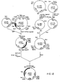
- E. coli K-12 JM103 was transformed by using 20 microliters of the resulting DNA solution by the method described in Example 5. Plasmids contained in ampicillin (50 micrograms/ml)-resistant colonies were examined. Plasmid pMAB1 in which a DNA fragment containing the acyB gene was inserted into the SacI site of pUJ350 was obtained. The foregoing plasmid constructing process is shown in Figure 12.
- S. lividans TK24 was transformed with plasmid pMAB1 by the method of Example 3.
- the transformants were inoculated in a GYM agar slant medium containing 50 micrograms/ml of thiopeptin (0.4 % of glucose, 0.4 % of yeast extract, 1 % of malt extract, and 1.5 % of agar; adjusted to pH 6.8).
- One loopful of the grown transformants was inoculated in 25 ml of a seed culture medium (2 % of soluble starch, 2 % of soybean meal, 0.1 % of yeast extract, 0.1 % of K 2 HPO 4 , 0.05 % of MgSO 4 .7H 2 O and 5 micrograms/ml of thiopeptin), and cultured with shaking at 28 o C for 48 hours.
- a seed culture medium (2 % of soluble starch, 2 % of soybean meal, 0.1 % of yeast extract, 0.1 % of K 2 HPO 4 , 0.05 % of MgSO 4 .7H 2 O and 5 micrograms/ml of thiopeptin
- One milliliter of the seed culture prepared was inoculated in a production medium containing 30 micrograms/ml of thiopeptin (4 % of galactose, 2 % of soybean meal, 0.1 % of yeast extract, 0.05 % of KH 2 PO 4 and 0.05 % of MgSO 4 .7H 2 O) and cultured at 28 o C for 48 hours.
- thiopeptin 4 % of galactose, 2 % of soybean meal, 0.1 % of yeast extract, 0.05 % of KH 2 PO 4 and 0.05 % of MgSO 4 .7H 2 O
- To the culture broth was added tylosin, spiramycin, angoramycin, leucomycin U, leucomycin V or deltamycin X as a macrolide having -OH group at the 4 ⁇ -position so that its concentration became 200 micrograms/ml.
- L-leucine was added so that its concentration became 200 micrograms/ml.
- the culturing was continued at 28 o C for 48 hours. After the psyng, the pH of the culture broth was adjusted to 9.0 with 0.1M Na 3 PO 4 , and it was extracted with an equivalent weight of toluene. Twenty microliters of the toluene layer was spotted on a silica gel thin layer plate (Art 5715 made by Merck and Co.) and a developing solvent of the following composition.
- Tylosin (0.22), 4 ⁇ -O-isovaleryl tylosin (0.49); spiramycin I (0.057), 4 ⁇ -O-isovaleryl spiramycin I (0.25); angoramycin (0.26), 4 ⁇ -O-isovaleryl angoramycin (0.51)
- Leucomycin U (0.12), 4 ⁇ -isovaleryl leucomycin U (0.65); leucomycin V (0.08), 4 ⁇ -isovaleryl leucomycin V (0.50).
- Deltamycin X (0.20), 4 ⁇ -isovaleryl deltamycin X (0.70).
- One loopful of a slant culture of Streptomyces fradiae was inoculated in a 250 ml Erlenmeyer flask holding GPY medium (1.0 % of glucose, 0.4 % of polypeptone, 0.4 % of yeast extract, 0.05 % of MgSO 4 .7H 2 O, 0.1 % of K 2 HPO 4 and 0.05 % of glycine, pH 7.2), and cultured with shaking at 28 o C for 48 hours.
- GPY medium 1.0 % of glucose, 0.4 % of polypeptone, 0.4 % of yeast extract, 0.05 % of MgSO 4 .7H 2 O, 0.1 % of K 2 HPO 4 and 0.05 % of glycine, pH 7.2
- 0.5 ml of the culture broth was inoculated in a 250 ml Erlenmeyer flask containing a spring and holding 25 ml of a YEME + 15 % sucrose medium (containing 5 mM of MgCl 2 and 0.5 % of glycine) and culture with shaking at 28 o C for 40 hours.
- the cells were collected by centrifugation (3,000 rpm, 15 minutes) washed with 10 ml of 10.3 % sucrose, suspended in 4 ml of P medium containing 1 mg/ml of lysozyme, and reacted at 30 o C for 1 hour.
- the composition of the P medium was as shown in Example 3.
- the reaction mixture was filtered through cotton, and the remaining mycelia were removed.
- Protoplasts were collected by centrifugation (3,000 rpm, 10 minutes) and suspended in P medium so that their concentration became 3 x 10 9 /100 microliters.
- the resulting protoplasts were transformed with 10 microliters of the aforesaid PMAB1 solution (15 micrograms/10 microliters in the presence of 25 % PEG 1000 (a product of BDH Chemical).
- the transformed protoplasts were washed with the P medium, and re-suspended in 1 ml of the P medium.
- One milliliter of the suspension was inoculated in an R3 medium [13.5 g of sucrose, 0.05 % of KCl, 1.0 g of glucose, 0.4 g of polypeptone, 0.4 g of yeast extract, 0.573 g of TES buffer, 6.1 ml of 10 % MgCl 2 , 2.0 g of bactoagar and 100 ml of distilled water; after autoclave sterilization, 1.0 ml of KH 2 PO 4 (0.5 %), 0.3 ml of CaCl 2 (5 M) and 1.8 ml of NaOH (1N) were added], and cultured at 28 o C for 16 hours.
- an R3 medium [13.5 g of sucrose, 0.05 % of KCl, 1.0 g of glucose, 0.4 g of polypeptone, 0.4 g of yeast extract, 0.573 g of TES buffer, 6.1 ml of 10 % MgCl 2 , 2.0 g of bactoagar and 100
- a soft agar medium containing thiopeptin was overlayed so that the final concentration of thiopeptin would become 5 micrograms/ml or 25 micrograms/ml.
- the culturing was further carried out at 28 o C for 7 days.
- the soft agar medium had been prepared by adding 1 liter of distilled water to 8 g of a nutrient broth and 3 g of bactoagar and sterilizing the mixture at 120 o C for 15 minutes in an autoclave.
- MAB-01 Streptomyces fradiae MAB-01
- One loopful of MAB-01 was inoculated in a 250 ml Erlenmeyer flask containing a coil and holding a production medium (0.5 % of soluble starch, 0.5 % of glucose, 0.5 % of yeast extract, 1.0 % of malt extract and 5 micrograms/ml of thiopeptin), and cultured with shaking at 28 o C for 21 days. Then, 1 ml of the culture broth was taken, and 0.2 g of K 2 HPO 4 and 0.2 ml of toluene were added. The mixture was vigorously stirred to extract the culture broth with toluene.
- a production medium 0.5 % of soluble starch, 0.5 % of glucose, 0.5 % of yeast extract, 1.0 % of malt extract and 5 micrograms/ml of thiopeptin
- the upper toluene layer was separated by centrifugation (3,000 rpm, 10 minutes), and subjected to thin-layer chromatography.
- Merck Art 5715 was used as a thin-layer plate, and a mixture of 75 ml of ethyl acetate, 1.5 ml of diethylamine, 1.5 ml of water and 0.75 ml of methanol was used as a developing solvent.
- the plate was dried and analyzed at a wavelength of 280 nm by a chromatoscanner (CR-930 of Shimadzu seisakusho). After immersion in 10 % H 2 SO 4 , the plate was heated at 105 °C for 5 minutes to color it, and the position of the macrolides produced was determined.
- Rf relative mobility
- the culture fluid from 1 liter of the above production medium (50 ml x 20) was extracted with ethyl acetate at a pH of 8.5.
- the extract was extracted with 200 ml of acidic water (0.01N-HCl), and again adjusted to pH 8.5, and back extracted with 50 ml of ethyl acetate.
- the extract was concentrated to 10 ml, and spotted in a band form to five thin-layer plates (Art 5715 produced by Merck & Co.) at a rate of 2 ml, followed by developing with the aforesaid developing solvent. After air drying, silica gel at a spot portion having an Rf value of 0.77 was scraped off and extracted with 10 ml of acetone.
- the extract was evaporated to dryness in a vacuum evaporator-concentrator to give 4 mg of of a white powder which had the following physical and chemical properties.
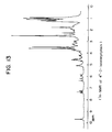
- Figure 13 shows the 1 H-NMR chart.
- Silica gel of a spot portion having an Rf of 0.69 was scraped off, and subjected to acetone extraction, concentration and drying by the same method as above to give 4.5 mg of a white powder. Its physical and chemical properties were examined.
- Figure 14 shows the 1 H-NMR chart.
Landscapes
- Chemical & Material Sciences (AREA)
- Organic Chemistry (AREA)
- Life Sciences & Earth Sciences (AREA)
- Engineering & Computer Science (AREA)
- Health & Medical Sciences (AREA)
- Zoology (AREA)
- Wood Science & Technology (AREA)
- Genetics & Genomics (AREA)
- Biotechnology (AREA)
- Biochemistry (AREA)
- Bioinformatics & Cheminformatics (AREA)
- General Health & Medical Sciences (AREA)
- General Engineering & Computer Science (AREA)
- Molecular Biology (AREA)
- Microbiology (AREA)
- Biomedical Technology (AREA)
- Medicinal Chemistry (AREA)
- Chemical Kinetics & Catalysis (AREA)
- General Chemical & Material Sciences (AREA)
- Micro-Organisms Or Cultivation Processes Thereof (AREA)
- Preparation Of Compounds By Using Micro-Organisms (AREA)
- Enzymes And Modification Thereof (AREA)
Description
- This invention relates to a DNA fragment containing a gene (named "acyB") which gives a host microorganism the enzyme activity of acylating the 4"-position of a macrolide antibiotic, a recombinant DNA plasmid containing this DNA fragment and a microorganism transformed with this recombinant plasmid.
- Macrolide antibiotics (having 14-membered and 16-membered rings) are useful antibiotics which are widely used as pharmaceuticals for man and animals (for domestic animals and fish culture). S. Omura et al. [Journal of Antibiotics, vol. 28, pages 401-433 (1980)] and R. Okamoto et al. [Journal of Antibiotics, vol. 27, pages 542-544 (1979)] reported that the antimicrobial activity of a 16-membered ring macrolide antibiotic is increased by acylation of the OH group at its 4"-position. In the paper of Okamoto et al., the 3- and 4"-positions of tylosin were acylated, and the pharmacological efficacies of the derivatives against various bacteria having resistance to macrolide antibiotics were examined; and it was concluded that 3- and 4"-acylated tylosins, particularly the 4"-acylated tylosins, have a higher pharmacological efficacy against these resistant bacteria than tylosin. However, the tylosin-producing microorganism strains produce only tylosin of the type in which -OH is attached to the 3- and 4"-positions. Acylation of -OH at these positions requires isolation of the tylosin and subsequent subjection of the isolated tylosin to a chemical or biological method, and no other suitable method has been available.
- The present invention provides an industrially very useful method in which a microorganism strain capable of producing a 4"-acylation product of a macrolide antibiotic is obtained by a genetic engineering technique from a microorganism strain which inherently cannot directly produce the 4"-acylation product.
- Some of the previously known macrolide antibiotics are shown below.


- Chemical methods are generally employed for the acylation of the 4˝-position of these macrolide antibiotics. Omura et al., however, reported that it was difficult to acylate a specific position in a molecule with good efficiency [Yakugaku Zasshi, vol. 106, pages 729-757 (1986)]. A biological method was proposed in Japanese Patent Publication No. 15160/1978. It is a method of acylating tylosin by using a microorganism having the ability to acylate tylosin, a culture of the microorganism, a treated product thereof, or an enzyme separated from the microorganism. According to this method, a macrolide antibiotic having -OH at the 3- and/or 4˝-position is added as a substrate to cells, a culture or a cell extract of, for example, Streptomyces thermotolerans (ATCC 11416), Streptomyces hygroscopicus (ATCC 21582) or Streptomyces kitasatoensis(IFO 13686). However, the macrolide antibiotic-producing microorganism differs from the microorganism having acylating activity. This is economically disadvantageous in industrial production.
- To acylate biologically a macrolide antibiotic of the type having OH at the 3-position and/or 4"-position, a two-step operation is necessary in which the macrolide-producing microorganism strain is cultured and the macrolide antibiotic is isolated, and then the macrolide antibiotic substance is transformed with a microorganism having an acylating enzyme. In order to enable an acylated macrolide to be produced only by a macrolide-producing microorganism strain, much work has been done to provide a method which comprises isolating DNA of a macrolide acylating enzyme from a microorganism containing the macrolide acylating enzyme DNA, introducing the isolated DNA into a macrolide-producing microorganism strain by a genetic engineering technique, and causing the resulting microorganism strain to produce an acylated macrolide directly.
- The recent advances in genetic engineering technique have made it possible to extract a desired DNA from a specific organism, and introduce the DNA into another desired microorganism either as such or after it is ligated with a suitable vector [D. A. Hopwood et al.: Genetic Manipulation of Streptomyces, The John Innes Foundation (1985)].
- EP-A-0 354 641 (prior art under Article 54(3) and (4) EPC) discloses a method for increasing the amount of 4"-O-isovalerylacylase enzyme in an organism that comprises (1) transforming said organism with a recombinant DNA vector that codes for expression of the carE gene product; and (2) culturing said organism transformed in step (1) under conditions that allow for gene expression.
- The present inventors made extensive investigations in order to isolate the DNA of an enzyme capable of acylating the 3- and/or 4"-position of a macrolide from a microorganism containing it, and consequently succeeded in isolating the 4"-position acylating enzyme gene acyB from a certain microorganism belonging to the genus Streptomyces. This has led to the accomplishment of the present invention.
- The invention will be described partly with reference to the accompanying drawings, in which
- Figure 1 is a restriction endonuclease map of a DNA fragment containing the gene acyB in accordance with this invention;
- Figure 2 is a map showing the restriction endonuclease cleavage sites and function of plasmid pAOY-17;
- Figure 3 is a map showing the restriction endonuclease cleavage sites and function of plasmid pAO207;
- Figure 4 is a map showing the restriction endounclease cleavage sites and function of plasmid pUY-17;
- Figure 5 is a map showing the restriction endonuclease cleavage sites and function of plasmid pY-17ΔEco;
- Figure 6 is a map showing the restriction endonuclease cleavage sites and function of plasmid pUS81;
- Figure 7 is a map showing the restriction endonuclease cleavage sites and function of plasmid pUS82;
- Figures 8-(a) and -(b) are maps showing the restriction endonuclease cleavage sites and functions of plasmid pS82ΔPst and plasmid pMAB7, respectively;
- Figure 9 is a map showing the endonuclease cleavage sites and function of plasmid pS82ΔSac;
- Figure 10 is the DNA base sequence of acyB;
- Figures 11-(a) and -(b) are the base sequence and amino acid sequence of acyB1 and acyB2, respectively;
- Figure 12 is a view showing the process of constructing plasmid pMAB1;
- Figure 13 is a 1H-NMR chart of 4"-O-isovaleryl tylosin; and
- Figure 14 is a 1H-NMR chart of 4"-O-acetyl tylosin.
- Thus, according to this invention, there is provided a purified and isolated DNA fragment derived from Streptomyces thermotolerans, having a size of about 3.1 kb and bounded by the Sau3AI and SphI restriction endonuclease sites depicted in Fig. 1, said fragment comprising DNA sequences encoding for an acyB1 gene and acyB2 gene and containing a DNA portion coding for the following amino acid sequence

 and a DNA portion coding for the following amino acid sequence


- The "DNA restriction fragments obtained by digesting the DNA fragment with restriction endonucleases", as used herein, denote DNA fragments of various sizes digested with restriction endonuleases to lengths necessary for expression of acylating enzyme activity.
- The DNA fragment containing gene acyB and DNA restriction fragments in accordance with this invention are useful for commercial production of macrolide antibiotic derivatives such as 4"-acylated tylosin. They are particularly useful in that they make it possible to utilize the recombinant DNA technique commercially for microorganisms of the genus Streptomyces and other antibiotic-producing microorganisms.
- The production and characteristics of the DNA fragment of the invention containing acyB gene, and the production of acylated macrolide antibiotics by using transformants obtained with the DNA fragment will be described in detail.
- Microorganism strains belonging to the genus Streptomyces as sources of the DNA fragment containing gene acyB are strains of Streptomyces thermotolerans.
- The following description relates to the case of using Streptomyces thermotolerans.
- S. thermotolerans is deposited, for example, at The American Type Culture Collection under the deposit No. ATCC 11416 and is easily available.
- This microorganism is aerobically cultured at 28 °C as described in detail in Example 1 given hereinafter, and the cells in the logarithmic growth period were obtained. The cells were treated in accordance with the method of Hopwood et al. (Genetic Manipulation of Streptomyces: A Laboratory Manual, The John Innes Foundation, Norwich, England, 1985) to prepare a genome DNA containing acyB gene.
- The resulting genome DNA containing acyB gene is ligated with a suitable vector plasmid such as pIJ702, pIJ943, pIJ350 or pUC18.
- As described in detail in Example 2 hereinbelow, the vector plasmid pIJ702 can be isolated from a microorganism containing it by phenol extraction and density gradient ultracentrifugation using cesium chloride. The resulting plasmid pIJ702 is digested with restriction endonuclease BalII. DNA fragment obtained by partially digesting the above genome DNA with restriction endonuclease Sau3AI is inserted into the resulting digested plasmid.
- Streptomyces lividans TK24 is transformed with the resulting plasmid containing part of the genome DNA of Streptomyces thermotolerans ATCC 11416 to obtain transformants which have acquired tylosin acylating ability (for details, see Example 3 given hereinafter). By extracting and purifying the plasmid from the transformants having tylosin acylating ability, plasmid pAOY-17 containing a DNA inserted fragment having a size of about 6 kb can be obtained.
- In the same way as in the preparation of the plasmid pAOY-17, plasmid pAO207 can be obtained in which an about 13 kb restriction endonuclease Sau3AI digested fragment derived from Streptomyces thermotolerans is inserted into the restriction endonuclease BglII-cleaved site of plasmid pIJ943 (see Example 4 given hereinafter).
- Plasmid pIJ702 is available from The American Type Culture Collection under deposit No. ATCC 35287. Plasmid pIJ943 is disclosed by Hopwood et al. [see Genetic Manipulation of Streptomyces: A Laboratory Manual, pages 314-315, 1985, The John Innes Foundation], and may be obtained under Streptomyces lividans TK24/pIJ943 from The John Innes Streptomyces Culture Collection, The John Innes Institute, Colney Lane, Norwich NR4 7UH, England. Streptomyces lividans TK24, its host organism, may likewise be obtained from The John Innes Streptomyces Culture Collection. Maps showing the restriction endonuclease cleavage sites and functions of plasmid pAOY-17 and pAO207 are shown in Figures 2 and 3 of the accompanying drawings.
- The acyB gene of the DNA fragment inserted into plasmids pAOY-17 and pAO207 can be further restricted. Specifically, the inserted DNA fragment is digested with a suitable restriction endonuclease, such as BglII, SacI and PstI to form a smaller DNA fragment, and the smaller DNA fragment is inserted into a suitable plasmid for the genus Streptomyces. Streptomyces lividans TK24 is transformed with the plasmid having inserted thereinto the smaller DNA fragment, and the ability of the transformant to acylate the 4˝-position of tylosin to determine whether the inserted DNA fragment contains the acyB gene (see Example 7 below for details).
- Thus, the acyB gene in the DNA fragment inserted in the plasmids pAOY-17 and pAO207 can be restricted as shown in Figure 1. This DNA fragment is contained in the common DNA region present in the plasmid pAOY-17 and pAO207.
- To analyze the origin of the DNA fragment inserted into the plasmid pAOY-17 and pAO207, a 13 kb PvuII-cleaved fragment is labelled with dCTP, and subjected to Southern hybridization with the genome DNA of Streptomyces thermotolerans ATCC 11416 or Streptomyces lividans TK24. The Southern hybridization is disclosed by Southern in Journal of Molecular Biology, vol. 98, page 503, 1975. This has led to the determination that the DNA fragments inserted into pAOY-17 and pAO207 are derived from S. thermotolerans ATCC 11416.
- The above plasmids pAOY-17 and pAO207 contain a Streptomyces replicon, a restriction fragment which imparts enzyme activity of acylating the 4˝-position of tylosin, and as a selection marker, a thiostrepton-resistant gene. The two plasmids can be utilized as useful starting substances for many plasmids of the invention containing acyB gene.
- Proliferation of plasmids derived from Streptomyces is more rapid and efficient than that of plasmids derived from Escherichia coli. It is advantageous therefore to introduce a DNA restriction fragment containing the acyB gene into a plasmid from Escherichia coli such as pBR322 and pUC18, and replicate the fragment in Escherichia coli. By inserting a DNA restriction fragment containing acyB gene into plasmid pUC18 by the procedures described in Examples 5 and 6 given hereinbelow, plasmids pUY-17, pY-17ΔEco, pUS81, pUS82, pS82ΔPst, or pS82ΔSac can be prepared. A commercial product is conveniently used as plasmid pUC18 (such as the one produced by Pharmacia). Maps showing the restriction endonuclease cleavage sites and functions of the above plasmids are shown in Figures 4 and 9 of the accompanying drawings.
- pUY-17 has both a replicon of Escherichia coli and a replicon of genus Streptomyces, and can transform Streptomyces lividans to impart the ability to acylate the 4˝-position of tylosin. The above plasmids except the pS82ΔPst completely retain the acyB gene. But in plasmid pS82ΔPst, part of the acyB-gene is deleted. Restriction fragments containing the acyB gene can be cut out from the above plasmids completely retaining the acyB gene by using restriction endonucleases such as SphI and SacI. The resulting restriction fragments can be inserted into such plasmids as pIJ702, pIJ943 and pIJ350 [Kieser et al.: Molecular and General Genetics, vol. 185, pages 223-238 (1982)]. The recombinant plasmids can transform Streptomyces lividans TK24 and impart to it the ability to acylate the 4˝-position of a macrolide antibiotic.
- The DNA fragments containing the acyB gene which are used for constructing plasmids in this invention may usually be modified in order to facilitate ligation. For example, the DNA fragment containing the acyB gene may be bound to a synthetic linker. Thus, a site specific for the subsequent ligation reaction can be constructed conveniently.
- pIJ702, pIJ943, pIJ350 and pUC18 may be cited as examples of vector plasmids that can be used to introduce the DNA fragment containing the acyB gene of this invention into host microorganisms, but these specific examples are not limitative. For example, vector plasmids containing Streptomyces replicons or Escherichia coli replicons which are known to be useful equivalently in various host ranges may also be utilized. Part of the above plasmids may be utilized as a vector so long as the function of the replicon is not destroyed.
- By using plasmids having part or the whole of the acyB gene-containing fragment, such as pS82ΔSac or pS82ΔPst as a template DNA, the DNA base sequence of the acyB gene can be determined by the dideoxy chain terminal method [F. Sanger et al.: Proc. Nat. Acad. Sci., vol. 74, pages 5463-5467 (1977)). In accordance with this method, a base sequence composed of 2749 bases beginning with G of the sequence GATC at the Sau3AI, one end of the acyB gene-containing fragment on plasmid pS82ΔSac can be determined as shown in Figure 10. By examining the determined base sequence for open reading frames, the translation region of the acyB gene is determined, and the amino acid sequence of an enzyme capable of acylating the 4˝-position of a macrolide antibiotic encoded by the translation region of the acyB gene can be anticipated. By this procedure, two open reading frames going in two opposite directions from the base sequence shown in Figure 10, namely going outwardly from the central part of the sequence, are discovered. The amino acid sequence of the upstream reading frame is shown in Figure 11-(a) as acyB1, and the amino acid sequence of the downstream reading frame is shown in Figure 11-(b) as acyB2.
- The DNA restriction fragment containing the acyB enzyme gene capable of acylating the 4˝-position of a macrolide antibiotic has a wide range of utility.
- The DNA fragment of this invention can be inserted into a vector plasmid, and introduced into a microorganism of the genus Streptomyces and a related microorganism. The restricting transformant is cultured, and by using the cultured cells, or the culture medium, or a treated product thereof, an enzyme capable of acylating the 4˝-position of a macrolide antibiotic, or a material containing the enzyme, a 4˝-acylated macrolide antibiotic can be produced.
- It is also possible to prepare a probe by using part of the DNA fragment of the invention, and thus provide an effective means for screening genes related to the acyB gene.
- An enzyme capable of acylating the 4˝-position of a macrolide antibiotic, which is expressed by using the acyB gene of this invention is especially useful for acylating the 4˝-position of tylosin. It has the ability to acylate the 4˝-positions of other macrolide antibiotics such as spiramycin, angoramycin, leucomycin and deltamycin.
- Accordingly, if a plasmid having the acyB gene of the invention inserted thereinto is introduced into a microorganism having the ability to produce a macrolide antibiotic, a 4˝-acylated macrolide antibiotic can be directly produced. It is also possible to introduce such a recombinant plasmid into microorganisms which do not produce macrolide antibiotics. If the transformants are cultured in a medium containing a macrolide antibiotic, a 4˝-acylated macrolide antibiotic can be produced.
- The 4˝-acylating enzyme produced by the transformants can of course be used as a catalyst for an enzyme reaction between the macrolide antibiotic and an acylating agent.
- A plasmid having inserted thereinto the acyB gene can be introduced into suitable host microorganisms according to the vector plasmid. The host microorganism may be those having the ability to produce macrolide antibiotics or those which do not produce macrolide antibiotics. Examples of the host organisms that can be used in this invention include Streptomyces kitasatoensis, S. narbonensis var josamyceticus, S. hygroscopicus, S. platensis, S. albireticuli, S. cinerochromogenes, S. djakartensis, S. macrosporeus, S. tendae, S. deltae, S. fradiae, S. eurythermus, S. ambofaciens, S. kasugaensis, S. erythreus and S. kanamyceticus.
- Generally, however, macrolide antibiotic-producing microorganisms substantially incapable of producing an enzyme capable of acylating the 4˝-position of a macrolide antibiotic, such as S. eurythermus ATCC 14975 (a microorganism producing angoramycin), and S. fradiae ATCC 19609 (a microorganism producing tylosin) are suitable, particularly the latter.
- The transformation of these host organisms with the recombinant plasmids containing the acyB gene can be performed by methods known per se for example the method described in Genetic Manipulation of Streptomyces: A Laboratory Manual, 1985, The John Innes Institute.
- By culturing the resulting transformants in a medium containing a macrolide antibiotic, a 4˝-acylated macrolide antibiotic can be formed. A transformant having the ability to produce a macrolide antibiotic may be acylated at the 4˝-position by culturing it in a medium free from a macrolide antibiotic.
- A 4˝-acylated macrolide antibiotic can also be produced by culturing the transformant and reacting the macrolide antibiotic with an acylating agent, such as acetyl coenzyme A, propionyl coenzyme A, n-butyryl coenzyme A, isovaleryl coenzyme A, phenylacetyl coenzyme A, or a biosynthesis precursor for donating an acyl group, such as leucine, in the presence of the cultured cells or a treated product thereof such as a cell-free extract obtained by disrupting the cultured cells by ultrasonic treatment.
- Thus, according to this invention, there can be produced macrolide antibiotics in which the 4˝-position is acylated with an alkanoyl group having 1 to 5 carbon atoms, such as acetyl, propionyl, n-butyryl or isovaleryl group, for example 4˝-acetyltylosin, 4˝-propionyltylosin, 4˝-n-butyryltylosin, 4˝-isovaleryltylosin, 4˝-acetylangoramycin, 4˝-propionylangoramycin, 4˝-n-butyrylangoramycin, 4˝-isovalerylangoramycin, 4˝-acetylspiramycin, 4˝-propionylspiramycin, 4˝-n-butyrylspiramycin, and 4˝-isovalerylspiramycin.
- A host microorganism, for example, a microorganism of the genus Streptomyces, transformed with a plasmid bearing the DNA fragment containing acyB gene of this invention by many methods using some different media.
- Preferred carbon sources to be added to the culture medium include, for example, molasses, glucose, starch, oils and fats and glycerol. Suitable nitrogen sources include, for example, soybean meal, amino acid mixtures, dry yeasts and peptone. Nutrient inorganic salts may also be added to the medium. They include ordinary salts capable of releasing potassium, sodium, magnesium, calcium, phosphate, chlorine, sulfate ions, etc. Trace components such as vitamins may also be added as required. These trace components can be usually fed as impurities incidental to the addition of the other components. A macrolide antibiotic to be acylated may also be added as required.
- The transformed microorganism of the genus Streptomyces can be cultured in a medium at a broad pH range of from about 5 to 9 at a temperature of about 20 to 40 oC under aerobic conditions. At this time, a condition necessary for maintaining the stability of the plasmid may be introduced. For example, a drug such as thiostrepton may be added as a selective pressure.
- The following examples illustrate the present invention in greater detail. These examples are merely for illustrating the present invention, and are not intended to limit the scope of the invention.
-
- (A) Cultivation of Streptomyces thermotolerans ATCC 11416
One loopful of the above-identified microorganism cultured at 38 oC for 2 weeks was inoculated in 25 ml of a seed culture medium composed of 2 % of soluble starch, 2 % of soybean meal, 0.1 % of yeast extract, 0.1 % of K2HPO4 and 0.05 % of MgSO4.7H2O. The seed culture medium had been sterilized at 120 oC for 15 minutes in a 250 ml Erlenmeyer flask. The flask containing the seed culture in which the above microorganism was inoculated was shaken at 38 oC for 48 hours to form a seed. One milliliter of the seed was inoculated in 25 ml of a TSB medium (tryptic soy broth), and then cultured at 28 oC for 48 hours. The TSB medium was obtained from Difco Laboratories, Detroit, Michigan, U. S. A. and adjusted to a concentration of 30 g/liter. - (B) Preparation of a genome DNA
The cells were collected and washed once with a 0.3 % sucrose solution. Then, 5 ml, per gram of the cells, of 25 % sucrose/Tris-HCl (50 mM, pH 8) was added. The cells were well dispersed and then the suspension was well mixed with a 10 mg/ml solution of lysozyme (grade 1, produced by Sigma Chemical Co.). The mixture was incubated at 30 °C for 30 minutes. Subsequently, 0.6 ml of an EDTA solution (0.5 M, pH 8) and 4.1 ml of a 10 mg/ml solution of pronase E (a product of Sigma Chemical Co.) were added, and the mixture was incubated at 30 °C for 5 minutes. Then, 3.6 ml of a 3.3 % SDS (a product of Sigma Chemical Co.) solution was added and the mixture was incubated at 37 °C for 1 hour. Six milliliters of a phenol layer obtained by mixing 500 g of phenol and 500 g of chloroform with 200 ml of Tris-HCl (50 mM, pH 8)/NaCl (100 mM)/EDTA (5 mM, pH 8) was added, and mixed well under mild conditions. Thereafter, the solution was centrifuged (10,000 rpm, 20 minutes) to perform layer separation. About 7 ml of an aqueous layer was obtained. Two milliliters of chloroform was added to the aqueous layer, and they were well mixed under mild conditions. The mixture was then centrifuged (10,000 rpm, 10 minutes) to obtain about 7 ml of an aqueous layer. A 10 mg/ml solution of 30 microliters of ribonuclease A type 1-AS (a product of Sigma Chemical Co.) was heat-treated at 90 °C for 10 minutes, and added to the aqueous layer, and the mixture was incubated at 37 °C for 1 hour. 0.1 Volume of a sodium acetate solution (3 M, pH 4.8) and 1 volume of isopropanol were added and well mixed. The mixture was left to stand at room temperature for 10 minutes, and centrifuged (6,000 rpm, 10 minutes). The precipitates were collected and dissolved in 5 ml of a TE buffer [Tris-HCl (10 mM, pH 8)/EDTA (1 mM, pH 8)], and then the solution was well mixed with 0.5 ml of a sodium acetate solution and 12.1 ml of ethanol. The mixture was left to stand overnight at -20 °C. The precipitates were collected by centrifugation (6,000 rpm, 10 minutes) and vacuum dried in a desiccator. About 800 micrograms of genome DNA was obtained from 25 ml of the culture broth. -
- (A) Preparation of pIJ702
About 108 spores of Streptomyces lividans ATCC 35287 were inoculated in 500 ml of a YEME + 34 % sucrose medium containing 5 micrograms/ml of thiopeptin (*) (comprising 0.3 % of yeast extract, 0.5 % of bactopeptone, 0.3 % of malt extract, 1 % of glucose and 34 % of sucrose; after sterilization, 1 ml of 2.5 M MgCl2 solution was added), and cultured under shaking at 28 °C for 48 hours.
(*) thiopeptin (prepared by extracting Thiofeed, a product of Fujisawa Pharmaceutical Industry Co., Ltd. with chloroform) was used as a selective pressure instead of thiostrepton [S. Pastka and J. W. Boldley, Antibiotics III, pages 551 to 573, 1975, Springer-Verlag].
The cells were collected, washed once with a 10.3 % solution of sucrose, and then suspended in 45 ml of a 10.3 % sucrose/Tris-HCl (25 mM, pH 8)/EDTA (21 mM, pH 8) solution. Five milliliters of a lysozyme solution (10 mg/ml, dissolved in the same solution as used in suspending the cells) and 250 microliters of a ribonuclease type 1-AS solution were added, and well mixed. The mixture was incubated at 37 °C for 30 minutes, then well mixed with 30 ml of 0.3 M NaOH/2 % SDS solution and further incubated at 55 °C for 15 minutes. Twenty milliliters of the same phenol solution as used in Example 1 was added and well mixed, and then centrifugally separated (15,000 rpm, 15 minutes) to give about 70 ml of an aqueous layer. The aqueous layer was well mixed with 7 ml of a sodium acetate solution (3 M, pH not adjusted) and 70 ml of isopropanol. The mixture was left to stand at room temperature for 10 minutes. The precipitates were collected by centrifugation (15,000 rpm, 15 minutes), and dissolved in 10 ml of TNE buffer [Tris-HCl (10 mM, pH 8)/EDTA (1 mM, pH 8)/NaCl (50 mM)]. Then, 5 ml of the same phenol solution as described above was added, and the mixture was well stirred. It was centrifuged (15,000 rpm, 15 minutes) to give about 10 ml of an upper layer. One milliliter of sodium acetate (3 M, pH 6) and 10 ml isopropanol were added, and well stirred. The mixture was centrifuged (15,000 rpm, 10 minutes). The precipitates were collected, washed with 1 ml of ethanol, and dried. The precipitates were dissolved in 11.9 ml of TE buffer, and 12.6 g of cesium chloride was added. Furthermore, 0.6 ml of an ethidium bromide solution (10 mg/ml) was added. The mixture was centrifuged at 6,000 rpm for 60 hours, and fractions containing a plasmid band were extracted five times with TE buffer and isopropanol saturated with cesium chloride to remove ethidium bromide. The extract was then dialyzed against TE buffer for 24 hours in a dialyzing tube to obtain about 130 micrograms of plasmid pIJ702. - (B) Preparation of pIJ943
By substantially the same method of preparing the plasmid pIJ702 as above, plasmid pIJ943 was prepared from Streptomyces lividans TK24/pIJ943. About 45 micrograms of pIJ943 was obtained from 1 liter of the culture broth. - Twenty micrograms of plasmid pIJ702 was dissolved in 19 microliters of TE buffer, and 10 microliters of 10 x BglII buffer [NaCl (1 M)/Tris-HCl (100 mM, pH 7.4)/MgCl2/dithiothreitol (100 mM)/bovine serum albumin (1 mg/ml)] and 80 microliters of water were added. Five micrograms of restriction endonuclease BglII (a product of Nippon Gene; 5 to 15 units/microliter) was added, and well mixed. The mixture was incubated at 37 oC for 15 hours. The reaction product was treated with a phenol/chloroform solution, and by using 0.1 volume of a 3 M sodium acetate solution and 3 volumes of ethanol, the digested plasmid was precipitated. The precipitates were washed with 70 % ethanol and ethanol. Plasmid pIJ702 digested with restriction endonuclease BglII was dissolved in TE buffer so that its final concentration became 0.1 microgram/microliter.
- Three hundred micrograms of the genome DNA of Streptomyces thermotolerans ATCC 11416 prepared as in Example 1 was dissolved in 900 milliliters of TE buffer, and 450 microliters of a 10 x Sau3AI buffer [NaCl (500 mM)/Tris-HCl (100 mM, pH 7.4)/MgCl2 (100 mM)/dithiothreitol (10 mM)] was added. Water was added to make the total volume 4.5 ml. Five units of restriction endonuclease Sau3AI were added to digest the genome DNA partially at 37 oC for 1 hour. The restriction endonuclease Sau3AI was obtained from New England Biolabs, Tozer Road Beverly Massachusetts, 01915-9990, U. S. A. After phenol/chloroform treatment, ethanol precipitation was carried out. The partially digested genome DNA was dissolved in 100 microliters of TE buffer, and then electrophoresed for 15 hours at a voltage of 3 V/cm using a 0.8 % agarose gel. After the electrophoresis, DNA was stained with ethidium bromide. On a transilluminator, a 5-10 kb fraction and a 10-20 kb fraction were cut out and put in a DNA cell (made by Daiichi Chemical Co., Ltd.), and DNA was electrically eluted from the gel. DNA fragments (about 50 micrograms) were obtained from the two fractions. After precipitation with ethanol, they were respectively dissolved in TE buffer to a final concentration of 0.5 microgram/microliter.
- Ninety microliters (9 micrograms of DNA) of the BglII-cleaved vector-pIJ702 solution and 80 microliters (40 micrograms of DNA) and the 5-10 kb fraction of the partially digested genome DNA were well mixed, and 150 microliters of 10 x ligation buffer [Tris-HCl (500 µM, pH 8), MgCl2 (100 mM), dithiothreitol (200 mM), ATP (100 mM) ] and 4 microliters of T4 ligase (a product of Nippon Gene; 500 units/microliter) were added, and a reaction of ligating DNA chains was carried out at 16 oC for about 15 hours. After the reaction, ethanol precipitation was carried out, and the precipitates were dissolved in 200 microliters of TE buffer.
- About 108 spores of Streptomyces lividans were inoculated in a 250 ml Erlenmeyer flask containing a spring and 25 ml of the same YEME + 34 % sucrose medium as used in Example 2 (containing 5 mM of MgCl2 and 0.5 % of glycine but being free from thiopeptin) and cultured under shaking at 28 oC for 32 hours. The cells were collected by centrifugal separation, washed with 10 ml of 10.3 % sucrose, suspended in 4 ml of a P medium containing 1 mg/ml of lysozyme, and incubated at 30 oC for 45 minutes.
- The P medium was prepared by mixing 10.3 g of sucrose, 0.025 g of K2SO4, 0.202 g of MgCl2.6H2O and 0.2 ml of a trace metal solution containing 40 mg of ZnCl2, 200 mg of FeCl3.6H2O, 10 mg of CuCl2.2H2O, 10 mg of MnCl2.4H2O, 10 mg of Na2B4O7.10 H2O and 10 mg of (NH4)6Mo7O24.4H2O, and adding 80 ml of deionized water to form 80 ml of a mixed solution, sterilizing the solution at 120 oC for 15 minutes, and then adding 1 ml of 0.5 % KH2PO4, 10 ml of CaCl2.2H2O, and 10 ml of TES [2-(tris-(hydroxymethyl(methyl)amino)methanesulfonic acid] buffer (0.25 M, pH 7.2) which were separately sterilized. The mixture was incubated, and filtered through a cotton. The remaining mycelia were removed. Protoplasts were collected by centrifugation (3,000 rpm, 10 minutes) and re-suspended in a P medium in a concentration of 3 to 4 x 10⁷/100 microliters. Ten microliters of the DNA solution after the ligation described above was added, and transformation was carried out in the presence of 25 % PEG 1000 (a product of BDH Chemical).
- The protoplasts transformed in the P medium were washed and re-suspended in 1 ml of P medium. 0.1 ml of the suspension was inoculated on a R2YE agar plate, and cultured at 28 oC for 16 hours. After the culturing, a soft agar medium containing thiopeptin was superimposed so that the final concentration of thiopeptin would become 50 micrograms/ml. The R2YE plate had been prepared by dissolving 10.3 g of sucrose, 0.025 g of K2SO4, 1.012 g of MgCl2.6H2O, 1 g of glucose, 0.01 g of Casamino acid (a product of Difco) and 2.2 g of bactoagar in 80 ml of distilled water, sterilizing the solution at 120 oC for 15 minutes, then adding 0.2 ml of each of the above sterilized trace metal solution, 1 ml of 0.5 % KH2PO4, 8 ml of 3.68 % CaCl2.2H2O, 1.5 ml of 20 % L-proline, 10 ml of TES buffer (0.25 M, pH 7.4), 0.5 ml of 1N NaOH and 5 ml of 10 % yeast extract, putting 20 ml of the mixture in a plastic petri dish having a diameter of 9 cm, and then drying it in a clean bench for about 2 hours. On the other hand, the soft agar medium had been prepared by adding distilled water to 8 g of a nutrient broth and 3 g of bactoagar to form 1 liter of a medium, and sterilizing it at 120 oC for 15 minutes.
- About 2,000 transformants obtained were inoculated in an agar medium composed of 1 % of glucose, 0.5 % of yeast extract, 1 % of malt extract, 0.02 % of L-leucine, 10 % of TES buffer (0.25 M, pH 7.2), 1.5 % of bactoagar, and 50 micrograms/ml of thiopeptin, and cultured at 28 oC for 1 week. A soft agar medium containing tylosin was overlayed so that the final concentration of tylosin would become 1000 micrograms/ml, and the culturing was continued for 24 hours. The center of each growing colony was punched out with a cork borer having a diameter of about 6 mm, and the agar piece was placed on a TLC plate (LK6DF produced by Watman Co.) and air dried. The plate was developed with a developing solvent composed of 150 ml of ethyl acetate, 3 ml of diethylamine, 1.5 ml of methanol and 3 ml of water, and acylated tylosin was detected under ultraviolet irradiation at 253.7 nm. Out of about 2,000 transformants, transformant No. Y-17 was found to produce acylated tylosin. The acylated tylosin produced by Y-17 was identified as 4˝-isovaleryl tylosin from the results of analysis by TLC, HPLC, UV spectrum, NMR spectrum and antimicrobial spectrum.
- Plasmid pAOY-17 was isolated from the transformant Streptomyces lividans #Y-17 substantially in accordance with the method described in Example 2. About 6 kb foreign DNA was seen to be inserted in this plasmid. Figure 2 shows maps showing its restriction endonuclease cleavege sites and function. Only those restriction endonuclease sites which were significant are shown in Figure 2, and the others were omitted.
- pIJ943 was used as a vector plasmid, and the 10-20 kb fraction described in Example 3 was used as a genome DNA digestion fragment. Substantially in accordance with the method of Example 3, the transformant Streptomyces lividans TK24/pAO207 having the ability to acylate tylosin was isolated. This transformant is deposited at Fermentation Research Institute, Agency of Industrial Science and Technology, Ministry of International Trade and Industry, 1-3, Higashi, 1-chome, Tsukuba-shi, Ibaraki-ken, Japan under FERM BP-1880. Plasmid pAO207 was isolated from this transformant in accordance with the method described in Example 2. Maps showing its restriction endonuclease cleavage sites and function are shown in Figure 3. Plasmid pAO207 contained an about 13 kb DNA fragments inserted thereinto.
- Plasmid pAOY-17 obtained by the method described in Example 3 was digested with restriction endonuclease BglII to prepare 0.6 microgram of a digestion fragment. On the other hand, E. coli plasmid pUC18 (a product of Toyobo) was completely digested with restriction endonuclease BamHI, and then treated with alkaline phosphatase derived from bovine intestines (a product of Boehringer Manheim Co.) to give about 0.2 microgram of a digestion fragment. The two fragments were mixed, and 2 microliters of a 10 x ligation buffer and 1 microliter of T4-ligase were added. Water was added to adjust the total amount of the mixture to 20 microliters. The mixture was reacted at 15 oC for 16 hours. The ligated DNA was used to transform E. coli K-12 JM103 by the method shown below.
- E. coli K-12 JM103 having the ability to undergo transformation used in this experiment was one produced by Pharmacia Co. A single colony was isolated from it, inoculated in 50 ml of an L-broth (prepared by adding distilled water to 10 g of bactotryptone, 10 g of sodium chloride and 5 g of bactoyeast extract to make 1 liter of a solution), and cultured at 37 oC for 5 hours. The optical density at 600 nm of the culture broth was about 0.5. The culture broth was centrifuged (3,000 rpm, 5 minutes). The cells were collected and suspended in 2 ml of 50 mM CaCl2. The suspension was cooled over an ice water bath for 30 minutes and then centrifuged (3,000 rpm, 5 minutes, 4 oC). The cells were collected and re-suspended in 5 ml of 50 mM CaCl2. Ten microliters of the ligated DNA was added to 100 microliters of the suspension. The mixture was incubated for 30 minutes over an ice water bath, and then given a thermal shock at 42 oC for 30 minutes. One milliliter of L-broth was added, and the mixture incubated at 37 oC for 1 hour.
- The BamHI site on the plasmid pUC18 is contained in a polylinker forming part of the DNA sequence coding for the lacZα-fragment. The expression of the lacZα-fragment in E. coli mutant JM103 revives the beta-galactosidase activity of this E. coli strain. Insertion of DNA in a restriction endonuclease cleavage site of the polylinker existing in pUC18 destructs the lacZα-fragment gene and causes a loss of the complementary ability of plasmid pUC18 with respect to JM103. β-Galactosidase hydrolyzes a colorless compound X-Gal (5-bromo-4-chloro-3-indolyl-β-D-galactopyranoside) to form a blue product, and therefore permits easy distinction between a transformant containing plasmid pUC18 and a transformant containing plasmid pUC18 having DNA inserted thereinto in the transformed cells.
- The transformed strain described above is inoculated on a plate of L-agar (containing 15 g of agar per liter of L-broth) containing 100 micrograms/ml of ampicillin, 40 micrograms/ml of X-Gal and 40 micrograms/ml of IPTG (isopropyl-β-D-thiogalactopyranoside), and cultured at 37 oC for 15 hours. IPTG has the function of inducing the expression of the lac gene on plasmid pUC18.
- A colony containing the plasmid having DNA inserted thereinto as was the case with plasmid pUY-17 was white. Two white colonies which showed resistance to ampicillin were separated, and the plasmids were isolated. The two colonies contained the same plasmid pUY-17. Maps showing the restriction endonuclease cleavage sites and function of plasmid pUY-17 are shown in Figure 4.
- Since plasmid pUY-17 contains a replicon of the genus Streptomyces, it can impart the ability to transform Streptomyces lividans TK24 and acylate the 4˝-position of tylosin.
- The plasmid pUY-17 was digested with restriction endonuclease EcoRI and then by using agarose gel electrophoresis and a DNA cell, about 9 kb fragment was obtained. About 0.1 microgram of the fragment was dissolved in 10 microliters of TE buffer, and 2 microliters of 10 x ligation buffer and 7 microliters of water were added. Furthermore, 1 microliter of T4-ligase was added. Ligation was carried out at 15 oC for 16 hours. Using the recombinant plasmid, E. coli K-12 JM103 was transformed.
- Plasmid pY-17ΔEco was isolated from the transformant. This plasmid does not have a replicon of the genus Streptomyces and cannot transform Streptomyces lividans TK24. Maps showing the restriction endonuclease cleavage sites and function of the plasmid pY-17ΔEco are shown in Figure 5.
- pAO207 obtained by the method described in Example 4 was completely digested with restriction endonuclease SphI, and by using agarose gel electrophoresis and a DNA cell, about 1 microgram of a 8.8 kb DNA fragment was obtained. This fragment contained a DNA fragment which contained part of the tyrosinase gene and all of the acyB gene of plasmid pIJ943. This DNA fragment was ligated with the restriction endonuclease SphI digestion product of pUS18 by the method described in Example 5. E. Coli K-12 JM103 was transformed with the resulting recombinant plasmid, and plasmids pUS81 and pUS82 were isolated from ampicillin-resistant white transformants. Figures 6 and 7 give maps showing the restriction endonuclease cleavage sites and functions of plasmids pUS81 and pUS82. The direction of the inserted DNA in plasmid pUS81 was reverse to that in pUS82.
- One microgram of the resulting plasmid pUS82 was digested with restriction endonuclease PstI, and agarose gel electrophoresis and a DNA cell, about 0.1 microgram of a 5.2 kb fragment was obtained. This fragment was dissolved in TE buffer, and ligated by using a T4 ligase. Then, by using the recombinant plasmid, E. coli K-12 JM103 was transformed by the method described in Example 5. About 20 colonies which were white and ampicillin-resistant were separated. Plasmids were isolated from these transformants. All plasmids obtained were pS82ΔPst. Figure 8-(a) shows a map of the restriction endonuclease cleavage sites and function of plasmid pS82ΔPst. Part of the acyB gene in the plasmid pS82ΔPst was deleted.
- By the same method, plasmid pUS82 was digested with restriction endonuclease SacI and self-ligated to obtain plasmid pS82ΔSac. Figure 9 gives a map showing the restriction endonuclease cleavage sites and function of plasmid pS82ΔSac. Plasmid pS82ΔSac completely retained the acyB gene.
- Plasmids pAOY-17 and pAO207 were prepared by the methods described in Examples 3 and 4 in order to restrict the acyB gene contained in the DNA fragments inserted into the plasmids pAOY-17 and pAO207. A detailed examination of the inserted DNA portions of the two plasmids showed that they have a DNA fragment having a size of about 4 kb in common (see Figures 2 and 3). Plasmids pS82ΔPst and pS82ΔSac having this common DNA region were prepared by the method described in Example 6.
- Plasmid pS82ΔSac (0.5 microgram) was dissolved in 10 microliters of TE buffer, and 2 microliters of 10 x SphI buffer and 10 units of restriction endonuclease SphI were added. Water was added to make the
total amount 20 microliters. The plasmid was digested at 37 oC for 4 hours. Two microliters of the reaction mixture was subjected to agarose gel electrophoresis, and it was determined that the digestion came to completion. - Separately, 0.5 microgram of plasmid pIJ702 prepared by the method described in Example 2 was digested in the same way as above, treated with alkaline phosphatase derived from bovine intestines at 37 oC for 30 minutes, and then heated at 70 oC for 15 minutes. 0.1 microgram of the SphI digested product of plasmid pS82ΔSac described above was added, and the mixture was subjected to phenol/chloroform treatment and precipitation with ethanol. The DNA pellets obtained by ethanol precipitation were dissolved in 10 microliters of TE buffer, and ligated by the method described in Example 3. Streptomyces lividans TK24 was transformed with the reaction product, and the ability of the resulting transformants to acylate the 4˝-position of tylosin was examined. All the transformants acylated the 4˝-position of tylosin.
- In quite the same way as in the case of plasmid pS82ΔSac, plasmid pS82ΔPst was inserted into the SphI site of plasmid pIJ702, and Streptomyces lividans TK24 was transformed with the resulting recombinant plasmid. None of the transformants showed the ability to acylate the 4˝-position of tylosin.
- From the results obtained, it seems that in the map shown in Figure 9, a region ranging from Sau3AI/BglII to PstI is essential for imparting the ability to acylate the 4˝-position of tylosin, and considered as part of the acyB gene.
- Plasmid pUS81 was prepared by the method described in Example 6. One microgram of it was dissolved in 10 microliters of TE buffer, and 2 microliters of 10 x SacI buffer and 10 units of a restriction endonuclease were added. Water was then added to make 20 microliters. The reaction solution was placed on 8 % agarose gel, and electrophoresed. An agarose containing an about 3.3 kb DNA fragment was cut out and electrically eluted by using a DNA cell thereby to give about 0.1 microgram of a DNA fragment. This DNA fragment contained part (240 bases) of the tyrosinase gene of plasmid pIJ943 and part of the multicloning sites of plasmid pUC18.
- The resulting DNA fragment having a size of about 3.3 kb was inserted into the SacI site of plasmid pIJ702 according to the method of Example 3, and Streptomyces lividans TK24 was transformed with the resulting recombinant plasmid. All transformants acylated the 4˝-position of tylosin. The plasmids held by the transformants were slightly unstable.
- The restriction endonuclease cleavage sites of the DNA fragment containing the acyB gene restricted to about 3.1 kb were determined (see Figure 1). In preparing the restriction endonuclease map shown in Figure 1, the DNA fragment was digested with various restriction endonucleases either singly or in combination. The digested products were subjected to agarose gel electrophoresis and the migration distance of each of the DNA fragments was measured and its size was determined. The agarose gel concentration was varied between 0.6 % and 1 % according to the size of the DNA fragment. λ/HindIII and øX174/HaeIII (both produced by Nippon Gene) were used as size markers for DNA.
- A PstI-SacI fragment (1.2 kb) of the acyB gene and its PstI-SphI fragment (2.1 kb) were subcloned into E. coli vector pUC18 to obtain plasmid pMAB7 [Figure 8-(b)] and plasmid pS82ΔPst (see Example 6) containing these fragments respectively were obtained. These two plasmids or new plasmids obtained by deleting a specified region from them were used for determination of DNA base sequence. To cause deletion, a method was used in which deletion took place between a suitable cloning site of the vector and a suitable restriction site of the acyB fragment. For example, a deleted plasmid was constructed by the following method in order to obtain a plasmid convenient for determining the sequence from the NcoI site of pS82ΔPst.


- As another deleting method, the DNA fragment was gradually decomposed from the specific terminal by exonuclease III and S1 nuclease in a time-dependent manner. Thus, a series of continuous deletion plasmids having an overlapping region convenient for sequence determination were obtained. For example, a series of deletion plasmids obtained by digesting from the ApaI site of pMAB7 were obtained by the following procedure.

- Subsequently, the same procedure as in the above method beginning with the reaction with the Klenow fragment was carried out.
- The two plasmids obtained by subcloning and deletion plasmids derived therefrom were modified with alkali to form single-stranded DNA. For one reaction, 1 to 2 micrograms of modified DNA was used as a template DNA for sequencing. The sequencing was in accordance with the method of Hattori et al. [Hattori, M. & Sasaki, Y: Anal. Biochem. 152, 232-328 (1986)]. The method of sequencing was basically in accordance with the Sanger's dideoxy chain terminal method [Sanger, F. et al.: Proceedings of the National Academy of Sciences, U. S. A., 74, 5463-5467 (1977)]. Specifically, the DNA base sequence containing a region coding for the acyB gene was determined by using a sequence kit (made by United States Biochemical Corporation) and [α-32P] dCTP (a product of Amersham). The DNA base sequence determined is shown in Figure 10 of the accompanying drawings. The determined DNA base sequence was examined for reading frames. Specifically, the base sequence was examined in accordance with the report of M. J. Bibb et al. [M. J. Bibb et al.; "Gene", vol. 30, pages 157-166 (1984)], and an open reading frame leading from the central part of the sequence towards the Sau3AI terminal and an open reading frame leading toward the SphI terminal were found. These two open reading frames were named acyB1 and acyB2. The acyB1 begins with the 1245th ATG and ends with the 81st TAG in Figure 10, whereas the acyB2 begins with the 1569th GTG or 1608th ATG and ends with the 2730th TGA. Figure 11-(a) and Figure 11-(b) respectively show the DNA base sequences and amino acid sequence of acyB1 and AcyB2.
- Productivity of 4˝-acylated tylosin using a microorganism transformed with a plasmid containing DNA fragment having the acyB gene of the invention:-
- Streptomyces lividans TK24 was transformed with plasmid pAO207 by the method of Example 3. The transformants were cultured by the following procedure. The transformants were inoculated in a GYM agar slant medium containing 50 micrograms/ml of thiopeptin (0.4 % of glucose, 0.4 % of yeast extract, 1 % of malt extract and 1.5 % of agar; adjusted to pH 6.8). One loopful of the grown transformants was inoculated in 25 ml of a seed culture medium (2 % of soluble starch, 2 % of soybean meal, 0.1 % of yeast extract, 0.1 % K2HPO4, 0.05 % MgSO4.7H2O and 5 micrograms/ml of thiopeptin), and cultured at 28 oC for 48 hours under shaking.
- One milliliter of the prepared seed culture was inoculated in 50 ml of a production medium containing 30 micrograms/ml of thiopeptin (4 % of galactose, 2 % of soybean meal, 0.1 % of yeast extract, 0.05 % of KH2PO4 and 0.05 % of MgSO4.7H2O), and cultured at 28 oC for 48 hours. A mixed solution of tylosin and L-leucine was added so that their final concentrations would be 1,000 micrograms/ml and 400 micrograms/ml, and the culturing was continued at 28 oC for 48 hours. After the culturing, the fermentation broth was adjusted to pH 9.0 with 0.1 M Na3PO4, and the broth was extracted with an equal amount of toluene. Twenty microliters of the toluene layer was spotted on a silica gel TLC plate [Art 5715 made by Merck Co.], and developed with a solvent composed of 150 ml of ethyl acetate, 3 ml of diethylamine, 1.5 ml of methanol and 3 ml of water. The solvent was removed by air drying. The amount of the acylated tylosin produced was measured by a TLC-chromatoscanner CS-920 (made by Shimadzu Seisakusho). The results are shown in Table 1.


- Specific activity of an enzyme capable of acylating the 4˝-position of tylosin in the case of using a microorganism transformed with a plasmid containing a DNA fragment containing the acyB gene of the invention:-
- In accordance with the methods described in Examples 1 and 4 Streptomyces lividans TK24 containing plasmid pAO207 and Streptomycesthermotolerans ATCC 11416 were cultured. About 10 g of the cells were suspended in 20 ml of TES buffer (0.25 M, pH 7.2). The suspension was treated at 80 KW for 5 minutes in an ice water bath by using an ultrasonic disrupter (model UR 200-P made by Tomy Seiko Co., Ltd.). It was centrifuged (14,000 rpm, 30 minutes), and the activity of an enzyme in the supernatant which was capable of acylating the 4˝-position of tylosin was measured. The amount of proteins in the supernatant was measured by UV absorption at 280 nm (calculated by taking
E 
- Crude enzyme solution: 50 microliter
- TES buffer (0.25 M, pH 7.2): 130 microliters
- Tylosin (10 mM): 10 microliters
- Isovaleryl CoA (10 mM): 10 microliters
- Incubated at 30 oC for 1 hour
- After the reaction, 20 microliters of 1N-NaOH was added, and the solution was extracted with 50 microliters of toluene. Twenty microliters of the toluene extract was spotted on a silica gel TLC plate, and the amount of acylated tylosin formed was measured by the method described in Example 8. The results are shown in Table 2.
-
Table 2 Microorganism strain Plasmid contained Specific activity of the enzyme capable of acylating the 4"-position of tylosin S. lividans TK24 none 0 Ditto pIJ943 0 Ditto pAO207 1.5 S. thermotolerans ATCC 11416 none 1 - The specfic activity was calculated as units/mg protein, and the relative value with respect to S. thermotolerans ATCC 11416 is shown in the table. One unit is defined as nmoles of acylated tylosin/hour.
- Production of 4"-acylated tylosin using a microorganism transformed with a plasmid containing a DNA fragment containing the acyB gene of the invention:-
- Two micrograms of DNA of plasmid pIJ702 was dissolved in 50 microliters of Bcl buffer [10 mM of Tris-HCl (pH 7.5), 7 mM of MgCl2, 60 mM of NaCl and 7 mM of 2-mercaptoethanol. Two units of restriction endonuclease BclI (a product of Toyobo) was added to diest the DNA partially at 50 °C for 5 minutes. The digestion product was treated with ethanol/chloroform, and precipitated with ethanol. The purified DNA was dissolved in 50 microliters of a ligation buffer, and 2 microliters of T4 ligase (a product of Nippon Gene; 500 units/microliter) was added. The ligation reaction was carried out at 16 °C for 15 hours. After the reaction, the reaction mixture was precipitated with ethanol, and then dissolved in 100 microliters of TE buffer.
- Protoplasts of Streptomyces lividans TK24 were prepared by the same method as in Example 3, and transformed with the partially BclI digested product of pIJ702 by the same method as in Example 3. Colonies which had resistance to thiopeptin (50 micrograms/ml) and did not produce melanin pigment were selected, and the plasmid was extracted and purified to give pIJ350 (4.1 kb).
- E. coli plasmid pUC18 was linked to plasmid pIJ350 of actinomycetes, and multicloning sites were introduced into pIJ350.
- pIJ350 was dissolved in 50 microliters of PstI buffer [20 mM of Tris-HCl (pH 7.5), 10 mM of MgCl2 and 150 mM of NaCl], and 1 microliter of restriction endonuclease PstI (a product of Nippon Gene; 20,000 units/ml) was added. The mixture was reacted at 37 oC for 4 hours. The reaction mixture was precipitated with ethanol, and dissolved in 50 microliters of distilled water. Separately, 2 micrograms of plasmid (pUC-Bg) resulting from changing the HincII (SalI) site of pUC18 to a BglII site was dissolved in 50 micrograms of Pst buffer, and digested with 20 units of restriction endonuclease PstI at 37 oC for 4 hours. The digestion product was purified by ethanol precipitation, and dissolved in 50 microliters of distilled water. The two DNA solutions were mixed, and 10 microliters (10-fold concentration) of a ligase buffer and 1,000 units of T4 ligase were added, and the reaction was carried out at 16 oC for 20 hours. The DNA was purified by ethanol precipitation treatment, and finally the precipitate was dissolved in 50 microliters of TE buffer. By using 10 microliters of the solution, E. coli K-12 JM103 was transformed. The plasmids in transformants which were ampicillin-resistant and white on an X-gal plate under IPTG inducing conditions were examined. There was obtained plasmid pUJ350 (6.8 kb) in which the PstI-digested fragment of plasmid pIJ350 was linked to the PstI site of plasmid pUC-Bg.
- Fifteen micrograms of DNA of the plasmid pUS81 shown in Example 6 was dissolved in 50 microliters of SacI buffer [10 mM of Tris-Hcl (pH 7.5), 7 mM of MgCl2, 20 mM of 2-mercaptoethanol, and 100 micrograms/ml of bovine serum albumin], and 2.5 microliters of restriction endonuclease SacI (a product of Nippon Gene, 15 units/ml) was added to digest the DNA at 37 oC for 4 hours. The reaction solution was subjected to 0.8 % agarose electrophoresis, and agarose containing a DNA fragment having a size of about 3.3 kb was cut out. The agarose gel was put in a DNA cell (made by Daiichi Chemical Co.) and the DNA was eluted and recovered by electrophoresis. It was precipitated with ethanol and then dissolved in 50 microliters of TE buffer. Twenty microliters of this solution was mixed with 2 micrograms of the SacI-digested DNA fragment of pUJ350, and 10 microliters of a ligation buffer (10-fold concentration), 4 microliters of T4 ligase (a product of Nippon Gene; 500 units/microliter) and 70 microliters of distilled water were added, and the reaction was carried out at 16 oC for 15 hours. After the reaction, precipitation with ethanol was carried out, and the precipitate was dissolved in 100 microliters of TE buffer. E. coli K-12 JM103 was transformed by using 20 microliters of the resulting DNA solution by the method described in Example 5. Plasmids contained in ampicillin (50 micrograms/ml)-resistant colonies were examined. Plasmid pMAB1 in which a DNA fragment containing the acyB gene was inserted into the SacI site of pUJ350 was obtained. The foregoing plasmid constructing process is shown in Figure 12.
- S. lividans TK24 was transformed with plasmid pMAB1 by the method of Example 3. The transformants were inoculated in a GYM agar slant medium containing 50 micrograms/ml of thiopeptin (0.4 % of glucose, 0.4 % of yeast extract, 1 % of malt extract, and 1.5 % of agar; adjusted to pH 6.8). One loopful of the grown transformants was inoculated in 25 ml of a seed culture medium (2 % of soluble starch, 2 % of soybean meal, 0.1 % of yeast extract, 0.1 % of K2HPO4, 0.05 % of MgSO4.7H2O and 5 micrograms/ml of thiopeptin), and cultured with shaking at 28 oC for 48 hours. One milliliter of the seed culture prepared was inoculated in a production medium containing 30 micrograms/ml of thiopeptin (4 % of galactose, 2 % of soybean meal, 0.1 % of yeast extract, 0.05 % of KH2PO4 and 0.05 % of MgSO4.7H2O) and cultured at 28 oC for 48 hours. To the culture broth was added tylosin, spiramycin, angoramycin, leucomycin U, leucomycin V or deltamycin X as a macrolide having -OH group at the 4˝-position so that its concentration became 200 micrograms/ml. Furthermore, L-leucine was added so that its concentration became 200 micrograms/ml. The culturing was continued at 28 oC for 48 hours. After the culturiing, the pH of the culture broth was adjusted to 9.0 with 0.1M Na3PO4, and it was extracted with an equivalent weight of toluene. Twenty microliters of the toluene layer was spotted on a silica gel thin layer plate (Art 5715 made by Merck and Co.) and a developing solvent of the following composition.
- (1) 30 ml of n-hexane, 10 ml of acetone, 8 ml of methanol, 25 ml of benzene and 20 ml of ethyl acetate.
- (2) 85 ml of benzene, 10 ml of methanol and 5 ml of toluene.
- or (3) 50 ml of benzene and 50 ml of acetone.
- After the development, the solvent was removed by air drying. The residue was analyzed by a TLC chromatoscanner (CS-930 of Shimadzu Seisakusho). The mobilities (Rf) of 4˝-OH macrolides and 4˝-O-isovaleryl macrolides are shown in the parentheses.
- Tylosin (0.22), 4˝-O-isovaleryl tylosin (0.49); spiramycin I (0.057), 4˝-O-isovaleryl spiramycin I (0.25); angoramycin (0.26), 4˝-O-isovaleryl angoramycin (0.51)
- The foregoing were obtained by using the developing solvent (1).
- Leucomycin U (0.12), 4˝-isovaleryl leucomycin U (0.65); leucomycin V (0.08), 4˝-isovaleryl leucomycin V (0.50).
- The foregoing were obtained by using the developing solvent (2).
- Deltamycin X (0.20), 4˝-isovaleryl deltamycin X (0.70).
- The foregoing were obtained by using the developing solvent (3).
- One loopful of a slant culture of Streptomyces fradiae was inoculated in a 250 ml Erlenmeyer flask holding GPY medium (1.0 % of glucose, 0.4 % of polypeptone, 0.4 % of yeast extract, 0.05 % of MgSO4.7H2O, 0.1 % of K2HPO4 and 0.05 % of glycine, pH 7.2), and cultured with shaking at 28 oC for 48 hours. 0.5 ml of the culture broth was inoculated in a 250 ml Erlenmeyer flask containing a spring and holding 25 ml of a YEME + 15 % sucrose medium (containing 5 mM of MgCl2 and 0.5 % of glycine) and culture with shaking at 28 oC for 40 hours. The cells were collected by centrifugation (3,000 rpm, 15 minutes) washed with 10 ml of 10.3 % sucrose, suspended in 4 ml of P medium containing 1 mg/ml of lysozyme, and reacted at 30 oC for 1 hour. The composition of the P medium was as shown in Example 3. After the reaction, the reaction mixture was filtered through cotton, and the remaining mycelia were removed. Protoplasts were collected by centrifugation (3,000 rpm, 10 minutes) and suspended in P medium so that their concentration became 3 x 10⁹/100 microliters. The resulting protoplasts were transformed with 10 microliters of the aforesaid PMAB1 solution (15 micrograms/10 microliters in the presence of 25 % PEG 1000 (a product of BDH Chemical). The transformed protoplasts were washed with the P medium, and re-suspended in 1 ml of the P medium. One milliliter of the suspension was inoculated in an R3 medium [13.5 g of sucrose, 0.05 % of KCl, 1.0 g of glucose, 0.4 g of polypeptone, 0.4 g of yeast extract, 0.573 g of TES buffer, 6.1 ml of 10 % MgCl2, 2.0 g of bactoagar and 100 ml of distilled water; after autoclave sterilization, 1.0 ml of KH2PO4 (0.5 %), 0.3 ml of CaCl2 (5 M) and 1.8 ml of NaOH (1N) were added], and cultured at 28 oC for 16 hours. After the culturing, a soft agar medium containing thiopeptin was overlayed so that the final concentration of thiopeptin would become 5 micrograms/ml or 25 micrograms/ml. The culturing was further carried out at 28 oC for 7 days. The soft agar medium had been prepared by adding 1 liter of distilled water to 8 g of a nutrient broth and 3 g of bactoagar and sterilizing the mixture at 120 oC for 15 minutes in an autoclave.
- Many colonies appeared on the medium containing 5 micrograms/ml of thiopeptin. One colony which showed resistance to 25 micrograms/ml of thiopeptin was named Streptomyces fradiae MAB-01 (to be referred to as MAB-01), and a production test was carried out.
- One loopful of MAB-01 was inoculated in a 250 ml Erlenmeyer flask containing a coil and holding a production medium (0.5 % of soluble starch, 0.5 % of glucose, 0.5 % of yeast extract, 1.0 % of malt extract and 5 micrograms/ml of thiopeptin), and cultured with shaking at 28 oC for 21 days. Then, 1 ml of the culture broth was taken, and 0.2 g of K2HPO4 and 0.2 ml of toluene were added. The mixture was vigorously stirred to extract the culture broth with toluene. The upper toluene layer was separated by centrifugation (3,000 rpm, 10 minutes), and subjected to thin-layer chromatography. Merck Art 5715 was used as a thin-layer plate, and a mixture of 75 ml of ethyl acetate, 1.5 ml of diethylamine, 1.5 ml of water and 0.75 ml of methanol was used as a developing solvent. The plate was dried and analyzed at a wavelength of 280 nm by a chromatoscanner (CR-930 of Shimadzu seisakusho). After immersion in 10 % H2SO4, the plate was heated at 105 °C for 5 minutes to color it, and the position of the macrolides produced was determined. The Rf (relative mobility) of tylosin was 0.62, and in addition to tylosin, 4"-acetyl tylosin (Rf=0.69), 4"-propionyl tylosin (Rf=0.74) and 4"-isovaleryl tylosin (Rf=0.77) were noted as the products of transformants.
- The corresponding microorganism not transformed with plasmid pMABl was not seen to produce acylated tylosin.
- The culture fluid from 1 liter of the above production medium (50 ml x 20) was extracted with ethyl acetate at a pH of 8.5. The extract was extracted with 200 ml of acidic water (0.01N-HCl), and again adjusted to pH 8.5, and back extracted with 50 ml of ethyl acetate. The extract was concentrated to 10 ml, and spotted in a band form to five thin-layer plates (Art 5715 produced by Merck & Co.) at a rate of 2 ml, followed by developing with the aforesaid developing solvent. After air drying, silica gel at a spot portion having an Rf value of 0.77 was scraped off and extracted with 10 ml of acetone. The extract was evaporated to dryness in a vacuum evaporator-concentrator to give 4 mg of of a white powder which had the following physical and chemical properties.
- Molecular weight: 999 (a dehydration ion peak 982 was obtained by a chemical impact mass spectrometer analysis)
- Specific optical rotation [α]

- Ultraviolet absorption peak λ

E 
- 1H-NMR (δ, ppm): 0.97 [OCCH2CH(CH 3)2], 1.8 (12-CH3), 2.28 [OCCH 2CH(CH3)2], 2.5 [N(CH3)2], 3.5 (3"-OCH3), 3.6 (2'''-OCH3), 4.15 (H1'), 4.55 (H1'''), 4.60 (H4"), 5.05 (H1"), 5.95 (H13), 6.25 (H10), 7.3 (H11), 9.7 (CHO).
- Figure 13 shows the 1H-NMR chart.
- By comparing the above results with the literature values [R. Okamoto et al.: The Journal of Antibiotics, vol. 33, pages 1300-1308 (1980)1, the above spotted portion was determined to be 4"-O-isovaleryl tylosin.
- Silica gel of a spot portion having an Rf of 0.69 was scraped off, and subjected to acetone extraction, concentration and drying by the same method as above to give 4.5 mg of a white powder. Its physical and chemical properties were examined.
- Molecular-weight: 957
- Specific optical rotation [α]

- Ultraviolet absorption peak γ

E 
- 1H-NMR (δ, ppm): 1.8 (12-CH3), 2.13 (-COCH3), 2.5 [N(CH3)2], 3.5 (3"-OCH3), 3.63 (2'''-OCH3), 4.2 (H1'), 4.52 (H1'''), 4.57 (H4"), 5.05 (H1"), 5.9 (H13), 6.23 (H10), 7.3 (H11), 9.7 (CHO).
- Figure 14 shows the 1H-NMR chart.
- The results led to the identification of the above substance was 4"-O-acetyl tylosin.
Claims (10)
- A purified and isolated DNA fragment derived from Streptomyces thermotolerans, having a size of about 3.1 kb and bounded by the Sau3AI and SphI restriction endonuclease sites depicted in Fig. 1, said fragment comprising DNA sequences encoding for an acyB1 gene and acyB2 gene and containing a DNA portion coding for the following amino acid sequence

 and a DNA portion coding for the following amino acid sequence

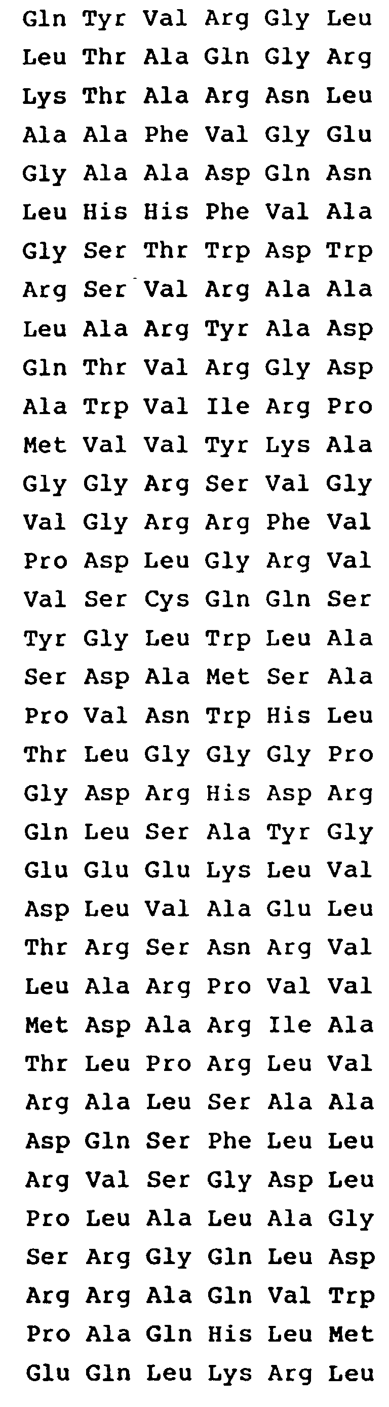

- A purified and isolated DNA fragment consisting
of a DNA sequence the acyB2 gene product according to the following amino acid sequence


- A purified and isolated DNA fragment consisting
of a DNA sequence encoding the acyB2 gene product according to the following base sequence


- The DNA fragment and the DNA restriction fragment according to claim 1 in which the microorganism of the genus Streptomyces is Streptomyces thermotolerans ATCC 11416.
- The DNA fragment and the DNA restriction fragment according to claim 1 which have the following base sequence


- A plasmid containing the DNA fragment of any one of claim 1, 4 or 5 which is a recombinant plasmid resulting from insertion of said DNA fragment into a vector plasmid selected from pIJ702, pIJ943, pIJ350 and pUC18.
- The plasmid of claim 6 which is pMABI according to Fig. 12.
- A microorganism transformed with the plasmid set forth in any one of claim 6 or 7, said microorganism being of the genus Streptomyces or of the species E. coli.
- The microorganism of claim 8 in which the host organism is Streptomyces lividans TK24 or Streptomyces fradiae ATCC 19609.
- The microorganism of claim 8 in which the host microorganism is a macrolide antibiotic with a mycarose substituent producing microorganism which does not substantially produce an enzyme capable of acylating the 4"-position of a macrolide antibiotic.
Applications Claiming Priority (4)
| Application Number | Priority Date | Filing Date | Title |
|---|---|---|---|
| JP125091/88 | 1988-05-24 | ||
| JP12509188 | 1988-05-24 | ||
| JP50120/88 | 1989-03-03 | ||
| JP1050120A JP2749616B2 (en) | 1988-05-24 | 1989-03-03 | Gene encoding 4 "acylase of macrolide antibiotic |
Publications (3)
| Publication Number | Publication Date |
|---|---|
| EP0345546A2 EP0345546A2 (en) | 1989-12-13 |
| EP0345546A3 EP0345546A3 (en) | 1991-02-27 |
| EP0345546B1 true EP0345546B1 (en) | 1996-09-04 |
Family
ID=26390569
Family Applications (1)
| Application Number | Title | Priority Date | Filing Date |
|---|---|---|---|
| EP89109425A Expired - Lifetime EP0345546B1 (en) | 1988-05-24 | 1989-05-24 | DNA coding for enzyme capable of acylating the 4''-position of macrolide antibiotics |
Country Status (4)
| Country | Link |
|---|---|
| US (1) | US5240849A (en) |
| EP (1) | EP0345546B1 (en) |
| JP (1) | JP2749616B2 (en) |
| DE (1) | DE68927074T2 (en) |
Cited By (1)
| Publication number | Priority date | Publication date | Assignee | Title |
|---|---|---|---|---|
| EP2597152B1 (en) * | 2010-07-23 | 2023-11-01 | Shen Yang Fuyang Medicine Technology Co., Ltd | Genetically engineered strain wsj-ia for producing isovaleryl spiramycin i. |
Families Citing this family (8)
| Publication number | Priority date | Publication date | Assignee | Title |
|---|---|---|---|---|
| US5322937A (en) * | 1990-06-01 | 1994-06-21 | Mercian Corporation | Genes encoding a 3-acylation enzyme for macrolide antibiotics |
| FR2739628B1 (en) * | 1995-10-05 | 1997-12-26 | Pasteur Institut | PROCESS FOR THE SYNTHESIS OF HYBRID MOLECULES CONSISTING OF THE CONDENSATION OF AT LEAST TWO SEGMENTS BELONGING TO DIFFERENT CHEMICAL SPECIES SEQUENCES AND PROTEINS FOR THE IMPLEMENTATION THEREOF |
| US7579167B2 (en) * | 2002-10-08 | 2009-08-25 | Aventis Pharma S. | Polypeptides involved in the biosynthesis of spiramycins, nucleotide sequences encoding these polypeptides and applications thereof |
| CA2501445C (en) | 2002-10-08 | 2016-01-26 | Aventis Pharma S.A. | Polypeptides involved in spiramycin biosynthesis, nucleotide sequences encoding said polypeptides and uses thereof |
| EP1905833B1 (en) * | 2002-10-08 | 2015-08-12 | Aventis Pharma S.A. | Polypeptides involved in the biosynthesis of spiramycins, nucleotide sequences coding these polypeptides and their uses |
| US20120258502A1 (en) * | 2011-04-08 | 2012-10-11 | Vinod Pandiripally | Method of producing recombinant plasmid dna using substantially solid growth medium |
| CN103626855B (en) * | 2013-11-14 | 2015-09-16 | 中国农业科学院植物保护研究所 | A kind of albumen relevant to Wuyiencin biosynthesizing and encoding gene thereof and application |
| EP3116888A1 (en) | 2014-03-11 | 2017-01-18 | AbbVie Inc. | 4"-0-substituted tylosin a derivatives |
Family Cites Families (5)
| Publication number | Priority date | Publication date | Assignee | Title |
|---|---|---|---|---|
| US4201843A (en) * | 1975-08-01 | 1980-05-06 | Sanraku Ocean Co., Ltd. | Process for manufacturing tylosin derivatives |
| DK144084A (en) * | 1983-03-03 | 1984-09-04 | Lilly Co Eli | MAKROLID, ITS MANUFACTURING AND USE |
| CA1340599C (en) * | 1986-03-21 | 1999-06-22 | Karen Leigh Cox | Antibiotic-producing microorganisms |
| US5068189A (en) * | 1988-05-13 | 1991-11-26 | Eli Lilly And Company | Recombinant dna vectors encoding a 4"-o-isovaleryl acylase derived from a carbomycin biosynthetic gene, designated care, for use in streptomyces and other organisms |
| US5098837A (en) * | 1988-06-07 | 1992-03-24 | Eli Lilly And Company | Macrolide biosynthetic genes for use in streptomyces and other organisms |
-
1989
- 1989-03-03 JP JP1050120A patent/JP2749616B2/en not_active Expired - Fee Related
- 1989-05-23 US US07/356,323 patent/US5240849A/en not_active Expired - Lifetime
- 1989-05-24 DE DE68927074T patent/DE68927074T2/en not_active Expired - Fee Related
- 1989-05-24 EP EP89109425A patent/EP0345546B1/en not_active Expired - Lifetime
Non-Patent Citations (2)
| Title |
|---|
| BIOL. ACTINOMYCETES, 1988; J.K. EPP et al., pp. 82-85 * |
| CHEMICAL ABSTRACTS, vol. 103, no. 5, 05 August 1985, Columbus, OH (US); J. VARA et al., p. 111, AN 32971d * |
Cited By (1)
| Publication number | Priority date | Publication date | Assignee | Title |
|---|---|---|---|---|
| EP2597152B1 (en) * | 2010-07-23 | 2023-11-01 | Shen Yang Fuyang Medicine Technology Co., Ltd | Genetically engineered strain wsj-ia for producing isovaleryl spiramycin i. |
Also Published As
| Publication number | Publication date |
|---|---|
| JPH0272880A (en) | 1990-03-13 |
| JP2749616B2 (en) | 1998-05-13 |
| EP0345546A2 (en) | 1989-12-13 |
| EP0345546A3 (en) | 1991-02-27 |
| US5240849A (en) | 1993-08-31 |
| DE68927074T2 (en) | 1997-01-09 |
| DE68927074D1 (en) | 1996-10-10 |
Similar Documents
| Publication | Publication Date | Title |
|---|---|---|
| EP0346000A2 (en) | Macrolide biosynthetic genes for use in streptomyces and other organisms | |
| US5240849A (en) | Dna coding for enzyme capable of acylating the 4"-position of macrolide antibiotic | |
| ES2206506T3 (en) | GEN THAT CODIFIES GLICOSIL-TRANSFERASA, AND ITS USE. | |
| US4332898A (en) | Hybrid plasmid and process of making same | |
| AU652766B2 (en) | Cloning of the biosynthetic pathway genes for chlortetracycline production from streptomyces aureofaciens and their expression in streptomyces lividans | |
| US5589385A (en) | Cloning of the biosynthetic pathway for chlortetracycline and tetracycline formation and cosmids useful therein | |
| OMURA et al. | Extraction and characterization of plasmids from macrolide antibiotic-producing streptomycetes | |
| US5068189A (en) | Recombinant dna vectors encoding a 4"-o-isovaleryl acylase derived from a carbomycin biosynthetic gene, designated care, for use in streptomyces and other organisms | |
| AU651982B2 (en) | A bifunctional cosmid useful for cloning actinomycete DNA | |
| AU602954B2 (en) | Carbomycin biosynthetic gene, designated carg, for use in streptomyces and other organisms | |
| EP0459525B1 (en) | Genes encoding a 3-acylation enzyme for macrolide antibiotics | |
| US4518698A (en) | Plasmid and production thereof | |
| EP0288200A1 (en) | Spiramycin resistance-conferring cloning vectors | |
| US4478937A (en) | Plasmid and production thereof | |
| JP2988735B2 (en) | Gane coding 3-acylation enzyme of macrolide antibiotic substance | |
| JPH0638750A (en) | Enzyme for acylating 3-position of macrolide antibiotic and gene coding the same | |
| PARAG et al. | A plasmid of the sisomicin producer Micromonospora inyoensis | |
| US4914030A (en) | Carbomycin resistance-conferring gene, designated carb, for use in streptomyces and other organisms | |
| EP0038156A2 (en) | A plasmid and its microbiological preparation | |
| EP0258046A2 (en) | Method for selecting recombinant DNA-containing streptomyces and other host cells | |
| Gräfe et al. | The platenolides I and II as precursors of turimycin | |
| GB2044773A (en) | A plasmid and its microbiological preparation | |
| JPH06121677A (en) | Method for high expression of acylating enzyme gene of macrolide antibiotics | |
| Woodman | Molecular and biochemical studies on thiopeptide antibiotic biosynthesis | |
| KOJIMA et al. | Mutual relation of three pock-forming plasmids resident in Streptomyces noursei |
Legal Events
| Date | Code | Title | Description |
|---|---|---|---|
| PUAI | Public reference made under article 153(3) epc to a published international application that has entered the european phase |
Free format text: ORIGINAL CODE: 0009012 |
|
| AK | Designated contracting states |
Kind code of ref document: A2 Designated state(s): DE FR GB IT |
|
| PUAL | Search report despatched |
Free format text: ORIGINAL CODE: 0009013 |
|
| AK | Designated contracting states |
Kind code of ref document: A3 Designated state(s): DE FR GB IT |
|
| 17P | Request for examination filed |
Effective date: 19910715 |
|
| 17Q | First examination report despatched |
Effective date: 19940526 |
|
| RAP1 | Party data changed (applicant data changed or rights of an application transferred) |
Owner name: MERCIAN CORPORATION |
|
| GRAH | Despatch of communication of intention to grant a patent |
Free format text: ORIGINAL CODE: EPIDOS IGRA |
|
| GRAA | (expected) grant |
Free format text: ORIGINAL CODE: 0009210 |
|
| ITF | It: translation for a ep patent filed | ||
| AK | Designated contracting states |
Kind code of ref document: B1 Designated state(s): DE FR GB IT |
|
| REF | Corresponds to: |
Ref document number: 68927074 Country of ref document: DE Date of ref document: 19961010 |
|
| ET | Fr: translation filed | ||
| PLBE | No opposition filed within time limit |
Free format text: ORIGINAL CODE: 0009261 |
|
| STAA | Information on the status of an ep patent application or granted ep patent |
Free format text: STATUS: NO OPPOSITION FILED WITHIN TIME LIMIT |
|
| 26N | No opposition filed | ||
| REG | Reference to a national code |
Ref country code: GB Ref legal event code: IF02 |
|
| REG | Reference to a national code |
Ref country code: FR Ref legal event code: TP |
|
| REG | Reference to a national code |
Ref country code: GB Ref legal event code: 732E |
|
| PGFP | Annual fee paid to national office [announced via postgrant information from national office to epo] |
Ref country code: DE Payment date: 20070621 Year of fee payment: 19 |
|
| PGFP | Annual fee paid to national office [announced via postgrant information from national office to epo] |
Ref country code: GB Payment date: 20070515 Year of fee payment: 19 |
|
| PGFP | Annual fee paid to national office [announced via postgrant information from national office to epo] |
Ref country code: IT Payment date: 20070616 Year of fee payment: 19 |
|
| PGFP | Annual fee paid to national office [announced via postgrant information from national office to epo] |
Ref country code: FR Payment date: 20070531 Year of fee payment: 19 |
|
| GBPC | Gb: european patent ceased through non-payment of renewal fee |
Effective date: 20080524 |
|
| REG | Reference to a national code |
Ref country code: FR Ref legal event code: ST Effective date: 20090119 |
|
| PG25 | Lapsed in a contracting state [announced via postgrant information from national office to epo] |
Ref country code: DE Free format text: LAPSE BECAUSE OF NON-PAYMENT OF DUE FEES Effective date: 20081202 Ref country code: FR Free format text: LAPSE BECAUSE OF NON-PAYMENT OF DUE FEES Effective date: 20080602 |
|
| PG25 | Lapsed in a contracting state [announced via postgrant information from national office to epo] |
Ref country code: GB Free format text: LAPSE BECAUSE OF NON-PAYMENT OF DUE FEES Effective date: 20080524 |
|
| PG25 | Lapsed in a contracting state [announced via postgrant information from national office to epo] |
Ref country code: IT Free format text: LAPSE BECAUSE OF NON-PAYMENT OF DUE FEES Effective date: 20080524 |