EP0345461A2 - Mouse monoclonal antibodies to HIV-1P24 and their use in diagnostic tests - Google Patents
Mouse monoclonal antibodies to HIV-1P24 and their use in diagnostic tests Download PDFInfo
- Publication number
- EP0345461A2 EP0345461A2 EP89108001A EP89108001A EP0345461A2 EP 0345461 A2 EP0345461 A2 EP 0345461A2 EP 89108001 A EP89108001 A EP 89108001A EP 89108001 A EP89108001 A EP 89108001A EP 0345461 A2 EP0345461 A2 EP 0345461A2
- Authority
- EP
- European Patent Office
- Prior art keywords
- hiv
- antibody
- monoclonal
- immunoassay
- monoclonal antibody
- Prior art date
- Legal status (The legal status is an assumption and is not a legal conclusion. Google has not performed a legal analysis and makes no representation as to the accuracy of the status listed.)
- Granted
Links
- 229940126619 mouse monoclonal antibody Drugs 0.000 title description 6
- 238000002405 diagnostic procedure Methods 0.000 title description 4
- 101710147327 Calcineurin B homologous protein 1 Proteins 0.000 claims abstract description 127
- 101710205625 Capsid protein p24 Proteins 0.000 claims abstract description 127
- 101710177166 Phosphoprotein Proteins 0.000 claims abstract description 127
- 101710149279 Small delta antigen Proteins 0.000 claims abstract description 127
- 102100022563 Tubulin polymerization-promoting protein Human genes 0.000 claims abstract description 127
- 239000000427 antigen Substances 0.000 claims abstract description 53
- 108091007433 antigens Proteins 0.000 claims abstract description 53
- 102000036639 antigens Human genes 0.000 claims abstract description 53
- 208000031886 HIV Infections Diseases 0.000 claims abstract description 31
- 238000003018 immunoassay Methods 0.000 claims abstract description 31
- 241000713340 Human immunodeficiency virus 2 Species 0.000 claims abstract description 30
- 239000000203 mixture Substances 0.000 claims abstract description 27
- 238000001514 detection method Methods 0.000 claims abstract description 23
- 102000004190 Enzymes Human genes 0.000 claims abstract description 9
- 108090000790 Enzymes Proteins 0.000 claims abstract description 9
- 239000012472 biological sample Substances 0.000 claims abstract description 7
- 210000004027 cell Anatomy 0.000 claims description 47
- 239000000523 sample Substances 0.000 claims description 39
- 239000011324 bead Substances 0.000 claims description 35
- 108010001336 Horseradish Peroxidase Proteins 0.000 claims description 16
- 238000002835 absorbance Methods 0.000 claims description 16
- 210000004408 hybridoma Anatomy 0.000 claims description 13
- 238000012360 testing method Methods 0.000 claims description 13
- 238000005406 washing Methods 0.000 claims description 13
- 230000027455 binding Effects 0.000 claims description 12
- 239000003153 chemical reaction reagent Substances 0.000 claims description 9
- 239000007787 solid Substances 0.000 claims description 8
- 210000004989 spleen cell Anatomy 0.000 claims description 8
- 238000013507 mapping Methods 0.000 claims description 7
- 230000003612 virological effect Effects 0.000 claims description 7
- 239000012634 fragment Substances 0.000 claims description 6
- 241001529936 Murinae Species 0.000 claims description 4
- 206010035226 Plasma cell myeloma Diseases 0.000 claims description 3
- 201000000050 myeloid neoplasm Diseases 0.000 claims description 3
- 210000000628 antibody-producing cell Anatomy 0.000 claims description 2
- 125000003178 carboxy group Chemical group [H]OC(*)=O 0.000 claims description 2
- 239000011248 coating agent Substances 0.000 claims 1
- 238000000576 coating method Methods 0.000 claims 1
- 150000001875 compounds Chemical class 0.000 claims 1
- 239000007850 fluorescent dye Substances 0.000 claims 1
- 230000009870 specific binding Effects 0.000 claims 1
- 238000000034 method Methods 0.000 abstract description 27
- 230000002163 immunogen Effects 0.000 abstract description 7
- 239000013060 biological fluid Substances 0.000 abstract description 5
- 230000009257 reactivity Effects 0.000 abstract description 3
- 241000725303 Human immunodeficiency virus Species 0.000 description 174
- 238000003556 assay Methods 0.000 description 91
- 210000002966 serum Anatomy 0.000 description 25
- 238000010790 dilution Methods 0.000 description 17
- 239000012895 dilution Substances 0.000 description 17
- 235000018102 proteins Nutrition 0.000 description 16
- 102000004169 proteins and genes Human genes 0.000 description 16
- 108090000623 proteins and genes Proteins 0.000 description 16
- 230000035945 sensitivity Effects 0.000 description 16
- 208000030507 AIDS Diseases 0.000 description 14
- 241000283707 Capra Species 0.000 description 14
- 241000699670 Mus sp. Species 0.000 description 14
- 241000700605 Viruses Species 0.000 description 14
- 241000699666 Mus <mouse, genus> Species 0.000 description 13
- 239000000243 solution Substances 0.000 description 12
- 239000004793 Polystyrene Substances 0.000 description 11
- LOKCTEFSRHRXRJ-UHFFFAOYSA-I dipotassium trisodium dihydrogen phosphate hydrogen phosphate dichloride Chemical compound P(=O)(O)(O)[O-].[K+].P(=O)(O)([O-])[O-].[Na+].[Na+].[Cl-].[K+].[Cl-].[Na+] LOKCTEFSRHRXRJ-UHFFFAOYSA-I 0.000 description 11
- 239000002953 phosphate buffered saline Substances 0.000 description 11
- 229920002223 polystyrene Polymers 0.000 description 11
- 206010003445 Ascites Diseases 0.000 description 10
- 108091003079 Bovine Serum Albumin Proteins 0.000 description 10
- FAPWRFPIFSIZLT-UHFFFAOYSA-M Sodium chloride Chemical compound [Na+].[Cl-] FAPWRFPIFSIZLT-UHFFFAOYSA-M 0.000 description 10
- 238000006243 chemical reaction Methods 0.000 description 10
- 230000004927 fusion Effects 0.000 description 10
- 238000001262 western blot Methods 0.000 description 8
- 210000004369 blood Anatomy 0.000 description 7
- 239000008280 blood Substances 0.000 description 7
- 208000015181 infectious disease Diseases 0.000 description 7
- CSCPPACGZOOCGX-UHFFFAOYSA-N Acetone Chemical compound CC(C)=O CSCPPACGZOOCGX-UHFFFAOYSA-N 0.000 description 6
- OKKJLVBELUTLKV-UHFFFAOYSA-N Methanol Chemical compound OC OKKJLVBELUTLKV-UHFFFAOYSA-N 0.000 description 6
- 239000013592 cell lysate Substances 0.000 description 6
- 238000005119 centrifugation Methods 0.000 description 6
- 239000012531 culture fluid Substances 0.000 description 6
- 238000011161 development Methods 0.000 description 6
- 239000006166 lysate Substances 0.000 description 6
- 239000013642 negative control Substances 0.000 description 6
- 210000002381 plasma Anatomy 0.000 description 6
- XOJVVFBFDXDTEG-UHFFFAOYSA-N pristane Chemical compound CC(C)CCCC(C)CCCC(C)CCCC(C)C XOJVVFBFDXDTEG-UHFFFAOYSA-N 0.000 description 6
- 239000011541 reaction mixture Substances 0.000 description 6
- GEYOCULIXLDCMW-UHFFFAOYSA-N 1,2-phenylenediamine Chemical compound NC1=CC=CC=C1N GEYOCULIXLDCMW-UHFFFAOYSA-N 0.000 description 5
- 241000588724 Escherichia coli Species 0.000 description 5
- 239000000020 Nitrocellulose Substances 0.000 description 5
- 229920002684 Sepharose Polymers 0.000 description 5
- DBMJMQXJHONAFJ-UHFFFAOYSA-M Sodium laurylsulphate Chemical compound [Na+].CCCCCCCCCCCCOS([O-])(=O)=O DBMJMQXJHONAFJ-UHFFFAOYSA-M 0.000 description 5
- 239000013504 Triton X-100 Substances 0.000 description 5
- 229920004890 Triton X-100 Polymers 0.000 description 5
- 108010067390 Viral Proteins Proteins 0.000 description 5
- 210000001124 body fluid Anatomy 0.000 description 5
- 239000010839 body fluid Substances 0.000 description 5
- 229940098773 bovine serum albumin Drugs 0.000 description 5
- 238000012512 characterization method Methods 0.000 description 5
- 230000002860 competitive effect Effects 0.000 description 5
- 239000012228 culture supernatant Substances 0.000 description 5
- 238000012217 deletion Methods 0.000 description 5
- 230000037430 deletion Effects 0.000 description 5
- 238000011534 incubation Methods 0.000 description 5
- 239000002609 medium Substances 0.000 description 5
- 229920001220 nitrocellulos Polymers 0.000 description 5
- 239000011780 sodium chloride Substances 0.000 description 5
- 210000000952 spleen Anatomy 0.000 description 5
- 210000001519 tissue Anatomy 0.000 description 5
- MHAJPDPJQMAIIY-UHFFFAOYSA-N Hydrogen peroxide Chemical compound OO MHAJPDPJQMAIIY-UHFFFAOYSA-N 0.000 description 4
- 102100034353 Integrase Human genes 0.000 description 4
- 241000283973 Oryctolagus cuniculus Species 0.000 description 4
- 239000002202 Polyethylene glycol Substances 0.000 description 4
- IQFYYKKMVGJFEH-XLPZGREQSA-N Thymidine Chemical compound O=C1NC(=O)C(C)=CN1[C@@H]1O[C@H](CO)[C@@H](O)C1 IQFYYKKMVGJFEH-XLPZGREQSA-N 0.000 description 4
- 239000007983 Tris buffer Substances 0.000 description 4
- 239000002671 adjuvant Substances 0.000 description 4
- 239000000872 buffer Substances 0.000 description 4
- 230000009260 cross reactivity Effects 0.000 description 4
- 230000000694 effects Effects 0.000 description 4
- 238000001114 immunoprecipitation Methods 0.000 description 4
- 238000000338 in vitro Methods 0.000 description 4
- 210000004940 nucleus Anatomy 0.000 description 4
- 229920001223 polyethylene glycol Polymers 0.000 description 4
- 238000013207 serial dilution Methods 0.000 description 4
- 239000007790 solid phase Substances 0.000 description 4
- LENZDBCJOHFCAS-UHFFFAOYSA-N tris Chemical compound OCC(N)(CO)CO LENZDBCJOHFCAS-UHFFFAOYSA-N 0.000 description 4
- DGVVWUTYPXICAM-UHFFFAOYSA-N β‐Mercaptoethanol Chemical compound OCCS DGVVWUTYPXICAM-UHFFFAOYSA-N 0.000 description 4
- WVDDGKGOMKODPV-UHFFFAOYSA-N Benzyl alcohol Chemical compound OCC1=CC=CC=C1 WVDDGKGOMKODPV-UHFFFAOYSA-N 0.000 description 3
- 101710132601 Capsid protein Proteins 0.000 description 3
- XUJNEKJLAYXESH-UHFFFAOYSA-N Cysteine Chemical compound SCC(N)C(O)=O XUJNEKJLAYXESH-UHFFFAOYSA-N 0.000 description 3
- 238000002965 ELISA Methods 0.000 description 3
- FFEARJCKVFRZRR-BYPYZUCNSA-N L-methionine Chemical compound CSCC[C@H](N)C(O)=O FFEARJCKVFRZRR-BYPYZUCNSA-N 0.000 description 3
- 108010092799 RNA-directed DNA polymerase Proteins 0.000 description 3
- 238000010367 cloning Methods 0.000 description 3
- 239000003599 detergent Substances 0.000 description 3
- 238000003745 diagnosis Methods 0.000 description 3
- 239000003085 diluting agent Substances 0.000 description 3
- 239000013024 dilution buffer Substances 0.000 description 3
- 238000002474 experimental method Methods 0.000 description 3
- 239000000284 extract Substances 0.000 description 3
- 239000012894 fetal calf serum Substances 0.000 description 3
- 239000012530 fluid Substances 0.000 description 3
- 238000006386 neutralization reaction Methods 0.000 description 3
- 125000002924 primary amino group Chemical group [H]N([H])* 0.000 description 3
- 230000009467 reduction Effects 0.000 description 3
- 238000012216 screening Methods 0.000 description 3
- YBJHBAHKTGYVGT-ZKWXMUAHSA-N (+)-Biotin Chemical compound N1C(=O)N[C@@H]2[C@H](CCCCC(=O)O)SC[C@@H]21 YBJHBAHKTGYVGT-ZKWXMUAHSA-N 0.000 description 2
- TVZGACDUOSZQKY-LBPRGKRZSA-N 4-aminofolic acid Chemical compound C1=NC2=NC(N)=NC(N)=C2N=C1CNC1=CC=C(C(=O)N[C@@H](CCC(O)=O)C(O)=O)C=C1 TVZGACDUOSZQKY-LBPRGKRZSA-N 0.000 description 2
- DWRXFEITVBNRMK-UHFFFAOYSA-N Beta-D-1-Arabinofuranosylthymine Natural products O=C1NC(=O)C(C)=CN1C1C(O)C(O)C(CO)O1 DWRXFEITVBNRMK-UHFFFAOYSA-N 0.000 description 2
- 206010061818 Disease progression Diseases 0.000 description 2
- KCXVZYZYPLLWCC-UHFFFAOYSA-N EDTA Chemical compound OC(=O)CN(CC(O)=O)CCN(CC(O)=O)CC(O)=O KCXVZYZYPLLWCC-UHFFFAOYSA-N 0.000 description 2
- DHMQDGOQFOQNFH-UHFFFAOYSA-N Glycine Chemical compound NCC(O)=O DHMQDGOQFOQNFH-UHFFFAOYSA-N 0.000 description 2
- 241000282412 Homo Species 0.000 description 2
- 108060003951 Immunoglobulin Proteins 0.000 description 2
- 108020004511 Recombinant DNA Proteins 0.000 description 2
- CDBYLPFSWZWCQE-UHFFFAOYSA-L Sodium Carbonate Chemical compound [Na+].[Na+].[O-]C([O-])=O CDBYLPFSWZWCQE-UHFFFAOYSA-L 0.000 description 2
- PXIPVTKHYLBLMZ-UHFFFAOYSA-N Sodium azide Chemical compound [Na+].[N-]=[N+]=[N-] PXIPVTKHYLBLMZ-UHFFFAOYSA-N 0.000 description 2
- 229930006000 Sucrose Natural products 0.000 description 2
- CZMRCDWAGMRECN-UGDNZRGBSA-N Sucrose Chemical compound O[C@H]1[C@H](O)[C@@H](CO)O[C@@]1(CO)O[C@@H]1[C@H](O)[C@@H](O)[C@H](O)[C@@H](CO)O1 CZMRCDWAGMRECN-UGDNZRGBSA-N 0.000 description 2
- 238000001042 affinity chromatography Methods 0.000 description 2
- 229960003896 aminopterin Drugs 0.000 description 2
- 230000003321 amplification Effects 0.000 description 2
- 238000002820 assay format Methods 0.000 description 2
- IQFYYKKMVGJFEH-UHFFFAOYSA-N beta-L-thymidine Natural products O=C1NC(=O)C(C)=CN1C1OC(CO)C(O)C1 IQFYYKKMVGJFEH-UHFFFAOYSA-N 0.000 description 2
- 230000015572 biosynthetic process Effects 0.000 description 2
- 210000004899 c-terminal region Anatomy 0.000 description 2
- 230000007910 cell fusion Effects 0.000 description 2
- 230000010261 cell growth Effects 0.000 description 2
- 239000006285 cell suspension Substances 0.000 description 2
- 230000001413 cellular effect Effects 0.000 description 2
- 230000007423 decrease Effects 0.000 description 2
- 238000013461 design Methods 0.000 description 2
- 230000005750 disease progression Effects 0.000 description 2
- -1 electron microscopy Proteins 0.000 description 2
- 238000001962 electrophoresis Methods 0.000 description 2
- 239000012091 fetal bovine serum Substances 0.000 description 2
- 210000004754 hybrid cell Anatomy 0.000 description 2
- 230000003053 immunization Effects 0.000 description 2
- 238000002649 immunization Methods 0.000 description 2
- 230000000984 immunochemical effect Effects 0.000 description 2
- 102000018358 immunoglobulin Human genes 0.000 description 2
- 230000002458 infectious effect Effects 0.000 description 2
- 239000003112 inhibitor Substances 0.000 description 2
- 238000002955 isolation Methods 0.000 description 2
- 238000002372 labelling Methods 0.000 description 2
- 210000004698 lymphocyte Anatomy 0.000 description 2
- 239000012139 lysis buffer Substances 0.000 description 2
- 238000003199 nucleic acid amplification method Methods 0.000 description 2
- 210000004303 peritoneum Anatomy 0.000 description 2
- 235000020030 perry Nutrition 0.000 description 2
- YBYRMVIVWMBXKQ-UHFFFAOYSA-N phenylmethanesulfonyl fluoride Chemical compound FS(=O)(=O)CC1=CC=CC=C1 YBYRMVIVWMBXKQ-UHFFFAOYSA-N 0.000 description 2
- 239000008363 phosphate buffer Substances 0.000 description 2
- 239000002243 precursor Substances 0.000 description 2
- 238000002360 preparation method Methods 0.000 description 2
- 238000003127 radioimmunoassay Methods 0.000 description 2
- 230000003362 replicative effect Effects 0.000 description 2
- 230000000405 serological effect Effects 0.000 description 2
- 229910000029 sodium carbonate Inorganic materials 0.000 description 2
- 238000010186 staining Methods 0.000 description 2
- 239000000758 substrate Substances 0.000 description 2
- 239000005720 sucrose Substances 0.000 description 2
- 230000002195 synergetic effect Effects 0.000 description 2
- 229940104230 thymidine Drugs 0.000 description 2
- LWIHDJKSTIGBAC-UHFFFAOYSA-K tripotassium phosphate Chemical compound [K+].[K+].[K+].[O-]P([O-])([O-])=O LWIHDJKSTIGBAC-UHFFFAOYSA-K 0.000 description 2
- 238000005199 ultracentrifugation Methods 0.000 description 2
- HZAXFHJVJLSVMW-UHFFFAOYSA-N 2-Aminoethan-1-ol Chemical compound NCCO HZAXFHJVJLSVMW-UHFFFAOYSA-N 0.000 description 1
- JKMHFZQWWAIEOD-UHFFFAOYSA-N 2-[4-(2-hydroxyethyl)piperazin-1-yl]ethanesulfonic acid Chemical compound OCC[NH+]1CCN(CCS([O-])(=O)=O)CC1 JKMHFZQWWAIEOD-UHFFFAOYSA-N 0.000 description 1
- 241000714195 Aids-associated retrovirus Species 0.000 description 1
- NLXLAEXVIDQMFP-UHFFFAOYSA-N Ammonia chloride Chemical compound [NH4+].[Cl-] NLXLAEXVIDQMFP-UHFFFAOYSA-N 0.000 description 1
- 108010039627 Aprotinin Proteins 0.000 description 1
- 240000003291 Armoracia rusticana Species 0.000 description 1
- CIWBSHSKHKDKBQ-JLAZNSOCSA-N Ascorbic acid Chemical compound OC[C@H](O)[C@H]1OC(=O)C(O)=C1O CIWBSHSKHKDKBQ-JLAZNSOCSA-N 0.000 description 1
- 239000004471 Glycine Substances 0.000 description 1
- 239000007995 HEPES buffer Substances 0.000 description 1
- 208000037357 HIV infectious disease Diseases 0.000 description 1
- 241000598436 Human T-cell lymphotropic virus Species 0.000 description 1
- 101900330621 Human immunodeficiency virus type 1 group M subtype B Transmembrane protein gp41 Proteins 0.000 description 1
- 108010091358 Hypoxanthine Phosphoribosyltransferase Proteins 0.000 description 1
- 102100029098 Hypoxanthine-guanine phosphoribosyltransferase Human genes 0.000 description 1
- 102000001399 Kallikrein Human genes 0.000 description 1
- 108060005987 Kallikrein Proteins 0.000 description 1
- XUJNEKJLAYXESH-REOHCLBHSA-N L-Cysteine Chemical compound SC[C@H](N)C(O)=O XUJNEKJLAYXESH-REOHCLBHSA-N 0.000 description 1
- 241000713666 Lentivirus Species 0.000 description 1
- 208000008771 Lymphadenopathy Diseases 0.000 description 1
- 241001465754 Metazoa Species 0.000 description 1
- 108020004711 Nucleic Acid Probes Proteins 0.000 description 1
- 108091028043 Nucleic acid sequence Proteins 0.000 description 1
- 241000276498 Pollachius virens Species 0.000 description 1
- 229920001213 Polysorbate 20 Polymers 0.000 description 1
- 239000012980 RPMI-1640 medium Substances 0.000 description 1
- 102000007056 Recombinant Fusion Proteins Human genes 0.000 description 1
- 108010008281 Recombinant Fusion Proteins Proteins 0.000 description 1
- 241000293869 Salmonella enterica subsp. enterica serovar Typhimurium Species 0.000 description 1
- 229920005654 Sephadex Polymers 0.000 description 1
- 239000012507 Sephadex™ Substances 0.000 description 1
- 101710172711 Structural protein Proteins 0.000 description 1
- QAOWNCQODCNURD-UHFFFAOYSA-N Sulfuric acid Chemical compound OS(O)(=O)=O QAOWNCQODCNURD-UHFFFAOYSA-N 0.000 description 1
- 210000001744 T-lymphocyte Anatomy 0.000 description 1
- FRTPPQMVPHJITG-UHFFFAOYSA-N [4-(4-hydrazinylphenyl)phenyl]hydrazine;hydrochloride Chemical compound Cl.C1=CC(NN)=CC=C1C1=CC=C(NN)C=C1 FRTPPQMVPHJITG-UHFFFAOYSA-N 0.000 description 1
- 235000019270 ammonium chloride Nutrition 0.000 description 1
- 230000001745 anti-biotin effect Effects 0.000 description 1
- 230000003302 anti-idiotype Effects 0.000 description 1
- 230000000840 anti-viral effect Effects 0.000 description 1
- 229960004405 aprotinin Drugs 0.000 description 1
- 238000000376 autoradiography Methods 0.000 description 1
- 235000019445 benzyl alcohol Nutrition 0.000 description 1
- 229960002685 biotin Drugs 0.000 description 1
- 235000020958 biotin Nutrition 0.000 description 1
- 239000011616 biotin Substances 0.000 description 1
- 230000000903 blocking effect Effects 0.000 description 1
- 239000010836 blood and blood product Substances 0.000 description 1
- 210000000601 blood cell Anatomy 0.000 description 1
- 229940125691 blood product Drugs 0.000 description 1
- 238000004113 cell culture Methods 0.000 description 1
- 239000012930 cell culture fluid Substances 0.000 description 1
- 210000000170 cell membrane Anatomy 0.000 description 1
- 210000001175 cerebrospinal fluid Anatomy 0.000 description 1
- 239000003795 chemical substances by application Substances 0.000 description 1
- 210000000349 chromosome Anatomy 0.000 description 1
- 230000005757 colony formation Effects 0.000 description 1
- 230000000052 comparative effect Effects 0.000 description 1
- 239000012141 concentrate Substances 0.000 description 1
- 239000000470 constituent Substances 0.000 description 1
- 230000008878 coupling Effects 0.000 description 1
- 238000010168 coupling process Methods 0.000 description 1
- 238000005859 coupling reaction Methods 0.000 description 1
- 238000012258 culturing Methods 0.000 description 1
- 235000018417 cysteine Nutrition 0.000 description 1
- 230000000120 cytopathologic effect Effects 0.000 description 1
- 230000002950 deficient Effects 0.000 description 1
- 229960003964 deoxycholic acid Drugs 0.000 description 1
- KXGVEGMKQFWNSR-LLQZFEROSA-N deoxycholic acid Chemical compound C([C@H]1CC2)[C@H](O)CC[C@]1(C)[C@@H]1[C@@H]2[C@@H]2CC[C@H]([C@@H](CCC(O)=O)C)[C@@]2(C)[C@@H](O)C1 KXGVEGMKQFWNSR-LLQZFEROSA-N 0.000 description 1
- 230000001419 dependent effect Effects 0.000 description 1
- 238000007865 diluting Methods 0.000 description 1
- 201000010099 disease Diseases 0.000 description 1
- 208000037265 diseases, disorders, signs and symptoms Diseases 0.000 description 1
- 238000001493 electron microscopy Methods 0.000 description 1
- 108010078428 env Gene Products Proteins 0.000 description 1
- 230000002255 enzymatic effect Effects 0.000 description 1
- 210000003743 erythrocyte Anatomy 0.000 description 1
- DEFVIWRASFVYLL-UHFFFAOYSA-N ethylene glycol bis(2-aminoethyl)tetraacetic acid Chemical compound OC(=O)CN(CC(O)=O)CCOCCOCCN(CC(O)=O)CC(O)=O DEFVIWRASFVYLL-UHFFFAOYSA-N 0.000 description 1
- 230000001747 exhibiting effect Effects 0.000 description 1
- 238000002523 gelfiltration Methods 0.000 description 1
- 230000012010 growth Effects 0.000 description 1
- 230000036541 health Effects 0.000 description 1
- 238000010438 heat treatment Methods 0.000 description 1
- 208000033519 human immunodeficiency virus infectious disease Diseases 0.000 description 1
- 238000009396 hybridization Methods 0.000 description 1
- 230000028993 immune response Effects 0.000 description 1
- 238000012760 immunocytochemical staining Methods 0.000 description 1
- 230000005847 immunogenicity Effects 0.000 description 1
- 239000012133 immunoprecipitate Substances 0.000 description 1
- 230000002779 inactivation Effects 0.000 description 1
- ZPNFWUPYTFPOJU-LPYSRVMUSA-N iniprol Chemical compound C([C@H]1C(=O)NCC(=O)NCC(=O)N[C@H]2CSSC[C@H]3C(=O)N[C@@H](CCCCN)C(=O)N[C@@H](C)C(=O)N[C@@H](CCCNC(N)=N)C(=O)N[C@H](C(N[C@H](C(=O)N[C@@H](CCCNC(N)=N)C(=O)N[C@@H](CC=4C=CC(O)=CC=4)C(=O)N[C@@H](CC=4C=CC=CC=4)C(=O)N[C@@H](CC=4C=CC(O)=CC=4)C(=O)N[C@@H](CC(N)=O)C(=O)N[C@@H](C)C(=O)N[C@@H](CCCCN)C(=O)N[C@@H](C)C(=O)NCC(=O)N[C@@H](CC(C)C)C(=O)N[C@@H](CSSC[C@H](NC(=O)[C@H](CC(O)=O)NC(=O)[C@H](CCC(O)=O)NC(=O)[C@H](C)NC(=O)[C@H](CO)NC(=O)[C@H](CCCCN)NC(=O)[C@H](CC=4C=CC=CC=4)NC(=O)[C@H](CC(N)=O)NC(=O)[C@H](CC(N)=O)NC(=O)[C@H](CCCNC(N)=N)NC(=O)[C@H](CCCCN)NC(=O)[C@H](C)NC(=O)[C@H](CCCNC(N)=N)NC2=O)C(=O)N[C@@H](CCSC)C(=O)N[C@@H](CCCNC(N)=N)C(=O)N[C@@H]([C@@H](C)O)C(=O)N[C@@H](CSSC[C@H](NC(=O)[C@H](CC=2C=CC=CC=2)NC(=O)[C@H](CC(O)=O)NC(=O)[C@H]2N(CCC2)C(=O)[C@@H](N)CCCNC(N)=N)C(=O)N[C@@H](CC(C)C)C(=O)N[C@@H](CCC(O)=O)C(=O)N2[C@@H](CCC2)C(=O)N2[C@@H](CCC2)C(=O)N[C@@H](CC=2C=CC(O)=CC=2)C(=O)N[C@@H]([C@@H](C)O)C(=O)NCC(=O)N2[C@@H](CCC2)C(=O)N3)C(=O)NCC(=O)NCC(=O)N[C@@H](C)C(O)=O)C(=O)N[C@@H](CCC(N)=O)C(=O)N[C@H](C(=O)N[C@@H](CC=2C=CC=CC=2)C(=O)N[C@H](C(=O)N1)C(C)C)[C@@H](C)O)[C@@H](C)CC)=O)[C@@H](C)CC)C1=CC=C(O)C=C1 ZPNFWUPYTFPOJU-LPYSRVMUSA-N 0.000 description 1
- 238000002347 injection Methods 0.000 description 1
- 239000007924 injection Substances 0.000 description 1
- 238000001990 intravenous administration Methods 0.000 description 1
- 238000011835 investigation Methods 0.000 description 1
- 238000012417 linear regression Methods 0.000 description 1
- 208000018555 lymphatic system disease Diseases 0.000 description 1
- 238000004519 manufacturing process Methods 0.000 description 1
- 210000004379 membrane Anatomy 0.000 description 1
- 239000012528 membrane Substances 0.000 description 1
- 238000005374 membrane filtration Methods 0.000 description 1
- 229930182817 methionine Natural products 0.000 description 1
- 239000011859 microparticle Substances 0.000 description 1
- 230000011278 mitosis Effects 0.000 description 1
- 238000012544 monitoring process Methods 0.000 description 1
- 230000000877 morphologic effect Effects 0.000 description 1
- 239000002853 nucleic acid probe Substances 0.000 description 1
- 239000008188 pellet Substances 0.000 description 1
- 102000013415 peroxidase activity proteins Human genes 0.000 description 1
- 108040007629 peroxidase activity proteins Proteins 0.000 description 1
- 239000013612 plasmid Substances 0.000 description 1
- 229920002401 polyacrylamide Polymers 0.000 description 1
- 238000002264 polyacrylamide gel electrophoresis Methods 0.000 description 1
- 239000000256 polyoxyethylene sorbitan monolaurate Substances 0.000 description 1
- 235000010486 polyoxyethylene sorbitan monolaurate Nutrition 0.000 description 1
- 239000013641 positive control Substances 0.000 description 1
- 229910000160 potassium phosphate Inorganic materials 0.000 description 1
- 235000011009 potassium phosphates Nutrition 0.000 description 1
- 238000011533 pre-incubation Methods 0.000 description 1
- 239000003755 preservative agent Substances 0.000 description 1
- 230000002335 preservative effect Effects 0.000 description 1
- 108090000765 processed proteins & peptides Proteins 0.000 description 1
- 230000000644 propagated effect Effects 0.000 description 1
- 238000010814 radioimmunoprecipitation assay Methods 0.000 description 1
- 238000002976 reverse transcriptase assay Methods 0.000 description 1
- 239000012723 sample buffer Substances 0.000 description 1
- 230000003248 secreting effect Effects 0.000 description 1
- 239000012679 serum free medium Substances 0.000 description 1
- 230000001568 sexual effect Effects 0.000 description 1
- 235000017550 sodium carbonate Nutrition 0.000 description 1
- JQWHASGSAFIOCM-UHFFFAOYSA-M sodium periodate Chemical compound [Na+].[O-]I(=O)(=O)=O JQWHASGSAFIOCM-UHFFFAOYSA-M 0.000 description 1
- 238000000527 sonication Methods 0.000 description 1
- 239000011550 stock solution Substances 0.000 description 1
- 235000011149 sulphuric acid Nutrition 0.000 description 1
- 239000006228 supernatant Substances 0.000 description 1
- 230000001360 synchronised effect Effects 0.000 description 1
- 230000008866 synergistic binding Effects 0.000 description 1
- 239000008399 tap water Substances 0.000 description 1
- 235000020679 tap water Nutrition 0.000 description 1
- 238000002560 therapeutic procedure Methods 0.000 description 1
- 238000012546 transfer Methods 0.000 description 1
- 239000003656 tris buffered saline Substances 0.000 description 1
- GPRLSGONYQIRFK-MNYXATJNSA-N triton Chemical compound [3H+] GPRLSGONYQIRFK-MNYXATJNSA-N 0.000 description 1
- 241001430294 unidentified retrovirus Species 0.000 description 1
- 210000003462 vein Anatomy 0.000 description 1
- 230000009385 viral infection Effects 0.000 description 1
- XLYOFNOQVPJJNP-UHFFFAOYSA-N water Substances O XLYOFNOQVPJJNP-UHFFFAOYSA-N 0.000 description 1
- 230000003442 weekly effect Effects 0.000 description 1
Images


Classifications
-
- C—CHEMISTRY; METALLURGY
- C07—ORGANIC CHEMISTRY
- C07K—PEPTIDES
- C07K16/00—Immunoglobulins [IGs], e.g. monoclonal or polyclonal antibodies
- C07K16/08—Immunoglobulins [IGs], e.g. monoclonal or polyclonal antibodies against material from viruses
- C07K16/10—Immunoglobulins [IGs], e.g. monoclonal or polyclonal antibodies against material from viruses from RNA viruses
- C07K16/1036—Retroviridae, e.g. leukemia viruses
- C07K16/1045—Lentiviridae, e.g. HIV, FIV, SIV
- C07K16/1054—Lentiviridae, e.g. HIV, FIV, SIV gag-pol, e.g. p17, p24
-
- Y—GENERAL TAGGING OF NEW TECHNOLOGICAL DEVELOPMENTS; GENERAL TAGGING OF CROSS-SECTIONAL TECHNOLOGIES SPANNING OVER SEVERAL SECTIONS OF THE IPC; TECHNICAL SUBJECTS COVERED BY FORMER USPC CROSS-REFERENCE ART COLLECTIONS [XRACs] AND DIGESTS
- Y10—TECHNICAL SUBJECTS COVERED BY FORMER USPC
- Y10S—TECHNICAL SUBJECTS COVERED BY FORMER USPC CROSS-REFERENCE ART COLLECTIONS [XRACs] AND DIGESTS
- Y10S435/00—Chemistry: molecular biology and microbiology
- Y10S435/971—Capture of complex after antigen-antibody reaction
-
- Y—GENERAL TAGGING OF NEW TECHNOLOGICAL DEVELOPMENTS; GENERAL TAGGING OF CROSS-SECTIONAL TECHNOLOGIES SPANNING OVER SEVERAL SECTIONS OF THE IPC; TECHNICAL SUBJECTS COVERED BY FORMER USPC CROSS-REFERENCE ART COLLECTIONS [XRACs] AND DIGESTS
- Y10—TECHNICAL SUBJECTS COVERED BY FORMER USPC
- Y10S—TECHNICAL SUBJECTS COVERED BY FORMER USPC CROSS-REFERENCE ART COLLECTIONS [XRACs] AND DIGESTS
- Y10S435/00—Chemistry: molecular biology and microbiology
- Y10S435/974—Aids related test
-
- Y—GENERAL TAGGING OF NEW TECHNOLOGICAL DEVELOPMENTS; GENERAL TAGGING OF CROSS-SECTIONAL TECHNOLOGIES SPANNING OVER SEVERAL SECTIONS OF THE IPC; TECHNICAL SUBJECTS COVERED BY FORMER USPC CROSS-REFERENCE ART COLLECTIONS [XRACs] AND DIGESTS
- Y10—TECHNICAL SUBJECTS COVERED BY FORMER USPC
- Y10S—TECHNICAL SUBJECTS COVERED BY FORMER USPC CROSS-REFERENCE ART COLLECTIONS [XRACs] AND DIGESTS
- Y10S435/00—Chemistry: molecular biology and microbiology
- Y10S435/975—Kit
Definitions
- the present invention relates to the detection of the Human Immunodeficiency Virus (HIV-I), the etiologic agent of Acquired Immunodeficiency Syndrome (AIDS), in serum, plasma or other body fluids.
- HIV-I Human Immunodeficiency Virus
- AIDS Acquired Immunodeficiency Syndrome
- this invention describes a diagnostic test which employs a combination of unique mouse monoclonal antibodies as a probe for the detection of HIV-I core protein p24.
- HIV-I HIV-I includes the formerly named viruses Human T-cell Lymphotrophic Virus Type III (HTLV III), Lymphadenopathy Associated Virus (LAV) and AIDS Associated Retrovirus (ARV). HIV-I is related to a group of cytopathic retroviruses, namely lentiviruses, on the basis of in vitro characteristics, morphologic features and nucleotide sequences (Gonda et al., Science (1985) 227:177-179; Stephan et al., Science (1986) 231 :589-594).
- HTLV III Human T-cell Lymphotrophic Virus Type III
- LAV Lymphadenopathy Associated Virus
- ARV AIDS Associated Retrovirus
- the most widely used methods for detecting HIV-I in infected individuals include the isolation of virus from infected blood or blood cells and subsequent in vitro propagation of the virus in lymphocyte cultures.
- In vitro replicating virus may be detected by measuring reverse transcriptase (RT) levels, immunocytochemical staining of viral proteins, electron microscopy, and nucleic acid probe hybridization.
- RT reverse transcriptase
- In vitro cultivation and isolation of virus are labor intensive, technique sensitive and may not be practical for use as a routine diagnostic method.
- enzyme immunoassays have been developed to detect HIV-I antigens in serum and other body fluids of infected people (Goudsmit et al., The Lancet (1986) 2:177-180; Allain et al., Brit. Med. J.
- HIV-I core antigens may be important serological markers for initial diagnosis of infection and disease progression, and as well may provide a tool for monitoring antiviral therapy in AIDS patients.
- Mouse monoclonal antibodies to HIV-I p24 are provided by the invention which are highly specific reagents employed in immunoassays designed to detect and/or capture HIV-I p24. Available information on the nature and consequence of HIV-I infection establishes a need for the early detection of HIV-I antigens, especially p24, prior to seroconversion in people infected with HIV-I.
- a highly sensitive diagnostic assay to detect HIV-I p24, using a mixture of two monoclonal antibodies as a probe, is provided by the present invention.
- One monoclonal antibody, 31-42-19 recognizes a unique epitope on HIV-I p24 which is not recognized by sera from seropositive individuals.
- monoclonal antibody 31-90-25 which recognizes an epitope within a highly immunogenic region of HIV-I p24, also is employed in the assay.
- monoclonal antibodies 31-42-19 and 31-90-25 are used together in solution as the probe to detect HIV-I p24, with polystyrene beads previously coated with anti-HIV-I human IgG as the capture antibodies. It has been found that 31-42-19 is the key monoclonal antibody which in combination with 31-90-25 results in optimal assay sensitivity for detection of HIV-I p24. Several alternate procedures can be employed to achieve the desired sensitivity and speed. In an alternate assay configuration, these two monoclonals can be successfully employed as capture antibodies for HIV-I p24 when coated on polystyrene beads.
- monoclonal antibody 31-42-19 also recognizes HIV-2 p24. This unique characteristic of detecting both HIV-I p24 and HIV-2 p24 can be exploited in an immunoassay designed to screen non-discriminately for HIV infection. In addition, monoclonal antibody 31-42-19 can be used as a probe for the detection of HIV-2 p24.
- monoclonal antibody 31-90-25 can be employed as a competitive probe to detect anti-HIV-I core antibodies in biological samples.
- the present invention provides a novel means for the detection of HIV-I p24 in picogram quantities in body fluids of infected individuals, using two monoclonal antibodies as a probe.
- This highly sensitive enzyme immunoassay is unique because of the characteristics of the monoclonal antibody 31-42-19.
- the epitope recognized by monoclonal antibody 31-42-19 maps toward the carboxy terminus of HIV-I p24 and is not immunogenic in humans.
- the second monoclonal antibody employed in this assay recognizes an epitope within a highly immunogenic region of HIV-I p24, and maps toward the amino terminus of HIV-I p24.
- Monoclonal antibodies 31-42-19 and 31-90-25 bind synergistically to HIV-I p24.
- the epitope recognized by monoclonal antibody 31-42-19 is antigenically cross-reactive with an epitope on HIV-2 p24, as shown by the ability of 31-42-19 to immunoprecipitate HIV-2 p24 from biosynthetically labeled HIV-2 infected cells.
- the monoclonal antibody 31-90-25 when appropriately labelled, can be employed as a competitive probe against HIV-I core antibodies in serum samples for binding to recombinant-derived HIV-I p24.
- HRPO labelled 31-90-25 can be employed in an immunoassay for antibodies to HIV as disclosed in U.S. Patent Application Serial No. 020,282, filed Feb. 27, 1987 by Dawson et al., and commonly assigned herewith.
- the present invention provides novel hybridoma cell lines, exemplified by murine-derived cell line ATCC HB 9726 and murine-derived cell line ATCC HB 9725, and novel monoclonal antibodies secreted thereby, exemplified by the above-noted monoclonal antibodies 31-42-19 and 31-90-25, respectively.
- a biological sample presumably containing HIV-I p24, is incubated with a mixture of monoclonal antibodies 31-42-19 and 31-90-25, and a polystyrene bead coated with anti-HIV-I IgG (purified from serum of seropositive individuals for HIV-I p24 antibodies).
- the amount of mouse monoclonal antibodies bound which is proportional to the amount of HIV-I p24 captured on the bead, is determined with horseradish peroxidase-labelled goat anti-mouse IgG.
- the monoclonal antibody mixture can also be coated on a solid phase to serve as capture antibodies.
- this mixture can be used to coat a solid support of an immunoassay to detect HIV-I (HTLV-III) antigens as disclosed in U.S. Patent No. 4,748,110, issued May 31, 1988.
- antibodies 31-42-19 and 31-90-25 can be employed in detection systems using fixed cells, with appropriate labelling of each monoclonal antibody.
- These antibodies also can be employed for purifying HIV-I p24, and the particular monoclonal 31-42-19 for purifying HIV-2 p24, by affinity chromatography.
- Biological samples which are easily tested by the method of the present invention include human and animal body fluids such as whole blood, serum, plasma, cerebrospinal fluid and lymphocyte or cell culture supernatants. Additionally, the test samples could be inactivated whole virus or partially purified native or recombinant HIV-I p24.
- Solid supports which can be used in immunoassays of the invention include wells of reaction trays, test tubes, polystyrene beads, strips, membranes, microparticles, and other solid matrices known to those skilled in the art. Any label capable of producing a detectable signal or an enzyme amplification system can be used in immunoassays of the invention. Representative labels include enzymatic, radioisotopic, fluorescent and chemiluminescent labels. Further, hapten/labelled anti-hapten systems such as a biotin/labelled anti-biotin system may be utilized in the inventive assays. Additionally, one can employ a labelled anti-idiotype antibody to detect the monoclonal antibodies described herein.
- reagents for the assays of the invention are ideally suited for preparation of a kit.
- a kit may comprise carrier means being compartmentalized to receive in close confinement, one or more container means such as vials, bottles, test tubes and the like.
- container means such as vials, bottles, test tubes and the like.
- Each of the container means comprises one of the separate elements to be used in the method of this invention.
- Examples 1 and 2 relate to the procedures whereby hybridoma cell lines secreting monoclonal antibodies were generated.
- Example 3 relates to the screening, cloning and characterization of monoclonal antibodies 31-42-19 and 31-90-25.
- Example 4 relates to the method used for amplifying antibody yields.
- Example 5 relates to assays performed to determine the activity, specificity and epitope mapping of the 31-42-19 and 31-90-25 monoclonal antibodies.
- Example 6 relates to the development of an enzyme immunoassay (EIA) for the detection of HIV-I p24 in biological fluids using the above-mentioned monoclonal antibodies.
- EIA enzyme immunoassay
- Example 7 is a summary of alternate assay procedures covering the clinical utility of these monoclonal antibodies for AIDS diagnostics.
- BZH mice obtained from Chella David, Department of Immunology, Mayo Clinic, Rochester, Minnesota
- partially purified, detergent disrupted HIV-I HTLV-III prototype strain obtained from R.C. Gallo, National Institute of Health
- recombinant derived purified p24 just before fusion.
- HIV-I was partially purified from infected H9 cells by (a) membrane filtration separating HIV-I from cells, followed by (b) concentration of cell culture fluid containing HIV-I, followed by (c) collection of virus by ultracentrifugation, followed by (d) resuspension of the virus and collection by centrifugation onto a 20% sucrose pad followed by (e) sucrose density gradient banding of HIV-I at a density of approximately 1.16 and (f) ultracentrifugation of banded virus to collect and concentrate HIV-I. HIV-I was disrupted by addition of 0.5% Triton X-100, followed by vigorous sonication at 4°C. Full-length recombinant HIV-I p24 was produced in E.
- coli by recombinant DNA methods and purified by affinity chromatography as disclosed in U.S. Patent Application Serial No. 020,282. Briefly, a plasmid designated pB1, containing a 951 bp Pvu II to Bg /II restriction fragment, was induced to produce full-length HIV-I p24, and then the recombinant HIV-I p24 was purified.
- mice received 10 ⁇ g disrupted HIV-I in 0.4 ml of Freund's Complete Adjuvant (Difco Laboratories) given subcutaneously (s.c.) and intraperitoneally (i.p.) in 0.1 ml portions at four different sites.
- the second immunization was performed 14 days later when mice received 10 ⁇ g of HIV-I in 0.3 ml of Freund's Incomplete Adjuvant, given s.c. and i.p.
- mice were immunized with 10 ⁇ g HIV-I and 4 ⁇ g of S. typhimurium extract (RIBI Immunochemicals) in 0.2 ml Freund's Complete Adjuvant.
- mice were immunized with 10 ⁇ g HIV-I in 0.2 ml Freund's Complete Adjuvant. Mice were bled on days 15, 36 and 70. The immune responses of the immunized mice were assessed by assaying their sera for anti-HIV-I antibody by enzyme-linked immunoassay and Western Blot. Approximately 14 months later, mice were immunized with a pre-fusion boost of recombinant HIV-I p24 antigen.
- EIA Enzyme-linked Immunoassay
- Sera from naive or immunized mice were serially diluted in dilution buffer containing 20 mM potassium phosphate, pH 7.4, 0.15 M NaCl, 20% normal goat serum, 10% fetal calf serum, 5 mM EDTA, 10 mM EGTA, 50 mM Tris buffer (pH 8.0), 0.2% Tween-20, and sodium azide as preservative.
- the diluted sera were reacted with 1/4" polystyrene beads directly coated with partially purified HIV-I, or, alternatively, with HIV-I bound to the bead via human anti-HIV-I antibody (purified from serum of an HIV-I seropositive individual).
- reaction mixtures consisted of a nitrocellulose strip incubated with an appropriate amount of test sample in 2.5 ml of buffer (20 mM Tris, 1 mM EDTA, 0.2M NaCl, 0.3% Triton X-100, and 2 mg/ml bovine serum albumin, pH 7.5) for 1-2 hours at room temperature.
- the strips were washed with buffered detergent (10 mM phosphate buffered saline (PBS), pH 7:5, containing 0.1% SDS and 0.5% Triton X-100), followed by addition of goat anti-mouse IgG antibody conjugated to HRPO.
- the strips were incubated for 1-2 hours at room temperature, followed by washing with buffered detergent.
- antibody bound to viral protein was visualized by addition of freshly prepared HRP color reagent (Biorad) (120 mg dissolved in 40 ml ice-cold methanol, then diluted into 200 ml Tris buffered saline, pH 7.8, containing 120 ⁇ l of 30% hydrogen peroxide). This assay demonstrated the presence of antibody to specific HIV-I proteins.
- mice Upon demonstration of specific anti-HIV-I antibody present at reasonable titers in sera of immunized mice, the mice were allowed to rest prior to a pre-fusion boost of antigen.
- the pre-fusion antigen boost was performed by intravenous (tail vein) injection of approximately 200 ⁇ l of purified recombinant HIV-I p24. Three days later the mice were sacrificed, and their spleens, containing anti-HIV-I antibody producing cells, were disrupted to single cells. The single cell suspensions were treated with 0.83% NH4Cl to remove red blood cells, and then mixed with SP2/0 cells at a 10:1 (SP2/0:spleen cells) ratio.
- the mixed cells were centrifuged, washed once with serum-free medium, then centrifuged again.
- the fusogen polyethylene glycol (PEG)
- PEG polyethylene glycol
- fusion of the spleen and SP2/0 cells was accomplished by exposing the pellet to 40% PEG (ATTC, MW 1300-1600) in serum-free Iscoe's Modified Dulbecco's Medium (IMDM) for two minutes.
- PEG serum-free Iscoe's Modified Dulbecco's Medium
- IMDM Iscoe's Modified Dulbecco's Medium
- the PEG and cell suspension was diluted slowly by the addition of 20 ml of serum-free IMDM over a period of five minutes, followed by colleciton of the cells by centrifugation.
- the supernatant was decanted and replaced with 30 ml IMDM containing 20% fetal bovine serum (Hyclone) with HAT (hypozanthine, aminopterin, and thymidine) to select for hybridomas.
- HAT hyperzanthine, aminopterin, and thymidine
- Spleen cells from one nonimmune Balb/c mouse also were added as a feeder layer.
- the cells were plated at 0.1 ml/well in three 96 well tissue culture plates. Three days later an additional 0.1 ml of HAT media was added to each well. At weekly intervals thereafter, one-half the media was replaced with IMDM containing 20% fetal bovine serum (Hyclone) with HT (hypozanthine and thymidine), and hybrids were allowed to grow for an additional 7-14 days.
- IMDM 20% fetal bovine serum
- HT hyperzanthine and thymidine
- hybrids were composed of spleen cells making antibody to HIV-I fused with SP2/0 cells. Briefly, the fusogen promotes fusion of spleen cell and SP2/0 cell membranes, forming a heterokaryon containing nuclei of both cells. Eventually, the dissimilar nuclei fuse producing a single nucleus capable of synchronous mitosis. As the fused cells divide, the hybrid stablizes by losing chromosomes of each nucleus. The fused cells are plated into multiple 96 well plates at 105 to 106 cells per well. Hybrid cells formed from SP2/0:spleen cell fusions are selectively propagated by culturing in HAT medium.
- SP2/0 or SP2/0:SP2/0 fused cells are prevented from growing by aminopterin, and unfused spleen cells or spleen:spleen fused cells die off in culture. Only SP2/0:spleen hybrids will grow in the HAT selection medium.
- culture fluids from wells containing hybridoma cell growth were screened for antibody to HIV-I p24 using EIA and Western Blot procedures described in Example 1.
- EIA using HIV-I p24 produced in E. coli by recombinant DNA methods (rp24) was employed for screening. Briefly, polystyrene beads coated with human IgG (purified from anti-p24 seropositive individuals) were reacted with crude E. coli extracts of full-length recombinant-derived HIV-I p24.
- Each expanded hybrid was plated into 96 well microtiter plates at dilutions of 105 to 106 and allowed to grow from 10-21 days.
- thirteen hybrids having clones identified as producing antibodies to HIV-I p24 were clones #19 from hybrid #42 of fusion #31, named accordingly 31-42-19, and #25 from hybrid #90 of fusion #31, named accordingly 31-90-25.
- the clones were obtained by limiting dilution using the guidelines outlined by J.W. Goding in Monoclonal Antibodies: Principles and Practice (Academic Press, N.Y., 1983).
- the isotype of monoclonal antibody 31-42-19 was determined to be IgG1 and that of monoclonal antibody 31-90-25 was determined to be IgG2a.
- An EIA isotyping procedure employed a microtiter plate coated with goat anti-mouse immunoglobulin, which was incubated with culture fluid of the clone to capture the secreted mouse antibody. After 2 hours, the plate was washed and rabbit anti-mouse isotype was applied for an additional 2 hours. The plate was washed again, and HRPO-conjugated goat anti-rabbit IgG was applied for 1 hr. The excess conjugate was removed by washing, then OPD substrate was added. The amount of rabbit anti-mouse isotype bound to mouse immunoglobulin was proportional to the absorbance measured at 492 nm. Further characterization of both monoclonals was performed with antibodies from mouse ascites.
- cloned cells of the desired antibody 31-42-19 or 31-90-25 were inoculated into a Balb/c mouse previously treated intraperitoneally with 0.5 ml pristane (2,6,10,14-tetramethylpentadecane) [method outlined in Hurrell et al., supra].
- Pristane treatment enhances growth of mouse myeloma hybrids within the peritoneum of the mouse, and the ascites fluids which form are rich in the monoclonal antibody secreted by the hybrid cells.
- mice After formation of monoclonal antibody enriched ascites (approximately 7 days) the mice were sacrificed and the ascites was withdrawn from the peritoneum, clarified by centrifugation and stored at -20°C. Other characterization procedures (described herein) were performed with culture fluid, clarified ascites or purified antibodies from ascites, using protein A sepharose (Hurrell et al., supra). The monoclonal antibody in ascites was titered by EIA (Example 5).
- human anti-HIV-I IgG was serially diluted in dilution buffer (described in Example 1) containing a constant amount of either 31-42-19 or 31-90-25.
- human anti-HIV-I IgG was added at a constant concentration (1 ⁇ g/ml) to serial dilutions of each of the monoclonals.
- the human IgG served as a competitive inhibitor for the binding of mouse monoclonal antibody to HIV-I rp24 on beads.
- Competition of anti-HIV-I IgG with each monoclonal was indicated by a reduction in the amount of monoclonal antibody bound to the bead when compared to the control which contained no competing human IgG.
- Tables 2 and 3 show that 31-42-19 recognizes an epitope not readily immunogenic in humans, while 31-90-25 recognizes an epitope within a highly immunogenic region of HIV-I p24.
- Table 3 data shows, however, that gross excesses of human anti-HIV-I IgG can block the binding of 31-42-19 to some extent.
- Table 1. Activity of Monoclonal Antibodies 31-42-19 and 31-90-25 against HIV-I rp24.
- Antibody Concentration( ⁇ g/ml) Absorbance (492 nm) 31-42-19 31-90-25 control +anti-HIV-I control +anti-HIV-I 1.0 1.436 1.412 1.767 0.612 0.5 1.305 1.315 1.771 0.461 0.25 1.250 1.223 1.717 0.383 0.125 1.215 1.069 1.731 0.255 0.0625 1.063 1.034 1.772 0.152 0.0312 0.848 0.663 1.799 0.108 0.0156 0.708 0.448 1.415 0.056 0.0078 0.484 0.353 1.131 0.053
- Cells were harvested from culture, washed once with RPMI 1640 deficient in methionine and cysteine (Gibco Laboratories), then suspended at 1-2.5 X 10⁶ cells/ml in the same medium. Washed cells were incubated for 30-45 minutes at 37°C in 6% CO2, followed by the addition of 50-100 ⁇ Ci each of [35S] methionine and [35S] cysteine (Amersham) to the medium.
- Cells were radiolabelled at 37°C for 4-8 hours, harvested by centrifugation and lysed in PBS, pH 7.4, containing 1 mM PMSF, aprotinin (100 kallikrein inactivation units per ml of buffer), 1.0% Triton X-100, 0.1% SDS and 0.5% sodium deoxycholate (all reagents from Sigma). The lysate was clarified by centrifugation at 100,000 x g for 40 minutes and stored at -70°C.
- Immunoprecipitation was performed by incubating 100 ⁇ l aliquots of cell lysates with 50 ⁇ g purified monoclonal antibody (1.0 mg/ml stock), 100 ⁇ l tissue culture supernatant or 3 ⁇ l serum for 30-60 minutes at 4°C.
- Antigen-antibody complexes were recovered by addition of 200 ⁇ l of preswollen protein A-Sepharose (Pharmacia) previously washed in lysis buffer containing 1.0 mg/ml bovine serum albumin (IgG binding capacity of 50-200 ⁇ g/200 ⁇ l protein A). The reaction mixture was shaken vigorously at 4°C for 1 hour, followed by 3 washes of Protein A-Sepharose using lysis buffer.
- Protein A-Sepharose was then collected by centrifugation and immune complexes were dissociated by heating at 95°C in SDS gel sample buffer containing 2-mercaptoethanol (Laemmli et al., supra). The sample was subjected to SDS-10% polyacrylamide gel electrophoresis. The gel was incubated for 30 minutes in Enhance (Dupont), dried and exposed to X-ray film for autoradiography of the immunoprecipitated radiolabelled proteins. The results are illustrated in Fig.
- each monoclonal antibody also was reacted with labelled cell lysates of HIV-I infected HUT 78 cell line (Panel A), HIV-2 infected HUT 78 cell lines (Panel B), and uninfected HUT-78 cell lines (Panel C). Results are illustrated in Fig. 2, wherein each of these cell lysates was reacted with (1) normal mouse serum, (2) normal human serum, (3) serum from an HIV-I seropositive individiual, (4) serum from an HIV-2 seropositive individual, (5) monoclonal antibody 31-90-25 and (6) monoclonal antibody 31-42-19. Results indicated that monoclonal antibody 31-42-19 specifically immunoprcipitates HIV-2 p24.
- Monoclonal antibodies 31-42-19 and 31-90-25 were employed in an immunocytochemical assay against uninfected and HIV-I infected H9 cells in order to further demonstrate their specificity for HIV-I. Briefly, uninfected or HIV-I infected H9 cells were fixed to microscope slides using 100% acetone for 10 min., then air dried, and stored at -20°C. For staining, the cells were rehydrated in PBS for 10 min., followed by addition of 100 ⁇ l of 2% normal goat serum in PBS containing 100 ⁇ g/ml normal human IgG for 10 min. Excess reagent was removed, and 100 ⁇ l of 1 ⁇ g/ml 31-42-19 IgG or 31-90-25 IgG were added for 30 min.
- the slides were washed in PBS, followed by addition of 100 ⁇ l of a second antibody, goat anti-mouse IgG (Pel Freez) at a 1:400 dilution, for 30 min.
- the slides were washed with PBS, followed by addition of 100 ⁇ l of peroxidase anti-peroxidase complex (Clonal PAP, Sternberger and Meyer) diluted 1:300 in PBS containing 2% normal goat serum and 100 ⁇ g/ml normal human IgG.
- the slides wre incubated (with the Clonal PAP) in a humidified chamber.
- EIA Enzyme-linked Immunoassay
- a panel of anti-HIV-I p24 monoclonal antibodies was tested in a three step assay configuration. Briefly, 200 ⁇ l HIV-I antigen sample (1.0 ng/ml partially purified HIV-I viral lysate, diluted in normal human plasma [NHP]) was incubated with a 1/4" polystyrene bead coated previously with anti-HIV-I human IgG overnight at room temperature. After washing, 200 ⁇ l of either a single monoclonal antibody solution or a monoclonal antibody mixture solution (each monoclonal in equal proportion), at a final concentration of 5 ⁇ g/ml diluted in NHP, was added. The reaction mixture was incubated at 40°C for 4 hr.
- Monoclonal antibodies 31-7-20, 31-32-9, 31-77-8, 31-89-8, 32-51-16 and 31-90-25 all recognize the same epitope on HIV-I p24. Therefore, 31-42-19 is the key monoclonal antibody which binds synergistically with 31-90-25, resulting in optimal binding between human anti-HIV-I IgG on the bead, HIV-I p24 and the monoclonals.
- Table 4 Synergy Between Monoclonal Antibodies to HIV-I p24 Tested as Probes for HIV-I p24 Antigen Assay. Probe Net O.D.
- Monoclonal antibody 31-79-18 recognized an epitope near the carboxy terminus of HIV-I p24.
- Table 5 Synergistic Characteristics of 31-42-19 and Epitope Specificity of Synergy Between 31-42-19 and a Group of Monoclonals to HIV-I p24.
- a sensitivity panel of p24 enriched antigen was prepared with partially purified HIV-I viral lysate diluted in NHP in the concentration range from 500 to 31.25 pg protein/ml. Each panel member was assayed using each of the below-listed monoclonal antibody mixtures as a probe. An absorbance value which was 0.05 0.D. units greater than that of the negative control was considered the cut-off value for the assay, and samples showing higher absorbance values than the cut-off were considered positive.
- the sensitivity defined as picograms of HIV-I p24 per ml that gave an absorbance value of 0.05 0.D. units greater than that of the negative control, was determined by linear regression.
- Monoclonal antibody 31-90-25 was selected from a group of monoclonals mapping to the same general region of HIV-I p24 because it produced the most sensitive results in the two step assay configuration, as demonstrated in Table 6.
- Table 6 Comparison of the Sensitivity of Two Step HIV-I p24 Antigen Assay, replacing 31-90-25 with Other Monoclonals which Map to Same Epitope.
- Experimental data detailed herein describe the preferred assay configurations for the detection of HIV-I p24 in biological fluids. Experimental conditions of incubation time and temperature can be varied according to the requirements of speed of the assay and sensitivity.
- Monoclonal antibodies 31-42-19 and 31-90-25 used in the assay were purified from either tissue culture supernatants or mouse ascites fluids using protein A-Sepharose. Protein determinations of purified IgG was done by measuring absorbance at 280 nm.
- a mixture of monoclonal antibodies was prepared as a stock solution containing 25 ⁇ g IgG/ml with a 31-42-19 to 31-90-25 ratio of 4:6, in a diluent of 20mM phosphate buffer, pH 7.4, 0.15M NaCl, 25% NHP (v/v), 1% bovine serum albumin (w/v) and 0.25% Triton X-100 (v/v).
- a stock solution containing 25 ⁇ g IgG/ml with a 31-42-19 to 31-90-25 ratio of 4:6, in a diluent of 20mM phosphate buffer, pH 7.4, 0.15M NaCl, 25% NHP (v/v), 1% bovine serum albumin (w/v) and 0.25% Triton X-100 (v/v).
- 50 ⁇ l of this solution was added to 200 ⁇ l of assay sample, it gave a final concentration of monoclonal mix at 5 ⁇ g/ml in the assay volume.
- reaction tray wells For the assay procedure, briefly, 50 ⁇ l of monoclonal mix was added to reaction tray wells followed by 200 ⁇ l of either (a) a sample presumably containing HIV-I p24, (b) a sensitivity panel member with known amounts of either partially purified HIV-I virus or native p24 diluted in NHP as positive control or (c) plasma from an established uninfected individual as negative control. A 1/4" polystyrene bead previously coated with anti-HIV-I human IgG was added to each reaction well. The reaction mixtures were incubated at room temperature overnight (12-18 hr). After washing beads with water, 200 ⁇ l of conjugate solution were added to each reaction well.
- the conjugate solution was prepared by diluting HRPO labelled goat anti-mouse (Y) IgG (Kirkegaard & Perry) or HRPO labelled goat anti-mouse (Fc) IgG (Jackson Immunochemicals) in a diluent containing 20mM phosphate buffer, pH 7.4, 0.15M NaCl, 10% fetal calf serum (v/v), 15% normal goat serum (v/v), 25% NHP (v/v), 5% normal rabbit serum (v/v), 0.025% benzyl alcohol (v/v), 0.5% Triton X-100 (v/v) and 20mM HEPES (N-2-Hydroxyethylpiperazine-N′-2-ethane sulphonic acid). The reaction mixtures were incubated at 40°C for 2 hr in a waterbath. After washing, the beads were transferred to reaction tubes and the addition of OPD carried out as described in Example 1.
- monoclonal antibodies 31-42-19 and 31-90-25 can be successfully coated on 1/4" polystyrene beads for use as capture antibodies for HIV-I p24.
- the monoclonal antibody mixture was coated at an appropriate concentration on polystyrene beads. Additional binding sites were overcoated with bovine serum albumin.
- the antigen sample containing HIV-I p24 was incubated with the coated beads. After appropriate incubation, beads were washed and HIV-I p24 bound was detects using a suitable probe.
- An especially preferred probe used comprised F(ab′)2 fragments prepared from purified anti-HIV-I rabbit IgG, as disclosed in an U.S. patent application, entitled “Immunoassay for HIV 1 Antigens Using F(ab′)2 Fragments as Probe,” filed by J. Stewart et al. concurrently with this application.
- Monoclonal antibody 31-42-19 exhibited similar synergistic characteristics when either used as a probe in solution or as a capture antibody on the bead in combination with monoclonal antibody 31-90-25.
- the assay described in section B.1 of this example was used to detect purified HIV-2 p24. Data indicated that the present assay configuration was able to detect 800 pg/ml HIV-2 p24 using anti-HIV-I human IgG as capture antibody on the bead. Increased sensitivity of the assay can be acheived by using HIV-2 specific capture antibodies. In addition, 31-42-19 monoclonal antibody used alone as a probe at 5 ⁇ g/ml assay concentration gave a sensitivity equivalent to that of the monoclonal mixture. These results were expected since 31-90-25 does not cross react with HIV-2 p24.
- the clinical utility of the monoclonal based HIV-I antigen assay was assessed by comparing it to the Abbott HIV-I antigen assay of established clinical efficacy (Paul et al., supra). Approximately 485 clinical samples from ARC, AIDS or high risk, asymptomatic patients were received from Rush Presbyterian St. Luke's Medical Center, Chicago, Illinois, and tested over a period of 1 year. Each of the samples was tested with the Abbott HIV-I antigen assay and the ON RT/2 hr 40°C Monoclonal assay. An absorbance value which was 0.05 0.D. units greater than that of the negative control was considered the cut-off value for both assays, and samples showing higher absorbance values than the cut-off were considered positive.
- HIV-I neutralization assay All positive samples were confirmed by the Abbott HIV-I neutralization assay procedure. Briefly, HIV-I antigen positive samples were incubated with an excess of serum or purified IgG from an established HIV-I seropositive individual with high HIV-I anti-p24 titers. After preincubation at room temperature, the residual (not neutralized) p24 was detected using either the Abbott HIV-I antigen assay or the Monoclonal assay. True HIV-I positive samples showed drastic reductions in O.D. units at 492 nm. Any sample that showed greater than 50% neutralization in the presence of anti-HIV-I antibodies compared to that of the control, which contained same amount of normal human IgG or plasma, was considered confirmed positive.
- Non-neutralizable positive samples were considered negative for HIV-I antigen. Data are summarized in Table 8. Table 8 Comparative Profile of Clinical Samples. Monoclonal HIV-I Ag Assay Pos Neg Abbott HIV-I Ag Assay Pos 165 7 a Neg 31 b 282 a Monoclonal assay missed 7 samples, of which 4 were serial bleeds from two patients. b Abbott antigen assay missed 31 samples, of which 22 samples were serial bleeds--2 samples from patient A, 3 samples from patient B, 4 samples from patient C, 2 samples from patient D and 11 samples from patient E.
- the assay configuration designed specifically for capture of HIV-I detects as low as 800 pg/ml of HIV-2 p24 due to a high degree of cross reactivity observed between the core antigens of HIV-I and HIV-2.
- monoclonal antibody 31-42-19 can be successfully used in the design of a highly sensitive HIV-2 p24 antigen assay.
- an antibody can be generated against native protein, synthetic peptide or recombinant protein including both HIV-I and HIV-2 p24 epitopes.
- monoclonal antibody 31-42-19 can also be used to design an immunoassay that would detect both HIV-I and HIV-2 p24 with equal efficiency.
- monoclonal antibody 31-42-19 can be employed in assays that detect both HIV-I and HIV-2 or assays that discriminate HIV-I and HIV-2 when used in appropriate enzyme immunoassay configurations.
- monoclonal antibody 31-90-25 can be employed as a labelled probe for the detection of anti-HIV-I core antibodies in biological samples. Labelling can be performed using horseradish peroxidase. Briefly, the HRPO was activated by addition of 1.6 ml of 5 mg/ml HRPO (Sigma) to 1.6 ml of NaIO4 at 17 mg/ml. The mixture was incubated 30 min in the dark followed by G25 Sephadex gel filtration. Fractions containing activated HRPO were pooled and protein concentration was adjusted to approximately to 0.6 mg/ml.
- test sample presumably containing anti-HIV-I p24
- partially purified recombinant p24 see Example 1
- the mixture was incubated with a bead, previously coated with anti-HIV-I p24 antibody purified from sera of HIV-I seropositive individuals, and [HRPO]31-90-25 simultaneously.
- the absence of bound recombinant p24 resulted in a reduction of O.D. values at 492 nm and indicated the presence of anti-HIV-I p24 antibodies in the test sample.
Abstract
Description
- The present invention relates to the detection of the Human Immunodeficiency Virus (HIV-I), the etiologic agent of Acquired Immunodeficiency Syndrome (AIDS), in serum, plasma or other body fluids. In particular, this invention describes a diagnostic test which employs a combination of unique mouse monoclonal antibodies as a probe for the detection of HIV-I core protein p24.
- AIDS is an infectious and fatal disease transmitted through intimate sexual contact and exposure to contaminated blood or blood products. HIV-I includes the formerly named viruses Human T-cell Lymphotrophic Virus Type III (HTLV III), Lymphadenopathy Associated Virus (LAV) and AIDS Associated Retrovirus (ARV). HIV-I is related to a group of cytopathic retroviruses, namely lentiviruses, on the basis of in vitro characteristics, morphologic features and nucleotide sequences (Gonda et al., Science (1985) 227:177-179; Stephan et al., Science (1986) 231 :589-594).
- Presently, the most reliable method for the diagnosis of AIDS is testing for serum antibodies to HIV-I structural proteins. Although these tests are highly sensitive, the presence of antibodies in blood only indicates prior exposure to HIV-I and not the presence of infection with HIV-I. In addition, once a primary infection is established, a time interval of up to 6-8 weeks may elapse before people infected with HIV-I seroconvert (Cooper et al., Lancet (1985) 1 :537-540; Ho et al., Ann. Int. Med. (1985) 103:880-883). During this time, virus may be actively replicating in the host, thus making the host a seronegative source of infectious blood. Therefore, there is a need to detect virus infection during the time that precedes seroconversion in order to prevent the spread of infection through blood transfusions.
- The most widely used methods for detecting HIV-I in infected individuals include the isolation of virus from infected blood or blood cells and subsequent in vitro propagation of the virus in lymphocyte cultures. In vitro replicating virus may be detected by measuring reverse transcriptase (RT) levels, immunocytochemical staining of viral proteins, electron microscopy, and nucleic acid probe hybridization. In vitro cultivation and isolation of virus are labor intensive, technique sensitive and may not be practical for use as a routine diagnostic method. Recently, enzyme immunoassays have been developed to detect HIV-I antigens in serum and other body fluids of infected people (Goudsmit et al., The Lancet (1986) 2:177-180; Allain et al., Brit. Med. J. (1986) 2993:1459-1462; Caruso et al., J. Virol. Methods (1987) 17:199-210). These enzyme immunoassays are easier to perform, specific for HIV-I, and more sensitive than conventional RT assays. Through the use of these assays, longitudinal studies carried out on high risk individuals have established a definite correlation between HIV-I antigenemia, the decline of antibodies to core protein p24, and disease progression from asymptomatic seropositivity to full blown AIDS. (Paul et al., J. Med. Virol. (1987) 22:357-363; Lange et al., Brit. Med. J. (1986) 293:1459-1462; Goudsmit et al., Concise Comm. (1986) 155:558-560; Lange et al., The Lancet (1987) Feb. :190.) In addition, a significant decrease in the level of HIV-I p24 antigen has been observed in patients treated with AZT (Chaisson et al., New Eng. J. Med. (1986) 315:1610-1611). These studies demonstrate that HIV-I core antigens may be important serological markers for initial diagnosis of infection and disease progression, and as well may provide a tool for monitoring antiviral therapy in AIDS patients.
- Mouse monoclonal antibodies to HIV-I p24 are provided by the invention which are highly specific reagents employed in immunoassays designed to detect and/or capture HIV-I p24. Available information on the nature and consequence of HIV-I infection establishes a need for the early detection of HIV-I antigens, especially p24, prior to seroconversion in people infected with HIV-I.
- A highly sensitive diagnostic assay to detect HIV-I p24, using a mixture of two monoclonal antibodies as a probe, is provided by the present invention. One monoclonal antibody, 31-42-19, recognizes a unique epitope on HIV-I p24 which is not recognized by sera from seropositive individuals. In addition, monoclonal antibody 31-90-25, which recognizes an epitope within a highly immunogenic region of HIV-I p24, also is employed in the assay. These two monoclonals have been found to bind synergistically to HIV-I p24; that is, the binding of one monoclonal antibody enhances the binding of the other, and this synergy has been exploited in an enzyme immunoassay to detect HIV-I p24 in serum, plasma or other body fluids of HIV-I infected individuals.
- In an especially preferred assay of the invention, monoclonal antibodies 31-42-19 and 31-90-25 are used together in solution as the probe to detect HIV-I p24, with polystyrene beads previously coated with anti-HIV-I human IgG as the capture antibodies. It has been found that 31-42-19 is the key monoclonal antibody which in combination with 31-90-25 results in optimal assay sensitivity for detection of HIV-I p24. Several alternate procedures can be employed to achieve the desired sensitivity and speed. In an alternate assay configuration, these two monoclonals can be successfully employed as capture antibodies for HIV-I p24 when coated on polystyrene beads.
- Furthermore, monoclonal antibody 31-42-19 also recognizes HIV-2 p24. This unique characteristic of detecting both HIV-I p24 and HIV-2 p24 can be exploited in an immunoassay designed to screen non-discriminately for HIV infection. In addition, monoclonal antibody 31-42-19 can be used as a probe for the detection of HIV-2 p24.
- Additionally, monoclonal antibody 31-90-25 can be employed as a competitive probe to detect anti-HIV-I core antibodies in biological samples.
-
- Fig. 1 is an autoradiograph showing the specificity of monoclonal antibodies 31-42-19 and 31-90-25 for HIV-I p24 by immunoprecipitation of metabolically radiolabelled HIV-I p55.
- Fig. 2 is an autoradiograph showing reactivity of 31-42-19 and 31-90-25 with HIV-I and HIV-2 as labeled antigen; more specifically, showing cross reactivity of 31-42-19 with HIV-2 p24.
- Fig. 3 is a schematic representation of the recombinant deletion clones used for epitope mapping and approximate location of epitopes recognized by monoclonal antibody 31-42-19 and 31-90-25.
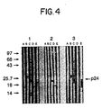
- Fig. 4 is a Western Blot showing specificity of monoclonal antibodies 31-42-19 and 31-90-25 for HIV-I p24 and their epitope recognition profiles.
- The present invention provides a novel means for the detection of HIV-I p24 in picogram quantities in body fluids of infected individuals, using two monoclonal antibodies as a probe. This highly sensitive enzyme immunoassay is unique because of the characteristics of the monoclonal antibody 31-42-19. The epitope recognized by monoclonal antibody 31-42-19 maps toward the carboxy terminus of HIV-I p24 and is not immunogenic in humans. The second monoclonal antibody employed in this assay recognizes an epitope within a highly immunogenic region of HIV-I p24, and maps toward the amino terminus of HIV-I p24. Monoclonal antibodies 31-42-19 and 31-90-25 bind synergistically to HIV-I p24. Alternatively, one can employ F(ab′)₂ or any antigen binding fragments of at least one of the monoclonal antibodies employed in the assay.
- In addition, the epitope recognized by monoclonal antibody 31-42-19 is antigenically cross-reactive with an epitope on HIV-2 p24, as shown by the ability of 31-42-19 to immunoprecipitate HIV-2 p24 from biosynthetically labeled HIV-2 infected cells.
- In addition to being employed as one of the components of the probe for HIV-I p24, the monoclonal antibody 31-90-25, when appropriately labelled, can be employed as a competitive probe against HIV-I core antibodies in serum samples for binding to recombinant-derived HIV-I p24. For example, HRPO labelled 31-90-25 can be employed in an immunoassay for antibodies to HIV as disclosed in U.S. Patent Application Serial No. 020,282, filed Feb. 27, 1987 by Dawson et al., and commonly assigned herewith.
- In another of its aspects, the present invention provides novel hybridoma cell lines, exemplified by murine-derived cell line ATCC HB 9726 and murine-derived cell line ATCC HB 9725, and novel monoclonal antibodies secreted thereby, exemplified by the above-noted monoclonal antibodies 31-42-19 and 31-90-25, respectively.
- In an especially preferred embodiment of the invention, a biological sample, presumably containing HIV-I p24, is incubated with a mixture of monoclonal antibodies 31-42-19 and 31-90-25, and a polystyrene bead coated with anti-HIV-I IgG (purified from serum of seropositive individuals for HIV-I p24 antibodies). After washing, the amount of mouse monoclonal antibodies bound, which is proportional to the amount of HIV-I p24 captured on the bead, is determined with horseradish peroxidase-labelled goat anti-mouse IgG. Alternatively, the monoclonal antibody mixture can also be coated on a solid phase to serve as capture antibodies. For example, this mixture can be used to coat a solid support of an immunoassay to detect HIV-I (HTLV-III) antigens as disclosed in U.S. Patent No. 4,748,110, issued May 31, 1988.
- In addition, the above-mentioned antibodies 31-42-19 and 31-90-25 can be employed in detection systems using fixed cells, with appropriate labelling of each monoclonal antibody. These antibodies also can be employed for purifying HIV-I p24, and the particular monoclonal 31-42-19 for purifying HIV-2 p24, by affinity chromatography.
- Biological samples which are easily tested by the method of the present invention include human and animal body fluids such as whole blood, serum, plasma, cerebrospinal fluid and lymphocyte or cell culture supernatants. Additionally, the test samples could be inactivated whole virus or partially purified native or recombinant HIV-I p24. Solid supports which can be used in immunoassays of the invention include wells of reaction trays, test tubes, polystyrene beads, strips, membranes, microparticles, and other solid matrices known to those skilled in the art. Any label capable of producing a detectable signal or an enzyme amplification system can be used in immunoassays of the invention. Representative labels include enzymatic, radioisotopic, fluorescent and chemiluminescent labels. Further, hapten/labelled anti-hapten systems such as a biotin/labelled anti-biotin system may be utilized in the inventive assays. Additionally, one can employ a labelled anti-idiotype antibody to detect the monoclonal antibodies described herein.
- In addition, reagents for the assays of the invention are ideally suited for preparation of a kit. Such a kit may comprise carrier means being compartmentalized to receive in close confinement, one or more container means such as vials, bottles, test tubes and the like. Each of the container means comprises one of the separate elements to be used in the method of this invention.
- The following illustrative examples serve to demonstrate the advantages of the invention, and relate (a) to the development and characterization of monoclonal antibodies which specifically react with certain epitopes of the HIV-I p24 core protein, and (b) to the development of a highly sensitive diagnostic test to detect HIV-I p24 in biological fluids. More specifically, Examples 1 and 2 relate to the procedures whereby hybridoma cell lines secreting monoclonal antibodies were generated. Example 3 relates to the screening, cloning and characterization of monoclonal antibodies 31-42-19 and 31-90-25. Example 4 relates to the method used for amplifying antibody yields. Example 5 relates to assays performed to determine the activity, specificity and epitope mapping of the 31-42-19 and 31-90-25 monoclonal antibodies. Example 6 relates to the development of an enzyme immunoassay (EIA) for the detection of HIV-I p24 in biological fluids using the above-mentioned monoclonal antibodies. Example 7 is a summary of alternate assay procedures covering the clinical utility of these monoclonal antibodies for AIDS diagnostics.
- In the procedure for production of hybridoma cell lines 31-42-19 and 31-90-25, BZH mice (obtained from Chella David, Department of Immunology, Mayo Clinic, Rochester, Minnesota) were initially immunized with partially purified, detergent disrupted HIV-I (HTLV-III prototype strain obtained from R.C. Gallo, National Institute of Health) and boosted with recombinant derived purified p24 just before fusion. HIV-I was partially purified from infected H9 cells by (a) membrane filtration separating HIV-I from cells, followed by (b) concentration of cell culture fluid containing HIV-I, followed by (c) collection of virus by ultracentrifugation, followed by (d) resuspension of the virus and collection by centrifugation onto a 20% sucrose pad followed by (e) sucrose density gradient banding of HIV-I at a density of approximately 1.16 and (f) ultracentrifugation of banded virus to collect and concentrate HIV-I. HIV-I was disrupted by addition of 0.5% Triton X-100, followed by vigorous sonication at 4°C. Full-length recombinant HIV-I p24 was produced in E. coli by recombinant DNA methods and purified by affinity chromatography as disclosed in U.S. Patent Application Serial No. 020,282. Briefly, a plasmid designated pB1, containing a 951 bp PvuII to Bg/II restriction fragment, was induced to produce full-length HIV-I p24, and then the recombinant HIV-I p24 was purified.
- On
day 1, mice received 10 µg disrupted HIV-I in 0.4 ml of Freund's Complete Adjuvant (Difco Laboratories) given subcutaneously (s.c.) and intraperitoneally (i.p.) in 0.1 ml portions at four different sites. The second immunization was performed 14 days later when mice received 10 µg of HIV-I in 0.3 ml of Freund's Incomplete Adjuvant, given s.c. and i.p. On day 29, mice were immunized with 10 µg HIV-I and 4 µg of S. typhimurium extract (RIBI Immunochemicals) in 0.2 ml Freund's Complete Adjuvant. On day 63, mice were immunized with 10 µg HIV-I in 0.2 ml Freund's Complete Adjuvant. Mice were bled on days 15, 36 and 70. The immune responses of the immunized mice were assessed by assaying their sera for anti-HIV-I antibody by enzyme-linked immunoassay and Western Blot. Approximately 14 months later, mice were immunized with a pre-fusion boost of recombinant HIV-I p24 antigen. - Sera from naive or immunized mice were serially diluted in dilution buffer containing 20 mM potassium phosphate, pH 7.4, 0.15 M NaCl, 20% normal goat serum, 10% fetal calf serum, 5 mM EDTA, 10 mM EGTA, 50 mM Tris buffer (pH 8.0), 0.2% Tween-20, and sodium azide as preservative. The diluted sera were reacted with 1/4" polystyrene beads directly coated with partially purified HIV-I, or, alternatively, with HIV-I bound to the bead via human anti-HIV-I antibody (purified from serum of an HIV-I seropositive individual). After 2 hours at 40°C, the beads were washed, and goat anti-mouse IgG (H+L) Horseradish Peroxidase (HRPO)-conjugated antibody (Kirkegaard & Perry Laboratories) was added. The beads were incubated at 40°C for 2 hr, washed and transferred to reaction tubes containing o-phenylenediamine:2HCl (OPD) color reagent. The reaction was carried out in the dark at room temperature for 30 minutes, and then stopped by the addition of 1 ml of 1N H₂SO₄ and the absorbance at 492/600 nm was recorded. The absorbance was directly proportional to the amount of HIV-I specific mouse antibody bound to the bead. The specificity of mouse antibody for HIV-I antigen on the bead was confirmed by the addition of human HIV-I seropositive serum (20-50 µl) in the reaction as a competitor for binding to HIV-I antigens on the bead.
-
- Approximately 500 µg of partially purified HIV-I were treated with sodium dodecyl sulfate (SDS) and 2-mercaptoethanol at 95°C, and electrophoresed in a 12% polyacrylamide-SDS gel (Laemmli et al., Nature (1970) 227:680-685). Proteins were transferred overnight from the gel to nitrocellulose by electrophoresis at 100 mamp, or transferred in 1-2 hr at 1.0 amp, in standard transfer buffer composed of 25 mM Tris (Hydroxymethyl Aminomethane), 192 mM glycine, and 2.0% methanol, pH 8.3. (Towbin et al., Proc. Natl. Acad. Sci. (1979) 73:4350-4354.) After transferring the viral proteins and blocking the nitrocellulose with 20% fetal calf serum diluted in 10 mM Tris buffer (pH 8.0) containing 0.15 M NaCl, the nitrocellulose was cut into strips (each strip containing approximately 15 µg of viral protein) which were then used to determine the presence of anti-HIV-I antibody in test sera (or other samples). Reaction mixtures consisted of a nitrocellulose strip incubated with an appropriate amount of test sample in 2.5 ml of buffer (20 mM Tris, 1 mM EDTA, 0.2M NaCl, 0.3% Triton X-100, and 2 mg/ml bovine serum albumin, pH 7.5) for 1-2 hours at room temperature. The strips were washed with buffered detergent (10 mM phosphate buffered saline (PBS), pH 7:5, containing 0.1% SDS and 0.5% Triton X-100), followed by addition of goat anti-mouse IgG antibody conjugated to HRPO. The strips were incubated for 1-2 hours at room temperature, followed by washing with buffered detergent. Finally, antibody bound to viral protein was visualized by addition of freshly prepared HRP color reagent (Biorad) (120 mg dissolved in 40 ml ice-cold methanol, then diluted into 200 ml Tris buffered saline, pH 7.8, containing 120 µl of 30% hydrogen peroxide). This assay demonstrated the presence of antibody to specific HIV-I proteins.
- Upon demonstration of specific anti-HIV-I antibody present at reasonable titers in sera of immunized mice, the mice were allowed to rest prior to a pre-fusion boost of antigen. The pre-fusion antigen boost was performed by intravenous (tail vein) injection of approximately 200 µl of purified recombinant HIV-I p24. Three days later the mice were sacrificed, and their spleens, containing anti-HIV-I antibody producing cells, were disrupted to single cells. The single cell suspensions were treated with 0.83% NH₄Cl to remove red blood cells, and then mixed with SP2/0 cells at a 10:1 (SP2/0:spleen cells) ratio. The mixed cells were centrifuged, washed once with serum-free medium, then centrifuged again. The fusogen, polyethylene glycol (PEG), was used to form hybrids of the immune donor spleen cells with the myeloma cell line SP2/0 (HPRT neg.) [Kohler and Milstein, Nature (1975) 256:494, and reviewed in Monoclonal Hybridoma Antibodies: Techniques and Applications, ed. Hurrell (CRC Press, Inc., 1982)]. Briefly, fusion of the spleen and SP2/0 cells was accomplished by exposing the pellet to 40% PEG (ATTC, MW 1300-1600) in serum-free Iscoe's Modified Dulbecco's Medium (IMDM) for two minutes. The PEG and cell suspension was diluted slowly by the addition of 20 ml of serum-free IMDM over a period of five minutes, followed by colleciton of the cells by centrifugation. The supernatant was decanted and replaced with 30 ml IMDM containing 20% fetal bovine serum (Hyclone) with HAT (hypozanthine, aminopterin, and thymidine) to select for hybridomas. Spleen cells from one nonimmune Balb/c mouse also were added as a feeder layer. The cells were plated at 0.1 ml/well in three 96 well tissue culture plates. Three days later an additional 0.1 ml of HAT media was added to each well. At weekly intervals thereafter, one-half the media was replaced with IMDM containing 20% fetal bovine serum (Hyclone) with HT (hypozanthine and thymidine), and hybrids were allowed to grow for an additional 7-14 days.
- Some of the hybrids were composed of spleen cells making antibody to HIV-I fused with SP2/0 cells. Briefly, the fusogen promotes fusion of spleen cell and SP2/0 cell membranes, forming a heterokaryon containing nuclei of both cells. Eventually, the dissimilar nuclei fuse producing a single nucleus capable of synchronous mitosis. As the fused cells divide, the hybrid stablizes by losing chromosomes of each nucleus. The fused cells are plated into multiple 96 well plates at 10⁵ to 10⁶ cells per well. Hybrid cells formed from SP2/0:spleen cell fusions are selectively propagated by culturing in HAT medium. All unfused SP2/0 or SP2/0:SP2/0 fused cells are prevented from growing by aminopterin, and unfused spleen cells or spleen:spleen fused cells die off in culture. Only SP2/0:spleen hybrids will grow in the HAT selection medium.
- After 10-14 days, culture fluids from wells containing hybridoma cell growth were screened for antibody to HIV-I p24 using EIA and Western Blot procedures described in Example 1. In addition, an EIA using HIV-I p24 produced in E. coli by recombinant DNA methods (rp24) was employed for screening. Briefly, polystyrene beads coated with human IgG (purified from anti-p24 seropositive individuals) were reacted with crude E. coli extracts of full-length recombinant-derived HIV-I p24. The resultant beads, bearing rp24 bound via human anti HIV-I antibody, were reacted with hybridoma culture fluids for 4 hr at 40°C or overnight at room temperature, washed and reacted with HRPO-labelled goat anti-mouse IgG (Fc) for 2 hr at 40°C. After washing again, beads were transferred to reaction tubes for the addition of OPD substrate as described in Example 1.
- From a single fusion, #31, twenty-one wells were identified as showing hybridoma cell growth and colony formation. Culture fluid from each well was screened for antibody to HIV-I p24 by EIA and Western Blot. Of 21 wells, 15 were identified as hybrids containing antibody to HIV-I p24. Cells from each of these 15 wells were expanded in multiple 24 well plates, and culture fluid was again assayed for the presence of anti-HIV-I p24 antibody, and confirmed by competition EIA with human anti-HIV-I serum as described in Example 1. All 15 were positive for anti-HIV-I p24 antibody and each hybrid was further expanded in T25 flasks for cloning by limiting dilution. Each expanded hybrid was plated into 96 well microtiter plates at dilutions of 10⁵ to 10⁶ and allowed to grow from 10-21 days. Among thirteen hybrids having clones identified as producing antibodies to HIV-I p24 were clones #19 from hybrid #42 of fusion #31, named accordingly 31-42-19, and #25 from hybrid #90 of fusion #31, named accordingly 31-90-25. The clones were obtained by limiting dilution using the guidelines outlined by J.W. Goding in Monoclonal Antibodies: Principles and Practice (Academic Press, N.Y., 1983).
- The isotype of monoclonal antibody 31-42-19 was determined to be IgG1 and that of monoclonal antibody 31-90-25 was determined to be IgG2a. An EIA isotyping procedure employed a microtiter plate coated with goat anti-mouse immunoglobulin, which was incubated with culture fluid of the clone to capture the secreted mouse antibody. After 2 hours, the plate was washed and rabbit anti-mouse isotype was applied for an additional 2 hours. The plate was washed again, and HRPO-conjugated goat anti-rabbit IgG was applied for 1 hr. The excess conjugate was removed by washing, then OPD substrate was added. The amount of rabbit anti-mouse isotype bound to mouse immunoglobulin was proportional to the absorbance measured at 492 nm. Further characterization of both monoclonals was performed with antibodies from mouse ascites.
- In order to obtain greater amounts of monoclonal antibodies, ten to 20 million cloned cells of the desired antibody (31-42-19 or 31-90-25) were inoculated into a Balb/c mouse previously treated intraperitoneally with 0.5 ml pristane (2,6,10,14-tetramethylpentadecane) [method outlined in Hurrell et al., supra]. Pristane treatment enhances growth of mouse myeloma hybrids within the peritoneum of the mouse, and the ascites fluids which form are rich in the monoclonal antibody secreted by the hybrid cells. After formation of monoclonal antibody enriched ascites (approximately 7 days) the mice were sacrificed and the ascites was withdrawn from the peritoneum, clarified by centrifugation and stored at -20°C. Other characterization procedures (described herein) were performed with culture fluid, clarified ascites or purified antibodies from ascites, using protein A sepharose (Hurrell et al., supra). The monoclonal antibody in ascites was titered by EIA (Example 5).
- The following experiments were carried out for both monoclonal antibodies 31-42-19 and 31-90-25 to assess their activity, specificity and epitope recognition profile.
- Assays described in Examples 1 and 3 were used to determine the titer of monoclonal antibodies. Briefly, clarified ascites fluids or protein A-purified IgG were reacted in serial dilutions against polystyrene beads coated with recombinant-derived HIV-I p24 bound to beads via human IgG (purified from sera of HIV-I seropositive individuals). Representative data, as illustrated in Table 1, shows the specific reactivity of the monoclonal antibodies when compared to the negative control. In addition, two assays were performed in which purified IgG from an established HIV-I seropositive individual was used as a competitive inhibitor to determine the human immunogenicity of the epitopes recognized by 31-42-19 and 31-90-25. In one procedure, human anti-HIV-I IgG was serially diluted in dilution buffer (described in Example 1) containing a constant amount of either 31-42-19 or 31-90-25. In the other procedure, human anti-HIV-I IgG was added at a constant concentration (1 µg/ml) to serial dilutions of each of the monoclonals. The human IgG served as a competitive inhibitor for the binding of mouse monoclonal antibody to HIV-I rp24 on beads. Competition of anti-HIV-I IgG with each monoclonal was indicated by a reduction in the amount of monoclonal antibody bound to the bead when compared to the control which contained no competing human IgG. Representative data, as illustrated in Tables 2 and 3, shows that 31-42-19 recognizes an epitope not readily immunogenic in humans, while 31-90-25 recognizes an epitope within a highly immunogenic region of HIV-I p24. Table 3 data shows, however, that gross excesses of human anti-HIV-I IgG can block the binding of 31-42-19 to some extent.
Table 1. Activity of Monoclonal Antibodies 31-42-19 and 31-90-25 Against HIV-I rp24. Antibody Concentration(µg/ml) Absorbance (492 nm) 31-42-19 31-90-25 1.0 1.436 1.767 0.5 1.305 1.771 0.25 1.250 1.717 0.125 1.215 1.731 0.0625 1.063 1.772 0.0312 0.848 1.719 0.0156 0.708 1.415 0.0078 0.484 1.131 Neg. Controla 0.028 0.044 aNegative control contained only dilution buffer and served as reagent blank. Table 2 Competition Profile with Serial Dilution of Human Anti-HIV-I IgG. Human IgG Absorbance (492 nm) 31-42-19 a 31-90-25 b Undiluted 2.300 0.116 Dilution # 12.320 0.181 Dilution # 22.216 0.181 Dilution # 32.500 0.264 Dilution # 42.134 0.377 Dilution # 52.043 0.595 Dilution # 62.305 0.822 Dilution #7 2.301 1.064 Dilution #8 2.165 1.467 Control 2.282 1.401 a31-42-19 added at 0.1 µg/ml, anti-HIV-I human IgG added at 1.0 µg/ml in the first well and serially diluted in the next eight wells. b31-90-25 added at 2.0 µg/ml, anti-HIV-I human IgG added at 20 µg/ml in the first well and serially diluted in the next eight wells. Table 3 Competition Profile with Serial Dilution of 31-42-19 and 31-90-25. Antibody Concentration(µg/ml) Absorbance (492 nm) 31-42-19 31-90-25 control +anti-HIV-I control +anti-HIV-I 1.0 1.436 1.412 1.767 0.612 0.5 1.305 1.315 1.771 0.461 0.25 1.250 1.223 1.717 0.383 0.125 1.215 1.069 1.731 0.255 0.0625 1.063 1.034 1.772 0.152 0.0312 0.848 0.663 1.799 0.108 0.0156 0.708 0.448 1.415 0.056 0.0078 0.484 0.353 1.131 0.053 - The specificity of monoclonal antibodies 31-42-19 and 31-90-25 was confirmed by immunoprecipitation of [³⁵S] methionine/[³⁵S] cysteine biosynthetically-labelled viral p55 (gag precursor protein) from HIV-I infected H9 cell lysates. Immunoprecipitation assays for viral proteins have been described previously (Devare et al., Proc. Natl. Acad. Sci., U.S.A. (1986) 83:5718-5722). Cell lines employed for these studies were uninfected H9 cells, HIV-I infected H9 cells or a cloned cell line S5G7 derived from infected H9 cells, which produces large quantities of virus. Cells were harvested from culture, washed once with RPMI 1640 deficient in methionine and cysteine (Gibco Laboratories), then suspended at 1-2.5 X 10⁶ cells/ml in the same medium. Washed cells were incubated for 30-45 minutes at 37°C in 6% CO₂, followed by the addition of 50-100 µCi each of [³⁵S] methionine and [³⁵S] cysteine (Amersham) to the medium. Cells were radiolabelled at 37°C for 4-8 hours, harvested by centrifugation and lysed in PBS, pH 7.4, containing 1 mM PMSF, aprotinin (100 kallikrein inactivation units per ml of buffer), 1.0% Triton X-100, 0.1% SDS and 0.5% sodium deoxycholate (all reagents from Sigma). The lysate was clarified by centrifugation at 100,000 x g for 40 minutes and stored at -70°C.
- Immunoprecipitation was performed by incubating 100 µl aliquots of cell lysates with 50 µg purified monoclonal antibody (1.0 mg/ml stock), 100 µl tissue culture supernatant or 3 µl serum for 30-60 minutes at 4°C. Antigen-antibody complexes were recovered by addition of 200 µl of preswollen protein A-Sepharose (Pharmacia) previously washed in lysis buffer containing 1.0 mg/ml bovine serum albumin (IgG binding capacity of 50-200 µg/200 µl protein A). The reaction mixture was shaken vigorously at 4°C for 1 hour, followed by 3 washes of Protein A-Sepharose using lysis buffer. Protein A-Sepharose was then collected by centrifugation and immune complexes were dissociated by heating at 95°C in SDS gel sample buffer containing 2-mercaptoethanol (Laemmli et al., supra). The sample was subjected to SDS-10% polyacrylamide gel electrophoresis. The gel was incubated for 30 minutes in Enhance (Dupont), dried and exposed to X-ray film for autoradiography of the immunoprecipitated radiolabelled proteins. The results are illustrated in Fig. 1 where, in Panel A, labelled cell lysates from uninfected H9 cells, and, in Panel B, lysates from HIV-I infected H9 cells (S5G7 cell line) were mixed with (1) normal mouse serum, (2) normal human serum, (3) HIV-I immune human serum, (4) monoclonal antibody 31-42-19 tissue culture supernatant and (5) monoclonal antibody 31-90-25 tissue culture supernatant. The results indicated that both monoclonal antibodies immunoprecipitated gag precursor p55 from HIV-I infected cells. No detectable cross reactivity was observed with any other radiolabelled viral or cellular protein. Thus, both monoclonal antibodies bound specifically to HIV-I p55.
- In addition, each monoclonal antibody also was reacted with labelled cell lysates of HIV-I infected HUT 78 cell line (Panel A), HIV-2 infected HUT 78 cell lines (Panel B), and uninfected HUT-78 cell lines (Panel C). Results are illustrated in Fig. 2, wherein each of these cell lysates was reacted with (1) normal mouse serum, (2) normal human serum, (3) serum from an HIV-I seropositive individiual, (4) serum from an HIV-2 seropositive individual, (5) monoclonal antibody 31-90-25 and (6) monoclonal antibody 31-42-19. Results indicated that monoclonal antibody 31-42-19 specifically immunoprcipitates HIV-2 p24. No cross reactivity with HIV-2 p24 was observed with monoclonal antibody 31-90-25, or with normal mouse serum, which indicates that it is a unique characteristic of monoclonal antibody 31-42-19 that allows it to cross react with an epitope on HIV-2 p24.
- Western blots (as described in Example 1) of purified monoclonal antibodies 31-42-19 and 31-90-25 were performed using partially purified HIV-I virus, and, recombinant full-length p24 and p24 deletion clones as antigens to further establish specificity and for epitope mapping, respectively. Partially purified HIV-I or crude cell extract of E. coli-derived recombinant HIV-I p24 clones (see Fig. 3 which shows a schematic representation of clones used) was electrophoretically transferred to each nitrocellulose strip after electrophoresis in a 12% polyacrylamide gel under reducing conditions. As shown in Fig. 4, Panel A was a control of E. coli (untransformed) cell lysates; Panel B used full-length recombinant clone pB1 as antigen; Panel C, HIV-I viral lysate; Panel D, recombinant deletion clone pRA10 and Panel E, recombinant deletion clone pRA13. The results confirmed that both monoclonal antibodies specifically react with HIV-I p24 when tested against lysates of partially purified virus. Further, the reaction of each monoclonal antibody with recombinant deletion clones of HIV-I p24 indicated that 31-42-19 maps toward the carboxy half of HIV-I p24 while 31-90-25 maps toward the amino terminus of HIV-I p24. These results also were confirmed by comparing serological data and competitive RIA (radioimmunoassays) with other members of HIV-I p24 monoclonal panel (data not shown here).
- Monoclonal antibodies 31-42-19 and 31-90-25 were employed in an immunocytochemical assay against uninfected and HIV-I infected H9 cells in order to further demonstrate their specificity for HIV-I. Briefly, uninfected or HIV-I infected H9 cells were fixed to microscope slides using 100% acetone for 10 min., then air dried, and stored at -20°C. For staining, the cells were rehydrated in PBS for 10 min., followed by addition of 100 µl of 2% normal goat serum in PBS containing 100 µg/ml normal human IgG for 10 min. Excess reagent was removed, and 100 µl of 1 µg/ml 31-42-19 IgG or 31-90-25 IgG were added for 30 min. to the slides maintained in a humidified chamber. The slides were washed in PBS, followed by addition of 100 µl of a second antibody, goat anti-mouse IgG (Pel Freez) at a 1:400 dilution, for 30 min. The slides were washed with PBS, followed by addition of 100 µl of peroxidase anti-peroxidase complex (Clonal PAP, Sternberger and Meyer) diluted 1:300 in PBS containing 2% normal goat serum and 100 µg/ml normal human IgG. The slides wre incubated (with the Clonal PAP) in a humidified chamber. After washing in PBS, color development was initiated by the addition of 100 µl of the chromagen (0.63 mg/ml tetra hydrochloride diamino-benzidine in 0.06% hydrogen peroxide in PBS). Color development was stopped after 6 min. by washing the slides in tap water. The slides were dehydrated and mounted for examination.
- Results indicated that the monoclonal antibodies did not cross react with any normal cellular constituents. Specific staining was only detected with HIV-I infected H9 cells which was characteristic of HIV-I anti-p24 monoclonals.
- A panel of anti-HIV-I p24 monoclonal antibodies was tested in a three step assay configuration. Briefly, 200 µl HIV-I antigen sample (1.0 ng/ml partially purified HIV-I viral lysate, diluted in normal human plasma [NHP]) was incubated with a 1/4" polystyrene bead coated previously with anti-HIV-I human IgG overnight at room temperature. After washing, 200 µl of either a single monoclonal antibody solution or a monoclonal antibody mixture solution (each monoclonal in equal proportion), at a final concentration of 5 µg/ml diluted in NHP, was added. The reaction mixture was incubated at 40°C for 4 hr. After washing, the amount of each monoclonal bound to the bead was detected using HRPO labelled goat anti-mouse IgG. The detection steps were carried out as described in Example 1. Absorbance at 492 nm was directly proportional to the amount of mouse monoclonal antibodies bound to the HIV-I p24 captured on the bead. Data in Tables 4 and 5 demonstrate that 31-42-19 drives the synergistic binding with 31-90-25 because any monoclonal mapping in the region of 31-90-25 bound synergistically to HIV-I p24 in the presence of 31-42-19. Monoclonal antibodies 31-7-20, 31-32-9, 31-77-8, 31-89-8, 32-51-16 and 31-90-25 all recognize the same epitope on HIV-I p24. Therefore, 31-42-19 is the key monoclonal antibody which binds synergistically with 31-90-25, resulting in optimal binding between human anti-HIV-I IgG on the bead, HIV-I p24 and the monoclonals.
Table 4 Synergy Between Monoclonal Antibodies to HIV-I p24 Tested as Probes for HIV-I p24 Antigen Assay. Probe Net O.D. (492 nm) 31-42-19 alone 0.498 31-90-25 alone 0.468 32-51-16 alonea 0.494 31-74-22 aloneb 0.095 31-79-18 alonec 0.034 31-42-19 + 31-90-25 1.211 31-42-19 + 32-51-16 1.189 32-51-16 + 31-90-25 0.484 31-42-19 + 31-90-25 + 32-51-16 1.241 aMonoclonal antibody 32-51-16 recognized the same epitope as 31-90-25 but was generated from a different fusion experiment. bMonoclonal antibody 31-74-22 recognized an epitope near the amino terminus of HIV-I p24, but different from the epitope of 31-90-25. cMonoclonal antibody 31-79-18 recognized an epitope near the carboxy terminus of HIV-I p24. Table 5 Synergistic Characteristics of 31-42-19 and Epitope Specificity of Synergy Between 31-42-19 and a Group of Monoclonals to HIV-I p24. Monoclonal Mixture Absorbance (492 nm) 31-42-19 + 31-7-20 1.219 31-42-19 + 31-32-9 1.156 31-42-19 + 31-77-8 1.128 31-42-19 + 31-89-8 1.354 31-42-19 + 32-51-16 1.190 31-42-19 + 31-90-25 1.355 31-42-19 + 31-79-18 0.658 31-90-25 + 31-77-8 0.452 - During further investigations, the monoclonals were employed in the development of a two step (overnight/2 hr) assay for the detection of HIV-I p24 (detailed herein). A sensitivity panel of p24 enriched antigen was prepared with partially purified HIV-I viral lysate diluted in NHP in the concentration range from 500 to 31.25 pg protein/ml. Each panel member was assayed using each of the below-listed monoclonal antibody mixtures as a probe. An absorbance value which was 0.05 0.D. units greater than that of the negative control was considered the cut-off value for the assay, and samples showing higher absorbance values than the cut-off were considered positive. The sensitivity, defined as picograms of HIV-I p24 per ml that gave an absorbance value of 0.05 0.D. units greater than that of the negative control, was determined by linear regression. Monoclonal antibody 31-90-25 was selected from a group of monoclonals mapping to the same general region of HIV-I p24 because it produced the most sensitive results in the two step assay configuration, as demonstrated in Table 6.
Table 6 Comparison of the Sensitivity of Two Step HIV-I p24 Antigen Assay, replacing 31-90-25 with Other Monoclonals which Map to Same Epitope. Monoclonal Mixture Sensitivity in pg/ml antigen 31-42-19 + 31-90-25 23.9 31-42-19 + 31-7-20 126.0 31-42-19 + 31-32-9 73.0 31-42-19 + 31-77-8 97.0 31-42-19 + 32-51-16 62.0 - Experimental data detailed herein describe the preferred assay configurations for the detection of HIV-I p24 in biological fluids. Experimental conditions of incubation time and temperature can be varied according to the requirements of speed of the assay and sensitivity.
- Monoclonal antibodies 31-42-19 and 31-90-25 used in the assay were purified from either tissue culture supernatants or mouse ascites fluids using protein A-Sepharose. Protein determinations of purified IgG was done by measuring absorbance at 280 nm.
- A mixture of monoclonal antibodies was prepared as a stock solution containing 25 µg IgG/ml with a 31-42-19 to 31-90-25 ratio of 4:6, in a diluent of 20mM phosphate buffer, pH 7.4, 0.15M NaCl, 25% NHP (v/v), 1% bovine serum albumin (w/v) and 0.25% Triton X-100 (v/v). When 50 µl of this solution was added to 200 µl of assay sample, it gave a final concentration of monoclonal mix at 5 µg/ml in the assay volume. The ratio of both monoclonal antibodies and the IgG concentration were found to be satisfactory under the assay conditions used, although variations in the mixture can be made to meet lot to lot variations of the monoclonal antibody preparations.
- For the assay procedure, briefly, 50 µl of monoclonal mix was added to reaction tray wells followed by 200 µl of either (a) a sample presumably containing HIV-I p24, (b) a sensitivity panel member with known amounts of either partially purified HIV-I virus or native p24 diluted in NHP as positive control or (c) plasma from an established uninfected individual as negative control. A 1/4" polystyrene bead previously coated with anti-HIV-I human IgG was added to each reaction well. The reaction mixtures were incubated at room temperature overnight (12-18 hr). After washing beads with water, 200 µl of conjugate solution were added to each reaction well. The conjugate solution was prepared by diluting HRPO labelled goat anti-mouse (Y) IgG (Kirkegaard & Perry) or HRPO labelled goat anti-mouse (Fc) IgG (Jackson Immunochemicals) in a diluent containing 20mM phosphate buffer, pH 7.4, 0.15M NaCl, 10% fetal calf serum (v/v), 15% normal goat serum (v/v), 25% NHP (v/v), 5% normal rabbit serum (v/v), 0.025% benzyl alcohol (v/v), 0.5% Triton X-100 (v/v) and 20mM HEPES (N-2-Hydroxyethylpiperazine-N′-2-ethane sulphonic acid). The reaction mixtures were incubated at 40°C for 2 hr in a waterbath. After washing, the beads were transferred to reaction tubes and the addition of OPD carried out as described in Example 1.
- Additionally, shorter assay incubation times are possible by incubating the antigen sample, monoclonal mix and beads at 40°C in a waterbath or in a dynamic incubator for 4-6 hr, and incubating the conjugate solution for 1 hr at 40°C. Table 7 shows that both Monoclonal assay procedures have improved sensitivity when compared to the current Polyclonal HIV-I antigen assay.

- Alternatively, monoclonal antibodies 31-42-19 and 31-90-25 can be successfully coated on 1/4" polystyrene beads for use as capture antibodies for HIV-I p24. Briefly, the monoclonal antibody mixture was coated at an appropriate concentration on polystyrene beads. Additional binding sites were overcoated with bovine serum albumin. In an assay procedure, the antigen sample containing HIV-I p24 was incubated with the coated beads. After appropriate incubation, beads were washed and HIV-I p24 bound was detects using a suitable probe. An especially preferred probe used comprised F(ab′)₂ fragments prepared from purified anti-HIV-I rabbit IgG, as disclosed in an U.S. patent application, entitled "Immunoassay for
HIV 1 Antigens Using F(ab′)₂ Fragments as Probe," filed by J. Stewart et al. concurrently with this application. - Monoclonal antibody 31-42-19 exhibited similar synergistic characteristics when either used as a probe in solution or as a capture antibody on the bead in combination with monoclonal antibody 31-90-25.
- The assay described in section B.1 of this example was used to detect purified HIV-2 p24. Data indicated that the present assay configuration was able to detect 800 pg/ml HIV-2 p24 using anti-HIV-I human IgG as capture antibody on the bead. Increased sensitivity of the assay can be acheived by using HIV-2 specific capture antibodies. In addition, 31-42-19 monoclonal antibody used alone as a probe at 5 µg/ml assay concentration gave a sensitivity equivalent to that of the monoclonal mixture. These results were expected since 31-90-25 does not cross react with HIV-2 p24.
- The clinical utility of the monoclonal based HIV-I antigen assay was assessed by comparing it to the Abbott HIV-I antigen assay of established clinical efficacy (Paul et al., supra). Approximately 485 clinical samples from ARC, AIDS or high risk, asymptomatic patients were received from Rush Presbyterian St. Luke's Medical Center, Chicago, Illinois, and tested over a period of 1 year. Each of the samples was tested with the Abbott HIV-I antigen assay and the ON RT/2 hr 40°C Monoclonal assay. An absorbance value which was 0.05 0.D. units greater than that of the negative control was considered the cut-off value for both assays, and samples showing higher absorbance values than the cut-off were considered positive. All positive samples were confirmed by the Abbott HIV-I neutralization assay procedure. Briefly, HIV-I antigen positive samples were incubated with an excess of serum or purified IgG from an established HIV-I seropositive individual with high HIV-I anti-p24 titers. After preincubation at room temperature, the residual (not neutralized) p24 was detected using either the Abbott HIV-I antigen assay or the Monoclonal assay. True HIV-I positive samples showed drastic reductions in O.D. units at 492 nm. Any sample that showed greater than 50% neutralization in the presence of anti-HIV-I antibodies compared to that of the control, which contained same amount of normal human IgG or plasma, was considered confirmed positive. Non-neutralizable positive samples were considered negative for HIV-I antigen. Data are summarized in Table 8.
Table 8 Comparative Profile of Clinical Samples. Monoclonal HIV-I Ag Assay Pos Neg Abbott HIV-I Ag Assay Pos 165 7a Neg 31b 282 aMonoclonal assay missed 7 samples, of which 4 were serial bleeds from two patients. bAbbott antigen assay missed 31 samples, of which 22 samples were serial bleeds--2 samples from patient A, 3 samples from patient B, 4 samples from patient C, 2 samples from patient D and 11 samples from patient E. - As shown in Table 8, the majority of samples are in good agreement with the Abbott HIV-I antigen assay. Those samples which were discrepant seemed to follow a pattern--(a) for the majority (4 of 7) of positive samples missed by the Monoclonal assay, their absorbance values were very close to the cut-off value of the Abbott HIV-I antigen assay, (b) the Monoclonal assay (with the neutralization assay) confirmed the samples missed by the Abbott HIV-I antigen assay (28 of 31, 3 samples could not be confirmed due to lack of sample volume) partly due to better sensitivity of the Monoclonal assay (see Table 7), (c) there seems to be a patient-specific pattern, i.e. all samples of that patient were missed, when either the Monoclonal assay or the Abbott HIV-I antigen assay missed a sample from a particular patient.
- In addition, several established seroconversion panels were tested. These panels were also tested by several other commercially available or in-house tests for HIV-I antibodies and antigen. These comparisons show that the Monoclonal antigen assay is highly effective in detecting seroconversion in longitudinal studies. For example, as shown in Table 9, using an in-house seroconversion panel, both the Abbott HIV-I antigen assay and the Monoclonal assay detected the seroconversion of an AIDS-infected individual at the same time point that antibodies to HIV-I env proteins were detected by an HIV-I antibody immunoassay, and before HIV-I core antibodies were detected. Similarly, in another seroconversion panel purchased from Boston Biomedica, the Monoclonal assay detected the presence of HIV-I antigen at the same time point antigen was also detected by the ENI antigen assay (EN antigen assay results were supplied by Boston Biomedica).
Table 9 In-house Seroconversion Panel. Bleed No. Anti-HIV-I Ab HIV-I Antigen Core Env Abbott Ag Monoclonal 1 - - - - 2 - - - - 3 - - - - 4 - - - - 5 - - - - 6 - - - - 7 - - - - 8 - - - - 9 - - - - 10 - - - - 11 - + + + 12 + + - - 13 + + - - 14 + + - - 15 + + - - 16 + + - - 17 + + - - 18 + + - - 19 + + - - - As described in Example 6, the assay configuration designed specifically for capture of HIV-I detects as low as 800 pg/ml of HIV-2 p24 due to a high degree of cross reactivity observed between the core antigens of HIV-I and HIV-2. Based on these findings, monoclonal antibody 31-42-19 can be successfully used in the design of a highly sensitive HIV-2 p24 antigen assay. In addition, an antibody can be generated against native protein, synthetic peptide or recombinant protein including both HIV-I and HIV-2 p24 epitopes. Such an antibody either on solid phase or in solution as a probe, with monoclonal antibody 31-42-19 as probe or on solid phase, respectively, can also be used to design an immunoassay that would detect both HIV-I and HIV-2 p24 with equal efficiency. Thus, monoclonal antibody 31-42-19 can be employed in assays that detect both HIV-I and HIV-2 or assays that discriminate HIV-I and HIV-2 when used in appropriate enzyme immunoassay configurations.
-
- In another embodiment of the invention, monoclonal antibody 31-90-25 can be employed as a labelled probe for the detection of anti-HIV-I core antibodies in biological samples. Labelling can be performed using horseradish peroxidase. Briefly, the HRPO was activated by addition of 1.6 ml of 5 mg/ml HRPO (Sigma) to 1.6 ml of NaIO₄ at 17 mg/ml. The mixture was incubated 30 min in the dark followed by G25 Sephadex gel filtration. Fractions containing activated HRPO were pooled and protein concentration was adjusted to approximately to 0.6 mg/ml. One ml of 1 mg/ml 31-90-25 IgG previously dialysed into 50 mM Na₂C0₃ buffer, pH9.5, was added to 1.1 ml of activated HRPO at 30°C and incubated for 2 hr in the dark, followed by a 30 minute incubation at 4°C. The theoretical coupling ratio of HRPO:IgG in the reaction mixture was 4:1. Freshly prepared Na₂BH₄ (0.08 ml of 5 mg/ml solution in cold 10 mM Na₂CO₃, pH 9.5) was added to the mixture and left overnight at 4°C, followed by addition of 0.08 ml acetone which was allowed to react for 30 min at 30°C. Finally, 7.7 ml of 1.43% bovine serum albumin in PBS (pH 7.2) were added to yield a final IgG concentration of 100 µg/ml; aliquots were stored at -70°C. An appropriate dilution of this conjugated antibody was made in a suitable diluent and employed in the assay described below.
- First, a test sample, presumably containing anti-HIV-I p24, was mixed with partially purified recombinant p24 (see Example 1), and then incubated. Next, the mixture was incubated with a bead, previously coated with anti-HIV-I p24 antibody purified from sera of HIV-I seropositive individuals, and [HRPO]31-90-25 simultaneously. The absence of bound recombinant p24 resulted in a reduction of O.D. values at 492 nm and indicated the presence of anti-HIV-I p24 antibodies in the test sample.
- To determine specificity of the assay, a population of blood donors having no known risk factors for exposure to HIV-I were tested in the assay. The data were compared with that of an assay, which employs a polyclonal human anti-HIV-I IgG as a probe, of established clinical efficacy (ENVACOR assay, Abbott Laboratories, North Chicago, Illinois). Data from both assays were in complete agreement. Out of 160 samples tested by both assays, all of them were negative for antibodies to HIV-I p24 (as well as negative for antibodies to HIV-I gp41 tested by the ENVACOR assay).
- In order to determine the sensitivity of the assay, eleven samples from persons known to have antibodies to HIV-I by Western Blot or other HIV-I detection assays, were serially diluted in NHP. Samples from each of these dilutions were tested by the above-mentioned assays and results were compared. The results, described in Table 10, clearly indicate that the monoclonal probe assay showed significantly higher endpoint titers than the ENVACOR assay. This clearly illustrates superior sensitivity of the monoclonal assay.
Table 10 Sensitivity of Monoclonal Probe Immunoassay for Detection of HIV-I anti-p24 Antibodies. Patient Diagnosis Endpoint Titers a ENVACOR Monoclonal 1 AIDS negb neg 2 AIDS NDc neg 3 AIDS ND ND 4 AIDS ND 32 5 AIDS 64 1024 6 ARC ND 2 7 ARC ND 2 8 ARC ND 4096 9 ASYM 4096 8192 10 ASYM 65,536 1,048,576 11 ASYM ND 512 aReciprocal of endpoint dilutions are given. End point dilutions defined as highest dilution of patient sera exhibiting positive results for antibodies to HIV-I p24. bneg--negative for antibodies to HIV-I p24 cND--not determinable (antibody titers may be too low to be detected by the assay) - The foregoing disclosure and experiments described herein set forth preferred assay configurations, but this is for illustrative purposes only and is not intended in any way to limit the scope of this invention. In addition, one skilled in the art will appreciate that the above-described monoclonal antibodies can be used for the detection of native HIV-I p24 or recombinant-derived HIV-I p24 in various assay formats under varied conditions of incubation, time and temperature to achieve optimum sensitivity and speed of the assay. Immunoassays within the scope of this invention include any assay format that is dependent upon the formation of an antigen/antibody complex, either in solution or on a solid phase, employing these antibodies individually or in a mixture.
Claims (23)
a. coating a solid support with human anti-HIV-I IgG;
b. contacting the coated support with the sample, incubating and washing;
c. contacting the bead with a monoclonal antibody mixture, incubating and washing;
d. contacting the support with an anti-mouse antibody or a fragment thereof conjugated to horseradish peroxidase, incubating and washing;
e. contacting the support with an o-phenylenediamine-hydrogen peroxide solution; and
f. measuring the absorbance of the color product formed at 492 nm to determine the presence of HIV-I p24 in the sample;
wherein said monoclonal antibody mixture comprises the monoclonal antibodies designated 31-42-19 and 31-90-25.
Applications Claiming Priority (2)
| Application Number | Priority Date | Filing Date | Title |
|---|---|---|---|
| US204798 | 1988-06-10 | ||
| US07/204,798 US5173399A (en) | 1988-06-10 | 1988-06-10 | Mouse monoclonal antibodies to hiv-1p24 and their use in diagnostic tests |
Publications (3)
| Publication Number | Publication Date |
|---|---|
| EP0345461A2 true EP0345461A2 (en) | 1989-12-13 |
| EP0345461A3 EP0345461A3 (en) | 1990-06-27 |
| EP0345461B1 EP0345461B1 (en) | 1994-09-28 |
Family
ID=22759480
Family Applications (1)
| Application Number | Title | Priority Date | Filing Date |
|---|---|---|---|
| EP89108001A Expired - Lifetime EP0345461B1 (en) | 1988-06-10 | 1989-05-03 | Mouse monoclonal antibodies to HIV-1P24 and their use in diagnostic tests |
Country Status (6)
| Country | Link |
|---|---|
| US (1) | US5173399A (en) |
| EP (1) | EP0345461B1 (en) |
| JP (1) | JP3102643B2 (en) |
| CA (1) | CA1340919C (en) |
| DE (1) | DE68918497T2 (en) |
| ES (1) | ES2065935T3 (en) |
Cited By (8)
| Publication number | Priority date | Publication date | Assignee | Title |
|---|---|---|---|---|
| EP0345462A2 (en) * | 1988-06-10 | 1989-12-13 | Abbott Laboratories | Immunoassay for HIV-1 antigens using F(AB')2 fragments as probe |
| EP0472659A1 (en) * | 1989-05-15 | 1992-03-04 | Akzo Nobel N.V. | T-lymphotropic retrovirus monoclonal antibodies |
| EP0519866A1 (en) * | 1991-06-18 | 1992-12-23 | Ciba-Geigy Ag | Novel anti-HIV antibodies |
| FR2777285A1 (en) * | 1998-04-10 | 1999-10-15 | Bio Merieux | PEPTIDE LIGAND HAVING SPECIFIC AFFINITY TO PROTEIN P24 OF RETROVIRUS HIV-1 |
| EP1072888A2 (en) * | 1999-07-28 | 2001-01-31 | Fujirebio Kabushiki Kaisha | Immunoassay method and reagent for HIV-1p24 antigen |
| WO2002064615A2 (en) * | 2000-12-06 | 2002-08-22 | Abbott Laboratories | Monoclonal antibodies to human immunodeficiency virus and uses thereof |
| WO2022013730A1 (en) * | 2020-07-13 | 2022-01-20 | Grifols Diagnostic Solutions Inc. | Anti-human immunodeficiency virus-1 antibodies and methods for uses thereof |
| WO2023088444A1 (en) * | 2021-11-20 | 2023-05-25 | 东莞市朋志生物科技有限公司 | Antibody against hiv-1 p24, and preparation method therefor and use thereof |
Families Citing this family (22)
| Publication number | Priority date | Publication date | Assignee | Title |
|---|---|---|---|---|
| US6319665B1 (en) | 1994-06-07 | 2001-11-20 | Inverness Medical Technology, Inc. | Home test kit and method with telephone verification of results |
| AU3541495A (en) * | 1994-09-01 | 1996-03-22 | Wisconsin Alumni Research Foundation | Therapeutic remodeling in aids |
| US6027731A (en) * | 1998-11-17 | 2000-02-22 | Wisconsin Alumni Research Foundation | Pertussis toxin induced lymphocytosis |
| US6828157B1 (en) | 1999-05-04 | 2004-12-07 | Dan A. Pankowsky | Products and methods for single parameter and multiparameter phenotyping of cells |
| US6682940B2 (en) | 1999-05-04 | 2004-01-27 | Dan A. Pankowsky | Products and methods for single parameter and multiparameter phenotyping of cells |
| WO2006126287A1 (en) * | 2005-05-27 | 2006-11-30 | Masami Moriyama | Method for detecting hiv-1 and kit to be used for the method |
| WO2006126286A1 (en) * | 2005-05-27 | 2006-11-30 | Masami Moriyama | Hybridoma producing antibody that recognizes hiv-1 |
| WO2008059553A1 (en) * | 2006-11-13 | 2008-05-22 | Masami Moriyama | Method for detecting hiv-1 without being affected by anti-mouse protein antibody existing in human and kit to be used for the same |
| JP2008170700A (en) * | 2007-01-11 | 2008-07-24 | Yamaha Corp | Keyboard-type percussion instrument |
| EP2239575A4 (en) * | 2007-12-28 | 2013-01-02 | Sysmex Corp | Reagent for detecting hiv-1 antigen and detection method |
| CA2736816C (en) * | 2008-09-12 | 2018-05-22 | Isis Innovation Limited | Pd-1 specific antibodies and uses thereof |
| CA2793959C (en) | 2010-03-25 | 2019-06-04 | Oregon Health & Science University | Cmv glycoproteins and recombinant vectors |
| CA2832109C (en) | 2011-06-10 | 2021-07-06 | Oregon Health & Science University | Cmv glycoproteins and recombinant vectors |
| EP2568289A3 (en) | 2011-09-12 | 2013-04-03 | International AIDS Vaccine Initiative | Immunoselection of recombinant vesicular stomatitis virus expressing hiv-1 proteins by broadly neutralizing antibodies |
| US9402894B2 (en) | 2011-10-27 | 2016-08-02 | International Aids Vaccine Initiative | Viral particles derived from an enveloped virus |
| ES2631608T3 (en) | 2012-06-27 | 2017-09-01 | International Aids Vaccine Initiative | Env-glycoprotein variant of HIV-1 |
| FR2994482B1 (en) * | 2012-08-13 | 2014-08-22 | Repropharm | COMPOSITION, METHOD AND KIT FOR DETECTION OF PREVULATORY PIC OF LH |
| US20150065381A1 (en) | 2013-09-05 | 2015-03-05 | International Aids Vaccine Initiative | Methods of identifying novel hiv-1 immunogens |
| DK3044339T3 (en) * | 2013-09-10 | 2019-08-12 | MockV Solutions | Methods and Kits for Quantifying the Removal of Fake (MOCK) Virus Particles from a Purified Solution |
| EP2873423B1 (en) | 2013-10-07 | 2017-05-31 | International Aids Vaccine Initiative | Soluble hiv-1 envelope glycoprotein trimers |
| US10174292B2 (en) | 2015-03-20 | 2019-01-08 | International Aids Vaccine Initiative | Soluble HIV-1 envelope glycoprotein trimers |
| US9931394B2 (en) | 2015-03-23 | 2018-04-03 | International Aids Vaccine Initiative | Soluble HIV-1 envelope glycoprotein trimers |
Citations (1)
| Publication number | Priority date | Publication date | Assignee | Title |
|---|---|---|---|---|
| EP0216191A1 (en) * | 1985-09-25 | 1987-04-01 | Abbott Laboratories | Immunoassay for HTLV-III antigens |
Family Cites Families (2)
| Publication number | Priority date | Publication date | Assignee | Title |
|---|---|---|---|---|
| US4376110A (en) * | 1980-08-04 | 1983-03-08 | Hybritech, Incorporated | Immunometric assays using monoclonal antibodies |
| US4755457A (en) * | 1985-02-05 | 1988-07-05 | Robert Guroff Marjorie | Method for detecting HTLV-III neutralizing antibodies in sera |
-
1988
- 1988-06-10 US US07/204,798 patent/US5173399A/en not_active Expired - Fee Related
-
1989
- 1989-05-03 EP EP89108001A patent/EP0345461B1/en not_active Expired - Lifetime
- 1989-05-03 ES ES89108001T patent/ES2065935T3/en not_active Expired - Lifetime
- 1989-05-03 DE DE68918497T patent/DE68918497T2/en not_active Expired - Fee Related
- 1989-05-25 JP JP01132515A patent/JP3102643B2/en not_active Expired - Fee Related
- 1989-06-08 CA CA000602167A patent/CA1340919C/en not_active Expired - Fee Related
Patent Citations (1)
| Publication number | Priority date | Publication date | Assignee | Title |
|---|---|---|---|---|
| EP0216191A1 (en) * | 1985-09-25 | 1987-04-01 | Abbott Laboratories | Immunoassay for HTLV-III antigens |
Non-Patent Citations (2)
| Title |
|---|
| JOURNAL OF CLINICAL MICROBIOLOGY, vol. 24, no. 3, September 1986, pages 424-430, American Society for Microbiology; J.R. HIGGINS et al.: "Detection and differentiation by sandwich enzyme-linked immunosorbent assay of human T-cell lymphotropic virus type III/Lymphadenopathy-associated virus- and acquired immunodeficiency syndrome-associated retroviruslike clinical isolates" * |
| JOURNAL OF VIROLOGICAL METHODS, vol. 17, 1987, pages 199-210, Elsevier Science Publishers B.V.; A. CARUSO et al.: "Liquid competition radioimmunoassay for the detection and quantitation of the HIV p24" * |
Cited By (14)
| Publication number | Priority date | Publication date | Assignee | Title |
|---|---|---|---|---|
| EP0345462A2 (en) * | 1988-06-10 | 1989-12-13 | Abbott Laboratories | Immunoassay for HIV-1 antigens using F(AB')2 fragments as probe |
| EP0345462A3 (en) * | 1988-06-10 | 1991-07-31 | Abbott Laboratories | Immunoassay for hiv-1 antigens using f(ab')2 fragments as probe |
| EP0472659A1 (en) * | 1989-05-15 | 1992-03-04 | Akzo Nobel N.V. | T-lymphotropic retrovirus monoclonal antibodies |
| EP0472659A4 (en) * | 1989-05-15 | 1992-04-08 | Akzo N.V. | T-lymphotropic retrovirus monoclonal antibodies |
| EP0519866A1 (en) * | 1991-06-18 | 1992-12-23 | Ciba-Geigy Ag | Novel anti-HIV antibodies |
| WO1999053063A1 (en) * | 1998-04-10 | 1999-10-21 | Bio Merieux | PEPTIDE LIGAND HAVING SPECIFIC AFFINITY FOR THE HIV1 RETROVIRUS p24 PROTEIN |
| FR2777285A1 (en) * | 1998-04-10 | 1999-10-15 | Bio Merieux | PEPTIDE LIGAND HAVING SPECIFIC AFFINITY TO PROTEIN P24 OF RETROVIRUS HIV-1 |
| EP1072888A2 (en) * | 1999-07-28 | 2001-01-31 | Fujirebio Kabushiki Kaisha | Immunoassay method and reagent for HIV-1p24 antigen |
| EP1072888A3 (en) * | 1999-07-28 | 2003-06-18 | Fujirebio Kabushiki Kaisha | Immunoassay method and reagent for HIV-1p24 antigen |
| WO2002064615A2 (en) * | 2000-12-06 | 2002-08-22 | Abbott Laboratories | Monoclonal antibodies to human immunodeficiency virus and uses thereof |
| WO2002064615A3 (en) * | 2000-12-06 | 2003-04-17 | Abbott Lab | Monoclonal antibodies to human immunodeficiency virus and uses thereof |
| EP2336175A1 (en) * | 2000-12-06 | 2011-06-22 | Abbott Laboratories | Monoclonal antibodies to human immunodeficiency virus and uses thereof |
| WO2022013730A1 (en) * | 2020-07-13 | 2022-01-20 | Grifols Diagnostic Solutions Inc. | Anti-human immunodeficiency virus-1 antibodies and methods for uses thereof |
| WO2023088444A1 (en) * | 2021-11-20 | 2023-05-25 | 东莞市朋志生物科技有限公司 | Antibody against hiv-1 p24, and preparation method therefor and use thereof |
Also Published As
| Publication number | Publication date |
|---|---|
| DE68918497D1 (en) | 1994-11-03 |
| JP3102643B2 (en) | 2000-10-23 |
| US5173399A (en) | 1992-12-22 |
| EP0345461B1 (en) | 1994-09-28 |
| DE68918497T2 (en) | 1995-05-04 |
| EP0345461A3 (en) | 1990-06-27 |
| JPH02107196A (en) | 1990-04-19 |
| CA1340919C (en) | 2000-03-07 |
| ES2065935T3 (en) | 1995-03-01 |
Similar Documents
| Publication | Publication Date | Title |
|---|---|---|
| EP0345461B1 (en) | Mouse monoclonal antibodies to HIV-1P24 and their use in diagnostic tests | |
| US9115189B2 (en) | Monoclonal antibodies to human immunodeficiency virus and uses thereof | |
| CA2009198C (en) | Monoclonal antibody for differentiating hiv-2 from hiv-1 seropositive individuals | |
| US5104790A (en) | Monoclonal antibodies to specific antigenic regions of the human immunodeficiency virus and methods for use | |
| US5219725A (en) | Monoclonal antibodies to feline-t-lymphotropic lentivirus | |
| CA1339830C (en) | Human monoclonal antibody to lymphadenopathy associated virus | |
| EP0335134B1 (en) | Mouse monoclonal antibody to human immunodeficiency virus gp41 protein | |
| CA1339363C (en) | Monoclonal antibodies to specific antigenic regions of the human immunodeficiency virus and methods for use | |
| US5514541A (en) | T-lymphotropic retrovirus monoclonal antibodies | |
| US5254457A (en) | Monoclonal antibodies and method for identifying different aids-related viruses | |
| CA2117378C (en) | Htlv-i/htlv-ii assay and method | |
| US20030118985A1 (en) | Mouse monoclonal antibody (5-21-3) to human immunodeficiency virus gp41 protein | |
| EP0386713B1 (en) | Immunoassay for antibodies to HIV | |
| WO1994010294A1 (en) | Human monoclonal antibodies to human parvovirus and methods of making and using thereof | |
| AU646460B2 (en) | Monoclonal antibody for differentiating HIV-2 from HIV-1 seropositive individuals | |
| AU625720B2 (en) | Monoclonal antibodies to specific antigenic regions of the human immunodeficiency virus and methods for use | |
| WO1988005824A1 (en) | Selection of monoclonal antibodies to viral glycoproteins | |
| EP0424931A2 (en) | Immunoassays which select virus-specific neutralizing antibodies |
Legal Events
| Date | Code | Title | Description |
|---|---|---|---|
| PUAI | Public reference made under article 153(3) epc to a published international application that has entered the european phase |
Free format text: ORIGINAL CODE: 0009012 |
|
| AK | Designated contracting states |
Kind code of ref document: A2 Designated state(s): DE ES FR IT |
|
| PUAL | Search report despatched |
Free format text: ORIGINAL CODE: 0009013 |
|
| RHK1 | Main classification (correction) |
Ipc: C12P 21/08 |
|
| AK | Designated contracting states |
Kind code of ref document: A3 Designated state(s): DE ES FR IT |
|
| 17P | Request for examination filed |
Effective date: 19901220 |
|
| 17Q | First examination report despatched |
Effective date: 19920928 |
|
| GRAA | (expected) grant |
Free format text: ORIGINAL CODE: 0009210 |
|
| AK | Designated contracting states |
Kind code of ref document: B1 Designated state(s): DE ES FR IT |
|
| REF | Corresponds to: |
Ref document number: 68918497 Country of ref document: DE Date of ref document: 19941103 |
|
| ITF | It: translation for a ep patent filed |
Owner name: MODIANO & ASSOCIATI S.R.L. |
|
| ET | Fr: translation filed | ||
| REG | Reference to a national code |
Ref country code: ES Ref legal event code: FG2A Ref document number: 2065935 Country of ref document: ES Kind code of ref document: T3 |
|
| PLBE | No opposition filed within time limit |
Free format text: ORIGINAL CODE: 0009261 |
|
| STAA | Information on the status of an ep patent application or granted ep patent |
Free format text: STATUS: NO OPPOSITION FILED WITHIN TIME LIMIT |
|
| 26N | No opposition filed | ||
| PGFP | Annual fee paid to national office [announced via postgrant information from national office to epo] |
Ref country code: FR Payment date: 20030505 Year of fee payment: 15 |
|
| PGFP | Annual fee paid to national office [announced via postgrant information from national office to epo] |
Ref country code: ES Payment date: 20030522 Year of fee payment: 15 |
|
| PGFP | Annual fee paid to national office [announced via postgrant information from national office to epo] |
Ref country code: DE Payment date: 20030530 Year of fee payment: 15 |
|
| PG25 | Lapsed in a contracting state [announced via postgrant information from national office to epo] |
Ref country code: ES Free format text: LAPSE BECAUSE OF NON-PAYMENT OF DUE FEES Effective date: 20040504 |
|
| PG25 | Lapsed in a contracting state [announced via postgrant information from national office to epo] |
Ref country code: DE Free format text: LAPSE BECAUSE OF NON-PAYMENT OF DUE FEES Effective date: 20041201 |
|
| PG25 | Lapsed in a contracting state [announced via postgrant information from national office to epo] |
Ref country code: FR Free format text: LAPSE BECAUSE OF NON-PAYMENT OF DUE FEES Effective date: 20050131 |
|
| REG | Reference to a national code |
Ref country code: FR Ref legal event code: ST |
|
| PG25 | Lapsed in a contracting state [announced via postgrant information from national office to epo] |
Ref country code: IT Free format text: LAPSE BECAUSE OF NON-PAYMENT OF DUE FEES;WARNING: LAPSES OF ITALIAN PATENTS WITH EFFECTIVE DATE BEFORE 2007 MAY HAVE OCCURRED AT ANY TIME BEFORE 2007. THE CORRECT EFFECTIVE DATE MAY BE DIFFERENT FROM THE ONE RECORDED. Effective date: 20050503 |
|
| REG | Reference to a national code |
Ref country code: ES Ref legal event code: FD2A Effective date: 20040504 |