EP0309558B1 - Method for detecting the expression of bioregulatory genes in microcultures of eukaryotic cells - Google Patents
Method for detecting the expression of bioregulatory genes in microcultures of eukaryotic cells Download PDFInfo
- Publication number
- EP0309558B1 EP0309558B1 EP88903742A EP88903742A EP0309558B1 EP 0309558 B1 EP0309558 B1 EP 0309558B1 EP 88903742 A EP88903742 A EP 88903742A EP 88903742 A EP88903742 A EP 88903742A EP 0309558 B1 EP0309558 B1 EP 0309558B1
- Authority
- EP
- European Patent Office
- Prior art keywords
- cells
- aliquot
- gene
- rna
- expression
- Prior art date
- Legal status (The legal status is an assumption and is not a legal conclusion. Google has not performed a legal analysis and makes no representation as to the accuracy of the status listed.)
- Expired - Lifetime
Links
- 230000014509 gene expression Effects 0.000 title claims abstract description 56
- 238000000034 method Methods 0.000 title claims abstract description 53
- 108090000623 proteins and genes Proteins 0.000 title claims abstract description 52
- 230000001164 bioregulatory effect Effects 0.000 title claims abstract description 22
- 210000003527 eukaryotic cell Anatomy 0.000 title claims abstract description 14
- 210000004027 cell Anatomy 0.000 claims abstract description 56
- 230000001939 inductive effect Effects 0.000 claims abstract description 12
- LFQSCWFLJHTTHZ-UHFFFAOYSA-N Ethanol Chemical compound CCO LFQSCWFLJHTTHZ-UHFFFAOYSA-N 0.000 claims abstract description 11
- SCVFZCLFOSHCOH-UHFFFAOYSA-M potassium acetate Chemical compound [K+].CC([O-])=O SCVFZCLFOSHCOH-UHFFFAOYSA-M 0.000 claims abstract description 11
- DGAQECJNVWCQMB-PUAWFVPOSA-M Ilexoside XXIX Chemical compound C[C@@H]1CC[C@@]2(CC[C@@]3(C(=CC[C@H]4[C@]3(CC[C@@H]5[C@@]4(CC[C@@H](C5(C)C)OS(=O)(=O)[O-])C)C)[C@@H]2[C@]1(C)O)C)C(=O)O[C@H]6[C@@H]([C@H]([C@@H]([C@H](O6)CO)O)O)O.[Na+] DGAQECJNVWCQMB-PUAWFVPOSA-M 0.000 claims abstract description 5
- PJJJBBJSCAKJQF-UHFFFAOYSA-N guanidinium chloride Chemical compound [Cl-].NC(N)=[NH2+] PJJJBBJSCAKJQF-UHFFFAOYSA-N 0.000 claims abstract description 5
- 230000002934 lysing effect Effects 0.000 claims abstract description 5
- 235000011056 potassium acetate Nutrition 0.000 claims abstract description 5
- 229910052708 sodium Inorganic materials 0.000 claims abstract description 5
- 239000001632 sodium acetate Substances 0.000 claims abstract description 5
- 235000017281 sodium acetate Nutrition 0.000 claims abstract description 5
- 239000011159 matrix material Substances 0.000 claims abstract description 4
- 238000012258 culturing Methods 0.000 claims abstract description 3
- 230000001376 precipitating effect Effects 0.000 claims abstract description 3
- YPHMISFOHDHNIV-FSZOTQKASA-N cycloheximide Chemical compound C1[C@@H](C)C[C@H](C)C(=O)[C@@H]1[C@H](O)CC1CC(=O)NC(=O)C1 YPHMISFOHDHNIV-FSZOTQKASA-N 0.000 claims description 28
- 108010047620 Phytohemagglutinins Proteins 0.000 claims description 24
- 230000001885 phytohemagglutinin Effects 0.000 claims description 24
- 108010002350 Interleukin-2 Proteins 0.000 claims description 19
- ZJYYHGLJYGJLLN-UHFFFAOYSA-N guanidinium thiocyanate Chemical compound SC#N.NC(N)=N ZJYYHGLJYGJLLN-UHFFFAOYSA-N 0.000 claims description 11
- 239000003795 chemical substances by application Substances 0.000 claims description 10
- KWGKDLIKAYFUFQ-UHFFFAOYSA-M lithium chloride Chemical compound [Li+].[Cl-] KWGKDLIKAYFUFQ-UHFFFAOYSA-M 0.000 claims description 7
- 210000004369 blood Anatomy 0.000 claims description 5
- 239000008280 blood Substances 0.000 claims description 5
- 201000010099 disease Diseases 0.000 claims description 5
- 208000037265 diseases, disorders, signs and symptoms Diseases 0.000 claims description 5
- 210000004698 lymphocyte Anatomy 0.000 claims description 5
- 230000000155 isotopic effect Effects 0.000 claims description 4
- 102000014150 Interferons Human genes 0.000 claims description 3
- 108010050904 Interferons Proteins 0.000 claims description 3
- 102000015696 Interleukins Human genes 0.000 claims description 3
- 108010063738 Interleukins Proteins 0.000 claims description 3
- 210000001130 astrocyte Anatomy 0.000 claims description 3
- 230000001580 bacterial effect Effects 0.000 claims description 3
- 210000001772 blood platelet Anatomy 0.000 claims description 3
- 210000002889 endothelial cell Anatomy 0.000 claims description 3
- 210000002950 fibroblast Anatomy 0.000 claims description 3
- 230000002538 fungal effect Effects 0.000 claims description 3
- 239000003102 growth factor Substances 0.000 claims description 3
- 230000028993 immune response Effects 0.000 claims description 3
- 230000004957 immunoregulator effect Effects 0.000 claims description 3
- 229940047124 interferons Drugs 0.000 claims description 3
- 229940047122 interleukins Drugs 0.000 claims description 3
- 210000002510 keratinocyte Anatomy 0.000 claims description 3
- 210000001616 monocyte Anatomy 0.000 claims description 3
- 210000004498 neuroglial cell Anatomy 0.000 claims description 3
- 230000003612 virological effect Effects 0.000 claims description 3
- 125000003636 chemical group Chemical group 0.000 claims description 2
- 210000004748 cultured cell Anatomy 0.000 claims description 2
- 229960000789 guanidine hydrochloride Drugs 0.000 claims description 2
- 231100000590 oncogenic Toxicity 0.000 claims description 2
- 230000002246 oncogenic effect Effects 0.000 claims description 2
- 238000002405 diagnostic procedure Methods 0.000 claims 2
- 206010054094 Tumour necrosis Diseases 0.000 claims 1
- 238000000338 in vitro Methods 0.000 claims 1
- 210000005087 mononuclear cell Anatomy 0.000 claims 1
- 230000002093 peripheral effect Effects 0.000 claims 1
- 230000006698 induction Effects 0.000 abstract description 7
- 238000002360 preparation method Methods 0.000 abstract description 5
- 230000008569 process Effects 0.000 abstract description 4
- 230000010474 transient expression Effects 0.000 abstract description 3
- 238000011084 recovery Methods 0.000 abstract description 2
- 229920002477 rna polymer Polymers 0.000 abstract description 2
- 238000011896 sensitive detection Methods 0.000 abstract description 2
- 108091032973 (ribonucleotides)n+m Proteins 0.000 description 33
- 102000000588 Interleukin-2 Human genes 0.000 description 17
- 238000007792 addition Methods 0.000 description 15
- 108090000695 Cytokines Proteins 0.000 description 14
- 102000004127 Cytokines Human genes 0.000 description 11
- 238000009396 hybridization Methods 0.000 description 8
- 239000008188 pellet Substances 0.000 description 8
- 108020004999 messenger RNA Proteins 0.000 description 7
- 239000000203 mixture Substances 0.000 description 7
- 239000000020 Nitrocellulose Substances 0.000 description 6
- FAPWRFPIFSIZLT-UHFFFAOYSA-M Sodium chloride Chemical compound [Na+].[Cl-] FAPWRFPIFSIZLT-UHFFFAOYSA-M 0.000 description 6
- DBMJMQXJHONAFJ-UHFFFAOYSA-M Sodium laurylsulphate Chemical compound [Na+].CCCCCCCCCCCCOS([O-])(=O)=O DBMJMQXJHONAFJ-UHFFFAOYSA-M 0.000 description 6
- 210000001744 T-lymphocyte Anatomy 0.000 description 6
- 238000004458 analytical method Methods 0.000 description 6
- 229920001220 nitrocellulos Polymers 0.000 description 6
- 239000012071 phase Substances 0.000 description 6
- 239000000243 solution Substances 0.000 description 6
- QKNYBSVHEMOAJP-UHFFFAOYSA-N 2-amino-2-(hydroxymethyl)propane-1,3-diol;hydron;chloride Chemical compound Cl.OCC(N)(CO)CO QKNYBSVHEMOAJP-UHFFFAOYSA-N 0.000 description 5
- 239000008346 aqueous phase Substances 0.000 description 5
- 239000000872 buffer Substances 0.000 description 5
- 238000003260 vortexing Methods 0.000 description 5
- HEDRZPFGACZZDS-UHFFFAOYSA-N Chloroform Chemical compound ClC(Cl)Cl HEDRZPFGACZZDS-UHFFFAOYSA-N 0.000 description 4
- KCXVZYZYPLLWCC-UHFFFAOYSA-N EDTA Chemical compound OC(=O)CN(CC(O)=O)CCN(CC(O)=O)CC(O)=O KCXVZYZYPLLWCC-UHFFFAOYSA-N 0.000 description 4
- 238000005119 centrifugation Methods 0.000 description 4
- 210000005259 peripheral blood Anatomy 0.000 description 4
- 239000011886 peripheral blood Substances 0.000 description 4
- 230000004044 response Effects 0.000 description 4
- 239000006228 supernatant Substances 0.000 description 4
- DGVVWUTYPXICAM-UHFFFAOYSA-N β‐Mercaptoethanol Chemical compound OCCS DGVVWUTYPXICAM-UHFFFAOYSA-N 0.000 description 4
- 241000588724 Escherichia coli Species 0.000 description 3
- WSFSSNUMVMOOMR-UHFFFAOYSA-N Formaldehyde Chemical compound O=C WSFSSNUMVMOOMR-UHFFFAOYSA-N 0.000 description 3
- 102000008070 Interferon-gamma Human genes 0.000 description 3
- 108010074328 Interferon-gamma Proteins 0.000 description 3
- 238000003556 assay Methods 0.000 description 3
- AIYUHDOJVYHVIT-UHFFFAOYSA-M caesium chloride Chemical compound [Cl-].[Cs+] AIYUHDOJVYHVIT-UHFFFAOYSA-M 0.000 description 3
- 239000002299 complementary DNA Substances 0.000 description 3
- 238000001514 detection method Methods 0.000 description 3
- 239000000411 inducer Substances 0.000 description 3
- 238000005259 measurement Methods 0.000 description 3
- 239000013612 plasmid Substances 0.000 description 3
- 102000004169 proteins and genes Human genes 0.000 description 3
- 230000001105 regulatory effect Effects 0.000 description 3
- VHJLVAABSRFDPM-UHFFFAOYSA-N 1,4-dithiothreitol Chemical compound SCC(O)C(O)CS VHJLVAABSRFDPM-UHFFFAOYSA-N 0.000 description 2
- 229920001917 Ficoll Polymers 0.000 description 2
- ZHNUHDYFZUAESO-UHFFFAOYSA-N Formamide Chemical compound NC=O ZHNUHDYFZUAESO-UHFFFAOYSA-N 0.000 description 2
- 102000008072 Lymphokines Human genes 0.000 description 2
- 108010074338 Lymphokines Proteins 0.000 description 2
- 206010028980 Neoplasm Diseases 0.000 description 2
- 238000000636 Northern blotting Methods 0.000 description 2
- 238000002123 RNA extraction Methods 0.000 description 2
- 101150085390 RPM1 gene Proteins 0.000 description 2
- VMHLLURERBWHNL-UHFFFAOYSA-M Sodium acetate Chemical compound [Na+].CC([O-])=O VMHLLURERBWHNL-UHFFFAOYSA-M 0.000 description 2
- 239000007984 Tris EDTA buffer Substances 0.000 description 2
- 108060008682 Tumor Necrosis Factor Proteins 0.000 description 2
- ZKHQWZAMYRWXGA-MVKANHKCSA-N [[(2R,3S,4R,5R)-5-(6-aminopurin-9-yl)-3,4-dihydroxyoxolan-2-yl]methoxy-hydroxy(32P)phosphoryl] phosphono hydrogen phosphate Chemical compound C1=NC=2C(N)=NC=NC=2N1[C@@H]1O[C@H](CO[32P](O)(=O)OP(O)(=O)OP(O)(O)=O)[C@@H](O)[C@H]1O ZKHQWZAMYRWXGA-MVKANHKCSA-N 0.000 description 2
- 230000002159 abnormal effect Effects 0.000 description 2
- 230000004913 activation Effects 0.000 description 2
- 230000033228 biological regulation Effects 0.000 description 2
- 238000001815 biotherapy Methods 0.000 description 2
- 201000011510 cancer Diseases 0.000 description 2
- 230000009089 cytolysis Effects 0.000 description 2
- 238000003745 diagnosis Methods 0.000 description 2
- LOKCTEFSRHRXRJ-UHFFFAOYSA-I dipotassium trisodium dihydrogen phosphate hydrogen phosphate dichloride Chemical compound P(=O)(O)(O)[O-].[K+].P(=O)(O)([O-])[O-].[Na+].[Na+].[Cl-].[K+].[Cl-].[Na+] LOKCTEFSRHRXRJ-UHFFFAOYSA-I 0.000 description 2
- ZRALSGWEFCBTJO-UHFFFAOYSA-O guanidinium Chemical compound NC(N)=[NH2+] ZRALSGWEFCBTJO-UHFFFAOYSA-O 0.000 description 2
- 210000000987 immune system Anatomy 0.000 description 2
- 210000005105 peripheral blood lymphocyte Anatomy 0.000 description 2
- 210000003819 peripheral blood mononuclear cell Anatomy 0.000 description 2
- 239000002953 phosphate buffered saline Substances 0.000 description 2
- 238000001556 precipitation Methods 0.000 description 2
- 238000012545 processing Methods 0.000 description 2
- 239000000047 product Substances 0.000 description 2
- 230000008844 regulatory mechanism Effects 0.000 description 2
- 238000013207 serial dilution Methods 0.000 description 2
- 239000011780 sodium chloride Substances 0.000 description 2
- DAEPDZWVDSPTHF-UHFFFAOYSA-M sodium pyruvate Chemical compound [Na+].CC(=O)C([O-])=O DAEPDZWVDSPTHF-UHFFFAOYSA-M 0.000 description 2
- UCSJYZPVAKXKNQ-HZYVHMACSA-N streptomycin Chemical compound CN[C@H]1[C@H](O)[C@@H](O)[C@H](CO)O[C@H]1O[C@@H]1[C@](C=O)(O)[C@H](C)O[C@H]1O[C@@H]1[C@@H](NC(N)=N)[C@H](O)[C@@H](NC(N)=N)[C@H](O)[C@H]1O UCSJYZPVAKXKNQ-HZYVHMACSA-N 0.000 description 2
- 238000002560 therapeutic procedure Methods 0.000 description 2
- 238000013518 transcription Methods 0.000 description 2
- 230000035897 transcription Effects 0.000 description 2
- 102000003390 tumor necrosis factor Human genes 0.000 description 2
- 238000005199 ultracentrifugation Methods 0.000 description 2
- KZDCMKVLEYCGQX-UDPGNSCCSA-N 2-(diethylamino)ethyl 4-aminobenzoate;(2s,5r,6r)-3,3-dimethyl-7-oxo-6-[(2-phenylacetyl)amino]-4-thia-1-azabicyclo[3.2.0]heptane-2-carboxylic acid;hydrate Chemical compound O.CCN(CC)CCOC(=O)C1=CC=C(N)C=C1.N([C@H]1[C@H]2SC([C@@H](N2C1=O)C(O)=O)(C)C)C(=O)CC1=CC=CC=C1 KZDCMKVLEYCGQX-UDPGNSCCSA-N 0.000 description 1
- JKMHFZQWWAIEOD-UHFFFAOYSA-N 2-[4-(2-hydroxyethyl)piperazin-1-yl]ethanesulfonic acid Chemical compound OCC[NH+]1CCN(CCS([O-])(=O)=O)CC1 JKMHFZQWWAIEOD-UHFFFAOYSA-N 0.000 description 1
- 241000972773 Aulopiformes Species 0.000 description 1
- 208000023275 Autoimmune disease Diseases 0.000 description 1
- 108091003079 Bovine Serum Albumin Proteins 0.000 description 1
- 102000016911 Deoxyribonucleases Human genes 0.000 description 1
- 108010053770 Deoxyribonucleases Proteins 0.000 description 1
- 238000009007 Diagnostic Kit Methods 0.000 description 1
- 229930182566 Gentamicin Natural products 0.000 description 1
- CEAZRRDELHUEMR-URQXQFDESA-N Gentamicin Chemical compound O1[C@H](C(C)NC)CC[C@@H](N)[C@H]1O[C@H]1[C@H](O)[C@@H](O[C@@H]2[C@@H]([C@@H](NC)[C@@](C)(O)CO2)O)[C@H](N)C[C@@H]1N CEAZRRDELHUEMR-URQXQFDESA-N 0.000 description 1
- ZRALSGWEFCBTJO-UHFFFAOYSA-N Guanidine Chemical class NC(N)=N ZRALSGWEFCBTJO-UHFFFAOYSA-N 0.000 description 1
- 239000007995 HEPES buffer Substances 0.000 description 1
- 206010021460 Immunodeficiency syndromes Diseases 0.000 description 1
- 241000347881 Kadua laxiflora Species 0.000 description 1
- ISWSIDIOOBJBQZ-UHFFFAOYSA-N Phenol Chemical compound OC1=CC=CC=C1 ISWSIDIOOBJBQZ-UHFFFAOYSA-N 0.000 description 1
- 102000006382 Ribonucleases Human genes 0.000 description 1
- 108010083644 Ribonucleases Proteins 0.000 description 1
- 229920005654 Sephadex Polymers 0.000 description 1
- 239000012507 Sephadex™ Substances 0.000 description 1
- 101710137500 T7 RNA polymerase Proteins 0.000 description 1
- 102000009618 Transforming Growth Factors Human genes 0.000 description 1
- 108010009583 Transforming Growth Factors Proteins 0.000 description 1
- 108020000999 Viral RNA Proteins 0.000 description 1
- 208000036142 Viral infection Diseases 0.000 description 1
- 239000002253 acid Substances 0.000 description 1
- 150000007513 acids Chemical class 0.000 description 1
- 238000013459 approach Methods 0.000 description 1
- 238000010420 art technique Methods 0.000 description 1
- 238000000211 autoradiogram Methods 0.000 description 1
- 238000000376 autoradiography Methods 0.000 description 1
- 230000015572 biosynthetic process Effects 0.000 description 1
- 230000015556 catabolic process Effects 0.000 description 1
- 238000004113 cell culture Methods 0.000 description 1
- 239000006285 cell suspension Substances 0.000 description 1
- 230000001413 cellular effect Effects 0.000 description 1
- YTRQFSDWAXHJCC-UHFFFAOYSA-N chloroform;phenol Chemical compound ClC(Cl)Cl.OC1=CC=CC=C1 YTRQFSDWAXHJCC-UHFFFAOYSA-N 0.000 description 1
- 230000001684 chronic effect Effects 0.000 description 1
- 150000001875 compounds Chemical class 0.000 description 1
- 238000007796 conventional method Methods 0.000 description 1
- 230000007123 defense Effects 0.000 description 1
- 230000007812 deficiency Effects 0.000 description 1
- 238000006731 degradation reaction Methods 0.000 description 1
- 238000011161 development Methods 0.000 description 1
- 229960000633 dextran sulfate Drugs 0.000 description 1
- 239000012153 distilled water Substances 0.000 description 1
- 230000002222 downregulating effect Effects 0.000 description 1
- 230000003828 downregulation Effects 0.000 description 1
- 239000003797 essential amino acid Substances 0.000 description 1
- 235000020776 essential amino acid Nutrition 0.000 description 1
- 238000000605 extraction Methods 0.000 description 1
- 239000012894 fetal calf serum Substances 0.000 description 1
- 229940044627 gamma-interferon Drugs 0.000 description 1
- 238000007429 general method Methods 0.000 description 1
- ZDXPYRJPNDTMRX-UHFFFAOYSA-N glutamine Natural products OC(=O)C(N)CCC(N)=O ZDXPYRJPNDTMRX-UHFFFAOYSA-N 0.000 description 1
- 239000005556 hormone Substances 0.000 description 1
- 229940088597 hormone Drugs 0.000 description 1
- 210000005104 human peripheral blood lymphocyte Anatomy 0.000 description 1
- 230000006872 improvement Effects 0.000 description 1
- 238000011534 incubation Methods 0.000 description 1
- 230000003993 interaction Effects 0.000 description 1
- 229960003130 interferon gamma Drugs 0.000 description 1
- 210000000265 leukocyte Anatomy 0.000 description 1
- 239000012139 lysis buffer Substances 0.000 description 1
- 238000010841 mRNA extraction Methods 0.000 description 1
- 230000000873 masking effect Effects 0.000 description 1
- 238000002156 mixing Methods 0.000 description 1
- 238000007479 molecular analysis Methods 0.000 description 1
- 238000010369 molecular cloning Methods 0.000 description 1
- 230000009456 molecular mechanism Effects 0.000 description 1
- 238000012544 monitoring process Methods 0.000 description 1
- 239000002773 nucleotide Substances 0.000 description 1
- 125000003729 nucleotide group Chemical group 0.000 description 1
- 229960000988 nystatin Drugs 0.000 description 1
- VQOXZBDYSJBXMA-NQTDYLQESA-N nystatin A1 Chemical compound O[C@H]1[C@@H](N)[C@H](O)[C@@H](C)O[C@H]1O[C@H]1/C=C/C=C/C=C/C=C/CC/C=C/C=C/[C@H](C)[C@@H](O)[C@@H](C)[C@H](C)OC(=O)C[C@H](O)C[C@H](O)C[C@H](O)CC[C@@H](O)[C@H](O)C[C@](O)(C[C@H](O)[C@H]2C(O)=O)O[C@H]2C1 VQOXZBDYSJBXMA-NQTDYLQESA-N 0.000 description 1
- 230000001575 pathological effect Effects 0.000 description 1
- 210000004976 peripheral blood cell Anatomy 0.000 description 1
- 239000001267 polyvinylpyrrolidone Substances 0.000 description 1
- 235000013855 polyvinylpyrrolidone Nutrition 0.000 description 1
- 239000002244 precipitate Substances 0.000 description 1
- 238000001243 protein synthesis Methods 0.000 description 1
- 230000004043 responsiveness Effects 0.000 description 1
- 235000019515 salmon Nutrition 0.000 description 1
- 230000035945 sensitivity Effects 0.000 description 1
- 210000002966 serum Anatomy 0.000 description 1
- 229940054269 sodium pyruvate Drugs 0.000 description 1
- 230000003381 solubilizing effect Effects 0.000 description 1
- 238000010561 standard procedure Methods 0.000 description 1
- 229960005322 streptomycin Drugs 0.000 description 1
- 239000000126 substance Substances 0.000 description 1
- 230000001225 therapeutic effect Effects 0.000 description 1
- 230000014616 translation Effects 0.000 description 1
- YNJBWRMUSHSURL-UHFFFAOYSA-N trichloroacetic acid Chemical compound OC(=O)C(Cl)(Cl)Cl YNJBWRMUSHSURL-UHFFFAOYSA-N 0.000 description 1
- 230000009385 viral infection Effects 0.000 description 1
- XLYOFNOQVPJJNP-UHFFFAOYSA-N water Chemical compound O XLYOFNOQVPJJNP-UHFFFAOYSA-N 0.000 description 1
Images




Classifications
-
- G—PHYSICS
- G01—MEASURING; TESTING
- G01N—INVESTIGATING OR ANALYSING MATERIALS BY DETERMINING THEIR CHEMICAL OR PHYSICAL PROPERTIES
- G01N33/00—Investigating or analysing materials by specific methods not covered by groups G01N1/00 - G01N31/00
- G01N33/48—Biological material, e.g. blood, urine; Haemocytometers
- G01N33/50—Chemical analysis of biological material, e.g. blood, urine; Testing involving biospecific ligand binding methods; Immunological testing
- G01N33/5005—Chemical analysis of biological material, e.g. blood, urine; Testing involving biospecific ligand binding methods; Immunological testing involving human or animal cells
-
- C—CHEMISTRY; METALLURGY
- C12—BIOCHEMISTRY; BEER; SPIRITS; WINE; VINEGAR; MICROBIOLOGY; ENZYMOLOGY; MUTATION OR GENETIC ENGINEERING
- C12Q—MEASURING OR TESTING PROCESSES INVOLVING ENZYMES, NUCLEIC ACIDS OR MICROORGANISMS; COMPOSITIONS OR TEST PAPERS THEREFOR; PROCESSES OF PREPARING SUCH COMPOSITIONS; CONDITION-RESPONSIVE CONTROL IN MICROBIOLOGICAL OR ENZYMOLOGICAL PROCESSES
- C12Q1/00—Measuring or testing processes involving enzymes, nucleic acids or microorganisms; Compositions therefor; Processes of preparing such compositions
- C12Q1/68—Measuring or testing processes involving enzymes, nucleic acids or microorganisms; Compositions therefor; Processes of preparing such compositions involving nucleic acids
- C12Q1/6806—Preparing nucleic acids for analysis, e.g. for polymerase chain reaction [PCR] assay
-
- C—CHEMISTRY; METALLURGY
- C12—BIOCHEMISTRY; BEER; SPIRITS; WINE; VINEGAR; MICROBIOLOGY; ENZYMOLOGY; MUTATION OR GENETIC ENGINEERING
- C12Q—MEASURING OR TESTING PROCESSES INVOLVING ENZYMES, NUCLEIC ACIDS OR MICROORGANISMS; COMPOSITIONS OR TEST PAPERS THEREFOR; PROCESSES OF PREPARING SUCH COMPOSITIONS; CONDITION-RESPONSIVE CONTROL IN MICROBIOLOGICAL OR ENZYMOLOGICAL PROCESSES
- C12Q1/00—Measuring or testing processes involving enzymes, nucleic acids or microorganisms; Compositions therefor; Processes of preparing such compositions
- C12Q1/68—Measuring or testing processes involving enzymes, nucleic acids or microorganisms; Compositions therefor; Processes of preparing such compositions involving nucleic acids
- C12Q1/6876—Nucleic acid products used in the analysis of nucleic acids, e.g. primers or probes
- C12Q1/6883—Nucleic acid products used in the analysis of nucleic acids, e.g. primers or probes for diseases caused by alterations of genetic material
-
- C—CHEMISTRY; METALLURGY
- C12—BIOCHEMISTRY; BEER; SPIRITS; WINE; VINEGAR; MICROBIOLOGY; ENZYMOLOGY; MUTATION OR GENETIC ENGINEERING
- C12Q—MEASURING OR TESTING PROCESSES INVOLVING ENZYMES, NUCLEIC ACIDS OR MICROORGANISMS; COMPOSITIONS OR TEST PAPERS THEREFOR; PROCESSES OF PREPARING SUCH COMPOSITIONS; CONDITION-RESPONSIVE CONTROL IN MICROBIOLOGICAL OR ENZYMOLOGICAL PROCESSES
- C12Q2600/00—Oligonucleotides characterized by their use
- C12Q2600/158—Expression markers
-
- G—PHYSICS
- G01—MEASURING; TESTING
- G01N—INVESTIGATING OR ANALYSING MATERIALS BY DETERMINING THEIR CHEMICAL OR PHYSICAL PROPERTIES
- G01N2800/00—Detection or diagnosis of diseases
- G01N2800/52—Predicting or monitoring the response to treatment, e.g. for selection of therapy based on assay results in personalised medicine; Prognosis
Definitions
- the present invention relates to a method for detecting the expression of bioregulatory genes in microcultures of eukaryotic cells.
- the present invention is particularly useful in diagnosis as well as gaining basic understanding of genesis and dynamics of viral, bacterial, fungal, oncogenic or immunoregulatory diseases. It is also useful for monitoring the effectiveness of therapies therefor. Diagnostic kits for carrying out the invented method are also provided.
- Cytokines soluble cell products that behave as local hormones, regulating the growth and behaviour of other cells, are involved in proper physiological functioning. Cytokines include the interferons, interleukins, tumor necrosis factors and similar substances, and the growth factors.
- Cytokine biotherapy offers improvement over the existing treatment of cancer, viral, fungal, bacterial and immuno-regulatory diseases.
- Eukaryotic cells such as lymphocytes, monocytes, fibroblasts, keratinocytes, astrocytes, endothelial cells, glial cells and platelets, are producing cytokines in response to various biological needs and functions.
- gene expression involves the formation of messenger RNA during the process of protein synthesis. Therefore, it is possible to measure gene expression by quantitatively determining the extent of presence of mRNA specific for various cytokines.
- IL-2 interleukin-2
- IL-2R receptor for IL-2
- IFN-y immune-interferon or gamma interferon
- RNA from cells generally include lysis of the cells and ultracentrifugation through a cesium chloride gradient, are time consuming, cumbersome and require large numbers of cells and heavy equipment. Procedures known to be suitable for small numbers of cells, when applied to peripheral blood cells, suffer from the deficiencies that the quantitative determination of the RNA encoded by a specific gene is not sufficiently accurate to allow precise measure to be made of gene expression.
- GCN Guanidinium thiocyanate
- RNA based on guanidinium such as GCN-LiCl (Cathala G. Savouret J.F. Mendez B. West B.L., Karin M., Martial J.A. and Baxter J.D. DNA 2 :239 1983) are yielding a pure mRNA but involve too many steps and cause some loss of RNA to allow convenient processing of large numbers of samples.
- GCN-LiCl Cathala G. Savouret J.F. Mendez B. West B.L., Karin M., Martial J.A. and Baxter J.D. DNA 2 :239 1983
- guanidinium hydrochloride A method based on guanidinium hydrochloride [Cheley S. and Anderson R. Anal. Biochem . 137:15 (1984)] has been used to detect viral RNA expressed in situations of chronic viral infections.
- the present inventors have discovered that an adaption of this method can be used for detecting the expression of bioregulatory genes in eukaryotic cells provided that appropriate steps are taken to induce expression of the bioregulatory gene or genes.
- the method according to this invention can be used in order to identify and quantify reliably and repeatedly the expression of bioregulatory genes in only a few millilitres of peripheral blood taken from a given patient.
- the present inventors have discovered that by using a plurality of induction processes on separate samples of the eukaryotic cells a much greater understanding can be obtained of the dynamic functionality of the genes in the cell sample than is possible by any known technique.
- the present inventors have selected and provided a method which overcomes the shortcomings of the prior art techniques for measuring gene expression in cytokine-producing cells, inducing the expression of cytokine cells and detecting such expression in a smaller number of cells than hitherto possible, in defined culture conditions that are capable of providing measurement not only of actual expression of cytokine genes but also of the functional intactness of regulatory mechanisms that control expression of these genes and their disturbance in pathological situations.
- the object of the invention is to provide a highly efficient method of mRNA extraction from appropriately cultured human peripheral blood mononuclear cell populations which increases the sensitivity for detecting cytokine gene expression and avoids the pool artifacts created by the use of standard Northern blot analyses, in order to assess the normal or abnormal expression of cytokine gene and their regulation.
- the present inventors have now succeeded in providing a method that allows accurate and sensitive detection of a transient expression of bioregulatory genes in situations where such expression is usually very low and almost undetectable and where only small cell samples are available.
- the technique according to this invention can, in preferred embodiments, accurately monitor the extent of gene expression in as little as half-a-million cells or up to 500-fold fewer cells than needed for the standard methods that have hitherto been used to detect the expression of such genes. This allows the study of the expression of various bioregulatory genes in only a few millilitres of peripheral blood which are easy to get from a patient and which express many genes of key bioregulatory importance.
- It is an object of the present invention to provide a general method for detecting the expression of bioregulatory genes in microcultures of eukaryotic cells, having an important diagnostic and therapeutic value comprising the steps of: (a) isolating a sample of eukaryotic cells and (b) dividing it into at least two aliquots (c) culturing the cells in each aliquot; (d) lysing said cultured cells with guanidinium thiocyanate or guanidinium hydrochloride; (e) homogenizing and thereafter selectively precipitating ribonucleic acid by adding to the homogenized cell preparation a sodium or potassium acetate solution followed by absolute ethanol; (f) recovering and blotting RNA from each aliquot onto support matrix; (g) hybridizing recovered and blotted RNA with a labelled riboprobe specific to at least one of said genes; and (h) quantitatively determining the extent of binding of said labelled riboprobe to said blotted RNA.
- said lysing step (b) is done by adding guanidinium thiocyanate and said selective precipitation in step (c) by addition of lithium chloride followed by sodium or potassium acetate solution and absolute ethanol.
- the method according to the present invention is preferably carried out for the detection of expression of bioregulatory genes, selected from the group comprising interferons, interleukins, tumor necrosis factors, and growth factors, wherein the eukaryotic cells comprise lymphocytes, monocytes, keratinocytes, fibroblasts, astrocytes, endothelial cells, glial cells and platelets.
- bioregulatory genes selected from the group comprising interferons, interleukins, tumor necrosis factors, and growth factors
- the eukaryotic cells comprise lymphocytes, monocytes, keratinocytes, fibroblasts, astrocytes, endothelial cells, glial cells and platelets.
- Inducing the expression of the bioregulatory genes is preferably carried out by a compound such as phytohemagglutinin or concavalin A, with or without cycloheximide and gamma-irradiation, or their appropriate combination.
- Gamma-irradiation is an example of a treatment that prevents the activation of suppressor T cells [Gullberg M., Larsson E.L., J. Immunol . 128:746 (1982)], which affects IFN-y gene expression [Lebendiker, M.A., Tal C., Sayer D., Pilo S., Banai Y., Eilon A. & Kaempfer R. EMBO 6:658 (1987)].
- Cycloheximide is an agent that leads to abnormal expression of the IL-2 gene [Efrat S. and Kaempfer R. Proc. Natl. Acad. Sci. USA .
- a suitable inducer is phytohemagglutinin.
- the ratio of the induced expression level to the basal level indicates whether gene regulation is normal or not. If desired inducement may be carried out on separate aliquots of the cell sample to provide kinetic information on the induction process.
- additional or alternative inducers such as cycloheximide are added to additional aliquots of the cell sample to super induce the cells.
- Such superinduction can indicate the maximum potential for gene expression. This in turn provides a guide as to the extent to which the basal expression level approaches the maximum potential expression of the gene.
- suppressor T cells down-regulate at least some cytokine genes. ⁇ -irradiation prevents activation of suppressor T cells. Thus, advantageously, one aliquot of the cell-sample is subjected to superinduction and ⁇ -irradiation. Such a sample will indicate the extent to which suppressor T cells are down-regulating expressions of the gene of interest.
- the method according to this invention requires the said riboprobe to be labelled with a detectable isotopic or non-isotopic chemical group.
- the invention offers convenient analysis of many samples (48 and more) without the use of heavy equipment and a rapid quantitation of data in numerical form.
- the method according to this invention may be carried out conveniently and without requiring the use of an ultracentifuge, making the rapid analysis of large numbers of samples feasible; moreover, it reproduceably detects gene expression over at least a 200-fold range.
- the equipment needed is standard and simple in nature: microfuge, cell centrifuge, CO2 incubator, dot-blot apparatus, waterbath.
- a 10-ml sample of blood yields usually 107 cells, permitting multiple analysis of gene expression (several genes, several conditions of induction).
- the ability to make many measurements on a single blood sample allows refinements under various conditions of induction to measure intactness of gene regulatory mechanisms in disease.
- RNA Response is linear with increasing RNA input. Response increases linearly both with cell number and amount of RNA. Three times more cells generate a signal equal to that of three times more RNA, i.e., threefold stronger. The gene expression signal is linear over at least a 200-fold range. Thus, both hybridization and film blackening are a quantitative measurement. These properties render the method highly reproducible and informative.
- the product is total cell RNA and mRNA intact with no more than 5% contaminating DNA and even less protein.
- hybridization signal is equally as strong as that of pure RNA prepared by guanidinium thiocyanate lysis and ultracentrifugation through CsCl, a long and cumbersome procedure.
- a signal of specific mRNA e.g., for Interleukin-2 (IL-2) or Interferon-gamma (IFN-y), can be detected in as little as 5 x 10⁵ cells within 24hr of film exposure. This is 20-to-100 fold fewer cells than are needed for a standard Northern blot signal for these genes.
- IL-2 Interleukin-2
- IFN-y Interferon-gamma
- a preferred method according to the present invention is as follows:
- the riboprobe is synthesized using Promega-Biotec riboprobe kit.
- IL-2 cDNA was excised from plasmid p3-16 (Taniguchi T., Matsui, H., Fujita, T., Takaoka, C., Kashima, N., Yoshimoto, R., Hamuro, J. Nature 302 (1983) p.305) with Pst I and inserted into pGEM-3 (Promega Biotec)at the Pst I site.
- pGEM-3 Promega Biotec
- the riboprobe for IFN-y gene expression was synthesized following the procedure described in Example 5.
- IFN-y cDNA was excised from plasmid pH11F-SV-y with Bam H1 (Devos, R., Cheroutre, H., Taya, Y., Degrave, W., Heuverswyn, H.Y., Fiers, W. Nucl. Acids Res . 10 (1982) p.2487) and inserted into pGEM-3 at the Bam H1 site.
- Bam H1 Devos, R., Cheroutre, H., Taya, Y., Degrave, W., Heuverswyn, H.Y., Fiers, W. Nucl. Acids Res . 10 (1982) p.2487
- the orientation in which transcription from the T7 promotor gives the desired RNA strand was selected.
- Filters are dried and exposed to X-ray film for 5-25 hr at -70°C.
- Results may be analyzed as follows: IL-2 IFN-Y No inducer 1A 1B PHA (16 hr) 2A 2B PHA (20 hr) 3A 3B PHA + cycloheximide 4A 4B PHA + Y-irradiation 5A 5B
Landscapes
- Chemical & Material Sciences (AREA)
- Life Sciences & Earth Sciences (AREA)
- Health & Medical Sciences (AREA)
- Engineering & Computer Science (AREA)
- Organic Chemistry (AREA)
- Immunology (AREA)
- Proteomics, Peptides & Aminoacids (AREA)
- Analytical Chemistry (AREA)
- Wood Science & Technology (AREA)
- Zoology (AREA)
- Molecular Biology (AREA)
- Biotechnology (AREA)
- Microbiology (AREA)
- Physics & Mathematics (AREA)
- Biochemistry (AREA)
- General Health & Medical Sciences (AREA)
- Genetics & Genomics (AREA)
- Urology & Nephrology (AREA)
- Bioinformatics & Cheminformatics (AREA)
- Hematology (AREA)
- Biomedical Technology (AREA)
- Pathology (AREA)
- General Engineering & Computer Science (AREA)
- Biophysics (AREA)
- Cell Biology (AREA)
- General Physics & Mathematics (AREA)
- Medicinal Chemistry (AREA)
- Food Science & Technology (AREA)
- Tropical Medicine & Parasitology (AREA)
- Chemical Kinetics & Catalysis (AREA)
- Measuring Or Testing Involving Enzymes Or Micro-Organisms (AREA)
Abstract
Description
- The present invention relates to a method for detecting the expression of bioregulatory genes in microcultures of eukaryotic cells. The present invention is particularly useful in diagnosis as well as gaining basic understanding of genesis and dynamics of viral, bacterial, fungal, oncogenic or immunoregulatory diseases. It is also useful for monitoring the effectiveness of therapies therefor. Diagnostic kits for carrying out the invented method are also provided.
- Cytokines, soluble cell products that behave as local hormones, regulating the growth and behaviour of other cells, are involved in proper physiological functioning. Cytokines include the interferons, interleukins, tumor necrosis factors and similar substances, and the growth factors.
- Study of cytokines has led to an important medical development of biotherapy. Cytokine biotherapy offers improvement over the existing treatment of cancer, viral, fungal, bacterial and immuno-regulatory diseases.
- Eukaryotic cells such as lymphocytes, monocytes, fibroblasts, keratinocytes, astrocytes, endothelial cells, glial cells and platelets, are producing cytokines in response to various biological needs and functions.
- In eukaryotic cells, gene expression involves the formation of messenger RNA during the process of protein synthesis. Therefore, it is possible to measure gene expression by quantitatively determining the extent of presence of mRNA specific for various cytokines.
- Currently, there exists no direct method of assessing the dynamics of the immune response in a given patient, even though this information could be of great clinical importance in diagnosis and the guiding of therapy in many diseases, including cancer, autoimmune diseases and immunodeficiency syndromes. Monoclonal antibodies can be used to count subsets of T cells, but fail to report the function and dynamic responsiveness of the immune system. Three human genes are of key importance in determining the strength of the immune response. They are encoding interleukin-2 (IL-2) or T-cell growth factor, the receptor for IL-2 (IL-2R), and immune-interferon or gamma interferon (IFN-y). The expression of these genes, induced by an immune stimulus, is normally down-regulated severely (up to 99%) by novel molecular mechanisms [Efrat, S. and Kaempfer, R. Proc. Natl. Acad. Sci. USA 81, 2601-2605 (1984); Efrat, S., Zelig, S., Yagen, B. and Kaempfer, R. Biochem. Biophys Res. Commun. 123, 842-848 (1984); Kaempfer, R. and Efrat, S. Cellular and Molecular Biology of Lymphokines (C. Sorg and A. Schimpl, eds.) Academic Press, Orlando, Fla., pp. 605-618 (1985); Kaempfer, R., Efrat, S. and Marsh, S. Molecular Cloning and Analysis of Lymphokines (D.R. Webb and D. Goeddel, ets.) Academic Press, Orlando, Fla., pp. 59-72 (1987); Lebendiker, M.A., Tal, C., Sayar, D., Pilo, S. Eilon, A., Banai, Y. and Kaempfer, R. EMBO J. 6, 585-589 (1987)] elicited, in part, by interactions between subsets of cells in the immune system [Kaempfer, R., and Efrat, S. Leukocytes and Host Defense (J.J. Oppenheim and D.M. Jacobs, eds.) Alan R. Liss, Inc., NY, pp. 57-68 (1986); Lebendiker, M.A., Tal, C., Sayar, D., Pilo, S., Eilon, A., Banai, Y. and Kaempfer, R. EMBO J. 6, 585-589 (1987); Kaempfer, R., Sayar, D., Efrat, S., Lebendiker, M., Ketzinel, M. and Tal, C. Lymphokine Research 6, Vol. 1 5th International Lymphokine Workshop Abstr. 1640 (1987)]. Moreover, expression of these three genes is functionally linked, creating a network of interacting genes that are expressed in different lymphocytes.
- Conventional methods for the recovery of RNA from cells generally include lysis of the cells and ultracentrifugation through a cesium chloride gradient, are time consuming, cumbersome and require large numbers of cells and heavy equipment. Procedures known to be suitable for small numbers of cells, when applied to peripheral blood cells, suffer from the deficiencies that the quantitative determination of the RNA encoded by a specific gene is not sufficiently accurate to allow precise measure to be made of gene expression.
- Conventional SDS and NP-40 methods of extraction of mRNA [White, B.A., S. Bancroft F.C. J. Biol. Chem. 257:8569 (1982)] are inappropriate for the detection of cytokines because the presence of contaminating protein masks the presence of RNA and some degradation of RNA occurs during the many steps involved in these procedures.
- Guanidinium thiocyanate (GCN) Direct Blot [Matsuyama M, Sugamura K, Kawade Y. and Hinuma Y.J. Immunology 129:450 (1982)] is inappropriate because the viscosity of DNA interferes with the processing of RNA fractions. This procedure has the limitation that no more than 10⁵ cells can be analyzed, too few for the detection of cytokine gene expression which is at lower levels than that of most other genes.
- Other methods of recovering RNA based on guanidinium, such as GCN-LiCl (Cathala G. Savouret J.F. Mendez B. West B.L., Karin M., Martial J.A. and Baxter J.D. DNA 2:239 1983) are yielding a pure mRNA but involve too many steps and cause some loss of RNA to allow convenient processing of large numbers of samples.
- A method based on guanidinium hydrochloride [Cheley S. and Anderson R. Anal. Biochem. 137:15 (1984)] has been used to detect viral RNA expressed in situations of chronic viral infections. Methods of isolating RNA using guanidinium salts, typically guanidine hydrochloride and guanidinium thiocyanate, have been disclosed [MacDonald R. J., Swift G. H., Przybyla A. E. and Chirgwin J. M. Methods in Enzymology 152:219 (1987); this reference, teaches that selective precipitations from guanidinium solutions can be nonquantitative and are affected in unforeseeable ways by components of the homogenate. This problem, it is disclosed, is generally more serious when the RNA concentration in the homogenate is very low.
- The present inventors have discovered that an adaption of this method can be used for detecting the expression of bioregulatory genes in eukaryotic cells provided that appropriate steps are taken to induce expression of the bioregulatory gene or genes. The method according to this invention can be used in order to identify and quantify reliably and repeatedly the expression of bioregulatory genes in only a few millilitres of peripheral blood taken from a given patient. Further, the present inventors have discovered that by using a plurality of induction processes on separate samples of the eukaryotic cells a much greater understanding can be obtained of the dynamic functionality of the genes in the cell sample than is possible by any known technique.
- Therefore, the present inventors have selected and provided a method which overcomes the shortcomings of the prior art techniques for measuring gene expression in cytokine-producing cells, inducing the expression of cytokine cells and detecting such expression in a smaller number of cells than hitherto possible, in defined culture conditions that are capable of providing measurement not only of actual expression of cytokine genes but also of the functional intactness of regulatory mechanisms that control expression of these genes and their disturbance in pathological situations. The object of the invention is to provide a highly efficient method of mRNA extraction from appropriately cultured human peripheral blood mononuclear cell populations which increases the sensitivity for detecting cytokine gene expression and avoids the pool artifacts created by the use of standard Northern blot analyses, in order to assess the normal or abnormal expression of cytokine gene and their regulation.
- The present inventors have now succeeded in providing a method that allows accurate and sensitive detection of a transient expression of bioregulatory genes in situations where such expression is usually very low and almost undetectable and where only small cell samples are available.
- The technique according to this invention can, in preferred embodiments, accurately monitor the extent of gene expression in as little as half-a-million cells or up to 500-fold fewer cells than needed for the standard methods that have hitherto been used to detect the expression of such genes. This allows the study of the expression of various bioregulatory genes in only a few millilitres of peripheral blood which are easy to get from a patient and which express many genes of key bioregulatory importance.
- It is an object of the present invention to provide a general method for detecting the expression of bioregulatory genes in microcultures of eukaryotic cells, having an important diagnostic and therapeutic value comprising the steps of:
(a) isolating a sample of eukaryotic cells and (b) dividing it into at least two aliquots (c) culturing the cells in each aliquot;
(d) lysing said cultured cells with guanidinium thiocyanate or guanidinium hydrochloride;
(e) homogenizing and thereafter selectively precipitating ribonucleic acid by adding to the homogenized cell preparation a sodium or potassium acetate solution followed by absolute ethanol;
(f) recovering and blotting RNA from each aliquot onto support matrix;
(g) hybridizing recovered and blotted RNA with a labelled riboprobe specific to at least one of said genes; and
(h) quantitatively determining the extent of binding of said labelled riboprobe to said blotted RNA. the method being characterised in that in step (c): - (c) (i) the cells in a first aliquot are cultured in the presence of an inducing agent for inducing expression of the bioregulatory gene; and
- (c) (ii) the cells in a second aliquot are cultured in the absence of an inducing agent;
- i) the ratio of the extent of gene expression in the first aliquot to the extent of gene expression in the second aliquot is calculated, said ratio providing a measure of the inductivity of the bioregulatory gene.
- In a preferred embodiment, said lysing step (b) is done by adding guanidinium thiocyanate and said selective precipitation in step (c) by addition of lithium chloride followed by sodium or potassium acetate solution and absolute ethanol.
- The method according to the present invention is preferably carried out for the detection of expression of bioregulatory genes, selected from the group comprising interferons, interleukins, tumor necrosis factors, and growth factors, wherein the eukaryotic cells comprise lymphocytes, monocytes, keratinocytes, fibroblasts, astrocytes, endothelial cells, glial cells and platelets. Inducing the expression of the bioregulatory genes is preferably carried out by a compound such as phytohemagglutinin or concavalin A, with or without cycloheximide and gamma-irradiation, or their appropriate combination. Gamma-irradiation is an example of a treatment that prevents the activation of suppressor T cells [Gullberg M., Larsson E.L., J. Immunol. 128:746 (1982)], which affects IFN-y gene expression [Lebendiker, M.A., Tal C., Sayer D., Pilo S., Banai Y., Eilon A. & Kaempfer R. EMBO 6:658 (1987)]. Cycloheximide is an agent that leads to abnormal expression of the IL-2 gene [Efrat S. and Kaempfer R. Proc. Natl. Acad. Sci. USA. 81; 2601 (1984)] and the IFN-y gene [Lebendiker, M.A., Tal C., Sayar D., Pilo S., Banai Y., Eilon A. & Kaempfer R. EMBO 6:585 (1987)].
- A suitable inducer is phytohemagglutinin. The ratio of the induced expression level to the basal level indicates whether gene regulation is normal or not. If desired inducement may be carried out on separate aliquots of the cell sample to provide kinetic information on the induction process.
- In a further preferment according to this invention additional or alternative inducers such as cycloheximide are added to additional aliquots of the cell sample to super induce the cells. Such superinduction can indicate the maximum potential for gene expression. This in turn provides a guide as to the extent to which the basal expression level approaches the maximum potential expression of the gene.
- It has been found that suppressor T cells down-regulate at least some cytokine genes. γ-irradiation prevents activation of suppressor T cells. Thus, advantageously, one aliquot of the cell-sample is subjected to superinduction and γ-irradiation. Such a sample will indicate the extent to which suppressor T cells are down-regulating expressions of the gene of interest.
- The method according to this invention requires the said riboprobe to be labelled with a detectable isotopic or non-isotopic chemical group.
- The invention offers convenient analysis of many samples (48 and more) without the use of heavy equipment and a rapid quantitation of data in numerical form.
- The method according to this invention may be carried out conveniently and without requiring the use of an ultracentifuge, making the rapid analysis of large numbers of samples feasible; moreover, it reproduceably detects gene expression over at least a 200-fold range. The equipment needed is standard and simple in nature: microfuge, cell centrifuge, CO₂ incubator, dot-blot apparatus, waterbath.
- A 10-ml sample of blood yields usually 10⁷ cells, permitting multiple analysis of gene expression (several genes, several conditions of induction). The ability to make many measurements on a single blood sample allows refinements under various conditions of induction to measure intactness of gene regulatory mechanisms in disease.
- Response is linear with increasing RNA input. Response increases linearly both with cell number and amount of RNA. Three times more cells generate a signal equal to that of three times more RNA, i.e., threefold stronger. The gene expression signal is linear over at least a 200-fold range. Thus, both hybridization and film blackening are a quantitative measurement. These properties render the method highly reproducible and informative.
- The product is total cell RNA and mRNA intact with no more than 5% contaminating DNA and even less protein.
- There is no masking of the signal of hybridization by protein. The hybridization signal is equally as strong as that of pure RNA prepared by guanidinium thiocyanate lysis and ultracentrifugation through CsCl, a long and cumbersome procedure.
- A signal of specific mRNA, e.g., for Interleukin-2 (IL-2) or Interferon-gamma (IFN-y), can be detected in as little as 5 x 10⁵ cells within 24hr of film exposure. This is 20-to-100 fold fewer cells than are needed for a standard Northern blot signal for these genes.
- As used herein the units "µg" and "µl" are to be understood as meaning respectively micrograms and microlitres.
-
- Fig. 1 is a diagrammatic outline of a procedure according to this invention;
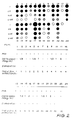
- Fig. 2 demonstrates the use of the assay for measuring IFN-y gene expression. 4 x 10⁶ human peripheral blood mononuclear cells were induced for IFN-y gene expression. RNA extracted from the cells was analyzed in four serial dilutions (0.125, 0.25, 0.5 and 1) using a manifold dot-blot device. The nitrocellulose filter was hybridized to IFN-y riboprobe.
Gamma-irradiation (γ-irradiation) was done before addition of 0.4% (v/v) phytohemagglutinin (PHA), the dose was 1500 rad. Cycloheximide (CHX) was added to several cultures after the addition of PHA. The filter was exposed to film after hybridization and the autoradiogram is shown. - Fig. 3 is a graph showing the linearity of the assay according to this invention. Samples expressing different levels of IL-2 RNA or IFN-y RNA were analyzed in four serial dilutions (0.125, 0.25, 0.5 and 1.0) using a manifold dot-blot device. After hybridization to specific riboprobes and autoradiography of the nitrocellulose filters, absorbency of individual dots at 630nm was measured in a micro-elisa autoreader.
- Fig. 4 is a graph showing the linearity and equivalent response of the assay as a function of increasing cell number or increasing RNA input. 4 x 10⁶ cells, induced for IL-2 gene expression, were serially diluted and RNA was then extracted (▲). Alternatively, RNA was extracted from 4 x 10⁶ cells, and then serially diluted (o).
- Fig. 5 is a graph showing the kinetics of IL-2 mRNA levels in human peripheral blood lymphocytes (PBL) induced with phytohemagglutinin (PHA). Cells were induced with PHA (0.4% v/v) at 0hr. Samples of 4 x 10⁶ cells were taken at each time point indicated, and RNA was extracted. Cycloheximide (CHX; 20ug/ml) was added at 21hr and RNA was analyzed at 24hr (▲).
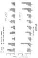
- Fig. 6 is a chart showing the quantitation of IL-2 and IFN-y gene expression in PBL of nine blood donors. 10ml of peripheral blood was taken from each donor. Cells from each donor were incubated with 1) no additions (background, BKG); 2) addition of 0.4% (v/v) PHA; 3) addition of PHA after Y-irradiation (1500 rad) of the cells; 4) addition of PHA, followed, at 16hr, by the addition of CHX (20ug/ml). RNA was extracted from all samples of 20hr after the addition of PHA and dot-blot filters were hybridized separately to IL-2 and IFN-y riboprobes. Bars denote film density for respective aasays, determining in a micro-elisa autoreader.
- Fig. 7 is a chart showing the distribution of IL-2 and IFN-y gene expression levels in cells from the nine donors described in Fig. 6, expressed as absorbency of film at 630nm which reflects the amount of hybridized RNA.
- A preferred method according to the present invention is as follows:
-
- 1. Take 10ml of peripheral blood from patient in heparized tube.
- 2. Dilute 2-fold with phosphate-buffered saline (PBS).
- 3. Layer over 2vol Ficoll® (Pharmacia).
- 4. Spin 30 min at 200g at room temperature in cell centrifuge.
- 5. Wash twice with RPM1 1640 medium without serum.
- 6. Resuspend cells at a density of 4 x 10⁶ in 10-ml screw cap round-bottom plastic culture tubes (Cel-Cult) into RPM1 1640 medium containing:
2 mM glutamine
10 mM MEM non-essential amino acids
100 mM MEM sodium pyruvate
10 mM HEPES pH 7.2
100 u/ml penicillin
100 ug/ml streptomycin
40 ug/ml gentamycin
5 x 10⁻⁵ M 2-mercaptoethanol
2% fetal calf serum
5 ug/ml nystatin - 7. Incubate overnight at 37°C in an atmosphere of 5% CO₂.
- Cells from each donor were incubated with 1) no additions (background, BKG): 2) addition of 0.4% (v/v) PHA; 3) addition of PHA after γ-irradiation (1500 rad) of the cells; 4) addition of PHA, followed, at 16hr, by the addition of CHX (20ug/ml). RNA was extracted from all samples at 20hr after the addition of PHA.
- All steps are performed in a single microcentrifuge tube of 1.5ml, using a standard table-top microcentrifuge (Eppendorf).
- 1. Spin down 1ml of cell suspension in an Eppendorf centrifuge at 500rpm for 5 min.
- 2. Add 0.4ml of 7.5M guanidinium thiocyanate (GTC) may be used as described in Example 4.
- 3. Homogenize by vortexing the tube for at least 30 sec, until dissolved completely.
- 4. Add 20µl of 2 M KOAc pH 5.1. Mix and then add 250µl of absolute ethanol. Mix again.
- 5. Leave overnight at -20°C.
- 6. Centrifuge at 15,000 rpm for 30 min at 4°C in an Eppendorf centrifuge.
- 7. Discard supernatant.
- 8. Dissolve pellet into 50µl of 37% formaldehyde. Mix on a vortex until dissolved. Then add 50µl of 3 M NaCL, 0.3 M Na-citrate.
- 9. Place for 15 min at 60°C.
- 10. Perform a dot-block procedure. The sample is pipetted into a single well of a 96-place microtiter plate (Nunc) and serially diluted, in 2-fold steps, into 1.5 M NaCl, 0.15 M Na-citrate (10 x SSC) and pipetted into adjacent wells. After all samples have been diluted, the contents of each well of a 96-place manifold dot-blot device (Schleicher & Schuell) under vacuum, containing a 9 x 13cm nitrocellulose sheet (Schleicher & Schuell GB 003 blotting paper. The nitrocellulose sheet and the blotting paper were presoaked in distilled water and then in 10 x SSC. Each well is washed once with 100ul of 10 x SSC. The nitrocellulose is baked at 80°C for 2hr under vacuum and probed with the desired gene riboprobe, as described in examples 5 or 6.
- 11. Quantitate film dots in micro-elisa autoreader. To this end, exposed and developed X-ray film is cut into the shape of a microtiter plate, placed on the plate such that dots align with wells, and introduced into the autoreader (e.g., Dynatech®). Scanning is at a wavelength of 630nm.
-
- 1. Spin down 1ml of cell culture in an Eppendorf centrifuge at 500rpm for 5 min.
- 2. Discard supernatant and drain pellet.
- 3. Add 100µl of lysis buffer, consisting of 5 M GTC, 10 mM EDTA, 50 mM Tris-HCl pH 7.5, and 8% (v/v) 2-mercaptoethanol (added freshly).
- 4. Mix three times on vortex for 10 sec. Add 700ul of 4 M LiCl and leave for 20hr (overnight) at 4°C.
- 5. Spin in an Eppendorf centrifuge for 90 min at 12,000 rpm at 4°C.
- 6. Discard supernatant, dry pellet and 50µl of RNA solubilizing buffer: 0.1% sodium dodecyl sulfate (SDS), 1 mM EDTA, 10 mM Tris-HCl pH 7.5
- 7. Mix by vortexing for 20 sec. Repeat after every 10 min for 40 min. This step is done at room temperature.
- 8. Extract solution by vortexing it with an equal volume of phenol/chloroform (1:1), collect upper phase.
- 9. Add 1/10 vol of 3 M NaOAc pH 5.1, mix, add 2.5 vol absolute ethanol and leave at -20°C for 20 hr.
- 10. Precipitate RNA at 4°C by a 45 min centrifugation at 15,000 rpm in an Eppendorf centrifuge.
- 11. Wash pellet with 70% ethanol.
- 12. Discard supernatant and dry pellet.
- 13. Follow steps 8-11 of Example 3.
- The riboprobe is synthesized using Promega-Biotec riboprobe kit. IL-2 cDNA was excised from plasmid p3-16 (Taniguchi T., Matsui, H., Fujita, T., Takaoka, C., Kashima, N., Yoshimoto, R., Hamuro, J. Nature 302 (1983) p.305) with Pst I and inserted into pGEM-3 (Promega Biotec)at the Pst I site. The orientation in which transcription from the T7 promoter gives the desired RNA strand was selected.
- a) Transcript was made as follows:-
- 1) 4µl transcript buffer.
- 2) 2µl dithiotreitol (DDT) (100 mM).
- 3) 0.6µl RNasin (350 u/ml).
- 4) 1µl each of the nucleotides UTP, CTP, GTP (0.5 mM).
- 5) 1µl T7 RNA polymerase (15 u/ml).
- 6) 1µl plasmid containing the IL-2 cDNA (1 µg/1 ul.
- 7) 10µl [α⁻³²P] ATP (10 mCi/ml).
- 8) 10µl of [α-³⁵ ] ATP (10 mCi/ml) (Amersham) may be substituted for [α⁻³²P] ATP in the previous step.
- b) The mixture is incubated for 1hr at 37°C.
- c) 1ul DNase (free of RNase; 1u/ml is added and incubation is continued for 15 min at 37°C.
- d) RNA is extracted as follows:
- 1) 20µl of phenolchloroform (1:1) is added, followed by mixing on vortex and centrifugation for 2 min in an Eppendorf centrifuge.
- 2) The upper (aqueous) phase is carefully collected.
- 3) The lower phase of phenochloroform is washed with 20ul of TE buffer: 10 mM Tris-HCl pH 7.6, 1 mM EDTA.
- 4) After vortexing and centrifugation of 2 min in an Eppendorf centrifuge, the upper phase is collected and added to the aqueous phase collected previously.
- 5) 40µl of chloroform is added to the combined aqueous phases.
- 6) After vortexing and centrifugation for 2 min in an Eppendorf centrifuge, the upper phase is collected.
- 7) The lower phase is extracted with 40µl of TE buffer, as described under
steps step 6 above. - 8) 8µl of 3 M NaOAc pH 5.1 and 2.5 vol of absolute ethanol are added to the combined aqueous phases.
- 9) The solution is stored for 20 hr (overnight) at -20°C, or for 2 hr at -70°C.
- 10) Centrifuge for 30 min at 4°C in an Eppendorf centrifuge at 15,000 rpm.
- 11) Wash pellet with 1ml of 70% ethanol.
- 12) Dry pellet in vacuo.
- 13) Resuspend pellet in 100µl TEN buffer containing: 10 mM Tris-HCl, pH 7.6, 1 mM EDTA, 0.1 M Na Cl and 20 ug/ml tRNA from E.coli MRE 600 (Boehringer).
- 14) Apply to a 1-ml Sephadex® G-50 column made in a 2-ml syringe and equilibrated with TEN buffer.
- 15) Centrifuge the column for 5 min at 1,000 rpm in a cell centrifuge and collect the flow-through in a tube.
- 16) Take a 1ul sample into 10% ice-cold trichloroacetic acid and determine precipitable radioactivity.
- 17) A good preparation yields 3 - 10 x 10⁵ cpm/µl.
- The riboprobe for IFN-y gene expression was synthesized following the procedure described in Example 5. IFN-y cDNA was excised from plasmid pH11F-SV-y with Bam H1 (Devos, R., Cheroutre, H., Taya, Y., Degrave, W., Heuverswyn, H.Y., Fiers, W. Nucl. Acids Res. 10 (1982) p.2487) and inserted into pGEM-3 at the Bam H1 site. The orientation in which transcription from the T7 promotor gives the desired RNA strand was selected.
-
- 1) Filters are prehybridized in a sealed plastic bag for 4 hr at 60°C. The prehybridization buffer contains:
25% formamide
10% dextran sulfate (5,000 mol weight)
1% SDS
50 mM Tris-HCl pH 7.5
100 µg/ml salmon sperm DNA that has been boiled for
10 min and cooled rapidly
100 µg/ml tRNA (E.coli MRE 600)
0.7 M NaCl - 2) For hybridization add: 5 x 10⁶ cpm/ml of riboprobe (see above) x 1
Denhardt solution: 0.02% (w/v) Ficoll, 0.02% (w/v)
polyvinyl-pyrrolidone, 0.02% (w/v) BSA fraction V
(Sigma)
100 µg/ml tRNA (E.coli MRE600)
Incubate for 16-18 hr, but not more, at 60°C. - 3) After hybridization, filters are washed:
- 1) twice with 2 x SSC (500 ml) at room temperature for 15 min
- 2) twice with 2 x SSC, 1% SDS (500 ml) at 68°C for 30 min
- 3) once with 0.2 x SSC, 0.2% SDS (500 ml) at 68°C for 30 min.
- Filters are dried and exposed to X-ray film for 5-25 hr at -70°C.
- Results may be analyzed as follows:
IL-2 IFN-Y No inducer 1A 1B PHA (16 hr) 2A 2B PHA (20 hr) 3A 3B PHA + cycloheximide 4A 4B PHA + Y-irradiation 5A 5B - (a) Basal level of gene expression:
1A, 1B. - (b) Inducibility of genes
Ratio 2A/1A, 2B/1B. Ratio 3A/1A, 3B/1B.
The difference betweensamples - (c) Superinducibility by cycloheximide:
A4/A3, B4/B3.
The ratio shows the potential for gene expression. Normally, this ratio should be considerably greater than 1 (1.5-5 or more). If not, this means that either basal expression is very high, in which case the ratios A4/A1, B4/B1 should be close to 1, or, alternatively, that induced gene expression is very high and uncontrolled, in which case the ratios A3/A1 and B3/B1 should be high. - (d) Superinducibility by Y-irradiation:
A5/A3, B5/B3.
Normally, this ratio should be well in excess of 1 (1.5-3). Normally, the ratio B5/B3 will exceed the ratio A5/A3. These values measure the extent to which suppressor T cells down-regulate IL-2 and IFN-Y gene expression; the latter gene is more strongly regulated in this manner. The higher these ratios, the stronger the down-regulation.
Claims (13)
- A method for calculating the inductivity of a bioregulatory gene in a sample of eukaryotic cells, the method comprising:a) isolating a sample of eukaryotic cells;b) dividing the sample into at least two aliquots;c) culturing the cells in each aliquot;d) lysing the cultured cells in each aliquot by the addition of guanidinium thiocyanate or guanidine hydrochloride lysing agent;e) homogenising each aliquot and thereafter selectively precipitating the RNA in each aliquot by the successive addition of sodium or potassium acetate and absolute ethanol;f) recovering and blotting the RNA from each of aliquot onto a support matrix;g) hybridising the recovered and blotted RNA from each aliquot with a labelled riboprobe specific to the bioregulatory gene; andh) quantitatively determining the extent of expression of the bioregulatory gene by measuring the extent of binding of the labelled riboprobe to the blotted RNA;the method being characterised in that in step (c):(c) (i) the cells in a first aliquot are cultured in the presence of an inducing agent for inducing expression of the bioregulatory gene; and(c) (ii) the cells in a second aliquot are cultured in the absence of an inducing agent;and in that:i) the ratio of the extent of gene expression in the first aliquot to the extent of gene expression in the second aliquot is calculated, said ratio providing a measure of the inductivity of the bioregulatory gene.
- A method as claimed in Claim 1 wherein said sample of eukaryotic cells is a sample of cells selected from peripheral human blood mononuclear cells, lymphocytes, monocytes, fibroblasts, astrocytes, endothelial cells, glial cells, keratinocytes and platelets.
- A method as claimed in Claim 1 or Claim 2 wherein said inducing agent is phytohemagglutinin (PHA) or concavalin A.
- A method as claimed in Claim 1, Claim 2 or Claim 3 characterised in that the cells in a third aliquot are superinduced; and in that the ratio of the extent of gene expression in the third aliquot to the extent of gene expression in the first aliquot is calculated to provide a measure of the maximum extent of expression of the bioregulatory gene.
- A method as claimed in Claim 4 wherein the cells in the third aliquot are cultured in the presence of a superinducing agent selected fom cycloheximide, cycloheximide + phytohemagglutinin and cycloheximide + concavalin A.
- A method as claimed in Claim 4 wherein the cells in the third aliquot are cultured in the presence of an inducing agent selected from phytohemagglutinin and concavalin A, and prior to adding said inducing agent are irradiated with gamma-radiation to superinduce said cells.
- A method as claimed in any preceding claim wherein in step (g) the riboprobe is specific to a bioregulatory gene selected from the genes coding for interferons, interleukins, tumour necrosis factors and growth factors.
- A method as claimed in any preceding claim wherein in step (g) the riboprobe is specific to a bioregulatory gene coding for IL-2 or IFN-y.
- A method as claimed in any preceding claim wherein step (e) lithium chloride and guanidinium thiocyanate are added to each aliquot prior to the sodium or potassium acetate.
- A method as claimed in any preceding claim wherein the riboprobe is labelled with a detectable isotopic or non-isotopic chemical group.
- A method as claimed in claim 10 wherein said riboprobe is isotopically labelled, and in step (h) the extent of binding is measured by measuring the absorbancy of a photographic film blackened by exposure to the support matrix carrying the RNA and hybridized labelled riboprobe.
- A method of evaluating the dynamic functionality of the immune response in a sample of eukaryotic cells, the method comprising a method as claimed in any of Claims 1 - 11 using said sample.
- An in vitro diagnostic method for diagnosing viral, bacterial, fungal, oncogenic and immunoregulatory diseases in a patient, the diagnostic method comprising a method as claimed in any of Claims 1 - 11 using a sample of blood from the patient.
Applications Claiming Priority (3)
| Application Number | Priority Date | Filing Date | Title |
|---|---|---|---|
| AU1455/87 | 1987-04-15 | ||
| AUPI145587 | 1987-04-15 | ||
| PCT/AU1988/000099 WO1988008038A1 (en) | 1987-04-15 | 1988-04-05 | Method for detecting the expression of bioregulatory genes in microcultures of eukaryotic cells and kit therefor |
Publications (3)
| Publication Number | Publication Date |
|---|---|
| EP0309558A1 EP0309558A1 (en) | 1989-04-05 |
| EP0309558A4 EP0309558A4 (en) | 1992-04-01 |
| EP0309558B1 true EP0309558B1 (en) | 1994-11-02 |
Family
ID=3772116
Family Applications (1)
| Application Number | Title | Priority Date | Filing Date |
|---|---|---|---|
| EP88903742A Expired - Lifetime EP0309558B1 (en) | 1987-04-15 | 1988-04-05 | Method for detecting the expression of bioregulatory genes in microcultures of eukaryotic cells |
Country Status (7)
| Country | Link |
|---|---|
| EP (1) | EP0309558B1 (en) |
| JP (1) | JPH01503598A (en) |
| AT (1) | ATE113665T1 (en) |
| DE (1) | DE3852004T2 (en) |
| ES (1) | ES2009261A6 (en) |
| PT (1) | PT87224B (en) |
| WO (1) | WO1988008038A1 (en) |
Families Citing this family (3)
| Publication number | Priority date | Publication date | Assignee | Title |
|---|---|---|---|---|
| ATE143700T1 (en) * | 1991-09-23 | 1996-10-15 | Pfizer | METHOD FOR THE DETECTION OF SPECIFIC MRNS AND DNA IN CELLS |
| US5837467A (en) * | 1997-01-21 | 1998-11-17 | Yissum Research Development Company Of The Hebrew University Of Jerusalem | Method for predicting the response to treatment with BCG of superficial bladder carcinoma |
| US20010046665A1 (en) * | 2000-01-21 | 2001-11-29 | Powell Thomas J. | Method of identifying the function of a test agent |
Family Cites Families (4)
| Publication number | Priority date | Publication date | Assignee | Title |
|---|---|---|---|---|
| US4483920A (en) * | 1982-05-17 | 1984-11-20 | Hahnemann University | Immobilization of message RNA directly from cells onto filter material |
| ES8600404A1 (en) * | 1983-05-02 | 1985-10-01 | Enzo Biochem Inc | Method and materials useful for the analysis and detection of genetic material. |
| FR2552879B1 (en) * | 1983-10-03 | 1987-08-14 | Pasteur Institut | PROCESS AND REAGENTS FOR THE DETECTION OF A GENETIC ANOMALY IN A BIOLOGICAL MATERIAL, IN PARTICULAR IN A PREPARATION OF LEUKOCYTES |
| CA1301606C (en) * | 1986-05-02 | 1992-05-26 | David H. Gillespie | Chaotropic method for evaluating nucleic acids in a biological sample |
-
1988
- 1988-04-05 AT AT88903742T patent/ATE113665T1/en not_active IP Right Cessation
- 1988-04-05 WO PCT/AU1988/000099 patent/WO1988008038A1/en not_active Ceased
- 1988-04-05 EP EP88903742A patent/EP0309558B1/en not_active Expired - Lifetime
- 1988-04-05 JP JP63503796A patent/JPH01503598A/en active Pending
- 1988-04-05 DE DE3852004T patent/DE3852004T2/en not_active Expired - Fee Related
- 1988-04-12 PT PT87224A patent/PT87224B/en not_active IP Right Cessation
- 1988-04-13 ES ES8801135A patent/ES2009261A6/en not_active Expired
Non-Patent Citations (6)
| Title |
|---|
| ANALYTICAL BIOCHEMISTRY, vol. 137, 1984; S. CHELEY et al., pp. 15-19# * |
| EMBO JOURNAL, vol. 6, no. 3, 1987, IRL Press Ltd., Oxford (GB); M.A. LEBENDIKER et al., pp. 585-589# * |
| JOURNAL OF IMMUNOLOGY, vol. 129, no. 2, August 1982, US; M. MATSUYAMA et al., pp. 450-451# * |
| METHODS IN ENZYMOLOGY, vol. 152, 1987, S.L. Berger et al. (eds.), Academic Pres, London (GB); pp. 219-226# * |
| NATURE, vol. 297, 20 May 1982, London (GB); S. EFRAT et al., pp. 236-239# * |
| PROCEEDINGS OF THE NATL. ACADEMY OF SCIENCES USA, vol. 1981, May 1984; S. EFRAT et al., pp. 2601-2605# * |
Also Published As
| Publication number | Publication date |
|---|---|
| WO1988008038A1 (en) | 1988-10-20 |
| EP0309558A4 (en) | 1992-04-01 |
| EP0309558A1 (en) | 1989-04-05 |
| DE3852004T2 (en) | 1995-06-01 |
| ES2009261A6 (en) | 1989-09-16 |
| PT87224B (en) | 1992-08-31 |
| DE3852004D1 (en) | 1994-12-08 |
| JPH01503598A (en) | 1989-12-07 |
| ATE113665T1 (en) | 1994-11-15 |
| PT87224A (en) | 1988-05-01 |
Similar Documents
| Publication | Publication Date | Title |
|---|---|---|
| Romano et al. | Haloperidol increases proenkephalin mRNA levels in the caudate-putamen of the rat: a quantitative study at the cellular level using in situ hybridization | |
| Breslin et al. | Multiple promoters exist in the human GR gene, one of which is activated by glucocorticoids | |
| Sur et al. | Localization of eosinophil‐derived neurotoxin and eosinophil cationic protein in neutrophilic leukocytes | |
| McChesney et al. | Measles virus infection of B lymphocytes permits cellular activation but blocks progression through the cell cycle | |
| De Webster et al. | Use of a biotinylated probe and in situ hybridization for light and electron microscopic localization of P 0 mRNA in myelin-forming Schwann cells | |
| Morley et al. | Human lymphocytes resistant to 6-thioguanine increase with age | |
| Rottman et al. | A reverse transcription-quantitative competitive polymerase chain reaction (RT-qcPCR) technique to measure cytokine gene expression in domestic mammals | |
| Pederson et al. | Relationship between protein synthesis and ribosome assembly in HeLa cells | |
| Bross et al. | Ribosomal cistrons and acrocentric chromosomes in man | |
| EP1523575A1 (en) | Nucleic acid array comprising selective monocytic macrophagic genes | |
| Lipsick et al. | c-myb protein expression is a late event during T-lymphocyte activation | |
| WO1988008038A1 (en) | Method for detecting the expression of bioregulatory genes in microcultures of eukaryotic cells and kit therefor | |
| Kravtsov et al. | Nuclear abnormalities of lymphocytes as the simplest markers for bioindication test in case of mass casualty events involving radiation exposure | |
| Longley et al. | Isolation, detection, and amplification of intact mRNA from dermatome strips, epidermal sheets, and sorted epidermal cells | |
| Ichihashi et al. | Biogenesis of poxviruses: relationship between a translation complex and formation of A-type inclusions | |
| AU1688188A (en) | Method for detecting the expression of bioregulatory genes in microcultures of eukaryotic cells and kit therefor | |
| Aviles et al. | Decreased expression of T-cell NF-κB p65 subunit in steroid-resistant nephrotic syndrome | |
| Wiedemann et al. | Differences among the polyadenylated RNA sequences of human leucocyte populations: an approach to the objective classification of human leukaemias. | |
| Monia et al. | Induction of metallothionein is correlated with resistance to auranofin, a gold compound, in Chinese hamster ovary cells. | |
| CN114317775A (en) | Application of RNA m6A modification of NCOA4 as gamma-ray radiation marker | |
| US20040152109A1 (en) | Methods and compositions for determining risk of treatment toxicity | |
| Kozma et al. | In situ fluorescence hybridization of Y translations: cytogenetic analysis using probes Y190 and Y431 | |
| EP4036233A1 (en) | Method for detecting brain tumor | |
| Dörmer et al. | Quantitation of globin mRNA in individual human erythroblasts by in situ hybridization | |
| Hwang et al. | DNA microarray analysis of transcriptional responses of human neuroblastoma IMR-32 cells to methylmercury |
Legal Events
| Date | Code | Title | Description |
|---|---|---|---|
| PUAI | Public reference made under article 153(3) epc to a published international application that has entered the european phase |
Free format text: ORIGINAL CODE: 0009012 |
|
| 17P | Request for examination filed |
Effective date: 19881216 |
|
| AK | Designated contracting states |
Kind code of ref document: A1 Designated state(s): AT BE CH DE FR GB IT LI LU NL SE |
|
| A4 | Supplementary search report drawn up and despatched |
Effective date: 19920211 |
|
| AK | Designated contracting states |
Kind code of ref document: A4 Designated state(s): AT BE CH DE FR GB IT LI LU NL SE |
|
| 17Q | First examination report despatched |
Effective date: 19921208 |
|
| GRAA | (expected) grant |
Free format text: ORIGINAL CODE: 0009210 |
|
| AK | Designated contracting states |
Kind code of ref document: B1 Designated state(s): AT BE CH DE FR GB IT LI LU NL SE |
|
| PG25 | Lapsed in a contracting state [announced via postgrant information from national office to epo] |
Ref country code: IT Free format text: LAPSE BECAUSE OF FAILURE TO SUBMIT A TRANSLATION OF THE DESCRIPTION OR TO PAY THE FEE WITHIN THE PRESCRIBED TIME-LIMIT;WARNING: LAPSES OF ITALIAN PATENTS WITH EFFECTIVE DATE BEFORE 2007 MAY HAVE OCCURRED AT ANY TIME BEFORE 2007. THE CORRECT EFFECTIVE DATE MAY BE DIFFERENT FROM THE ONE RECORDED. Effective date: 19941102 Ref country code: NL Effective date: 19941102 Ref country code: BE Effective date: 19941102 Ref country code: CH Effective date: 19941102 Ref country code: LI Effective date: 19941102 Ref country code: AT Effective date: 19941102 |
|
| REF | Corresponds to: |
Ref document number: 113665 Country of ref document: AT Date of ref document: 19941115 Kind code of ref document: T |
|
| REF | Corresponds to: |
Ref document number: 3852004 Country of ref document: DE Date of ref document: 19941208 |
|
| PG25 | Lapsed in a contracting state [announced via postgrant information from national office to epo] |
Ref country code: SE Effective date: 19950202 |
|
| REG | Reference to a national code |
Ref country code: CH Ref legal event code: PL |
|
| ET | Fr: translation filed | ||
| NLV1 | Nl: lapsed or annulled due to failure to fulfill the requirements of art. 29p and 29m of the patents act | ||
| PG25 | Lapsed in a contracting state [announced via postgrant information from national office to epo] |
Ref country code: LU Free format text: LAPSE BECAUSE OF NON-PAYMENT OF DUE FEES Effective date: 19950430 |
|
| PLBE | No opposition filed within time limit |
Free format text: ORIGINAL CODE: 0009261 |
|
| STAA | Information on the status of an ep patent application or granted ep patent |
Free format text: STATUS: NO OPPOSITION FILED WITHIN TIME LIMIT |
|
| 26N | No opposition filed | ||
| REG | Reference to a national code |
Ref country code: GB Ref legal event code: IF02 |
|
| PGFP | Annual fee paid to national office [announced via postgrant information from national office to epo] |
Ref country code: GB Payment date: 20040331 Year of fee payment: 17 |
|
| PGFP | Annual fee paid to national office [announced via postgrant information from national office to epo] |
Ref country code: FR Payment date: 20040408 Year of fee payment: 17 |
|
| PGFP | Annual fee paid to national office [announced via postgrant information from national office to epo] |
Ref country code: DE Payment date: 20040415 Year of fee payment: 17 |
|
| PG25 | Lapsed in a contracting state [announced via postgrant information from national office to epo] |
Ref country code: GB Free format text: LAPSE BECAUSE OF NON-PAYMENT OF DUE FEES Effective date: 20050405 |
|
| PG25 | Lapsed in a contracting state [announced via postgrant information from national office to epo] |
Ref country code: DE Free format text: LAPSE BECAUSE OF NON-PAYMENT OF DUE FEES Effective date: 20051101 |
|
| GBPC | Gb: european patent ceased through non-payment of renewal fee |
Effective date: 20050405 |
|
| PG25 | Lapsed in a contracting state [announced via postgrant information from national office to epo] |
Ref country code: FR Free format text: LAPSE BECAUSE OF NON-PAYMENT OF DUE FEES Effective date: 20051230 |
|
| REG | Reference to a national code |
Ref country code: FR Ref legal event code: ST Effective date: 20051230 |