EP0274517B1 - Hypodermically implantable genitourinary prosthesis - Google Patents
Hypodermically implantable genitourinary prosthesis Download PDFInfo
- Publication number
- EP0274517B1 EP0274517B1 EP87905032A EP87905032A EP0274517B1 EP 0274517 B1 EP0274517 B1 EP 0274517B1 EP 87905032 A EP87905032 A EP 87905032A EP 87905032 A EP87905032 A EP 87905032A EP 0274517 B1 EP0274517 B1 EP 0274517B1
- Authority
- EP
- European Patent Office
- Prior art keywords
- membrane
- containment
- containment membrane
- urethra
- fluid
- Prior art date
- Legal status (The legal status is an assumption and is not a legal conclusion. Google has not performed a legal analysis and makes no representation as to the accuracy of the status listed.)
- Expired
Links
Images
Classifications
-
- A—HUMAN NECESSITIES
- A61—MEDICAL OR VETERINARY SCIENCE; HYGIENE
- A61F—FILTERS IMPLANTABLE INTO BLOOD VESSELS; PROSTHESES; DEVICES PROVIDING PATENCY TO, OR PREVENTING COLLAPSING OF, TUBULAR STRUCTURES OF THE BODY, e.g. STENTS; ORTHOPAEDIC, NURSING OR CONTRACEPTIVE DEVICES; FOMENTATION; TREATMENT OR PROTECTION OF EYES OR EARS; BANDAGES, DRESSINGS OR ABSORBENT PADS; FIRST-AID KITS
- A61F2/00—Filters implantable into blood vessels; Prostheses, i.e. artificial substitutes or replacements for parts of the body; Appliances for connecting them with the body; Devices providing patency to, or preventing collapsing of, tubular structures of the body, e.g. stents
- A61F2/0004—Closure means for urethra or rectum, i.e. anti-incontinence devices or support slings against pelvic prolapse
- A61F2/0031—Closure means for urethra or rectum, i.e. anti-incontinence devices or support slings against pelvic prolapse for constricting the lumen; Support slings for the urethra
- A61F2/0036—Closure means for urethra or rectum, i.e. anti-incontinence devices or support slings against pelvic prolapse for constricting the lumen; Support slings for the urethra implantable
- A61F2/004—Closure means for urethra or rectum, i.e. anti-incontinence devices or support slings against pelvic prolapse for constricting the lumen; Support slings for the urethra implantable inflatable
-
- A—HUMAN NECESSITIES
- A61—MEDICAL OR VETERINARY SCIENCE; HYGIENE
- A61B—DIAGNOSIS; SURGERY; IDENTIFICATION
- A61B17/00—Surgical instruments, devices or methods
- A61B17/34—Trocars; Puncturing needles
- A61B17/3468—Trocars; Puncturing needles for implanting or removing devices, e.g. prostheses, implants, seeds, wires
-
- Y—GENERAL TAGGING OF NEW TECHNOLOGICAL DEVELOPMENTS; GENERAL TAGGING OF CROSS-SECTIONAL TECHNOLOGIES SPANNING OVER SEVERAL SECTIONS OF THE IPC; TECHNICAL SUBJECTS COVERED BY FORMER USPC CROSS-REFERENCE ART COLLECTIONS [XRACs] AND DIGESTS
- Y10—TECHNICAL SUBJECTS COVERED BY FORMER USPC
- Y10S—TECHNICAL SUBJECTS COVERED BY FORMER USPC CROSS-REFERENCE ART COLLECTIONS [XRACs] AND DIGESTS
- Y10S128/00—Surgery
- Y10S128/25—Artificial sphincters and devices for controlling urinary incontinence
Definitions
- This invention relates to hypodermic system as set forth in the preamble of claim 1.
- an artificial prosthetic sphincter has often been implanted so that occlusive pressure may be applied to the urethra to restore continence.
- Artificial sphincters are well-known and specific examples thereof will not be listed.
- the implantation of an artificial sphincter commonly requires a major surgical procedure which necessitates the hospitalization of the patient.
- Such a procedure is relatively complex and expensive, and usually requires six to eight weeks or more of recovery time.
- the patient also faces approximately two months delay before activation of the prosthesis to even ascertain whether the surgery has been successful (i.e.
- the patient is continent). More particularly, because of the swollen and aggravated condition of edema of the urethral tissues during, and for a period subsequent to surgery, the physician cannot precisely match the occlusive pressure to the patient's urethra. Therefore, the physician must estimate the required minimal occlusive pressure needed to achieve continence in that particular patient. As a consequence of such estimate, sphincteric mechanisms are often improperly selected or fitted, so that the occlusive pressures generated by such mechanisms are either insufficient to successfully achieve continence or excessive to the point of causing ischemia and subsequent erosion of urethral tissue. Excessive circumferential occlusive forces will eventually effect blood flow to the urethra and thereby cause ischemia and subsequent erosion.
- US-A-4 240 433 and US-A-4 585 457 disclose medical devices comprising an inflatable containment membrane, an outer tube having a cutting end and an inner tube communicating with said membrane. Also, US-A-3 834 394 discloses an inflatable contaiment membrane being in a normally uninflated membrane located within said outer tube and communicating with said membrane and with a source of material for inflating said membrane. US-A-4 373 218 suggustsd to provide corresponding outer tubes with such cutting ends in order to pierce channels through human tissues.
- this invention relates to a hypodermic system as claimed in claim 1.
- a hypodermic system as claimed in claim 1.
- Preferred embodiments of the invention are disclosed in the dependent claims.
- an extensible and inflatable elliptoidal or pyriform-shaped membrane is provided to be periurethrally or perineally injected or implanted to form an enclosure for receiving and containing a supply of fluid or suspended particulate matter.
- the membrane is precisely and controllably inflated while in situ so as to add bulk and thereby apply increased localized tissue volume and proportionately greater external occlusive pressure to the urethral mucosa to restore the patient's continence.
- the system of this invention is implanted by means of specialized dual hypodermic positioning, injecting and inflating instrument.
- the dual hypodermic instrument includes a hollow outer trocar to surround the containment membrane and dilate a suitably sized insertion channel through the targeted patient tissues.
- Located within the outer trocar is a hollow stylus which communicates with the interior of the containment membrane for placing the membrane in communication with a source of fluid or suspended particulate matter.
- An inflation of the membrane proportionately increases local tissue volume in the periurethral tissues to correspondingly increase the occlusive pressure applied to the proximal urethra tissues for restoring a patient's continence.
- the positioning, injecting and inflating instrument is then withdrawn from the periurethral tissues leaving the inflated containment membrane in the form of an envelope for preventing the undesirable migration of the fluid or particulate matter while adding the desired tissue bulk to cause a partial obstruction.
- One or more of the genitourinary prostheses of the present invention may be implanted, as just described, depending upon etiology, residual sphincteric function, vascularity and physical properties of that individual patient's urethral tissues.
- FIG. 1 of the drawings there is shown an expandable and inflatable prosthesis 1 in an expanded and inflated state (i.e. filled with fluid or suspended particulate matter).
- the prosthesis 1 of this invention will be described in its preferred embodiment as a genitourinary prosthesis for returning a patient to continence by producing a controlled increase in localized tissue volume and a corresponding greater passive occlusive pressure.
- the prosthesis 1 is not limited to the treatment of urinary continence, but has other applications including, but not limited to, a variable volume mass to replace surgically removed tissue and/or organ excisions or as an injectable anti-urethral reflux mass.
- Still further applications of the present invention include an injectable testicular prosthesis, injectable prosthetic eye, prosthetic sphincter and injectable intraocular lens.
- the prosthesis may be formed from a selectively permeable (by a gas or liquid) material which can be implanted as a controlled release drug delivery system.
- the genitourinary prosthesis 1 comprises a generally elliptoidal or pyriform-shaped containment membrane that is preferably formed from a suitable tear-resistant, biocompatible, elastomeric material, such as, for example, polyurethane, silicone, latex, or the like.
- the containment membrane 2 contains a normally closed orifice 4 through which biomeric fluid or suspended particulate matter 6 is percutaneously injected to inflate membrane 2.
- the material 6 which is injected through orifice 4 may include TEFLON (a trademark) particles or spheres, radio-opaque isotonic fluid, physiologic saline solution, and the like. Hypodermic instruments for positioning, injecting and inflating containment membrane 2 will be disclosed in greater detail hereinafter when referring to FIGs. 2 and 3.
- the containment membrane 2 serves as a non-permeable envelope for preventing the migration of the material 6 contained therewithin.
- the containment membrane may be precisely and controllably infused with the minimum volume of fluid or suspended particulate matter necessary for achieving continence while simultaneously minimizing the risk of impeding arteriovascular blood flow through the urethra, as a consequence of ischemia.
- the dual hypodermic needle 8 includes a hollow, cylindrical outer trocar 10.
- Outer trocar 10 is formed from a structurally sound and corrosion-resistant material, such as titanium, or the like.
- Trocar 10 functions as an outer casing to protect the integrity of and guide the contaimment membrane 2 during implantation.
- Trocar 10 also functions as a cutting instrument by which to dialate a suitably sized channel through the patient's urethral mucosa, so that the prosthesis 1 may be suitably located (e.g. within the bulbar urethra of the corpus spongiousum).
- trocar 10 is provided with a sharp, oblique cutting end 12 for establishing the subcutaneous channel (not shown) through which to insert prosthesis 1.
- Stylus 14 is also preferrably formed from a suitable material, such as titanium, or the like. Outer trocar 10 and inner stylus 14 are concentrically aligned with and spaced from one another, so that a narrow channel 16 is formed therebetween. Located within channel 16 at the cutting end of dual hypodermic needle 8 is the containment membrane 10 in an uninflated state (i.e. devoid of fluid or particulate matter).
- the neck of containment membrane 2 is disposed within channel 16, such that membrane 2 extends over and around the distal end of inner stylus 14.
- the interior of containment membrane 2 is retained in fluid communication with a central passage 18 extending through hollow stylus 14.
- the distal end of inner stylus 14 extends into the interior of containment membrane 2 via the normally closed orifice 4 (of FIG. 1), which is now opened by the receipt therethrough of stylus 14.
- the elastic nature and spring-like memory of containment membrane 2 closes the orifice tightly around and prevents the inadvertent removal of membrane 2 from the distal end of stylus 14.
- containment membrane 2 extending over the distal end of inner stylus 14 makes dual hypodermic needle 8 non-coring and thereby prevents the patient's tissue from entering the central passage 18 of stylus 14 during such time as when the outer trocar 10 is used to penetrate the urethral mucosa. Hence, trauma will be minimized since none of the patient's tissue will be removed with stylus 14 and the conclusion of the nonsurgical process for implanting the prosthesis 1, which is now described while referring concurrently to FIGs. 2 and 3 of the drawings.
- the physician initially exerts a downward force on dual hypodermic needle 8, so that the cutting end 12 of outer trocar 10 transurethrally or perineally penetrates the patient's urethral tissue and dialates an insertion channel to the bulbar urethra of the corpus spongiousum.
- the physician verifies the location of cutting end 12, so that containment membrane 2 may be properly positioned within the periurethral tissues.
- hypodermic syringe (not shown), or the like, to the proximal end of inner stylus 14.
- the hypodermic syringe contains the aforementioned fluid or suspended particulate matter 6, a regulated amount of which is percutaneously infused to the interior of containment membrane 2. Accordingly, a flow path is established between the hypodermic syringe and the containment membrane 2 by way of stylus 14, so that membrane 2 may be inflated, while in situ, with a controlled and measured supply of fluid or suspended particulate matter to thereby produce an increase in localized tissue volume and a correspondingly greater occlusive pressure necessary to achieve coaptive continence of the urethral mucosa.
- containment membrane 2 Following the implantation and inflation of containment membrane 2, the physician exerts an upward pulling force on dual hypodermic needle 8 to remove the needle from the patient's tissue.
- the cavity produced during inflation of the containment membrane 2 within the patient's tissue produces a locating effect which removes the neck of the inflated membrane from its receipt by channel 16 at such time as when the hypodermic needle 8 is withdrawn from the patient's tissue. Accordingly, the normally closed orifice 4 (of FIG. 1) of containment membrane 2 automatically returns to the closed position whereby to prevent the migration of fluid or particles and avoid the possible deflation of membrane 2.
- the channel 16 between outer trocar 10 and inner stylus 16, in which the neck of containment membrane 2 is received may be interconnected with an external source of mechanical or hydraulic pressure 20.
- the application of sufficient pressure to the neck of containment membrane 2 by way of channel 16 will aid the physician in dispensing and positioning the membrane, after its inflation and just prior to the removal of hypodermic needle 8.
- hypodermic needle 8 removal of the hypodermic needle 8 from the patient's urethral tissue will leave behind a relatively minor puncture wound.
- the patient will require a substantially shorter recovery time or no recovery time at all as compared to approximately two months or more if a prosthetic sphincter had been surgically implanted in a hospital.
- the high cost, confinement and inconvenience commonly associated with such a hospital is stay minimized, since the present prosthesis 1 can be implanted by means of injection under a local anesthetic and the patient treated on a relatively cost efficient basis with a reduced hospital stay (e.g. about 24-48 hours).
- the present genitourinary prosthesis prevents the previous problem of small particle migration by virtue of the anti-migration containment membrane 2. Moreover, the prosthesis increases the pressure around the urethra in limited areas, thus reducing the risk of vascular ischemia and eventual urethral erosion, as are commonly caused by conventional surgically implanted genitourinary cuff sphincters.
Landscapes
- Health & Medical Sciences (AREA)
- Life Sciences & Earth Sciences (AREA)
- Animal Behavior & Ethology (AREA)
- General Health & Medical Sciences (AREA)
- Surgery (AREA)
- Engineering & Computer Science (AREA)
- Biomedical Technology (AREA)
- Heart & Thoracic Surgery (AREA)
- Veterinary Medicine (AREA)
- Public Health (AREA)
- Urology & Nephrology (AREA)
- Oral & Maxillofacial Surgery (AREA)
- Cardiology (AREA)
- Vascular Medicine (AREA)
- Pathology (AREA)
- Nuclear Medicine, Radiotherapy & Molecular Imaging (AREA)
- Transplantation (AREA)
- Medical Informatics (AREA)
- Molecular Biology (AREA)
- Prostheses (AREA)
Abstract
Description
- This invention relates to hypodermic system as set forth in the preamble of claim 1.
- As will be known to those skilled in the art, in cases where the natural sphincter muscles of a patient have been surgically excised, damaged by disease or compromised by physical trauma, an artificial prosthetic sphincter has often been implanted so that occlusive pressure may be applied to the urethra to restore continence. Artificial sphincters are well-known and specific examples thereof will not be listed. However, the implantation of an artificial sphincter commonly requires a major surgical procedure which necessitates the hospitalization of the patient. Such a procedure is relatively complex and expensive, and usually requires six to eight weeks or more of recovery time. In most cases, the patient also faces approximately two months delay before activation of the prosthesis to even ascertain whether the surgery has been successful (i.e. the patient is continent). More particularly, because of the swollen and aggravated condition of edema of the urethral tissues during, and for a period subsequent to surgery, the physician cannot precisely match the occlusive pressure to the patient's urethra. Therefore, the physician must estimate the required minimal occlusive pressure needed to achieve continence in that particular patient. As a consequence of such estimate, sphincteric mechanisms are often improperly selected or fitted, so that the occlusive pressures generated by such mechanisms are either insufficient to successfully achieve continence or excessive to the point of causing ischemia and subsequent erosion of urethral tissue. Excessive circumferential occlusive forces will eventually effect blood flow to the urethra and thereby cause ischemia and subsequent erosion. Also, if the implant surgery should prove to be unsuccessful (i.e. the maximum occlusive pressure to be generated by the sphincter is insufficient to hold the patient continent or the sphincter malfunctions mechanically), then additional surgery becomes necessary to provide sphincter adjustment, repair or explant.
- Reference is now made to "Migration And Granulomatous Reaction After Periurethral Injection Of Polytef (Teflon)" by Anthony A. Malizia, Jr. et al, Journal of the A.M.A., June 22/29, 1984, Vol. 251, No. 24, pp. 3277-3281. It has been demonstrated in humans that urinary incontinence may be successfully treated by non-surgical means with periurethral injection of polytef paste to increase localized tissue volume and thereby increase external occlusive pressure to the urethra for causing a partial obstruction and, thus, continence. However, this article reveals wide spread migration of polytef particles from the injection site. In addition, such paste was found to induce tissue reaction with TEFLON (a trademark) granulomas. Because of the possible toxicity of TEFLON-based paste, concern for patient safety has also been expressed. Hence, an otherwise sound, non-surgical procedure has now experienced reduced application.
- US-A-4 240 433 and US-A-4 585 457 disclose medical devices comprising an inflatable containment membrane, an outer tube having a cutting end and an inner tube communicating with said membrane. Also, US-A-3 834 394 discloses an inflatable contaiment membrane being in a normally uninflated membrane located within said outer tube and communicating with said membrane and with a source of material for inflating said membrane. US-A-4 373 218 suggustsd to provide corresponding outer tubes with such cutting ends in order to pierce channels through human tissues.
- Briefly, and in general terms, this invention relates to a hypodermic system as claimed in claim 1. Preferred embodiments of the invention are disclosed in the dependent claims. Preferably, an extensible and inflatable elliptoidal or pyriform-shaped membrane is provided to be periurethrally or perineally injected or implanted to form an enclosure for receiving and containing a supply of fluid or suspended particulate matter. In this manner, the membrane is precisely and controllably inflated while in situ so as to add bulk and thereby apply increased localized tissue volume and proportionately greater external occlusive pressure to the urethral mucosa to restore the patient's continence. Adding bulk to the periurethral tissues increases the external pressure around the proximal urethra which restores continence by causing a partial obstruction. However, the previous problem of particle migration is solved by virtue of the anti-migration membrane in which the fluid or particulate matter are retained.
- The system of this invention is implanted by means of specialized dual hypodermic positioning, injecting and inflating instrument. The dual hypodermic instrument includes a hollow outer trocar to surround the containment membrane and dilate a suitably sized insertion channel through the targeted patient tissues. Located within the outer trocar is a hollow stylus which communicates with the interior of the containment membrane for placing the membrane in communication with a source of fluid or suspended particulate matter. With the containment membrane percutaneously implanted in the proximal urethra, the membrane is then infused, via the stylus, with a measured supply of fluid or particulate matter, whereby to inflate the membrane. An inflation of the membrane proportionately increases local tissue volume in the periurethral tissues to correspondingly increase the occlusive pressure applied to the proximal urethra tissues for restoring a patient's continence. The positioning, injecting and inflating instrument is then withdrawn from the periurethral tissues leaving the inflated containment membrane in the form of an envelope for preventing the undesirable migration of the fluid or particulate matter while adding the desired tissue bulk to cause a partial obstruction. One or more of the genitourinary prostheses of the present invention may be implanted, as just described, depending upon etiology, residual sphincteric function, vascularity and physical properties of that individual patient's urethral tissues.
-
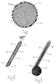
- FIG. 1 shows the genitourinary prosthesis of the present invention in an inflated state and filled with fluid or suspended particulate matter;
- FIG. 2 shows the dual hypodermic positioning, injecting and inflating instrument for implanting the prosthesis of FIG. 1; and
- FIG. 3 shows the dual hypodermic instrument of FIG. 2 with the prosthesis of FIG. 1 being percutaneously infused with a measured supply of fluid or suspended particulate matter.
- Referring to FIG. 1 of the drawings, there is shown an expandable and inflatable prosthesis 1 in an expanded and inflated state (i.e. filled with fluid or suspended particulate matter). The prosthesis 1 of this invention will be described in its preferred embodiment as a genitourinary prosthesis for returning a patient to continence by producing a controlled increase in localized tissue volume and a corresponding greater passive occlusive pressure. However, it is to be expressly understood that the prosthesis 1 is not limited to the treatment of urinary continence, but has other applications including, but not limited to, a variable volume mass to replace surgically removed tissue and/or organ excisions or as an injectable anti-urethral reflux mass. Still further applications of the present invention include an injectable testicular prosthesis, injectable prosthetic eye, prosthetic sphincter and injectable intraocular lens. What is more, the prosthesis may be formed from a selectively permeable (by a gas or liquid) material which can be implanted as a controlled release drug delivery system.
- According to the preferred embodiment of this invention, the genitourinary prosthesis 1 comprises a generally elliptoidal or pyriform-shaped containment membrane that is preferably formed from a suitable tear-resistant, biocompatible, elastomeric material, such as, for example, polyurethane, silicone, latex, or the like. The
containment membrane 2 contains a normally closed orifice 4 through which biomeric fluid or suspendedparticulate matter 6 is percutaneously injected to inflatemembrane 2. By way of example, thematerial 6 which is injected through orifice 4 may include TEFLON (a trademark) particles or spheres, radio-opaque isotonic fluid, physiologic saline solution, and the like. Hypodermic instruments for positioning, injecting and inflatingcontainment membrane 2 will be disclosed in greater detail hereinafter when referring to FIGs. 2 and 3. - In the inflated condition of FIG. 1, the
containment membrane 2 serves as a non-permeable envelope for preventing the migration of thematerial 6 contained therewithin. Moreover, the containment membrane may be precisely and controllably infused with the minimum volume of fluid or suspended particulate matter necessary for achieving continence while simultaneously minimizing the risk of impeding arteriovascular blood flow through the urethra, as a consequence of ischemia. - A dual
hypodermic needle 8 for implanting the prosthesis 1 is now described while referring to FIG. 2 of the drawings. The dualhypodermic needle 8 includes a hollow, cylindrical outer trocar 10. Outer trocar 10 is formed from a structurally sound and corrosion-resistant material, such as titanium, or the like. Trocar 10 functions as an outer casing to protect the integrity of and guide thecontaimment membrane 2 during implantation. Trocar 10 also functions as a cutting instrument by which to dialate a suitably sized channel through the patient's urethral mucosa, so that the prosthesis 1 may be suitably located (e.g. within the bulbar urethra of the corpus spongiousum). In this regard, trocar 10 is provided with a sharp,oblique cutting end 12 for establishing the subcutaneous channel (not shown) through which to insert prosthesis 1. - Located within outer trocar 10 is a hollow, cylindrical
inner stylus 14.Stylus 14 is also preferrably formed from a suitable material, such as titanium, or the like. Outer trocar 10 andinner stylus 14 are concentrically aligned with and spaced from one another, so that anarrow channel 16 is formed therebetween. Located withinchannel 16 at the cutting end of dualhypodermic needle 8 is the containment membrane 10 in an uninflated state (i.e. devoid of fluid or particulate matter). - During manufacture of the dual
hypodermic needle 8, the neck ofcontainment membrane 2 is disposed withinchannel 16, such thatmembrane 2 extends over and around the distal end ofinner stylus 14. Thus, the interior ofcontainment membrane 2 is retained in fluid communication with acentral passage 18 extending throughhollow stylus 14. More particularly, the distal end ofinner stylus 14 extends into the interior ofcontainment membrane 2 via the normally closed orifice 4 (of FIG. 1), which is now opened by the receipt therethrough ofstylus 14. However, the elastic nature and spring-like memory ofcontainment membrane 2 closes the orifice tightly around and prevents the inadvertent removal ofmembrane 2 from the distal end ofstylus 14. - The presence of
containment membrane 2 extending over the distal end ofinner stylus 14 makes dualhypodermic needle 8 non-coring and thereby prevents the patient's tissue from entering thecentral passage 18 ofstylus 14 during such time as when the outer trocar 10 is used to penetrate the urethral mucosa. Hence, trauma will be minimized since none of the patient's tissue will be removed withstylus 14 and the conclusion of the nonsurgical process for implanting the prosthesis 1, which is now described while referring concurrently to FIGs. 2 and 3 of the drawings. - In operation, the physician initially exerts a downward force on dual
hypodermic needle 8, so that the cuttingend 12 of outer trocar 10 transurethrally or perineally penetrates the patient's urethral tissue and dialates an insertion channel to the bulbar urethra of the corpus spongiousum. The physician verifies the location of cuttingend 12, so thatcontainment membrane 2 may be properly positioned within the periurethral tissues. - Once a proper subcutaneous location has been achieved, the physician attaches a high pressure hypodermic syringe (not shown), or the like, to the proximal end of
inner stylus 14. The hypodermic syringe contains the aforementioned fluid or suspendedparticulate matter 6, a regulated amount of which is percutaneously infused to the interior ofcontainment membrane 2. Accordingly, a flow path is established between the hypodermic syringe and thecontainment membrane 2 by way ofstylus 14, so thatmembrane 2 may be inflated, while in situ, with a controlled and measured supply of fluid or suspended particulate matter to thereby produce an increase in localized tissue volume and a correspondingly greater occlusive pressure necessary to achieve coaptive continence of the urethral mucosa. - Following the implantation and inflation of
containment membrane 2, the physician exerts an upward pulling force on dualhypodermic needle 8 to remove the needle from the patient's tissue. The cavity produced during inflation of thecontainment membrane 2 within the patient's tissue produces a locating effect which removes the neck of the inflated membrane from its receipt bychannel 16 at such time as when thehypodermic needle 8 is withdrawn from the patient's tissue. Accordingly, the normally closed orifice 4 (of FIG. 1) ofcontainment membrane 2 automatically returns to the closed position whereby to prevent the migration of fluid or particles and avoid the possible deflation ofmembrane 2. - What is more, the
channel 16 between outer trocar 10 andinner stylus 16, in which the neck ofcontainment membrane 2 is received, may be interconnected with an external source of mechanical orhydraulic pressure 20. The application of sufficient pressure to the neck ofcontainment membrane 2 by way ofchannel 16 will aid the physician in dispensing and positioning the membrane, after its inflation and just prior to the removal ofhypodermic needle 8. - It may be noted that removal of the
hypodermic needle 8 from the patient's urethral tissue will leave behind a relatively minor puncture wound. Thus, as another important advantage of this invention the patient will require a substantially shorter recovery time or no recovery time at all as compared to approximately two months or more if a prosthetic sphincter had been surgically implanted in a hospital. Moreover, the high cost, confinement and inconvenience commonly associated with such a hospital is stay minimized, since the present prosthesis 1 can be implanted by means of injection under a local anesthetic and the patient treated on a relatively cost efficient basis with a reduced hospital stay (e.g. about 24-48 hours). - Although the presently disclosed invention has been explained with reference to a single, implantable tissue expanding
containment membrane 2, it is to be expressly understood that additional numbers of such containment membranes will usually be implanted, depending on the increased tissue volume and the resulting occlusive pressure which are required to permit the patient to be restored to continence. During and after one or more prostheses have been implanted, the physician will cystoscopically monitor the degree of the urethral mucosa. In the event that greater occlusive pressure is needed, the physician may implant a corresponding additional number of prostheses until patient continence is restored. - Thus, the present genitourinary prosthesis prevents the previous problem of small particle migration by virtue of the
anti-migration containment membrane 2. Moreover, the prosthesis increases the pressure around the urethra in limited areas, thus reducing the risk of vascular ischemia and eventual urethral erosion, as are commonly caused by conventional surgically implanted genitourinary cuff sphincters.
Claims (7)
- A hypodermic system (8) for treating urinary incontinence by means of applying increased occlusive pressure to the urethra by producing an increase of localized tissue volume proximal to the urethra, said system including an inflatable containment membrane (1) being in a normally uninflated condition, an outer tube (10) having a cutting end (2) for piercing a channel through the patient's urethral tissue, said uninflated containment membrane located within said outer tube means behind the cutting end thereof, and an inner tube (14) located within said outer tube and extending at a first end thereof into the interior of said containment membrane and communicating at the other end with a source of material, so that said membrane may be percutaneously infused with a supply of material from said source for inflating said membrane and thereby increasing both the local tissue volume proximal to the urethra and the occlusive pressure applied to the urethra,
said system being characterized by said inner and outer tubes concentrically aligned with and spaced from one another, said containment membrane being received within the space (16) between said inner and outer tubes and extending across the first end of said inner tube (14) to prevent the patient's tissue from entering said first end. - The system recited in claim 1, further characterized by means (20) connected to apply a force through the space (16) between said inner and outer tubes (14 and 10) for dispensing said containment membrane from said space after said membrane is inflated.
- The system recited in claim 1, wherein said containment membrane (1) has an orifice (4) biased toward a normally closed condition, said inner tube (14) extending through said orifice and holding said orifice open for percutaneously infusing and thereby inflating said containment membrane with material from said source.
- The system recited in claim 1, wherein said membrane is characterized as an envelope in the inflated condition to prevent the migration of said material from the injection site.
- The system recited in claim 1, wherein the material by which to inflate said membrane is a fluid.
- The system recited in claim 5, wherein said fluid is a saline solution.
- The system recited in claim 1, wherein the material by which to inflate said membrane includes a supply of particles.
Applications Claiming Priority (2)
| Application Number | Priority Date | Filing Date | Title |
|---|---|---|---|
| US881830 | 1986-07-03 | ||
| US06/881,830 US4773393A (en) | 1986-07-03 | 1986-07-03 | Hypodermically implantable genitourinary prosthesis |
Publications (3)
| Publication Number | Publication Date |
|---|---|
| EP0274517A1 EP0274517A1 (en) | 1988-07-20 |
| EP0274517A4 EP0274517A4 (en) | 1989-11-07 |
| EP0274517B1 true EP0274517B1 (en) | 1992-09-16 |
Family
ID=25379306
Family Applications (1)
| Application Number | Title | Priority Date | Filing Date |
|---|---|---|---|
| EP87905032A Expired EP0274517B1 (en) | 1986-07-03 | 1987-07-02 | Hypodermically implantable genitourinary prosthesis |
Country Status (6)
| Country | Link |
|---|---|
| US (1) | US4773393A (en) |
| EP (1) | EP0274517B1 (en) |
| JP (1) | JP2652164B2 (en) |
| AU (1) | AU603130B2 (en) |
| DE (1) | DE3781786T2 (en) |
| WO (1) | WO1988000064A1 (en) |
Families Citing this family (112)
| Publication number | Priority date | Publication date | Assignee | Title |
|---|---|---|---|---|
| US5041090A (en) * | 1988-01-12 | 1991-08-20 | Scheglov Viktor I | Occluding device |
| US5123428A (en) * | 1988-10-11 | 1992-06-23 | Schwarz Gerald R | Laparoscopically implanting bladder control apparatus |
| US5097848A (en) * | 1988-10-11 | 1992-03-24 | Schwarz Gerald R | Incontinence bladder control method and apparatus |
| US5012822A (en) * | 1988-10-11 | 1991-05-07 | Schwarz Gerald R | Method for controlling urinary incontinence |
| US4969474A (en) * | 1988-10-11 | 1990-11-13 | Schwarz Gerald R | Incontinence bladder control method and apparatus |
| US5007940A (en) * | 1989-06-09 | 1991-04-16 | American Medical Systems, Inc. | Injectable polymeric bodies |
| US5064434A (en) * | 1990-04-04 | 1991-11-12 | Haber Terry M | Genitourinary implant |
| US6461346B1 (en) | 1990-08-29 | 2002-10-08 | Applied Medical Resources Corp. | Sealing occlusion catheter and method of using same |
| US5149329A (en) * | 1990-12-12 | 1992-09-22 | Wayne State University | Surgical suture carrier and method for urinary bladder neck suspension |
| US5304123A (en) | 1991-10-24 | 1994-04-19 | Children's Medical Center Corporation | Detachable balloon catheter for endoscopic treatment of vesicoureteral reflux |
| SE503716C2 (en) * | 1991-12-12 | 1996-08-12 | Nobel Biocare Ab | Device for stabilizing the nipple base of an intestinal reservoir |
| US5295960A (en) * | 1992-04-01 | 1994-03-22 | Applied Medical Resources Corporation | Catheter with interior balloon |
| DE69318835T2 (en) * | 1992-04-06 | 1998-11-05 | Uroplasty, Inc., Minneapolis, Minn. | TREATMENT OF REFLUX DISORDER BY INJECTION OF MICROPARTICLES |
| US6592859B1 (en) * | 1992-08-20 | 2003-07-15 | Ethicon, Inc. | Controlled expansion sphincter augmentation media |
| US5334210A (en) * | 1993-04-09 | 1994-08-02 | Cook Incorporated | Vascular occlusion assembly |
| US5437603A (en) * | 1993-09-14 | 1995-08-01 | C.R. Bard, Inc. | Apparatus and method for implanting prostheses within periurethral tissues |
| US6060639A (en) * | 1994-03-04 | 2000-05-09 | Mentor Corporation | Testicular prosthesis and method of manufacturing and filling |
| US5451406A (en) * | 1994-07-14 | 1995-09-19 | Advanced Uroscience, Inc. | Tissue injectable composition and method of use |
| US5830228A (en) | 1996-05-29 | 1998-11-03 | Urosurge, Inc. | Methods and systems for deployment of a detachable balloon at a target site in vivo |
| US5855615A (en) * | 1996-06-07 | 1999-01-05 | Menlo Care, Inc. | Controller expansion sphincter augmentation media |
| US5792478A (en) * | 1996-07-08 | 1998-08-11 | Advanced Uro Science | Tissue injectable composition and method of use |
| US5813411A (en) * | 1996-08-20 | 1998-09-29 | Menlo Care, Inc. | Method of deforming tissue with a swollen hydrogel |
| US7049346B1 (en) | 1996-08-20 | 2006-05-23 | Menlo Care Div Of Ethicon, Inc. | Swollen hydrogel for sphincter augmentation |
| US6480746B1 (en) | 1997-08-13 | 2002-11-12 | Surx, Inc. | Noninvasive devices, methods, and systems for shrinking of tissues |
| US6035238A (en) * | 1997-08-13 | 2000-03-07 | Surx, Inc. | Noninvasive devices, methods, and systems for shrinking of tissues |
| US6091995A (en) | 1996-11-08 | 2000-07-18 | Surx, Inc. | Devices, methods, and systems for shrinking tissues |
| US6081749A (en) * | 1997-08-13 | 2000-06-27 | Surx, Inc. | Noninvasive devices, methods, and systems for shrinking of tissues |
| US6292700B1 (en) | 1999-09-10 | 2001-09-18 | Surx, Inc. | Endopelvic fascia treatment for incontinence |
| US7317949B2 (en) * | 1996-11-08 | 2008-01-08 | Ams Research Corporation | Energy induced bulking and buttressing of tissues for incontinence |
| AU2912797A (en) * | 1997-01-15 | 1998-08-07 | Shantha Jayatilake Bandara Lenadora | Device for genuine stress incontinence in the female |
| US6645138B2 (en) | 1997-09-12 | 2003-11-11 | Uromedica, Inc. | Adjustable implantable genitourinary device |
| US6579224B1 (en) | 1999-10-11 | 2003-06-17 | Uromedica, Inc. | Apparatus and method for inserting an adjustable implantable genitourinary device |
| US7364540B1 (en) * | 1997-06-12 | 2008-04-29 | Uromedica, Inc. | Implantable device and method for adjustably restricting a body lumen |
| US6045498A (en) | 1997-06-12 | 2000-04-04 | Uromedica, Inc. | Method for adjustably restricting a body lumen |
| US6419624B1 (en) * | 1999-10-11 | 2002-07-16 | Uromedica, Inc. | Apparatus and method for inserting an adjustable implantable genitourinary device |
| CA2300152A1 (en) | 1997-08-13 | 1999-02-25 | Surx, Inc. | Noninvasive devices, methods, and systems for shrinking of tissues |
| US20030178032A1 (en) * | 1997-08-13 | 2003-09-25 | Surx, Inc. | Noninvasive devices, methods, and systems for shrinking of tissues |
| US9023031B2 (en) | 1997-08-13 | 2015-05-05 | Verathon Inc. | Noninvasive devices, methods, and systems for modifying tissues |
| EP1047347B1 (en) | 1998-01-14 | 2009-08-12 | AMS Research Corporation | Ribbed electrodes |
| US6044847A (en) | 1998-06-23 | 2000-04-04 | Surx, Inc. | Tuck and fold fascia shortening for incontinence |
| US6251064B1 (en) | 1998-12-11 | 2001-06-26 | Enteric Medical Technologies, Inc. | Method for creating valve-like mechanism in natural body passageway |
| US7132582B2 (en) | 2003-05-30 | 2006-11-07 | Council Of Scientific And Industrial Research | Catalytic process for the preparation of isolongifolene |
| US6238335B1 (en) * | 1998-12-11 | 2001-05-29 | Enteric Medical Technologies, Inc. | Method for treating gastroesophageal reflux disease and apparatus for use therewith |
| US6595910B2 (en) * | 1998-12-11 | 2003-07-22 | Scimed Life Systems, Inc. | Method for treating fecal incontinence |
| US6312405B1 (en) | 1999-02-02 | 2001-11-06 | American Medical Systems, Inc. | Self-sealing detachable balloon |
| US6572532B1 (en) * | 1999-03-01 | 2003-06-03 | Advanced Biomedical Devices Inc. | Implant positioning system and method |
| EP1189552B1 (en) | 1999-04-30 | 2009-04-01 | Uromedica Inc. | Apparatus with an adjustable sling for treatment of urinary stress incontinence |
| US6358197B1 (en) | 1999-08-13 | 2002-03-19 | Enteric Medical Technologies, Inc. | Apparatus for forming implants in gastrointestinal tract and kit for use therewith |
| US6964669B1 (en) | 2000-04-12 | 2005-11-15 | Ams Research Corporation | Linear delivery system for deployment of a detachable balloon at a target site in vivo |
| US6976950B2 (en) * | 2000-04-14 | 2005-12-20 | Solace Therapeutics, Inc. | Implantable valved pressure attenuation device |
| US7374532B2 (en) | 2000-04-14 | 2008-05-20 | Attenuex Technologies, Inc. | High vapor pressure attenuation device |
| US6682473B1 (en) | 2000-04-14 | 2004-01-27 | Solace Therapeutics, Inc. | Devices and methods for attenuation of pressure waves in the body |
| US8574146B2 (en) | 2000-04-14 | 2013-11-05 | Attenuex Technologies, Inc. | Implant with high vapor pressure medium |
| US10327880B2 (en) | 2000-04-14 | 2019-06-25 | Attenuex Technologies, Inc. | Attenuation device for use in an anatomical structure |
| US7771482B1 (en) * | 2000-05-09 | 2010-08-10 | Ben-Zion Karmon | Method for tissue expansion and regeneration using bioresorbable inflatable devices |
| US6423332B1 (en) | 2000-05-26 | 2002-07-23 | Ethicon, Inc. | Method and composition for deforming soft tissues |
| US6540789B1 (en) * | 2000-06-15 | 2003-04-01 | Scimed Life Systems, Inc. | Method for treating morbid obesity |
| WO2002011696A2 (en) * | 2000-08-08 | 2002-02-14 | Ev & M | Active tissue augmentation materials and method |
| US7306591B2 (en) | 2000-10-02 | 2007-12-11 | Novasys Medical, Inc. | Apparatus and methods for treating female urinary incontinence |
| US6425854B1 (en) * | 2000-10-02 | 2002-07-30 | Genyx Medical, Inc. | Method for delivering non-biodegradable bulking composition to a urological site |
| US7033373B2 (en) | 2000-11-03 | 2006-04-25 | Satiety, Inc. | Method and device for use in minimally invasive placement of space-occupying intragastric devices |
| US7160325B2 (en) | 2001-05-15 | 2007-01-09 | Ams Research Corporation | Implantable medical balloon and valve |
| US7083629B2 (en) | 2001-05-30 | 2006-08-01 | Satiety, Inc. | Overtube apparatus for insertion into a body |
| US6558400B2 (en) | 2001-05-30 | 2003-05-06 | Satiety, Inc. | Obesity treatment tools and methods |
| US20030004534A1 (en) * | 2001-06-01 | 2003-01-02 | George Stephanie A. | Balloon transporter |
| US7695427B2 (en) | 2002-04-26 | 2010-04-13 | Torax Medical, Inc. | Methods and apparatus for treating body tissue sphincters and the like |
| AU2003240000A1 (en) * | 2002-06-12 | 2003-12-31 | Boston Scientific Limited | Bulking agents |
| US20040019388A1 (en) * | 2002-07-24 | 2004-01-29 | Starkebaum Warren L. | Methods and implants for retarding stomach emptying to treat eating disorders |
| US6746460B2 (en) | 2002-08-07 | 2004-06-08 | Satiety, Inc. | Intra-gastric fastening devices |
| US7033384B2 (en) * | 2002-08-30 | 2006-04-25 | Satiety, Inc. | Stented anchoring of gastric space-occupying devices |
| US6981978B2 (en) * | 2002-08-30 | 2006-01-03 | Satiety, Inc. | Methods and devices for maintaining a space occupying device in a relatively fixed location within a stomach |
| US7214233B2 (en) | 2002-08-30 | 2007-05-08 | Satiety, Inc. | Methods and devices for maintaining a space occupying device in a relatively fixed location within a stomach |
| US7229428B2 (en) | 2002-10-23 | 2007-06-12 | Satiety, Inc. | Method and device for use in endoscopic organ procedures |
| US7220237B2 (en) | 2002-10-23 | 2007-05-22 | Satiety, Inc. | Method and device for use in endoscopic organ procedures |
| US6656194B1 (en) | 2002-11-05 | 2003-12-02 | Satiety, Inc. | Magnetic anchoring devices |
| US7175638B2 (en) | 2003-04-16 | 2007-02-13 | Satiety, Inc. | Method and devices for modifying the function of a body organ |
| WO2005009291A2 (en) | 2003-07-23 | 2005-02-03 | Synapse Biomedical, Inc. | System and method for conditioning a diaphragm of a patient |
| US20050113855A1 (en) * | 2003-08-11 | 2005-05-26 | Kennedy Kenneth C.Ii | Surgical implant |
| US7097650B2 (en) | 2003-10-14 | 2006-08-29 | Satiety, Inc. | System for tissue approximation and fixation |
| US7914543B2 (en) | 2003-10-14 | 2011-03-29 | Satiety, Inc. | Single fold device for tissue fixation |
| US20050177176A1 (en) | 2004-02-05 | 2005-08-11 | Craig Gerbi | Single-fold system for tissue approximation and fixation |
| EP1713402B1 (en) | 2004-02-13 | 2018-07-04 | Ethicon Endo-Surgery, Inc. | Device for reducing stomach volume |
| US20100261951A1 (en) * | 2004-02-23 | 2010-10-14 | Uromedica, Inc. | Method and apparatus for an adjustable implantable continence device |
| MXPA06009971A (en) | 2004-02-27 | 2007-08-08 | Satiety Inc | Methods and devices for reducing hollow organ volume. |
| US8449560B2 (en) | 2004-03-09 | 2013-05-28 | Satiety, Inc. | Devices and methods for placement of partitions within a hollow body organ |
| US9028511B2 (en) | 2004-03-09 | 2015-05-12 | Ethicon Endo-Surgery, Inc. | Devices and methods for placement of partitions within a hollow body organ |
| US8628547B2 (en) | 2004-03-09 | 2014-01-14 | Ethicon Endo-Surgery, Inc. | Devices and methods for placement of partitions within a hollow body organ |
| US8252009B2 (en) | 2004-03-09 | 2012-08-28 | Ethicon Endo-Surgery, Inc. | Devices and methods for placement of partitions within a hollow body organ |
| US7753870B2 (en) | 2004-03-26 | 2010-07-13 | Satiety, Inc. | Systems and methods for treating obesity |
| US20060106288A1 (en) | 2004-11-17 | 2006-05-18 | Roth Alex T | Remote tissue retraction device |
| US7536225B2 (en) | 2005-01-21 | 2009-05-19 | Ams Research Corporation | Endo-pelvic fascia penetrating heating systems and methods for incontinence treatment |
| EP1863401A1 (en) * | 2005-02-23 | 2007-12-12 | Uromedica, Inc. | Method and apparatus for an adjustable implantable continence device |
| US20060189940A1 (en) * | 2005-02-24 | 2006-08-24 | Kirsch Andrew J | Implant positioning system and method |
| US20070021648A1 (en) * | 2005-06-29 | 2007-01-25 | Jay Lenker | Transluminal sheath hub |
| US9050005B2 (en) * | 2005-08-25 | 2015-06-09 | Synapse Biomedical, Inc. | Method and apparatus for transgastric neurostimulation |
| WO2007038476A2 (en) | 2005-09-26 | 2007-04-05 | Atteneux Technologies, Inc. | Pressure attenuation device |
| US9079016B2 (en) | 2007-02-05 | 2015-07-14 | Synapse Biomedical, Inc. | Removable intramuscular electrode |
| US9820671B2 (en) | 2007-05-17 | 2017-11-21 | Synapse Biomedical, Inc. | Devices and methods for assessing motor point electromyogram as a biomarker |
| US8428726B2 (en) | 2007-10-30 | 2013-04-23 | Synapse Biomedical, Inc. | Device and method of neuromodulation to effect a functionally restorative adaption of the neuromuscular system |
| EP2288376B1 (en) | 2008-04-18 | 2016-01-13 | Collplant Ltd. | Methods of generating and using procollagen |
| US8221349B2 (en) * | 2009-10-29 | 2012-07-17 | Kyphon Sarl | Anterior inflation balloon |
| US20120296365A1 (en) * | 2011-05-17 | 2012-11-22 | Endobese, Inc. | Method and Apparatus for Buoyant Gastric Implant |
| US8894563B2 (en) | 2012-08-10 | 2014-11-25 | Attenuex Technologies, Inc. | Methods and systems for performing a medical procedure |
| US9295510B2 (en) | 2013-02-06 | 2016-03-29 | Kyphon SÀRL | Device for performing a surgical procedure and methods of use |
| US10231770B2 (en) | 2015-01-09 | 2019-03-19 | Medtronic Holding Company Sárl | Tumor ablation system |
| US10265111B2 (en) | 2016-04-26 | 2019-04-23 | Medtronic Holding Company Sárl | Inflatable bone tamp with flow control and methods of use |
| SE541401C2 (en) * | 2017-08-16 | 2019-09-17 | Implantica Patent Ltd | Abdominal instrument |
| CN108543165B (en) * | 2018-02-27 | 2021-06-18 | 宁波胜杰康生物科技有限公司 | Carrier-based muscle function auxiliary device |
| US11471683B2 (en) | 2019-01-29 | 2022-10-18 | Synapse Biomedical, Inc. | Systems and methods for treating sleep apnea using neuromodulation |
| EP3920771A4 (en) | 2019-02-07 | 2022-11-02 | Solace Therapeutics, Inc. | Pressure attenuation device |
| US11510766B2 (en) | 2019-02-14 | 2022-11-29 | Uromedica, Inc. | Method and apparatus for monitoring implantable device for urinary continence |
| US11484355B2 (en) | 2020-03-02 | 2022-11-01 | Medtronic Holding Company Sàrl | Inflatable bone tamp and method for use of inflatable bone tamp |
Family Cites Families (10)
| Publication number | Priority date | Publication date | Assignee | Title |
|---|---|---|---|---|
| US3512528A (en) * | 1967-05-29 | 1970-05-19 | Kimberly Clark Co | Expandable tampon |
| US3834394A (en) * | 1969-11-21 | 1974-09-10 | R Sessions | Occlusion device and method and apparatus for inserting the same |
| US3833003A (en) * | 1972-07-05 | 1974-09-03 | A Taricco | Intravascular occluding catheter |
| US3903894A (en) * | 1974-07-26 | 1975-09-09 | Isaac Michael Rosen | Implantable clamp |
| US4240433A (en) * | 1977-07-22 | 1980-12-23 | Bordow Richard A | Fluid aspiration device and technique for reducing the risk of complications |
| US4373218A (en) * | 1980-11-17 | 1983-02-15 | Schachar Ronald A | Variable power intraocular lens and method of implanting into the posterior chamber |
| US4545367A (en) * | 1982-07-16 | 1985-10-08 | Cordis Corporation | Detachable balloon catheter and method of use |
| JPS59186553A (en) * | 1983-04-06 | 1984-10-23 | 有限会社友信開発 | Bone cement mixing container |
| US4585457A (en) * | 1985-05-16 | 1986-04-29 | Kalb Irvin M | Inflatable intraocular lens |
| US4832680A (en) * | 1986-07-03 | 1989-05-23 | C.R. Bard, Inc. | Apparatus for hypodermically implanting a genitourinary prosthesis |
-
1986
- 1986-07-03 US US06/881,830 patent/US4773393A/en not_active Expired - Lifetime
-
1987
- 1987-07-02 AU AU77509/87A patent/AU603130B2/en not_active Expired
- 1987-07-02 WO PCT/US1987/001586 patent/WO1988000064A1/en not_active Ceased
- 1987-07-02 DE DE8787905032T patent/DE3781786T2/en not_active Expired - Lifetime
- 1987-07-02 EP EP87905032A patent/EP0274517B1/en not_active Expired
- 1987-07-03 JP JP62165555A patent/JP2652164B2/en not_active Expired - Lifetime
Also Published As
| Publication number | Publication date |
|---|---|
| WO1988000064A1 (en) | 1988-01-14 |
| DE3781786D1 (en) | 1992-10-22 |
| EP0274517A1 (en) | 1988-07-20 |
| US4773393A (en) | 1988-09-27 |
| AU7750987A (en) | 1988-01-29 |
| DE3781786T2 (en) | 1993-04-29 |
| AU603130B2 (en) | 1990-11-08 |
| JP2652164B2 (en) | 1997-09-10 |
| EP0274517A4 (en) | 1989-11-07 |
| JPS6324940A (en) | 1988-02-02 |
Similar Documents
| Publication | Publication Date | Title |
|---|---|---|
| US4773393A (en) | Hypodermically implantable genitourinary prosthesis | |
| US4832680A (en) | Apparatus for hypodermically implanting a genitourinary prosthesis | |
| AU588477B2 (en) | Hypodermic implantation of a genito urinary prosthesis | |
| US4846784A (en) | Manually adjustable sphincteric system | |
| US4802479A (en) | Hand-held instrument for implanting, dispensing, and inflating an inflatable membrane | |
| EP0996389B1 (en) | Adjustable implantable genitourinary device | |
| US5012822A (en) | Method for controlling urinary incontinence | |
| US4883485A (en) | Expandable lens replacement | |
| US6328687B1 (en) | Female incontinence control device actuated by abdominal pressure | |
| US5097848A (en) | Incontinence bladder control method and apparatus | |
| EP0553075B1 (en) | Incontinence bladder control apparatus | |
| US4584990A (en) | Prosthetic sphincter having a diametric occlusion geometry | |
| EP1117342A1 (en) | Distensible sling for urinary incontinence | |
| Augspurger | Pitfalls of the Rosen anti-incontinence prosthesis | |
| HK1027738B (en) | Adjustable implantable genitourinary device |
Legal Events
| Date | Code | Title | Description |
|---|---|---|---|
| PUAI | Public reference made under article 153(3) epc to a published international application that has entered the european phase |
Free format text: ORIGINAL CODE: 0009012 |
|
| AK | Designated contracting states |
Kind code of ref document: A1 Designated state(s): DE FR GB IT SE |
|
| 17P | Request for examination filed |
Effective date: 19880707 |
|
| RAP1 | Party data changed (applicant data changed or rights of an application transferred) |
Owner name: C.R. BARD, INC. |
|
| A4 | Supplementary search report drawn up and despatched |
Effective date: 19891107 |
|
| 17Q | First examination report despatched |
Effective date: 19910405 |
|
| GRAA | (expected) grant |
Free format text: ORIGINAL CODE: 0009210 |
|
| AK | Designated contracting states |
Kind code of ref document: B1 Designated state(s): DE FR GB IT SE |
|
| REF | Corresponds to: |
Ref document number: 3781786 Country of ref document: DE Date of ref document: 19921022 |
|
| ITF | It: translation for a ep patent filed | ||
| ET | Fr: translation filed | ||
| PLBE | No opposition filed within time limit |
Free format text: ORIGINAL CODE: 0009261 |
|
| STAA | Information on the status of an ep patent application or granted ep patent |
Free format text: STATUS: NO OPPOSITION FILED WITHIN TIME LIMIT |
|
| 26N | No opposition filed | ||
| EAL | Se: european patent in force in sweden |
Ref document number: 87905032.6 |
|
| REG | Reference to a national code |
Ref country code: GB Ref legal event code: IF02 |
|
| PGFP | Annual fee paid to national office [announced via postgrant information from national office to epo] |
Ref country code: GB Payment date: 20060628 Year of fee payment: 20 |
|
| PGFP | Annual fee paid to national office [announced via postgrant information from national office to epo] |
Ref country code: DE Payment date: 20060629 Year of fee payment: 20 |
|
| PGFP | Annual fee paid to national office [announced via postgrant information from national office to epo] |
Ref country code: FR Payment date: 20060719 Year of fee payment: 20 |
|
| PGFP | Annual fee paid to national office [announced via postgrant information from national office to epo] |
Ref country code: IT Payment date: 20060731 Year of fee payment: 20 |
|
| REG | Reference to a national code |
Ref country code: GB Ref legal event code: PE20 |
|
| EUG | Se: european patent has lapsed | ||
| PG25 | Lapsed in a contracting state [announced via postgrant information from national office to epo] |
Ref country code: GB Free format text: LAPSE BECAUSE OF EXPIRATION OF PROTECTION Effective date: 20070701 |
|
| PGFP | Annual fee paid to national office [announced via postgrant information from national office to epo] |
Ref country code: SE Payment date: 20060705 Year of fee payment: 20 |