EP0259833A2 - Réactif et procédé pour classifier des leucocytes par cytometrie à écoulement - Google Patents
Réactif et procédé pour classifier des leucocytes par cytometrie à écoulement Download PDFInfo
- Publication number
- EP0259833A2 EP0259833A2 EP87113112A EP87113112A EP0259833A2 EP 0259833 A2 EP0259833 A2 EP 0259833A2 EP 87113112 A EP87113112 A EP 87113112A EP 87113112 A EP87113112 A EP 87113112A EP 0259833 A2 EP0259833 A2 EP 0259833A2
- Authority
- EP
- European Patent Office
- Prior art keywords
- leukocytes
- fluorescence
- dye
- staining
- eosinophils
- Prior art date
- Legal status (The legal status is an assumption and is not a legal conclusion. Google has not performed a legal analysis and makes no representation as to the accuracy of the status listed.)
- Granted
Links
Images


Classifications
-
- G—PHYSICS
- G01—MEASURING; TESTING
- G01N—INVESTIGATING OR ANALYSING MATERIALS BY DETERMINING THEIR CHEMICAL OR PHYSICAL PROPERTIES
- G01N15/00—Investigating characteristics of particles; Investigating permeability, pore-volume, or surface-area of porous materials
- G01N15/10—Investigating individual particles
- G01N15/14—Electro-optical investigation, e.g. flow cytometers
- G01N15/1456—Electro-optical investigation, e.g. flow cytometers without spatial resolution of the texture or inner structure of the particle, e.g. processing of pulse signals
-
- G—PHYSICS
- G01—MEASURING; TESTING
- G01N—INVESTIGATING OR ANALYSING MATERIALS BY DETERMINING THEIR CHEMICAL OR PHYSICAL PROPERTIES
- G01N33/00—Investigating or analysing materials by specific methods not covered by groups G01N1/00 - G01N31/00
- G01N33/48—Biological material, e.g. blood, urine; Haemocytometers
- G01N33/50—Chemical analysis of biological material, e.g. blood, urine; Testing involving biospecific ligand binding methods; Immunological testing
- G01N33/5005—Chemical analysis of biological material, e.g. blood, urine; Testing involving biospecific ligand binding methods; Immunological testing involving human or animal cells
- G01N33/5094—Chemical analysis of biological material, e.g. blood, urine; Testing involving biospecific ligand binding methods; Immunological testing involving human or animal cells for blood cell populations
Definitions
- the present invention relates to a reagent and a method for classifying leukocytes in the practice of clinical testing. More particularly, the present invention relates to a reagent and a method for classifying leukocytes with a flow cytometer by means of optical measurements on fluorochrome-stained blood cells.
- Leukocytes in the peripheral blood of normal subjects can be classified as being of five types consisting of lymphocytes, monocytes, neutrophils, eosinophils, and basophils.
- Different leukocyte types have different functions and counting of leukocytes in the blood according to their type will provide valuable information for diagnostic purposes. For instance, an increase in the number of neutorphils is associated with such diseases as inflammations, myocardial infarction and leukemia, and a decrease in their number is associated with viral diseases, hypoplastic anemia, agranulocytosis, etc.
- an increase in the number of eosinophils is found in such diseases as parasitosis, Hodgkin's disease and allergosis. An increased number of monocytes occurs either during the convalescence period of patients suffering from infectious diseases or in such diseases as monocytic leukemia.
- Classification and counting of leukocytes have been made most commonly by the differential counting method which is also referred to as the visual counting method or simply as the manual method.
- a blood sample is measured on a glass slide and the blood corpuscles in the smear are fixed and stained for examination by miscroscopyl.
- the technician identifies the type of individual leukocytes according to their morphological features (e.g., their size, the morphology of their nucleus and cytoplasm, and the presence or absence of granules) or the degree of dye uptake and performs classification and counting of them.
- l00 - 200 leukocytes are usually counted for each sample and the percentage of the total leukocyte count occupied by each type of corpuscle is recorded as a measured value.
- the differential counting method has several disadvantages.
- microscopic observation must be preceded by cumbersome procedures for preparing a specimen that involve such steps as smearing a blood sample on a glass slide, fixing the corpuscles and staining them.
- the first method consists of recording the images of corpuscles with a video camera or some other suitable imaging device and classifying the leukocytes by means of image processing on a computer.
- the operating principle of this method is similar to that of the conventional visual counting method but primarily due to the existence of many corpuscles that defy classification by processing with a computer, this method has not yet become an ideal alternative to the manual method. Furthermore, this method is not economically feasible since it requires sophisticated equipment which is large and costly.
- the other approach toward automatic classification and counting of leukocytes is based on a flow system.
- a blood sample having corpuscles suspended in a diluent is permitted to flow in such a way that the corpuscles will individually (one by one) pass through a constricted detector and leukocyte classification is made by analyzing the signal generated by the detector.
- This second method of leukocyte counting which makes use of a flow system is further subdivided into two categories.
- an electrolyte in which all red cells that were present have been destroyed with a lysing agent so that only leukocytes will be suspended is permitted to flow through an orifice and the change in electrical impedance that occurs at the orifice when each corpuscle passes through it is detected, with the magnitude of the detected signal being used as a basis for classification of leukocytes.
- a method of the second category is characterized by the use of a flow cytometer that comprises a flight source, a flow cell that permits the blood cells in a sample to flow one by one through a constricted channel, a photometric unit that detects light issuing from each blood cell, and an analyzer for analyzing the detected signal.
- the corpuscles in the sample which are stained are illuminated under light and the fluorescence emitted from the irradiated corpuscles is detected, optionally together with scattered light, with leukocyte classification being made in accordance with the intensity of the detected signal.
- the flow cytometer employed in these methods used either a mercury lamp that produces three different wavelengths of light or three lasers, so as to excite the three fluorescent stains in each of the dye solutions.
- the parameters measured are three kinds of fluorescence, forward scattered light, 90° scattered light and absorbed light. Based on these six parameters, two-dimensional plots are constructed in four stages and analyzed to make leukocyte classification and counting.
- Japanese Patent Application No. 2l37l5/1986 filed on September l0, l986 discloses a one-step staining process consisting of staining a blood sample with a dye solution comprised of a buffer solution, inorganic salts and fluorescent stains. But this method has the problem that unlyzed erythrocytes may adversely affect measurement data to produce unreliable resutls.
- the disruption of erythrocytes is a perequisite but depending on a blood sample, it is impossible to effect complete lysis of erythrocytes and the accuracy of measurements may be impaired in such a case.
- leukocytes only the corpuscles that emit fluorescence having an intensity greater than a certain level.
- the scattered light from erythrocytes is superposed on the scattered light from leukocytes, thereby making it difficult to accomplish accurate measurement of scattered light from the leukocytes.
- a blood sample is diluted by, for example, 20 folds so that the probability of coincidence of erythrocytes and leukocytes is reduced but the potential interference by erythrocytes cannot be completely prevented.
- eosinophils and basophils are excluded by measurement of the intensity of fluorescence and if the intensities of right-angle scattered light from the remaining three types of leukocytes, i.e., lymphocytes, monocytes and neutrophils, are potted, the populations of the three leukocyte types cannot be completely separated from one another as shown in Fig. 2b.
- Fig. 2c is a plot of the intensities of right-angle scattered light from these three types of leukocytes.
- Fig. 2c is a plot of the intensities of right-angle scattered light from these three types of leukocytes.
- Blood samples for leukocyte counting that are free from erythrocytes are commonloy prepared by the following methods.
- This method inhibits subsequent staining and in addition to lysing of erythrocytes, it causes morphological changes in leukocytes such as the loss of nuclei, swellig and shrinking, thereby making it difficult to achieve 3-part differentiation of leukocytes by signals of scattered light.
- leukocytes in the sample treated with a surfactant will experience morphological changes with time.
- ammonium salt does not have a great ability to lyse erythrocytes and a thick sample that is a 20-fold dilution of the whole blood is difficult to prepare by this method. Furthermore, it takes as many as 3 - 5 minutes to achieve complete lysis of erythrocytes by method (b).
- This method leaves leukocytes intact while lysing erythorocytes by making use of the fact that leukocytes are more resistant than erythrocytes in hypotonic solutions. However, at a physiological pH and under conditions that cause complete lysis of erythorocytes, part of the leukocytes will be destroyed.
- the present invention has been accomplished in order to solve the aforementioned problems of the prior art techniques for leukocyte classification and counting and it provides a reagent and a method that enable accurate classification and counting of leukocytes by simple procedures.
- the present invention provides a reagent system of the following composition for use in clasifying leukocytes into five types by flow cytometry:
- the dyes used as constituents (l), (2) and (5) respectively have the following chemical structural formulae:
- the reagent system of the present invention is used, no complicated preliminary treatments are necessary and selective classification and counting of leukocytes can be accomplished with a flow cytometer by simply performing a one-step staining operation on the blood sample.
- the present inventors found that there were l7 dyes with which leukocytes could be stained for classification into at least 4 different types based on two-dimensional plots of two of the parameters for measurement that consist of right-angle scattered light and several fluorescence emissions, with an argon ion laser that operates at 488 nm being employed as the sole light source.
- l7 dyes with which leukocytes could be stained for classification into at least 4 different types based on two-dimensional plots of two of the parameters for measurement that consist of right-angle scattered light and several fluorescence emissions, with an argon ion laser that operates at 488 nm being employed as the sole light source.
- Leukocytes can be classified into five or more types if acridine dyes such as Acridine Orange and Rhoduline Orange are used.
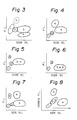
- the separation pattern shown in Fig. 3 is typical of staining with Xanthene dyes, oxacarbocyanine dyes or acridine dyes. Simiar patterns are obtained by constructing two-dimensional plots of the intensities of green and red fluorescence from leukocytes stained with acridine dyes.
- the separation pattern shown in Fig. 4 is typical of staining with Neutral Red.
- the separation pattern shown in Fig. 5 is typical of staining with Astrazon orange G or Auramine O.
- the present inventors also found that there were about 20 dyes with which luekocytes could be stained for classification into three types and the separation pattern that is typical of staining with these dyes is shown in Fig. 6.
- a pattern of the type shown in Fig. 7 is produced by measurement of the intensities of fluorescence and right-angle scattered light. If two fluorescence emissions (i.e., green fluorescence and red fluorescence) that have wavelength appropriate for specific dyes are employed, eosinophils and basophils can be separated from other leukocyte types as shown in Fig. 8 (eosinophils 4 and basophils 5). The remaining components of leukocytes (i.e., lymphocytes, monocytes and neutrophils) can be separated from one another by the intensities of fluorescence and right-angle scattered light as shown in Fig. 9.
- leukocytes i.e., lymphocytes, monocytes and neutrophils
- a dye that produces a pattern of the type shown in Fig. 6 is added to a dye that produces the pattern of Fig. 7, a better resolution of lymphocytes, monocytes and neutrophils is achieved to produce a pattern of the type shown in Fig l0 (in which the respective leukocyte populations are designated by l, 2 and 3).
- a two-stage analysis can be effected by first emplying green and red fluorescence (Fig. 8), then employing fluorescence and right-angle scattered light (Fig. ll).
- a two-dimensional plot of the intensities of right-angle scattered light and red fluorescence from leukocytes stained with Neutral Red is shown in Fig. 4.
- Fig. l2 shows the excitation and emission spectra of fluorescence of Neutral Red.
- Neutral Red produces a specific staining of eosinophils in the band of 580 - 640 nm (orange to red).
- a two-dimensional plot of the pattern shown in Fig. 4 is produced by using a dye solution having a pH of 5 - ll and a dye concentration of 3 - 300 ⁇ g/ml. Even if the dye concentration is less than 3 ⁇ g/ml, a specific pattern of the distribution of eosinophils is produced but the other leukocytes are too noisy to be accurately measured. If one needs to obtain only the signal of eosinophils, the dye concentration may be at least about 0.l ⁇ g/ml.
- a two-dimensional plot of the intensities of right-angle scattered light and green fluorescence from leukocytes stained with Astrazon Orange G is shown in Fig. 5.
- Fig. l3 shows the excitation and emission spectra of fluorescence of Astrazon Orange G.
- Astrazon Orange G produces a specific staining of basophils in the yellow-green band having a central wavelength of about 540 nm.
- a two-dimensional plot of the pattern shown in Fig. 5 is produced by using a dye solution having a pH of 5 - ll and a dye concentration of l - 300 ⁇ g/ml.
- a similar separation pattern is obtained with Auramine O.
- fluorochrome dyes that selectively stain leukocytes can also be used. They stain monocytes to a greater extent than other leukocytes. They are capable of differentiating leukocytes into at least three types in terms of right-angle scattered light and fluorescence as shown in Fig. 6.
- Neutral Red produces a specific staining of eosinophils while Astrazon orange G specifically stains basophils, thereby producing a two-dimensional plot of the intensities of right-angle scattered light and yellow to red fluorescence as shown in Fig. 7.
- This plot is obtained by using a dye solution having a pH of 5 - ll, a Neutral Red concentration of 0.l - 30 ⁇ g/ml, and an Astrazon Orange G concentration of l - 300 ⁇ g/ml.
- leukocytes can be stained in such a way that a better resolution of monocytes (less contamination by lymphocytes and neutrophils) can be attained as compared with the case of using dyes of group d. alone.
- a two-dimensional plot of the intensities of right-angle scattered light and yellow to red fluorescence form leukocytes stained with combinations of neutral Red, Astrazon Orange and other appropriate dyes is shown in Fig. l0.
- Illustrative dyes that fall under category c. and which can be used to produce a separation pattern of the type shown in Fig. l0 include oxacarbocyanine dyes such as DiOC1(3), DiOC2(3), DiOC3(3), DiOC5(3) and DiOC6(3), TA-2 (a styryl dye produced by Nippon Kankoh-Shikiso Kenkyusho Co., Ltd., Okayama, Japan), and cyanine dyes such as 2-[ ⁇ -(l ⁇ -ethyl-4 ⁇ -5 ⁇ -benzothiazolylidene)-propenyl]-l-ethyl-4,5-benzoxazolium iodide.
- oxacarbocyanine dyes such as DiOC1(3), DiOC2(3), DiOC3(3), DiOC5(3) and DiOC6(3)
- TA-2 a styryl dye produced by Nippon Kankoh-Shikiso Kenkyusho Co., Ltd.
- oxacarbocyanine dyes used alone will allow leukocytes to be classified into 4 types, with eosinophils (4) stained to a smaller extent than neutrophils (3). If such dyes are mixed with Neutral Red which has a strong specificity for staining of eosinophils, a plot of the pattern shown in Fig. l0 is obtained, in which eosinophils (4) are distributed above neutrophils (3).
- the buffer is used to maintain the pH of the dye solution at an optimum level. It is important that the pH of the dye solution be maintained at an optimum level since a dye's adsorption mass and this specificity to cytoplasmic proteins vary with pH. Blood itself has a buffering action to maintain a pH near 7.4, so the buffer must be added in an amount sufficient to cancel this action and provide a desired pH.
- buffers such as phosphate, citrate, borate, Tris, Hepes, glycine, carbonate, collidine and taurine are used in amounts ranging from 5 to 200 ppm.
- the osmolarity compensating agent is used to prevent leukocytes from experiencing such defects as extreme deformation and lysis.
- alkaline metal salts are used in amounts of 60 - 380 mM so as to provide an osmolarity that is within the range of 40 - 250% of the physiological osmolarity of human blood (280 mOsm/kg).
- Natural Red and Astrazon Orange G used in the method of the present invention have the following chemical formulae:
- the right-angle scattered light signal reflects the structural information of an individual white cell.
- a lymphocyte contains very few or no granules, so the scattered light produced from the lymphocyte is the weakest of all leukocytes.
- a neutrophil contains many granules and has a large nucleus, so that it produces the most intense scattered light.
- Monocytes, eosinophils and basophils produce scattered light the intensity of which is intermediate between the intensities of scattered light from lymphocytes and neutrophils. For these reasons, the relative intensites of right-angle scattered light from individual leukocyte types are plotted as shown in Fig. 2a.
- the fluorescence signal reflects the cytochemical characters of leukocytes and depending on the interaction between stains and individual leukocyte types, signals of different intensities are produced form the leukocytes.
- leukocytes can be classified into five types by first performing selective staining of eosinophils and basophils so that the clusteres of these two types of leukocytes can be separated from each other by the intensity of fluorescence, and subsequently differentiating the remaining leukocytes (i.e., lymphocytes, monocytes and neutrophils) by means of the intensity of right-angle scattered light.
- leukocytes i.e., lymphocytes, monocytes and neutrophils
- the method of the present invention has the advantage that no cumbersome operations involving a complicated preliminary treatment are required and that the leukocytes in blood alone can be classified and counted with a flow cytometer after a simple two-stage staining operation has been completed.
- FIG. l A specific example of the optics of a flow cytometer employed in the present invention is hereunder described with reference to Fig. l.
- the optics shown in Fig. l is used in a flow cytometer designed for measuring right-angle scattered light, red fluorescence and green fluorescence.
- the optics generally indicated by l0 uses an argon ion laser l2 as a light source and it operates at a wavelength of 488 nm, producing an output of l0 mW.
- Light emitted from the laser l2 is converged by a cylindrical lens l6 and illuminates a blood sample flowing through a flow cell l4.
- the stained leukocytes in the sample When the stained leukocytes in the sample are irradiated by the laser light, they produce scattered light and fluorescence.
- the right-angle scattered light and the fluorescence are converged with a condenser lens l8 and pass through an aperture 20 to fall upon a dichroic mirror 22.
- the dichroic mirror 22 reflects the right-angle scattered light 24 and transmits the flurescence 26.
- the right angle scattered light 24 reflected from the dichroic mirror 22 is detected in a photomultiplier tube 28.
- red fluorescence 32 is reflected by a dichroic mirror 30 and green fluorescence 38 is transmitted through that mirror.
- the reflected red fluorescence 32 passes through a color filter 34 and is detected in a photomultiplier tube 36.
- the transmitted green fluorescence 38 passes through a color filter 40 and is detected in a photomultiplier tube 42.
- erythrocytes in a blood sample are disrupted by an acidic and hypotonic treatment such as to reduce the disturbance that occurs in the intensity distribution of right-angle scattered light on account of coincidence of red and white blood cells.
- a hypotonic treatment is performed in the physiological pH range, not only the erythrocytes but also some leukocytes will be destroyed.
- a hypotonic treatment is performed in an acidic pH range, for example, at a pH between 2.0 and 5.0, leukocytes will remain intact and only erythrocytes will be disrupted. In this case, no morphological changes such as the loss of nuclei, swelling and shrinkage will occur in leukocytes.
- Astrazon Orange G and neutral Red used as fluorochrome dyes in the present invention are described below.
- a sample of anti-coagulated blood is first mixed with the first fluid so that the erythrocytes in the blood are reduced to ghosts and fragments. Subsequently, the second fluid is added so as to stain the leukocytes and platelets in the blood.
- Astrazon Orange G would bind strongly to acidic substances such as heparin and histamine in basophilic granules and, as a consequence, the wavelength of fluorescence emitted from Astrazon Orange G shifts from 520 - 540 nm to 560 - 580 nm (this phenomenon is generally referred to as metachromasia).
- Astrazon Orange G also binds to the granules in the other leukocytes (i.e...
- Astrazon Orange G binds weakly to the surfaces of nuclei and cells and emits fluorescence in the wavelength range of 520 - 540 nm.
- Neutral Red also principally stains granules and emits fluorescence of 620 nm. This dye binds to eosinophilic granules to a greater extent than the granules in other leukocytes, thereby emitting a stronger fluorescence radiation than that emitted from any other leukocytes.
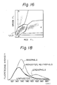
- FIG. l6 A two-dimensional plot constructed from the measurement with a flow cytometer of a blood sample to which both the first and second fluids have been added is shown in Fig. l6, in which Red FL. signifies the relative intensity of red fluorescence and Green FL. denotes the relative intensity of green fluorescence.
- the numerals used in Fig. l6 have the following meanings: l, lymphocytes, 2, monocytes; 3, neutrophils; 4, eosinophils; 5, basophils; and 6, non-leukocytes, namely, platelets and erythrocytic ghosts and fragments (the same symbols and numerals used hereinafter have the same definitions).
- Fig. l6 the leuykocytes are clearly distinguished from platelets and erythrocytic ghosts and fragments denoted by 6 since the latter emit a lower intensity of green fluorescence.
- Eosinophils (4) are completley separated from basophils (5) in the two-dimensional plot of Fig. l6.
- the other leukocytes i.e., lymphocytes l, monocytes 2 and neutrophils 3
- lymphocytes l, monocytes 2 and neutrophils 3 which do not emit any specific fluorescence cannot be separated from one another on the two-dimensional plot of the intensities of green and red fluorescences and can be classified as shown in Fig. 3 based on the intensities of fluorescence and right-angle scattered light.
- compositions, pHs and osmolarities of the first and second fluids used in the method of the present invention are described below in detail.
- Astrazon Orange G produces the best separation of basophils and neutrophils when its final concenration is l5 ppm with the staining pH being at 9.0. If the final concnetration of Astrazon orange G is less than l5 ppm, a lower resolution results because of the decrease in the intensity of green fluorescence from basophils. The same result also occurs if the final concentration of Astrazon Orange G is more than l5 ppm and this is because of the combined effect of the decrease in the intensity of green fluorescence from basophils and the increase in the intensity of green fluorescence from neutrophils. The concentration of Astrazon Orange G that provides an optimum resolution varies with pH. The adsorption mass of Astrazon Orange G decreases with decreasing pH.
- a good resolution between eosinophils and neutrophils can be attained at the higher end of the concentration range of neutral Red from l to l0 ppm.
- Eosinophils have better staining characteristics at lower pHs.
- Neutral Red also stains the granulesin basophils (i.e., the intensity of fluorescence it emits has no specificity to basophils), so it inhibits selective staining of basophils by Astrazon orange G. It is therefore necessary to determine a concentration of neutral Red that provides for good resolution between neutrophils and each of basophils and eosinophils.
- Fig. l5 shows the profiles of resolution between eosinophils and neutrophils and between basophils and neutrophils as a function of the concentration of neutral Red with the concentration of Astrazon Orange G and pH fixed at l5 ppm and 9.0, respecitvely.
- the term "green fluorescence ratio of basophils/neutrophils” means the ratio of the intensity of green fluorescence from basophils to that from neutrophils
- the term “red fluorescence ratio of eosinophils/neutrophils” means the ratio of the intensity of red fluorescence from eosinophils to that from neutrophils (the same expressions used hereinafter have the same meanings). The higher the points in the figure, the better separation that can be achieved between neutrophils and basophils or eosinophils.
- Fig. l5 the separation between basophils and neutrophils coincides with that between eosinophils and neutrophils but in practice, there usually are fewer basophils in leukocytes than eosinophils, so in order to improve the resolution of basophils from neutrophils, it is desirable to set the concentration of neutral Red at a comparatively low level, say 2 ppm.
- the concentrations of Astrazon Orange G and neutral red in the first fluid may be adjusted to l6.5 ppm and 2.2 ppm, respecitvely, in order that their final concentrations will be at l5 ppm and 2 ppm, respectively.
- Fig. l7 shows the profile of resolution between neutrophils and basophils or eosinophils as a function of pH, with the concentrations of Astrazon Orange G and Neutral Red being fixed at l5.0 ppm and 3.0 ppm, respectively.
- the resolution of eosinophils from neutrophils decreases with increasing pH.
- the resolution of basophils from neutrophils increases with the increase in pH up to about 9.0 - 9.5 and decreases thereafter.
- the rate at which basophils are stained i.e., the time required for the intensity of fluorescence to reach a maximum
- the rate at which basophils are stained increases, but once a maximum fluorescence intensity has been reached, the subsequent decrease in fluorescence intensity is rapid.
- the staining rate of eosinophils does not vary greatly with pH.
- the final pH is referred to as the "staining pH”.
- the pH of the first fluid influences the lysing efficiency of erythrocytes. Erythrocytes lyse rapidly at pHs of 5.0 and below, and the lower the pH, the faster the rate of lysis. However, at pHs below 2.0, proteins such as hemoglobin begin to denature as the lysing of erythrocytes progresses, and the rate of protein denaturation increases as pH decreases. A denatured protein will clog at the time when the final "staining" pH has been attained. In consideration of these facts, it is desirable to adjust the pH of the first fluid to be at a value between 2.0 and 5.0.
- the buffer in the first fluid is used to maintain the pH of the first fluid at a level suitable for lysing erythrocytes, and any buffer that has a pKa value of 3.5 ⁇ l.5 may be employed for this purpose.
- Illustrative examples include maleic acid, malonic acid, phthalic acid, diglycolic acid, saliyclic acid, fumaric acid, tartaric acid, citric acid and malic acid.
- the concentration of the buffer is desirably held as low as possible.
- the concentration of the buffer in the first fluid is preferably at 50 mM and below, more preferably at 5 - 30 mM.
- the buffer in the second fluid is used to neutralize the acid in the buffer in the first fluid and to maintain the pH of the resulting dye solution at the staining pH.
- Any buffer that has pKa value of 8.0 - 9.5 may be employed for this purpose.
- Illustrative examples include Tris, tricin, bicine, 2-amino-2-methyl-l,3-propanediol, taurine, boric acid and srine.
- These buffers are preferably used at concentrations of at least l0 mM in terms of the final concentration which is attained as a result of mixing of the first and second fluids.
- the buffer in the second fluid advantageously has a final concentration of 30 - l00 mM.
- the osmolarity of the first fluid is preferably adjusted to a value in the range of 0 - l00 mOsm/kg, more preferably in the range of 0 - 50 mOsm/kg.
- the osmoraltiy of the second fluid determines the fianl osmorarity which is to be attained as a result of mixing the first and second fluids.
- the final osmorality influences the ability of leukocytes to retain their own shape and is preferably within the range of l50 - 600 mOsm/kg, more preferably in the range of l50 - 300 mOsm/kg.
- a dye solution having a pH of 8.0 was prepared by adding l0 ⁇ g/ml of Astrazon Orange G and l ⁇ g/ml of neutral Red to a l0 mM borate buffer solution containing 75 mM of NaCl.
- Two additional dye solutions were prepared in the same manner as desdcibed above except that their pHs were adjusted to 9.0 and l0.0, respectively.
- flow cytometry was conducted as in Example l. With the dye solution having a pH of l0.0, 5-part differentiation of leukocytes could not be successfully achieved by measurement of the intensities of red fluorescence and right-angle scattered light.
- the intended results could be attained by first differentiating basophils from eosinophils in terms of green fluorescence and red fluorescence and then distinguishing between the remaining three types of leukocytes based on green fluorescence and right-angle scattered light.
- the dye solution having a pH of 9.0 5-part differentiation of leukocytes could be accomplished based on red fluorescence and right-angle scattered light.
- the dye solution having a pH of 8.0 4-part differentiation was possible on the basis of the red fluorescence and right-angle scattered light.
- a dye solution was prepared by adding 75 mM NaCI, l0 ⁇ g/ml of Astrazon Orange G and l ⁇ g/ml of Neutral Red to a borate buffer soloution (pH, 9.0) wherein the buffer was incorporated in an amount of 3 mM.
- Two additional dye solutions were prepared in the same manner as described above except that the buffer concentration was adjusted to l0 mM and 30 mM, respectively. Using these dye solutions, flow cytometry was conducted as in Example l. No significant changes in separation pattern were observed within the tested range of buffer concentrations and 5-part differentiation of leukocytes could successfully be achieved with each of the dye solutions.
- Example l Flow cytometry was conducted as in Example l using a dye solution that was composed of a l0 mM borate buffer solution (pH,9.0) containing 75 mM NaCl, l0 ⁇ g/ml of Astrazon orange G and l ⁇ g/ml of Neutral Red.
- the analysis was based on the measurement of the intensities of right-angle scattered light and six fluorescence emissions not shorter in wavelength than 520 nm, 540 nm, 560 nm, 580 nm, 600 nm and 620 nm, respectively, that were collected with a photomultiplier tube 36 in the optics shown in Fig. l.
- a total refelction mirror was used as a dichroic mirror 30, and a long-pass filter as a color filter 34.
- the resolution between basophils and lymphocytes decreased whereas the resolution between eosinophils and neutrophils increased.
- the efficiency of 5-part differentiation of leukocytes was particularly high when fluorescence emissions having wavelengths not shorter than 560 nm and 580 mn were collected.
- Astrazon Orange G l6.5 ppm (selective dye for basophils) Neutral Red 2.2 ppm (selective dye for eosinophils) Citric acid/sodium hydroxide l0 mM (buffer) pH, 3.0; osmolarity, l0 mOsm/kg
- FIG. l6 A two-dimensional plot of the intensities of red and green fluorescences as measured with a flow cytometer under the conditions described above is shown in Fig. l6.
- Population 6 (consisting of platelets, red cell ghosts and fragments) was successfully separated from leukocytes, and it was possible for both an eosinophil cluster 4 and a basophil cluster 5 to be separated from all other leukocytes with high resolution.
- the remaining leukocytes will also successfully separated from one another with good resolution, as indicated in Fig. 2c which is a frequency distribution curve for lymphocytes l, monocytes 2 and neutrophils 3.
- Side Sc. signifies the relative intensity of right-angle scatterd light and Freq. stands for frequency.
- the light source in the flow cytometer used in the present invention is not limited to the aforementioned argon ion laser of low output and any of the other light sources can be emplyed, such as a mercury arc lamp, xenon arc lamp, a He-Cd laser, a He-Ne laser and a krypton ion laer, as well as an argon ion laser of high output. If these light sources are used, the conditions of staining, reaction and measurement may be selected as appropriate.
- the reagent system and the method of the present invention as applied to classify and count leukocytes in blood by flow cytometry have the following advantages.
Applications Claiming Priority (4)
| Application Number | Priority Date | Filing Date | Title |
|---|---|---|---|
| JP213716/86 | 1986-09-10 | ||
| JP61213716A JPH076979B2 (ja) | 1986-09-10 | 1986-09-10 | フロ−サイトメトリ−による白血球分類に使用される試薬 |
| JP282697/86 | 1986-11-27 | ||
| JP61282697A JPH0635972B2 (ja) | 1986-11-27 | 1986-11-27 | フロ−サイトメトリ−による白血球の分類方法 |
Publications (3)
| Publication Number | Publication Date |
|---|---|
| EP0259833A2 true EP0259833A2 (fr) | 1988-03-16 |
| EP0259833A3 EP0259833A3 (en) | 1988-12-07 |
| EP0259833B1 EP0259833B1 (fr) | 1993-01-27 |
Family
ID=26519957
Family Applications (1)
| Application Number | Title | Priority Date | Filing Date |
|---|---|---|---|
| EP19870113112 Expired - Lifetime EP0259833B1 (fr) | 1986-09-10 | 1987-09-08 | Réactif et procédé pour classifier des leucocytes par cytometrie à écoulement |
Country Status (3)
| Country | Link |
|---|---|
| EP (1) | EP0259833B1 (fr) |
| CA (1) | CA1309327C (fr) |
| DE (1) | DE3783838T2 (fr) |
Cited By (9)
| Publication number | Priority date | Publication date | Assignee | Title |
|---|---|---|---|---|
| EP0525398A2 (fr) * | 1991-07-29 | 1993-02-03 | Toa Medical Electronics Co., Ltd. | Méthode pour préparer un échantillon pour la classification et le comptage de leucocytes |
| EP0525397A2 (fr) * | 1991-07-29 | 1993-02-03 | Toa Medical Electronics Co., Ltd. | Méthode de préparation d'échantillon pour la classification et le comptage de leucocytes |
| DE4309328A1 (de) * | 1993-03-18 | 1994-09-22 | Volker Ost | Verfahren zur Unterscheidung von Erythrozyten und Leukozyten im Vollblut mit Methoden der Streulichtmessung in einem Durchflußzytometer zur Zellzählung und Zellsortierung |
| US5378633A (en) * | 1992-02-07 | 1995-01-03 | Sequoia-Turner Corporation, A Corp. Of Ca | Method for accurately enumerating and sensitively qualifying heterogenous cell populations in cytolytic processing conditions |
| US5389549A (en) * | 1987-05-29 | 1995-02-14 | Toa Medical Electronics Co., Ltd. | Method for classifying leukocytes and a reagent used therefor |
| US5693484A (en) * | 1991-05-14 | 1997-12-02 | Toa Medical Electronics Co., Ltd. | Method of classifying and counting cells in urine |
| EP1953526A3 (fr) * | 2007-02-01 | 2013-05-01 | Sysmex Corporation | Analyseur hématologique, procédé d'analyse de liquide organique et produit de programme informatique |
| US10203275B2 (en) | 2010-08-05 | 2019-02-12 | Abbott Point Of Care, Inc. | Method and apparatus for automated whole blood sample analyses from microscopy images |
| EP3392343A4 (fr) * | 2015-12-17 | 2019-07-17 | Quintino De Oliveira, Henrique Jesus | Composition colorante de cellules à base aqueuse, procédé correspondant de préparation et d'utilisation de celle-ci, procédés de préparation d'échantillon et de comptage de cellules |
Citations (4)
| Publication number | Priority date | Publication date | Assignee | Title |
|---|---|---|---|---|
| US3684377A (en) * | 1970-07-13 | 1972-08-15 | Bio Physics Systems Inc | Method for analysis of blood by optical analysis of living cells |
| DE2628468A1 (de) * | 1975-06-27 | 1976-12-30 | Inst Nat Sante Rech Med | Reagens und verfahren zur zaehlung von basophilen |
| EP0106339A2 (fr) * | 1982-10-15 | 1984-04-25 | Max-Planck-Gesellschaft zur Förderung der Wissenschaften e.V. | Procédé pour la détermination quantitative simultanée de cellules et réactif utilisé à cette fin |
| US4581223A (en) * | 1980-03-12 | 1986-04-08 | Lawrence Kass | Individual leukocyte determination by means of differential metachromatic dye sorption |
-
1987
- 1987-09-04 CA CA000546200A patent/CA1309327C/fr not_active Expired - Fee Related
- 1987-09-08 EP EP19870113112 patent/EP0259833B1/fr not_active Expired - Lifetime
- 1987-09-08 DE DE19873783838 patent/DE3783838T2/de not_active Expired - Fee Related
Patent Citations (4)
| Publication number | Priority date | Publication date | Assignee | Title |
|---|---|---|---|---|
| US3684377A (en) * | 1970-07-13 | 1972-08-15 | Bio Physics Systems Inc | Method for analysis of blood by optical analysis of living cells |
| DE2628468A1 (de) * | 1975-06-27 | 1976-12-30 | Inst Nat Sante Rech Med | Reagens und verfahren zur zaehlung von basophilen |
| US4581223A (en) * | 1980-03-12 | 1986-04-08 | Lawrence Kass | Individual leukocyte determination by means of differential metachromatic dye sorption |
| EP0106339A2 (fr) * | 1982-10-15 | 1984-04-25 | Max-Planck-Gesellschaft zur Förderung der Wissenschaften e.V. | Procédé pour la détermination quantitative simultanée de cellules et réactif utilisé à cette fin |
Cited By (15)
| Publication number | Priority date | Publication date | Assignee | Title |
|---|---|---|---|---|
| US5389549A (en) * | 1987-05-29 | 1995-02-14 | Toa Medical Electronics Co., Ltd. | Method for classifying leukocytes and a reagent used therefor |
| US5693484A (en) * | 1991-05-14 | 1997-12-02 | Toa Medical Electronics Co., Ltd. | Method of classifying and counting cells in urine |
| EP0525397A2 (fr) * | 1991-07-29 | 1993-02-03 | Toa Medical Electronics Co., Ltd. | Méthode de préparation d'échantillon pour la classification et le comptage de leucocytes |
| EP0525397A3 (en) * | 1991-07-29 | 1993-06-30 | Toa Medical Electronics Co., Ltd. | Method of preparing specimen for classifying and counting leukocytes |
| EP0525398A3 (en) * | 1991-07-29 | 1993-06-30 | Toa Medical Electronics Co., Ltd. | Method of preparing specimen for classifying and counting leukocytes |
| US5308772A (en) * | 1991-07-29 | 1994-05-03 | Toa Medical Electronics Co. Ltd. | Method for classifying and counting leukocytes |
| EP0525398A2 (fr) * | 1991-07-29 | 1993-02-03 | Toa Medical Electronics Co., Ltd. | Méthode pour préparer un échantillon pour la classification et le comptage de leucocytes |
| US5378633A (en) * | 1992-02-07 | 1995-01-03 | Sequoia-Turner Corporation, A Corp. Of Ca | Method for accurately enumerating and sensitively qualifying heterogenous cell populations in cytolytic processing conditions |
| DE4309328A1 (de) * | 1993-03-18 | 1994-09-22 | Volker Ost | Verfahren zur Unterscheidung von Erythrozyten und Leukozyten im Vollblut mit Methoden der Streulichtmessung in einem Durchflußzytometer zur Zellzählung und Zellsortierung |
| DE4309328C2 (de) * | 1993-03-18 | 1998-03-12 | Volker Ost | Verfahren zur Differenzierung, Konzentrationsbestimmung und Sortierung von Erythrozyten, Thrombozyten und Leukozyten |
| EP1953526A3 (fr) * | 2007-02-01 | 2013-05-01 | Sysmex Corporation | Analyseur hématologique, procédé d'analyse de liquide organique et produit de programme informatique |
| US10203275B2 (en) | 2010-08-05 | 2019-02-12 | Abbott Point Of Care, Inc. | Method and apparatus for automated whole blood sample analyses from microscopy images |
| US10365203B2 (en) | 2010-08-05 | 2019-07-30 | Abbott Point Of Care, Inc. | Method and apparatus for automated whole blood sample analyses from microscopy images |
| US10677711B2 (en) | 2010-08-05 | 2020-06-09 | Abbott Point Of Care, Inc. | Method and apparatus for automated whole blood sample analyses from microscopy images |
| EP3392343A4 (fr) * | 2015-12-17 | 2019-07-17 | Quintino De Oliveira, Henrique Jesus | Composition colorante de cellules à base aqueuse, procédé correspondant de préparation et d'utilisation de celle-ci, procédés de préparation d'échantillon et de comptage de cellules |
Also Published As
| Publication number | Publication date |
|---|---|
| EP0259833A3 (en) | 1988-12-07 |
| EP0259833B1 (fr) | 1993-01-27 |
| CA1309327C (fr) | 1992-10-27 |
| DE3783838T2 (de) | 1993-07-22 |
| DE3783838D1 (de) | 1993-03-11 |
Similar Documents
| Publication | Publication Date | Title |
|---|---|---|
| US5175109A (en) | Reagent for classifying leukocytes by flow cytometry | |
| EP0483116B1 (fr) | Méthode pour classifier des leucocytes par cytométrie de flux et réactifs à cet effet | |
| US5434081A (en) | Method of classifying leukocytes by flow cytometry | |
| US5693484A (en) | Method of classifying and counting cells in urine | |
| EP0545315B1 (fr) | Compositions réactives et leur utilisation dans l'identification et caractérisation de réticulocytes du sang entier | |
| JP3048260B2 (ja) | 白血球分類計数用試料調製方法 | |
| CA1309326C (fr) | Methode de classification des leucocytes par cytometrie de flux, et reactifs utilises dans le cadre de cette methode | |
| US9797824B2 (en) | Method for hematology analysis | |
| US5179026A (en) | Method of classifying leukocytes by flow cytometry and reagents used in the method | |
| CA1309327C (fr) | Reactif et methode de classification des leucocytes par cytometrie de flux | |
| JPH0611506A (ja) | 白血球測定用試薬及び白血球測定用試料調製方法 | |
| JPH079423B2 (ja) | フローサイトメトリーによる白血球分類試薬 | |
| JPH0650310B2 (ja) | フローサイトメトリーによる白血球の分類方法 | |
| JPH0664053B2 (ja) | フロ−サイトメトリ−による白血球の分類方法 | |
| JPS6370167A (ja) | フロ−サイトメトリ−による白血球分類に使用される試薬 |
Legal Events
| Date | Code | Title | Description |
|---|---|---|---|
| PUAI | Public reference made under article 153(3) epc to a published international application that has entered the european phase |
Free format text: ORIGINAL CODE: 0009012 |
|
| AK | Designated contracting states |
Kind code of ref document: A2 Designated state(s): DE FR GB IT NL |
|
| PUAL | Search report despatched |
Free format text: ORIGINAL CODE: 0009013 |
|
| AK | Designated contracting states |
Kind code of ref document: A3 Designated state(s): DE FR GB IT NL |
|
| 17P | Request for examination filed |
Effective date: 19881229 |
|
| 17Q | First examination report despatched |
Effective date: 19910607 |
|
| RAP1 | Party data changed (applicant data changed or rights of an application transferred) |
Owner name: TOA MEDICAL ELECTRONICS CO., LTD. |
|
| ITF | It: translation for a ep patent filed |
Owner name: STUDIO TORTA SOCIETA' SEMPLICE |
|
| GRAA | (expected) grant |
Free format text: ORIGINAL CODE: 0009210 |
|
| AK | Designated contracting states |
Kind code of ref document: B1 Designated state(s): DE FR GB IT NL |
|
| REF | Corresponds to: |
Ref document number: 3783838 Country of ref document: DE Date of ref document: 19930311 |
|
| ET | Fr: translation filed | ||
| PLBE | No opposition filed within time limit |
Free format text: ORIGINAL CODE: 0009261 |
|
| STAA | Information on the status of an ep patent application or granted ep patent |
Free format text: STATUS: NO OPPOSITION FILED WITHIN TIME LIMIT |
|
| 26N | No opposition filed | ||
| PGFP | Annual fee paid to national office [announced via postgrant information from national office to epo] |
Ref country code: GB Payment date: 19970901 Year of fee payment: 11 |
|
| PGFP | Annual fee paid to national office [announced via postgrant information from national office to epo] |
Ref country code: NL Payment date: 19970929 Year of fee payment: 11 |
|
| PG25 | Lapsed in a contracting state [announced via postgrant information from national office to epo] |
Ref country code: GB Free format text: LAPSE BECAUSE OF NON-PAYMENT OF DUE FEES Effective date: 19980908 |
|
| PG25 | Lapsed in a contracting state [announced via postgrant information from national office to epo] |
Ref country code: NL Free format text: LAPSE BECAUSE OF NON-PAYMENT OF DUE FEES Effective date: 19990401 |
|
| GBPC | Gb: european patent ceased through non-payment of renewal fee |
Effective date: 19980908 |
|
| NLV4 | Nl: lapsed or anulled due to non-payment of the annual fee |
Effective date: 19990401 |
|
| PGFP | Annual fee paid to national office [announced via postgrant information from national office to epo] |
Ref country code: FR Payment date: 20020910 Year of fee payment: 16 |
|
| PGFP | Annual fee paid to national office [announced via postgrant information from national office to epo] |
Ref country code: DE Payment date: 20020911 Year of fee payment: 16 |
|
| PG25 | Lapsed in a contracting state [announced via postgrant information from national office to epo] |
Ref country code: DE Free format text: LAPSE BECAUSE OF NON-PAYMENT OF DUE FEES Effective date: 20040401 |
|
| PG25 | Lapsed in a contracting state [announced via postgrant information from national office to epo] |
Ref country code: FR Free format text: LAPSE BECAUSE OF NON-PAYMENT OF DUE FEES Effective date: 20040528 |
|
| REG | Reference to a national code |
Ref country code: FR Ref legal event code: ST |
|
| PG25 | Lapsed in a contracting state [announced via postgrant information from national office to epo] |
Ref country code: IT Free format text: LAPSE BECAUSE OF NON-PAYMENT OF DUE FEES;WARNING: LAPSES OF ITALIAN PATENTS WITH EFFECTIVE DATE BEFORE 2007 MAY HAVE OCCURRED AT ANY TIME BEFORE 2007. THE CORRECT EFFECTIVE DATE MAY BE DIFFERENT FROM THE ONE RECORDED. Effective date: 20050908 |