EP0258221B1 - Coaxial arteriovenous grafting tube - Google Patents
Coaxial arteriovenous grafting tube Download PDFInfo
- Publication number
- EP0258221B1 EP0258221B1 EP86901236A EP86901236A EP0258221B1 EP 0258221 B1 EP0258221 B1 EP 0258221B1 EP 86901236 A EP86901236 A EP 86901236A EP 86901236 A EP86901236 A EP 86901236A EP 0258221 B1 EP0258221 B1 EP 0258221B1
- Authority
- EP
- European Patent Office
- Prior art keywords
- tube
- inner tube
- outer tube
- internal diameter
- tubes
- Prior art date
- Legal status (The legal status is an assumption and is not a legal conclusion. Google has not performed a legal analysis and makes no representation as to the accuracy of the status listed.)
- Expired - Lifetime
Links
Images

Classifications
-
- A—HUMAN NECESSITIES
- A61—MEDICAL OR VETERINARY SCIENCE; HYGIENE
- A61M—DEVICES FOR INTRODUCING MEDIA INTO, OR ONTO, THE BODY; DEVICES FOR TRANSDUCING BODY MEDIA OR FOR TAKING MEDIA FROM THE BODY; DEVICES FOR PRODUCING OR ENDING SLEEP OR STUPOR
- A61M39/00—Tubes, tube connectors, tube couplings, valves, access sites or the like, specially adapted for medical use
- A61M39/02—Access sites
- A61M39/0208—Subcutaneous access sites
-
- A—HUMAN NECESSITIES
- A61—MEDICAL OR VETERINARY SCIENCE; HYGIENE
- A61F—FILTERS IMPLANTABLE INTO BLOOD VESSELS; PROSTHESES; DEVICES PROVIDING PATENCY TO, OR PREVENTING COLLAPSING OF, TUBULAR STRUCTURES OF THE BODY, e.g. STENTS; ORTHOPAEDIC, NURSING OR CONTRACEPTIVE DEVICES; FOMENTATION; TREATMENT OR PROTECTION OF EYES OR EARS; BANDAGES, DRESSINGS OR ABSORBENT PADS; FIRST-AID KITS
- A61F2/00—Filters implantable into blood vessels; Prostheses, i.e. artificial substitutes or replacements for parts of the body; Appliances for connecting them with the body; Devices providing patency to, or preventing collapsing of, tubular structures of the body, e.g. stents
- A61F2/02—Prostheses implantable into the body
- A61F2/04—Hollow or tubular parts of organs, e.g. bladders, tracheae, bronchi or bile ducts
- A61F2/06—Blood vessels
Definitions
- This invention relates to a tube for use as a vascular graft to permit in vivo hemoaccess.
- Dialysis treatment for patients with renal failure require a ready access to blood vessels in order for blood to be continuously withdrawn from the patient in amounts of over 200 ml/min.
- the blood withdrawn from the patient is passed through a dailysis machine, and returned to the patient.
- a very common method of producing this hemoaccess is the use of expanded polytetrafluorethylene (PTFE) tubes as grafts which are surgically placed between an artery and a vein (PTFE AV fistula). This procedure is especially useful in patients who do not have blood vessels which will support the construction of a more traditional primary fistula on the forearm.
- PTFE expanded polytetrafluorethylene
- a coaxial double tube for use in hemoaccess wherein the space between the two tubes is filled with a self-sealing, nonbiodegradable polymer (hereinafter "filled double tubes") has now been discovered.
- the filled double tube does not bleed upon puncture with a dialysis needle even immediately after surgery.
- Ex vivo perfusion tests and AV fistula experiments in dogs clearly show that the filled double tubes are superior as grafts to the known single PTFE tube in the amount of bleeding resulting from puncture of the tube with a dialysis needle.
- the advantages of the filled double tube are that (1) it can be used for hemoaccess immediately after implantation, (2) the lack of bleeding results in a lower incidence of perigraft hematomas and a lower incidence of pseudoaneurysms, (3) the lack of bleeding upon puncture obviates the need for prolonged compression of the puncture site with secondary occlusion of the tube, and (4) due to the increased rigidity of the filled double tube, puncturing of the tube is easier.
- FR-A-2 215 248 discloses a double tube having improved leakage resistance which is intended for use as part of the conduits of a dialysis or similar device but which is not for implantation.
- the present invention provides a tube for use as a vascular graft to permit in vivo hemoaccess, which tube comprises an outer tube positioned over an inner tube, wherein the outer tube has an internal diameter which is at least 1 mm larger than the external diameter of the inner tube creating a space between the outer tube and the inner tube and that a self-sealing material evenly fills the space between the outer tube and the inner tube, said material comprising a nonbiodegradable, biocompatible polymer adhesive.
- the filled double tube comprises an inner tube 11 and an outer tube 13 which is positioned over the center portion of the inner tube leaving a space 15 between the inner and outer tube 13 and allowing the ends of the inner tube to extend equally beyond the ends of the outer tube.
- the space 15 is filled with a self-sealing, nonbiodegradeable, biocompatible polymer (hereinafter "polymer”) for example a silicone rubber sealant (Dow Corning Catalogue No. 732-3).
- the inner tube 11 can have an internal diameter from about 4 mm to 8 mm with 6 mm being the preferred internal diameter and a wall thickness of about 0.5 to 1 mm.
- the outer tube 13 can have an internal diameter from about 6 mm to 10 mm which is at least 1 mm larger than the external diameter (internal diameter + wall thickness) of the inner tube with 8 mm being the preferred internal diameter and a wall thickness of about 0.5 mm to 1 mm, preferably 1 mm.
- the wall thickness of the inner and outer tubes may be the same or different.
- the length of the inner tube 11 depends on the specific application for the tube, and the site in the patient at which the filled double tube is implanted. As a result, the inner tube 11 can vary in length from 10 mm to 500 mm with 150 mm being the preferred length for looped forearm grafts.
- the length of the outer tube 13 is equal to or less than that of the inner tube 11 and can vary in length from 60 mm to 400 mm with 100 mm being the preferred length for forearm grafts.
- the inner tube 11 can be made from any hemocompatible material suitable as a conduit. Suitable material for the inner and outer tubes are, for example, Teflon PTFE and Dacron synthetic polyester fiber.
- the outer tube 13 can be made from any biocompatible material suitable as a conduit.
- the filled double tube is made entirely from GORE-TEX reinforced PTFE tubing developed by W. F. Gore & Associates, Inc. by rapidly stretching highly crystalline unsintered polytetrafluorethylene.
- the filled double tube can be used for any medical technique in which repeated hemoaccess is required, for example, but without intending to limit the possible applications, intravenous drug administration, chronic insulin injections, chemotherapy, frequent blood samples, connection to artificial lungs and hyperalimentation.
- the filled double tube is ideally suited for use in chronic hemodialysis access for example in a looped forearm graft fistula, straight forearm graft fistula, straight arm graft fistula, looped thigh graft fistula and axillaryaxillary graft fistula or any other AV fistula application.
- the filled double tube may also be useful for among other things, arterial bypasses subjected to external pressure (for example, axillo-femoral, axillo-axillo bypasses) and other replacement surgery.
- the inner surface of the outer tube and the outer surface of the inner tube may be rough to prevent possible separation of the polymer from the tube surface during use.
- Example 2 The filled double tubes used in the following Examples (2-3) were prepared by the procedures outlined in Example 1. The same procedure can be used to prepare filled double tubes of various sizes and lengths.
- a 150 mm segment of a 6 mm internal diameter thick GORE-TEX reinforced PTFE tube was placed over a metallic rod.
- the rod had a diameter of 6 mm and fit the lumen of the tube tightly.
- a 70 mm segment of a 8 mm internal diameter thick GORE-TEX reinforced PTFE tube was placed over the inner tube with the rod in its lumen.
- the outer tube was centered over the inner tube in order to leave about 40 mm of the inner tube free on both ends.
- One of the ends of the outer tube was sealed with silicone rubber sealant (Dow Corning Cat. #732-3) by using a 5 ml syringe filled with silicone sealant and equipped with an angiocatheter. After allowing the tubes to stand for 24 hours the sealant was cured and the opening at one end was completely sealed. The remaining space between the inner and outer tubes was filled by diluting silicone sealant 3:1 (w/v) with acetic acid and placing the diluted silicone sealant in a 5 cc syringe equipped with a 2 mm diameter catheter, The catheter was inserted into the remaining space between the tubes from the unsealed end and the syringe was placed in a Harvard pump.
- silicone rubber sealant Dow Corning Cat. #732-3
- the pump injected the silicone into the unsealed end and the catheter was slowly withdrawn. As soon as excess sealant began to appear at the open end, injection was discontinued, The filled double tube was then rolled on a smooth surface to distribute the silicone uniformly in the space between the tubes and after setting for an additional 24 hours the silicone sealant was cured and the silicone filled double tube was removed from the metallic rod and was ready for use.
- a 60 mm double tube having as an inner tube a 4 mm internal diameter GORE-TEX PTFE thin tube and as an outer tube a coaxial 6 mm thick GORE-TEX PTFE tube with the space between the tubes filled with silicone rubber sealant (“filled double") was employed.
- the following control tubes of the same lengths were used: (1) an unfilled double tube using a 4 mm inner thin wall GORE-TEX PTFE tube and a 6 mm outer thick wall GORE-TEX PTFE tube without silicone in the space between the tubes (“double"), (2) a thin 4 mm conventional GORE-TEX PTFE tube (“thin”), and (3) a thick 6 mm conventional GORE-TEX PTFE tube ("thick”).
- a Scribner shunt was placed between the femoral artery and the femoral vein in each dog.
- Each of the tubes to be tested was placed between the arterial and venous branches of the shunts.
- the four different types of tubes were inserted three times in various sequences in each dog (12 measurements per dog).
- Puncturing was performed in each case by piercing the tube with an 18-gauge needle. Intraluminal pressure measurements were carried out with each puncture in order to be certain that the tube was open and that the pressure condition remained constant. After removing the needle, each tube puncture site was allowed to bleed freely into a cup for 30 seconds. The Scribner shunt was then clamped and the tube changed. The amount of bleeding was measured by weighing the cup before and after bleeding. The results of these experiments are set forth in Tables 1, 2 and 3 and Figure 3 and clearly demonstrate a very significant reduction in bleeding after puncture ex vivo using the new filled double tube ("filled double") for the graft.
- Eleven dogs had a fistula constructed between the right carotid artery and the left external jugular vein, using a graft going through a subcutaneous tunnel in the posterior aspect of the neck.
- a 6 mm diameter conventional GORE-TEX PTFE tube 120 mm in length was used in 5 dogs and a 120 mm filled double tube prepared according to Example 1 was tested on 6 dogs.
- Each graft was punctured on days 1, 3-4, and 7-8 after construction of the fistula. Five minutes after removing the needle, a small incision over the puncture area was made, thereby exposing the tube. The amount of blood present was estimated by absorbing it with sponges and weighing them. The results are shown in Table 4.
- Example 2 The results described in Examples 2 and 3 clearly demonstrate the superior characteristics of the filled double tube over conventional grafts for hemoaccess in terms of the amount of bleeding resulting from a needle puncture. This was clearly verified both in the ex vivo experiment (Example 2) and in the in vivo interposition AV fistulas (Example 3).
Landscapes
- Health & Medical Sciences (AREA)
- Heart & Thoracic Surgery (AREA)
- Life Sciences & Earth Sciences (AREA)
- Veterinary Medicine (AREA)
- Pulmonology (AREA)
- Public Health (AREA)
- General Health & Medical Sciences (AREA)
- Engineering & Computer Science (AREA)
- Biomedical Technology (AREA)
- Animal Behavior & Ethology (AREA)
- Transplantation (AREA)
- Vascular Medicine (AREA)
- Oral & Maxillofacial Surgery (AREA)
- Cardiology (AREA)
- Gastroenterology & Hepatology (AREA)
- Anesthesiology (AREA)
- Hematology (AREA)
- Materials For Medical Uses (AREA)
- Prostheses (AREA)
- Waveguide Connection Structure (AREA)
Abstract
Description
- This invention relates to a tube for use as a vascular graft to permit in vivo hemoaccess.
- Dialysis treatment for patients with renal failure require a ready access to blood vessels in order for blood to be continuously withdrawn from the patient in amounts of over 200 ml/min. The blood withdrawn from the patient is passed through a dailysis machine, and returned to the patient. A very common method of producing this hemoaccess is the use of expanded polytetrafluorethylene (PTFE) tubes as grafts which are surgically placed between an artery and a vein (PTFE AV fistula). This procedure is especially useful in patients who do not have blood vessels which will support the construction of a more traditional primary fistula on the forearm.
- These PTFE grafts cannot be used safely to withdraw blood however until they have been in place for a minimum of 14 days after surgery and have become surrounded by fibrotic tissue. Because of the bleeding which occurs at the site of a needle puncture in these grafts if fibrotic tissue is absent, common complications encountered with early puncturing of PTFE AV fistulas are a hematoma surrounding the graft, false aneurysm and possible graft occlusion. The Atlas of Angioaccess Surgery by Pedro A. Rubio and Edward M. Farrell (Year Book Medical Publishers, Inc., 1983) states that "graft fistulas should not be punctured before the third postoperative week. Earlier puncture risks hematoma formation in the subcutaneous tunnel following needle withdrawal, which will result in further delay in healing." Various materials in addition to PTFE have been tried including autologous saphenous vein, Dacron synthetic polyester fiber velour, modified bovine carotid xenograft, and modified human umbilical vein, but none have overcome the problems associated with early puncture of the graft.
- A coaxial double tube for use in hemoaccess wherein the space between the two tubes is filled with a self-sealing, nonbiodegradable polymer (hereinafter "filled double tubes") has now been discovered. The filled double tube does not bleed upon puncture with a dialysis needle even immediately after surgery. Ex vivo perfusion tests and AV fistula experiments in dogs clearly show that the filled double tubes are superior as grafts to the known single PTFE tube in the amount of bleeding resulting from puncture of the tube with a dialysis needle.
- The advantages of the filled double tube are that (1) it can be used for hemoaccess immediately after implantation, (2) the lack of bleeding results in a lower incidence of perigraft hematomas and a lower incidence of pseudoaneurysms, (3) the lack of bleeding upon puncture obviates the need for prolonged compression of the puncture site with secondary occlusion of the tube, and (4) due to the increased rigidity of the filled double tube, puncturing of the tube is easier.
- FR-A-2 215 248 discloses a double tube having improved leakage resistance which is intended for use as part of the conduits of a dialysis or similar device but which is not for implantation.
- The present invention provides a tube for use as a vascular graft to permit in vivo hemoaccess, which tube comprises an outer tube positioned over an inner tube, wherein the outer tube has an internal diameter which is at least 1 mm larger than the external diameter of the inner tube creating a space between the outer tube and the inner tube and that a self-sealing material evenly fills the space between the outer tube and the inner tube, said material comprising a nonbiodegradable, biocompatible polymer adhesive.
-
- Figure 1 is a plan view of the claimed filled double tube;
- Figure 2 is a cross-sectional view of the claimed filled double tube; and
- Figure 3 is a chart which compares the blood loss from the filled double tube when used as a graft for hemoaccess with the blood loss from other conventional grafts.
- As illustrated in Figures 1 and 2, the filled double tube comprises an inner tube 11 and an
outer tube 13 which is positioned over the center portion of the inner tube leaving aspace 15 between the inner andouter tube 13 and allowing the ends of the inner tube to extend equally beyond the ends of the outer tube. Thespace 15 is filled with a self-sealing, nonbiodegradeable, biocompatible polymer (hereinafter "polymer") for example a silicone rubber sealant (Dow Corning Catalogue No. 732-3). - The inner tube 11 can have an internal diameter from about 4 mm to 8 mm with 6 mm being the preferred internal diameter and a wall thickness of about 0.5 to 1 mm. The
outer tube 13 can have an internal diameter from about 6 mm to 10 mm which is at least 1 mm larger than the external diameter (internal diameter + wall thickness) of the inner tube with 8 mm being the preferred internal diameter and a wall thickness of about 0.5 mm to 1 mm, preferably 1 mm. The wall thickness of the inner and outer tubes may be the same or different. - The length of the inner tube 11 depends on the specific application for the tube, and the site in the patient at which the filled double tube is implanted. As a result, the inner tube 11 can vary in length from 10 mm to 500 mm with 150 mm being the preferred length for looped forearm grafts. The length of the
outer tube 13 is equal to or less than that of the inner tube 11 and can vary in length from 60 mm to 400 mm with 100 mm being the preferred length for forearm grafts. The inner tube 11 can be made from any hemocompatible material suitable as a conduit. Suitable material for the inner and outer tubes are, for example, Teflon PTFE and Dacron synthetic polyester fiber. Theouter tube 13 can be made from any biocompatible material suitable as a conduit. Examples of suitable materials are Teflon PTFE and Dacron synthetic polyester fiber. Preferably, the filled double tube is made entirely from GORE-TEX reinforced PTFE tubing developed by W. F. Gore & Associates, Inc. by rapidly stretching highly crystalline unsintered polytetrafluorethylene. - The filled double tube can be used for any medical technique in which repeated hemoaccess is required, for example, but without intending to limit the possible applications, intravenous drug administration, chronic insulin injections, chemotherapy, frequent blood samples, connection to artificial lungs and hyperalimentation.
- The filled double tube is ideally suited for use in chronic hemodialysis access for example in a looped forearm graft fistula, straight forearm graft fistula, straight arm graft fistula, looped thigh graft fistula and axillaryaxillary graft fistula or any other AV fistula application. Because of its hard consistency and resistance to collapse, the filled double tube may also be useful for among other things, arterial bypasses subjected to external pressure (for example, axillo-femoral, axillo-axillo bypasses) and other replacement surgery.
- In an alternate embodiment of the invention, the inner surface of the outer tube and the outer surface of the inner tube may be rough to prevent possible separation of the polymer from the tube surface during use.
- The filled double tubes used in the following Examples (2-3) were prepared by the procedures outlined in Example 1. The same procedure can be used to prepare filled double tubes of various sizes and lengths.
- A 150 mm segment of a 6 mm internal diameter thick GORE-TEX reinforced PTFE tube was placed over a metallic rod. The rod had a diameter of 6 mm and fit the lumen of the tube tightly. A 70 mm segment of a 8 mm internal diameter thick GORE-TEX reinforced PTFE tube was placed over the inner tube with the rod in its lumen. The outer tube was centered over the inner tube in order to leave about 40 mm of the inner tube free on both ends.
- One of the ends of the outer tube was sealed with silicone rubber sealant (Dow Corning Cat. #732-3) by using a 5 ml syringe filled with silicone sealant and equipped with an angiocatheter. After allowing the tubes to stand for 24 hours the sealant was cured and the opening at one end was completely sealed. The remaining space between the inner and outer tubes was filled by diluting silicone sealant 3:1 (w/v) with acetic acid and placing the diluted silicone sealant in a 5 cc syringe equipped with a 2 mm diameter catheter, The catheter was inserted into the remaining space between the tubes from the unsealed end and the syringe was placed in a Harvard pump.
- The pump injected the silicone into the unsealed end and the catheter was slowly withdrawn. As soon as excess sealant began to appear at the open end, injection was discontinued, The filled double tube was then rolled on a smooth surface to distribute the silicone uniformly in the space between the tubes and after setting for an additional 24 hours the silicone sealant was cured and the silicone filled double tube was removed from the metallic rod and was ready for use.
- A 60 mm double tube having as an inner tube a 4 mm internal diameter GORE-TEX PTFE thin tube and as an outer tube a coaxial 6 mm thick GORE-TEX PTFE tube with the space between the tubes filled with silicone rubber sealant ("filled double") was employed. For comparison the following control tubes of the same lengths were used: (1) an unfilled double tube using a 4 mm inner thin wall GORE-TEX PTFE tube and a 6 mm outer thick wall GORE-TEX PTFE tube without silicone in the space between the tubes ("double"), (2) a thin 4 mm conventional GORE-TEX PTFE tube ("thin"), and (3) a thick 6 mm conventional GORE-TEX PTFE tube ("thick"). Four dogs were used in this experiment.
- A Scribner shunt was placed between the femoral artery and the femoral vein in each dog. Each of the tubes to be tested was placed between the arterial and venous branches of the shunts. The four different types of tubes were inserted three times in various sequences in each dog (12 measurements per dog).
- Puncturing was performed in each case by piercing the tube with an 18-gauge needle. Intraluminal pressure measurements were carried out with each puncture in order to be certain that the tube was open and that the pressure condition remained constant. After removing the needle, each tube puncture site was allowed to bleed freely into a cup for 30 seconds. The Scribner shunt was then clamped and the tube changed. The amount of bleeding was measured by weighing the cup before and after bleeding. The results of these experiments are set forth in Tables 1, 2 and 3 and Figure 3 and clearly demonstrate a very significant reduction in bleeding after puncture ex vivo using the new filled double tube ("filled double") for the graft.
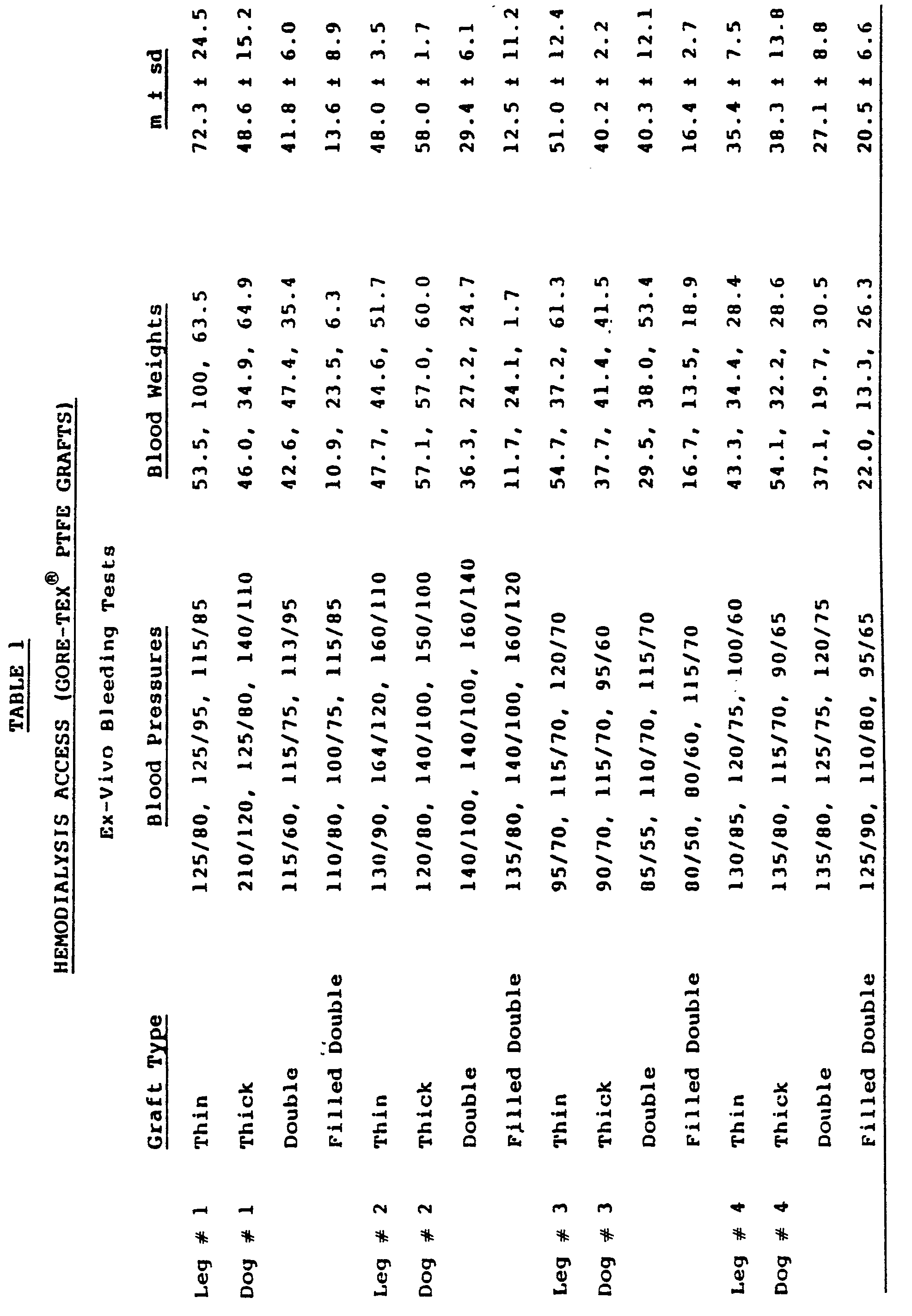


- Eleven dogs had a fistula constructed between the right carotid artery and the left external jugular vein, using a graft going through a subcutaneous tunnel in the posterior aspect of the neck. A 6 mm diameter conventional GORE-TEX PTFE tube 120 mm in length was used in 5 dogs and a 120 mm filled double tube prepared according to Example 1 was tested on 6 dogs. Each graft was punctured on days 1, 3-4, and 7-8 after construction of the fistula. Five minutes after removing the needle, a small incision over the puncture area was made, thereby exposing the tube. The amount of blood present was estimated by absorbing it with sponges and weighing them. The results are shown in Table 4.
- In the control group, 8 punctures resulted in the loss of over 100 ml of blood. Two punctures bled between 40 and 100 ml, 2 punctures bled 20 ml and 40 ml and only one puncture bled less than 20 ml. In 10 instances, it was necessary to place a stitch in the tube in order to stop the bleeding, In the filled double tube, only 3 out of 14 punctures bled. Two bled between 20 ml and 40 ml and the third one bled less than 20 ml, Not one puncture required stitches for hemostasis.
- The results described in Examples 2 and 3 clearly demonstrate the superior characteristics of the filled double tube over conventional grafts for hemoaccess in terms of the amount of bleeding resulting from a needle puncture. This was clearly verified both in the ex vivo experiment (Example 2) and in the in vivo interposition AV fistulas (Example 3).

Claims (7)
Priority Applications (1)
| Application Number | Priority Date | Filing Date | Title |
|---|---|---|---|
| AT86901236T ATE52698T1 (en) | 1986-01-29 | 1986-01-29 | COAXIAL NEEDLES FOR VENOUS GRAFT. |
Applications Claiming Priority (1)
| Application Number | Priority Date | Filing Date | Title |
|---|---|---|---|
| PCT/US1986/000211 WO1987004627A1 (en) | 1986-01-29 | 1986-01-29 | Coaxial double lumen anteriovenous grafting tube |
Publications (2)
| Publication Number | Publication Date |
|---|---|
| EP0258221A1 EP0258221A1 (en) | 1988-03-09 |
| EP0258221B1 true EP0258221B1 (en) | 1990-05-16 |
Family
ID=22195360
Family Applications (1)
| Application Number | Title | Priority Date | Filing Date |
|---|---|---|---|
| EP86901236A Expired - Lifetime EP0258221B1 (en) | 1986-01-29 | 1986-01-29 | Coaxial arteriovenous grafting tube |
Country Status (6)
| Country | Link |
|---|---|
| EP (1) | EP0258221B1 (en) |
| JP (1) | JPS63502640A (en) |
| AT (1) | ATE52698T1 (en) |
| AU (1) | AU584397B2 (en) |
| DE (1) | DE3671171D1 (en) |
| WO (1) | WO1987004627A1 (en) |
Families Citing this family (3)
| Publication number | Priority date | Publication date | Assignee | Title |
|---|---|---|---|---|
| US5100422A (en) * | 1989-05-26 | 1992-03-31 | Impra, Inc. | Blood vessel patch |
| FR2694688B1 (en) * | 1992-08-11 | 1994-11-10 | Novadis Sarl | Expandable tubular prosthesis. |
| GB0524087D0 (en) * | 2005-11-25 | 2006-01-04 | Symetis Ag | Biodegradable scaffold |
Family Cites Families (3)
| Publication number | Priority date | Publication date | Assignee | Title |
|---|---|---|---|---|
| US3112748A (en) * | 1960-04-04 | 1963-12-03 | Pharmaseal Lab | Surgical tube |
| US3814137A (en) * | 1973-01-26 | 1974-06-04 | Baxter Laboratories Inc | Injection site for flow conduits containing biological fluids |
| AU512689B2 (en) * | 1976-10-06 | 1980-10-23 | Dow Chemical Company, The | Infusion tube access site |
-
1986
- 1986-01-29 AT AT86901236T patent/ATE52698T1/en not_active IP Right Cessation
- 1986-01-29 EP EP86901236A patent/EP0258221B1/en not_active Expired - Lifetime
- 1986-01-29 DE DE8686901236T patent/DE3671171D1/en not_active Expired - Lifetime
- 1986-01-29 WO PCT/US1986/000211 patent/WO1987004627A1/en not_active Ceased
- 1986-01-29 AU AU54527/86A patent/AU584397B2/en not_active Ceased
- 1986-01-29 JP JP61501126A patent/JPS63502640A/en active Pending
Also Published As
| Publication number | Publication date |
|---|---|
| ATE52698T1 (en) | 1990-06-15 |
| WO1987004627A1 (en) | 1987-08-13 |
| AU5452786A (en) | 1987-08-25 |
| AU584397B2 (en) | 1989-05-25 |
| JPS63502640A (en) | 1988-10-06 |
| EP0258221A1 (en) | 1988-03-09 |
| DE3671171D1 (en) | 1990-06-21 |
Similar Documents
| Publication | Publication Date | Title |
|---|---|---|
| US4619641A (en) | Coaxial double lumen anteriovenous grafts | |
| JP3748881B2 (en) | Porosity controlled implantable first lumen device | |
| US3713441A (en) | Method of using an artery vein shunt applique | |
| CA2377483C (en) | Graft-catheter vascular access system | |
| US6053901A (en) | Subcutaneously implanted cannula and method for arterial access | |
| CA1150122A (en) | Double-lumen cannula | |
| US3818511A (en) | Medical prosthesis for ducts or conduits | |
| US5562617A (en) | Implantable vascular device | |
| EP1007128A1 (en) | Subcutaneously implanted cannula and method for arterial access | |
| US4484912A (en) | Atraumatic blood access device sealing mechanism | |
| KR20030036687A (en) | Distally narrowed vascular grafts | |
| Flores et al. | Dacron arterio-venous shunts for vascular access in hemodialysis | |
| AU584397B2 (en) | Coaxial double lumen anteriovenous grafting tube | |
| Levowitz et al. | Prosthetic arteriovenous fistula for vascular access in hemodialysis | |
| Schanzer et al. | A self-sealing dialysis prosthesis. Coaxial double PTFE-silicone graft | |
| CN215019710U (en) | an artificial blood vessel | |
| NAKAZONO et al. | New concepts in blood access devices (preliminary study) | |
| Ross | Insertion of short introducer sheaths into a polyurethane vascular access graft for immediate hemodialysis access | |
| Jacob et al. | Dacron arterio-venous interposition graft: An access to circulation in patients on chronic hemodialysis | |
| SITES | Vascular access and other techniques for dialysis and chemotherapy | |
| Tellis | Recent techniques in vascular access | |
| Kootstra et al. | Abstracts of posters in Unusual procedures and desperate cases | |
| US20040064079A1 (en) | Transcutaneous hemodialysis access system | |
| McEvoy et al. | Development of a self-sealing blood access device | |
| CN111921037A (en) | Intelligent blood transfusion device and method for hematology department |
Legal Events
| Date | Code | Title | Description |
|---|---|---|---|
| PUAI | Public reference made under article 153(3) epc to a published international application that has entered the european phase |
Free format text: ORIGINAL CODE: 0009012 |
|
| 17P | Request for examination filed |
Effective date: 19871027 |
|
| AK | Designated contracting states |
Kind code of ref document: A1 Designated state(s): AT BE CH DE FR GB IT LI LU NL SE |
|
| 17Q | First examination report despatched |
Effective date: 19880831 |
|
| ITF | It: translation for a ep patent filed | ||
| RAP1 | Party data changed (applicant data changed or rights of an application transferred) |
Owner name: MOUNT SINAI SCHOOL OF MEDICINE OF THE CITY UNIVERS |
|
| GRAA | (expected) grant |
Free format text: ORIGINAL CODE: 0009210 |
|
| AK | Designated contracting states |
Kind code of ref document: B1 Designated state(s): AT BE CH DE FR GB IT LI LU NL SE |
|
| REF | Corresponds to: |
Ref document number: 52698 Country of ref document: AT Date of ref document: 19900615 Kind code of ref document: T |
|
| REF | Corresponds to: |
Ref document number: 3671171 Country of ref document: DE Date of ref document: 19900621 |
|
| ET | Fr: translation filed | ||
| PG25 | Lapsed in a contracting state [announced via postgrant information from national office to epo] |
Ref country code: AT Effective date: 19910129 |
|
| PG25 | Lapsed in a contracting state [announced via postgrant information from national office to epo] |
Ref country code: SE Effective date: 19910130 |
|
| PG25 | Lapsed in a contracting state [announced via postgrant information from national office to epo] |
Ref country code: LU Free format text: LAPSE BECAUSE OF NON-PAYMENT OF DUE FEES Effective date: 19910131 Ref country code: LI Effective date: 19910131 Ref country code: CH Effective date: 19910131 Ref country code: BE Effective date: 19910131 |
|
| PLBE | No opposition filed within time limit |
Free format text: ORIGINAL CODE: 0009261 |
|
| STAA | Information on the status of an ep patent application or granted ep patent |
Free format text: STATUS: NO OPPOSITION FILED WITHIN TIME LIMIT |
|
| 26N | No opposition filed | ||
| PG25 | Lapsed in a contracting state [announced via postgrant information from national office to epo] |
Ref country code: NL Effective date: 19910801 |
|
| NLV4 | Nl: lapsed or anulled due to non-payment of the annual fee | ||
| REG | Reference to a national code |
Ref country code: CH Ref legal event code: PL |
|
| PGFP | Annual fee paid to national office [announced via postgrant information from national office to epo] |
Ref country code: GB Payment date: 19920109 Year of fee payment: 7 |
|
| PGFP | Annual fee paid to national office [announced via postgrant information from national office to epo] |
Ref country code: FR Payment date: 19920130 Year of fee payment: 7 |
|
| PGFP | Annual fee paid to national office [announced via postgrant information from national office to epo] |
Ref country code: DE Payment date: 19920131 Year of fee payment: 7 |
|
| PG25 | Lapsed in a contracting state [announced via postgrant information from national office to epo] |
Ref country code: GB Effective date: 19930129 |
|
| GBPC | Gb: european patent ceased through non-payment of renewal fee |
Effective date: 19930129 |
|
| PG25 | Lapsed in a contracting state [announced via postgrant information from national office to epo] |
Ref country code: FR Effective date: 19930930 |
|
| PG25 | Lapsed in a contracting state [announced via postgrant information from national office to epo] |
Ref country code: DE Effective date: 19931001 |
|
| REG | Reference to a national code |
Ref country code: FR Ref legal event code: ST |
|
| EUG | Se: european patent has lapsed |
Ref document number: 86901236.9 Effective date: 19910910 |
|
| PG25 | Lapsed in a contracting state [announced via postgrant information from national office to epo] |
Ref country code: IT Free format text: LAPSE BECAUSE OF NON-PAYMENT OF DUE FEES;WARNING: LAPSES OF ITALIAN PATENTS WITH EFFECTIVE DATE BEFORE 2007 MAY HAVE OCCURRED AT ANY TIME BEFORE 2007. THE CORRECT EFFECTIVE DATE MAY BE DIFFERENT FROM THE ONE RECORDED. Effective date: 20050129 |