EP0256536A2 - Coccidiosis vaccine - Google Patents
Coccidiosis vaccine Download PDFInfo
- Publication number
- EP0256536A2 EP0256536A2 EP87111820A EP87111820A EP0256536A2 EP 0256536 A2 EP0256536 A2 EP 0256536A2 EP 87111820 A EP87111820 A EP 87111820A EP 87111820 A EP87111820 A EP 87111820A EP 0256536 A2 EP0256536 A2 EP 0256536A2
- Authority
- EP
- European Patent Office
- Prior art keywords
- gametocytes
- eimeria
- extract
- chicken
- vaccine
- Prior art date
- Legal status (The legal status is an assumption and is not a legal conclusion. Google has not performed a legal analysis and makes no representation as to the accuracy of the status listed.)
- Granted
Links
- 229960005486 vaccine Drugs 0.000 title claims abstract description 43
- 208000003495 Coccidiosis Diseases 0.000 title description 6
- 206010023076 Isosporiasis Diseases 0.000 title description 6
- 210000000973 gametocyte Anatomy 0.000 claims abstract description 267
- 108090000623 proteins and genes Proteins 0.000 claims abstract description 145
- 102000004169 proteins and genes Human genes 0.000 claims abstract description 144
- 238000000034 method Methods 0.000 claims abstract description 112
- 241000287828 Gallus gallus Species 0.000 claims abstract description 110
- 241000223934 Eimeria maxima Species 0.000 claims abstract description 91
- 210000003250 oocyst Anatomy 0.000 claims abstract description 88
- 239000000284 extract Substances 0.000 claims abstract description 83
- 208000015181 infectious disease Diseases 0.000 claims abstract description 74
- 241000223924 Eimeria Species 0.000 claims abstract description 62
- 230000003053 immunization Effects 0.000 claims abstract description 36
- 238000000338 in vitro Methods 0.000 claims abstract description 36
- 238000011161 development Methods 0.000 claims abstract description 32
- 230000036039 immunity Effects 0.000 claims abstract description 29
- 229940079593 drug Drugs 0.000 claims abstract description 21
- 239000003814 drug Substances 0.000 claims abstract description 21
- 235000018102 proteins Nutrition 0.000 claims description 137
- 235000013330 chicken meat Nutrition 0.000 claims description 108
- 239000000427 antigen Substances 0.000 claims description 92
- 102000036639 antigens Human genes 0.000 claims description 92
- 108091007433 antigens Proteins 0.000 claims description 92
- 210000002966 serum Anatomy 0.000 claims description 51
- 210000001519 tissue Anatomy 0.000 claims description 40
- 210000000936 intestine Anatomy 0.000 claims description 38
- 230000000968 intestinal effect Effects 0.000 claims description 27
- 239000000872 buffer Substances 0.000 claims description 25
- 230000004224 protection Effects 0.000 claims description 23
- 230000000890 antigenic effect Effects 0.000 claims description 20
- 239000011148 porous material Substances 0.000 claims description 17
- 239000000243 solution Substances 0.000 claims description 17
- 108010048090 soybean lectin Proteins 0.000 claims description 17
- 239000003599 detergent Substances 0.000 claims description 16
- 238000004519 manufacturing process Methods 0.000 claims description 16
- 108020004999 messenger RNA Proteins 0.000 claims description 16
- YBYRMVIVWMBXKQ-UHFFFAOYSA-N phenylmethanesulfonyl fluoride Chemical compound FS(=O)(=O)CC1=CC=CC=C1 YBYRMVIVWMBXKQ-UHFFFAOYSA-N 0.000 claims description 14
- 238000002415 sodium dodecyl sulfate polyacrylamide gel electrophoresis Methods 0.000 claims description 14
- 241001465754 Metazoa Species 0.000 claims description 13
- FAPWRFPIFSIZLT-UHFFFAOYSA-M Sodium chloride Chemical compound [Na+].[Cl-] FAPWRFPIFSIZLT-UHFFFAOYSA-M 0.000 claims description 13
- 238000013519 translation Methods 0.000 claims description 13
- 239000000306 component Substances 0.000 claims description 12
- 238000011534 incubation Methods 0.000 claims description 11
- UXVMQQNJUSDDNG-UHFFFAOYSA-L Calcium chloride Chemical compound [Cl-].[Cl-].[Ca+2] UXVMQQNJUSDDNG-UHFFFAOYSA-L 0.000 claims description 9
- 239000001110 calcium chloride Substances 0.000 claims description 9
- 235000011148 calcium chloride Nutrition 0.000 claims description 9
- 229910001628 calcium chloride Inorganic materials 0.000 claims description 9
- 210000004408 hybridoma Anatomy 0.000 claims description 9
- 239000002609 medium Substances 0.000 claims description 9
- 108010003272 Hyaluronate lyase Proteins 0.000 claims description 8
- 102000001974 Hyaluronidases Human genes 0.000 claims description 8
- 229960002773 hyaluronidase Drugs 0.000 claims description 8
- 239000000463 material Substances 0.000 claims description 8
- 210000004877 mucosa Anatomy 0.000 claims description 8
- 238000011084 recovery Methods 0.000 claims description 8
- UIIMBOGNXHQVGW-UHFFFAOYSA-M Sodium bicarbonate Chemical compound [Na+].OC([O-])=O UIIMBOGNXHQVGW-UHFFFAOYSA-M 0.000 claims description 6
- 239000007983 Tris buffer Substances 0.000 claims description 6
- 230000028993 immune response Effects 0.000 claims description 6
- 210000000055 macrogametocyte Anatomy 0.000 claims description 6
- 229940070376 protein Drugs 0.000 claims description 6
- 239000011780 sodium chloride Substances 0.000 claims description 6
- LENZDBCJOHFCAS-UHFFFAOYSA-N tris Chemical compound OCC(N)(CO)CO LENZDBCJOHFCAS-UHFFFAOYSA-N 0.000 claims description 6
- 229920000936 Agarose Polymers 0.000 claims description 5
- 108091003079 Bovine Serum Albumin Proteins 0.000 claims description 5
- WQZGKKKJIJFFOK-GASJEMHNSA-N Glucose Natural products OC[C@H]1OC(O)[C@H](O)[C@@H](O)[C@@H]1O WQZGKKKJIJFFOK-GASJEMHNSA-N 0.000 claims description 5
- 229940098773 bovine serum albumin Drugs 0.000 claims description 5
- 238000001914 filtration Methods 0.000 claims description 5
- 239000008103 glucose Substances 0.000 claims description 5
- 210000000052 microgametocyte Anatomy 0.000 claims description 5
- 239000000203 mixture Substances 0.000 claims description 5
- ZDXPYRJPNDTMRX-VKHMYHEASA-N L-glutamine Chemical compound OC(=O)[C@@H](N)CCC(N)=O ZDXPYRJPNDTMRX-VKHMYHEASA-N 0.000 claims description 4
- 239000012980 RPMI-1640 medium Substances 0.000 claims description 4
- 238000004587 chromatography analysis Methods 0.000 claims description 4
- 229960005322 streptomycin Drugs 0.000 claims description 4
- KIUKXJAPPMFGSW-DNGZLQJQSA-N (2S,3S,4S,5R,6R)-6-[(2S,3R,4R,5S,6R)-3-Acetamido-2-[(2S,3S,4R,5R,6R)-6-[(2R,3R,4R,5S,6R)-3-acetamido-2,5-dihydroxy-6-(hydroxymethyl)oxan-4-yl]oxy-2-carboxy-4,5-dihydroxyoxan-3-yl]oxy-5-hydroxy-6-(hydroxymethyl)oxan-4-yl]oxy-3,4,5-trihydroxyoxane-2-carboxylic acid Chemical compound CC(=O)N[C@H]1[C@H](O)O[C@H](CO)[C@@H](O)[C@@H]1O[C@H]1[C@H](O)[C@@H](O)[C@H](O[C@H]2[C@@H]([C@@H](O[C@H]3[C@@H]([C@@H](O)[C@H](O)[C@H](O3)C(O)=O)O)[C@H](O)[C@@H](CO)O2)NC(C)=O)[C@@H](C(O)=O)O1 KIUKXJAPPMFGSW-DNGZLQJQSA-N 0.000 claims description 3
- 229930182816 L-glutamine Natural products 0.000 claims description 3
- 239000002253 acid Substances 0.000 claims description 3
- 238000005119 centrifugation Methods 0.000 claims description 3
- 230000003247 decreasing effect Effects 0.000 claims description 3
- LOKCTEFSRHRXRJ-UHFFFAOYSA-I dipotassium trisodium dihydrogen phosphate hydrogen phosphate dichloride Chemical group P(=O)(O)(O)[O-].[K+].P(=O)(O)([O-])[O-].[Na+].[Na+].[Cl-].[K+].[Cl-].[Na+] LOKCTEFSRHRXRJ-UHFFFAOYSA-I 0.000 claims description 3
- 239000012737 fresh medium Substances 0.000 claims description 3
- 229920002674 hyaluronan Polymers 0.000 claims description 3
- 229960003160 hyaluronic acid Drugs 0.000 claims description 3
- 230000001939 inductive effect Effects 0.000 claims description 3
- 239000007928 intraperitoneal injection Substances 0.000 claims description 3
- 238000002156 mixing Methods 0.000 claims description 3
- 239000002953 phosphate buffered saline Substances 0.000 claims description 3
- 235000017557 sodium bicarbonate Nutrition 0.000 claims description 3
- 229910000030 sodium bicarbonate Inorganic materials 0.000 claims description 3
- 238000001042 affinity chromatography Methods 0.000 claims description 2
- 239000012298 atmosphere Substances 0.000 claims description 2
- 238000011049 filling Methods 0.000 claims description 2
- 238000007918 intramuscular administration Methods 0.000 claims description 2
- 239000007927 intramuscular injection Substances 0.000 claims description 2
- 238000001990 intravenous administration Methods 0.000 claims description 2
- 238000000527 sonication Methods 0.000 claims description 2
- WQZGKKKJIJFFOK-VFUOTHLCSA-N beta-D-glucose Chemical compound OC[C@H]1O[C@@H](O)[C@H](O)[C@@H](O)[C@@H]1O WQZGKKKJIJFFOK-VFUOTHLCSA-N 0.000 claims 1
- 239000000969 carrier Substances 0.000 claims 1
- 238000002360 preparation method Methods 0.000 abstract description 18
- 241000271566 Aves Species 0.000 description 49
- 238000002474 experimental method Methods 0.000 description 31
- 210000003046 sporozoite Anatomy 0.000 description 28
- 230000018109 developmental process Effects 0.000 description 27
- 244000045947 parasite Species 0.000 description 27
- 238000001262 western blot Methods 0.000 description 27
- 241000283973 Oryctolagus cuniculus Species 0.000 description 24
- 108091032973 (ribonucleotides)n+m Proteins 0.000 description 21
- 239000000047 product Substances 0.000 description 18
- 238000002965 ELISA Methods 0.000 description 16
- 241000699666 Mus <mouse, genus> Species 0.000 description 16
- 210000004027 cell Anatomy 0.000 description 16
- 210000003936 merozoite Anatomy 0.000 description 16
- 108090001090 Lectins Proteins 0.000 description 15
- 102000004856 Lectins Human genes 0.000 description 15
- 238000001727 in vivo Methods 0.000 description 15
- 239000002523 lectin Substances 0.000 description 15
- 238000012360 testing method Methods 0.000 description 15
- 230000012010 growth Effects 0.000 description 14
- 238000002649 immunization Methods 0.000 description 13
- 210000000056 organ Anatomy 0.000 description 13
- 241000894007 species Species 0.000 description 13
- 239000007924 injection Substances 0.000 description 12
- 238000002347 injection Methods 0.000 description 12
- 230000001681 protective effect Effects 0.000 description 12
- 230000027455 binding Effects 0.000 description 11
- 238000009739 binding Methods 0.000 description 11
- 230000000694 effects Effects 0.000 description 11
- 150000002632 lipids Chemical class 0.000 description 9
- 230000036961 partial effect Effects 0.000 description 8
- 238000001556 precipitation Methods 0.000 description 8
- 239000000020 Nitrocellulose Substances 0.000 description 6
- FJWGYAHXMCUOOM-QHOUIDNNSA-N [(2s,3r,4s,5r,6r)-2-[(2r,3r,4s,5r,6s)-4,5-dinitrooxy-2-(nitrooxymethyl)-6-[(2r,3r,4s,5r,6s)-4,5,6-trinitrooxy-2-(nitrooxymethyl)oxan-3-yl]oxyoxan-3-yl]oxy-3,5-dinitrooxy-6-(nitrooxymethyl)oxan-4-yl] nitrate Chemical compound O([C@@H]1O[C@@H]([C@H]([C@H](O[N+]([O-])=O)[C@H]1O[N+]([O-])=O)O[C@H]1[C@@H]([C@@H](O[N+]([O-])=O)[C@H](O[N+]([O-])=O)[C@@H](CO[N+]([O-])=O)O1)O[N+]([O-])=O)CO[N+](=O)[O-])[C@@H]1[C@@H](CO[N+]([O-])=O)O[C@@H](O[N+]([O-])=O)[C@H](O[N+]([O-])=O)[C@H]1O[N+]([O-])=O FJWGYAHXMCUOOM-QHOUIDNNSA-N 0.000 description 6
- 238000006243 chemical reaction Methods 0.000 description 6
- 235000013601 eggs Nutrition 0.000 description 6
- 238000000605 extraction Methods 0.000 description 6
- 238000010166 immunofluorescence Methods 0.000 description 6
- 238000002955 isolation Methods 0.000 description 6
- 239000013642 negative control Substances 0.000 description 6
- 229940079938 nitrocellulose Drugs 0.000 description 6
- 241000224483 Coccidia Species 0.000 description 5
- 241000223932 Eimeria tenella Species 0.000 description 5
- 241000699670 Mus sp. Species 0.000 description 5
- 230000015572 biosynthetic process Effects 0.000 description 5
- 239000002299 complementary DNA Substances 0.000 description 5
- 230000004927 fusion Effects 0.000 description 5
- 229920001220 nitrocellulos Polymers 0.000 description 5
- 239000013641 positive control Substances 0.000 description 5
- 230000001568 sexual effect Effects 0.000 description 5
- 241000220317 Rosa Species 0.000 description 4
- 239000002671 adjuvant Substances 0.000 description 4
- BFNBIHQBYMNNAN-UHFFFAOYSA-N ammonium sulfate Chemical compound N.N.OS(O)(=O)=O BFNBIHQBYMNNAN-UHFFFAOYSA-N 0.000 description 4
- 229910052921 ammonium sulfate Inorganic materials 0.000 description 4
- 239000001166 ammonium sulphate Substances 0.000 description 4
- 235000011130 ammonium sulphate Nutrition 0.000 description 4
- 238000004458 analytical method Methods 0.000 description 4
- 239000003224 coccidiostatic agent Substances 0.000 description 4
- 210000003608 fece Anatomy 0.000 description 4
- 230000009545 invasion Effects 0.000 description 4
- 229940126619 mouse monoclonal antibody Drugs 0.000 description 4
- 230000001717 pathogenic effect Effects 0.000 description 4
- 239000006228 supernatant Substances 0.000 description 4
- XLYOFNOQVPJJNP-UHFFFAOYSA-N water Substances O XLYOFNOQVPJJNP-UHFFFAOYSA-N 0.000 description 4
- 230000003442 weekly effect Effects 0.000 description 4
- 102000003886 Glycoproteins Human genes 0.000 description 3
- 108090000288 Glycoproteins Proteins 0.000 description 3
- 238000007792 addition Methods 0.000 description 3
- 238000013459 approach Methods 0.000 description 3
- 210000004671 cell-free system Anatomy 0.000 description 3
- 238000010367 cloning Methods 0.000 description 3
- 238000004440 column chromatography Methods 0.000 description 3
- 238000011109 contamination Methods 0.000 description 3
- MHMNJMPURVTYEJ-UHFFFAOYSA-N fluorescein-5-isothiocyanate Chemical compound O1C(=O)C2=CC(N=C=S)=CC=C2C21C1=CC=C(O)C=C1OC1=CC(O)=CC=C21 MHMNJMPURVTYEJ-UHFFFAOYSA-N 0.000 description 3
- 238000005194 fractionation Methods 0.000 description 3
- 239000011521 glass Substances 0.000 description 3
- 210000004201 immune sera Anatomy 0.000 description 3
- 229940042743 immune sera Drugs 0.000 description 3
- 102000013415 peroxidase activity proteins Human genes 0.000 description 3
- 108040007629 peroxidase activity proteins Proteins 0.000 description 3
- 239000002244 precipitate Substances 0.000 description 3
- 230000009257 reactivity Effects 0.000 description 3
- 102000005962 receptors Human genes 0.000 description 3
- 108020003175 receptors Proteins 0.000 description 3
- 230000004044 response Effects 0.000 description 3
- 210000001995 reticulocyte Anatomy 0.000 description 3
- 210000001563 schizont Anatomy 0.000 description 3
- 229940031626 subunit vaccine Drugs 0.000 description 3
- 206010003445 Ascites Diseases 0.000 description 2
- 108010062580 Concanavalin A Proteins 0.000 description 2
- 206010059866 Drug resistance Diseases 0.000 description 2
- LFQSCWFLJHTTHZ-UHFFFAOYSA-N Ethanol Chemical compound CCO LFQSCWFLJHTTHZ-UHFFFAOYSA-N 0.000 description 2
- 102000009109 Fc receptors Human genes 0.000 description 2
- 108010087819 Fc receptors Proteins 0.000 description 2
- DHMQDGOQFOQNFH-UHFFFAOYSA-N Glycine Chemical compound NCC(O)=O DHMQDGOQFOQNFH-UHFFFAOYSA-N 0.000 description 2
- 102000028555 IgG binding proteins Human genes 0.000 description 2
- 108091009325 IgG binding proteins Proteins 0.000 description 2
- 108010057081 Merozoite Surface Protein 1 Proteins 0.000 description 2
- OVRNDRQMDRJTHS-CBQIKETKSA-N N-Acetyl-D-Galactosamine Chemical compound CC(=O)N[C@H]1[C@@H](O)O[C@H](CO)[C@H](O)[C@@H]1O OVRNDRQMDRJTHS-CBQIKETKSA-N 0.000 description 2
- MBLBDJOUHNCFQT-UHFFFAOYSA-N N-acetyl-D-galactosamine Natural products CC(=O)NC(C=O)C(O)C(O)C(O)CO MBLBDJOUHNCFQT-UHFFFAOYSA-N 0.000 description 2
- 241001494479 Pecora Species 0.000 description 2
- 229920005654 Sephadex Polymers 0.000 description 2
- 239000012507 Sephadex™ Substances 0.000 description 2
- 108010046516 Wheat Germ Agglutinins Proteins 0.000 description 2
- 229940037003 alum Drugs 0.000 description 2
- 238000003556 assay Methods 0.000 description 2
- QVGXLLKOCUKJST-UHFFFAOYSA-N atomic oxygen Chemical compound [O] QVGXLLKOCUKJST-UHFFFAOYSA-N 0.000 description 2
- 230000002238 attenuated effect Effects 0.000 description 2
- 230000000903 blocking effect Effects 0.000 description 2
- 210000004369 blood Anatomy 0.000 description 2
- 239000008280 blood Substances 0.000 description 2
- 210000004556 brain Anatomy 0.000 description 2
- 239000003153 chemical reaction reagent Substances 0.000 description 2
- 210000004748 cultured cell Anatomy 0.000 description 2
- 238000003745 diagnosis Methods 0.000 description 2
- 201000010099 disease Diseases 0.000 description 2
- 208000037265 diseases, disorders, signs and symptoms Diseases 0.000 description 2
- 230000004720 fertilization Effects 0.000 description 2
- 238000004108 freeze drying Methods 0.000 description 2
- ZJYYHGLJYGJLLN-UHFFFAOYSA-N guanidinium thiocyanate Chemical compound SC#N.NC(N)=N ZJYYHGLJYGJLLN-UHFFFAOYSA-N 0.000 description 2
- 230000002163 immunogen Effects 0.000 description 2
- 230000001506 immunosuppresive effect Effects 0.000 description 2
- 230000005764 inhibitory process Effects 0.000 description 2
- 230000003993 interaction Effects 0.000 description 2
- 210000004698 lymphocyte Anatomy 0.000 description 2
- 201000004792 malaria Diseases 0.000 description 2
- 210000004379 membrane Anatomy 0.000 description 2
- 239000012528 membrane Substances 0.000 description 2
- 229910052760 oxygen Inorganic materials 0.000 description 2
- 239000001301 oxygen Substances 0.000 description 2
- 239000008363 phosphate buffer Substances 0.000 description 2
- 238000011533 pre-incubation Methods 0.000 description 2
- 238000000746 purification Methods 0.000 description 2
- 238000003127 radioimmunoassay Methods 0.000 description 2
- 230000009467 reduction Effects 0.000 description 2
- 239000012146 running buffer Substances 0.000 description 2
- 238000003860 storage Methods 0.000 description 2
- 239000000758 substrate Substances 0.000 description 2
- 238000000954 titration curve Methods 0.000 description 2
- 102000040650 (ribonucleotides)n+m Human genes 0.000 description 1
- UMCMPZBLKLEWAF-BCTGSCMUSA-N 3-[(3-cholamidopropyl)dimethylammonio]propane-1-sulfonate Chemical compound C([C@H]1C[C@H]2O)[C@H](O)CC[C@]1(C)[C@@H]1[C@@H]2[C@@H]2CC[C@H]([C@@H](CCC(=O)NCCC[N+](C)(C)CCCS([O-])(=O)=O)C)[C@@]2(C)[C@@H](O)C1 UMCMPZBLKLEWAF-BCTGSCMUSA-N 0.000 description 1
- QFVHZQCOUORWEI-UHFFFAOYSA-N 4-[(4-anilino-5-sulfonaphthalen-1-yl)diazenyl]-5-hydroxynaphthalene-2,7-disulfonic acid Chemical compound C=12C(O)=CC(S(O)(=O)=O)=CC2=CC(S(O)(=O)=O)=CC=1N=NC(C1=CC=CC(=C11)S(O)(=O)=O)=CC=C1NC1=CC=CC=C1 QFVHZQCOUORWEI-UHFFFAOYSA-N 0.000 description 1
- HRPVXLWXLXDGHG-UHFFFAOYSA-N Acrylamide Chemical compound NC(=O)C=C HRPVXLWXLXDGHG-UHFFFAOYSA-N 0.000 description 1
- 241000224482 Apicomplexa Species 0.000 description 1
- 240000003291 Armoracia rusticana Species 0.000 description 1
- 235000011330 Armoracia rusticana Nutrition 0.000 description 1
- 241000238421 Arthropoda Species 0.000 description 1
- 208000031504 Asymptomatic Infections Diseases 0.000 description 1
- 241000894006 Bacteria Species 0.000 description 1
- 241000700198 Cavia Species 0.000 description 1
- XZMCDFZZKTWFGF-UHFFFAOYSA-N Cyanamide Chemical compound NC#N XZMCDFZZKTWFGF-UHFFFAOYSA-N 0.000 description 1
- KCXVZYZYPLLWCC-UHFFFAOYSA-N EDTA Chemical compound OC(=O)CN(CC(O)=O)CCN(CC(O)=O)CC(O)=O KCXVZYZYPLLWCC-UHFFFAOYSA-N 0.000 description 1
- 241000223931 Eimeria acervulina Species 0.000 description 1
- 241000499566 Eimeria brunetti Species 0.000 description 1
- 241000224484 Eimeriidae Species 0.000 description 1
- 241001342769 Eimeriorina Species 0.000 description 1
- 241000196324 Embryophyta Species 0.000 description 1
- 244000068988 Glycine max Species 0.000 description 1
- 235000010469 Glycine max Nutrition 0.000 description 1
- 229910021380 Manganese Chloride Inorganic materials 0.000 description 1
- GLFNIEUTAYBVOC-UHFFFAOYSA-L Manganese chloride Chemical compound Cl[Mn]Cl GLFNIEUTAYBVOC-UHFFFAOYSA-L 0.000 description 1
- 206010048685 Oral infection Diseases 0.000 description 1
- 102000035195 Peptidases Human genes 0.000 description 1
- 108091005804 Peptidases Proteins 0.000 description 1
- 208000012641 Pigmentation disease Diseases 0.000 description 1
- 241000224016 Plasmodium Species 0.000 description 1
- 241000276498 Pollachius virens Species 0.000 description 1
- 208000035415 Reinfection Diseases 0.000 description 1
- 239000012506 Sephacryl® Substances 0.000 description 1
- 244000098338 Triticum aestivum Species 0.000 description 1
- 241000251539 Vertebrata <Metazoa> Species 0.000 description 1
- 229940117913 acrylamide Drugs 0.000 description 1
- 239000000654 additive Substances 0.000 description 1
- 239000011543 agarose gel Substances 0.000 description 1
- 230000001165 anti-coccidial effect Effects 0.000 description 1
- 230000027645 antigenic variation Effects 0.000 description 1
- 230000005540 biological transmission Effects 0.000 description 1
- -1 by sonication Substances 0.000 description 1
- 238000004113 cell culture Methods 0.000 description 1
- 229920002678 cellulose Polymers 0.000 description 1
- 239000001913 cellulose Substances 0.000 description 1
- 238000012512 characterization method Methods 0.000 description 1
- 239000003795 chemical substances by application Substances 0.000 description 1
- WORJEOGGNQDSOE-UHFFFAOYSA-N chloroform;methanol Chemical compound OC.ClC(Cl)Cl WORJEOGGNQDSOE-UHFFFAOYSA-N 0.000 description 1
- 210000003555 cloaca Anatomy 0.000 description 1
- 229940000425 combination drug Drugs 0.000 description 1
- 238000009833 condensation Methods 0.000 description 1
- 230000005494 condensation Effects 0.000 description 1
- 238000005520 cutting process Methods 0.000 description 1
- 238000001514 detection method Methods 0.000 description 1
- 230000029087 digestion Effects 0.000 description 1
- 238000010790 dilution Methods 0.000 description 1
- 239000012895 dilution Substances 0.000 description 1
- 238000009509 drug development Methods 0.000 description 1
- 238000003255 drug test Methods 0.000 description 1
- 210000000990 duodenal loop Anatomy 0.000 description 1
- 238000001493 electron microscopy Methods 0.000 description 1
- 210000002257 embryonic structure Anatomy 0.000 description 1
- 210000002919 epithelial cell Anatomy 0.000 description 1
- ZMMJGEGLRURXTF-UHFFFAOYSA-N ethidium bromide Chemical compound [Br-].C12=CC(N)=CC=C2C2=CC=C(N)C=C2[N+](CC)=C1C1=CC=CC=C1 ZMMJGEGLRURXTF-UHFFFAOYSA-N 0.000 description 1
- 229960005542 ethidium bromide Drugs 0.000 description 1
- 239000004744 fabric Substances 0.000 description 1
- 230000002550 fecal effect Effects 0.000 description 1
- 239000012530 fluid Substances 0.000 description 1
- GNBHRKFJIUUOQI-UHFFFAOYSA-N fluorescein Chemical compound O1C(=O)C2=CC=CC=C2C21C1=CC=C(O)C=C1OC1=CC(O)=CC=C21 GNBHRKFJIUUOQI-UHFFFAOYSA-N 0.000 description 1
- 238000000799 fluorescence microscopy Methods 0.000 description 1
- 210000001035 gastrointestinal tract Anatomy 0.000 description 1
- 239000000499 gel Substances 0.000 description 1
- 238000001502 gel electrophoresis Methods 0.000 description 1
- 210000004317 gizzard Anatomy 0.000 description 1
- 150000004676 glycans Chemical class 0.000 description 1
- 238000000227 grinding Methods 0.000 description 1
- 230000008004 immune attack Effects 0.000 description 1
- 230000016784 immunoglobulin production Effects 0.000 description 1
- 238000010348 incorporation Methods 0.000 description 1
- 230000006698 induction Effects 0.000 description 1
- 230000002458 infectious effect Effects 0.000 description 1
- 230000001524 infective effect Effects 0.000 description 1
- 230000002401 inhibitory effect Effects 0.000 description 1
- 210000004347 intestinal mucosa Anatomy 0.000 description 1
- 244000000056 intracellular parasite Species 0.000 description 1
- 238000007912 intraperitoneal administration Methods 0.000 description 1
- 210000003292 kidney cell Anatomy 0.000 description 1
- 229940124590 live attenuated vaccine Drugs 0.000 description 1
- 229940023012 live-attenuated vaccine Drugs 0.000 description 1
- 210000004185 liver Anatomy 0.000 description 1
- 230000007774 longterm Effects 0.000 description 1
- 210000000054 macrogamete Anatomy 0.000 description 1
- 239000011565 manganese chloride Substances 0.000 description 1
- 235000002867 manganese chloride Nutrition 0.000 description 1
- 239000003550 marker Substances 0.000 description 1
- 238000005259 measurement Methods 0.000 description 1
- 230000007246 mechanism Effects 0.000 description 1
- 238000001466 metabolic labeling Methods 0.000 description 1
- 238000007479 molecular analysis Methods 0.000 description 1
- 230000035772 mutation Effects 0.000 description 1
- 238000006386 neutralization reaction Methods 0.000 description 1
- 230000003472 neutralizing effect Effects 0.000 description 1
- 244000052769 pathogen Species 0.000 description 1
- 229920001282 polysaccharide Polymers 0.000 description 1
- 239000005017 polysaccharide Substances 0.000 description 1
- 239000000843 powder Substances 0.000 description 1
- 230000037452 priming Effects 0.000 description 1
- 230000008569 process Effects 0.000 description 1
- 102000004196 processed proteins & peptides Human genes 0.000 description 1
- 108090000765 processed proteins & peptides Proteins 0.000 description 1
- 229940024999 proteolytic enzymes for treatment of wounds and ulcers Drugs 0.000 description 1
- 230000000384 rearing effect Effects 0.000 description 1
- 230000010076 replication Effects 0.000 description 1
- 230000000717 retained effect Effects 0.000 description 1
- 230000002441 reversible effect Effects 0.000 description 1
- 238000012216 screening Methods 0.000 description 1
- SUKJFIGYRHOWBL-UHFFFAOYSA-N sodium hypochlorite Chemical compound [Na+].Cl[O-] SUKJFIGYRHOWBL-UHFFFAOYSA-N 0.000 description 1
- 230000009870 specific binding Effects 0.000 description 1
- 210000000952 spleen Anatomy 0.000 description 1
- 210000004989 spleen cell Anatomy 0.000 description 1
- 230000004936 stimulating effect Effects 0.000 description 1
- 230000002311 subsequent effect Effects 0.000 description 1
- 238000004809 thin layer chromatography Methods 0.000 description 1
- 238000004448 titration Methods 0.000 description 1
- 230000007704 transition Effects 0.000 description 1
- 230000004580 weight loss Effects 0.000 description 1
Images


Classifications
-
- C—CHEMISTRY; METALLURGY
- C07—ORGANIC CHEMISTRY
- C07K—PEPTIDES
- C07K16/00—Immunoglobulins [IGs], e.g. monoclonal or polyclonal antibodies
- C07K16/18—Immunoglobulins [IGs], e.g. monoclonal or polyclonal antibodies against material from animals or humans
- C07K16/20—Immunoglobulins [IGs], e.g. monoclonal or polyclonal antibodies against material from animals or humans from protozoa
-
- A—HUMAN NECESSITIES
- A61—MEDICAL OR VETERINARY SCIENCE; HYGIENE
- A61K—PREPARATIONS FOR MEDICAL, DENTAL OR TOILETRY PURPOSES
- A61K39/00—Medicinal preparations containing antigens or antibodies
- A61K39/002—Protozoa antigens
- A61K39/012—Coccidia antigens
-
- A—HUMAN NECESSITIES
- A61—MEDICAL OR VETERINARY SCIENCE; HYGIENE
- A61P—SPECIFIC THERAPEUTIC ACTIVITY OF CHEMICAL COMPOUNDS OR MEDICINAL PREPARATIONS
- A61P31/00—Antiinfectives, i.e. antibiotics, antiseptics, chemotherapeutics
- A61P31/04—Antibacterial agents
-
- A—HUMAN NECESSITIES
- A61—MEDICAL OR VETERINARY SCIENCE; HYGIENE
- A61P—SPECIFIC THERAPEUTIC ACTIVITY OF CHEMICAL COMPOUNDS OR MEDICINAL PREPARATIONS
- A61P33/00—Antiparasitic agents
- A61P33/02—Antiprotozoals, e.g. for leishmaniasis, trichomoniasis, toxoplasmosis
-
- C—CHEMISTRY; METALLURGY
- C07—ORGANIC CHEMISTRY
- C07K—PEPTIDES
- C07K14/00—Peptides having more than 20 amino acids; Gastrins; Somatostatins; Melanotropins; Derivatives thereof
- C07K14/435—Peptides having more than 20 amino acids; Gastrins; Somatostatins; Melanotropins; Derivatives thereof from animals; from humans
- C07K14/44—Peptides having more than 20 amino acids; Gastrins; Somatostatins; Melanotropins; Derivatives thereof from animals; from humans from protozoa
- C07K14/455—Eimeria
-
- A—HUMAN NECESSITIES
- A61—MEDICAL OR VETERINARY SCIENCE; HYGIENE
- A61K—PREPARATIONS FOR MEDICAL, DENTAL OR TOILETRY PURPOSES
- A61K39/00—Medicinal preparations containing antigens or antibodies
-
- F—MECHANICAL ENGINEERING; LIGHTING; HEATING; WEAPONS; BLASTING
- F02—COMBUSTION ENGINES; HOT-GAS OR COMBUSTION-PRODUCT ENGINE PLANTS
- F02B—INTERNAL-COMBUSTION PISTON ENGINES; COMBUSTION ENGINES IN GENERAL
- F02B75/00—Other engines
- F02B75/02—Engines characterised by their cycles, e.g. six-stroke
- F02B2075/022—Engines characterised by their cycles, e.g. six-stroke having less than six strokes per cycle
- F02B2075/027—Engines characterised by their cycles, e.g. six-stroke having less than six strokes per cycle four
Definitions
- the organisms which cause the disease known as "coccidiosis" in chickens belong to the phylum Apicomplexa , class Sporozoa , subclass Coccidia , order Eucoccidia , suborder Eimeriorina , family Eimeriidae , genus Eimeria .
- Within the Eimerian genus there are many species, several of which are pathogenic in chickens.
- the species of major concern to the chicken industry are Eimeria tenella , Eimeria maxima , Eimeria acervulina , Eimeria nacatrix and Eimeria brunetti .
- Coccidiosis has become a major economic problem in the chicken industry over the past few decades, mainly due to the overcrowding of chicken houses and drug resistance.
- the rearing of chickens under crowded condi tions on a litter floor provides optimal conditions for the growth and spread of coccidia. Under such circumstances, sanitary control becomes nearly impossible and the farmer must rely on the effectiveness of coccidiostat drugs.
- drugs must be kept in the feed at all times and therefore are very expensive, certain drugs have costly side effects, and drug resistance has become a major problem under field conditions.
- Several large suppliers of these agents have come to realize that perhaps the only viable approach to the control of coccidiosis is by vaccine development.
- the Eimerian parasites undergo a complex life cycle in the mucosa of the intestinal tract. They show a great deal of specificity both in terms of the species they infect and in their location within the intestine. In fact, site specificity of infection is used as the major criterion for diagnosis. Other parameters for diagnosis include size and shape of oocysts, characteristics of the infected intestine, weight loss, and skin pigmentation changes.
- Eimeria The life cycle of Eimeria is very similar to that of the hemosporidian parasites (i.e. plasmodium) except for the lack of an arthropod vector.
- Oocysts sporulate on the litter floor producing four sporocysts, each containing two sporozoites. These are ingested by the chicken and the sporozoites are released by the mechanical grinding of the oocysts in the gizzard and the subsequent digestion of the sporocyst wall by proteolytic enzymes. Sporozoites then invade lymphocytes and go on to invade epithelial cells where the asexual cycle begins.
- the parasite goes through 2-4 cycles of replication and division (each species having a defined number of divisions) leading to the production of large numbers of merozoites.
- the sexual cycle begins with the production of macrogametocytes (female) and microgametocytes.
- the macrogametocyte is characterized by the production of wall forming bodies which are involved in the production of the oocyst wall.
- Microgametocytes contain the components involved in the formation of microgametes which bud off from the surface of the intracellular parasite. Microgametes are flagellated and are responsible for the fertilization of the macrogamete.
- a zygote is formed which matures into the oocyst by fusion of the wall forming bodies and condensation of the nucleus. Oocysts are secreted in the feces, thus completing the cycle.
- the attenuated live vaccine may be used in the field until a more defined, noninfective, subunit vaccine can be developed.
- the extracellular stages of the life cycle (the sporozoite, the merozoite and the gametes - micro and macro) are the most vulnerable to immune attack.
- the sporozoite is the first stage of development after the parasite is released from the oocyst and after a short time in the lumen invades a lymphocyte.
- high titers of antibody to sporozoites have been found and this stage is considered to be most promising for vaccine development.
- several laboratories have been working towards a sporozoite vaccine.
- a vaccine using antigens from the merozoite stage is also being tested (European patent publication No. 0 135 073).
- Several laboratories are making monoclonal antibodies to merozoite surface antigens in order to test their ability to inhibit invasion in vitro and in vivo .
- Most of these antigens were also found to be present in sporozoites as well as in different Eimerian species (9,10).
- In vivo protection results using these monoclonal antibodies again only showed partial inhibition and only with very low numbers of parasites (challenge dosage of 200 sporozoites) (European patent publication No. 0,135,172).
- Eimeria in cultured cells In the area of in vitro cultivation, of all coccidial species, the developement of Eimeria in cultured cells has been most extensively studied. To date, those Eimeria species studied in vitro have been either parasites of avian or mammalian hosts. While various cell types have been used to support the development of Eimerias , the best development occurs in cell types obtained from the natural host. In addition to cultured cells, avian Eimerias are grown in the chlorioallantoic membrane (CAM) of embryonated eggs or in tissue culture cells there. In general, cell cultures (e.g.
- the number of oocysts formed in the tissue cultured CAM cells were small compared to the numbers of gametocytes grown, suggesting that fertilization and the development of oocysts in vitro is more difficult to induce than the growth of macrogametocytes.
- the present invention involves a method for the isolation of E . maxima macrogametocytes and microgametocytes.
- the isolation procedure of the present invention allows for the purification, to a very high degree (over 90%) of E . maxima gametocytes, with little or no host contamination (based on SDS-PAGE analysis).
- the present invention also concerns the identification of protective gametocyte antigens and the RNA which encodes then. These antigens were found to give partial protection against challenge infections with E . maxima in vivo . Antisera from immunized and recovered birds as well as immunized mice, guinea pigs, and rabbits have been used to identify the antigens involved by immune precipitation and Western blotting techniques. These antisera as well as monoclonal antibodies and soybean lectin can be used to isolate these antigens either from protein extracts of gametocytes or by cloning the genes from the RNA encoding these antigens. These antibodies or antigens can then be used by passive or active immunization respectively to protect birds against coccidial infections.
- the present invention concerns a method for the growth of E . maxima parasites in an in vitro organ culture system.
- the organ culture system is based on growing parasites in whole pieces of infected intestine and closely reflects the growth conditions in vivo . It can be used to very efficiently grow E . maxima from the merozoite stage to the oocyst stage with rates of growth being nearly as high as that found in vivo .
- This system can be used to assay the effectiveness of both antibodies and drugs, as well as in the development of drugs which inhibit the sexual stages of coccidial development.
- the present invention provides substantially purified Eimeria ssp. gametocytes and a method of preparing them which comprises:
- a vaccine for conferring upon a chicken active immunity against infection by Eimeria ssp which comprises an effective immunizing amount of gametocytes of the present invention and a carrier.
- a vaccine for conferring upon a chicken active immunity against infection by Eimeria maxima comprises an effective immunizing amount of Eimeria maxima gametocytes and a carrier.
- a proteinaceous extract of the present invention derived from Eimeria maxima gametocytes is capable of inducing in a chicken an immune response conferring protection against infection by Eimeria maxima and other Eimeria spp.
- the extract is soluble in a detergent containing buffer, is present as an antigen of Eimeria maxima gametocytes, is specifically recognized by polyclonal antibodies of recovered chicken serum and comprises at least nine proteins having molecular weights of 250 ⁇ 20 kd, 116 ⁇ 10 kd, 82 ⁇ 10 kd, 78 ⁇ 5 kd, 56 ⁇ 5 kd, 54 ⁇ 5 kd, 52 ⁇ 5 kd, 43 ⁇ kd, and 36 ⁇ 5 kd.
- a method of preparing the extract of the present invention comprises:
- the present invention also provides a vaccine for conferring upon a chicken active immunity against infection by Eimeria maxima and other Eimeria spp. which comprises an effective immunizing amount of the extract of the present invention and a carrier or any combination of one or more of the proteins of the present invention and a carrier.
- Also provided is a method of in vitro development of Eimeria spp. gametocytes or oocysts which comprises:
- the present invention also provides an extract of mRNA derived from Eimeria maxima gametocytes which, when contacted with a cell-free translation system, translates at least seven immune-precipitable proteins having molecular weights of 225 ⁇ 20 kd, 100 ⁇ 10 k, 90 ⁇ 10 kd, 65 ⁇ 5 kd, 45 ⁇ 5 kd, 40 ⁇ 5 kd and 34 ⁇ 5 kd.
- the present invention provides substantially purified Eimeria spp. gametocytes and a method of preparing them which comprises:
- the gametocytes so produced may be microgametocytes or macrogametocytes.
- a desirable species of Eimeria for use in the method of the present invention is Eimeria maxima , thereby producing Eimeria maxima gametocytes.
- a desirable host animal is a chicken.
- the suitable time postinfection is between about 121 and about 144 hours, desirably about 136-138 hours.
- the treating comprises filling the removed intestine of the animal with a suitable buffer, e.g. a SAC buffer comprising about 170 mM NaCl, about 10 mM Tris, pH 7.0, about 10 mM glucose, about 5 mM CaCl2 about 1 mM PMSF and about 1 mg/ml bovine serum albumin, containing hyaluronidase at a concentration from about 200 to about 1000 units/ml, e.g. 350 units/ml.
- a suitable buffer i.e.
- the recovering comprises filtering said wash solution containing gametocytes from the tissue through a first filter and the flow through is filtered through a second filter having a smaller pore size than the first filter.
- the pore size of the first filter is from about 15 to about 20 microns, e.g. 17 microns.
- the pore size of the second filter is from about 8 to about 12 microns, e.g. 10 microns.
- An alternative method of preparing the gametocytes of the present invention comprises:
- the first filter of the set of filters may have a pore size of about 150 microns and the last filter may have a pore size of about 10 microns.
- the present invention concerns substantially purified Eimeria spp. gametocytes prepared by the methods of the present invention.
- substantially purified Eimeria maxima gametocytes may be prepared by the present methods.
- a vaccine for conferring upon a chicken active immunity against infection by Eimeria spp. comprises an effective immunizing amount of Eimeria spp. gametocytes and a suitable carrier.
- An effective immunizing amount is from about 0.5 ⁇ 106 to about 2.0 ⁇ 106 gametocytes.
- a suitable carrier is a SAC buffer, a phosphate buffer (0.01 to 0.1M) or a saline solution (0.8%).
- the carrier may also contain a suitable adjuvant, e.g. Freund's adjuvant.
- a vaccine for conferring upon a chicken active immunity against infection by Eimeria maxima comprises an effective immunizing amount of Eimeria maxima gametocytes and a suitable carrier.
- An effective immunizing amount is from about 0.5 ⁇ 106 to about 2.0 ⁇ 106 gametocytes.
- the gametocyte vaccine of the present invention may contain whole gametocytes, fractionated gametocytes, wherein the gametocytes have been treated so as to cause the release of their internal components, e.g. by sonication, or a mixture of whole plus fractionated gametocytes.
- a desirable ratio of whole gametocytes to sonicated gametocytes is 1:1.
- a method of conferring upon a chicken active immunity against infection by Eimeria spp. comprises administering to the chicken an effective amount of the vaccine of the present invention.
- a method of conferring upon a chicken active immunity against infection by Eimeria maxima comprises administering to the chicken an effective amount of the vaccine containing Eimeria maxima gametocytes.
- the administration may be by intravenous, intramuscular or intraperitoneal injection.
- An effective amount of vaccine is that amount derived from about 0.5 ⁇ 106 to about 2.0 ⁇ 106 gametoctytes or that which causes the animal's sera to have an antisera titer of about 1:000 (as determined by ELISA).
- the present invention concerns a proteinaceous extract derived from Eimeria maxima gametocytes which is capable of inducing in a chicken an immune response conferring protection against infection by Eimeria maxima , which is soluble in a detergent containing buffer, is present as an antigen of Eimeria maxima gametocytes, is specifically recognized by polyclonal antibodies of recovered chicken serum and comprises at least nine proteins having molecular weights of 250 ⁇ 20 kd, 116 ⁇ 10 kd, 82 ⁇ 10 kd, 78 ⁇ 5 kd, 56 ⁇ 5 kd, 54 ⁇ 5 kd, 52 ⁇ 5 kd, 43 ⁇ kd, and 36 ⁇ 5 kd.
- a method of preparing the extract of the present invention comprises:
- the buffered detergent solution comprises 0.5% NP-40 or 0.5% DOC in a SAC buffer.
- the suitable conditions comprise incubating the gametocytes with the buffered detergent solution at about 4°C for about one hour.
- Another vaccine for conferring upon a chicken active immunity against infection by Eimeria maxima comprises an effective immunizing amount of the extract of the present invention and a carrier.
- An effective immunizing amount of extract is an amount of extract that is derived from about 0.5 ⁇ 106 to about 2.0 ⁇ 106 gametocytes.
- a method of conferring upon a chicken active immunity against infection by Eimeria maxima comprises administering to the chicken an effective amount of the vaccine derived from an Eimeria maxima gametocyte extract.
- An effective amount of the vaccine is that which causes the sera to have an antisera titer of about 1:1000.
- the present invention also provides individual antigenic proteins, derived from the gametocyte extract of the present invention, which have the following respective molecular weights: 250 ⁇ 20 kd, 116 ⁇ 10 kd, 82 ⁇ 10 kd, 78 ⁇ 5 kd, 56 ⁇ 5 kd, 54 ⁇ 5 kd,52 ⁇ 5 kd, 43 ⁇ kd, and 36 ⁇ 5 kd.
- a method of preparing the antigenic protein derived from the gametocyte extract of the present invention having molecular weight of 82 ⁇ 10 kd comprises separately recovering the protein from the proteinaceous extract of the present invention. Recovery of the protein may be accomplished by affinity purifying the extract of the present invention over a column containing soybean lectin bound to agarose or by immunoaffinity chromatography with the monoclonal antibodies produced by hybridoma cell lines 1A-1, 1A-2 or 1A-3.
- a method of preparing the antigenic protein derived from the gametocyte extract of the present invention having a molecular weight of 56 ⁇ 5 kd comprises separately recovering the protein from the proteinaceous extract of the present invention. Recovery of the protein may be accomplished by affinity purifying the extract of the present invention over a column containing soybean lectin bound to agarose or by immunoaffinity chromatography with any one of the monoclonal antibodies 1E11-11, 1C3-23, 6B3-27 and E9-10.
- a method of preparing the antigenic protein derived from the gametocyte extract of the present invention having a molecular weight of 43 ⁇ 5 kd comprises separately recovering the protein from the proteinaceous extract of the present invention. Recovery of the protein is accomplished by affinity purifying the extract of the present invention over a column containing mono specific polyclonal chicken IgG.
- the present invention also provides monoclonal antibodies produced by the hybridoma cell lines 1A-1, 1A-2, 1A-3, 1E11-11, 1C3-23, 6B3-27 and E9-10. Also provided are monoclonal antibodies directed against the protein derived from the gametocyte extract of the present invention having molecular weight of 82 ⁇ 10 kd, as well as monoclonal antibodies directed against the protein derived from the gametocyte extract of the present invention having molecular weight of 56 ⁇ 5 kd.
- a further vaccine for conferring upon a chicken active immunity against infection by Eimeria maxima comprises an effective immunizing amount of any one of these proteins and a suitable carrier, or an effective immunizing amount of two or more of these proteins and a suitable carrier.
- a method of in vitro development of Eimeria spp. gametocytes or oocysts comprises:
- the medium of step (b) comprises RPMI 1640 medium containing 5% inactivated normal chicken serum, penicillin-streptomycin and L-glutamine.
- the suitable conditions comprise 40° C and a 95% O2/5% CO2 atmosphere.
- the ionic components of step (c) comprise 40 mM NaHCO3 and 20 mM CaCl2.
- the present invention also provides an extract of mRNA derived from Eimeria maxima gametocytes which, when contacted with a cell-free translation system, translates at least seven immune-precipitable proteins having molecular weights of 225 ⁇ 20 kd, 100 ⁇ 10 kd, 90 ⁇ 10 kd, 65 ⁇ 5 kd, 45 ⁇ 5 kd, 40 ⁇ 5 kd and 34 ⁇ 5 kd.
- the present invention contemplates preparing cDNA from any of the mRNA molecules present in the mRNA extract.
- hyaluronidase Type III from Sigma, 700 units/mg (19)
- the gametocytes were then washed off the intestinal mucosa with SAC through a 17 micron polymon filter (Swiss Silk Bolting Cloth Mfg. Co. Ltd., Zurich, Switzerland). The flowthrough was filtered through a 10 micron polymon filter and the gametocytes accumulating on the filter were washed with SAC and collected by centrifugation at 800 ⁇ g for five minutes. They were then examined microscopically and counted in a modified Fuchs-Rosenthal counting chamber. The yield was 0.5-2 ⁇ 106 gametocytes per infected intestine, however, on a few occations much higher yields of 10-20 ⁇ 106 gametocytes per infected bird were obtained. The reason for this is not clear and does not appear to be related to the intensity of the infection or the size of the birds.
- control birds gave an average oocyst output of 52.2 ⁇ 106/bird while the immunized birds gave an average of 30.5 ⁇ 106 oocyst/bird. Three out of four of the immunized birds were much lower than the average control.
- Results also showed that gametocytes prepared by Method I were heavily contaminated with host proteins, while in contrast, using Method II there was very little host contamination. This is based on both SDS-PAGE of Coomassie blue stained proteins as ell as 35S-methionine metabolically labeled gametocyte proteins.
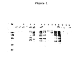
- the proteins had molecular weights between 10,000-300,000 daltons with 5 major metabolically labeled proteins of molecular weights of about 85,000, 73,000, 58,000, 52,000 and 35,000 daltons (Table 5 and Fig. 1).
- Poly A containing mRNA was prepared from the gametocyte total RNA by oligo (dT) - cellulose chromatography (22). Gametocyte poly A+ mRNA and total RNA as well as uninfected control intestine RNA were translated in a rabbit reticulocyte cell-free system (23) where it was found that the gametocyte cell-free products contained little if any contaminating host cell-free products. There were eight major bands with molecular weights of about 34,000, 40,000, 45,000, 50,000, 65,000, 90,000, 100,000 and 225,000 daltons, some of which were similar in size to the five major metabolically labeled gametocyte bands (35,000, 52,000, 58,000, 73,000, and 85,000) (see Fig. 1). The relationship of these bands was analyzed by immune precipitation and Western blotting as described in Example 4.
- RNA from infected mucosa every four hours starting on day five post infection with oocysts were translated in the rabbit reticulocyte cell-free system and compared to products directed by day six purified gametocyte RNA as well as control mucosa RNA. It appeared that various gametocyte bands were present at different times during development, however no major bands were detected early in gametocyte development which were absent from the mature gametocytes. It was concluded, therefore, that the preparations contained most if not all of the detectable gametocyte protein mRNAs.
- ELISA was performed using NP-40 extracts of purified E. maxima gametocytes as antigen. Most of the various sera tested showed titers of at least 1:1000 and some had titers as high as 1:10,000. Antisera preabsorbed with antigens from normal intestine NP-40 extracts showed no decrease in its antigametocyte titer indica ing the reaction is specific to parasite antigens. The finding that sera from birds which recovered from infections also gave strong titers by ELISA indicates that these antigens are also immunogenic during the course of an infection. The results of the ELISA were used to select antisera for the further screening tests described below.
- the immunofluorescence test was used to determine localization and stage specificity of the gametocyte antigens. Briefly, freshly prepared gametocytes were incubated with sera for 20 minutes at 37°C, washed 3 times with PBS, and then incubated with FITC conjugated second antibody. In control experiments where gametocytes were incubated with FITC conjugated rabbit anti-chicken IgG, rabbit anti-human IgG, rabbit anti-mouse IgG, or sheep anti-rabbit IgG (without antisera to the parasite present), a very high specific background was found even at dilutions as high as 1:1000.
- Noninfected chicken intestinal tissue did not react with any of the FITC-conjugates and preabsorption of these antibodies with either a chicken liver homogenate or purified gametocytes did not reduce this binding.
- preincubation with normal rabbit IgG did not effect the background.
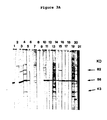
- rabbit antiNP-40 gametocyte extract serum strongly reacted with bands in the gametocyte protein extract and weakly with control intestine protein extracts.
- rabbit anti-gametocyte extract serum consistently detected 6 major (90K, 76K, 75K, 73K, 58K and 56K) and 6 minor (94K, 48K, 45K, 41K, 39K and 34K) gametocyte specific proteins (Fig. 2).
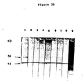
- the reactivity of normal chicken sera with the 43 kd protein can be explained in two possible ways. Either the sera contain a certain level of antibody against this gametocyte protein even though the chickens were never exposed to E. maxima , or the 43 kd protein binds chicken IgC irrespective of its source. That the latter possibility is correct was shown by using affinity purified chicken anti-cytochrome IgG on a Western blot where we found that it also bound to the 43 kd protein band. Thus, it was concluded that the 43 kd protein is an IgG binding protein, and may in fact be the Fc receptor responsible for the binding of IgG seen in the immunofluorescence studies (see above).
- antisera were used to identify antigens synthesized in vitro from gametocyte messenger RNA.
- mRNA was extracted by the guanidinium thiocyanate method (21) and translated in the rabbit reticulocyte cell free system (BRL, Bethesda, Maryland). Cell-free products were immune precipitated with the various antisera and the proteins analyzed by SDS-PAGE (Fig. 4). It was found that antisera to gametocytes from a variety of immunized animals reacted with gametocyte directed cell-free products and not with cell-free products from uninfected intestine, merozoite stage infected intestine, sporulated oocysts, or chick brain.
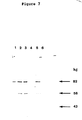
- Gametocyte specific proteins show a characteristic pattern of 5 (metabolic labeling) or 8 (cell-free products) major bands. Of these, most are the same size as the major antigenic proteins recognized by immune precipitation or Western blotting with anti-gametocyte or recovered sera. As defined by the Western blotting technique there are three major antigens recognized by recovered chicken serum having apparent molecular weight of 82 kd, 56 kd and 43 kd. One of these (56 kd) also reacted strongly with immune rabbit serum and based on the time course experiment using post-infection sera (Fig. 3) plays an important role in protective immunity. The 43 kd protein appears to be an IgG binding protein and is likely to be an Fc receptor (as discussed above).
- Lectins are proteins, found mainly in plants, which bind very specifically to receptors on cell surfaces. More accurately, they bind to distinct sugar moities of the receptors. Most lectins interact preferentially with a single sugar structure on cell surfaces. This interaction with cells is so selective that lectins can be used to distinguish e.g. between different human blood groups (32). This specificity of lectins was used to test gametocytes for surface antigen binding.
- lectin blots were used.
- the principle of lectin blots is similar to Western blots, however the lectin is allowed to react directly with the immobilized antigens without prior antiserum incubation.
- Lyophilized DOC-extracts of gametocyte antigens and normal chicken intestines were dissolved in water at a concentration of 10mg/ml.
- the antigens were separated on SDS-PAGE and blotted electrophoretically onto nitrocellulose paper. The paper was then cut into equal strips and tested for binding to soybean lectin.
- soybean lectin conjugated to peroxidase was used (Sigma, St. Lousi).
- nitrocellulose strips were incubated in blocking solution (TBS pH 7.0, 10% FCS) for 10 minutes, then transferred to trays containing in addition to blocking solution, 5 mM MnCl2, 5 mM CaCl2 and 10 ⁇ g/ml soybean lectin-peroxidase; the strips were incubated for 1 hour at room-temperature.
- blocking solution TBS pH 7.0, 10% FCS
- Control strips were treated as above, but all solutions contain in addition 100 mM N-acetyl-D-galactosamine.
- Negative controls consisted of nitrocellulose blots with immobilized normal chicken intestinal proteins.
- the gametocyte antigens contain 10 bands, 5 major and 5 minor, which can be detected with soybean lectin.
- the major bands have apparent molecular weights of 82, 78, 56, 54 & 52 all of which are identical in size to the major bands detected by recovered chicken serum in Western blots (See Fig. 6 and Table 5).
- the sugar inhibited the soybean lectin binding completely, proving, that the binding is indeed lectin specific.
- soybean lectin we have hereby clearly identified some of the major antigens of E. maxima gametocytes as being surface specific glycoproteins. The isolation of these proteins can easily be achieved by soybean lectin agarose column chromatography.
- Freeze dried material can usually be stored for long periods of time without losing activity.
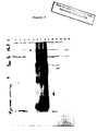
- Gametocytes purified according to method II were extracted with a variety of detergents. The extracts were analyzed by Western Blotting using recovered chicken serum, where it was found that extraction with Na2 DOC gave the best results (see Figure 7). Consequently, large scale extraction was performed using Na2 DOC. The extract was subsequently dialyzed against a 10 mM phosphate buffer pH 8.0 containing PMSF at 4°C. The dialyzed material was frozen at -80°C and then lyophilized to dryness. The antigens are stored with dessicant at -20°C.
- Gametocyte extracts contain a mixture of proteins lipids a.s.o.
- Pure gametocytes were extracted with 0.5% NP-40 (see Example 3). Proteins were precipitated by addition ammonium sulphate to the extract to a concentration of 10%. The precipitate was removed and the supernatant was brought to an ammonium sulphate concentration of 50%. The precipitated proteins (50%-cut) were tested in both ELISA and Western blots and shown to retain their activity. For column chromatography the 50% cut was dissolved in the column running buffer, namely: 10 mM Tris pH 8.0, 150 mM NaCl, 1 mM PMSF, 0.05% DOC and 1mM EDTA.
- the main component of the second peak was shown by thin layer chromatography to be mostly of lipid nature.
- gametocyte lipids as well as normal chick intestinal lipids were extracted by chloroform-methanol 2:1.
- Ethanol soluble lipids were analyzed for activity with recovered chick serum, normal chick serum, immune mouse serum and normal mouse serum using both radioimmunoassay (RIA) and ELISA. Water soluble lipids were analyzed with the same sera using the ELISA technique.
- the first monoclonal antibody was isolated by priming a Balb/C mouse 4 times with an intraperitoneal injection of 150,000 purified gametocytes. A fifth injection of a gametocyte NP-40 extract (see Example 1) was given intraspleenic. The mouse was sacrificed 4 days after the last injection and its spleen cells fused to myeloma cells according to published procedures (34). Monoclonal antibody 2B8-10 (2nd plate, row B, well 8, 10 th fusion) was tested for activity by the ELISA technique and found to react strongly to gametocyte antigen, to a lesser extent also to control chicken intestine.
- the next monoclonal antibody we described is 1C3-23. It was derived by injecting a Balb/C mouse twice with fractions of a Sephadex G-200 column (see Example 6) containing a pool of the first peak and the trough fractions. The third and last injection into this mouse was with a 50% ammonium sulphate cut (see Example 6). 1C3-23 reacted on ELISA plates with gametocyte antigen only and did not shown and response to control intestine. On Western clots we found, that IC3-23, lie E9-10, react with one single band to the 56 kd protein.
- mice were injected with gelpieces from SDS-PAGE.
- the first such fusion was done with the spleen of a mouse which had received three injections of gelpieces that contained the protein of 82 kd (see Example 4).
- the polyclonal serum of this mouse, taken before sacrifice showed a very strong response to the 82 kd protein band on Western blots as expected.
- the central well of organ culture dishes contained intestinal pieces (2 mm2, mucosa side down on GF/C filters ) from chickens four or five days post infection.
- the well was filled with 0.8 ml of RPMI 1640 medium containing 5% inactivated normal chicken serum plus penicillin-streptomycin plus L-glutamine.
- Two ml of phosphate buffered saline was placed in the outer ring of the dish which then gassed 5-15 min with 95% oxygen, 5% CO2 in an airtight sterile chamber and incubated at 40°C.
- On day six post infection i.e.
- the medium was replaced with 0.8 ml of RPMI 1640 containing 5% normal chick serum, 40 mM NaHCO3, 20 mM CaCl2, penicillin-streptomycin plus L-glutamine, and the dishes were incubated one more day at 40°C in a 5% CO2 incubator. At varying times during the incubations pieces were smeared on glass slides and examined for the appearance of gametocytes and/or oocysts.
- organ culture system for in vitro antigametocyte antibody testing and drug testing.
- the organ culture system was shown in Example 5 to be efficient in the growth of E . maxima gametocytes and oocysts in vitro .
- pieces of organ culture on the second of incubation were assayed by immunofluorescence for gametocyte antigens and compared with intestinal smears from a six-day infected chicken intestine. It was observed that in both cases gametocytes were stained with very strong intensity whereas surrounding tissue was not fluorescent at all. This result illustrates the specificity of the antisera as well as showing normal development of the parasite in vitro .
- this system may be used to isolate gametocytes, test the effects of anti-gametocyte antibody and drugs on parasite development in vitro , and develop new strategies for vaccine and drug development.
- the intestinal pieces were analyzed for gametocyte production and compared to the positive control plates.
- 14 day post-infection sera (referred to in this patent as “recovered chicken serum”) can be used to passively immunize chickens against E. maxima challenge infections (Rose et al(5,7,8)).
- These monoclonal antibodies are described in Example 7, and one of them (1E11-11) has already shown its ability to inhibit growth of gametocytes in vitro (Example 9).
- the conditions for the challenge infection will be established, namely, a titration curve will be carried out using 50, 100, 200, 500, and 2,000 oocysts of the Houghton strain of E. maxima to infect 2-3 week old birds. Oocyst counts will then be made on days 6-7, 7-8, and 8-9 post-infection as described in Example 2. Each group will consist of 20 chickens and the mean and standard deviation will be calculated. Based on these results, the challenge dosage for the immunization experiment will be determined.
- the immunization experiment will be carried out as above except that on days 4-8 post-infection either recovered chicken serum (1-3 ml), normal chicken serum (1-3 ml), mouse monoclonal antibody to a gametocyte protein (either as ascites or the Ig fraction thereof), and a monoclonal antibody to some irrelevant antigen as negative control will be administered i.m., i.v., or i.p. at 24 hours intervals. Oocyst counts will be taken as above, and the statistics determined for each group. A significant reduction in oocyst output by birds receiving recovered chicken serum or mouse monoclonal antibody to gametocyte antigen as compared to the negative controls will be considered a positive result. Passive immunization can also be used as a basis for studying cross-species protection by injecting the same monoclonal antibodies to protect against challenge infections with other Eimerian species.
- Example 10 a titration experiment will be performed as described in Example 10, however, chickens will be challenged with the FS110 strain of E . maxima isolated at Merck, Inc., Rahway, New Jersey, U.S.A. The results of the titration curve will again be used to choose the challenge dose in the immunization experiment.
- the actual immunization protocol is as follows: Birds used in test will be 2-day old Peterson X Arbor Acre males. Twenty birds per group will be immunized with either 100 ng, 300 ng or 1,000 ng of test antigen precipitated with alum and injected i.m. on days 2, 9 and 16. On day 17 the birds will be challenged with FS110 oocyst and feces will be collected on days 23, 24 and 25 for measurement of oocyst output (i.e. days 6,7 and 8 post-challenge).
- Control groups will be immunized by trickle infection on days 2, 9 and 16 with a predetermined number of oocysts, or shamimmunized with alum alone. An additional group will serve as non-infected controls. Birds will also be bled on days 2 and 25, and sera will be tested for their reactivity with gametocyte antigen on Western blots.
- the first test antigen used in these studies will be a pool of the 3 gametocyte antigens (82 kd, 56 kd and 43 kd) which were affinity purified. Afterwards each of these antigens will be tested individually. Based on these results, the antigen(s) showing the strongest protection will be produced by genetically engineered bacteria containing a cDNA encoding gametocyte antigen insert. These cloned antigens will be tested for their ability to protect birds and those shown to give good protection will be used to produce large amounts of the antigen for testing against other Eimerian species as well as in field trials.
Abstract
Description
- This application is a continuation-in-part of U.S. Serial No. 896,611, filed August 14, 1986, the contents of which are hereby incorporated by reference into the present application.
- Throughout this application, various publications are referenced by Arabic numerals within parentheses. Full citations for these references may be found at the end of the specification immediately preceding the claims. The disclosures of these publications in their entireties are hereby incorporated by reference into this application in order to more fully describe the state of the art as known to those skilled therein as of the date of the invention described and claimed herein.
- The organisms which cause the disease known as "coccidiosis" in chickens belong to the phylum Apicomplexa, class Sporozoa, subclass Coccidia, order Eucoccidia, suborder Eimeriorina, family Eimeriidae, genus Eimeria. Within the Eimerian genus there are many species, several of which are pathogenic in chickens. The species of major concern to the chicken industry are Eimeria tenella, Eimeria maxima, Eimeria acervulina, Eimeria nacatrix and Eimeria brunetti.
- Coccidiosis has become a major economic problem in the chicken industry over the past few decades, mainly due to the overcrowding of chicken houses and drug resistance. The rearing of chickens under crowded condi tions on a litter floor provides optimal conditions for the growth and spread of coccidia. Under such circumstances, sanitary control becomes nearly impossible and the farmer must rely on the effectiveness of coccidiostat drugs. However, drugs must be kept in the feed at all times and therefore are very expensive, certain drugs have costly side effects, and drug resistance has become a major problem under field conditions. Several large suppliers of these agents have come to realize that perhaps the only viable approach to the control of coccidiosis is by vaccine development.
- The Eimerian parasites undergo a complex life cycle in the mucosa of the intestinal tract. They show a great deal of specificity both in terms of the species they infect and in their location within the intestine. In fact, site specificity of infection is used as the major criterion for diagnosis. Other parameters for diagnosis include size and shape of oocysts, characteristics of the infected intestine, weight loss, and skin pigmentation changes.
- The life cycle of Eimeria is very similar to that of the hemosporidian parasites (i.e. plasmodium) except for the lack of an arthropod vector. Oocysts sporulate on the litter floor producing four sporocysts, each containing two sporozoites. These are ingested by the chicken and the sporozoites are released by the mechanical grinding of the oocysts in the gizzard and the subsequent digestion of the sporocyst wall by proteolytic enzymes. Sporozoites then invade lymphocytes and go on to invade epithelial cells where the asexual cycle begins. The parasite goes through 2-4 cycles of replication and division (each species having a defined number of divisions) leading to the production of large numbers of merozoites. After the final cycle of merozoite production the sexual cycle begins with the production of macrogametocytes (female) and microgametocytes. The macrogametocyte is characterized by the production of wall forming bodies which are involved in the production of the oocyst wall. Microgametocytes contain the components involved in the formation of microgametes which bud off from the surface of the intracellular parasite. Microgametes are flagellated and are responsible for the fertilization of the macrogamete. A zygote is formed which matures into the oocyst by fusion of the wall forming bodies and condensation of the nucleus. Oocysts are secreted in the feces, thus completing the cycle.
- A single infection with Eimeria can lead to protection against subsequent reinfection with the same strain of that species. This finding was used as the basis for producing a live vaccine against coccidiosis. Small numbers of oocysts from all the major pathogenic species are provided either in water (COCCIVAC ™) or are encapsulated in a water soluble polysaccharide (British patent GB 2,008,404A) giving rise to subclinical infections. However, even with small numbers of oocysts severe outbreaks of coccidiosis have occurred and farmers are reluctant to introduce live, pathogenic parasites into their chicken houses.
- In order to solve the problem of introducing pathogenic coccidia into a live vaccine, several laboratories have been working towards the production of live attenuated vaccines. By repeated passages in egg embryos or isolation of precocious lines (i.e. parasites that go through the life cycle in five days rather than six of seven), much less virulent and nonpathogenic strains of most of the major species have been isolated. Good protection has been observed using this approach, however there are still problems which include: shelf life of live parasites; back mutations of attenuated lines to pathogenic strains; strain variation especially in the case of Eimeria maxima; inability to protect for long periods of time; and introduction of any live coccidia into a chicken house would be resisted by farmers. With all these problems, the attenuated live vaccine may be used in the field until a more defined, noninfective, subunit vaccine can be developed.
- In the area of subunit vaccines the extracellular stages of the life cycle (the sporozoite, the merozoite and the gametes - micro and macro) are the most vulnerable to immune attack. The sporozoite is the first stage of development after the parasite is released from the oocyst and after a short time in the lumen invades a lymphocyte. In natural infection, high titers of antibody to sporozoites have been found and this stage is considered to be most promising for vaccine development. As a result, several laboratories have been working towards a sporozoite vaccine.
- E. tennella sporozoite extracts as well as supernatants of ground, sporulated oocysts were shown to protect broilers up to 7 weeks of age from challenge infections (1). In work using monoclonal antibodies, results have indicated that preincubation of sporozoites with monoclonal antibodies directed against surface antigens and injected into the cloaca of chickens, can give partial protection against the infection. Antigens recognized by these monoclonal antibodies have been identified by Western blotting and one of them, a protein of 25,000 molecular weight, has been cloned and sequenced (European patent publication No. 0 164 176, published December 11, 1985). This antigen was tested as an immunogen for protection against challenge infections where partial immunity was observed. American Cyanamid has also made several monoclonal antibodies to sporozoite surface antigens, some of which exhibited partial protection against Eimeria tenella infections (European patent publication No. 0 135 712, published April 3, 1985, and European patent publication No. 0 135 073, published March 27, 1985). The reason that purified antigens gave only partial protection in those studies may be due to the finding, that Eimeria tenella sporozoites can cap and shed their surface antigens, which is an important mechanism of host immune invasion (2). Thus it appears that the sporozoite stage may contain some antigens which can give partial protection, however, it seems unlikely that full protection will be achieved using only a sporozoite vaccine.
- In several studies carried out by Rose & Long (3-8), it was found that serum taken from birds which had recovered from infections with E. tenella or E. maxima can give passive protection against challenge infections with these species. These sera were also tested for their effect on invasion by sporozoites in vitro where it was found that there is a correlation between sporozoite neutralization in vitro and protection in vivo. However, birds which were immunized with extracts of sporozoites or merozoites showed no resistance to oral infections with E. tennella oocysts, nor could the sera from the same birds be used to protect naive birds (all this in spite of the fact that these sera were capable of neutralizing sporozoites and merozoites in vitro).
- A vaccine using antigens from the merozoite stage is also being tested (European patent publication No. 0 135 073). Several laboratories are making monoclonal antibodies to merozoite surface antigens in order to test their ability to inhibit invasion in vitro and in vivo. Most of these antigens were also found to be present in sporozoites as well as in different Eimerian species (9,10). In vivo protection results using these monoclonal antibodies again only showed partial inhibition and only with very low numbers of parasites (challenge dosage of 200 sporozoites) (European patent publication No. 0,135,172). Similar studies have been carried out for malarial merozoite surface antigens where excellent results using monoclonal antibodies to inhibit invasion in vitro have been obtained (11,12), however poor results were found in vivo. Problems of antigenic variation and diversity at this developmental stage of malaria is probably the major reason. Currently, attempts are being made to identify constant epitopes within these antigens and produce small peptides which contain the important antigenic determinants.
- The gametocyte stages of Eimerian development are the most difficult to analyze and very little has appeared in the literature regarding gamete subunit vaccines due to the following reasons: despite much effort Eimerian gametes have never been isolated before and, therefore, new methods need to be developed in order to study this developmental stage at the molecular level; no efficient in vitro system has been described for working with the sexual stages of Eimerian development; and most previous reports by leading authors in the field have led to the conclusion that gametes play little or no role in protective immunity (13,14,30).
- In work carried out by Rose on immunity to E. maxima (5-8), it was found that sera taken from recovered
birds 14 days post infection can give passive protection of up to 93% (based on oocyst output) against challenge with E. maxima, and these sera by Ouchterlony were found to precipitate an antigen prepared from mucosa containing gametocyte stages and not with sporozoite proteins (8). However, in these studies no direct proof of gametes playing a role in protective immunity was reported, nor were experiments done using the recovered antisera to identify the antigens seen by Ouchterlony. In fact, gametocytes were not purified and tested; only a mucosal extract was tested. In subsequent work Rose and others did not conclude that sexual stages, i.e., gametes play any role in induction of protective immunity to E. maxima (14) or to any other Eimeria species in spite of the results they obtained. In order to assess the role of the recovered sera in protective immunity, studies at the molecular level are required to characterize the proteins being recognized and the particular stage(s) at which they are being synthesized. - In studies carried out on malarial gametocytes and gametes, it was found that antigens from these stages can be used to protect birds against transmission of the disease (15). Furthermore, monoclonal antibodies to these antigens can be used to block the further development of the malaria parasite (16).
- One of the prerequisites for studying the role of gametocytes in protective immunity, is their isolation. No prior report has been made on isolating gametocytes of Eimeria. Most of the published work done on gameto cytes prior to the subject invention involved using electron microscopy to observe the growth and development of gametocytes in vivo. No studies have been published on the identification of stage specific gametocyte antigens. In early attempts to analyze gametocyte antigens in either whole infected intestine or partially purified preparations, little or no difference was seen by SDS polyacrylamide gel electrophoresis between infected intestine and normal intestine. It was therefore necessary for us to develop a method for isolation of gametocytes to a very high degree of purity in order to carry out molecular studies.
- In the area of in vitro cultivation, of all coccidial species, the developement of Eimeria in cultured cells has been most extensively studied. To date, those Eimeria species studiedin vitro have been either parasites of avian or mammalian hosts. While various cell types have been used to support the development of Eimerias, the best development occurs in cell types obtained from the natural host. In addition to cultured cells, avian Eimerias are grown in the chlorioallantoic membrane (CAM) of embryonated eggs or in tissue culture cells there. In general, cell cultures (e.g. chicken kidney cells, CK) or CAM are infected with either sporozoites or merozoites of the parasite to be studied and the development to asexual or sexual stages is monitored. The most thoroughly studied parasite in vitro is E. tenella which is also the most successful one, i.e., sporozoite infection of CAM or CK cells lead to oocyst formation (17). These oocysts are infectious when orally given to chickens. Many of the first and second generations schizonts of these cultured parasites, however, do not resemble schizonts of natural infections (6). The number of oocysts formed in the tissue cultured CAM cells were small compared to the numbers of gametocytes grown, suggesting that fertilization and the development of oocysts in vitro is more difficult to induce than the growth of macrogametocytes.
- To date, E. maxima has never been successfully cultured in vitro starting with sporozoites and leading to oocysts. Work by Shirley et.al. (18) showed that the CAM of embryonated eggs can be infected with merozoites of E. maxima which subsequently lead to oocyst formation. Also oocysts produced in this system were infective. However, sporozoites obtained from these oocysts and inoculated into the allantoic cavity of eggs penetrated cells of the CAM but did not develop further. It appears therefore, that for E. maxima the process of development from the sporozoites is fundamentally different from that of the subsequent stages. The specific requirements cannot be met by either cells of the CAM or by a variety of cells in culture. Although Shirley et. al. have shown that E. maxima can be cultured, albeit from the merozoite stage only, in embryonated eggs, their system is not suitable for further studies of the parasite, mainly because the egg per se is a closed system. Effects of drugs or monoclonal antibodies would be very difficult to study.
- The present invention involves a method for the isolation of E. maxima macrogametocytes and microgametocytes. The isolation procedure of the present invention allows for the purification, to a very high degree (over 90%) of E. maxima gametocytes, with little or no host contamination (based on SDS-PAGE analysis).
- The present invention also concerns the identification of protective gametocyte antigens and the RNA which encodes then. These antigens were found to give partial protection against challenge infections with E. maxima in vivo. Antisera from immunized and recovered birds as well as immunized mice, guinea pigs, and rabbits have been used to identify the antigens involved by immune precipitation and Western blotting techniques. These antisera as well as monoclonal antibodies and soybean lectin can be used to isolate these antigens either from protein extracts of gametocytes or by cloning the genes from the RNA encoding these antigens. These antibodies or antigens can then be used by passive or active immunization respectively to protect birds against coccidial infections.
- The present invention concerns a method for the growth of E. maxima parasites in an in vitro organ culture system. The organ culture system is based on growing parasites in whole pieces of infected intestine and closely reflects the growth conditions in vivo. It can be used to very efficiently grow E. maxima from the merozoite stage to the oocyst stage with rates of growth being nearly as high as that found in vivo. This system can be used to assay the effectiveness of both antibodies and drugs, as well as in the development of drugs which inhibit the sexual stages of coccidial development.
- The present invention provides substantially purified Eimeria ssp. gametocytes and a method of preparing them which comprises:
- a. removing intestinal tissue from a host animal which has been infected with an Eimeria spp. at a suitable time postinfection;
- b. contacting the intestinal tissue so removed with hyaluronidase so as to digest hyaluronic acid and certain other acid mucopolysaccharides present in the tissue;
- c. treating the digested intestinal tissue so that gametocytes are released from the digested tissue; and
- d. recovering substantially purified Eimeria spp. gametocytes.
- Also provided is a vaccine for conferring upon a chicken active immunity against infection by Eimeria ssp. which comprises an effective immunizing amount of gametocytes of the present invention and a carrier. A vaccine for conferring upon a chicken active immunity against infection by Eimeria maxima comprises an effective immunizing amount of Eimeria maxima gametocytes and a carrier.
- A proteinaceous extract of the present invention derived from Eimeria maxima gametocytes is capable of inducing in a chicken an immune response conferring protection against infection by Eimeria maxima and other Eimeria spp. The extract is soluble in a detergent containing buffer, is present as an antigen of Eimeria maxima gametocytes, is specifically recognized by polyclonal antibodies of recovered chicken serum and comprises at least nine proteins having molecular weights of 250±20 kd, 116±10 kd, 82±10 kd, 78±5 kd, 56±5 kd, 54±5 kd, 52±5 kd, 43± kd, and 36±5 kd.
- A method of preparing the extract of the present invention comprises:
- a. contacting Eimeria maxima gametocytes with a buffered detergent solution under suitable conditions so as to extract all of the proteins from the gametocytes; and
- b. isolating that portion of the extract having nine protein bands of molecular weights of 250±20 kd, 116±10 kd, 82±10 kd, 78±5 kd, 56±5 kd, 54±5 kd, 52±5 kd, 43±5 kd, and 36±5 kd respectively, as determined by SDS-PAGE.
- The present invention also provides a vaccine for conferring upon a chicken active immunity against infection by Eimeria maxima and other Eimeria spp. which comprises an effective immunizing amount of the extract of the present invention and a carrier or any combination of one or more of the proteins of the present invention and a carrier.
- Also provided is a method of in vitro development of Eimeria spp. gametocytes or oocysts which comprises:
- a. removing intestinal tissue from Eimeria
infected chickens - b. treating the intestinal tissue so removed with a suitable medium and under suitable conditions so as to promote production of gametocytes or oocysts within the tissue;
- c. replacing the medium with fresh medium containing additional ionic components after about one day of incubation time and further treating the tissue about one more day until gametocytes or oocysts are produced; and
- d. recovering the gametocytes or oocysts so produced.
- A method of assaying drug or anti-gametocyte antibody effectivity on Eimeria spp. development in vitro comprises:
- a. preparing Eimeria spp. gametocytes by the in vitro method of the present invention in the presence of the drug or antibody;
- b. determining the presence or absence of gametocytes and/or oocysts at a suitable time postinfection; and
- c. comparing the results of step (b) with the results obtained from preparing Eimeria spp. gametocytes and/or oocysts by the in vitro method of the present invention in the absence of the drug or anti-gametocyte antibody and thereby determining the effectiveness of the drug or antibody on Eimeria spp. development.
- The present invention also provides an extract of mRNA derived from Eimeria maxima gametocytes which, when contacted with a cell-free translation system, translates at least seven immune-precipitable proteins having molecular weights of 225±20 kd, 100±10 k, 90±10 kd, 65±5 kd, 45±5 kd, 40±5 kd and 34±5 kd.
-
- Figure 1 depicts the SDS-PAGE of metabolically labeled and cell-free translated gametocyte and host proteins. Metabolically labelled (³⁵S-methionine incorporation) protein extracts of purified E. maxima gametocytes (
Lanes 1,5) and chick uninfected intestinal tissue (Lanes 2,6) were compared with the pattern of labeled proteins from cell-free translation of mRNA from purified gametocytes (Lanes - Figure 2 depicts the immunodetection of gametocyte and control proteins with rabbit antigametocyte NP-40 extract serum. Gametocyte and host tissue proteins were blotted on to nitrocellulose paper and reacted with normal rabbit serum (
Lanes Lanes Lanes 1,4) is hardly reactive compared with gametocyte protein extracts (Lanes - Figure 3 depicts the immunodetection of gametocyte antigens with recovered chicken serum and normal chicken serum.
- Figure 3A presents a preparative western blot containing gametocyte proteins where individual strips were reacted with a variety of recovered chicken sera (
Lanes 1 to 20) and as a control chicken anti-cytochrome serum (Lane 21). - Figure 3B presents a preparative Western Blot containing gametocyte proteins which were reacted with a variety of normal chicken sera (
Lanes 1 to 8) and compared to antigens detected by recovered chicken serum (Lane 9). - Figure 3C presents a preparative Western Blot containing gametocyte proteins which were reacted with chicken serum taken at various times post-infection. Birds were infected with 1 × 10⁴ E. Maxima oocysts and blood was taken on day 0 (Lane 1), day 6 (Lane 2), day 11 (Lane 3) and day 14 (Lane 4) post-infection.
- Figure 4 depicts the immune precipitation of E. maxima cell-free proteins with rabbit and chick antisera. Cell free translation products of total sporulated E. maxima oocyst RNA (Lane 6), purified gametocyte RNA (Lane 8) and uninfected chick intestine RNA (Lane 7) were reacted with chick and rabbit antisera. Gametocyte cell-free proteins were immunoprecipitated with recovered chick serum (
Lanes 9,10), chick antigametocyte NP-40 extract serum (Lane 11), normal chick sera (Lanes 1-3, 12), rabbit antigametocyte extract serum (Lane 13) and normal rabbit serum (Lane 14). Sporulated oocyst cell-free proteins were immunoprecipitated with recovered chick serum (Lane 4) and normal chick serum (Lane 5). - Figure 5 depicts the immune precipitation of gametocyte cell-free products directed by mRNA of infected intesttine extracted at various time points as compared to pure merozoite or gametocyte preparations. Gametocyte specific products derived from cell-free translation of total intestinal mRNA extracted during merozoite and gametocyte stages of E. maxima infections were immune-precipitated with recovered chick serum. Immune-precipitates of cell-free translation of mRNA from: merozoites (Lane 1), infected intestine at 118 h (Lane 2), 122 h (Lane 3), 126 h (Lane 4), 130 h (Lane 5), 134 h (Lane 6) and 138 h (Lane 7) post infection with E. maxima; purified E. maxima gametocytes (Lane 8) and chick brain (Lane 9).
- Figure 6 presents a Western Blot of gametocyte proteins detected by soybean lectin conjugated to peroxidase. The most prominent proteins as depicted are those having molecular weights of the 82 kd, the 56 kd and 40 kd.
- Figure 7 presents extracts of gametocytes using a variety of detergents. The same number of gametocytes were extracted with 0.5% CHAPS (3ʹ-[(3ʹ-cholamidopropyl) dimethylammonio] -1-propanesulfonate) (Lane 1), 0.5% NP-40 (Lane 2), 0.5% TRITOM X-100 (Lane 3), 0.5% SDS (Lane 4) and 0.5% Na₂ DOC (Lane 5) were blotted on to nitrocellulose paper and reacted with recovered chicken serum.
- The present invention provides substantially purified Eimeria spp. gametocytes and a method of preparing them which comprises:
- a. removing intestinal tissue from a host animal which has been infected with an Eimeria spp. at a suitable time postinfection;
- b. contacting the intestinal tissue so removed with hyaluronidase so as to digest hyaluronic acid and certain other acid mucopolysaccharides present in the tissue;
- c. treating the digested intestinal tissue so that gametocytes are released from the digested tissue; and
- d. recovering substantially purified Eimeria spp. gametocytes.
- The gametocytes so produced may be microgametocytes or macrogametocytes. A desirable species of Eimeria for use in the method of the present invention is Eimeria maxima, thereby producing Eimeria maxima gametocytes. A desirable host animal is a chicken.
- In the gametocyte preparation method of the present invention, the suitable time postinfection is between about 121 and about 144 hours, desirably about 136-138 hours. The treating comprises filling the removed intestine of the animal with a suitable buffer, e.g. a SAC buffer comprising about 170 mM NaCl, about 10 mM Tris, pH 7.0, about 10 mM glucose, about 5 mM CaCl₂ about 1 mM PMSF and about 1 mg/ml bovine serum albumin, containing hyaluronidase at a concentration from about 200 to about 1000 units/ml, e.g. 350 units/ml. Each end of the removed intestinal section is closed off and the tissue is placed in a suitable buffer, i.e. phos phate buffered saline (PBS), for a suitable time, e.g. about 15 to about 25 minutes, at about 37 °C, and then cutting the intestinal section open and contacting the tissue with a suitable wash solution, e.g. said SAC buffer. The recovering comprises filtering said wash solution containing gametocytes from the tissue through a first filter and the flow through is filtered through a second filter having a smaller pore size than the first filter. The pore size of the first filter is from about 15 to about 20 microns, e.g. 17 microns. The pore size of the second filter is from about 8 to about 12 microns, e.g. 10 microns. The isolated gametocytes which accumulate on top of the 10 micron filter, may be washed further with a suitable buffer and collected by centrifugation.
- An alternative method of preparing the gametocytes of the present invention comprises:
- a. removing the intestine from a host animal which has been infected with an Eimeria spp. at a suitable time postinfection;
- b. removing the mucosa from the infected intesttines into a suitable buffer;
- c. mixing the mucosal material and buffer;
- d. filtering the mucosal-buffer mixture through a set of filters having decreasing pore sizes; and
- e. recovering substantially purified Eimeria spp. gametocytes on the last filter of the set which has the smallest pore size.
- The first filter of the set of filters may have a pore size of about 150 microns and the last filter may have a pore size of about 10 microns.
- The present invention concerns substantially purified Eimeria spp. gametocytes prepared by the methods of the present invention. Desirably, substantially purified Eimeria maxima gametocytes may be prepared by the present methods.
- A vaccine for conferring upon a chicken active immunity against infection by Eimeria spp. comprises an effective immunizing amount of Eimeria spp. gametocytes and a suitable carrier. An effective immunizing amount is from about 0.5 × 10⁶ to about 2.0 × 10⁶ gametocytes. A suitable carrier is a SAC buffer, a phosphate buffer (0.01 to 0.1M) or a saline solution (0.8%). The carrier may also contain a suitable adjuvant, e.g. Freund's adjuvant.
- A vaccine for conferring upon a chicken active immunity against infection by Eimeria maxima comprises an effective immunizing amount of Eimeria maxima gametocytes and a suitable carrier. An effective immunizing amount is from about 0.5 × 10⁶ to about 2.0 × 10⁶ gametocytes.
- The gametocyte vaccine of the present invention may contain whole gametocytes, fractionated gametocytes, wherein the gametocytes have been treated so as to cause the release of their internal components, e.g. by sonication, or a mixture of whole plus fractionated gametocytes. A desirable ratio of whole gametocytes to sonicated gametocytes is 1:1.
- A method of conferring upon a chicken active immunity against infection by Eimeria spp. comprises administering to the chicken an effective amount of the vaccine of the present invention. A method of conferring upon a chicken active immunity against infection by Eimeria maxima comprises administering to the chicken an effective amount of the vaccine containing Eimeria maxima gametocytes. The administration may be by intravenous, intramuscular or intraperitoneal injection. An effective amount of vaccine is that amount derived from about 0.5 × 10⁶ to about 2.0 × 10⁶ gametoctytes or that which causes the animal's sera to have an antisera titer of about 1:000 (as determined by ELISA).
- The present invention concerns a proteinaceous extract derived from Eimeria maxima gametocytes which is capable of inducing in a chicken an immune response conferring protection against infection by Eimeria maxima, which is soluble in a detergent containing buffer, is present as an antigen of Eimeria maxima gametocytes, is specifically recognized by polyclonal antibodies of recovered chicken serum and comprises at least nine proteins having molecular weights of 250±20 kd, 116±10 kd, 82±10 kd, 78±5 kd, 56±5 kd, 54±5 kd, 52±5 kd, 43± kd, and 36±5 kd.
- A method of preparing the extract of the present invention comprises:
- a. contacting Eimeria maxima gametocytes with a buffered detergent solution under suitable conditions so as to extract all of the proteins from the gametocytes; and
- b. isolating that portion of the extract having nine protein bands of molecular weights 250±20 kd, 116±10 kd, 82±10 kd, 78±5
kd 56±5 kd, 54±5 kd, 52±5 kd, 43±5 kd, and 36±5 kd respectively, as determined by SDS-PAGE. - In the extraction method of the present invention, the buffered detergent solution comprises 0.5% NP-40 or 0.5% DOC in a SAC buffer. The suitable conditions comprise incubating the gametocytes with the buffered detergent solution at about 4°C for about one hour.
- Another vaccine for conferring upon a chicken active immunity against infection by Eimeria maxima comprises an effective immunizing amount of the extract of the present invention and a carrier. An effective immunizing amount of extract is an amount of extract that is derived from about 0.5 × 10⁶ to about 2.0 × 10⁶ gametocytes.
- A method of conferring upon a chicken active immunity against infection by Eimeria maxima comprises administering to the chicken an effective amount of the vaccine derived from an Eimeria maxima gametocyte extract. An effective amount of the vaccine is that which causes the sera to have an antisera titer of about 1:1000.
- The present invention also provides individual antigenic proteins, derived from the gametocyte extract of the present invention, which have the following respective molecular weights: 250±20 kd, 116±10 kd, 82±10 kd, 78±5 kd, 56±5 kd, 54±5 kd,52±5 kd, 43± kd, and 36±5 kd.
- A method of preparing the antigenic protein derived from the gametocyte extract of the present invention having molecular weight of 82±10 kd, comprises separately recovering the protein from the proteinaceous extract of the present invention. Recovery of the protein may be accomplished by affinity purifying the extract of the present invention over a column containing soybean lectin bound to agarose or by immunoaffinity chromatography with the monoclonal antibodies produced by hybridoma cell lines 1A-1, 1A-2 or 1A-3.
- A method of preparing the antigenic protein derived from the gametocyte extract of the present invention having a molecular weight of 56±5 kd comprises separately recovering the protein from the proteinaceous extract of the present invention. Recovery of the protein may be accomplished by affinity purifying the extract of the present invention over a column containing soybean lectin bound to agarose or by immunoaffinity chromatography with any one of the monoclonal antibodies 1E11-11, 1C3-23, 6B3-27 and E9-10.
- A method of preparing the antigenic protein derived from the gametocyte extract of the present invention having a molecular weight of 43±5 kd comprises separately recovering the protein from the proteinaceous extract of the present invention. Recovery of the protein is accomplished by affinity purifying the extract of the present invention over a column containing mono specific polyclonal chicken IgG.
- The present invention also provides monoclonal antibodies produced by the hybridoma cell lines 1A-1, 1A-2, 1A-3, 1E11-11, 1C3-23, 6B3-27 and E9-10. Also provided are monoclonal antibodies directed against the protein derived from the gametocyte extract of the present invention having molecular weight of 82±10 kd, as well as monoclonal antibodies directed against the protein derived from the gametocyte extract of the present invention having molecular weight of 56±5 kd.
- A further vaccine for conferring upon a chicken active immunity against infection by Eimeria maxima comprises an effective immunizing amount of any one of these proteins and a suitable carrier, or an effective immunizing amount of two or more of these proteins and a suitable carrier.
- A method of in vitro development of Eimeria spp. gametocytes or oocysts comprises:
- a. removing intestinal tissue from Eimeria
infected chickens - b. treating the intestinal tissue so removed with a suitable medium and under suitable conditions so as to promote production of gametocytes or oocysts within the tissue;
- c. replacing the medium with fresh medium containing additional ionic components after about one day of incubation time and further treating the tissue about one more day until gametocytes or oocysts are produced; and
- d. recovering the gametocytes or oocysts so produced.
- In the in vitro development method of the present invention, the medium of step (b) comprises RPMI 1640 medium containing 5% inactivated normal chicken serum, penicillin-streptomycin and L-glutamine. The suitable conditions comprise 40° C and a 95% O₂/5% CO₂ atmosphere. The ionic components of step (c) comprise 40 mM NaHCO₃ and 20 mM CaCl₂.
- A method of assaying drug or anti-gametocyte antibody effectivity on Eimeria spp. development in vitro comprises:
- a. preparing Eimeria spp. gametocytes by the in vitro development method of the present invention in the presence of the drug or antibody;
- b. determining the presence or absence of gametocytes and/or oocysts at a suitable time postinfection;
- c. comparing the results of step (b) with the results obtained from preparing Eimeria spp. gametocytes and/or oocysts by the in vitro development method of the present invention in the absence of the drug or anti-gametocyte antibody and thereby determining the effectiveness of the drug or antibody on Eimeria spp. development.
- The present invention also provides an extract of mRNA derived from Eimeria maxima gametocytes which, when contacted with a cell-free translation system, translates at least seven immune-precipitable proteins having molecular weights of 225±20 kd, 100±10 kd, 90±10 kd, 65±5 kd, 45±5 kd, 40±5 kd and 34±5 kd. The present invention contemplates preparing cDNA from any of the mRNA molecules present in the mRNA extract.
- Two methods were employed in order to purify gametocytes from E. maxima. Method I was used in the early studies, however, it was found that these preparations were highly contaminated with host protein in spite of the fact that they looked fairly clean microscopically. Method II was far superior and resulted in gametocyte preparations which were greater than 90% pure. The methods are described below in detail.
- Chickens were infected with 10,000 oocysts each and then sacrificed on day six post infection. Their intestines were cut open and the mucosa was scraped with glass slides into SAC buffer (170 mM NaCl, 10
mM Tris pH - Chickens (2-6 weeks) were infected as above. The optimal time for gametocyte production was determined to be 136-138 hours post infection. The chickens were then sacrificed, their intestines removed and washed with ice cold SAC. One end of the organ was tied with a string and the intestine was filled with 0.5 mg/ml hyaluronidase (Type III from Sigma, 700 units/mg (19)) in SAC. The other end was then tied and the intestines placed in warm 37°C PBS in a beaker and subsequently incubated for 20 minutes at 37°C with shaking. During this time the gametocytes were released from the intestine. After incubation the intestine was cut open and the contents discarded. The gametocytes were then washed off the intestinal mucosa with SAC through a 17 micron polymon filter (Swiss Silk Bolting Cloth Mfg. Co. Ltd., Zurich, Switzerland). The flowthrough was filtered through a 10 micron polymon filter and the gametocytes accumulating on the filter were washed with SAC and collected by centrifugation at 800 × g for five minutes. They were then examined microscopically and counted in a modified Fuchs-Rosenthal counting chamber. The yield was 0.5-2 × 10⁶ gametocytes per infected intestine, however, on a few occations much higher yields of 10-20 × 10⁶ gametocytes per infected bird were obtained. The reason for this is not clear and does not appear to be related to the intensity of the infection or the size of the birds.
- Experiments were carried out in order to test the purified E. maxima gametocytes as an immunogen in birds which were subsequently challenged with sporulated E. maxima oocysts. These data are summarized in four experiments:
- In this experiment one-day old chickens kept in one cage were immunized with 0.5-1.0 × 10⁶ gametocytes prepared by Method I (half of which were sonicated in order to release internal components). Five birds received three weekly injections of gametocytes in Freund's adjuvant IM. Five received three weekly injections of Freund's adjuvant alone. Five received injections of gametocytes IV and five were not immunized and used as negative controls. One week after the last immunization, the birds were challenged per os with 2000 sporulated E. maxima oocysts (apart from the negative controls which were not challenged). Two day fecal samples were collected five days after infection and oocyst output was determined. As can be seen in Table 1 the five negative controls were all zero as expected. Birds given Freund's alone secreted an average of 26 × 10⁶ oocysts/bird, birds immunized with gametocytes IV or IM actually produced by average more oocysts than the controls (37 × 10⁶/bird in both groups). From these results it was concluded that the antigen preparation used in this experiment showed an immunosuppressive effect on the ability of the chicks to resist infection. Similar results were found by others who tried immunizing with pure oocysts and, since the preparations used in the present experiments are contaminated with small numbers of oocysts, this may explain the effect.
- When the gametocyte proteins were analyzed on SDS acrylamide gels it was found that this type of preparation (Method I) was highly contaminated with host proteins. In subsequent experiments much cleaner gametocytes were used prepared by Method II, older birds were used, since they are more immunocompetent, and the challenge dose was lowered to 500 oocysts/bird.

- In this experiment, gametocytes (prepared by using Method II as described above) were used which were shown to contain little contaminating host protein by gel electrophoresis (see Example 3). 2 × 10⁶ E. maxima gametocytes were injected into the duodenal loop of six week old birds twice at weekly intervals and one week later the birds were challenged with 500 E. maxima oocysts per os. Control birds were kept in the same cage as the immunized birds and oocyst output of all the birds was found to be zero prior to challenge. On days seven to nine after infection, feces were collected and oocyst output data are shown in Table 2. The control birds gave an average oocyst output of 52.2 × 10⁶/bird while the immunized birds gave an average of 30.5 × 10⁶ oocyst/bird. Three out of four of the immunized birds were much lower than the average control.

- In this experiment, three 5-week old birds were immunized IV and IP with three weekly injections of 1-2 × 10⁶ E. maxima gametocytes. Three positive controls were kept in the same cage. One day after the last injection, birds were challenged with 500 oocysts per os. The results (Table 3) show that the three positive controls had an average two-day oocyst output of about 14 × 10⁶/bird. One immunized bird did not secrete any oocysts, one bird showed a very high oocyst output and the other immunized bird was similar to the controls. In order to be certain that all the birds were indeed infected, all of the chickens were reinfected. The second infection showed that all the birds were immune including the "0" bird.

- In this experiment birds were immunized with pure gametocytes, half of which were sonicated, given either IP, IM or IV. There were five positive controls. One day after the last immunization the birds were challenged with 500 oocysts and seven-ten days later feces were collected. As can be seen in Table 4, in the IP, IM group four out of seven birds showed a reduction down to 25% of the average control. In the IV group five out of seven birds were low. The average output of the groups showed the following: the IP, IM and IV groups were lower than the average control (36.7 × 10⁶, 32.6 × 10⁶, vs 55.6 × 10⁶ respectively). Two control birds in this experiment were low and one of them was particularly low (#5). This bird had a broken wing which may have contributed to this result.

- The experiments described above show that birds immunized with purified gametocytes injected IV, IP, IM or intraintestinally were partially protected against challenge infections. Preparations which were unclean (Experiment 1) seemed to have an immunosuppressive effect as has been reported previously using oocyst preparations to immunize chickens (26). Several of the sera from birds immunized in these experiments were analyzed for their titer and specificity as described below (Example 4). Both immune sera and sera from recovered birds (which was shown previously to confer a good level of protection when given passively) recognized the same antigens as determined by immune precipitation and Western blotting. Therefore, it was concluded that these antigens are protective and can be used to confer immunity to coccidial infections in chickens.
- Extraction of proteins from gametocytes prepared by Methods I and II (see Example 1) was carried out using various detergents. The yield of protein was determined by the method of Bradford (31) was 1-2 mg per 1×10⁶ pure gametocytes. Extracted proteins were examined by SDS polyacrylamide gel electrophoresis (SDS-PAGE) and the efficiency of extraction using the various detergents was compared. It was found that by incubating gametocytes in a solution of 0.5% NP-40 in SAC at 4°C for 1 hour, all of the protein extractable using the very strong detergent SDS was present in the solution (as determined by SDS PAGE analysis). Results also showed that gametocytes prepared by Method I were heavily contaminated with host proteins, while in contrast, using Method II there was very little host contamination. This is based on both SDS-PAGE of Coomassie blue stained proteins as ell as ³⁵S-methionine metabolically labeled gametocyte proteins. The proteins had molecular weights between 10,000-300,000 daltons with 5 major metabolically labeled proteins of molecular weights of about 85,000, 73,000, 58,000, 52,000 and 35,000 daltons (Table 5 and Fig. 1). Most of the major labeled bands seen in the gametocyte preparations were totally absent from the labeled uninfected control intestine, and the major bands seen in the uninfected control were either very weak or absent from the gametocyte protein preparation. Therefore, it was concluded that these proteins must be parasite derived and that the isolated parasites are metabolically active.
- RNA was also extracted from E. maxima gametocytes using two procedures; one based on SDS-phenol (20) and one using guanidinium thiocyanate (21). The yields were 1-2 mg total RNA per 10⁷ gametocytes using either method. RNA from gametocytes, sporulated oocysts and uninfected chicken intestine were analyzed on 1.5% agarose gels where it was found that the rRNAs of E. maxima migrated differently from the host RNA. Based on the intensity of the ethidium bromide stained major rRNA bands, it was found that the gametocyte RNA preparations contained very little host RNA. In some preparations the host RNA contamination was negligible. These results corroborate those found at the protein level and indicate that the parasite preparations were greater than 90% pure.
- Poly A containing mRNA was prepared from the gametocyte total RNA by oligo (dT) - cellulose chromatography (22). Gametocyte poly A⁺ mRNA and total RNA as well as uninfected control intestine RNA were translated in a rabbit reticulocyte cell-free system (23) where it was found that the gametocyte cell-free products contained little if any contaminating host cell-free products. There were eight major bands with molecular weights of about 34,000, 40,000, 45,000, 50,000, 65,000, 90,000, 100,000 and 225,000 daltons, some of which were similar in size to the five major metabolically labeled gametocyte bands (35,000, 52,000, 58,000, 73,000, and 85,000) (see Fig. 1). The relationship of these bands was analyzed by immune precipitation and Western blotting as described in Example 4.
- A time course experiment was performed by extracting RNA from infected mucosa every four hours starting on day five post infection with oocysts. These RNAs were translated in the rabbit reticulocyte cell-free system and compared to products directed by day six purified gametocyte RNA as well as control mucosa RNA. It appeared that various gametocyte bands were present at different times during development, however no major bands were detected early in gametocyte development which were absent from the mature gametocytes. It was concluded, therefore, that the preparations contained most if not all of the detectable gametocyte protein mRNAs.
- Several methods were employed in analyzing the antigens extracted from E. maxima gametocytes. These include ELISA (enzyme linked immunosorbent assay), immunofluorescence, Western blotting, and immune precipitation of cell-free translation products. The antisera employed in the studies were from chickens which recovered from E. maxima infections and were bled 14 days post infection (this type of sera was shown by Rose to be potentially protective in vivo (5-8) hereinafter "recovered sera"). The following groups were studied: chickens immunized with sonicated and whole pure gametocytes, mice immunized with NP-40 extracts of pure gametocytes, and rabbits immunized with NP-40 extracts of pure gametocytes.
- ELISA was performed using NP-40 extracts of purified E. maxima gametocytes as antigen. Most of the various sera tested showed titers of at least 1:1000 and some had titers as high as 1:10,000. Antisera preabsorbed with antigens from normal intestine NP-40 extracts showed no decrease in its antigametocyte titer indica ing the reaction is specific to parasite antigens. The finding that sera from birds which recovered from infections also gave strong titers by ELISA indicates that these antigens are also immunogenic during the course of an infection. The results of the ELISA were used to select antisera for the further screening tests described below.
- The immunofluorescence test was used to determine localization and stage specificity of the gametocyte antigens. Briefly, freshly prepared gametocytes were incubated with sera for 20 minutes at 37°C, washed 3 times with PBS, and then incubated with FITC conjugated second antibody. In control experiments where gametocytes were incubated with FITC conjugated rabbit anti-chicken IgG, rabbit anti-human IgG, rabbit anti-mouse IgG, or sheep anti-rabbit IgG (without antisera to the parasite present), a very high specific background was found even at dilutions as high as 1:1000. Noninfected chicken intestinal tissue did not react with any of the FITC-conjugates and preabsorption of these antibodies with either a chicken liver homogenate or purified gametocytes did not reduce this binding. In addition, preincubation with normal rabbit IgG did not effect the background.
- In order to avoid the background problem, the F(abʹ)₂ fraction of affinity purified sheep anti-mouse IgG FITC conjugated (SIGMA, St. Louis, Mo.) was used as the fluorescent reagent. Control experiments indeed showed that this reagent by itself or with normal mouse serum gave a very low background. This result indicates that the background found with IgG may be due to the presence of Fc receptors on the surface of the gametocytes.
- Thirteen immune mouse sera were then tested from mice immunized with either whole plus sonicated gametocytes or gametocyte NP-40 extracts. It was found that the various immune sera showed different intensities in their response with no significant difference between sera against whole gametocytes or gametocyte NP-40 extracts. In all instances the fluorescence was concentrated on the surface of the gametocytes indicating that the antigens are surface membrane associated. Some of the sera also reacted with the surface of oocysts.
- The stage specificity of the mouse anti-gametocyte sera was also tested. E. maxima sporozoites were isolated according to published procedures (22) and were dried on IFA glass slides. The immunofluorescence assay was carried out where it was found that the sporozoites fluoresced with a few sera, however not nearly with the intensity seen with purified gametocytes. Therefore, it was concluded that there may be come antigens present in both sporozoites and gametocytes which cross-react, however, the bulk of these proteins are present mainly in gametocytes.
- Identification of the individual antigens recognized by immune and recovered sera was performed by immunodetection of immobilized antigens (Western blotting (25)) and by immune precipitation of the cell-free synthesized gametocyte proteins. NP-40 extracted gametocyte proteins were first separated by SDS-PAGE and then transferred to a nitrocellulose filter. In one method for immunodectection the filter was incubated with rabbit antiserum, washed, and then overlaid with ¹²⁵I- protein A. The unbound protein A was washed off and the filter paper was exposed to X-ray film. It was found that normal rabbit sera reacted very weakly with only a few bands in both control and gametocyte antigen preparations. In contrast, the rabbit antiNP-40 gametocyte extract sera strongly reacted with bands in the gametocyte protein extract and weakly with control intestine protein extracts. Although some variability exists between different protein extracts, rabbit anti-gametocyte extract serum consistently detected 6 major (90K, 76K, 75K, 73K, 58K and 56K) and 6 minor (94K, 48K, 45K, 41K, 39K and 34K) gametocyte specific proteins (Fig. 2).
- In a second method the filters were overlaid with chicken antiserum followed by rabbit anti-chicken IgG linked to horseradish perodixase, which upon addition of the appropriate substrate gives rise to a red color reaction. Using several recovered chicken sera we consistently detected 3 major bands having apparent molecular weights of 82 kd, 56 kd and 43 kd regardless of the detergent used to extract the gametocyte protein (Figure 3A). In contrast, normal chicken serum reacted mainly with the 43 kd protein and much more inconsistently with the other 2 major bands (Figure 3B). Uninfected control intestine proteins did not react at all with either recovered or normal chicken sera. In addition to the 3 major bands described above 6 minor bands were detected (250 kd, 116 kd, 78 kd, 54 kd, 52 kd, and 36 kd) which appeared less consistently. These results are summarized in Table 5.
- The reactivity of normal chicken sera with the 43 kd protein can be explained in two possible ways. Either the sera contain a certain level of antibody against this gametocyte protein even though the chickens were never exposed to E. maxima, or the 43 kd protein binds chicken IgC irrespective of its source. That the latter possibility is correct was shown by using affinity purified chicken anti-cytochrome IgG on a Western blot where we found that it also bound to the 43 kd protein band. Thus, it was concluded that the 43 kd protein is an IgG binding protein, and may in fact be the Fc receptor responsible for the binding of IgG seen in the immunofluorescence studies (see above).
- An experiment was performed to determine on which day post-infection antibodies to the major gametocyte protein bands appear in vivo. Chickens were inoculated per os with 10, 000 E. maxima oocysts on day 0 and were bled on
days days 0 and 6 the only major band visible was the 43 kd protein, whereas sera taken ondays - Since it was reported by Rose (5), that sera taken prior to
day 10 post-infection give no protection by passive immunization against challenge infections in naive birds, whileday 14 sera give good protection, our results indicate that the 56 kd protein plays an important role in protective immunity. - Finally, antisera were used to identify antigens synthesized in vitro from gametocyte messenger RNA. mRNA was extracted by the guanidinium thiocyanate method (21) and translated in the rabbit reticulocyte cell free system (BRL, Bethesda, Maryland). Cell-free products were immune precipitated with the various antisera and the proteins analyzed by SDS-PAGE (Fig. 4). It was found that antisera to gametocytes from a variety of immunized animals reacted with gametocyte directed cell-free products and not with cell-free products from uninfected intestine, merozoite stage infected intestine, sporulated oocysts, or chick brain. Recovered and immune chick sera recognized several gametocyte cell-free translation products. Long exposures of the autoradiographs showed more than ten immune precipitable bands having molecular weights from about 34,000-100,000 daltons. Of the ten bands, six bands are prominent representing proteins, having molecular weights of about 100,000, 90,000, 65,000, 45,000, 40,000 and 34,000 daltons, respectively (Table 5 and Fig. 4). These bands corresponded to most of the major bands in the total cell-free products. Some of the sera recognized only the 100,000 band, others only the 90,000 band, while still others precipitated both bands. In all cases, the 45,000 and 65,000 dalton proteins were immune precipitated.
- Serum from animals immunized with NP-40 extracts precipitated the same bands apart from some variations. Rabbit anti-NP-40 extract clearly precipitated a 225,000 dalton protein along with the 90,000 and 65,000 dalton proteins, while the other bands were very weak. Mouse anti-NP-40 extract gave similar results, whereas those immunized with whole and sonicated gametocytes recognized two additional low molecular weight bands at about 10,000-20,000 daltons.
- When recovered chicken serum was reacted with cell-free translation products of total infected chick intestine RNA (taken at various times post infection) the proteins immune precipitated corresponded to those seen in pure gametocyte cell-free products (Fig.5). Antigenic proteins started to appear at about 130 hours (approximately eight hours prior to the peak of gametocyte production in vivo 138 hours), labeled most strongly at 134 hours, and then decreased in intensity at 138 hours. Thus, the entire range of mRNAs encoding antigens stimulating the immune response during infection with E. maxima are absent from the merozoite stage (96 hours) and only are first detectable at the late gametocyte stage. In addition, it would appear that none of these antigens are shared with the earlier developmental stages. The decrease in antigens precipitated with recovered chicken sera at 138 hours may reflect the transition of mature macrogameocytes to oocysts.
- Table 5 summarizes the results described above. Gametocyte specific proteins show a characteristic pattern of 5 (metabolic labeling) or 8 (cell-free products) major bands. Of these, most are the same size as the major antigenic proteins recognized by immune precipitation or Western blotting with anti-gametocyte or recovered sera. As defined by the Western blotting technique there are three major antigens recognized by recovered chicken serum having apparent molecular weight of 82 kd, 56 kd and 43 kd. One of these (56 kd) also reacted strongly with immune rabbit serum and based on the time course experiment using post-infection sera (Fig. 3) plays an important role in protective immunity. The 43 kd protein appears to be an IgG binding protein and is likely to be an Fc receptor (as discussed above).
- Using recovered chicken serum to immune precipitate cell-free translation products, we also found 3 major bands which appear consistenly, having apparent molecular weights of 100 kd, 65 kd, and 45 kd (see Figures 4 & 5). These three bands most likely correspond to the three major bands seen on the Western blots. It is therefore possible to use the recovered chicken serum to screen a gametocyte cDNA library in order to isolate clones for these three proteins. Furthermore, these cloned antigens should be capable of arousing an immune response to the native gametocyte antigen and protect chickens against challenge infections. It is interesting to note, that normal chicken serum from uninfected birds both in the laboratory and from the field reacted consistently with the 100 kd and 65 kd proteins in cell-free translation products (Figure 4) as well as (although less consistently) with the 82 kd and 56 kd proteins on Western blots (Figure 3). Surprisingly, even normal mouse serum and normal rabbit serum reacted with the 65 kd protein in cell-free translation products. These results corroborate reports of the ubiquity of anti-coccidia antibodies in the serum of most vertebrates. In addition, it indicates that these antigens are highly conserved between various species of coccidia, even those infecting animals other than chickens.


- Lectins are proteins, found mainly in plants, which bind very specifically to receptors on cell surfaces. More accurately, they bind to distinct sugar moities of the receptors. Most lectins interact preferentially with a single sugar structure on cell surfaces. This interaction with cells is so selective that lectins can be used to distinguish e.g. between different human blood groups (32). This specificity of lectins was used to test gametocytes for surface antigen binding.
- To facilitate the analysis, several commercially available lectins, all conjugated to fluorescein, were checked, namely wheatgerm lectin (Triticum vulgare), soybean lectin (Glycin max) and concanavalin A (all Bio-Makor, Rehovot). The lectins were each incubated for 35 minutes at room temperature with 50,000 gametocytes in 100 µl PBS at a concentration, for each lectin, of 10⁻² µg/µl. After repeated washes with PBS the gametocytes were mounted on slides in PBS: Glycerol 1:1, and analyzed by fluorescence microscopy for surface binding to the lectins. Neither with wheatgerm lectin nor with concanavalin A could any specific binding be detected, i.e. both gametocytes and cell debris fluoresced to the same extent. Soybean lectin, however, bound very specifically to the surface of gametocytes only.
- The specific sugar recognized by soybean lectin, N-acetyl-D-galactosamine (32), should inhibit the binding, if indeed the interaction observed is a true lectin-cell receptor reactions. In a competition experiment using this sugar at a concentration of 100 mM, the binding of soybean lectin could be completely inhibited. As expected for true lectin binding the inhibition was reversible. This result shows clearly, that some of the gametocyte surface moieties are indeed glycosylated.
- In order to determine whether or not these surface moities are glycoproteins and to relate them to the antigens detected by immune sera (see Example 4), lectin blots were used. The principle of lectin blots is similar to Western blots, however the lectin is allowed to react directly with the immobilized antigens without prior antiserum incubation. Lyophilized DOC-extracts of gametocyte antigens and normal chicken intestines (see Example 6) were dissolved in water at a concentration of 10mg/ml. The antigens were separated on SDS-PAGE and blotted electrophoretically onto nitrocellulose paper. The paper was then cut into equal strips and tested for binding to soybean lectin. In order to visualize reactive glycoprotein bands, soybean lectin conjugated to peroxidase was used (Sigma, St. Lousi).
- The nitrocellulose strips were incubated in blocking solution (TBS pH 7.0, 10% FCS) for 10 minutes, then transferred to trays containing in addition to blocking solution, 5 mM MnCl₂, 5 mM CaCl₂ and 10 µg/ml soybean lectin-peroxidase; the strips were incubated for 1 hour at room-temperature. Control strips were treated as above, but all solutions contain in addition 100 mM N-acetyl-D-galactosamine. Negative controls consisted of nitrocellulose blots with immobilized normal chicken intestinal proteins.
- After incubation the strips were washed extensively with TBS or TBS + sugar respectively. Binding was detected by incubating the strips in a solution containing a substrate that upon conversion by peroxidase gives rise to a colour reaction.
- By lectin blot analysis the normal chicken intestinal proteins did not react with soybean lectin. The gametocyte antigens, however, contain 10 bands, 5 major and 5 minor, which can be detected with soybean lectin. The major bands have apparent molecular weights of 82, 78, 56, 54 & 52 all of which are identical in size to the major bands detected by recovered chicken serum in Western blots (See Fig. 6 and Table 5). As already expected from the immunofluorescence studies above, the sugar inhibited the soybean lectin binding completely, proving, that the binding is indeed lectin specific. With the help of soybean lectin we have hereby clearly identified some of the major antigens of E. maxima gametocytes as being surface specific glycoproteins. The isolation of these proteins can easily be achieved by soybean lectin agarose column chromatography.
- One of the most commonly used methods to store proteins is the freeze-drying or lyophilization technique. Freeze dried material can usually be stored for long periods of time without losing activity. Gametocytes purified according to method II (see Example 1) were extracted with a variety of detergents. The extracts were analyzed by Western Blotting using recovered chicken serum, where it was found that extraction with Na₂ DOC gave the best results (see Figure 7). Consequently, large scale extraction was performed using Na₂ DOC. The extract was subsequently dialyzed against a 10 mM phosphate buffer pH 8.0 containing PMSF at 4°C. The dialyzed material was frozen at -80°C and then lyophilized to dryness. The antigens are stored with dessicant at -20°C. Western blots of the lyophilized material when developed with recovered chick serum, showed that all antigenic proteins were retained. In a comparison of ELISA from fresh material and freeze dried antigen no difference was found. We concluded therefore that the best way to store total gametocyte antigen is in the form of a freeze dried powder.
- Gametocyte extracts contain a mixture of proteins lipids a.s.o. In order to separate the different components, but also at the same time fractionate the various antigens, we employed the technique of column chromatography as described below.
- Pure gametocytes were extracted with 0.5% NP-40 (see Example 3). Proteins were precipitated by addition ammonium sulphate to the extract to a concentration of 10%. The precipitate was removed and the supernatant was brought to an ammonium sulphate concentration of 50%. The precipitated proteins (50%-cut) were tested in both ELISA and Western blots and shown to retain their activity. For column chromatography the 50% cut was dissolved in the column running buffer, namely: 10 mM Tris pH 8.0, 150 mM NaCl, 1 mM PMSF, 0.05% DOC and 1mM EDTA.
- Two different column media were used; both are separating on the principle of size fractionation, namely Sephadex G-200 and Sephacryl S-300 (both Pharmacia, Finland). The same column running buffer (see above) was used for either column. The fractionation achieved was in both cases very similar, i.e., the material separated into two major peaks. The gametocyte proteins active in ELISA and Western blots were shown to be concentrated on the descending slope of the first peak and in the trough between peaks. Antigen containing fractions were pooled, dialyzed an either used immediately or stored for short terms at 4°C. For long term storage the fractions were lyophilized as described above.
- The main component of the second peak was shown by thin layer chromatography to be mostly of lipid nature. To ensure that the main antigenic activity is indeed found in the protein fractions and not in the lipid fractions gametocyte lipids as well as normal chick intestinal lipids were extracted by chloroform-methanol 2:1. Ethanol soluble lipids were analyzed for activity with recovered chick serum, normal chick serum, immune mouse serum and normal mouse serum using both radioimmunoassay (RIA) and ELISA. Water soluble lipids were analyzed with the same sera using the ELISA technique.
- Using either technique no specific reactivity to gametocyte lipids was detected using recovered chicken sera or immune mouse sera. Thus it is concluded that gametocyte lipids are not an important component of the immune response to the parasite.
- The theory of monclonal antibody production is based on the clonal hypothesis of Mcfarlane Burnet (32). Ever since Kohler and Milstein routinely achieved hybridomas (33), their protocol is the method of choice for the production of monoclonal antibodies. We have used the Kohler-Milstein method to produce monoclonal antibodies to gametocyte antigens. The monoclonal antibodies obtained will be immobilized on affinity columns and used to obtain large quantities of the required antigens from DOC-extracts (see Example 6).
- The first monoclonal antibody was isolated by priming a Balb/
C mouse 4 times with an intraperitoneal injection of 150,000 purified gametocytes. A fifth injection of a gametocyte NP-40 extract (see Example 1) was given intraspleenic. The mouse was sacrificed 4 days after the last injection and its spleen cells fused to myeloma cells according to published procedures (34). Monoclonal antibody 2B8-10 (2nd plate, row B, well 8, 10th fusion) was tested for activity by the ELISA technique and found to react strongly to gametocyte antigen, to a lesser extent also to control chicken intestine. To exclude the possibility that 2B8-10 might consist of more then one clone, the cells were subcloned and retested. The second monoclonal antibody E9-10 was thus isolated, tested by ELISA and shown to react to gametocyte antigen only. Both supernatant of E9-10 cells ascites fluid and the IgG-fraction thereof (precipitated by 40% ammonium sulphate) showed one single band in Western blots reacting only with the 56 kd protein described in Example 6. - The next monoclonal antibody we described is 1C3-23. It was derived by injecting a Balb/C mouse twice with fractions of a Sephadex G-200 column (see Example 6) containing a pool of the first peak and the trough fractions. The third and last injection into this mouse was with a 50% ammonium sulphate cut (see Example 6). 1C3-23 reacted on ELISA plates with gametocyte antigen only and did not shown and response to control intestine. On Western clots we found, that IC3-23, lie E9-10, react with one single band to the 56 kd protein.
- An additional monoclonal antibody was isolated, which also reacted to the 56 kd protein band namely 6B3-27. In contrast to 1C3-23 the third injection into the recipient mouse was with pooled trough fractions only. In neither ELISA test nor in Western blots was any reaction to control intestine detected.
- From three different mice, treated with three different injection protocols we have thus received 4 monoclonal antibodies that all recognize the same 56 kd protein. It seems therefore, that this protein is one of the major antigens in gametocytes from E. maxima. With monoclonal antibody 1E11-11 a clone was isolated that also recognizes the 56 kd protein. This monoclonal antibody was derived from a Balb/C mouse that had been injected 5 times intraperitoneally with an average of 150,000 purified gametoyctes per injection. In both ELISA and Western blots this monoclonal antibody reacted very strongly to gametocyte antigen only, in Western blots one heavy band of 56 kd was detected.
- A very different approach was taken for a new series of fusions namely, mice were injected with gelpieces from SDS-PAGE. The first such fusion was done with the spleen of a mouse which had received three injections of gelpieces that contained the protein of 82 kd (see Example 4). The polyclonal serum of this mouse, taken before sacrifice showed a very strong response to the 82 kd protein band on Western blots as expected.
- Three monoclonal antibodies to the 82 kd protein were obtained from this fusion experiment (1A-1, 1A-2 and 1A-3). These monoclonal antibodies along with the 4 others described above can be used to affinity purify the 56 kd and 82 kd antigens from a gametocyte detergent extract.
- The above seven monoclonal antibodies were deposited with the ATCC on August , 1987 and allotted the following numbers:
- E9-10
1C3 - 23
6B3 - 27
1E11 - 11
1A - 1
1A - 2
1A - 3
- In order to establish in vitro growth conditions to reflect as closely as possible conditions in vivo, work was carried out to grow E. maxima in an organ culture system. This system was set up according to the basic principle of organ culture (27, 28) along with the find ings on growth conditions for other coccidial parasites (17), as well as those required for growth of malarial gametocytes in culture (29). A combination of all these conditions along with empirical testing of the various parameters such as temperature, oxygen tension, etc. led to the establishment of the system described below.
- The central well of organ culture dishes (Falcon) contained intestinal pieces (2 mm², mucosa side down on GF/C filters ) from chickens four or five days post infection. The well was filled with 0.8 ml of RPMI 1640 medium containing 5% inactivated normal chicken serum plus penicillin-streptomycin plus L-glutamine. Two ml of phosphate buffered saline was placed in the outer ring of the dish which then gassed 5-15 min with 95% oxygen, 5% CO₂ in an airtight sterile chamber and incubated at 40°C. On day six post infection (i.e. two days later for four day pieces, one day later for five day pieces) the medium was replaced with 0.8 ml of RPMI 1640 containing 5% normal chick serum, 40 mM NaHCO₃, 20 mM CaCl₂, penicillin-streptomycin plus L-glutamine, and the dishes were incubated one more day at 40°C in a 5% CO₂ incubator. At varying times during the incubations pieces were smeared on glass slides and examined for the appearance of gametocytes and/or oocysts.
- It was found that incubation of either day four or day five infected intestine led to the production of gametocytes and oocysts (oocysts appeared on day seven post infection, in vitro). Therefore, these parasites may be cultured from the merozoite to the oocyst stage in the organ culture system.
- Work was then carried out to isolate the oocysts from the pieces and quantitate the efficiency of the system. Organ culture pieces were transferred into tubes (four pieces per tube) and 0.5
ml 20% chlorox was added. The tubes were incubated for 1 hr at room temperature and the supernatant was taken off the pieces. The released oocysts were spun down and counted. Each tube contained between 1000-6000 oocysts. Similar sized pieces from in vivo infected birds were processed in the same way and approximately the same number of oocysts were extracted using this method. Therefore, it was concluded that the system efficiently produces oocysts and very closely reflects the growth rate occuring in vivo. - The organ culture system was shown in Example 5 to be efficient in the growth of E. maxima gametocytes and oocysts in vitro. In an experiment to test for the production for gametocyte antigens in vitro, pieces of organ culture on the second of incubation (six days post infection) were assayed by immunofluorescence for gametocyte antigens and compared with intestinal smears from a six-day infected chicken intestine. It was observed that in both cases gametocytes were stained with very strong intensity whereas surrounding tissue was not fluorescent at all. This result illustrates the specificity of the antisera as well as showing normal development of the parasite in vitro. Therefore, this system may be used to isolate gametocytes, test the effects of anti-gametocyte antibody and drugs on parasite development in vitro, and develop new strategies for vaccine and drug development.
- Two anticoccidial drugs namely Kokzid and Korforan (both sulpha-based coccidiostats) known to work at the schizont stage and one monoclonal antibody 1E11-11 (see Example 7) were tested in the organ culture system for their effect on gametocyte and/or oocyst production. Intestinal pieces were prepared as described in Example 8. Duplicate plates contained either 20 mg/ml Korforan or 4 µl/ml Kokzid. The monoclonal antibody was tested at a concentration of 0.5%, 1.0% and 5.0% in the presence or absence of normal chicken serum. Control plates did not contain any additives. All parameters were kept as described (see Example 8).
- After 48 hours of incubation the intestinal pieces were analyzed for gametocyte production and compared to the positive control plates.
- As expected both coccidiostats did not affect gametocyte formation and were comparable to the controls. The monoclonal antibody 1E11-11 however, both in the presence and absence of normal chicken serum was inhibiting gametocyte formation already at a concentration of 0.5%. Thus, showing the effectivity of the monoclonal antibody 1E11-11 to inhibit parasite growth in vitro.
- 14 day post-infection sera (referred to in this patent as "recovered chicken serum") can be used to passively immunize chickens against E. maxima challenge infections (Rose et al(5,7,8)). We will use this type of sera as the positive control for the mouse monoclonal antibodies, which will be tested for their ability to passively immunize chickens against E. maxima. These monoclonal antibodies are described in Example 7, and one of them (1E11-11) has already shown its ability to inhibit growth of gametocytes in vitro (Example 9).
- First, the conditions for the challenge infection will be established, namely, a titration curve will be carried out using 50, 100, 200, 500, and 2,000 oocysts of the Houghton strain of E. maxima to infect 2-3 week old birds. Oocyst counts will then be made on days 6-7, 7-8, and 8-9 post-infection as described in Example 2. Each group will consist of 20 chickens and the mean and standard deviation will be calculated. Based on these results, the challenge dosage for the immunization experiment will be determined.
- The immunization experiment will be carried out as above except that on days 4-8 post-infection either recovered chicken serum (1-3 ml), normal chicken serum (1-3 ml), mouse monoclonal antibody to a gametocyte protein (either as ascites or the Ig fraction thereof), and a monoclonal antibody to some irrelevant antigen as negative control will be administered i.m., i.v., or i.p. at 24 hours intervals. Oocyst counts will be taken as above, and the statistics determined for each group. A significant reduction in oocyst output by birds receiving recovered chicken serum or mouse monoclonal antibody to gametocyte antigen as compared to the negative controls will be considered a positive result. Passive immunization can also be used as a basis for studying cross-species protection by injecting the same monoclonal antibodies to protect against challenge infections with other Eimerian species.
- Active immunization experiments will be carried out using the major protective antigens described in this patent application. We have already shown that whole gametocytes can give partial protection against challenge infections (Example 2), and we will test the ability of the 3 major gametocyte antigens having molecular weights of 82 kd, 56 kd, and 43 kd to protect against E. maxima infections. These antigens will be prepared by affinity chromatography (Example 5 and 7), and by cDNA cloning.
- To establish the conditions, a titration experiment will be performed as described in Example 10, however, chickens will be challenged with the FS110 strain of E. maxima isolated at Merck, Inc., Rahway, New Jersey, U.S.A. The results of the titration curve will again be used to choose the challenge dose in the immunization experiment.
- The actual immunization protocol is as follows: Birds used in test will be 2-day old Peterson X Arbor Acre males. Twenty birds per group will be immunized with either 100 ng, 300 ng or 1,000 ng of test antigen precipitated with alum and injected i.m. on
days day 17 the birds will be challenged with FS110 oocyst and feces will be collected on days 23, 24 and 25 for measurement of oocyst output (i.e.days days days 2 and 25, and sera will be tested for their reactivity with gametocyte antigen on Western blots. - The first test antigen used in these studies will be a pool of the 3 gametocyte antigens (82 kd, 56 kd and 43 kd) which were affinity purified. Afterwards each of these antigens will be tested individually. Based on these results, the antigen(s) showing the strongest protection will be produced by genetically engineered bacteria containing a cDNA encoding gametocyte antigen insert. These cloned antigens will be tested for their ability to protect birds and those shown to give good protection will be used to produce large amounts of the antigen for testing against other Eimerian species as well as in field trials.
-
- 1. Tamas, T., Schleim, K.D. and Olson, G. (1985). Chicken battery study on host immunity and the coccidial life cycle. Poultry Science 64 (Supplement 1), 187.
- 2. Speer, C.A., Wong, R.B., Blixt, J.A. and Schenkel, R.H. (1985). Capping of immune complexes by sporozoites of Eimeria tenella. J.Parasit. 7: 33-42.
- 3. Long, P.L., Rose , M.E. and Pierce, A.E. (1963). Effects of fowl sera on some stages in the life cycle of Eimeria tenella. Exp. Parasit. 14: 210-217.
- 4. Rose, M.E. and Long, P.L (1971). Immunity to coccidiosis: protective effects of transferred serum and cells investigated in chick embryos infected with Eimeria tenella. Parasitology 63: 299-313.
- 5. Rose, M.E. (1971) Immunity to coccidiosis: protective effect of transferred serum in Eimeria maxima infections. Parasitology 62: 11-25.
- 6. Long, P.L. and Rose, M.E. (1972). Immunity to coccidiosis: effect of serum antibodies on cell invasion by sporozoite of Eimeria in vitro. Parasitology 65: 437-445.
- 7. Rose, M.E. (1974). Protective antibodies in infections with Eimeria maxima: the reduction of pathogenic effects in vivo and a comparison between oral and subcutaneous administration of antiserum. Parasitology 68: 285-292.
- 8. Rose, M.E. (1974). Immunity to Eimeria Maxima: Reactions of antisera in vitro and protection in vivo. J. of Parasit. 60: 528-530.
- 9. Danforth, H.D. and Augustine, P.C. (1983). Specificity and cross-reactivity of immune serum and hybridoma antibodies to various species of avian coccidia. Poultry Sci. 62: 2145-2151.
- 10. Augustine, P.C. and Danforth, H.D. (1986). Effects of hybridoma antibodies on invasion of cultured cells by sporozoites of Eimeria. Avian diseases 29: 1212-1223.
- 11. Freeman, R.R., Trejdosiewicz, A.J. and Cross, G.A.M. (1980). Protective monoclonal antibodies recognizing stage-specific merozoite antigens of a rodent malaria parasite. Nature 284: 366-368.
- 12. Perrin, L.H., Ramirez, E., Lambert, P.H. and Miescher, P.A. (1981). Inhibition of P. falciparum growth in human erythrocytes by monoclonal antibodies. Nature 289: 301-303.
- 13. Kouwenhoven, B. and Kuil, H. (1976). Demonstration of circulating antibodies to Eimeria tenella by the indirect immunofluorescent antibody test using sporozoites an second-stage schizonts as antigen. Vet. Parasit. 2: 283-292.
- 14. Rose, M.E. and Hesketh,P. (1976). Immunity to coccidiosis: stages of the life-cycle of Eimeria maxima which induce, and are affected by, the response of the host. Parasit. 73: 25-37.
- 15. Carter, R., Gwadz, R.W. and McAuliffe, F.M. (1979). Plasmodium gallinaceum: Transmission-blocking immunity in chickens I. Comparative immunnogenicity of gametocyte - and gamete - containing preparations. Exp. Parasit. 47: 185-193.
- 16. Harte, P.G., Rogers, N.C. and Targett, G.A.T. (1985). Monoclonal anti-gamete antibodies prevent transmission of murine malaria. Parasit. immunol. 7: 607-615.
- 17. Doran, D.J. and Augustine, P.C. (1973). Comparative development of Eimeria tenella from sporozoites to oocysts in primary kidney cell cultures from gallinaceous birds. J. Protozool. 20: 658-661.
- 18. Shirley, M.W., McDonald, V. and Ballingall, S. (1981). Eimeria ssp. from the chicken: from merozoites to oocysts in embryonated eggs. Parasitology 83: 259-267.
- 19. Stotish, R.L and Wang, C.C. (1975). Preparation and purification of merozoites of Eimeria tenella. J.Parasit. 61: 700-703.
- 20. Wallach, M. (1982). Efficient extraction and translation of Plasmodium falciparum messenger RNA. Mol. Biochem. Parasitol. 6: 335-342.
- 21. Chirgwin, J.M., Prezbybyla, A.E., MacDonald, R.J. and Rutter, W.J. (1979). Isolation of biologically active ribonucleic acid from sources enriched in ribonuclease. Biochemistry 18: 5294-5299.
- 22. Krystosek, A., Cawthon, M. and Kabat, D. (1975). Improved methods for purification and assay of eucaryotic messenger ribonucleic acids and ribosomes. J. Biol. Chem. 250: 6077-6084.
- 23. Pelham, H.R.B. and Jackson, R.J. (1976). An efficient mRNA-dependent translation system from reticulocyte lysates. Eur. J. Biochem. 67: 247-256.
- 24. Wagenbach, G.E. (1969). Purification of Eimeria tenella sporozoites with glass bead columns. J. Parasitol. 55: 833-838.
- 25. Towbin, H. and Gordon, J. (1984). Immunoblotting and dot immunoblotting - Current status and outlook. J. Immunol. Methods 72: 313-340.
- 26. Horton-Smith, C., Long, P.L., Pierce, A.E. and Rose, M.E. (1983) In: Immunity to coccidia in domestic animals, pp. 273-293 (Ed. P.C.C. Garnham and A.E. Pierce) Oxford: Blackwell Scientific Publications.
- 27. Browning, T.H. and Trier, J.S. (1969). Organ culture of mucosal biopsies of human small intestine. J.Clin. Invest. 48: 1423-1432.
- 28. Neutra, M.R. (1980). In: In vitro epithellia and birth defects Vol. XVI -2, pp. 261-273. (Ed. B. Shannon Danes) Alan R. Liss Inc., New York, NY.
- 29 Nijhout, M.M., and Carter, R. (1978). Gamete development in malaria parasites: bicarbonatedependent stimulation by pH in vitro. Parasitology 76: 39-53.
- 30. Danforth, H. (1983). Use of monoclonal antibodies directed against Eimeria tenella sporozoites to determine stage specificity and in vitro effect on parasite penetration and development. Am. J. Vet. Res. 44: 1722-1727.
- 31. Bradford, M. (1976). Anal Biochem. 72: 248
- 32. Burnet F.M. (1959) The clonal selection theory of acquired immunity, Cambridge University Press.
- 33. Kohler, G. and Milstein C. (1975). Nature 256: 495.
- 34. Kohler, G. and Milstein C. (1976) Eur. J., Immunol. 6: 511.
Claims (84)
Applications Claiming Priority (2)
| Application Number | Priority Date | Filing Date | Title |
|---|---|---|---|
| US89661186A | 1986-08-14 | 1986-08-14 | |
| US896611 | 1986-08-14 |
Publications (3)
| Publication Number | Publication Date |
|---|---|
| EP0256536A2 true EP0256536A2 (en) | 1988-02-24 |
| EP0256536A3 EP0256536A3 (en) | 1989-11-02 |
| EP0256536B1 EP0256536B1 (en) | 1996-01-10 |
Family
ID=25406502
Family Applications (1)
| Application Number | Title | Priority Date | Filing Date |
|---|---|---|---|
| EP87111820A Expired - Lifetime EP0256536B1 (en) | 1986-08-14 | 1987-08-14 | Coccidiosis vaccine |
Country Status (10)
| Country | Link |
|---|---|
| EP (1) | EP0256536B1 (en) |
| JP (3) | JP2617946B2 (en) |
| AT (1) | ATE132754T1 (en) |
| AU (1) | AU633244B2 (en) |
| CA (1) | CA1340838C (en) |
| DE (1) | DE3751670T2 (en) |
| ES (1) | ES2084576T3 (en) |
| GR (1) | GR3019619T3 (en) |
| IL (2) | IL83523A (en) |
| ZA (1) | ZA876027B (en) |
Cited By (12)
| Publication number | Priority date | Publication date | Assignee | Title |
|---|---|---|---|---|
| EP0349071A1 (en) * | 1988-06-27 | 1990-01-03 | Akzo Nobel N.V. | Coccidiosis vaccine |
| EP0382531A1 (en) * | 1989-02-10 | 1990-08-16 | Merck & Co. Inc. | Eimeria membrane associated protein immunogens |
| US5403581A (en) * | 1991-07-12 | 1995-04-04 | Hoffmann-La Roche Inc. | Coccidiosis vaccines |
| US5496550A (en) * | 1986-08-14 | 1996-03-05 | Chilwalner | Method of reducing the output of Eimeria oocysts from a newborn chick |
| AU707356B2 (en) * | 1994-09-16 | 1999-07-08 | Akzo Nobel N.V. | Coccidiosis poultry vaccine |
| US6008342A (en) * | 1991-07-12 | 1999-12-28 | Roche Vitamins Inc. | DNA encoding eimeria antigen |
| WO1999066953A2 (en) * | 1998-06-25 | 1999-12-29 | Akzo Nobel N.V. | Coccidiosis vaccine, method for the production thereof, and the use thereof |
| EP1432722A2 (en) * | 2001-07-06 | 2004-06-30 | Sabina I. Belli | Nucleic acids encoding recombinant 56 and 82 kda antigens from gametocytes of eimeria maxima and their uses |
| US7462707B1 (en) | 2001-07-06 | 2008-12-09 | University Of Technology, Sydney | Nucleic acids encoding a recombinant 250 kDa antigen from sporozoites/merozoites of Eimeria maxima and their uses |
| CN106457090A (en) * | 2014-04-10 | 2017-02-22 | Mep马解决方案有限责任公司 | Method for the quantification of parasite eggs in feces |
| CN113528348A (en) * | 2021-08-17 | 2021-10-22 | 中国农业科学院兰州兽医研究所 | Method for separating Eimeria small gametes and application |
| CN114058511A (en) * | 2021-11-30 | 2022-02-18 | 中国农业科学院兰州兽医研究所 | Method for separating coccidian gamete by using DNA probe |
Citations (5)
| Publication number | Priority date | Publication date | Assignee | Title |
|---|---|---|---|---|
| GB2008404A (en) | 1977-11-14 | 1979-06-06 | Unilever Ltd | Method and compositions for the control of coccidiosis in poultry |
| EP0135172A2 (en) | 1983-08-24 | 1985-03-27 | Takeda Chemical Industries, Ltd. | Method for treatment of osteoporosis |
| EP0135073A2 (en) | 1983-08-19 | 1985-03-27 | American Cyanamid Company | Antigens and monoclonal antibodies reactive against merozites of eimeria spp. |
| EP0135712A2 (en) | 1983-08-19 | 1985-04-03 | American Cyanamid Company | Antigens and monoclonal antibodies reactive against sporozoites of eimeria spp. |
| EP0164176A2 (en) | 1984-06-05 | 1985-12-11 | Solvay | Antigenic proteins and vaccines containing them and protective antibodies directed to them for prevention of coccidiosis caused by Eimeria tenella |
-
1987
- 1987-08-13 CA CA000544427A patent/CA1340838C/en not_active Expired - Fee Related
- 1987-08-13 IL IL8352387A patent/IL83523A/en not_active IP Right Cessation
- 1987-08-14 JP JP62203006A patent/JP2617946B2/en not_active Expired - Lifetime
- 1987-08-14 DE DE3751670T patent/DE3751670T2/en not_active Expired - Lifetime
- 1987-08-14 AT AT87111820T patent/ATE132754T1/en not_active IP Right Cessation
- 1987-08-14 ZA ZA876027A patent/ZA876027B/en unknown
- 1987-08-14 ES ES87111820T patent/ES2084576T3/en not_active Expired - Lifetime
- 1987-08-14 EP EP87111820A patent/EP0256536B1/en not_active Expired - Lifetime
- 1987-08-17 AU AU77121/87A patent/AU633244B2/en not_active Expired
-
1994
- 1994-01-24 IL IL10841994A patent/IL108419A0/en unknown
-
1996
- 1996-04-10 GR GR960401016T patent/GR3019619T3/en unknown
- 1996-08-20 JP JP8218953A patent/JP2769313B2/en not_active Expired - Lifetime
- 1996-08-20 JP JP8218952A patent/JPH09117282A/en active Pending
Patent Citations (5)
| Publication number | Priority date | Publication date | Assignee | Title |
|---|---|---|---|---|
| GB2008404A (en) | 1977-11-14 | 1979-06-06 | Unilever Ltd | Method and compositions for the control of coccidiosis in poultry |
| EP0135073A2 (en) | 1983-08-19 | 1985-03-27 | American Cyanamid Company | Antigens and monoclonal antibodies reactive against merozites of eimeria spp. |
| EP0135712A2 (en) | 1983-08-19 | 1985-04-03 | American Cyanamid Company | Antigens and monoclonal antibodies reactive against sporozoites of eimeria spp. |
| EP0135172A2 (en) | 1983-08-24 | 1985-03-27 | Takeda Chemical Industries, Ltd. | Method for treatment of osteoporosis |
| EP0164176A2 (en) | 1984-06-05 | 1985-12-11 | Solvay | Antigenic proteins and vaccines containing them and protective antibodies directed to them for prevention of coccidiosis caused by Eimeria tenella |
Cited By (27)
| Publication number | Priority date | Publication date | Assignee | Title |
|---|---|---|---|---|
| US5496550A (en) * | 1986-08-14 | 1996-03-05 | Chilwalner | Method of reducing the output of Eimeria oocysts from a newborn chick |
| US5932225A (en) * | 1986-08-14 | 1999-08-03 | Chilwalner | Vaccine comprising eimeria spp. gametocyte antigen |
| EP0349071A1 (en) * | 1988-06-27 | 1990-01-03 | Akzo Nobel N.V. | Coccidiosis vaccine |
| US5602033A (en) * | 1988-06-27 | 1997-02-11 | Akzo Nobel N.V. | Coccidiosis vaccine |
| US5783197A (en) * | 1988-06-27 | 1998-07-21 | Akzo Nobel N.V. | Eimeria polypeptide antigen and vaccines containing the same |
| EP0382531A1 (en) * | 1989-02-10 | 1990-08-16 | Merck & Co. Inc. | Eimeria membrane associated protein immunogens |
| US5403581A (en) * | 1991-07-12 | 1995-04-04 | Hoffmann-La Roche Inc. | Coccidiosis vaccines |
| US5688513A (en) * | 1991-07-12 | 1997-11-18 | Hoffmann-La Roche Inc. | Coccidiosis vaccines |
| US6008342A (en) * | 1991-07-12 | 1999-12-28 | Roche Vitamins Inc. | DNA encoding eimeria antigen |
| AU707356B2 (en) * | 1994-09-16 | 1999-07-08 | Akzo Nobel N.V. | Coccidiosis poultry vaccine |
| WO1999066953A2 (en) * | 1998-06-25 | 1999-12-29 | Akzo Nobel N.V. | Coccidiosis vaccine, method for the production thereof, and the use thereof |
| WO1999066953A3 (en) * | 1998-06-25 | 2000-03-16 | Hoechst Roussel Vet Gmbh | Coccidiosis vaccine, method for the production thereof, and the use thereof |
| EP1432722A2 (en) * | 2001-07-06 | 2004-06-30 | Sabina I. Belli | Nucleic acids encoding recombinant 56 and 82 kda antigens from gametocytes of eimeria maxima and their uses |
| EP1432722A4 (en) * | 2001-07-06 | 2005-08-10 | Abic Biolog Lab Teva Ltd | Nucleic acids encoding recombinant 56 and 82 kda antigens from gametocytes of eimeria maxima and their uses |
| US7423137B2 (en) | 2001-07-06 | 2008-09-09 | Abic Biological Laboratories Teva, Ltd. | Nucleic acids encoding recombinant 56 and 82 kDa antigens from gametocytes of Eimeria maxima and their uses |
| US7462707B1 (en) | 2001-07-06 | 2008-12-09 | University Of Technology, Sydney | Nucleic acids encoding a recombinant 250 kDa antigen from sporozoites/merozoites of Eimeria maxima and their uses |
| US7968695B2 (en) | 2001-07-06 | 2011-06-28 | Abic Biological Laboratories Ltd. | Nucleic acids encoding recombinant 56 and 82 kDa antigens from gametocytes of Eimeria maxima and their uses |
| CN106457090A (en) * | 2014-04-10 | 2017-02-22 | Mep马解决方案有限责任公司 | Method for the quantification of parasite eggs in feces |
| EP3131655A4 (en) * | 2014-04-10 | 2018-02-14 | Mep Equine Solutions LLC | Method for the quantification of parasite eggs in feces |
| US9933425B2 (en) | 2014-04-10 | 2018-04-03 | MEP Equine Solutions LLC | Method for the quantification of parasite eggs in feces |
| US10094829B2 (en) | 2014-04-10 | 2018-10-09 | MEP Equine Solutions LLC | Method for the quantification of parasite eggs in feces |
| AU2014389998B2 (en) * | 2014-04-10 | 2019-10-31 | MEP Equine Solutions LLC | Method for the quantification of parasite eggs in feces |
| US10677796B2 (en) | 2014-04-10 | 2020-06-09 | MEP Equine Solutions LLC | Method for the quantification of parasite eggs in feces |
| CN106457090B (en) * | 2014-04-10 | 2020-09-29 | Mep马解决方案有限责任公司 | Method for quantifying parasite eggs in excrement |
| AU2020200599B2 (en) * | 2014-04-10 | 2021-09-16 | MEP Equine Solutions LLC | Method for the quantification of parasite eggs in feces |
| CN113528348A (en) * | 2021-08-17 | 2021-10-22 | 中国农业科学院兰州兽医研究所 | Method for separating Eimeria small gametes and application |
| CN114058511A (en) * | 2021-11-30 | 2022-02-18 | 中国农业科学院兰州兽医研究所 | Method for separating coccidian gamete by using DNA probe |
Also Published As
| Publication number | Publication date |
|---|---|
| AU7712187A (en) | 1988-05-19 |
| DE3751670D1 (en) | 1996-02-22 |
| IL108419A0 (en) | 1994-04-12 |
| IL83523A (en) | 1994-10-21 |
| GR3019619T3 (en) | 1996-07-31 |
| EP0256536B1 (en) | 1996-01-10 |
| ATE132754T1 (en) | 1996-01-15 |
| AU633244B2 (en) | 1993-01-28 |
| JPH09117282A (en) | 1997-05-06 |
| JPS63233928A (en) | 1988-09-29 |
| CA1340838C (en) | 1999-12-07 |
| JPH09118700A (en) | 1997-05-06 |
| JP2617946B2 (en) | 1997-06-11 |
| ES2084576T3 (en) | 1996-05-16 |
| DE3751670T2 (en) | 1996-10-24 |
| ZA876027B (en) | 1988-04-27 |
| EP0256536A3 (en) | 1989-11-02 |
| JP2769313B2 (en) | 1998-06-25 |
Similar Documents
| Publication | Publication Date | Title |
|---|---|---|
| US5932225A (en) | Vaccine comprising eimeria spp. gametocyte antigen | |
| EP0164176B1 (en) | Antigenic proteins and vaccines containing them and protective antibodies directed to them for prevention of coccidiosis caused by eimeria tenella | |
| US6682737B1 (en) | Anti-cryptosporidium parvum preparations | |
| JPS6118724A (en) | Coccidial vaccine | |
| EP0231537A2 (en) | Antigenic proteins and vaccines containing them for prevention of coccidiosis | |
| US5028694A (en) | Antigenic proteins and vaccines containing them for prevention of coccidiosis caused by eimeria Eimeria necatrix and Eimeria tenella | |
| IE863029L (en) | Eimeria acervulina coccidiosis immunogens | |
| EP0594743B1 (en) | Continuous cell line and vaccine against avian coccidia | |
| EP0256536B1 (en) | Coccidiosis vaccine | |
| US5677438A (en) | Coccidiosis vaccine | |
| Horton-Smith et al. | Behavior of invasive stages of Eimeria tenella in the immune fowl (Gallus domesticus) | |
| DK171259B1 (en) | Specific excretion-secretion antigens of Toxoplasma gondii, their translational products, their method of preparation and their diagnostic applications | |
| US5843722A (en) | Coccidiosis poultry vaccine | |
| EP0709460B1 (en) | Coccidiosis poultry vaccine | |
| Ellis et al. | Control of intracellular parasites: the coccidia | |
| WO2001013923A1 (en) | Malaria gpi anchors as vaccines anti-parasitic drugs and for use in diagnostics | |
| CA1261285A (en) | Antigens and monoclonal antibodies reactive against sporozoites of eimeria spp | |
| Griffith | Towards the immunological control of protozoal diseases: 22–23 February 1984, The Royal Society, London |
Legal Events
| Date | Code | Title | Description |
|---|---|---|---|
| PUAI | Public reference made under article 153(3) epc to a published international application that has entered the european phase |
Free format text: ORIGINAL CODE: 0009012 |
|
| AK | Designated contracting states |
Kind code of ref document: A2 Designated state(s): AT BE CH DE ES FR GB GR IT LI LU NL SE |
|
| PUAL | Search report despatched |
Free format text: ORIGINAL CODE: 0009013 |
|
| AK | Designated contracting states |
Kind code of ref document: A3 Designated state(s): AT BE CH DE ES FR GB GR IT LI LU NL SE |
|
| 17P | Request for examination filed |
Effective date: 19900423 |
|
| 17Q | First examination report despatched |
Effective date: 19911206 |
|
| GRAA | (expected) grant |
Free format text: ORIGINAL CODE: 0009210 |
|
| AK | Designated contracting states |
Kind code of ref document: B1 Designated state(s): AT BE CH DE ES FR GB GR IT LI LU NL SE |
|
| REF | Corresponds to: |
Ref document number: 132754 Country of ref document: AT Date of ref document: 19960115 Kind code of ref document: T |
|
| ITF | It: translation for a ep patent filed |
Owner name: JACOBACCI & PERANI S.P.A. |
|
| REF | Corresponds to: |
Ref document number: 3751670 Country of ref document: DE Date of ref document: 19960222 |
|
| ET | Fr: translation filed | ||
| REG | Reference to a national code |
Ref country code: CH Ref legal event code: NV Representative=s name: SCHMAUDER & WANN PATENTANWALTSBUERO, INHABER KLAUS |
|
| REG | Reference to a national code |
Ref country code: ES Ref legal event code: FG2A Ref document number: 2084576 Country of ref document: ES Kind code of ref document: T3 |
|
| REG | Reference to a national code |
Ref country code: GR Ref legal event code: FG4A Free format text: 3019619 |
|
| PLBE | No opposition filed within time limit |
Free format text: ORIGINAL CODE: 0009261 |
|
| STAA | Information on the status of an ep patent application or granted ep patent |
Free format text: STATUS: NO OPPOSITION FILED WITHIN TIME LIMIT |
|
| 26N | No opposition filed | ||
| REG | Reference to a national code |
Ref country code: GB Ref legal event code: IF02 |
|
| NLS | Nl: assignments of ep-patents |
Owner name: BIOLOGICAL LABORATORIES TEVA LTD., |
|
| NLT1 | Nl: modifications of names registered in virtue of documents presented to the patent office pursuant to art. 16 a, paragraph 1 |
Owner name: ABIC BIOLOGICAL LABOTATORIES LTD. |
|
| REG | Reference to a national code |
Ref country code: FR Ref legal event code: CD Ref country code: FR Ref legal event code: TP |
|
| PGFP | Annual fee paid to national office [announced via postgrant information from national office to epo] |
Ref country code: LU Payment date: 20060814 Year of fee payment: 20 Ref country code: NL Payment date: 20060814 Year of fee payment: 20 |
|
| PGFP | Annual fee paid to national office [announced via postgrant information from national office to epo] |
Ref country code: CH Payment date: 20060815 Year of fee payment: 20 |
|
| PGFP | Annual fee paid to national office [announced via postgrant information from national office to epo] |
Ref country code: AT Payment date: 20060816 Year of fee payment: 20 Ref country code: DE Payment date: 20060816 Year of fee payment: 20 |
|
| PGFP | Annual fee paid to national office [announced via postgrant information from national office to epo] |
Ref country code: FR Payment date: 20060817 Year of fee payment: 20 |
|
| PGFP | Annual fee paid to national office [announced via postgrant information from national office to epo] |
Ref country code: GB Payment date: 20060824 Year of fee payment: 20 |
|
| PGFP | Annual fee paid to national office [announced via postgrant information from national office to epo] |
Ref country code: ES Payment date: 20060830 Year of fee payment: 20 |
|
| PGFP | Annual fee paid to national office [announced via postgrant information from national office to epo] |
Ref country code: GR Payment date: 20060831 Year of fee payment: 20 Ref country code: IT Payment date: 20060831 Year of fee payment: 20 |
|
| PGFP | Annual fee paid to national office [announced via postgrant information from national office to epo] |
Ref country code: BE Payment date: 20060914 Year of fee payment: 20 |
|
| REG | Reference to a national code |
Ref country code: GB Ref legal event code: PE20 |
|
| REG | Reference to a national code |
Ref country code: CH Ref legal event code: PL |
|
| NLV7 | Nl: ceased due to reaching the maximum lifetime of a patent |
Effective date: 20070814 |
|
| EUG | Se: european patent has lapsed | ||
| REG | Reference to a national code |
Ref country code: ES Ref legal event code: FD2A Effective date: 20070816 |
|
| PG25 | Lapsed in a contracting state [announced via postgrant information from national office to epo] |
Ref country code: GB Free format text: LAPSE BECAUSE OF EXPIRATION OF PROTECTION Effective date: 20070813 |
|
| BE20 | Be: patent expired |
Owner name: *CHILWALNER PARTNERSHIP Effective date: 20070814 |
|
| PG25 | Lapsed in a contracting state [announced via postgrant information from national office to epo] |
Ref country code: ES Free format text: LAPSE BECAUSE OF EXPIRATION OF PROTECTION Effective date: 20070816 Ref country code: NL Free format text: LAPSE BECAUSE OF EXPIRATION OF PROTECTION Effective date: 20070814 |
|
| PGFP | Annual fee paid to national office [announced via postgrant information from national office to epo] |
Ref country code: SE Payment date: 20060814 Year of fee payment: 20 |