EP0255537B1 - Assays for myasthenia gravis and substrates and kits for use therein - Google Patents
Assays for myasthenia gravis and substrates and kits for use therein Download PDFInfo
- Publication number
- EP0255537B1 EP0255537B1 EP87900919A EP87900919A EP0255537B1 EP 0255537 B1 EP0255537 B1 EP 0255537B1 EP 87900919 A EP87900919 A EP 87900919A EP 87900919 A EP87900919 A EP 87900919A EP 0255537 B1 EP0255537 B1 EP 0255537B1
- Authority
- EP
- European Patent Office
- Prior art keywords
- achr
- cells
- myasthenia gravis
- autoantibodies
- serum
- Prior art date
- Legal status (The legal status is an assumption and is not a legal conclusion. Google has not performed a legal analysis and makes no representation as to the accuracy of the status listed.)
- Expired - Lifetime
Links
- 238000003556 assay Methods 0.000 title claims abstract description 44
- 206010028417 myasthenia gravis Diseases 0.000 title claims abstract description 34
- 239000000758 substrate Substances 0.000 title claims description 12
- 102000034337 acetylcholine receptors Human genes 0.000 claims abstract description 181
- 238000003018 immunoassay Methods 0.000 claims abstract description 35
- 108020000715 acetylcholine receptors Proteins 0.000 claims description 172
- 210000004027 cell Anatomy 0.000 claims description 65
- 210000002966 serum Anatomy 0.000 claims description 48
- 238000000034 method Methods 0.000 claims description 27
- 239000007790 solid phase Substances 0.000 claims description 25
- 231100000765 toxin Toxicity 0.000 claims description 22
- 239000003053 toxin Substances 0.000 claims description 22
- 108700012359 toxins Proteins 0.000 claims description 22
- OIPILFWXSMYKGL-UHFFFAOYSA-N acetylcholine Chemical compound CC(=O)OCC[N+](C)(C)C OIPILFWXSMYKGL-UHFFFAOYSA-N 0.000 claims description 16
- 229960004373 acetylcholine Drugs 0.000 claims description 16
- 239000000126 substance Substances 0.000 claims description 16
- 101710195183 Alpha-bungarotoxin Proteins 0.000 claims description 12
- XLTANAWLDBYGFU-UHFFFAOYSA-N methyllycaconitine hydrochloride Natural products C1CC(OC)C2(C3C4OC)C5CC(C(C6)OC)C(OC)C5C6(O)C4(O)C2N(CC)CC31COC(=O)C1=CC=CC=C1N1C(=O)CC(C)C1=O XLTANAWLDBYGFU-UHFFFAOYSA-N 0.000 claims description 12
- 239000007787 solid Substances 0.000 claims description 12
- LYTCVQQGCSNFJU-LKGYBJPKSA-N α-bungarotoxin Chemical compound C(/[C@H]1O[C@H]2C[C@H]3O[C@@H](CC(=C)C=O)C[C@H](O)[C@]3(C)O[C@@H]2C[C@@H]1O[C@@H]1C2)=C/C[C@]1(C)O[C@H]1[C@@]2(C)O[C@]2(C)CC[C@@H]3O[C@@H]4C[C@]5(C)O[C@@H]6C(C)=CC(=O)O[C@H]6C[C@H]5O[C@H]4C[C@@H](C)[C@H]3O[C@H]2C1 LYTCVQQGCSNFJU-LKGYBJPKSA-N 0.000 claims description 12
- 230000002163 immunogen Effects 0.000 claims description 11
- 230000008569 process Effects 0.000 claims description 11
- 239000002581 neurotoxin Substances 0.000 claims description 10
- 231100000618 neurotoxin Toxicity 0.000 claims description 10
- 238000011534 incubation Methods 0.000 claims description 8
- 101710138657 Neurotoxin Proteins 0.000 claims description 7
- 230000001713 cholinergic effect Effects 0.000 claims description 7
- 238000003745 diagnosis Methods 0.000 claims description 7
- 239000003446 ligand Substances 0.000 claims description 7
- VPJXQGSRWJZDOB-UHFFFAOYSA-O 2-carbamoyloxyethyl(trimethyl)azanium Chemical compound C[N+](C)(C)CCOC(N)=O VPJXQGSRWJZDOB-UHFFFAOYSA-O 0.000 claims description 5
- 210000004408 hybridoma Anatomy 0.000 claims description 5
- 230000017854 proteolysis Effects 0.000 claims description 4
- 230000002285 radioactive effect Effects 0.000 claims description 3
- 238000010256 biochemical assay Methods 0.000 claims description 2
- 239000012535 impurity Substances 0.000 claims description 2
- 239000002244 precipitate Substances 0.000 claims 1
- 230000001376 precipitating effect Effects 0.000 claims 1
- 210000003205 muscle Anatomy 0.000 abstract description 43
- 108010009685 Cholinergic Receptors Proteins 0.000 abstract description 9
- 102000005962 receptors Human genes 0.000 abstract description 5
- 108020003175 receptors Proteins 0.000 abstract description 5
- 230000001004 anti-acetylcholinic effect Effects 0.000 abstract 1
- 102000004169 proteins and genes Human genes 0.000 description 15
- 108090000623 proteins and genes Proteins 0.000 description 15
- 238000001556 precipitation Methods 0.000 description 12
- 239000000872 buffer Substances 0.000 description 11
- 239000000243 solution Substances 0.000 description 10
- PXIPVTKHYLBLMZ-UHFFFAOYSA-N Sodium azide Chemical compound [Na+].[N-]=[N+]=[N-] PXIPVTKHYLBLMZ-UHFFFAOYSA-N 0.000 description 8
- FAPWRFPIFSIZLT-UHFFFAOYSA-M Sodium chloride Chemical compound [Na+].[Cl-] FAPWRFPIFSIZLT-UHFFFAOYSA-M 0.000 description 8
- 238000001514 detection method Methods 0.000 description 8
- 238000010790 dilution Methods 0.000 description 8
- 239000012895 dilution Substances 0.000 description 8
- 239000000284 extract Substances 0.000 description 8
- 239000013504 Triton X-100 Substances 0.000 description 7
- 229920004890 Triton X-100 Polymers 0.000 description 7
- 238000012360 testing method Methods 0.000 description 7
- 230000005540 biological transmission Effects 0.000 description 6
- YBYRMVIVWMBXKQ-UHFFFAOYSA-N phenylmethanesulfonyl fluoride Chemical compound FS(=O)(=O)CC1=CC=CC=C1 YBYRMVIVWMBXKQ-UHFFFAOYSA-N 0.000 description 6
- 238000010791 quenching Methods 0.000 description 6
- 102000004190 Enzymes Human genes 0.000 description 5
- 108090000790 Enzymes Proteins 0.000 description 5
- 239000000427 antigen Substances 0.000 description 5
- 230000000890 antigenic effect Effects 0.000 description 5
- 108091007433 antigens Proteins 0.000 description 5
- 102000036639 antigens Human genes 0.000 description 5
- 239000000287 crude extract Substances 0.000 description 5
- 239000002609 medium Substances 0.000 description 5
- 239000008188 pellet Substances 0.000 description 5
- 238000002360 preparation method Methods 0.000 description 5
- 229920000936 Agarose Polymers 0.000 description 4
- 241000283707 Capra Species 0.000 description 4
- 238000002965 ELISA Methods 0.000 description 4
- 108060003951 Immunoglobulin Proteins 0.000 description 4
- 239000012131 assay buffer Substances 0.000 description 4
- 230000000903 blocking effect Effects 0.000 description 4
- 230000009260 cross reactivity Effects 0.000 description 4
- 238000012258 culturing Methods 0.000 description 4
- 201000010099 disease Diseases 0.000 description 4
- 208000037265 diseases, disorders, signs and symptoms Diseases 0.000 description 4
- 102000018358 immunoglobulin Human genes 0.000 description 4
- 238000002372 labelling Methods 0.000 description 4
- 239000000463 material Substances 0.000 description 4
- 230000002232 neuromuscular Effects 0.000 description 4
- 239000008363 phosphate buffer Substances 0.000 description 4
- 239000011780 sodium chloride Substances 0.000 description 4
- 210000001519 tissue Anatomy 0.000 description 4
- 108091003079 Bovine Serum Albumin Proteins 0.000 description 3
- KCXVZYZYPLLWCC-UHFFFAOYSA-N EDTA Chemical compound OC(=O)CN(CC(O)=O)CCN(CC(O)=O)CC(O)=O KCXVZYZYPLLWCC-UHFFFAOYSA-N 0.000 description 3
- LYCAIKOWRPUZTN-UHFFFAOYSA-N Ethylene glycol Chemical compound OCCO LYCAIKOWRPUZTN-UHFFFAOYSA-N 0.000 description 3
- 229910019142 PO4 Inorganic materials 0.000 description 3
- 238000002474 experimental method Methods 0.000 description 3
- 230000000984 immunochemical effect Effects 0.000 description 3
- JDNTWHVOXJZDSN-UHFFFAOYSA-N iodoacetic acid Chemical compound OC(=O)CI JDNTWHVOXJZDSN-UHFFFAOYSA-N 0.000 description 3
- 239000007788 liquid Substances 0.000 description 3
- NBIIXXVUZAFLBC-UHFFFAOYSA-K phosphate Chemical compound [O-]P([O-])([O-])=O NBIIXXVUZAFLBC-UHFFFAOYSA-K 0.000 description 3
- 239000010452 phosphate Substances 0.000 description 3
- -1 polyethylene Polymers 0.000 description 3
- 238000010561 standard procedure Methods 0.000 description 3
- 238000005406 washing Methods 0.000 description 3
- YBJHBAHKTGYVGT-ZKWXMUAHSA-N (+)-Biotin Chemical compound N1C(=O)N[C@@H]2[C@H](CCCCC(=O)O)SC[C@@H]21 YBJHBAHKTGYVGT-ZKWXMUAHSA-N 0.000 description 2
- 208000010428 Muscle Weakness Diseases 0.000 description 2
- 206010028372 Muscular weakness Diseases 0.000 description 2
- 241001494875 Naja naja Species 0.000 description 2
- 241000283973 Oryctolagus cuniculus Species 0.000 description 2
- 239000011324 bead Substances 0.000 description 2
- PXXJHWLDUBFPOL-UHFFFAOYSA-N benzamidine Chemical compound NC(=N)C1=CC=CC=C1 PXXJHWLDUBFPOL-UHFFFAOYSA-N 0.000 description 2
- 229940098773 bovine serum albumin Drugs 0.000 description 2
- 238000012512 characterization method Methods 0.000 description 2
- 230000000295 complement effect Effects 0.000 description 2
- 238000012937 correction Methods 0.000 description 2
- 230000006735 deficit Effects 0.000 description 2
- 239000012149 elution buffer Substances 0.000 description 2
- 239000011536 extraction buffer Substances 0.000 description 2
- 238000003306 harvesting Methods 0.000 description 2
- 238000002955 isolation Methods 0.000 description 2
- 230000007246 mechanism Effects 0.000 description 2
- 230000001537 neural effect Effects 0.000 description 2
- 210000000715 neuromuscular junction Anatomy 0.000 description 2
- 229920003023 plastic Polymers 0.000 description 2
- 239000004033 plastic Substances 0.000 description 2
- 150000003384 small molecules Chemical class 0.000 description 2
- 239000006228 supernatant Substances 0.000 description 2
- 102000013563 Acid Phosphatase Human genes 0.000 description 1
- 108010051457 Acid Phosphatase Proteins 0.000 description 1
- 102000002260 Alkaline Phosphatase Human genes 0.000 description 1
- 108020004774 Alkaline Phosphatase Proteins 0.000 description 1
- 208000023275 Autoimmune disease Diseases 0.000 description 1
- 241000272079 Bungarus multicinctus Species 0.000 description 1
- 239000012983 Dulbecco’s minimal essential medium Substances 0.000 description 1
- 241000272060 Elapidae Species 0.000 description 1
- 101000588275 Heteractis magnifica Delta-stichotoxin-Rpa1b Proteins 0.000 description 1
- 241001136306 Hydrophiidae Species 0.000 description 1
- 241000270322 Lepidosauria Species 0.000 description 1
- 241000124008 Mammalia Species 0.000 description 1
- 241000699666 Mus <mouse, genus> Species 0.000 description 1
- 101000783409 Naja anchietae Long neurotoxin 1 Proteins 0.000 description 1
- 206010028980 Neoplasm Diseases 0.000 description 1
- 102000019315 Nicotinic acetylcholine receptors Human genes 0.000 description 1
- 108050006807 Nicotinic acetylcholine receptors Proteins 0.000 description 1
- 239000000020 Nitrocellulose Substances 0.000 description 1
- 239000004698 Polyethylene Substances 0.000 description 1
- 239000004743 Polypropylene Substances 0.000 description 1
- 229920001213 Polysorbate 20 Polymers 0.000 description 1
- 239000004793 Polystyrene Substances 0.000 description 1
- 241000700159 Rattus Species 0.000 description 1
- 239000012506 Sephacryl® Substances 0.000 description 1
- 229920002684 Sepharose Polymers 0.000 description 1
- 239000002253 acid Substances 0.000 description 1
- 230000003044 adaptive effect Effects 0.000 description 1
- 238000004458 analytical method Methods 0.000 description 1
- 230000001363 autoimmune Effects 0.000 description 1
- 230000006472 autoimmune response Effects 0.000 description 1
- 102000005936 beta-Galactosidase Human genes 0.000 description 1
- 108010005774 beta-Galactosidase Proteins 0.000 description 1
- 229960002685 biotin Drugs 0.000 description 1
- 235000020958 biotin Nutrition 0.000 description 1
- 239000011616 biotin Substances 0.000 description 1
- 210000004556 brain Anatomy 0.000 description 1
- 201000011510 cancer Diseases 0.000 description 1
- 230000007910 cell fusion Effects 0.000 description 1
- 229920002678 cellulose Polymers 0.000 description 1
- 239000001913 cellulose Substances 0.000 description 1
- 238000005119 centrifugation Methods 0.000 description 1
- 201000007990 cerebellar medulloblastoma Diseases 0.000 description 1
- 238000006243 chemical reaction Methods 0.000 description 1
- 230000000536 complexating effect Effects 0.000 description 1
- 150000001875 compounds Chemical class 0.000 description 1
- 238000011109 contamination Methods 0.000 description 1
- 230000009089 cytolysis Effects 0.000 description 1
- 238000010494 dissociation reaction Methods 0.000 description 1
- 230000005593 dissociations Effects 0.000 description 1
- 238000009826 distribution Methods 0.000 description 1
- 230000000694 effects Effects 0.000 description 1
- 238000010828 elution Methods 0.000 description 1
- DEFVIWRASFVYLL-UHFFFAOYSA-N ethylene glycol bis(2-aminoethyl)tetraacetic acid Chemical compound OC(=O)CN(CC(O)=O)CCOCCOCCN(CC(O)=O)CC(O)=O DEFVIWRASFVYLL-UHFFFAOYSA-N 0.000 description 1
- 238000000605 extraction Methods 0.000 description 1
- 206010016256 fatigue Diseases 0.000 description 1
- 239000012091 fetal bovine serum Substances 0.000 description 1
- 238000001914 filtration Methods 0.000 description 1
- 238000005187 foaming Methods 0.000 description 1
- 239000011521 glass Substances 0.000 description 1
- 230000001900 immune effect Effects 0.000 description 1
- 229940072221 immunoglobulins Drugs 0.000 description 1
- 229940088592 immunologic factor Drugs 0.000 description 1
- 239000000367 immunologic factor Substances 0.000 description 1
- 230000001771 impaired effect Effects 0.000 description 1
- 238000001727 in vivo Methods 0.000 description 1
- 239000004816 latex Substances 0.000 description 1
- 229920000126 latex Polymers 0.000 description 1
- 210000004962 mammalian cell Anatomy 0.000 description 1
- 238000005259 measurement Methods 0.000 description 1
- 230000001404 mediated effect Effects 0.000 description 1
- 239000012528 membrane Substances 0.000 description 1
- 244000005700 microbiome Species 0.000 description 1
- 238000000520 microinjection Methods 0.000 description 1
- 239000011859 microparticle Substances 0.000 description 1
- 239000004005 microsphere Substances 0.000 description 1
- 229920001220 nitrocellulos Polymers 0.000 description 1
- 102000039446 nucleic acids Human genes 0.000 description 1
- 108020004707 nucleic acids Proteins 0.000 description 1
- 150000007523 nucleic acids Chemical class 0.000 description 1
- 102000013415 peroxidase activity proteins Human genes 0.000 description 1
- 108040007629 peroxidase activity proteins Proteins 0.000 description 1
- 231100000614 poison Toxicity 0.000 description 1
- 230000007096 poisonous effect Effects 0.000 description 1
- 229920002401 polyacrylamide Polymers 0.000 description 1
- 229920001748 polybutylene Polymers 0.000 description 1
- 229920000573 polyethylene Polymers 0.000 description 1
- 229920000193 polymethacrylate Polymers 0.000 description 1
- 239000000256 polyoxyethylene sorbitan monolaurate Substances 0.000 description 1
- 235000010486 polyoxyethylene sorbitan monolaurate Nutrition 0.000 description 1
- 229920001155 polypropylene Polymers 0.000 description 1
- 229920002223 polystyrene Polymers 0.000 description 1
- 238000011533 pre-incubation Methods 0.000 description 1
- 239000000047 product Substances 0.000 description 1
- 239000011541 reaction mixture Substances 0.000 description 1
- 230000009467 reduction Effects 0.000 description 1
- 238000011160 research Methods 0.000 description 1
- 238000002764 solid phase assay Methods 0.000 description 1
- 241000894007 species Species 0.000 description 1
- 238000002560 therapeutic procedure Methods 0.000 description 1
- 238000001890 transfection Methods 0.000 description 1
- 230000009466 transformation Effects 0.000 description 1
- 238000003260 vortexing Methods 0.000 description 1
Images

Classifications
-
- G—PHYSICS
- G01—MEASURING; TESTING
- G01N—INVESTIGATING OR ANALYSING MATERIALS BY DETERMINING THEIR CHEMICAL OR PHYSICAL PROPERTIES
- G01N33/00—Investigating or analysing materials by specific methods not covered by groups G01N1/00 - G01N31/00
- G01N33/48—Biological material, e.g. blood, urine; Haemocytometers
- G01N33/50—Chemical analysis of biological material, e.g. blood, urine; Testing involving biospecific ligand binding methods; Immunological testing
- G01N33/53—Immunoassay; Biospecific binding assay; Materials therefor
- G01N33/564—Immunoassay; Biospecific binding assay; Materials therefor for pre-existing immune complex or autoimmune disease, i.e. systemic lupus erythematosus, rheumatoid arthritis, multiple sclerosis, rheumatoid factors or complement components C1-C9
Definitions
- the present invention relates to immunochemical assays for diagnosis of myasthenia gravis.
- Myasthenia gravis is an autoimmune disease.
- autoantibodies are generated against epitopes on acetylcholine receptors at neuromuscular junctions. This autoimmune response impairs neuromuscular transmission. This impairment causes the muscular weakness and fatigability which characterize the disease.
- the assays in the above-cited patents involve immune precipitation, with anti-human immunoglobulin, of labeled acetylcholine receptor protein complexed with autoantibodies to the receptor protein from serum of a myasthenia gravis patient.
- the acetylcholine receptor protein (hereinafter "AChR") that is employed is part of a crude extract of mammalian muscle.
- the labeling of the AChR in these assays is accomplished by binding to the protein in the crude extract radioactively labeled, curaremimetic neurotoxins, such as Naja naja siamensis toxin and alpha-bungarotoxin. These toxins complex with the AChR by binding in the acetylcholine binding site on the protein.
- the assays described in the above-cited patents have found widespread commercial application and are at present the preferred assays for diagnosing myasthenia gravis, they involve a number of drawbacks.
- the preferred source for the AChR utilized in these assays is muscle from amputated human legs. This tissue is in short supply and, on account of a variety of legal complications, difficult to obtain for commercial applications even when available. Further, it is possible to obtain from the tissue only very small quantities of AChR suitable for the assays because the protein is present at only very low concentration in even healthy, unamputated tissue and because substantial proteolytic degradation of the protein occurs unavoidably before a crude extract of the muscle containing the protein can be isolated from the amputated tissue.
- AChR mammalian-muscle sources for the AChR are available; but these other sources are not entirely free of the drawbacks, of sparse amount and proteolytic degradation, of the human leg-muscle source and, more importantly, the AChR from non-human sources is less acceptable for the assays because human autoantibodies to human AChR cross react poorly with AChR's from other mammals (e.g., to the extent of only 2.1% in the case of AChR from denervated rat muscle, a typical alternative to AChR from human leg muscle).
- autoantibodies bind to various epitopes on various regions of AChR.
- One of these regions is called the "main immunogenic region”. This region is located on the extracellular surface of alpha-subunits of the AChR.
- Another of these regions is the acetylcholine (hereinafter "ACh") binding sites, which are also located on the alpha-subunits.
- ACh acetylcholine
- Autoantibodies which bind to the main immunogenic region can cause antigenic modulation and fix complement in vivo and thereby reduce the number of ACh receptors.
- Antibodies which are directed at the ACh binding sites would, at very low concentrations, be very effective at directly impairing AChR function in neuromuscular transmission. It would be useful if one could measure not only total anti-AChR antibody concentration in sera of myasthenia gravis patients but also the fractions of these antibodies which are directed at the main immunogenic region and the ACh-binding sites of AChR.
- the assay of the above-cited patents depends on the labeling to high specific activity of the muscle AChR by complexing with it radioactively labeled toxins which bind in the ACh-binding site of the protein. It has not been possible to assay specifically for autoantibodies directed against the ACh-binding site with the assay of the above-cited patents because, in such assays, the binding site and nearby areas are blocked by toxin and, thereby, the epitopes in the binding site, and other epitopes near it, are inaccessible to binding by autoantibody.
- the invention described herein based on the discovery of a source for large quantities of easily isolated AChR which, for practical purposes, is immunologically indistinguishable from human muscle AChR, makes feasible immunoassay methods which do not require blocking of some epitopes on the AChR from binding by autoantibodies.
- measurement is possible of not only the total concentration of anti-AChR autoantibody in the sera of a patient, permitting diagnosis, but also the fractions of these antibodies directed at pathologically significant sites on the AChR, permitting better characterization of the patient's disease and improved basis for planning therapy to treat the disease.
- TE671 Line a cell line, designated TE671, Subline No. 2, from a human cerebellar medulloblastoma. This and equivalent AChR-producing cell lines are referred to in this specification as "the TE671 Line”.
- Hybridomas which secrete rat monoclonal antibodies, including that designated mAb 35, to the main immunogenic region of muscle AChR have been described by Tzartos et al., J. Biol. Chem. 256, 8635-8645 (1981) and Proc. Natl. Acad. Sci. U.S.A. 79, 188 (1982).
- Other hybridomas, including that designated mAb 64, which secrete monoclonal antibodies against muscle AChR have also been developed (Tzartos et al., J. Neuroimmunology, 10, 235-253 (1986)).
- the AChR from cells of the TE671 Line is readily available, in virtually unlimited amounts, at relatively high concentrations, and substantially free of heterogeneities and impurities due to proteolysis and other factors which lead to contamination of AChR from muscle sources.
- Figure 1 illustrates the practical equivalence of AChR from human leg muscle and AChR from cells of the TE671 Line in the immune precipitation assay for serum anti-AChR autoantibodies described in the above-cited patents.
- the Figure is a graph of the logarithms (to base 10) of the anti-AChR autoantibody concentrations, in nM, in the sera of 45 myasthenia gravis patients, as determined in such an immune precipitation assay with human leg muscle AChR labeled with 125I-labeled alpha-bungarotoxin, as a function of the logarithms (to base 10) of the anti-AChR autoantibody concentrations, in nM, in the same sera as determined by an assay procedure that was the same but for replacement of human leg muscle AChR with AChR from cells of the TE671 Line.
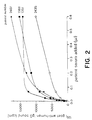
- Figure 2 illustrates the data from immunoassays, for serum anti-AChR autoantibody in various myasthenia gravis patients, employing AChR from cells of the TE671 Line as the solid-phase bound antigen and 125I-labeled goat anti-human IgG for detection of antigen-bound autoantibody.
- the present invention entails improvements in the assays and processes disclosed in the above-cited patents for diagnosing myasthenia gravis. These improvements reside in the use of AChR derived from cells of the TE671 Line rather than AChR in crude muscle extracts.
- the label on the AChR is a radioactively labeled, preferably an 125I-labeled, curaremimetic neurotoxin which binds with high affinity in the ACh binding site of the AChR.
- Curaremimetic toxins which can be employed to label AChR are naturally occurring proteins found in various poisonous reptiles, such as cobras and sea snakes. Many such toxins have been isolated and sequenced. See, e.g., Karlsson, Handbook of Experimental Pharmacology 52, 159-212 (1979). The preferred toxins for labeling are the so-called "long neurotoxins," as described by Karlsson, supra. Most preferred is alpha-bungarotoxin from Bungarus multicinctus.
- the present invention involves processes for making an AChR, that is suitable for diagnosis of myasthenia gravis by immunoassays of anti-AChR autoantibodies in human serum, by culturing cells of the TE671 Line and for obtaining, from cells of the TE671 Line cultures, preparations of such AChR that are suitable for use in such immunoassays.
- cells of the TE671 Line in the instant specification is meant (i) cells which are from a culture of any TE671 cell line, including a culture of subline no. 2 and including cultures of said subline that are on deposit at the ATCC under deposit no. CRL 8805; or (ii) cells of a subculture of any culture specified in (i); or (iii) mutants of any of the cells specified in (i) or (ii); provided that said cells (under (i), (ii) and (iii)) produce AChR that, for practical purposes in immunoassays, is immunologically essentially the same as human muscle AChR.
- subculture is meant a culture grown from cells of a culture (source culture) or any subculture of the source culture, regardless of the number of subculturings between the subculture of interest and the source culture.
- Mutants of cells specified in points (i) and (ii) of the definition of "cells of the TE671 Line” include mutants that arise spontaneously or from human intervention, as by repeated passaging, chemical treatment, irradiation, cell fusion, transformation, transfection, microinjection of nucleic acid into cell nucleii, and the like.
- Example II A method is illustrated in Example II for establishing that an AChR, for practical purposes in immunoassays, is immunologically essentially the same as human muscle AChR. Generally, this criterion is established if the anti-human muscle AChR antibodies in human sera are highly cross-reactive (to the extent of at least 50%) with the AChR of interest.
- the criterion obtains can be ascertained by testing both human muscle AChR and the AChR of interest with a battery of monoclonal antibodies known to be reactive against antigenic determinants on one or the other of the AChR's; if all or nearly all (i.e., more than 9 of 10) of the antigenic determinants corresponding to the monoclonal antibodies are found in the testing to be on both of the AChR's, it can be concluded that, for practical purposes in immunoassays, the AChR's are immunologically indistinguishable (i.e., essentially the same).
- the high level of cross-reactivity with monoclonal antibodies which characterizes such immunologically indistinguishable AChR's also implies that, after correction for any known non-immunological factors, such as differences in affinity for labeled toxin in the immune precipitation assay described in the above-cited patents, the titer of anti-AChR antibody in a serum sample as determined in an immunoassay with human muscle AChR will be no more than 25% (plus or minus some due to inevitable experimental uncertainty) greater than the titer with AChR from cells of the TE671 Line.
- anti-AChR antibody titers determined with two AChR's can be reliably ascertained by comparing the titers, determined in immune precipitation assays according to the above-cited patents and illustrated in Example II, with at least 10 sera which range over at least an order of magnitude in their anti-AChR antibody titers.
- the AChR of cells of the cultures of the TE671 Line, subline 2, deposited at ATCC under deposit no. CRL 8805, is, for practical purposes in immunoassays, immunologically indistinguishable from human muscle AChR. Indeed, as illustrated by the data plotted in Figure 1, after correction for differences in affinity for toxin (see Example II), the cross-reactivity of anti-AChR autoantibodies in human serum for AChR from cells of such cultures and AChR from human leg muscle is, within experimental error, nearly 100%; and the AChR's from these two sources are interchangeable in immunoassays of sera for diagnosing myasthenia gravis in persons suffering therefrom.
- the invention further entails solid-phase immunoassays for autoantibodies against human muscle AChR, employing as antigen AChR derived from cells of the TE671 Line.
- Immunoassays for anti-AChR autoantibodies which were not practicable heretofore because of the great difficulty and associated high cost of obtaining sufficient amounts of human muscle AChR, are made readily available by the availability of AChR from TE671 cells.
- the solid-phase immunoassays are carried out with AChR from cells of the TE671 Line following methods known in the solid-phase immunoassay art.
- a "substrate" for the solid-phase immunoassay is made by affixing the AChR covalently or non-covalently to a solid support by any of numerous methods known in the immunoassay art.
- solid supports known in the art that can be employed are those made of polyethylene, polypropylene, polybutylene, polystyrene, polymethacrylate, polyacrylamide, other synthetic polymeric materials to which the AChR can be affixed, glass, nitrocellulose, cellulose, and agarose.
- the solid support can be a "macroporous" gel-type support such as formed by, for example, certain agarose materials (e.g., Sephacryl S-500 from Pharmacia, Inc., Piscataway, N.J., U. S. A.).
- the solid support can be in the form of microparticles such as microspheres or beads (e.g., "latex" beads) or can be the inside wall of a tube, such as a microfuge tube or a well of a microtiter or Millititer plate.
- the solid support with AChR bound after appropriate washings and incubation with materials to reduce background by blocking sites on the support not occupied by AChR, as understood in the art, and after optional preincubation with a substance (such as a curaremimetic neurotoxin, a small cholinergic ligand or an antibody) which blocks certain epitopes on the AChR, is incubated with serum or, more commonly, a dilution thereof in a suitable buffer, as illustrated in Example III and otherwise understood in the art.
- a substance such as a curaremimetic neurotoxin, a small cholinergic ligand or an antibody
- the serum prior to being incubated with the solid-phase-bound AChR, can be mixed with a substance (such as a curaremimetic neurotoxin, a low molecular weight cholinergic compound, or an antibody) at concentrations sufficient to out-compete any autoantibody in the serum from binding to certain epitopes on the AChR.
- a substance such as a curaremimetic neurotoxin, a low molecular weight cholinergic compound, or an antibody
- Autoantibody in the serum against epitopes on the AChR that are not blocked by such epitope-blocking substances that might be employed will bind to the AChR.
- the autoantibody bound to the AChR is then detected by incubation with a labeled anti-autoantibody or labeled Protein A from Staphyloccocus aureus, or any protein or other substance which is labeled for detection and will bind to autoantibody but not significantly to other substances in the system.
- labeled anti-autoantibody is employed for detection.
- the labeled anti-autoantibody will be provided as labeled, polyclonal anti-human IgG.
- polyclonal subclass specific anti-immunoglobulins such as anti-human IgGl or anti-human IgM, or anti-human immunoglobulin that is not specific for a particular class or subclass, can also be used.
- the antisera employed for detection will typically be from goat, rabbit, rat or mouse, although other, non-human mammalian species may also be used as the source.
- the label on the anti-human immunoglobulin, S. aureus Protein A or other substance bound to anti-AChR autoantibody will be an enzyme, such as an acid or alkaline phosphatase, a peroxidase, or a beta-galactosidase, and detection will be by means of a product produced in a reaction catalyzed by the enzyme label, as known in the art.
- the label will usually be different from an enzyme.
- non-enzyme labels include radioactive atoms, chromophores or fluorophores, and biotin; detection methods for each of these labels, as well as other labels known in the art that may also be employed, are known in the art. Preferred is 125I-labeling.
- the AChR with anti-AChR autoantibody bound and with unlabeled anti-autoantibody also bound is detected by incubation with labeled S. aureus Protein A followed by detection of the label on the Protein A.
- the label on the Protein A can be an enzyme or any of the other labels described above or otherwise known in the art.
- Another aspect of the invention is a method to characterize the anti-AChR autoantibodies of a patient suffering from myasthenia gravis according to the titers of fractions of such autoantibodies that bind to various subsets of epitopes on the AChR.
- This method comprises carrying out at least two of four solid-phase immunoassays on the serum of a patient, which immunoassays are substantially the same except that: one is with solid-phase bound AChR alone; one with solid-phase bound AChR incubated with a monoclonal antibody (e.g., mAb 35) to the main immunogenic region, either prior to or simultaneously with incubation with a serum sample; one with solid-phase bound AChR incubated, prior to or simultaneously with incubation with a serum sample, with a curaremimetic neurotoxin; and one with solid-phase bound AChR incubated, prior to or simultaneously with incubation with a serum sample, with a low molecular weight compound (e.g., nicotinic cholinergic ligand such as carbamylcholine) that binds tightly in the ACh binding site but does not, like the neurotoxins, block nearby sites.
- a low molecular weight compound e.g., nicotin
- the assay with AChR alone will yield information on the titer of all autoantibodies to epitopes on the AChR.
- the assay together with the monoclonal antibody to the main immunogenic region will yield information on titer of autoantibodies to epitopes outside the main immunogenic region of the AChR.
- the assay together with the curaremimetic neurotoxin will yield information on the titer of autoantibodies to epitopes outside the ACh binding site and nearby sites blocked by the toxin.
- the assay together with the low molecular weight compound which binds in the ACh binding site will yield information on titer of autoantibodies to the ACh binding site itself.
- the incubation of the epitope-blocking substance with AChR is accomplished by combining the blocking substance with the serum sample, which is incubated with the immunoassay substrate (i.e., solid support with AChR bound), at a high enough concentration in the sample to out-compete in binding nearly all autoantibody in the sample that might bind to the same epitope or epitopes as the blocking substance.
- the immunoassay substrate i.e., solid support with AChR bound
- the basis for the solid-phase immunoassays is a substrate material to which AChR derived from cells of the TE671 Line is complexed by covalent or non-covalent binding.
- These solid-phase substrates with AChR complexed are also part of the instant invention.
- These substrates are prepared by any means known in the art for affixing an antigen to a solid support in a manner suitable for solid-phase immunoassay for antibody against the antigen.
- kits facilitate the preparation of kits for the diagnosis of myasthenia gravis.
- kits are also encompassed by the present invention.
- Said kits comprise AChR from cells of the TE671 Line and a solid support to which the AChR can be affixed to prepare a substrate according to the invention.
- the kits may comprise a substrate according to the invention (i.e., a solid support with, already affixed thereto, AChR from cells of the TE671 Line).
- kits of the invention may also include other components necessary for a solid-phase immunoassay of a patient's serum for anti-AChR autoantibodies, such as, for example, ligands or monoclonal antibodies which block parts of the AChR from binding by anti-AChR autoantibody (if the kit is to be employed in an assay to characterize the sites on the AChR to which autoantibody binds), buffers, anti-human IgG labeled for detection, and other components as illustrated in Example III and otherwise understood in the solid phase immunoassay art.
- other components necessary for a solid-phase immunoassay of a patient's serum for anti-AChR autoantibodies such as, for example, ligands or monoclonal antibodies which block parts of the AChR from binding by anti-AChR autoantibody (if the kit is to be employed in an assay to characterize the sites on the AChR to which autoantibody binds), buffers, anti-human IgG labeled for detection, and
- Any technique known in the art for culturing mammalian cells can be employed to culture cells of the TE671 Line.
- a preferred technique for growing TE671 cells is to use Iscove's modified Dulbecco's minimal essential medium (Irvine Scientific, Santa Ana, California) containing 10% fetal bovine serum in an atmosphere of 90% air and 10% CO2 at 37°C.
- Iscove's modified Dulbecco's minimal essential medium Irvine Scientific, Santa Ana, California
- plastic roller bottles (Falcon Labware, Becton-Dickinson, Inc., Oxnard, California U.S.A.) are used.
- 3 X 10⁷ cells are inoculated in 150 ml of medium; whereas, for 1750 cm2 bottles, 6 X 10⁷ cells are inoculated in 300 ml of medium.
- the medium is changed on day five, and AChR yield is optimal on days nine to ten.
- AChR has typically been isolated from the cultures from six or seven 850 cm2 roller bottles at a time, although much larger volumes of culture can easily be processed simultaneously. The following procedure has been used routinely for the isolation of AChR from cultures of cells of the TE671 Line, subline No. 2, samples of which are on deposit at the ATCC under deposit No.
- CRL 8805 To each 850 cm2 bottle of culture on day nine or ten, after growth as described in Example I, is added 25 ml of harvest buffer (100 mM NaCl, 10 mM Na phosphate buffer, 10 mM NaN3, 15 mM EDTA (ethylenediaminetetraacetic acid), 2 mM PMSF (phenylmethanesulfonylfluoride), 15 mM IAA (iodoacetic acid), and 5 mM benzamidine, pH 7.5). The bottles are then vigorously shaken until the cells come off of the plastic.
- harvest buffer 100 mM NaCl, 10 mM Na phosphate buffer, 10 mM NaN3, 15 mM EDTA (ethylenediaminetetraacetic acid), 2 mM PMSF (phenylmethanesulfonylfluoride), 15 mM IAA (iodoacetic acid), and 5 mM benzamidine, pH 7.5.
- the buffer and cells are collected in a bottle on ice, and the roller bottles are rinsed from bottle to bottle with an additional 100 ml of harvest buffer, which is then pooled with the rest.
- a Polytron homogenizer is then used to disrupt the cells for 15 seconds at a speed just below that at which foaming occurs.
- Membranes and other particulates are collected by centrifuging the homogenate for 30 minutes at 45,000 rpm in a Beckman Ti50.2 rotor (Beckman Instruments, Inc., Spinco Division, Palo Alto, California, U.S.A.) (302,000 X g) at 4°C. Typically, about 1.5 gm of pellet is recovered per 850 cm2 bottle.
- the pellets are placed in four volumes of extraction buffer (10 mM Na phosphate, 5 mM EDTA, 5 mM EGTA (ethylene glycol, bis-(2-aminoethylether)-N,N,N',N'-tetraacetic acid), 5 mM IAA, 5 mM benzamidine, 2 mM PMSF, 2% Triton X-100, pH 7.5) and gently suspended with the Polytron for 15 seconds. After extraction for 30 minutes with gentle shaking at 4°C, the preparation is centrifuged as before. The supernate typically contains AChR at about 1.1 X 10 ⁇ 8 M in a yield of about 6.6 X 10 ⁇ 11 moles per 850 cm2 bottle. A comparable crude extract of minced muscle tissue contains AChR at only about 2 x 10 ⁇ 10 M to about 2 X 10 ⁇ 9 M and generally less than 1 X 10 ⁇ 9 M.
- extraction buffer 10 mM Na phosphate, 5 mM EDTA, 5 mM EG
- Immune precipitation experiments were carried out on sera from 45 patients essentially as described in the above-cited patents using AChR at 2 nM from the 2% Triton X-100 extracts (as described above in this Example) of either TE671 cells or muscle from amputated human leg.
- the concentration of AChR in the TE671 cell extract was reduced to 2 nM by dilution with assay buffer (100 mM NaCl, 10 mM Na phosphate buffer, 10 mM NaN3, 0.5% Triton X-100, pH 7.5).
- assay buffer 100 mM NaCl, 10 mM Na phosphate buffer, 10 mM NaN3, 0.5% Triton X-100, pH 7.5.
- the muscle extract employed was a rare one, in which the concentration of AChR reached about 2 nM, and was used without dilution.
- the concentration of AChR in the muscle extracts would have been significantly lower and would have been increased by a standard technique (e.g., employing an Amicon concentrator) (Amicon, Danvers, Massachusetts, U.S.A.) until the AChR concentration reached 2 nM.
- a standard technique e.g., employing an Amicon concentrator (Amicon, Danvers, Massachusetts, U.S.A.) until the AChR concentration reached 2 nM.
- the AChR's were labeled by combining the AChR solutions with an amount of a 2 micromolar solution of 125I-labeled alpha-bungarotoxin, in Na phosphate buffer, pH 7-7.5, to bring the 125I-labeled alpha-bungarotoxin concentration to 4 nM, and then incubating the resulting solutions for 4 hours at 4°C.
- Analyses were carried out in triplicate on various dilutions with assay buffer of each serum being tested. Supplementary normal human serum was added to each sample so that there was a total of 5 microliters of serum in the final reaction mix.
- the purpose of the dilutions is to bring the concentration of anti-AChR autoantibody from a serum being tested within the measurable range of the assay and preferably within a range such that 10-20% of the labeled AChR in an assay solution is complexed by anti-AChR autoantibody.
- the dilutions employed may vary widely, but will be determined by taking into account the concentration of labeled AChR in the assay solution with which serum samples are combined and the fact that anti-AChR autoantibodies in sera of persons suffering from myasthenia gravis can be as high as about 1,500 nM.
- the supplementary normal serum is added so that the total amount of immunoglobulin in each sample is approximately the same.
- each of the three samples of each dilution of each test serum was combined with 100 microliters of the labeled AChR solution (2 nM) and the combination was incubated overnight at 4°C. Then 100 microliters of goat anti-human IgG (at a concentration sufficient to precipitate all IgG in 5 microliters of serum) was added and the resulting solution incubated for 30 minutes at 4°C to precipitate all of the human serum IgG. After addition of 1 ml of assay buffer, the solution was centrifuged for two minutes at 10,000 X g at 20°C.
- the supernatant was aspirated and the pellet washed twice, each time with 1 ml of assay buffer, by vortexing followed by 2 minute centrifugation at 10,000 X g and 20°C and aspiration of supernatant. Finally, the 125I in the pellet was determined by gamma-counting by a standard technique.
- comparisons of AChR from human leg muscle with AChR from cells of the TE671 Line using monoclonal antibodies have not identified any antigenic determinant on the leg muscle-derived AChR that is not also on the TE671 cell-derived AChR.
- Triton X-100 extract of AChR was obtained from cultures of cells of the TE671 Line, subline No. 2, of which samples are deposited at the ATCC under deposit No. CRL 8805.
- the AChR of the extract was then affinity-purified on toxin-agarose following the procedure of Lindstrom et al, Methods in Enzymology 74C, 432-459 (1981) as follows:
- AChR concentration is assayed by immune precipitation of the AChR labeled with 125 I-labeled-alpha-bungarotoxin (Lindstrom et al., supra).
- the assay can be carried out with a polyclonal anti-AChR antiserum, as from a rabbit, mouse or rat, with serum from a myasthenia gravis patient with a high serum titer of anti-AChR autoantibody, or with an anti-AChR monoclonal antibody, such as mAb 35 or mAb 64.
- the purified AChR was bound to wells of a Millipore Millititer-HA 96 well filtration plate (Millipore, Bedford, Massachusetts, U.S.A., Cat. No. STHA096NS) by applying 30 microliter aliquots overnight at 4°C (more than 400 fmoles AChR can be bound per well). Further binding was then quenched by adding 50 microliters of quench buffer (3% bovine serum albumin (BSA), 0.2% Tween-20, 100 mM NaCl, 10 mM NaN3, 10 mM Na phosphate pH 7.5) and letting stand for 2 hrs at 4°C. All of the liquid was then removed using a Millipore Millititer Vacuum Holder (Millipore Cat.
- quench buffer 3% bovine serum albumin (BSA), 0.2% Tween-20, 100 mM NaCl, 10 mM NaN3, 10 mM Na phosphate pH 7.5
- the concentration of myasthenia gravis patient serum autoantibodies against AChR can be calculated, in terms of grams or moles of autoimmune IgG/liter of patient serum, from the gamma emission data obtained as described above.
- the results can also be normalized to values obtained using the immune precipitation assay illustrated in Example II. Rather than using only anti-IgG to detect autoantibody bound to AChR, labeled subclass-specific antisera could be used and, thereby, the subclass distribution of the autoantibodies determined.
- the fraction of autoantibodies in a patient's serum which are directed at or near the ACh binding site can be determined by carrying out two immunoassays on the serum: one as described above and one wherein the AChR bound to wells is incubated with serum samples which include an excess of alpha-bungarotoxin (or similar toxin), or a small cholinergic ligand such as carbamylcholine, to out-compete from binding, at the toxin or cholinergic ligand binding sites, substantially all of any autoantibody in the serum that binds to such sites.
- the fraction of autoantibodies which are inhibited from binding to the AChR by the toxin or cholinergic ligand can be determined; this fraction is, or is a close approximation to, the fraction of autoantibodies directed at and near the ACh binding site.
- the fraction of the autoantibodies directed to the main immunogenic region of the AChR can be determined by determining the fraction which are inhibited from binding to AChR upon incubating the AChR bound to the wells with serum combined with an excess of a monoclonal antibody to the main immunogenic region of the AChR, such as the rat monoclonal antibody mAb 35.
Landscapes
- Health & Medical Sciences (AREA)
- Immunology (AREA)
- Life Sciences & Earth Sciences (AREA)
- Hematology (AREA)
- Engineering & Computer Science (AREA)
- Molecular Biology (AREA)
- Chemical & Material Sciences (AREA)
- Biomedical Technology (AREA)
- Urology & Nephrology (AREA)
- Food Science & Technology (AREA)
- Biochemistry (AREA)
- Cell Biology (AREA)
- Biotechnology (AREA)
- Rheumatology (AREA)
- Rehabilitation Therapy (AREA)
- Medicinal Chemistry (AREA)
- Physics & Mathematics (AREA)
- Analytical Chemistry (AREA)
- Microbiology (AREA)
- General Health & Medical Sciences (AREA)
- General Physics & Mathematics (AREA)
- Pathology (AREA)
- Peptides Or Proteins (AREA)
- Preparation Of Compounds By Using Micro-Organisms (AREA)
- Compositions Of Oxide Ceramics (AREA)
- Ultra Sonic Daignosis Equipment (AREA)
- Road Paving Structures (AREA)
- Warehouses Or Storage Devices (AREA)
- Investigating Or Analysing Biological Materials (AREA)
Abstract
Description
- The present invention relates to immunochemical assays for diagnosis of myasthenia gravis.
- Myasthenia gravis is an autoimmune disease. In a patient suffering from the disease, autoantibodies are generated against epitopes on acetylcholine receptors at neuromuscular junctions. This autoimmune response impairs neuromuscular transmission. This impairment causes the muscular weakness and fatigability which characterize the disease.
- In U.S. Patent Nos. 4,202,875 and RE 30,059, biochemical assays for the diagnosis of myasthenia gravis are described.
- The assays in the above-cited patents involve immune precipitation, with anti-human immunoglobulin, of labeled acetylcholine receptor protein complexed with autoantibodies to the receptor protein from serum of a myasthenia gravis patient. The acetylcholine receptor protein (hereinafter "AChR") that is employed is part of a crude extract of mammalian muscle. The labeling of the AChR in these assays is accomplished by binding to the protein in the crude extract radioactively labeled, curaremimetic neurotoxins, such as Naja naja siamensis toxin and alpha-bungarotoxin. These toxins complex with the AChR by binding in the acetylcholine binding site on the protein.
- While the assays described in the above-cited patents have found widespread commercial application and are at present the preferred assays for diagnosing myasthenia gravis, they involve a number of drawbacks. The preferred source for the AChR utilized in these assays is muscle from amputated human legs. This tissue is in short supply and, on account of a variety of legal complications, difficult to obtain for commercial applications even when available. Further, it is possible to obtain from the tissue only very small quantities of AChR suitable for the assays because the protein is present at only very low concentration in even healthy, unamputated tissue and because substantial proteolytic degradation of the protein occurs unavoidably before a crude extract of the muscle containing the protein can be isolated from the amputated tissue. Other, more plentiful, mammalian-muscle sources for the AChR are available; but these other sources are not entirely free of the drawbacks, of sparse amount and proteolytic degradation, of the human leg-muscle source and, more importantly, the AChR from non-human sources is less acceptable for the assays because human autoantibodies to human AChR cross react poorly with AChR's from other mammals (e.g., to the extent of only 2.1% in the case of AChR from denervated rat muscle, a typical alternative to AChR from human leg muscle).
- The limited supply of human muscle AChR suitable for immunochemical assay has made it impracticable to assay for myasthenia gravis-related autoantibody in human serum other than by the immune precipitation assay using crude extracts of muscle described in the above-cited patents. The ready availability of a more plentiful source of AChR, that reacts with human autoantibodies to human muscle AChR to substantially the same extent as human muscle AChR itself, would make practical other, more sensitive immunoassay techniques, such as solid-phase and enzyme-linked immunosorbent assay (ELISA) techniques, for the diagnosis of myasthenia gravis.
- The severity of muscle weakness in myasthenia gravis patients does not correlate closely with the concentration of anti-AChR antibodies in their sera. This is probably largely due to the multiple, complex mechanisms by which these antibodies impair transmission (reduction of number of receptors by antigenic modulation, complement-mediated focal lysis, direct impairment of AChR function by bound antibody) as well as the nature of neuromuscular transmission (occurring in an all or none fashion with a large safety factor to insure transmission) and the complex adaptive mechanisms which are available to the body to compensate for impaired neuromuscular transmission (increasing acetylcholine release, increasing the area of neuromuscular junctions).
- In myasthenia gravis patients, autoantibodies bind to various epitopes on various regions of AChR. One of these regions is called the "main immunogenic region". This region is located on the extracellular surface of alpha-subunits of the AChR. Another of these regions is the acetylcholine (hereinafter "ACh") binding sites, which are also located on the alpha-subunits. In the average myasthenia gravis patient, about half of the anti-AChR-autoantibodies are directed at the main immunogenic region. Autoantibodies which bind to the main immunogenic region can cause antigenic modulation and fix complement in vivo and thereby reduce the number of ACh receptors. Antibodies which are directed at the ACh binding sites would, at very low concentrations, be very effective at directly impairing AChR function in neuromuscular transmission. It would be useful if one could measure not only total anti-AChR antibody concentration in sera of myasthenia gravis patients but also the fractions of these antibodies which are directed at the main immunogenic region and the ACh-binding sites of AChR.
- The assay of the above-cited patents depends on the labeling to high specific activity of the muscle AChR by complexing with it radioactively labeled toxins which bind in the ACh-binding site of the protein. It has not been possible to assay specifically for autoantibodies directed against the ACh-binding site with the assay of the above-cited patents because, in such assays, the binding site and nearby areas are blocked by toxin and, thereby, the epitopes in the binding site, and other epitopes near it, are inaccessible to binding by autoantibody. The invention described herein, based on the discovery of a source for large quantities of easily isolated AChR which, for practical purposes, is immunologically indistinguishable from human muscle AChR, makes feasible immunoassay methods which do not require blocking of some epitopes on the AChR from binding by autoantibodies. Thus, with the present invention, measurement is possible of not only the total concentration of anti-AChR autoantibody in the sera of a patient, permitting diagnosis, but also the fractions of these antibodies directed at pathologically significant sites on the AChR, permitting better characterization of the patient's disease and improved basis for planning therapy to treat the disease.
- McAllister et al., Int. J. Cancer 20, 206-212 (1977) reported the establishment of a cell line, designated TE671, Subline No. 2, from a human cerebellar medulloblastoma. This and equivalent AChR-producing cell lines are referred to in this specification as "the TE671 Line".
- Syapin et al., Brain Research 231, 365-377 (1982) have characterized the cells of the TE671 Line as possessing mammalian neuronal nicotinic acetylcholine receptors. It is thought in the art that the proteins of such receptors are immunologically substantially different from (i.e., have low immunological cross-reactivity with) the AChR against which myasthenia gravis autoantibodies react (i.e., that antibodies against one of neuronal AChR and muscle AChR have little or no cross-reactivity with the other) (Patrick and Stallcup, Proc. Natl. Acad. Sci. (U.S.A.) 74, 4689 (1977); Swanson et al., Proc. Natl. Acad. Sci. (U.S.A.) 80, 4532 (1983); Smith et al., J. Neuroscience 5, 2726 (1985)).
- Hybridomas which secrete rat monoclonal antibodies, including that designated mAb 35, to the main immunogenic region of muscle AChR have been described by Tzartos et al., J. Biol. Chem. 256, 8635-8645 (1981) and Proc. Natl. Acad. Sci. U.S.A. 79, 188 (1982). Other hybridomas, including that designated mAb 64, which secrete monoclonal antibodies against muscle AChR have also been developed (Tzartos et al., J. Neuroimmunology, 10, 235-253 (1986)).
- It has now been found, that the AChR on cells of the TE671 Line is, for practical purposes in immunoassays, immunologically indistinguishable from human muscle AChR and functions substantially the same as human muscle AChR derived from amputated human leg muscle in the immune precipitation assay, for diagnosing myasthenia gravis, that is described in the above-cited patents.
- However, unlike the AChR derived from human leg muscle, the AChR from cells of the TE671 Line is readily available, in virtually unlimited amounts, at relatively high concentrations, and substantially free of heterogeneities and impurities due to proteolysis and other factors which lead to contamination of AChR from muscle sources.
- This discovery makes practical substantially improved immunochemical assay systems for diagnosing myasthenia gravis.
- Figure 1 illustrates the practical equivalence of AChR from human leg muscle and AChR from cells of the TE671 Line in the immune precipitation assay for serum anti-AChR autoantibodies described in the above-cited patents. The Figure is a graph of the logarithms (to base 10) of the anti-AChR autoantibody concentrations, in nM, in the sera of 45 myasthenia gravis patients, as determined in such an immune precipitation assay with human leg muscle AChR labeled with ¹²⁵I-labeled alpha-bungarotoxin, as a function of the logarithms (to base 10) of the anti-AChR autoantibody concentrations, in nM, in the same sera as determined by an assay procedure that was the same but for replacement of human leg muscle AChR with AChR from cells of the TE671 Line.
- Figure 2 illustrates the data from immunoassays, for serum anti-AChR autoantibody in various myasthenia gravis patients, employing AChR from cells of the TE671 Line as the solid-phase bound antigen and ¹²⁵I-labeled goat anti-human IgG for detection of antigen-bound autoantibody.
- In one aspect, the present invention entails improvements in the assays and processes disclosed in the above-cited patents for diagnosing myasthenia gravis. These improvements reside in the use of AChR derived from cells of the TE671 Line rather than AChR in crude muscle extracts.
- As noted above, the assays and processes of the above-cited patents depend on immune precipitation of labeled AChR complexed with anti-AChR autoantibody in the serum of a person suffering from myasthenia gravis. The label on the AChR is a radioactively labeled, preferably an ¹²⁵I-labeled, curaremimetic neurotoxin which binds with high affinity in the ACh binding site of the AChR. Curaremimetic toxins which can be employed to label AChR are naturally occurring proteins found in various poisonous reptiles, such as cobras and sea snakes. Many such toxins have been isolated and sequenced. See, e.g., Karlsson, Handbook of Experimental Pharmacology 52, 159-212 (1979). The preferred toxins for labeling are the so-called "long neurotoxins," as described by Karlsson, supra. Most preferred is alpha-bungarotoxin from Bungarus multicinctus.
- In another aspect, the present invention involves processes for making an AChR, that is suitable for diagnosis of myasthenia gravis by immunoassays of anti-AChR autoantibodies in human serum, by culturing cells of the TE671 Line and for obtaining, from cells of the TE671 Line cultures, preparations of such AChR that are suitable for use in such immunoassays.
- The culturing of cells of the TE671 Line, and obtaining preparations of AChR therefrom, are described in the Examples below.
- By "cells of the TE671 Line" in the instant specification is meant (i) cells which are from a culture of any TE671 cell line, including a culture of subline no. 2 and including cultures of said subline that are on deposit at the ATCC under deposit no. CRL 8805; or (ii) cells of a subculture of any culture specified in (i); or (iii) mutants of any of the cells specified in (i) or (ii); provided that said cells (under (i), (ii) and (iii)) produce AChR that, for practical purposes in immunoassays, is immunologically essentially the same as human muscle AChR. By "subculture" is meant a culture grown from cells of a culture (source culture) or any subculture of the source culture, regardless of the number of subculturings between the subculture of interest and the source culture. Mutants of cells specified in points (i) and (ii) of the definition of "cells of the TE671 Line" include mutants that arise spontaneously or from human intervention, as by repeated passaging, chemical treatment, irradiation, cell fusion, transformation, transfection, microinjection of nucleic acid into cell nucleii, and the like.
- A method is illustrated in Example II for establishing that an AChR, for practical purposes in immunoassays, is immunologically essentially the same as human muscle AChR. Generally, this criterion is established if the anti-human muscle AChR antibodies in human sera are highly cross-reactive (to the extent of at least 50%) with the AChR of interest. Whether the criterion obtains can be ascertained by testing both human muscle AChR and the AChR of interest with a battery of monoclonal antibodies known to be reactive against antigenic determinants on one or the other of the AChR's; if all or nearly all (i.e., more than 9 of 10) of the antigenic determinants corresponding to the monoclonal antibodies are found in the testing to be on both of the AChR's, it can be concluded that, for practical purposes in immunoassays, the AChR's are immunologically indistinguishable (i.e., essentially the same). The high level of cross-reactivity with monoclonal antibodies which characterizes such immunologically indistinguishable AChR's also implies that, after correction for any known non-immunological factors, such as differences in affinity for labeled toxin in the immune precipitation assay described in the above-cited patents, the titer of anti-AChR antibody in a serum sample as determined in an immunoassay with human muscle AChR will be no more than 25% (plus or minus some due to inevitable experimental uncertainty) greater than the titer with AChR from cells of the TE671 Line. The relationship of anti-AChR antibody titers determined with two AChR's can be reliably ascertained by comparing the titers, determined in immune precipitation assays according to the above-cited patents and illustrated in Example II, with at least 10 sera which range over at least an order of magnitude in their anti-AChR antibody titers.
- The AChR of cells of the cultures of the TE671 Line,
subline 2, deposited at ATCC under deposit no. CRL 8805, is, for practical purposes in immunoassays, immunologically indistinguishable from human muscle AChR. Indeed, as illustrated by the data plotted in Figure 1, after correction for differences in affinity for toxin (see Example II), the cross-reactivity of anti-AChR autoantibodies in human serum for AChR from cells of such cultures and AChR from human leg muscle is, within experimental error, nearly 100%; and the AChR's from these two sources are interchangeable in immunoassays of sera for diagnosing myasthenia gravis in persons suffering therefrom. - The invention further entails solid-phase immunoassays for autoantibodies against human muscle AChR, employing as antigen AChR derived from cells of the TE671 Line. Immunoassays for anti-AChR autoantibodies, which were not practicable heretofore because of the great difficulty and associated high cost of obtaining sufficient amounts of human muscle AChR, are made readily available by the availability of AChR from TE671 cells. The solid-phase immunoassays are carried out with AChR from cells of the TE671 Line following methods known in the solid-phase immunoassay art.
- A "substrate" for the solid-phase immunoassay is made by affixing the AChR covalently or non-covalently to a solid support by any of numerous methods known in the immunoassay art. Among the solid supports known in the art that can be employed are those made of polyethylene, polypropylene, polybutylene, polystyrene, polymethacrylate, polyacrylamide, other synthetic polymeric materials to which the AChR can be affixed, glass, nitrocellulose, cellulose, and agarose. The solid support can be a "macroporous" gel-type support such as formed by, for example, certain agarose materials (e.g., Sephacryl S-500 from Pharmacia, Inc., Piscataway, N.J., U. S. A.). The solid support can be in the form of microparticles such as microspheres or beads (e.g., "latex" beads) or can be the inside wall of a tube, such as a microfuge tube or a well of a microtiter or Millititer plate. The solid support with AChR bound, after appropriate washings and incubation with materials to reduce background by blocking sites on the support not occupied by AChR, as understood in the art, and after optional preincubation with a substance (such as a curaremimetic neurotoxin, a small cholinergic ligand or an antibody) which blocks certain epitopes on the AChR, is incubated with serum or, more commonly, a dilution thereof in a suitable buffer, as illustrated in Example III and otherwise understood in the art. Optionally, prior to being incubated with the solid-phase-bound AChR, the serum (or dilution) can be mixed with a substance (such as a curaremimetic neurotoxin, a low molecular weight cholinergic compound, or an antibody) at concentrations sufficient to out-compete any autoantibody in the serum from binding to certain epitopes on the AChR. Autoantibody in the serum against epitopes on the AChR that are not blocked by such epitope-blocking substances that might be employed will bind to the AChR. After appropriate washing as understood in the art, the autoantibody bound to the AChR is then detected by incubation with a labeled anti-autoantibody or labeled Protein A from Staphyloccocus aureus, or any protein or other substance which is labeled for detection and will bind to autoantibody but not significantly to other substances in the system.
- Preferably, labeled anti-autoantibody is employed for detection. Typically the labeled anti-autoantibody will be provided as labeled, polyclonal anti-human IgG. However, polyclonal subclass specific anti-immunoglobulins, such as anti-human IgGl or anti-human IgM, or anti-human immunoglobulin that is not specific for a particular class or subclass, can also be used. The antisera employed for detection will typically be from goat, rabbit, rat or mouse, although other, non-human mammalian species may also be used as the source.
- If the solid-phase immunoassay is an ELISA, the label on the anti-human immunoglobulin, S. aureus Protein A or other substance bound to anti-AChR autoantibody will be an enzyme, such as an acid or alkaline phosphatase, a peroxidase, or a beta-galactosidase, and detection will be by means of a product produced in a reaction catalyzed by the enzyme label, as known in the art.
- If the solid-phase immunoassay is not an ELISA and there is a label on the anti-immunoglobulin, S. aureus Protein A or other substance, the label will usually be different from an enzyme. Such non-enzyme labels include radioactive atoms, chromophores or fluorophores, and biotin; detection methods for each of these labels, as well as other labels known in the art that may also be employed, are known in the art. Preferred is ¹²⁵I-labeling.
- In another alternative, the AChR with anti-AChR autoantibody bound and with unlabeled anti-autoantibody also bound, is detected by incubation with labeled S. aureus Protein A followed by detection of the label on the Protein A. The label on the Protein A can be an enzyme or any of the other labels described above or otherwise known in the art.
- Another aspect of the invention is a method to characterize the anti-AChR autoantibodies of a patient suffering from myasthenia gravis according to the titers of fractions of such autoantibodies that bind to various subsets of epitopes on the AChR. This method comprises carrying out at least two of four solid-phase immunoassays on the serum of a patient, which immunoassays are substantially the same except that: one is with solid-phase bound AChR alone; one with solid-phase bound AChR incubated with a monoclonal antibody (e.g., mAb 35) to the main immunogenic region, either prior to or simultaneously with incubation with a serum sample; one with solid-phase bound AChR incubated, prior to or simultaneously with incubation with a serum sample, with a curaremimetic neurotoxin; and one with solid-phase bound AChR incubated, prior to or simultaneously with incubation with a serum sample, with a low molecular weight compound (e.g., nicotinic cholinergic ligand such as carbamylcholine) that binds tightly in the ACh binding site but does not, like the neurotoxins, block nearby sites. The assay with AChR alone will yield information on the titer of all autoantibodies to epitopes on the AChR. The assay together with the monoclonal antibody to the main immunogenic region will yield information on titer of autoantibodies to epitopes outside the main immunogenic region of the AChR. The assay together with the curaremimetic neurotoxin will yield information on the titer of autoantibodies to epitopes outside the ACh binding site and nearby sites blocked by the toxin. Finally, the assay together with the low molecular weight compound which binds in the ACh binding site will yield information on titer of autoantibodies to the ACh binding site itself. Usually in these assays, the incubation of the epitope-blocking substance with AChR is accomplished by combining the blocking substance with the serum sample, which is incubated with the immunoassay substrate (i.e., solid support with AChR bound), at a high enough concentration in the sample to out-compete in binding nearly all autoantibody in the sample that might bind to the same epitope or epitopes as the blocking substance.
- As indicated above, the basis for the solid-phase immunoassays is a substrate material to which AChR derived from cells of the TE671 Line is complexed by covalent or non-covalent binding. These solid-phase substrates with AChR complexed are also part of the instant invention. These substrates are prepared by any means known in the art for affixing an antigen to a solid support in a manner suitable for solid-phase immunoassay for antibody against the antigen.
- AChR from cells of the TE671 Line facilitate the preparation of kits for the diagnosis of myasthenia gravis. Such kits are also encompassed by the present invention. Said kits comprise AChR from cells of the TE671 Line and a solid support to which the AChR can be affixed to prepare a substrate according to the invention. Optionally, the kits may comprise a substrate according to the invention (i.e., a solid support with, already affixed thereto, AChR from cells of the TE671 Line). The kits of the invention may also include other components necessary for a solid-phase immunoassay of a patient's serum for anti-AChR autoantibodies, such as, for example, ligands or monoclonal antibodies which block parts of the AChR from binding by anti-AChR autoantibody (if the kit is to be employed in an assay to characterize the sites on the AChR to which autoantibody binds), buffers, anti-human IgG labeled for detection, and other components as illustrated in Example III and otherwise understood in the solid phase immunoassay art.
- The invention is described in greater detail in the following examples.
- Any technique known in the art for culturing mammalian cells can be employed to culture cells of the TE671 Line.
- A preferred technique for growing TE671 cells is to use Iscove's modified Dulbecco's minimal essential medium (Irvine Scientific, Santa Ana, California) containing 10% fetal bovine serum in an atmosphere of 90% air and 10% CO₂ at 37°C.
- Small volumes are grown in 35 mm dishes using an innoculum of 3 X 10⁵ cells in 1.5 ml of medium. After three days the medium is changed, and optimal AChR yield is obtained on day six or seven.
- For large volumes, plastic roller bottles (Falcon Labware, Becton-Dickinson, Inc., Oxnard, California U.S.A.) are used. In the case of 850 cm² bottles, 3 X 10⁷ cells are inoculated in 150 ml of medium; whereas, for 1750 cm² bottles, 6 X 10⁷ cells are inoculated in 300 ml of medium. The medium is changed on day five, and AChR yield is optimal on days nine to ten.
- AChR has typically been isolated from the cultures from six or seven 850 cm² roller bottles at a time, although much larger volumes of culture can easily be processed simultaneously. The following procedure has been used routinely for the isolation of AChR from cultures of cells of the TE671 Line, subline No. 2, samples of which are on deposit at the ATCC under deposit No. CRL 8805:
To each 850 cm² bottle of culture on day nine or ten, after growth as described in Example I, is added 25 ml of harvest buffer (100 mM NaCl, 10 mM Na phosphate buffer, 10 mM NaN₃, 15 mM EDTA (ethylenediaminetetraacetic acid), 2 mM PMSF (phenylmethanesulfonylfluoride), 15 mM IAA (iodoacetic acid), and 5 mM benzamidine, pH 7.5). The bottles are then vigorously shaken until the cells come off of the plastic. The buffer and cells are collected in a bottle on ice, and the roller bottles are rinsed from bottle to bottle with an additional 100 ml of harvest buffer, which is then pooled with the rest. A Polytron homogenizer is then used to disrupt the cells for 15 seconds at a speed just below that at which foaming occurs. Membranes and other particulates are collected by centrifuging the homogenate for 30 minutes at 45,000 rpm in a Beckman Ti50.2 rotor (Beckman Instruments, Inc., Spinco Division, Palo Alto, California, U.S.A.) (302,000 X g) at 4°C. Typically, about 1.5 gm of pellet is recovered per 850 cm² bottle. The pellets are placed in four volumes of extraction buffer (10 mM Na phosphate, 5 mM EDTA, 5 mM EGTA (ethylene glycol, bis-(2-aminoethylether)-N,N,N',N'-tetraacetic acid), 5 mM IAA, 5 mM benzamidine, 2 mM PMSF, 2% Triton X-100, pH 7.5) and gently suspended with the Polytron for 15 seconds. After extraction for 30 minutes with gentle shaking at 4°C, the preparation is centrifuged as before. The supernate typically contains AChR at about 1.1 X 10⁻⁸ M in a yield of about 6.6 X 10⁻¹¹ moles per 850 cm² bottle. A comparable crude extract of minced muscle tissue contains AChR at only about 2 x 10⁻¹⁰ M to about 2 X 10⁻⁹ M and generally less than 1 X 10⁻⁹ M. - Immune precipitation experiments were carried out on sera from 45 patients essentially as described in the above-cited patents using AChR at 2 nM from the 2% Triton X-100 extracts (as described above in this Example) of either TE671 cells or muscle from amputated human leg. The concentration of AChR in the TE671 cell extract was reduced to 2 nM by dilution with assay buffer (100 mM NaCl, 10 mM Na phosphate buffer, 10 mM NaN₃, 0.5% Triton X-100, pH 7.5). The muscle extract employed was a rare one, in which the concentration of AChR reached about 2 nM, and was used without dilution. More typically, the concentration of AChR in the muscle extracts would have been significantly lower and would have been increased by a standard technique (e.g., employing an Amicon concentrator) (Amicon, Danvers, Massachusetts, U.S.A.) until the AChR concentration reached 2 nM.
- The AChR's were labeled by combining the AChR solutions with an amount of a 2 micromolar solution of ¹²⁵I-labeled alpha-bungarotoxin, in Na phosphate buffer, pH 7-7.5, to bring the ¹²⁵I-labeled alpha-bungarotoxin concentration to 4 nM, and then incubating the resulting solutions for 4 hours at 4°C.
- Analyses were carried out in triplicate on various dilutions with assay buffer of each serum being tested. Supplementary normal human serum was added to each sample so that there was a total of 5 microliters of serum in the final reaction mix. The purpose of the dilutions is to bring the concentration of anti-AChR autoantibody from a serum being tested within the measurable range of the assay and preferably within a range such that 10-20% of the labeled AChR in an assay solution is complexed by anti-AChR autoantibody. The dilutions employed may vary widely, but will be determined by taking into account the concentration of labeled AChR in the assay solution with which serum samples are combined and the fact that anti-AChR autoantibodies in sera of persons suffering from myasthenia gravis can be as high as about 1,500 nM. The supplementary normal serum is added so that the total amount of immunoglobulin in each sample is approximately the same.
- Each of the three samples of each dilution of each test serum was combined with 100 microliters of the labeled AChR solution (2 nM) and the combination was incubated overnight at 4°C. Then 100 microliters of goat anti-human IgG (at a concentration sufficient to precipitate all IgG in 5 microliters of serum) was added and the resulting solution incubated for 30 minutes at 4°C to precipitate all of the human serum IgG. After addition of 1 ml of assay buffer, the solution was centrifuged for two minutes at 10,000 X g at 20°C. The supernatant was aspirated and the pellet washed twice, each time with 1 ml of assay buffer, by vortexing followed by 2 minute centrifugation at 10,000 X g and 20°C and aspiration of supernatant. Finally, the ¹²⁵I in the pellet was determined by gamma-counting by a standard technique.
- A blank amount, obtained using 5 microliters of normal serum, was subtracted from the amount determined for each sample.
- The results of this experiment are illustrated in Figure 1. In the Figure, the logarithm of the serum concentration of anti-AChR antibody precipitable by the anti-IgG, determined with human leg muscle AChR, is plotted as a function of the logarithm of the same concentration determined with the TE671 cell-derived AChR. The difference from equality of these two concentrations for each sample is accounted for by experimental error in the assay procedures and the reported difference between the dissociation constant of alpha-bungarotoxin complexed with AChR from human muscle ( 1.8 X 10⁻¹⁰ M, Luky and Morgan-Hughes, Ann. N.Y., Acad. Sci. 377, 61 (1981)) and that of alpha-bungarotoxin complexed with AChR from cells of the TE671 Line, subline no. 2 (1.4 X 10⁻⁹ M, Syapin et al., supra). The results of the experiment establish that serum autoantibodies against AChR from human muscle are greater than 70% cross-reactive with the AChR that was derived from TE671 cells and that, for practical purposes in immunoassays, the AChR's from the two sources can be used interchangeably.
- As further support for the conclusion that the AChR's from the two sources are immunologically the same, at least for purposes of immunoassays, comparisons of AChR from human leg muscle with AChR from cells of the TE671 Line using monoclonal antibodies have not identified any antigenic determinant on the leg muscle-derived AChR that is not also on the TE671 cell-derived AChR.
- Following the procedure described in Example II, but employing eight volumes rather than four volumes of extraction buffer, relative to the volume of pellets, 75 ml of Triton X-100 extract of AChR was obtained from cultures of cells of the TE671 Line, subline No. 2, of which samples are deposited at the ATCC under deposit No. CRL 8805. The AChR of the extract was then affinity-purified on toxin-agarose following the procedure of Lindstrom et al, Methods in Enzymology 74C, 432-459 (1981) as follows:
The Triton X-100 extract, containing about 5.9 X 10⁻⁹ M AChR, was recirculated overnight at 4°C through a 3 ml column of toxin-agarose (0.5 mg Naja naja siamensis toxin III per ml of Sepharose C14B (Pharmacia, Inc., Piscataway, New Jersey, U.S.A.)) at a rate of 42 ml/hr. (Improved binding of TE671 AChR, with its apparently low affinity for the toxin, could probably have been obtained by using an affinity column containing 5-10 mg/ml of toxin.) The column was then washed with 10 ml of 0.1% Triton X-100 in 10 nM Na phosphate buffer, pH 7.5. Elution of AChR was achieved using 1 M carbamylcholine in elution buffer (0.1% Triton X-100, 100 mM NaCl, 10 mM NaN₃, 10 mM Na phosphate, pH 7.5) recirculated in separate 3 ml aliquots at 4°C for 8 hrs, then 18 hrs, and finally 36 hrs. The three eluates were then combined and dialyzed at 4°C against elution buffer to remove carbamylcholine. Under these conditions, 0.226 nmoles of AChR, of the 0.44 nmoles applied, were bound, and a total of 0.101 nmoles were eluted in 8.1 ml of a 13.8 nM solution. - AChR concentration is assayed by immune precipitation of the AChR labeled with ¹²⁵ I-labeled-alpha-bungarotoxin (Lindstrom et al., supra). The assay can be carried out with a polyclonal anti-AChR antiserum, as from a rabbit, mouse or rat, with serum from a myasthenia gravis patient with a high serum titer of anti-AChR autoantibody, or with an anti-AChR monoclonal antibody, such as mAb 35 or mAb 64.
- The purified AChR was bound to wells of a Millipore Millititer-HA 96 well filtration plate (Millipore, Bedford, Massachusetts, U.S.A., Cat. No. STHA096NS) by applying 30 microliter aliquots overnight at 4°C (more than 400 fmoles AChR can be bound per well). Further binding was then quenched by adding 50 microliters of quench buffer (3% bovine serum albumin (BSA), 0.2% Tween-20, 100 mM NaCl, 10 mM NaN₃, 10 mM Na phosphate pH 7.5) and letting stand for 2 hrs at 4°C. All of the liquid was then removed using a Millipore Millititer Vacuum Holder (Millipore Cat. No. XX 28096 00). Then a sample of a test serum (diluted appropriately to 30 microliters with quench buffer) was added to each well and let stand for 4 hours at 4°C. After removing the liquid as before, the wells were washed twice with 300 microliters of the quench buffer. Then ¹²⁵I-labeled, affinity-purified goat anti-human IgG (30 microliters of a 1 X 10⁻⁸ M solution in quench buffer) was added to the wells and let stand for 2 hours at 4°C. After removing this liquid and washing twice with 300 microliters of quench buffer, the filters were punched out of the wells (using a Millipore filter punch, Millipore Cat. No. XX 28096 20) and placed in a gamma-counter for gamma-counting by a standard technique. Normal human serum diluted in quench buffer was employed as control, and values for gamma emission reflecting binding of anti-ACHR autoantibody to AChR in test samples were obtained by subtracting the appropriate normal serum values from the values obtained for the test samples. Data obtained using this assay are shown in Figure 2, wherein the ordinate is in units of counts per minute (cpm) from gamma emission and the abscissa is in units of microliters of test serum assayed.
- On the basis of standards, in which normal human IgG is applied to the wells instead of AChR and the gamma emission from ¹²⁵I-labeled anti-human IgG bound to the normal human IgG is measured, the concentration of myasthenia gravis patient serum autoantibodies against AChR can be calculated, in terms of grams or moles of autoimmune IgG/liter of patient serum, from the gamma emission data obtained as described above. The results can also be normalized to values obtained using the immune precipitation assay illustrated in Example II. Rather than using only anti-IgG to detect autoantibody bound to AChR, labeled subclass-specific antisera could be used and, thereby, the subclass distribution of the autoantibodies determined.
- Apparently, about half of the AChRs which bind to the millititer plate wells retain their ability to bind ¹²⁵I-labeled alpha-bungarotoxin. Thus, the fraction of autoantibodies in a patient's serum which are directed at or near the ACh binding site can be determined by carrying out two immunoassays on the serum: one as described above and one wherein the AChR bound to wells is incubated with serum samples which include an excess of alpha-bungarotoxin (or similar toxin), or a small cholinergic ligand such as carbamylcholine, to out-compete from binding, at the toxin or cholinergic ligand binding sites, substantially all of any autoantibody in the serum that binds to such sites. From the results of the two assays, the fraction of autoantibodies which are inhibited from binding to the AChR by the toxin or cholinergic ligand can be determined; this fraction is, or is a close approximation to, the fraction of autoantibodies directed at and near the ACh binding site. Similarly, the fraction of the autoantibodies directed to the main immunogenic region of the AChR can be determined by determining the fraction which are inhibited from binding to AChR upon incubating the AChR bound to the wells with serum combined with an excess of a monoclonal antibody to the main immunogenic region of the AChR, such as the rat monoclonal antibody mAb 35.
- Cultures of the TE671 Cell Line (Subline No. 2) and of the hybridoma lines mAb 35 (which produces monoclonal antibody mAb 35) were deposited, prior to January 6, 1986, at the American Type Culture Collection, Rockville, Maryland, U. S. A. (ATCC) under the terms of the Budapest Treaty on the Deposit of Microorganisms for the Purposes of Patent Procedure and the Regulations promulgated thereunder. The cultures of TE671 cells have been assigned ATCC deposit no. CRL 8805. The cultures of hybridoma mAb 35 have been assigned ATCC deposit no. HB 8857. Samples of all of the cultures are and will be available to industrial property offices and other parties legally entitled to receive them.
Claims (15)
- A biochemical assay for myasthenia gravis comprising as a component a complex of AChR with curaremimetic toxin labeled with a radioactive isotope, wherein the AChR is derived from cells of the TE671 Line, also referred to as cells of the TE671 Subline no. 2 as deposited at the ATCC under the deposit accession no. CRL 8805.
- The assay according to claim 1 wherein the radioactively labeled toxin is a curaremimetic neurotoxin labeled with ¹²⁵I.
- The assay according to claim 1 wherein the toxin is alpha-bungarotoxin.
- A process for diagnosing myasthenia gravis which comprises the steps of preparing a complex of AChR with curaremimetic toxin labeled with a radioactive isotope, incubating said complex with a serum sample from a patient so as to join antibodies engendered in connection with myasthenia gravis to said complex, precipitating said complex joined with antibody by addition of anti-immunoglobulin and measuring the radioactivity of said precipitate, wherein the AChR is derived from cells of the TE671 Line, also referred to as cells of the TE671 Subline no. 2 as deposited at the ATCC under the deposit accession no. CRL 8805.
- The process according to Claim 4 wherein the radio labeled toxin is a curaremimetic neurotoxin labeled with ¹²⁵I.
- The process according to Claim 4 wherein the toxin is alpha-bungarotoxin.
- A substrate for solid-phase immunoassay of serum for myasthenia gravis-associated anti-AChR autoantibodies, wherein said substrate comprises a solid support complexed with AChR from cells of the TE671 Line, also referred to as cells of the TE671 Subline no. 2 as deposited at the ATCC under the deposit accession no. CRL 8805.
- A substrate according to Claim 7, which is a portion of the inside wall of a tube or a well of a millititer or microtiter plate.
- A process for diagnosing myasthenia gravis, which comprises incubating serum of a patient with a solid-phase bound AChR and measuring the titer of anti-AChR autoantibodies bound to the AChR resulting from said incubation wherein the AChR is derived from cells of the TE671 Line, also referred to as cells of the TE671 Subline no. 2 as deposited at the ATCC under the deposit accession no. CRL 8805.
- A kit for the diagnosis of myasthenia gravis by solid-phase immunoassay of serum for myasthenia gravis-associated anti-AChR autoantibodies which comprises AChR derived from cells of the TE671 Line, also referred to as cells of the TE671 Subline no. 2 as deposited at the ATCC under the deposit accession no. CRL 8805 and is substantially free of heterogeneities and impurities due to proteolysis and a solid support to which said AChR can be affixed to prepare a substrate for said solid-phase immunoassay.
- The kit according to Claim 10 wherein the AChR is affixed to the solid support.
- The process according to Claim 9 wherein the titer of anti-AChR autoantibodies bound to a subset of epitopes on AChRs is measured by comparing the titer of anti-AChR autoantibodies that binds to the AChRs in the presence of an epitope-blocking substance that binds to said subset of epitopes with the titer of anti-AChR autoantibodies that binds to the AChRs in the absence of said epitope-blocking substance.
- The process according to Claim 12 wherein said epitope-blocking substance is selected from curaremimetic toxins, nicotinic cholinergic ligands or monoclonal antibodies which block parts of the AChR from binding by anti-AChR autoantibody.
- The process according to Claim 12 wherein said subset of epitopes is the acetylcholine binding site and said epitope-blocking substance is alpha-bungarotoxin or carbamylcholine.
- The process according to Claim 12 wherein said subset of epitopes is the main immunogenic region of the AChR and wherein said epitope-blocking substance is antibody mAb35 obtainable from the hybridoma cell line having the deposit accession number ATCC HB8857.
Applications Claiming Priority (3)
| Application Number | Priority Date | Filing Date | Title |
|---|---|---|---|
| US06/816,383 US4789640A (en) | 1986-01-06 | 1986-01-06 | Assays for myasthenia gravis |
| US816383 | 1986-01-06 | ||
| PCT/US1987/000011 WO1987004251A1 (en) | 1986-01-06 | 1987-01-05 | Assays for myasthenia gravis and substrates and kits for use therein |
Publications (3)
| Publication Number | Publication Date |
|---|---|
| EP0255537A1 EP0255537A1 (en) | 1988-02-10 |
| EP0255537A4 EP0255537A4 (en) | 1989-10-17 |
| EP0255537B1 true EP0255537B1 (en) | 1994-08-03 |
Family
ID=25220450
Family Applications (1)
| Application Number | Title | Priority Date | Filing Date |
|---|---|---|---|
| EP87900919A Expired - Lifetime EP0255537B1 (en) | 1986-01-06 | 1987-01-05 | Assays for myasthenia gravis and substrates and kits for use therein |
Country Status (9)
| Country | Link |
|---|---|
| US (1) | US4789640A (en) |
| EP (1) | EP0255537B1 (en) |
| JP (2) | JP2573636B2 (en) |
| AT (1) | ATE109569T1 (en) |
| AU (1) | AU602883B2 (en) |
| CA (1) | CA1287797C (en) |
| DE (1) | DE3750317T2 (en) |
| DK (1) | DK172659B1 (en) |
| WO (1) | WO1987004251A1 (en) |
Families Citing this family (12)
| Publication number | Priority date | Publication date | Assignee | Title |
|---|---|---|---|---|
| GB8822552D0 (en) * | 1988-09-26 | 1988-11-02 | Dow Chemical Nederland | Polymer polyol dispersions process for making them & polyurethane foams prepared using such polyol dispersions |
| USH1212H (en) | 1988-09-29 | 1993-07-06 | The United States Of America As Represented By The Secretary Of The Army | Generic detection with a receptor-based fiber optic sensor |
| US6489357B1 (en) | 1998-06-04 | 2002-12-03 | University Of Puerto Rico | Tobacco cembranoids block the expression of the behavioral sensitization to nicotine and inhibit neuronal acetylcholine receptors |
| US7622259B1 (en) * | 1999-07-05 | 2009-11-24 | K.U. Leuven Research & Development | Detection of von-Willebrand factor (vWF) activity |
| JP2006521532A (en) * | 2002-11-08 | 2006-09-21 | シェルドン ピー. ローゼンバーグ、 | Assays for autoantibodies against folate receptors |
| US7807378B2 (en) * | 2005-12-29 | 2010-10-05 | Industrial Technology Research Institute (Itri) | Method of diagnosing myasthenia gravis and kits therefor |
| JP4734647B2 (en) * | 2006-08-02 | 2011-07-27 | 国立大学法人金沢大学 | Diagnosis method of myasthenia gravis with thymoma |
| WO2009131435A1 (en) * | 2008-04-23 | 2009-10-29 | Erasmus University Medical Center Rotterdam | Linker containing bungarotoxin and a binding peptide |
| GR1007341B (en) | 2010-04-21 | 2011-07-05 | ΕΛΛΗΝΙΚΟ ΙΝΣΤΙΤΟΥΤΟ ΠΑΣΤΕΡ (κατά ποσοστό 40%), | Diagnostic assay |
| CN111676282B (en) * | 2020-01-13 | 2022-05-06 | 中南大学湘雅医院 | Application of G0S2 gene in the detection and treatment of myasthenia gravis patients |
| CN111693693B (en) * | 2020-06-29 | 2022-03-04 | 陕西脉元生物科技有限公司 | Kit and method for detecting myositis and myasthenia gravis autoantibodies in human body fluid |
| CN115656505B (en) * | 2022-10-19 | 2023-09-15 | 陕西脉元生物科技有限公司 | A kit, detection method and application for detecting AChR blocking antibodies |
Citations (1)
| Publication number | Priority date | Publication date | Assignee | Title |
|---|---|---|---|---|
| US30059A (en) * | 1860-09-18 | Improvement in machine-gearing |
Family Cites Families (4)
| Publication number | Priority date | Publication date | Assignee | Title |
|---|---|---|---|---|
| US4033722A (en) | 1975-08-08 | 1977-07-05 | The Salk Institute For Biological Studies | Assay for myasthenia gravis |
| US4078049A (en) * | 1977-02-28 | 1978-03-07 | Hoffmann-La Roche Inc. | Acetylcholine assay |
| US4202875A (en) * | 1978-03-20 | 1980-05-13 | The Salk Institute For Biological Studies | Receptor for biochemical assay system |
| US4518527A (en) * | 1983-08-16 | 1985-05-21 | Mitsubishi Chemical Industries Limited | Polypeptides related to the pre-acetylcholine receptor-α of the electric organ of Torpedo californica |
-
1986
- 1986-01-06 US US06/816,383 patent/US4789640A/en not_active Expired - Lifetime
-
1987
- 1987-01-05 JP JP62500859A patent/JP2573636B2/en not_active Expired - Fee Related
- 1987-01-05 EP EP87900919A patent/EP0255537B1/en not_active Expired - Lifetime
- 1987-01-05 AT AT87900919T patent/ATE109569T1/en not_active IP Right Cessation
- 1987-01-05 AU AU69396/87A patent/AU602883B2/en not_active Ceased
- 1987-01-05 DE DE3750317T patent/DE3750317T2/en not_active Expired - Fee Related
- 1987-01-05 WO PCT/US1987/000011 patent/WO1987004251A1/en not_active Ceased
- 1987-01-06 CA CA000526741A patent/CA1287797C/en not_active Expired
- 1987-09-04 DK DK198704628A patent/DK172659B1/en not_active IP Right Cessation
-
1996
- 1996-06-14 JP JP8154101A patent/JP2636824B2/en not_active Expired - Fee Related
Patent Citations (1)
| Publication number | Priority date | Publication date | Assignee | Title |
|---|---|---|---|---|
| US30059A (en) * | 1860-09-18 | Improvement in machine-gearing |
Non-Patent Citations (1)
| Title |
|---|
| JOURNAL OF NEUROIMMUNOLOGY, vol. 16, 1987; P.J. WHITING et al., pp. 205-213# * |
Also Published As
| Publication number | Publication date |
|---|---|
| CA1287797C (en) | 1991-08-20 |
| JPH0926424A (en) | 1997-01-28 |
| JP2636824B2 (en) | 1997-07-30 |
| AU602883B2 (en) | 1990-11-01 |
| DK462887D0 (en) | 1987-09-04 |
| JPS63502530A (en) | 1988-09-22 |
| WO1987004251A1 (en) | 1987-07-16 |
| ATE109569T1 (en) | 1994-08-15 |
| JP2573636B2 (en) | 1997-01-22 |
| US4789640A (en) | 1988-12-06 |
| DE3750317D1 (en) | 1994-09-08 |
| DE3750317T2 (en) | 1994-11-17 |
| EP0255537A4 (en) | 1989-10-17 |
| DK462887A (en) | 1987-09-04 |
| DK172659B1 (en) | 1999-05-03 |
| AU6939687A (en) | 1987-07-28 |
| EP0255537A1 (en) | 1988-02-10 |
Similar Documents
| Publication | Publication Date | Title |
|---|---|---|
| US4536479A (en) | Use of anti-idiotype antibodies in immunoassays | |
| US4511662A (en) | Simultaneous assay for T and B lymphocyte populations and subpopulations | |
| Carroll et al. | Antibodies to calf thymus RNA polymerase II from egg yolks of immunized hens. | |
| Tait et al. | Solid phase HLA antibody detection technology–challenges in interpretation | |
| US4789640A (en) | Assays for myasthenia gravis | |
| US4434227A (en) | Immunoassay for class specific immunoglobulin antibodies | |
| CA1185526A (en) | Determination of an antigen | |
| Banowetz et al. | Immunological comparisons of teliospores of two wheat bunt fungi, Tilletia species, using monoclonal antibodies and antisera | |
| JPH0720437B2 (en) | Monoclonal antibody | |
| US5041389A (en) | Assays for myasthenia gravis | |
| Morris et al. | Cellular enzyme-linked immunospecific assay (CELISA). I a new micromethod that detects antibodies to cell-surface antigens | |
| US4562160A (en) | Melanoma tumor antigen and autologous antibody | |
| AU1409688A (en) | Ckmb assay and monoclonal antibodies for use in same | |
| WO1998000696A1 (en) | Pretreatment reagents and methods using the same | |
| EP1347301A1 (en) | Method for ultra-rapid and ultra-sensitive measurement | |
| GB2171999A (en) | Antibodies | |
| EP0447490B1 (en) | Detection of basement membrane components as diagnostic of cancer and other diseases | |
| EP0133540A1 (en) | Receptor assays using labeled monoclonal anti-idiotypic antibodies | |
| US6764827B1 (en) | Anti-human medullasin monoclonal antibody process for producing the same and immunoassay using the same | |
| CA1220147A (en) | Detection of gonococcal infections using monoclonal antibodies | |
| US4900661A (en) | Method for immunological determination of amines, monoclonal antibodies and kit of reagents for carrying out the method | |
| CA2110019C (en) | Immunoassays for determining vitamin b12, and reagents and kits therefor | |
| US5264370A (en) | Detection of basement membrane components as diagnostic of cancer and other diseases | |
| EP0519536B1 (en) | Diagnostic method | |
| Davoli et al. | Immunoreactive properties of anti-thyroglobulin autoantibodies isolated by affinity chromatography from human thyroiditis serum |
Legal Events
| Date | Code | Title | Description |
|---|---|---|---|
| PUAI | Public reference made under article 153(3) epc to a published international application that has entered the european phase |
Free format text: ORIGINAL CODE: 0009012 |
|
| 17P | Request for examination filed |
Effective date: 19870904 |
|
| AK | Designated contracting states |
Kind code of ref document: A1 Designated state(s): AT BE CH DE FR GB IT LI LU NL SE |
|
| A4 | Supplementary search report drawn up and despatched |
Effective date: 19891017 |
|
| 17Q | First examination report despatched |
Effective date: 19910808 |
|
| GRAA | (expected) grant |
Free format text: ORIGINAL CODE: 0009210 |
|
| AK | Designated contracting states |
Kind code of ref document: B1 Designated state(s): AT BE CH DE FR GB IT LI LU NL SE |
|
| PG25 | Lapsed in a contracting state [announced via postgrant information from national office to epo] |
Ref country code: NL Effective date: 19940803 Ref country code: BE Effective date: 19940803 |
|
| REF | Corresponds to: |
Ref document number: 109569 Country of ref document: AT Date of ref document: 19940815 Kind code of ref document: T |
|
| REF | Corresponds to: |
Ref document number: 3750317 Country of ref document: DE Date of ref document: 19940908 |
|
| ITF | It: translation for a ep patent filed | ||
| ET | Fr: translation filed | ||
| PG25 | Lapsed in a contracting state [announced via postgrant information from national office to epo] |
Ref country code: SE Effective date: 19941103 |
|
| NLV1 | Nl: lapsed or annulled due to failure to fulfill the requirements of art. 29p and 29m of the patents act | ||
| PG25 | Lapsed in a contracting state [announced via postgrant information from national office to epo] |
Ref country code: LU Free format text: LAPSE BECAUSE OF NON-PAYMENT OF DUE FEES Effective date: 19950131 |
|
| PLBE | No opposition filed within time limit |
Free format text: ORIGINAL CODE: 0009261 |
|
| STAA | Information on the status of an ep patent application or granted ep patent |
Free format text: STATUS: NO OPPOSITION FILED WITHIN TIME LIMIT |
|
| 26N | No opposition filed | ||
| REG | Reference to a national code |
Ref country code: GB Ref legal event code: IF02 |
|
| PGFP | Annual fee paid to national office [announced via postgrant information from national office to epo] |
Ref country code: FR Payment date: 20021230 Year of fee payment: 17 |
|
| PGFP | Annual fee paid to national office [announced via postgrant information from national office to epo] |
Ref country code: DE Payment date: 20021231 Year of fee payment: 17 |
|
| PGFP | Annual fee paid to national office [announced via postgrant information from national office to epo] |
Ref country code: GB Payment date: 20030103 Year of fee payment: 17 |
|
| PGFP | Annual fee paid to national office [announced via postgrant information from national office to epo] |
Ref country code: AT Payment date: 20030108 Year of fee payment: 17 |
|
| PGFP | Annual fee paid to national office [announced via postgrant information from national office to epo] |
Ref country code: CH Payment date: 20030113 Year of fee payment: 17 |
|
| PG25 | Lapsed in a contracting state [announced via postgrant information from national office to epo] |
Ref country code: GB Free format text: LAPSE BECAUSE OF NON-PAYMENT OF DUE FEES Effective date: 20040105 Ref country code: AT Free format text: LAPSE BECAUSE OF NON-PAYMENT OF DUE FEES Effective date: 20040105 |
|
| PG25 | Lapsed in a contracting state [announced via postgrant information from national office to epo] |
Ref country code: LI Free format text: LAPSE BECAUSE OF NON-PAYMENT OF DUE FEES Effective date: 20040131 Ref country code: CH Free format text: LAPSE BECAUSE OF NON-PAYMENT OF DUE FEES Effective date: 20040131 |
|
| PG25 | Lapsed in a contracting state [announced via postgrant information from national office to epo] |
Ref country code: DE Free format text: LAPSE BECAUSE OF NON-PAYMENT OF DUE FEES Effective date: 20040803 |
|
| GBPC | Gb: european patent ceased through non-payment of renewal fee |
Effective date: 20040105 |
|
| REG | Reference to a national code |
Ref country code: CH Ref legal event code: PL |
|
| PG25 | Lapsed in a contracting state [announced via postgrant information from national office to epo] |
Ref country code: FR Free format text: LAPSE BECAUSE OF NON-PAYMENT OF DUE FEES Effective date: 20040930 |
|
| REG | Reference to a national code |
Ref country code: FR Ref legal event code: ST |
|
| PG25 | Lapsed in a contracting state [announced via postgrant information from national office to epo] |
Ref country code: IT Free format text: LAPSE BECAUSE OF NON-PAYMENT OF DUE FEES;WARNING: LAPSES OF ITALIAN PATENTS WITH EFFECTIVE DATE BEFORE 2007 MAY HAVE OCCURRED AT ANY TIME BEFORE 2007. THE CORRECT EFFECTIVE DATE MAY BE DIFFERENT FROM THE ONE RECORDED. Effective date: 20050105 |