EP0254315B1 - Mammalian cell derived autonomously replicating sequence DNA fragments having affinity to DNA binding proteins - Google Patents
Mammalian cell derived autonomously replicating sequence DNA fragments having affinity to DNA binding proteins Download PDFInfo
- Publication number
- EP0254315B1 EP0254315B1 EP87110696A EP87110696A EP0254315B1 EP 0254315 B1 EP0254315 B1 EP 0254315B1 EP 87110696 A EP87110696 A EP 87110696A EP 87110696 A EP87110696 A EP 87110696A EP 0254315 B1 EP0254315 B1 EP 0254315B1
- Authority
- EP
- European Patent Office
- Prior art keywords
- dna
- protein
- mammalian cell
- fragment
- plasmid
- Prior art date
- Legal status (The legal status is an assumption and is not a legal conclusion. Google has not performed a legal analysis and makes no representation as to the accuracy of the status listed.)
- Expired - Lifetime
Links
- 108020004414 DNA Proteins 0.000 title claims abstract description 90
- 210000004962 mammalian cell Anatomy 0.000 title claims abstract description 44
- 230000003362 replicative effect Effects 0.000 title claims abstract description 27
- 239000012634 fragment Substances 0.000 title claims description 73
- 102000052510 DNA-Binding Proteins Human genes 0.000 title claims description 30
- 108700020911 DNA-Binding Proteins Proteins 0.000 title description 4
- 239000013612 plasmid Substances 0.000 claims abstract description 75
- 108090000623 proteins and genes Proteins 0.000 claims abstract description 47
- 108090000765 processed proteins & peptides Proteins 0.000 claims abstract description 28
- 238000004519 manufacturing process Methods 0.000 claims abstract description 15
- 108091081024 Start codon Proteins 0.000 claims abstract description 9
- 230000014621 translational initiation Effects 0.000 claims abstract description 9
- 210000004027 cell Anatomy 0.000 claims description 89
- 238000000034 method Methods 0.000 claims description 57
- 102000004169 proteins and genes Human genes 0.000 claims description 33
- 101710096438 DNA-binding protein Proteins 0.000 claims description 26
- 241000282414 Homo sapiens Species 0.000 claims description 15
- 108010087705 Proto-Oncogene Proteins c-myc Proteins 0.000 claims description 12
- 102000009092 Proto-Oncogene Proteins c-myc Human genes 0.000 claims description 12
- 230000027455 binding Effects 0.000 claims description 11
- 101710150912 Myc protein Proteins 0.000 claims description 8
- 102000023732 binding proteins Human genes 0.000 claims description 7
- 108091008324 binding proteins Proteins 0.000 claims description 7
- 230000003172 anti-dna Effects 0.000 claims description 6
- 239000000126 substance Substances 0.000 claims description 6
- 238000000926 separation method Methods 0.000 claims description 5
- 102100038895 Myc proto-oncogene protein Human genes 0.000 claims description 4
- 108010043991 Oncogene Proteins v-fos Proteins 0.000 claims description 4
- 108010054076 Oncogene Proteins v-myb Proteins 0.000 claims description 4
- 108010071563 Proto-Oncogene Proteins c-fos Proteins 0.000 claims description 4
- 102000007568 Proto-Oncogene Proteins c-fos Human genes 0.000 claims description 4
- 108010087776 Proto-Oncogene Proteins c-myb Proteins 0.000 claims description 4
- 102000009096 Proto-Oncogene Proteins c-myb Human genes 0.000 claims description 4
- 210000005260 human cell Anatomy 0.000 claims description 4
- 238000011144 upstream manufacturing Methods 0.000 claims description 4
- 230000001902 propagating effect Effects 0.000 claims description 3
- 101001030211 Homo sapiens Myc proto-oncogene protein Proteins 0.000 claims description 2
- 238000001890 transfection Methods 0.000 claims 1
- 102000004196 processed proteins & peptides Human genes 0.000 description 14
- CURLTUGMZLYLDI-UHFFFAOYSA-N Carbon dioxide Chemical compound O=C=O CURLTUGMZLYLDI-UHFFFAOYSA-N 0.000 description 8
- 239000002609 medium Substances 0.000 description 8
- 108700024542 myc Genes Proteins 0.000 description 7
- DGVVWUTYPXICAM-UHFFFAOYSA-N β‐Mercaptoethanol Chemical compound OCCS DGVVWUTYPXICAM-UHFFFAOYSA-N 0.000 description 7
- 101150039798 MYC gene Proteins 0.000 description 6
- 108020004440 Thymidine kinase Proteins 0.000 description 6
- 239000000047 product Substances 0.000 description 6
- 230000010076 replication Effects 0.000 description 6
- 108091008146 restriction endonucleases Proteins 0.000 description 6
- 241000894007 species Species 0.000 description 6
- 108091003079 Bovine Serum Albumin Proteins 0.000 description 5
- 102000055056 N-Myc Proto-Oncogene Human genes 0.000 description 5
- 108091028043 Nucleic acid sequence Proteins 0.000 description 5
- 108091007433 antigens Proteins 0.000 description 5
- 239000012894 fetal calf serum Substances 0.000 description 5
- 239000002244 precipitate Substances 0.000 description 5
- 239000006144 Dulbecco’s modified Eagle's medium Substances 0.000 description 4
- 241000588724 Escherichia coli Species 0.000 description 4
- 101000721661 Homo sapiens Cellular tumor antigen p53 Proteins 0.000 description 4
- 101710087370 N-myc protein Proteins 0.000 description 4
- FAPWRFPIFSIZLT-UHFFFAOYSA-M Sodium chloride Chemical compound [Na+].[Cl-] FAPWRFPIFSIZLT-UHFFFAOYSA-M 0.000 description 4
- 239000007864 aqueous solution Substances 0.000 description 4
- 239000001569 carbon dioxide Substances 0.000 description 4
- 229910002092 carbon dioxide Inorganic materials 0.000 description 4
- 238000010276 construction Methods 0.000 description 4
- 239000002502 liposome Substances 0.000 description 4
- 239000000203 mixture Substances 0.000 description 4
- 239000000523 sample Substances 0.000 description 4
- 108010067770 Endopeptidase K Proteins 0.000 description 3
- 102000004190 Enzymes Human genes 0.000 description 3
- 241001646716 Escherichia coli K-12 Species 0.000 description 3
- 241001465754 Metazoa Species 0.000 description 3
- 229930006000 Sucrose Natural products 0.000 description 3
- CZMRCDWAGMRECN-UGDNZRGBSA-N Sucrose Chemical compound O[C@H]1[C@H](O)[C@@H](CO)O[C@@]1(CO)O[C@@H]1[C@H](O)[C@@H](O)[C@H](O)[C@@H](CO)O1 CZMRCDWAGMRECN-UGDNZRGBSA-N 0.000 description 3
- 102000006601 Thymidine Kinase Human genes 0.000 description 3
- 238000000246 agarose gel electrophoresis Methods 0.000 description 3
- 239000000427 antigen Substances 0.000 description 3
- 102000036639 antigens Human genes 0.000 description 3
- 238000006243 chemical reaction Methods 0.000 description 3
- 238000007796 conventional method Methods 0.000 description 3
- 239000000499 gel Substances 0.000 description 3
- 108020004999 messenger RNA Proteins 0.000 description 3
- 239000005720 sucrose Substances 0.000 description 3
- 230000009466 transformation Effects 0.000 description 3
- JKMHFZQWWAIEOD-UHFFFAOYSA-N 2-[4-(2-hydroxyethyl)piperazin-1-yl]ethanesulfonic acid Chemical compound OCC[NH+]1CCN(CCS([O-])(=O)=O)CC1 JKMHFZQWWAIEOD-UHFFFAOYSA-N 0.000 description 2
- IJGRMHOSHXDMSA-UHFFFAOYSA-N Atomic nitrogen Chemical compound N#N IJGRMHOSHXDMSA-UHFFFAOYSA-N 0.000 description 2
- 108090000790 Enzymes Proteins 0.000 description 2
- 102000003886 Glycoproteins Human genes 0.000 description 2
- 108090000288 Glycoproteins Proteins 0.000 description 2
- 239000007995 HEPES buffer Substances 0.000 description 2
- TWRXJAOTZQYOKJ-UHFFFAOYSA-L Magnesium chloride Chemical compound [Mg+2].[Cl-].[Cl-] TWRXJAOTZQYOKJ-UHFFFAOYSA-L 0.000 description 2
- 101710135898 Myc proto-oncogene protein Proteins 0.000 description 2
- 108091005804 Peptidases Proteins 0.000 description 2
- 102000035195 Peptidases Human genes 0.000 description 2
- WCUXLLCKKVVCTQ-UHFFFAOYSA-M Potassium chloride Chemical compound [Cl-].[K+] WCUXLLCKKVVCTQ-UHFFFAOYSA-M 0.000 description 2
- 108050002653 Retinoblastoma protein Proteins 0.000 description 2
- 239000006146 Roswell Park Memorial Institute medium Substances 0.000 description 2
- 238000002105 Southern blotting Methods 0.000 description 2
- 241000191967 Staphylococcus aureus Species 0.000 description 2
- 101710150448 Transcriptional regulator Myc Proteins 0.000 description 2
- 239000007983 Tris buffer Substances 0.000 description 2
- 101710101493 Viral myc transforming protein Proteins 0.000 description 2
- 241000700605 Viruses Species 0.000 description 2
- GFFGJBXGBJISGV-UHFFFAOYSA-N adenyl group Chemical group N1=CN=C2N=CNC2=C1N GFFGJBXGBJISGV-UHFFFAOYSA-N 0.000 description 2
- 238000004113 cell culture Methods 0.000 description 2
- 238000005119 centrifugation Methods 0.000 description 2
- 210000004748 cultured cell Anatomy 0.000 description 2
- 238000000855 fermentation Methods 0.000 description 2
- 230000004151 fermentation Effects 0.000 description 2
- 238000005194 fractionation Methods 0.000 description 2
- 238000010353 genetic engineering Methods 0.000 description 2
- 238000009396 hybridization Methods 0.000 description 2
- NOESYZHRGYRDHS-UHFFFAOYSA-N insulin Chemical compound N1C(=O)C(NC(=O)C(CCC(N)=O)NC(=O)C(CCC(O)=O)NC(=O)C(C(C)C)NC(=O)C(NC(=O)CN)C(C)CC)CSSCC(C(NC(CO)C(=O)NC(CC(C)C)C(=O)NC(CC=2C=CC(O)=CC=2)C(=O)NC(CCC(N)=O)C(=O)NC(CC(C)C)C(=O)NC(CCC(O)=O)C(=O)NC(CC(N)=O)C(=O)NC(CC=2C=CC(O)=CC=2)C(=O)NC(CSSCC(NC(=O)C(C(C)C)NC(=O)C(CC(C)C)NC(=O)C(CC=2C=CC(O)=CC=2)NC(=O)C(CC(C)C)NC(=O)C(C)NC(=O)C(CCC(O)=O)NC(=O)C(C(C)C)NC(=O)C(CC(C)C)NC(=O)C(CC=2NC=NC=2)NC(=O)C(CO)NC(=O)CNC2=O)C(=O)NCC(=O)NC(CCC(O)=O)C(=O)NC(CCCNC(N)=N)C(=O)NCC(=O)NC(CC=3C=CC=CC=3)C(=O)NC(CC=3C=CC=CC=3)C(=O)NC(CC=3C=CC(O)=CC=3)C(=O)NC(C(C)O)C(=O)N3C(CCC3)C(=O)NC(CCCCN)C(=O)NC(C)C(O)=O)C(=O)NC(CC(N)=O)C(O)=O)=O)NC(=O)C(C(C)CC)NC(=O)C(CO)NC(=O)C(C(C)O)NC(=O)C1CSSCC2NC(=O)C(CC(C)C)NC(=O)C(NC(=O)C(CCC(N)=O)NC(=O)C(CC(N)=O)NC(=O)C(NC(=O)C(N)CC=1C=CC=CC=1)C(C)C)CC1=CN=CN1 NOESYZHRGYRDHS-UHFFFAOYSA-N 0.000 description 2
- 238000002955 isolation Methods 0.000 description 2
- 238000012986 modification Methods 0.000 description 2
- 230000004048 modification Effects 0.000 description 2
- 238000007500 overflow downdraw method Methods 0.000 description 2
- 229920002401 polyacrylamide Polymers 0.000 description 2
- 238000011160 research Methods 0.000 description 2
- 239000011780 sodium chloride Substances 0.000 description 2
- 239000000243 solution Substances 0.000 description 2
- 239000006228 supernatant Substances 0.000 description 2
- 238000012360 testing method Methods 0.000 description 2
- 210000001519 tissue Anatomy 0.000 description 2
- LENZDBCJOHFCAS-UHFFFAOYSA-N tris Chemical compound OCC(N)(CO)CO LENZDBCJOHFCAS-UHFFFAOYSA-N 0.000 description 2
- 241001529453 unidentified herpesvirus Species 0.000 description 2
- TZCPCKNHXULUIY-RGULYWFUSA-N 1,2-distearoyl-sn-glycero-3-phosphoserine Chemical compound CCCCCCCCCCCCCCCCCC(=O)OC[C@H](COP(O)(=O)OC[C@H](N)C(O)=O)OC(=O)CCCCCCCCCCCCCCCCC TZCPCKNHXULUIY-RGULYWFUSA-N 0.000 description 1
- 229920000936 Agarose Polymers 0.000 description 1
- 244000063299 Bacillus subtilis Species 0.000 description 1
- 235000014469 Bacillus subtilis Nutrition 0.000 description 1
- 241000283690 Bos taurus Species 0.000 description 1
- 101100479039 Caenorhabditis elegans aars-1 gene Proteins 0.000 description 1
- 101100479031 Caenorhabditis elegans aars-2 gene Proteins 0.000 description 1
- OYPRJOBELJOOCE-UHFFFAOYSA-N Calcium Chemical compound [Ca] OYPRJOBELJOOCE-UHFFFAOYSA-N 0.000 description 1
- 241000283707 Capra Species 0.000 description 1
- 241000700199 Cavia porcellus Species 0.000 description 1
- 241000282693 Cercopithecidae Species 0.000 description 1
- 230000004568 DNA-binding Effects 0.000 description 1
- KCXVZYZYPLLWCC-UHFFFAOYSA-N EDTA Chemical compound OC(=O)CN(CC(O)=O)CCN(CC(O)=O)CC(O)=O KCXVZYZYPLLWCC-UHFFFAOYSA-N 0.000 description 1
- 241000283073 Equus caballus Species 0.000 description 1
- 108091092566 Extrachromosomal DNA Proteins 0.000 description 1
- ZWZWYGMENQVNFU-UHFFFAOYSA-N Glycerophosphorylserin Natural products OC(=O)C(N)COP(O)(=O)OCC(O)CO ZWZWYGMENQVNFU-UHFFFAOYSA-N 0.000 description 1
- 108010051696 Growth Hormone Proteins 0.000 description 1
- 102100039869 Histone H2B type F-S Human genes 0.000 description 1
- 101001035372 Homo sapiens Histone H2B type F-S Proteins 0.000 description 1
- 101000664418 Homo sapiens Secreted Ly-6/uPAR-related protein 1 Proteins 0.000 description 1
- 108060003951 Immunoglobulin Proteins 0.000 description 1
- 108090001061 Insulin Proteins 0.000 description 1
- 102000004877 Insulin Human genes 0.000 description 1
- 102000014150 Interferons Human genes 0.000 description 1
- 108010050904 Interferons Proteins 0.000 description 1
- 102000015696 Interleukins Human genes 0.000 description 1
- 108010063738 Interleukins Proteins 0.000 description 1
- 102000008072 Lymphokines Human genes 0.000 description 1
- 108010074338 Lymphokines Proteins 0.000 description 1
- 241000699666 Mus <mouse, genus> Species 0.000 description 1
- 108700026495 N-Myc Proto-Oncogene Proteins 0.000 description 1
- 108091093105 Nuclear DNA Proteins 0.000 description 1
- 108010089610 Nuclear Proteins Proteins 0.000 description 1
- 241000283973 Oryctolagus cuniculus Species 0.000 description 1
- 229910019142 PO4 Inorganic materials 0.000 description 1
- 241000282577 Pan troglodytes Species 0.000 description 1
- 241001494479 Pecora Species 0.000 description 1
- ISWSIDIOOBJBQZ-UHFFFAOYSA-N Phenol Chemical compound OC1=CC=CC=C1 ISWSIDIOOBJBQZ-UHFFFAOYSA-N 0.000 description 1
- 108091034057 RNA (poly(A)) Proteins 0.000 description 1
- 241000700159 Rattus Species 0.000 description 1
- 241000714474 Rous sarcoma virus Species 0.000 description 1
- 240000004808 Saccharomyces cerevisiae Species 0.000 description 1
- 235000014680 Saccharomyces cerevisiae Nutrition 0.000 description 1
- 229920002684 Sepharose Polymers 0.000 description 1
- 238000012300 Sequence Analysis Methods 0.000 description 1
- 102100038803 Somatotropin Human genes 0.000 description 1
- 108060008682 Tumor Necrosis Factor Proteins 0.000 description 1
- 108020005202 Viral DNA Proteins 0.000 description 1
- 239000002253 acid Substances 0.000 description 1
- 150000001413 amino acids Chemical group 0.000 description 1
- 210000003567 ascitic fluid Anatomy 0.000 description 1
- 238000003556 assay Methods 0.000 description 1
- 238000000376 autoradiography Methods 0.000 description 1
- 230000001580 bacterial effect Effects 0.000 description 1
- 230000037429 base substitution Effects 0.000 description 1
- 230000015572 biosynthetic process Effects 0.000 description 1
- 239000011575 calcium Substances 0.000 description 1
- 229910052791 calcium Inorganic materials 0.000 description 1
- 239000001506 calcium phosphate Substances 0.000 description 1
- 229910000389 calcium phosphate Inorganic materials 0.000 description 1
- 235000011010 calcium phosphates Nutrition 0.000 description 1
- 239000012930 cell culture fluid Substances 0.000 description 1
- 238000012832 cell culture technique Methods 0.000 description 1
- 210000003855 cell nucleus Anatomy 0.000 description 1
- 230000004663 cell proliferation Effects 0.000 description 1
- 239000006285 cell suspension Substances 0.000 description 1
- 238000012512 characterization method Methods 0.000 description 1
- 239000003153 chemical reaction reagent Substances 0.000 description 1
- 239000013611 chromosomal DNA Substances 0.000 description 1
- 210000000349 chromosome Anatomy 0.000 description 1
- 238000003776 cleavage reaction Methods 0.000 description 1
- 238000010367 cloning Methods 0.000 description 1
- 238000012790 confirmation Methods 0.000 description 1
- 238000012217 deletion Methods 0.000 description 1
- 230000037430 deletion Effects 0.000 description 1
- 238000001514 detection method Methods 0.000 description 1
- 230000029087 digestion Effects 0.000 description 1
- VHJLVAABSRFDPM-QWWZWVQMSA-N dithiothreitol Chemical compound SC[C@@H](O)[C@H](O)CS VHJLVAABSRFDPM-QWWZWVQMSA-N 0.000 description 1
- 229940079593 drug Drugs 0.000 description 1
- 239000003814 drug Substances 0.000 description 1
- 238000001962 electrophoresis Methods 0.000 description 1
- 239000003623 enhancer Substances 0.000 description 1
- DNJIEGIFACGWOD-UHFFFAOYSA-N ethyl mercaptane Natural products CCS DNJIEGIFACGWOD-UHFFFAOYSA-N 0.000 description 1
- 238000001914 filtration Methods 0.000 description 1
- -1 for example Substances 0.000 description 1
- 230000004927 fusion Effects 0.000 description 1
- 238000002523 gelfiltration Methods 0.000 description 1
- 238000007429 general method Methods 0.000 description 1
- 239000000122 growth hormone Substances 0.000 description 1
- 238000000265 homogenisation Methods 0.000 description 1
- 102000049578 human SLURP1 Human genes 0.000 description 1
- 230000003053 immunization Effects 0.000 description 1
- 102000018358 immunoglobulin Human genes 0.000 description 1
- 238000011534 incubation Methods 0.000 description 1
- 208000015181 infectious disease Diseases 0.000 description 1
- 230000002458 infectious effect Effects 0.000 description 1
- 229940125396 insulin Drugs 0.000 description 1
- 229940047124 interferons Drugs 0.000 description 1
- 229940047122 interleukins Drugs 0.000 description 1
- 238000011835 investigation Methods 0.000 description 1
- 239000007788 liquid Substances 0.000 description 1
- 229910001629 magnesium chloride Inorganic materials 0.000 description 1
- 244000005700 microbiome Species 0.000 description 1
- 238000000520 microinjection Methods 0.000 description 1
- 238000010369 molecular cloning Methods 0.000 description 1
- 229910052757 nitrogen Inorganic materials 0.000 description 1
- 102000039446 nucleic acids Human genes 0.000 description 1
- 108020004707 nucleic acids Proteins 0.000 description 1
- 150000007523 nucleic acids Chemical class 0.000 description 1
- 210000004940 nucleus Anatomy 0.000 description 1
- 210000003200 peritoneal cavity Anatomy 0.000 description 1
- NBIIXXVUZAFLBC-UHFFFAOYSA-K phosphate Chemical compound [O-]P([O-])([O-])=O NBIIXXVUZAFLBC-UHFFFAOYSA-K 0.000 description 1
- 239000010452 phosphate Substances 0.000 description 1
- 239000002504 physiological saline solution Substances 0.000 description 1
- 239000001103 potassium chloride Substances 0.000 description 1
- 235000011164 potassium chloride Nutrition 0.000 description 1
- 230000000644 propagated effect Effects 0.000 description 1
- 230000017854 proteolysis Effects 0.000 description 1
- 210000001938 protoplast Anatomy 0.000 description 1
- 239000002994 raw material Substances 0.000 description 1
- 239000011541 reaction mixture Substances 0.000 description 1
- 230000008707 rearrangement Effects 0.000 description 1
- 230000006798 recombination Effects 0.000 description 1
- 238000005215 recombination Methods 0.000 description 1
- 230000000717 retained effect Effects 0.000 description 1
- 230000007017 scission Effects 0.000 description 1
- 238000012216 screening Methods 0.000 description 1
- 239000007787 solid Substances 0.000 description 1
- 239000000725 suspension Substances 0.000 description 1
- 238000010257 thawing Methods 0.000 description 1
- 238000013518 transcription Methods 0.000 description 1
- 230000035897 transcription Effects 0.000 description 1
- 238000003151 transfection method Methods 0.000 description 1
- 238000013519 translation Methods 0.000 description 1
- QORWJWZARLRLPR-UHFFFAOYSA-H tricalcium bis(phosphate) Chemical compound [Ca+2].[Ca+2].[Ca+2].[O-]P([O-])([O-])=O.[O-]P([O-])([O-])=O QORWJWZARLRLPR-UHFFFAOYSA-H 0.000 description 1
- 102000003390 tumor necrosis factor Human genes 0.000 description 1
- 241000701161 unidentified adenovirus Species 0.000 description 1
- 239000002691 unilamellar liposome Substances 0.000 description 1
Images



Classifications
-
- C—CHEMISTRY; METALLURGY
- C12—BIOCHEMISTRY; BEER; SPIRITS; WINE; VINEGAR; MICROBIOLOGY; ENZYMOLOGY; MUTATION OR GENETIC ENGINEERING
- C12N—MICROORGANISMS OR ENZYMES; COMPOSITIONS THEREOF; PROPAGATING, PRESERVING, OR MAINTAINING MICROORGANISMS; MUTATION OR GENETIC ENGINEERING; CULTURE MEDIA
- C12N15/00—Mutation or genetic engineering; DNA or RNA concerning genetic engineering, vectors, e.g. plasmids, or their isolation, preparation or purification; Use of hosts therefor
- C12N15/09—Recombinant DNA-technology
- C12N15/63—Introduction of foreign genetic material using vectors; Vectors; Use of hosts therefor; Regulation of expression
- C12N15/79—Vectors or expression systems specially adapted for eukaryotic hosts
- C12N15/85—Vectors or expression systems specially adapted for eukaryotic hosts for animal cells
-
- C—CHEMISTRY; METALLURGY
- C07—ORGANIC CHEMISTRY
- C07K—PEPTIDES
- C07K14/00—Peptides having more than 20 amino acids; Gastrins; Somatostatins; Melanotropins; Derivatives thereof
- C07K14/82—Translation products from oncogenes
Definitions
- This invention relates to mammalian cell-derived autonomously replicating sequence DNA fragments having affinity to DNA-binding proteins, to plasmids containing those DNA fragments, to mammalian cells transfected with such a plasmid, to a method of producing peptides applying those transfected cells, and to a method of recovering mammalian cell-derived autonomously replicating sequence DNAs.
- Known methods of producing peptides by utilizing genetic engineering techniques include, among others, causing microorganisms such as Escherichia coli and Bacillus subtilis to produce peptides, proteins and the like by utilizing plasmids and causing hosts to viruses to produce peptides, proteins and the like by utilizing viral DNA.
- the present inventor previously succeeded in isolating a mouse cell-derived DNA fragment containing an autonomously replicating sequence (ARS) and reported that this fragment is an Eco RI- Bgl II fragment of about 2,500 base pairs ( Mol. Cell. Biol. , 5 , 563-568 (1985)).
- ARS autonomously replicating sequence
- J.M. Montiel et al provide in Nucl. Acid Res., Jan. 1984, 12(2), 1049-1068 a characterization of human chromosomal DNA sequences which replicate autonomously in Saccharomyces cerevisiae.
- EP 0 240 373 which is a document relevant under Art 54(3) EPC discloses certain plasmids containing mammalian cell derived autonomously replicating sequence DNA fragments.
- the present inventors carried out further investigations into the properties of this ARS fragment and found that the ARS has affinity for the c-myc protein which is the product of the c-myc gene and that the c-myc protein binds with c-myc gene itself and controls expression thereof. Furthermore the inventor found that not only myc protein but also various DNA-binding proteins have affinity for the ARS. This finding made it possible to separate and obtain the ARS from various mammalian cells using the myc protein and the like. The inventor further found that useful peptides, proteins or glycoproteins, among others, can be produced efficiently by utilizing such a mammalian cell-derived autonomously replicating sequence (ARS), and have now completed the present invention.
- ARS mammalian cell-derived autonomously replicating sequence
- the invention provides a method of recovering mammalian cell-derived autonomously replicating sequence DNAs which comprises binding a mammalian cell-derived DNA fragment to a DNA-binding protein, separating the binding product and isolating the DNA from the binding product; plasmids which contain a mammalian cell-derived autonomously replicating sequence DNA, a promoter and a gene for peptide production (inclusive of the translation initiation codon); mammalian cells transfected with these plasmids; and a method of producing peptides which comprises utilizing the mammalian cell-derived autonomously replicating sequence DNAs, the plasmids or the transformed cells.
- a mammalian cell-derived autonomously replicating sequence DNA fragment having affinity for the human DNA-binding protein c-myc.
- the autonomously replicating sequence is selected from the group consisting of the following DNA sequences: and functional equivalents thereto, having affinity for the DNA-binding protein as defined above.
- Another embodiment relates to a method of recovering mammalian cell-derived autonomously replicating sequence DNA fragments which comprises binding a mammalian cell-derived DNA fragment to the DNA-binding protein, separating the DNA/DNA-binding protein complex and isolating the DNA from said DNA/ DNA-binding protein complex, wherein the DNA-binding protein is selected from the group consisting of myc proteins, c-myb protein, v-myb protein, c-fos protein, v-fos protein and p 53, and wherein the ARS DNA originates from a mammalian cell line producing said DNA-binding protein at a high level.
- a nuclear extract of the cell line is employed as a DNA-binding protein source.
- This method may be performed in a way, wherein
- the substance as applied in step b) is selected from the group consisting of protein A and an antibody directed to the anti-DNA-binding protein antibody in the complex obtained from step a).
- Another embodiment relates to a mammalian cell-derived autonomously replicating sequence DNA fragment, obtainable by a method as disclosed above.
- plasmids which contain a mammalian cell-derived autonomously replicating sequence DNA fragment as defined above, a promoter and a gene for peptide production inclusive of the translation initiation codon.
- Another aspect of the present invention refers to a method of producing a peptide which comprises propagating in cells a plasmid containing a mammalian cell-derived autonomously replicating sequence DNA fragment as defined above, a promoter and a gene for peptide production inclusive of the translation initiation codon.
- a plasmid which contains a mammalian cell-derived autonomously replicating sequence DNA fragment as defined above,being obtainable by
- the invention further relates to mammalian cells transfected with a plasmid as defined above.
- the autonomous replicating sequence or ARS is the origin of replication in autonomous replication of eucaryotic chromosomes.
- the ARS can be obtained in the following manner: DNA is separated from mammalian cells and treated with an appropriate restriction enzyme or enzymes, preferably six-base restriction enzyme such as Hin dIII, Eco RI, Bam HI, etc. The thus-obtained DNA fragments which are normally in the range of 1,000-10,000 base pairs in length are mixed with the DNA-binding protein to thereby form a DNA-binding protein complex.
- DNA-binding protein is defined as the protein that binds with DNA, exists in the mammalian cell nucleus and controls replication or expression of the DNA in the cell, which may be called non-histone protein, and examples of the DNA-binding protein include myc proteins (such as c-myc protein ( Science 225 , 718-720 (1984)), v-myc protein ( Nature 296 , 262-264 (1982)), N-myc protein ( Annu. Rev. Biochem. , 52 , 301-310 (1983)), c-myb protein, v-myb protein, c-fos protein, v-fos protein, p53 (each described in Annu. Rev. Biochem.
- myc proteins such as c-myc protein ( Science 225 , 718-720 (1984)), v-myc protein ( Nature 296 , 262-264 (1982)), N-myc protein ( Annu. Rev. Biochem. , 52 , 301-310 (1983)
- proteins may be available in cell nuclear extract as described in the above references, for example, c-myc protein is available in HL-60 cell nuclear extract.
- conventional means of separation or isolation of proteins such as the procedure for antigen-antibody reactions or the like may be used.
- this complex is further bound to an antibody to the DNA-binding protein and the resultant complex is further bound to a substance capable of specifically binding to the antibody, for example, the second antibody or protein A, to produce a complex having molecular weight as large as forming precipitate.
- Precipitate of the complex can be collected with ease by a conventional manner such as filtration or centrifugation.
- the antibody to the DNA-binding protein can be produced by a general manner such as by immunizing an animal with the protein but it is convenient to obtain from commercial sources such as Oncor Inc., U.S.A. and etc.
- the third exon of N-myc gene (the specific moiety of N-myc) is expressed in E. coli to obtain N-myc protein then a mouse is immuninized with the protein by a general method.
- Anti-serum is collected from the mouse and the IgG fraction thereof is used as the anti-N-myc protein antibody. (see also J. Virol. 34 752-763 (1980)).
- the second antibody can also be produced by a method known per se, and protein A are provided as a form of cell suspension of Staphylococcus aureus (P-7l55 (Sigma)) or a combined form with some solid (Protein A-Sepharose CL-4B (Pharmacia) or Protein A-Agarose FPA-l (Cosmo-Bio Ltd.).
- Separation of the DNA from the complex can be carried out in a conventional manner, for example, by treatment with a reagent such as 2-mercaptoethanol or a proteolytic enzyme ( Mol. Cell. Biol. 3 , 1958-1966 (1983)) such as Proteinase K (Proteinase K P0390 (Sigma), Proteinase K No. 24568 (Merck)), however, 2-mercaptoethanol is preferable since the proteolytic enzyme may sometimes degradate DNA chain.
- a reagent such as 2-mercaptoethanol or a proteolytic enzyme ( Mol. Cell. Biol. 3 , 1958-1966 (1983)) such as Proteinase K (Proteinase K P0390 (Sigma), Proteinase K No. 24568 (Merck)
- 2-mercaptoethanol is preferable since the proteolytic enzyme may sometimes degradate DNA chain.
- the thus-isolated DNA fragment having specific affinity for the DNA-binding protein contains ARS DNA, and that the DNA-binding protein and ARS have very strong affinity for each other.
- the ARS-containing DNA fragment can be utilized either in the form of ARS itself or of an ARS-containing DNA fragment having an appropriate length, as desired.
- ARS DNA size of possible ARS DNA is considered in about one hundred base pairs, however, it is also supposed that sometimes the ARS DNA is partially overlapped with an enhancer DNA. So, use can be made of a DNA having several hundreds base pairs as ARS-containing DNA fragment.
- the techniques of DNA treatment, screening and so on for producing useful substances, for example, peptides, by utilizing the ARS will be apparent and readily understandable to those with ordinary skill in the art when they use conventional or known techniques with reference to the examples given herein and so forth (T. Maniatis et al., Molecular Cloning , Cold Spring Harbor Laboratory (1982), hereinafter Mol. Clon .).
- the base sequence of the ARS might vary slightly depending on the species of animal used for providing cells from which the ARS is derived, depending on the cell species, or depending on the site which serves as the source of the ARS (when cells of the same strain are used). Therefore, it is not always necessary that an ARS to be used should be completely identical in base sequence to the ARS obtained in the above manner. On the contrary, it is possible to perform partial base substitution in or deletion from or, further, base addition to the ARS. Whether the resulting ARS is suited for efficient production of the desired product can be determined by conventional testing and study with reference to the description given herein.
- an ARS derived from mammalian cells such as cells of a mouse, rat, guinea pig, cattle, horse, sheep, goat, rabbit, monkey, chimpanzee or human is exemplary.
- a mouse or human cell-derived ARS may be considered appropriate since advanced cell culture techniques are available for mouse and human cells.
- any mammalian cells can be employed as an origin for the production of ARS, it is convenient to use such cells much producing the DNA-binding proteins as c-myc protein as described in Science 225 , 718-720 (1984), v-myc protein described in Nature 296 , 262-264 (1982), N-myc protein, c-myb protein, v-myb protein, c-fos protein, v-fos protein, p53 which are described in Annu. Rev. Biochem. , 52 , 301-310 (1983), SV40 T antigen described in Cell , 13 , 165 (1978) and RB gene product described in Science , 235 , 1394-1399 (1987).
- ARS ARS-specific recombinant virus
- mammalian cells from which ARS is isolated include human HL-60 cell, human IMR-32 cell, human Raji cell, mouse FM3A cell and the like.
- the ARS-containing plasmids according to the invention which are suited for peptide production, can be constructed by utilizing conventional gene recombination techniques (cf. Mol. Clon. ).
- promoters which are usable are cell-derived promoters of various kinds, such as the thymidine kinase promoter and immunoglobulin promoter, and promoters from viruses infectious to mammalian cells, such as the SV40 T antigen promoter or SV40 early promoter, SV40 late promoter, Adenovirus promoters and the herpes virus thymidine kinase promoter.
- An ordinary ATG can be used as the translation initiation codon.
- peptides to be produced include among others, those peptides, proteins, glycoproteins and the like that are usable as drugs, for example, insulin, growth hormone, interferons, tumor necrosis factor, interleukins, other lymphokines, and various enzymes.
- genes for peptide production include among others, DNAs coding for respective amino acid sequences of the above-mentioned peptides, and sequences composed of the DNAs and a poly(A) signal for terminating translation as added thereto on the 3 ⁇ downstream side.
- the plasmids according to the invention are produced by joining a DNA fragment having an appropriate number of base pairs and one or more sites cleavable with restriction enzymes in common use, a promoter, the translation initiation codon and a gene for peptide production in an appropriate, desired order, further adding an ARS and circularizing the construct.
- the direction or location of the ARS need not be considered as particularly limited.
- the plasmids thus obtained are introduced into various mammalian cells e.g., by transfection method using calcium phosphate, the microinjection method, the liposome method, the protoplast fusion method or the like, whereby transformed cells can be produced.
- Mouse NS-l and FM3A and human HL-60, U937, Daudi and Raji are examples of suitable cultured cells but not limited to those.
- the desired peptides can be produced by growing and multiplying these transformed cells.
- a characteristic feature of the invention is that the cells into which the plasmids are to be introduced need not be from the same species as the species from which the ARS is derived. That is, when an ARS DNA is obtained from a mouse cell, the DNA can be used for the expression in another kind of mouse cells and in human cells or in other mammalian cells.
- the transformed cells are grown and multiplied by conventional methods of cultivating the cells used, for example, in DMEM (Dulbecco modified Eagle's medium) containing an appropriate amount of fetal calf serum at 37°C in the presence of 5% carbon dioxide, or in the peritoneal cavity of an animal (cf. " Manual of Cell Culture”, edited by Munemura, Kodansha, Tokyo (1982), Fundamental Technique in Virology , Tissue Culture).
- the peptides produced when they have been released into the medium, can be isolated by conventional fractionation techniques, for example, by centrifugation and/or gel filtration. When the peptides accumulate in cells, the peptides can be recovered by ordinary fractionation after disruption of the cells. Also when they are in the ascitic fluid, they can be isolated using conventional techniques.
- the plasmid pMU85 ( Mol. Cell. Biol. , 5 563-568 (1985)) was treated with Eco RI and Bgl II and a DNA fragment about 2,500 base pairs in length was isolated from this plasmid. This fragment was inserted into the plasmid pKSVl0 (commercially available from Pharmacia) in the Eco RI- Bgl II region thereof, whereby a plasmid, pARS65, was constructed.
- the plasmid pAGO Proc. Natl. Acad. Sci. USA, 76 , 3755 (1979)
- Bam HI a DNA fragment containing the herpes virus-derived thymidine kinase gene was isolated.
- This DNA fragment was inserted into the above-mentioned plasmid pARS65 at the Bam HI site thereof.
- the thus-constructed plasmid p65-tk was allowed to multiply in E. coli (the transformant containing the plasmid E. coli K12 C600 ARS-1 having been deposited with the Fermentation Research Institute under the deposit number FERM BP-1443).
- the plasmid pMTIOD J. Virology , 48 , 48l-49l (1981) was treated with Bam HI and Pvu II and the SV40 T antigen gene with the early promoter therefor was isolated.
- the Bam HI linker (Takara Shuzo, Japan) was joined to the Pvu II site of this gene.
- the fragment thus obtained was inserted into the plasmid pARS65 at the Bam HI site thereof to obtain a plasmid, p65-T.
- the plasmid p65-tk obtained in process (1) above was used to transfect FM3A tk- cells by the liposome fusion method.
- the plasmid p65-tk was encapsulated in large unilamellar vesicles (LUVs) prepared by the phosphatidylserine calcium induced fusion case.
- LUVs large unilamellar vesicles
- 20 mM Tris buffer (pH 7.5) containing 50 mM EDTA was used (cf. Science 2l5 , 166 (1982) and Proc. Natl. Acad. Sci. USA, 75 , 4194 (1978).
- the cells were grown in DMEM containing l0% fetal calf serum at 37°C in the presence of 5% carbon dioxide. After 2 days, the medium was replaced with HAT medium.
- Transformed cells namely FM3A tk+ cells, were observed in approximately a week. After 2 weeks of cultivation, each colony was isolated and transferred to HAT medium. For each colony, cultivation in HAT medium was continued until the number of cells amounted to about l07 per colony. The number of doublings during this cultivation period amounted to about 60.
- the above-mentioned l07 cells were extracted and obtained low molecular weight DNA fraction by the Hirt method ( J. Mol. Biol. , 26 , 365-369 (1967)). To confirm plasmid replication in the transformants, the low-molecular-weight DNA fraction was treated with the restriction enzyme Dpn I.
- DNAs were separated by agarose gel electrophoresis ( Mol. Clon. ) and subjected to Southern blot testing ( J. Mol. Biol. , 98 , 503-517 (1975)).
- hybridization was carried out using 3P-labeled p65-tk as a probe, followed by X-ray autoradiography. Detection of bands hybridized to the probe confirmed that p65-tk had been replicated as an extrachromosomal DNA in all of the 20 clones investigated.
- the copy number per cell estimated from the band intensity was l00-200.
- This plasmid was replicable in a stable manner also when the above cells were grown in DMEM containing l0% fetal calf serum, which was used in lieu of HAT medium. No modification was noted in the DNA level. It was also confirmed that this plasmid remained in the cells in a stable manner even after the lapse of the time for 150 doublings.
- pARS-65 was transfected into mouse NS-1 cells, human HL-60 cells, and human U937 cells, respectively, using the method described above. The cells were then grown in RPMI l640 medium containing l0% fetal calf serum at 37°C in the presence of 5% carbon dioxide. After 40 hours, the extrachromosomal low-molecular-weight DNA was analyzed by the method mentioned above in (4). As a result, about 500 copies of pARS-65 were confirmed to have been replicated in each NS-1 cell, about 10,000 copies were confirmed to have been replicated in each HL-60 cell, and about 100 copies were confirmed to have been replicated in each U937 cell.
- Human HL-60 cell DNA was extracted using the SDS-proteinase K method ( Mol. Clon. ) and treated with Hin dIII.
- a DNA fragment having affinity for the myc protein was obtained from the DNA fragment mixture thus obtained, using a conventional method ( Mol. Cell. Biol. , 3 , 1958-1966 (1983)) as follows:
- the above fragment mixture was mixed with an HL-60 nuclear extract (rich in myc protein), and the reaction was allowed to proceed at 0°C for 30 minutes, yielding a DNA-myc protein complex.
- an anti-myc protein antibody ( ⁇ -myc, commercially available from Oncor Inc. U.S.A.) was added, whereby a DNA-myc protein- ⁇ myc complex was formed. Furthermore, an aqueous solution of Staphylococcus aureus , which contained protein A capable of specifically binding to ⁇ myc, was added to form a DNA-myc protein- ⁇ myc-protein A complex.
- the plasmid obtained was multiplied in E . coli (the transformant with this plasmid E . coli K12 C600 ARS-2 having been deposited with the Fermentation Research Institute under the deposit number FERM BP-1444).
- the above-described HL-60 nuclear extract was prepared in the following manner.
- l liter of HL-60 cell culture fluid (5 ⁇ l05 cells/ml) was centrifuged and the cells harvested were washed with phosphate-buffered physiological saline.
- the cells were further washed with a hypotonic aqueous solution (20 mM HEPES, pH 7.5, 5 mM potassium chloride, 0.05 mM magnesium chloride, 0.5 mM dithiothreitol, 0.2 mM sucrose) and then suspended in a hypotonic aqueous solution (the former solution without any sucrose), and the suspension was allowed to stand for l0 minutes.
- a hypotonic aqueous solution the former solution without any sucrose
- the precipitate comprising cell nuclei was dissolved in 2.5 ml of an aqueous solution (5 mM HEPES, pH 7.5, l0% sucrose) and stored temporarily in liquid nitrogen. After thawing this gradually at 0°C, 5 M aqueous sodium chloride was added to a final concentration of 0.1 M and, after 5 minutes of treatment at 0°C, the mixture was centrifuged at 15,000 ⁇ g for 20 minutes to obtain the nuclear extract as a supernatant.
- an aqueous solution 5 mM HEPES, pH 7.5, l0% sucrose
- the plasmid pUCl9 has other restriction enzyme cleavage sites (e.g. Bam HI and Nar I sites) than the Hin dIII site. Therefore, the above plasmid was treated with Bam HI and Nar I, and the smaller, in molecular weight of the two DNA fragments thus formed was isolated.
- Bam HI and Nar I sites other restriction enzyme cleavage sites
- a 3P-labeled probe was prepared using this smaller fragment as the template and used for Southern hybridization ( J. Mol. Biol. , 98 , 503-517 (1975)) to the HL-60 cell DNA.
- This probe hybridized to the HL-60 cell DNA and it was thus confirmed that the fragment having an Nar I site at one end and a Bam HI site at the other ( Nar I- Bam HI fragment) contained an HL-60 cell-derived DNA.
- the length of the Nar I- Bam HI fragment as estimated by agarose gel electrophoresis was about 200-300 based pairs.
- the Hind III site is l3l base pairs apart from the Nar I site and 35 base pairs apart from the Bam HI site, and a Pst I site is present 25 base pairs apart from the Bam HI site.
- the Pst I site was retained. Therefore, the number of base pairs in the HL-60 cell-derived DNA contained in the plasmid pHLmyc was estimated to be about 120.
- HL-60 cells were transfected with pHLmyc by the liposome method, and the copy number was estimated by the method described above and found to be 10,000 per cell.
- the plasmid pmyc (commercially available from Oncor Inc. U.S.A.; containing the myc structural gene and the Rous sarcoma virus-derived long terminal repeat promoter (LTR)) was treated with Bam HI, Hin dIII and Eco RI, and the myc structural gene and LTR were isolated and inserted into pHLmyc in the Eco RI- Bam HI region thereof.
- the thus-constructed plasmid pARSmyc was transfected into human U937 cells by the liposome method and the cells were grown in RPMI l640 medium containing l0% fetal calf serum at 37°C in the presence of 5% carbon dioxide.
- the quantity of mRNA corresponding to the myc gene as occurring in the cells was determined by the Northern hybridization method ( Mol. Clon. ) and found to be about l00 times greater as compared with the quantity of the mRNA in pARS-free U937 cells. Therefore, it was concluded that mRNA transcription from the myc gene in the plasmid pARSmyc had taken place.
- Hin dIII- Kpn I fragment and the Hin dIII- Pst I fragment were obtained and DNA sequence of the Hin dIII- Pst I fragment was determined by the dideoxy chain termination method, which is shown below.
- Figure 7 shows comparison of the sequence between the fragment of pHLmyc and pmyc (H-P) and between the fragment of pARS65 and pmyc (H-P).
- Stars indicates matched sequences and sequence with bar is supposed to make stem of hairpin loop. The DNA sequence to make the stem, especially the nine base pairs of GTGAATAGT is considered most important.
- Plasmids pmyc (H-K), pmyc (H-P), pIMR-N and pRJ-53 were introduced into cell in a similar manner to Example l (3) and the copy number was investigated. The results are shown below. Plasmid Cell Copy Number pmyc (H-K) HL60 1000-5000 pmyc (H-P) HL60 1000-5000 pIMR-N IMR-32 1000-5000 pRJ-53 Raji 1000-5000
- the plasmids according to the invention can be replicated with good efficiency in various mammalian cells due to the function of the ARS; the plasmid copy number per cell is high and the gene product production efficiency is high. Therefore, the present invention also provides a peptide production which is excellent from the genetic engineering viewpoint.
Landscapes
- Health & Medical Sciences (AREA)
- Genetics & Genomics (AREA)
- Chemical & Material Sciences (AREA)
- Organic Chemistry (AREA)
- Life Sciences & Earth Sciences (AREA)
- Engineering & Computer Science (AREA)
- Biophysics (AREA)
- Biochemistry (AREA)
- General Engineering & Computer Science (AREA)
- Molecular Biology (AREA)
- General Health & Medical Sciences (AREA)
- Bioinformatics & Cheminformatics (AREA)
- Biotechnology (AREA)
- Wood Science & Technology (AREA)
- Zoology (AREA)
- Biomedical Technology (AREA)
- Microbiology (AREA)
- Plant Pathology (AREA)
- Physics & Mathematics (AREA)
- Oncology (AREA)
- Gastroenterology & Hepatology (AREA)
- Medicinal Chemistry (AREA)
- Proteomics, Peptides & Aminoacids (AREA)
- Preparation Of Compounds By Using Micro-Organisms (AREA)
- Peptides Or Proteins (AREA)
- Micro-Organisms Or Cultivation Processes Thereof (AREA)
- Measuring Or Testing Involving Enzymes Or Micro-Organisms (AREA)
Abstract
Description
- This invention relates to mammalian cell-derived autonomously replicating sequence DNA fragments having affinity to DNA-binding proteins, to plasmids containing those DNA fragments, to mammalian cells transfected with such a plasmid, to a method of producing peptides applying those transfected cells, and to a method of recovering mammalian cell-derived autonomously replicating sequence DNAs.
- Known methods of producing peptides by utilizing genetic engineering techniques include, among others, causing microorganisms such as Escherichia coli and Bacillus subtilis to produce peptides, proteins and the like by utilizing plasmids and causing hosts to viruses to produce peptides, proteins and the like by utilizing viral DNA.
- However, these known methods are not entirely satisfactory with respect to the stability of plasmids, the variety of usable cell species, and production efficiency.
- The present inventor previously succeeded in isolating a mouse cell-derived DNA fragment containing an autonomously replicating sequence (ARS) and reported that this fragment is an EcoRI-BglII fragment of about 2,500 base pairs (Mol. Cell. Biol., 5, 563-568 (1985)).
- J.M. Montiel et al provide in Nucl. Acid Res., Jan. 1984, 12(2), 1049-1068 a characterization of human chromosomal DNA sequences which replicate autonomously in Saccharomyces cerevisiae.
- EP 0 240 373 which is a document relevant under Art 54(3) EPC discloses certain plasmids containing mammalian cell derived autonomously replicating sequence DNA fragments.
- The present inventors carried out further investigations into the properties of this ARS fragment and found that the ARS has affinity for the c-myc protein which is the product of the c-myc gene and that the c-myc protein binds with c-myc gene itself and controls expression thereof. Furthermore the inventor found that not only myc protein but also various DNA-binding proteins have affinity for the ARS. This finding made it possible to separate and obtain the ARS from various mammalian cells using the myc protein and the like. The inventor further found that useful peptides, proteins or glycoproteins, among others, can be produced efficiently by utilizing such a mammalian cell-derived autonomously replicating sequence (ARS), and have now completed the present invention.
- The invention provides a method of recovering mammalian cell-derived autonomously replicating sequence DNAs which comprises binding a mammalian cell-derived DNA fragment to a DNA-binding protein, separating the binding product and isolating the DNA from the binding product; plasmids which contain a mammalian cell-derived autonomously replicating sequence DNA, a promoter and a gene for peptide production (inclusive of the translation initiation codon); mammalian cells transfected with these plasmids; and a method of producing peptides which comprises utilizing the mammalian cell-derived autonomously replicating sequence DNAs, the plasmids or the transformed cells.
- In the accompanying drawings,
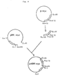
- Figs. 1-4 each shows an outlined construction scheme for the respective plasmid, and
- Figs. 5 to 7 each shows possible secondary structure of a certain DNA portion to be referred to later herein.
- Fig. 8 shows comparison of the DNA sequences between three plasmids.
- According to one embodiment of the present invention a mammalian cell-derived autonomously replicating sequence DNA fragment is provided having affinity for the human DNA-binding protein c-myc.
- The autonomously replicating sequence is selected from the group consisting of the following DNA sequences:and functional equivalents thereto, having affinity for the DNA-binding protein as defined above.

- Another embodiment relates to a method of recovering mammalian cell-derived autonomously replicating sequence DNA fragments which comprises binding a mammalian cell-derived DNA fragment to the DNA-binding protein, separating the DNA/DNA-binding protein complex and isolating the DNA from said DNA/ DNA-binding protein complex, wherein the DNA-binding protein is selected from the group consisting of myc proteins, c-myb protein, v-myb protein, c-fos protein, v-fos protein and p 53, and wherein the ARS DNA originates from a mammalian cell line producing said DNA-binding protein at a high level. Preferably, a nuclear extract of the cell line is employed as a DNA-binding protein source.
- This method may be performed in a way, wherein
- a) prior to the separation step the DNA/DNA-binding protein complex is further bound to an anti-DNA-binding protein antibody in order to obtain a DNA/DNA-binding protein/anti-DNA-binding protein antibody complex; and
- b) the complex obtained from step a) is further bound to a substance capable of specifically binding to the antibody of the complex.
- Preferably the substance as applied in step b) is selected from the group consisting of protein A and an antibody directed to the anti-DNA-binding protein antibody in the complex obtained from step a).
- Another embodiment relates to a mammalian cell-derived autonomously replicating sequence DNA fragment, obtainable by a method as disclosed above.
- According to the present invention plasmids are provided which contain a mammalian cell-derived autonomously replicating sequence DNA fragment as defined above, a promoter and a gene for peptide production inclusive of the translation initiation codon.
- Another aspect of the present invention refers to a method of producing a peptide which comprises propagating in cells a plasmid containing a mammalian cell-derived autonomously replicating sequence DNA fragment as defined above, a promoter and a gene for peptide production inclusive of the translation initiation codon.
- According to a further preferred embodiment of the present invention a plasmid is provided which contains a mammalian cell-derived autonomously replicating sequence DNA fragment as defined above,being obtainable by
- a) treating the human c-myc gene with HindIII and PstI;
- b) separating the upstream HindIII-PstI fragment; and
- c) subcloning the fragment into pUC19 at the corresponding site.
- Moreover,mammalian cell-derived autonomously replicating sequence DNA fragments are provided which are obtainable by
- a) excising from the plasmid as obtained according to the above-mentioned method the HindIII-PstI fragment of about 200bp; and
- b) separating the fragment by forming a complex which comprises the c-myc-protein.
- The invention further relates to mammalian cells transfected with a plasmid as defined above.
- The autonomous replicating sequence or ARS is the origin of replication in autonomous replication of eucaryotic chromosomes. The ARS can be obtained in the following manner: DNA is separated from mammalian cells and treated with an appropriate restriction enzyme or enzymes, preferably six-base restriction enzyme such as HindIII, EcoRI, BamHI, etc. The thus-obtained DNA fragments which are normally in the range of 1,000-10,000 base pairs in length are mixed with the DNA-binding protein to thereby form a DNA-binding protein complex.
- In this specification and claims the DNA-binding protein is defined as the protein that binds with DNA, exists in the mammalian cell nucleus and controls replication or expression of the DNA in the cell, which may be called non-histone protein, and examples of the DNA-binding protein include myc proteins (such as c-myc protein (Science 225, 718-720 (1984)), v-myc protein (Nature 296, 262-264 (1982)), N-myc protein (Annu. Rev. Biochem., 52, 301-310 (1983)), c-myb protein, v-myb protein, c-fos protein, v-fos protein, p53 (each described in Annu. Rev. Biochem. 52 301-310 (1983)), SV40 T antigen (Cell 13 165 (1978)) and RB gene product (Science 235 1394-1399 (1987)). These proteins may be available in cell nuclear extract as described in the above references, for example, c-myc protein is available in HL-60 cell nuclear extract. For the separation procedure after formation of such DNA-protein complex, conventional means of separation or isolation of proteins, such as the procedure for antigen-antibody reactions or the like may be used.
- For example, it is convenient that this complex is further bound to an antibody to the DNA-binding protein and the resultant complex is further bound to a substance capable of specifically binding to the antibody, for example, the second antibody or protein A, to produce a complex having molecular weight as large as forming precipitate. Precipitate of the complex can be collected with ease by a conventional manner such as filtration or centrifugation.
- The antibody to the DNA-binding protein can be produced by a general manner such as by immunizing an animal with the protein but it is convenient to obtain from commercial sources such as Oncor Inc., U.S.A. and etc. For example, the third exon of N-myc gene (the specific moiety of N-myc) is expressed in E. coli to obtain N-myc protein then a mouse is immuninized with the protein by a general method. Anti-serum is collected from the mouse and the IgG fraction thereof is used as the anti-N-myc protein antibody. (see also J. Virol. 34 752-763 (1980)).
- The second antibody can also be produced by a method known per se, and protein A are provided as a form of cell suspension of Staphylococcus aureus (P-7l55 (Sigma)) or a combined form with some solid (Protein A-Sepharose CL-4B (Pharmacia) or Protein A-Agarose FPA-l (Cosmo-Bio Ltd.).
- Separation of the DNA from the complex (e.g., DNA=binding protein=antibody=protein A) can be carried out in a conventional manner, for example, by treatment with a reagent such as 2-mercaptoethanol or a proteolytic enzyme (Mol. Cell. Biol. 3, 1958-1966 (1983)) such as Proteinase K (Proteinase K P0390 (Sigma), Proteinase K No. 24568 (Merck)), however, 2-mercaptoethanol is preferable since the proteolytic enzyme may sometimes degradate DNA chain.
- It is a very important fact discovered by the present inventor that the thus-isolated DNA fragment having specific affinity for the DNA-binding protein contains ARS DNA, and that the DNA-binding protein and ARS have very strong affinity for each other.
- The ARS-containing DNA fragment can be utilized either in the form of ARS itself or of an ARS-containing DNA fragment having an appropriate length, as desired.
- The size of possible ARS DNA is considered in about one hundred base pairs, however, it is also supposed that sometimes the ARS DNA is partially overlapped with an enhancer DNA. So, use can be made of a DNA having several hundreds base pairs as ARS-containing DNA fragment. The techniques of DNA treatment, screening and so on for producing useful substances, for example, peptides, by utilizing the ARS will be apparent and readily understandable to those with ordinary skill in the art when they use conventional or known techniques with reference to the examples given herein and so forth (T. Maniatis et al., Molecular Cloning, Cold Spring Harbor Laboratory (1982), hereinafter Mol. Clon.).
- It is quite possible that the base sequence of the ARS might vary slightly depending on the species of animal used for providing cells from which the ARS is derived, depending on the cell species, or depending on the site which serves as the source of the ARS (when cells of the same strain are used). Therefore, it is not always necessary that an ARS to be used should be completely identical in base sequence to the ARS obtained in the above manner. On the contrary, it is possible to perform partial base substitution in or deletion from or, further, base addition to the ARS. Whether the resulting ARS is suited for efficient production of the desired product can be determined by conventional testing and study with reference to the description given herein.
- Referring to the invention, as the isolable or usable ARS, an ARS derived from mammalian cells such as cells of a mouse, rat, guinea pig, cattle, horse, sheep, goat, rabbit, monkey, chimpanzee or human is exemplary. In particular, a mouse or human cell-derived ARS may be considered appropriate since advanced cell culture techniques are available for mouse and human cells.
- Though any mammalian cells can be employed as an origin for the production of ARS, it is convenient to use such cells much producing the DNA-binding proteins as c-myc protein as described in Science 225, 718-720 (1984), v-myc protein described in Nature 296, 262-264 (1982), N-myc protein, c-myb protein, v-myb protein, c-fos protein, v-fos protein, p53 which are described in Annu. Rev. Biochem., 52, 301-310 (1983), SV40 T antigen described in Cell, 13, 165 (1978) and RB gene product described in Science, 235, 1394-1399 (1987).
- Representative examples of such mammalian cells from which ARS is isolated include human HL-60 cell, human IMR-32 cell, human Raji cell, mouse FM3A cell and the like.
- The ARS-containing plasmids according to the invention, which are suited for peptide production, can be constructed by utilizing conventional gene recombination techniques (cf. Mol. Clon.).
- Introduction of the plasmids into cells, selection of the kind of cells to be used, cell proliferation and other necessary procedures can be carried out or made by utilizing those mammalian cell lines or strains and techniques that are generally known in the art. It will be also possible to utilize those techniques, cultured cells, media, and so forth which might be improved or developed in the future. (cf. "Manual of Cell Culture", edited by Munemura, Kodansha, Tokyo (1982), K. Habel and N. P. Salzman, Fundamental Technique in Virology, Academic Press, N.Y. (1969), Kruse and Patterson, Tissue Culture, Academic Press, N.Y. (1973) and Virology, 52 456-467 (1973)).
- Several examples of the general procedures are given below for further illustration.
- Examples of promoters which are usable are cell-derived promoters of various kinds, such as the thymidine kinase promoter and immunoglobulin promoter, and promoters from viruses infectious to mammalian cells, such as the SV40 T antigen promoter or SV40 early promoter, SV40 late promoter, Adenovirus promoters and the herpes virus thymidine kinase promoter. An ordinary ATG can be used as the translation initiation codon.
- Examples of peptides to be produced include among others, those peptides, proteins, glycoproteins and the like that are usable as drugs, for example, insulin, growth hormone, interferons, tumor necrosis factor, interleukins, other lymphokines, and various enzymes.
- Examples of genes for peptide production include among others, DNAs coding for respective amino acid sequences of the above-mentioned peptides, and sequences composed of the DNAs and a poly(A) signal for terminating translation as added thereto on the 3ʹ downstream side.
- The plasmids according to the invention are produced by joining a DNA fragment having an appropriate number of base pairs and one or more sites cleavable with restriction enzymes in common use, a promoter, the translation initiation codon and a gene for peptide production in an appropriate, desired order, further adding an ARS and circularizing the construct.
- The direction or location of the ARS need not be considered as particularly limited.
- The plasmids thus obtained are introduced into various mammalian cells e.g., by transfection method using calcium phosphate, the microinjection method, the liposome method, the protoplast fusion method or the like, whereby transformed cells can be produced. Mouse NS-l and FM3A and human HL-60, U937, Daudi and Raji are examples of suitable cultured cells but not limited to those. The desired peptides can be produced by growing and multiplying these transformed cells.
- A characteristic feature of the invention is that the cells into which the plasmids are to be introduced need not be from the same species as the species from which the ARS is derived. That is, when an ARS DNA is obtained from a mouse cell, the DNA can be used for the expression in another kind of mouse cells and in human cells or in other mammalian cells.
- The transformed cells are grown and multiplied by conventional methods of cultivating the cells used, for example, in DMEM (Dulbecco modified Eagle's medium) containing an appropriate amount of fetal calf serum at 37°C in the presence of 5% carbon dioxide, or in the peritoneal cavity of an animal (cf. "Manual of Cell Culture", edited by Munemura, Kodansha, Tokyo (1982), Fundamental Technique in Virology, Tissue Culture).
- The peptides produced, when they have been released into the medium, can be isolated by conventional fractionation techniques, for example, by centrifugation and/or gel filtration. When the peptides accumulate in cells, the peptides can be recovered by ordinary fractionation after disruption of the cells. Also when they are in the ascitic fluid, they can be isolated using conventional techniques.
- The following examples are given to further illustrate the present invention but are by no means to be construed to be limitative thereof. Unless otherwise indicated, all parts, percents, ratios and the like are by weight.
- The plasmid pMU85 (Mol. Cell. Biol., 5 563-568 (1985)) was treated with EcoRI and BglII and a DNA fragment about 2,500 base pairs in length was isolated from this plasmid. This fragment was inserted into the plasmid pKSVl0 (commercially available from Pharmacia) in the EcoRI-BglII region thereof, whereby a plasmid, pARS65, was constructed.
- The plasmid pAGO (Proc. Natl. Acad. Sci. USA, 76, 3755 (1979)) was treated with BamHI and a DNA fragment containing the herpes virus-derived thymidine kinase gene was isolated. This DNA fragment was inserted into the above-mentioned plasmid pARS65 at the BamHI site thereof. The thus-constructed plasmid p65-tk was allowed to multiply in E. coli (the transformant containing the plasmid E. coli K12 C600 ARS-1 having been deposited with the Fermentation Research Institute under the deposit number FERM BP-1443).
- The plasmid pMTIOD (J. Virology, 48, 48l-49l (1981)) was treated with BamHI and PvuII and the SV40 T antigen gene with the early promoter therefor was isolated. The BamHI linker (Takara Shuzo, Japan) was joined to the PvuII site of this gene. The fragment thus obtained was inserted into the plasmid pARS65 at the BamHI site thereof to obtain a plasmid, p65-T.
- The plasmid p65-tk obtained in process (1) above was used to transfect FM3Atk- cells by the liposome fusion method. The plasmid p65-tk was encapsulated in large unilamellar vesicles (LUVs) prepared by the phosphatidylserine calcium induced fusion case. In performing the transformation, 20 mM Tris buffer (pH 7.5) containing 50 mM EDTA was used (cf. Science 2l5, 166 (1982) and Proc. Natl. Acad. Sci. USA, 75, 4194 (1978).
- After transformation, the cells were grown in DMEM containing l0% fetal calf serum at 37°C in the presence of 5% carbon dioxide. After 2 days, the medium was replaced with HAT medium.
- Transformed cells, namely FM3Atk+ cells, were observed in approximately a week. After 2 weeks of cultivation, each colony was isolated and transferred to HAT medium. For each colony, cultivation in HAT medium was continued until the number of cells amounted to about l0⁷ per colony. The number of doublings during this cultivation period amounted to about 60.
- One thousand cells were subjected to transformation by the above method, whereupon about 300-400 transformant cells were obtained. It was thus confirmed that p65-tk had propagated in FM3Atk- cells and the thymidine kinase gene had been expressed.
- The above-mentioned l0⁷ cells were extracted and obtained low molecular weight DNA fraction by the Hirt method (J. Mol. Biol., 26, 365-369 (1967)). To confirm plasmid replication in the transformants, the low-molecular-weight DNA fraction was treated with the restriction enzyme DpnI.
- While p65-tk introduced into FM3Atk- cells contains a methylated adenine moiety, the replication product yielded in these cells did not contain any methylated adenine group. Since DpnI cleaves methylated DNA selectively, it cleaves exclusively the plasmid introduced into the cells to give a plurality of DNA fragments, with the plasmid copies replicated in the cells remaining uncleaved. Therefore, the plasmid copies replicated in the cells can be easily distinguished by electrophoresis.
- After DpnI digestion, DNAs were separated by agarose gel electrophoresis (Mol. Clon.) and subjected to Southern blot testing (J. Mol. Biol., 98, 503-517 (1975)). Thus, hybridization was carried out using ³P-labeled p65-tk as a probe, followed by X-ray autoradiography. Detection of bands hybridized to the probe confirmed that p65-tk had been replicated as an extrachromosomal DNA in all of the 20 clones investigated.
- The copy number per cell estimated from the band intensity was l00-200. This plasmid was replicable in a stable manner also when the above cells were grown in DMEM containing l0% fetal calf serum, which was used in lieu of HAT medium. No modification was noted in the DNA level. It was also confirmed that this plasmid remained in the cells in a stable manner even after the lapse of the time for 150 doublings.
- pARS-65 was transfected into mouse NS-1 cells, human HL-60 cells, and human U937 cells, respectively, using the method described above. The cells were then grown in RPMI l640 medium containing l0% fetal calf serum at 37°C in the presence of 5% carbon dioxide. After 40 hours, the extrachromosomal low-molecular-weight DNA was analyzed by the method mentioned above in (4). As a result, about 500 copies of pARS-65 were confirmed to have been replicated in each NS-1 cell, about 10,000 copies were confirmed to have been replicated in each HL-60 cell, and about 100 copies were confirmed to have been replicated in each U937 cell.
- It was thus confirmed that pARS-65 is replicable also in cells of other species than mouse. At the same time, the results suggest that p65-tk might have such replicability.
- Human HL-60 cell DNA was extracted using the SDS-proteinase K method (Mol. Clon.) and treated with HindIII. A DNA fragment having affinity for the myc protein was obtained from the DNA fragment mixture thus obtained, using a conventional method (Mol. Cell. Biol., 3, 1958-1966 (1983)) as follows:
- The above fragment mixture was mixed with an HL-60 nuclear extract (rich in myc protein), and the reaction was allowed to proceed at 0°C for 30 minutes, yielding a DNA-myc protein complex.
- Then, an anti-myc protein antibody (α-myc, commercially available from Oncor Inc. U.S.A.) was added, whereby a DNA-myc protein-α myc complex was formed. Furthermore, an aqueous solution of Staphylococcus aureus, which contained protein A capable of specifically binding to α myc, was added to form a DNA-myc protein-α myc-protein A complex.
- This complex, which occurred as a precipitate, was collected by centrifuging, washed with Tris buffer (pH 7.5) containing 0.l% SDS and 0.l M sodium chloride, and then the DNA was released by adding a 7 mM aqueous mercaptoethanol solution containing l% SDS and allowing the reaction to proceed at 30°C for 30 minutes. The reaction mixture was centrifuged at l5,000 × g for 5 minutes. The supernatant thus separated was extracted with phenol to remove proteins. The desired DNA fragment thus obtained was inserted into pUCl9 (commercially available from Pharmacia) at the HindIII site thereof. The number of base pairs in the above DNA fragment was estimated at about 200 by agarose gel electrophoresis.
- The plasmid obtained was multiplied in E. coli (the transformant with this plasmid E. coli K12 C600 ARS-2 having been deposited with the Fermentation Research Institute under the deposit number FERM BP-1444).
- The above-described HL-60 nuclear extract was prepared in the following manner. Thus, l liter of HL-60 cell culture fluid (5 × l0⁵ cells/ml) was centrifuged and the cells harvested were washed with phosphate-buffered physiological saline. The cells were further washed with a hypotonic aqueous solution (20 mM HEPES, pH 7.5, 5 mM potassium chloride, 0.05 mM magnesium chloride, 0.5 mM dithiothreitol, 0.2 mM sucrose) and then suspended in a hypotonic aqueous solution (the former solution without any sucrose), and the suspension was allowed to stand for l0 minutes. After homogenization by 40 strokes in a Dounce homogenizer, the homogenate was centrifuged at 3,000 × g for l0 minutes to obtain a precipitate.
- The precipitate comprising cell nuclei was dissolved in 2.5 ml of an aqueous solution (5 mM HEPES, pH 7.5, l0% sucrose) and stored temporarily in liquid nitrogen. After thawing this gradually at 0°C, 5 M aqueous sodium chloride was added to a final concentration of 0.1 M and, after 5 minutes of treatment at 0°C, the mixture was centrifuged at 15,000 × g for 20 minutes to obtain the nuclear extract as a supernatant.
- The above-mentioned plasmid as multiplied in E. coli K12 C600 was isolated (Mol. Clon.). Treatment with HindIII failed to cleave this plasmid. Hence it was considered that bacterial rearrangement of the plasmid had occurred. This plasmid was designated pHLmyc.
- The plasmid pUCl9 has other restriction enzyme cleavage sites (e.g. BamHI and NarI sites) than the HindIII site. Therefore, the above plasmid was treated with BamHI and NarI, and the smaller, in molecular weight of the two DNA fragments thus formed was isolated.
- A ³P-labeled probe was prepared using this smaller fragment as the template and used for Southern hybridization (J. Mol. Biol., 98, 503-517 (1975)) to the HL-60 cell DNA. This probe hybridized to the HL-60 cell DNA and it was thus confirmed that the fragment having an NarI site at one end and a BamHI site at the other (NarI-BamHI fragment) contained an HL-60 cell-derived DNA.
- The length of the NarI-BamHI fragment as estimated by agarose gel electrophoresis was about 200-300 based pairs.
- In pUCl9, the HindIII site is l3l base pairs apart from the NarI site and 35 base pairs apart from the BamHI site, and a PstI site is present 25 base pairs apart from the BamHI site. In the plasmid pHLmyc, the PstI site was retained. Therefore, the number of base pairs in the HL-60 cell-derived DNA contained in the plasmid pHLmyc was estimated to be about 120.
- This base sequence was determined and 99 bases were identified in the HindIII site within the polylinker region. The sequence thereof is shown below:

- Examination of this sequence for inverted repeats suggested the possibility that it might have hairpin structures as shown in Fig. 5 and Fig. 6. On that basis, it was presumed, of the above 99 bases, bases 12 to 59 or
bases 17 to 74 constituted the most important portion of the autonomously replicating sequence (ARS). - HL-60 cells were transfected with pHLmyc by the liposome method, and the copy number was estimated by the method described above and found to be 10,000 per cell.
- The plasmid pmyc (commercially available from Oncor Inc. U.S.A.; containing the myc structural gene and the Rous sarcoma virus-derived long terminal repeat promoter (LTR)) was treated with BamHI, HindIII and EcoRI, and the myc structural gene and LTR were isolated and inserted into pHLmyc in the EcoRI-BamHI region thereof. The thus-constructed plasmid pARSmyc was transfected into human U937 cells by the liposome method and the cells were grown in RPMI l640 medium containing l0% fetal calf serum at 37°C in the presence of 5% carbon dioxide.
- After 3 days of incubation, the quantity of mRNA corresponding to the myc gene as occurring in the cells was determined by the Northern hybridization method (Mol. Clon.) and found to be about l00 times greater as compared with the quantity of the mRNA in pARS-free U937 cells. Therefore, it was concluded that mRNA transcription from the myc gene in the plasmid pARSmyc had taken place.
-
- (1) Sub-Cloning of Human c-myc Gene
Human c-myc gene (Mol. Cell Biol., 5, 414-418 (1985)) was digested with HindIII and KpnI and the upstream HindIII-KpnI region (about 1,200 bp) was separated. Similarly, the upstream HindIII-PstI fragment was obtained by treating the gene with HindIII and PstI. These fragments were subcloned into pUC18 and pUC19 at the corresponding site, respectively, and the resultant plasmids of the clone were designated pmyc (H-K) and pmyc (H-P), respectively. - (2) Binding of the Hin dIII- Kpn I Fragment and HindIII-PstI Fragment with c-myc Protein
Gel-shift assay method has been recently employed to examine binding of DNA with a protein (Nucleic Acid Res., 9, 3047-3060 (1981)). In the method, an isotope-labelled DNA is treated with a protein to bind and eluted on polyacrylamide gel, so the DNA-protein complex is late to move on the gel.
The nuclear extract of HL-60 cell which was much producing c-myc protein was used as the c-myc protein source. Each HindIII-KpnI fragment and HindIII-PstI fragment described above was mixed with the nuclear extract and warmed at 30°C for 15 minutes. The resulting mixture was eluted on polyacrylamide gel. On the other hand, the nuclear extract was pre-treated with anti-myc protein antibody (Oncor Inc. U.S.A), which was then treated in the same manner as above with the above fragments, respectively.
The each fragment was bound with protein, however, pretreated protein with antibody were not bound with the fragment. From the fact it was concluded that there is a binding site with the c-myc protein in the HindIII-PstI fragment (about 200 bp). - (3) Plasmid pmyc (H-K) was digested with HindIII and KpnI, whereas pmyc (H-P) was digested with HindIII and PstI to produce DNA fragments. Each fragment was treated similarly to Example 2 with c-myc protein, antibody and protein A in turn and the complexes were treated with 2-mercaptoethanol to cause protein degradation and to remove resulted proteins.
- Thus, the HindIII-KpnI fragment and the HindIII-PstI fragment were obtained and DNA sequence of the HindIII-PstI fragment was determined by the dideoxy chain termination method, which is shown below.

- A possible secondary structue of the sequence was estimated by computor-assisted sequence analysis and is shown in Figure 7. Figure 8 shows comparison of the sequence between the fragment of pHLmyc and pmyc (H-P) and between the fragment of pARS65 and pmyc (H-P). Stars indicates matched sequences and sequence with bar is supposed to make stem of hairpin loop. The DNA sequence to make the stem, especially the nine base pairs of GTGAATAGT is considered most important.
- Similarly to the procedure in Example 2, several plasmids were obtained from various cell nuclear DNA, proteins and antibodies. The name of plasmid and the raw materials were shown in the following table.
Plasmid DNA/Enzyme Used Nuclear Extract of (Protein) Antibody pIMR-N IMR 32 DNA/HindIII IMR 32 cell (N-myc protein) anti-N-myc protein pRJ-53 Raji DNA/HindIII Raji cell (p53) anti p53 pHLmyc (Ex. 2) HL-60 DNA/HindIII HL-60 cell (c-myc protein) anti-c-myc protein - Plasmids pmyc (H-K), pmyc (H-P), pIMR-N and pRJ-53 were introduced into cell in a similar manner to Example l (3) and the copy number was investigated. The results are shown below.
Plasmid Cell Copy Number pmyc (H-K) HL60 1000-5000 pmyc (H-P) HL60 1000-5000 pIMR-N IMR-32 1000-5000 pRJ-53 Raji 1000-5000 - As described in detail hereinabove, the plasmids according to the invention can be replicated with good efficiency in various mammalian cells due to the function of the ARS; the plasmid copy number per cell is high and the gene product production efficiency is high. Therefore, the present invention also provides a peptide production which is excellent from the genetic engineering viewpoint.
- While the invention has been described in detail and with reference to specific embodiments thereof, it will be apparent to one skilled in the art that various changes and modifications can be made therein without departing from the spirit and scope thereof.
Claims (13)
- A method of recovering a mammalian cell-derived autonomously replicating sequence (ARS) DNA fragment which comprises binding a mammalian cell-derived DNA fragment to a DNA-binding protein, separating the DNA/DNA-binding protein complex and isolating the DNA from said DNA/DNA-binding protein complex,
wherein the DNA-binding protein is selected from the group consisting of myc proteins, c-myb protein, v-myb protein, c-fos protein, v-fos protein and p 53, and wherein the ARS DNA originates from a mammalian cell line producing said DNA-binding protein at a high level. - The method of claim 1, whereina) prior to the separation step the DNA/DNA-binding protein complex is further bound to an anti-DNA-binding protein antibody in order to obtain a DNA/DNA-binding protein/anti-DNA-binding protein antibody complex; andb) the complex obtained from step a) is further bound to a substance capable of specifically binding to the antibody of the complex.
- The method of claim 2, wherein the substance as applied in step b) is selected from the group consisting of protein A and an antibody directed to the anti-DNA-binding protein antibody in the complex obtained from step a).
- The method of one of claims 1 to 3, wherein a nuclear extract of a cell line is employed as a DNA-binding protein source.
- The method of one of claims 1 to 4, wherein a human cell-derived autonomously replicating sequence DNA fragment having affinity for the human DNA-binding protein c-myc is obtained, selected from the group consisting of the following sequences:and functional equivalents thereto.

- A method of preparing a plasmid, which method comprises providing a plasmid which contains a mammalian cell-derived autonomously replicating sequence DNA fragment according to claim 5, a promoter and a gene for peptide production inclusive of the translation initiation codon.
- A method of producing a peptide which comprises propagating in cells a plasmid containing a mammalian cell-derived autonomously replicating sequence DNA fragment according to claim 5, a promoter and a gene for peptide production inclusive of the translation initiation codon.
- A method of preparing a transfected mammalian cell, which method comprises transfection of a mammalian cell with a plasmid containing a mammalian cell-derived autonomously replicating sequence DNA fragment according to claim 5, a promoter and a gene for peptide production inclusive of the translation initiation codon.
- A method of preparing a plasmid which contains a mammalian cell-derived autonomously replicating sequence DNA fragment, which method comprises the steps ofa) treating the human c-myc gene with HindIII and PstI;b) separating the upstream HindIII-PstI fragment; andc) subcloning the fragment into pUC19 at the corresponding site.
- A method of preparing a mammalian cell-derived autonomously replicating sequence DNA fragment, which method comprises the steps ofa) excising from the plasmid obtainable according to claim 9 the HindIII-PstI fragment of about 200 bp; andb) separating the fragment by forming a complex which comprises the c-myc protein.
- The method of claim 5 or 10, wherein an ARS DNA fragment, characterized by the sequence GTGAATAGT, is obtained.
- A method of producing a peptide which comprises propagating in cells a plasmid according to claim 9.
- A method of preparing a transfected mammalian cell which method comprises transfecting a mammlian cell with a plasmid according to claim 9.
Applications Claiming Priority (4)
| Application Number | Priority Date | Filing Date | Title |
|---|---|---|---|
| JP174036/86 | 1986-07-24 | ||
| JP17403686 | 1986-07-24 | ||
| JP227455/86 | 1986-09-26 | ||
| JP22745586 | 1986-09-26 |
Publications (3)
| Publication Number | Publication Date |
|---|---|
| EP0254315A2 EP0254315A2 (en) | 1988-01-27 |
| EP0254315A3 EP0254315A3 (en) | 1989-12-13 |
| EP0254315B1 true EP0254315B1 (en) | 1996-03-27 |
Family
ID=26495785
Family Applications (1)
| Application Number | Title | Priority Date | Filing Date |
|---|---|---|---|
| EP87110696A Expired - Lifetime EP0254315B1 (en) | 1986-07-24 | 1987-07-23 | Mammalian cell derived autonomously replicating sequence DNA fragments having affinity to DNA binding proteins |
Country Status (6)
| Country | Link |
|---|---|
| EP (1) | EP0254315B1 (en) |
| JP (1) | JP2700458B2 (en) |
| AT (1) | ATE136057T1 (en) |
| DE (1) | DE3751757T2 (en) |
| ES (1) | ES2091179T3 (en) |
| GR (1) | GR3020035T3 (en) |
Cited By (4)
| Publication number | Priority date | Publication date | Assignee | Title |
|---|---|---|---|---|
| US5891691A (en) * | 1990-05-09 | 1999-04-06 | The Biological Research Center Of The Hungarian Academy Of Sciences | Method of producing a cell carrying an excess of mammalian centromeres and the cell line carrying an excess of mammalian centromeres |
| US6025155A (en) * | 1996-04-10 | 2000-02-15 | Chromos Molecular Systems, Inc. | Artificial chromosomes, uses thereof and methods for preparing artificial chromosomes |
| US6077697A (en) * | 1996-04-10 | 2000-06-20 | Chromos Molecular Systems, Inc. | Artificial chromosomes, uses thereof and methods for preparing artificial chromosomes |
| US8288610B2 (en) | 1996-04-10 | 2012-10-16 | The Biological Research Center Of The Hungarian Academy Of Sciences | Artificial chromosomes, uses thereof and methods for preparing artificial chromosomes |
Families Citing this family (2)
| Publication number | Priority date | Publication date | Assignee | Title |
|---|---|---|---|---|
| EP0350052A3 (en) * | 1988-07-06 | 1991-04-03 | Daiichi Pharmaceutical Co., Ltd. | Transgenic animals transformed with autonomously replicating sequence-containing plasmid |
| AU8904691A (en) * | 1990-10-17 | 1992-05-20 | Board Of Regents, The University Of Texas System | A eukaryotic episomal dna cloning and expression vector |
Family Cites Families (3)
| Publication number | Priority date | Publication date | Assignee | Title |
|---|---|---|---|---|
| IL63270A0 (en) * | 1980-08-05 | 1981-10-30 | Univ Leland Stanford Junior | Eukaryotic autonomously replicating segment |
| JP2516737B2 (en) * | 1984-10-04 | 1996-07-24 | 維紹 谷口 | Plasmid vector, its production and use |
| FR2593827B1 (en) * | 1986-01-31 | 1989-12-22 | Inst Nat Sante Rech Med | DNA FRAGMENTS FOR USE IN VECTORS FOR THE HEREDITARY MAINTENANCE OF AUTONOMOUS FOREIGN GENES IN TRANSGENIC ANIMALS, PROCESS FOR THEIR PRODUCTION AND BIOLOGICAL APPLICATIONS |
-
1987
- 1987-07-23 AT AT87110696T patent/ATE136057T1/en not_active IP Right Cessation
- 1987-07-23 ES ES87110696T patent/ES2091179T3/en not_active Expired - Lifetime
- 1987-07-23 EP EP87110696A patent/EP0254315B1/en not_active Expired - Lifetime
- 1987-07-23 DE DE3751757T patent/DE3751757T2/en not_active Expired - Fee Related
- 1987-07-24 JP JP62184749A patent/JP2700458B2/en not_active Expired - Fee Related
-
1996
- 1996-05-27 GR GR960401398T patent/GR3020035T3/en unknown
Cited By (6)
| Publication number | Priority date | Publication date | Assignee | Title |
|---|---|---|---|---|
| US5891691A (en) * | 1990-05-09 | 1999-04-06 | The Biological Research Center Of The Hungarian Academy Of Sciences | Method of producing a cell carrying an excess of mammalian centromeres and the cell line carrying an excess of mammalian centromeres |
| US6025155A (en) * | 1996-04-10 | 2000-02-15 | Chromos Molecular Systems, Inc. | Artificial chromosomes, uses thereof and methods for preparing artificial chromosomes |
| US6077697A (en) * | 1996-04-10 | 2000-06-20 | Chromos Molecular Systems, Inc. | Artificial chromosomes, uses thereof and methods for preparing artificial chromosomes |
| US6743967B2 (en) | 1996-04-10 | 2004-06-01 | Chromos Molecular Systems Inc. | Artificial chromosomes, uses thereof and methods for preparing artificial chromosomes |
| US8288610B2 (en) | 1996-04-10 | 2012-10-16 | The Biological Research Center Of The Hungarian Academy Of Sciences | Artificial chromosomes, uses thereof and methods for preparing artificial chromosomes |
| US8389802B2 (en) | 1996-04-10 | 2013-03-05 | The Biological Research Center Of The Hungarian Academy Of Sciences | Artificial chromosomes, uses thereof and methods for preparing artificial chromosomes |
Also Published As
| Publication number | Publication date |
|---|---|
| EP0254315A2 (en) | 1988-01-27 |
| ES2091179T3 (en) | 1996-11-01 |
| JPS63185386A (en) | 1988-07-30 |
| JP2700458B2 (en) | 1998-01-21 |
| DE3751757D1 (en) | 1996-05-02 |
| DE3751757T2 (en) | 1996-11-28 |
| EP0254315A3 (en) | 1989-12-13 |
| GR3020035T3 (en) | 1996-08-31 |
| ATE136057T1 (en) | 1996-04-15 |
Similar Documents
| Publication | Publication Date | Title |
|---|---|---|
| US4601978A (en) | Mammalian metallothionein promoter system | |
| US4359535A (en) | Autonomously replicating DNA containing inserted DNA sequences | |
| JP3148121B2 (en) | Vectors having stabilizing sequences and eukaryotic host cells | |
| Hanahan et al. | Characteristics of an SV40-plasmid recombinant and its movement into and out of the genome of a murine cell | |
| Hesse et al. | Regulated gene expression in transfected primary chicken erythrocytes. | |
| US5843643A (en) | Site-specific transfection of eukaryotic cells using polypeptide-linked recombinant nucleic acid | |
| US4797359A (en) | Heat shock regulated production of selected and fused proteins in yeast | |
| DE69333669T2 (en) | MUTAGENESIS AND SELECTION METHODS IN THE SAME HOST CELLS | |
| Weiss et al. | Expression of differentiated functions in hepatoma cell hybrids: IX extinction and reexpression of liver-specific enzymes in rat hepatoma-Chinese hamster fibroblast hybrids | |
| CA2357538A1 (en) | Expression of exogenous polynucleotide sequences in a vertebrate | |
| US4528266A (en) | Method of inserting unique DNA sequences into DNA vectors | |
| JP3844656B2 (en) | Methods for transformation of animal cells | |
| JPH0376580A (en) | Escherichia coli manifestation vector and production of antiviral protein using the same | |
| Aloni et al. | Attenuation May Regulate Gene Expression in Animal Viruses and Cell | |
| JPH05503015A (en) | mammalian expression vector | |
| JPH04228066A (en) | Culture cell for expressing exogenote | |
| Klionsky et al. | Assembly of a functional F1 of the proton-translocating ATPase of Escherichia coli. | |
| US5364761A (en) | Method for isolating a DNA encoding an autonomously replicating sequence | |
| EP0254315A2 (en) | Mammalian cell derived autonomously replicating sequence DNA fragments having affinity to DNA binding proteins | |
| Lo et al. | High level expression of human proteins in murine hybridoma cells: induction by methotrexate in the absence of gene amplification | |
| Hynes et al. | Structure, stability, methylation, expression and glucocorticoid induction of endogenous and transfected proviral genes of mouse mammary tumor virus in mouse fibroblasts | |
| EP0162319B1 (en) | Method of joining heterologous genes | |
| EP0055742A1 (en) | System for amplification of eukaryotic genes | |
| US4830965A (en) | Transposable linkers to transfer genetic information | |
| JPH012584A (en) | DNA and plasmids and microorganisms containing them |
Legal Events
| Date | Code | Title | Description |
|---|---|---|---|
| PUAI | Public reference made under article 153(3) epc to a published international application that has entered the european phase |
Free format text: ORIGINAL CODE: 0009012 |
|
| AK | Designated contracting states |
Kind code of ref document: A2 Designated state(s): AT BE CH DE ES FR GB GR IT LI NL SE |
|
| PUAL | Search report despatched |
Free format text: ORIGINAL CODE: 0009013 |
|
| AK | Designated contracting states |
Kind code of ref document: A3 Designated state(s): AT BE CH DE ES FR GB GR IT LI NL SE |
|
| 17P | Request for examination filed |
Effective date: 19900613 |
|
| 17Q | First examination report despatched |
Effective date: 19910819 |
|
| RAP1 | Party data changed (applicant data changed or rights of an application transferred) |
Owner name: DAIICHI PHARMACEUTICAL CO., LTD. |
|
| GRAA | (expected) grant |
Free format text: ORIGINAL CODE: 0009210 |
|
| AK | Designated contracting states |
Kind code of ref document: B1 Designated state(s): AT BE CH DE ES FR GB GR IT LI NL SE |
|
| REF | Corresponds to: |
Ref document number: 136057 Country of ref document: AT Date of ref document: 19960415 Kind code of ref document: T |
|
| REF | Corresponds to: |
Ref document number: 3751757 Country of ref document: DE Date of ref document: 19960502 |
|
| ITF | It: translation for a ep patent filed | ||
| REG | Reference to a national code |
Ref country code: ES Ref legal event code: BA2A Ref document number: 2091179 Country of ref document: ES Kind code of ref document: T3 |
|
| ET | Fr: translation filed | ||
| REG | Reference to a national code |
Ref country code: GR Ref legal event code: FG4A Free format text: 3020035 |
|
| GRAH | Despatch of communication of intention to grant a patent |
Free format text: ORIGINAL CODE: EPIDOS IGRA |
|
| REG | Reference to a national code |
Ref country code: ES Ref legal event code: FG2A Ref document number: 2091179 Country of ref document: ES Kind code of ref document: T3 |
|
| PLBE | No opposition filed within time limit |
Free format text: ORIGINAL CODE: 0009261 |
|
| STAA | Information on the status of an ep patent application or granted ep patent |
Free format text: STATUS: NO OPPOSITION FILED WITHIN TIME LIMIT |
|
| 26N | No opposition filed | ||
| K2C3 | Correction of patent specification (complete document) published |
Effective date: 19960327 |
|
| PGFP | Annual fee paid to national office [announced via postgrant information from national office to epo] |
Ref country code: GR Payment date: 19990527 Year of fee payment: 13 |
|
| PGFP | Annual fee paid to national office [announced via postgrant information from national office to epo] |
Ref country code: SE Payment date: 19990609 Year of fee payment: 13 |
|
| PGFP | Annual fee paid to national office [announced via postgrant information from national office to epo] |
Ref country code: BE Payment date: 19990614 Year of fee payment: 13 |
|
| PGFP | Annual fee paid to national office [announced via postgrant information from national office to epo] |
Ref country code: AT Payment date: 19990714 Year of fee payment: 13 |
|
| PGFP | Annual fee paid to national office [announced via postgrant information from national office to epo] |
Ref country code: CH Payment date: 19990715 Year of fee payment: 13 |
|
| PGFP | Annual fee paid to national office [announced via postgrant information from national office to epo] |
Ref country code: GB Payment date: 19990721 Year of fee payment: 13 |
|
| PGFP | Annual fee paid to national office [announced via postgrant information from national office to epo] |
Ref country code: NL Payment date: 19990730 Year of fee payment: 13 Ref country code: FR Payment date: 19990730 Year of fee payment: 13 Ref country code: ES Payment date: 19990730 Year of fee payment: 13 |
|
| PGFP | Annual fee paid to national office [announced via postgrant information from national office to epo] |
Ref country code: DE Payment date: 19990929 Year of fee payment: 13 |
|
| PG25 | Lapsed in a contracting state [announced via postgrant information from national office to epo] |
Ref country code: GB Free format text: LAPSE BECAUSE OF NON-PAYMENT OF DUE FEES Effective date: 20000723 Ref country code: AT Free format text: LAPSE BECAUSE OF NON-PAYMENT OF DUE FEES Effective date: 20000723 |
|
| PG25 | Lapsed in a contracting state [announced via postgrant information from national office to epo] |
Ref country code: SE Free format text: LAPSE BECAUSE OF NON-PAYMENT OF DUE FEES Effective date: 20000724 Ref country code: ES Free format text: LAPSE BECAUSE OF NON-PAYMENT OF DUE FEES Effective date: 20000724 |
|
| PG25 | Lapsed in a contracting state [announced via postgrant information from national office to epo] |
Ref country code: LI Free format text: LAPSE BECAUSE OF NON-PAYMENT OF DUE FEES Effective date: 20000731 Ref country code: GR Free format text: LAPSE BECAUSE OF NON-PAYMENT OF DUE FEES Effective date: 20000731 Ref country code: CH Free format text: LAPSE BECAUSE OF NON-PAYMENT OF DUE FEES Effective date: 20000731 Ref country code: BE Free format text: LAPSE BECAUSE OF NON-PAYMENT OF DUE FEES Effective date: 20000731 |
|
| BERE | Be: lapsed |
Owner name: DAIICHI PHARMACEUTICAL CO. LTD Effective date: 20000731 |
|
| PG25 | Lapsed in a contracting state [announced via postgrant information from national office to epo] |
Ref country code: NL Free format text: LAPSE BECAUSE OF NON-PAYMENT OF DUE FEES Effective date: 20010201 |
|
| GBPC | Gb: european patent ceased through non-payment of renewal fee |
Effective date: 20000723 |
|
| EUG | Se: european patent has lapsed |
Ref document number: 87110696.9 |
|
| PG25 | Lapsed in a contracting state [announced via postgrant information from national office to epo] |
Ref country code: FR Free format text: LAPSE BECAUSE OF NON-PAYMENT OF DUE FEES Effective date: 20010330 |
|
| NLV4 | Nl: lapsed or anulled due to non-payment of the annual fee |
Effective date: 20010201 |
|
| REG | Reference to a national code |
Ref country code: FR Ref legal event code: ST |
|
| PG25 | Lapsed in a contracting state [announced via postgrant information from national office to epo] |
Ref country code: DE Free format text: LAPSE BECAUSE OF NON-PAYMENT OF DUE FEES Effective date: 20010501 |
|
| REG | Reference to a national code |
Ref country code: ES Ref legal event code: FD2A Effective date: 20010810 |
|
| PG25 | Lapsed in a contracting state [announced via postgrant information from national office to epo] |
Ref country code: IT Free format text: LAPSE BECAUSE OF NON-PAYMENT OF DUE FEES;WARNING: LAPSES OF ITALIAN PATENTS WITH EFFECTIVE DATE BEFORE 2007 MAY HAVE OCCURRED AT ANY TIME BEFORE 2007. THE CORRECT EFFECTIVE DATE MAY BE DIFFERENT FROM THE ONE RECORDED. Effective date: 20050723 |
|
| REG | Reference to a national code |
Ref country code: CH Ref legal event code: NV Representative=s name: BOVARD AG PATENTANWAELTE |