EP0250137A2 - Colloidal gold immunoassay - Google Patents
Colloidal gold immunoassay Download PDFInfo
- Publication number
- EP0250137A2 EP0250137A2 EP87305027A EP87305027A EP0250137A2 EP 0250137 A2 EP0250137 A2 EP 0250137A2 EP 87305027 A EP87305027 A EP 87305027A EP 87305027 A EP87305027 A EP 87305027A EP 0250137 A2 EP0250137 A2 EP 0250137A2
- Authority
- EP
- European Patent Office
- Prior art keywords
- ligand
- sample
- membrane
- colloidal gold
- strip
- Prior art date
- Legal status (The legal status is an assumption and is not a legal conclusion. Google has not performed a legal analysis and makes no representation as to the accuracy of the status listed.)
- Granted
Links
- PCHJSUWPFVWCPO-UHFFFAOYSA-N gold Chemical compound [Au] PCHJSUWPFVWCPO-UHFFFAOYSA-N 0.000 title claims abstract description 67
- 238000003018 immunoassay Methods 0.000 title abstract description 14
- 239000012528 membrane Substances 0.000 claims abstract description 87
- 239000003446 ligand Substances 0.000 claims abstract description 53
- 239000003153 chemical reaction reagent Substances 0.000 claims abstract description 37
- 239000012530 fluid Substances 0.000 claims abstract description 19
- 238000001514 detection method Methods 0.000 claims abstract description 3
- 238000000034 method Methods 0.000 claims description 62
- 238000003556 assay Methods 0.000 claims description 34
- 230000027455 binding Effects 0.000 claims description 27
- 239000000427 antigen Substances 0.000 claims description 18
- 102000036639 antigens Human genes 0.000 claims description 18
- 108091007433 antigens Proteins 0.000 claims description 18
- 108060003951 Immunoglobulin Proteins 0.000 claims description 9
- 102000018358 immunoglobulin Human genes 0.000 claims description 9
- 239000007788 liquid Substances 0.000 claims description 9
- 238000011534 incubation Methods 0.000 claims description 5
- 230000003287 optical effect Effects 0.000 claims description 5
- 238000012875 competitive assay Methods 0.000 claims description 3
- 230000001900 immune effect Effects 0.000 claims description 3
- 230000008105 immune reaction Effects 0.000 claims description 3
- 229920002302 Nylon 6,6 Polymers 0.000 claims description 2
- 239000005556 hormone Substances 0.000 claims description 2
- 229940088597 hormone Drugs 0.000 claims description 2
- 230000009257 reactivity Effects 0.000 claims 2
- 230000005012 migration Effects 0.000 claims 1
- 238000013508 migration Methods 0.000 claims 1
- 239000011159 matrix material Substances 0.000 abstract description 7
- 210000001124 body fluid Anatomy 0.000 abstract description 2
- 239000010839 body fluid Substances 0.000 abstract description 2
- 230000002596 correlated effect Effects 0.000 abstract description 2
- 238000012360 testing method Methods 0.000 description 49
- 239000000523 sample Substances 0.000 description 42
- ZFXYFBGIUFBOJW-UHFFFAOYSA-N theophylline Chemical compound O=C1N(C)C(=O)N(C)C2=C1NC=N2 ZFXYFBGIUFBOJW-UHFFFAOYSA-N 0.000 description 31
- 229960000278 theophylline Drugs 0.000 description 21
- 239000000243 solution Substances 0.000 description 16
- 230000035945 sensitivity Effects 0.000 description 15
- 210000002700 urine Anatomy 0.000 description 15
- 239000000306 component Substances 0.000 description 14
- 239000000203 mixture Substances 0.000 description 14
- 239000008279 sol Substances 0.000 description 11
- 238000006243 chemical reaction Methods 0.000 description 9
- 239000008280 blood Substances 0.000 description 8
- 210000004369 blood Anatomy 0.000 description 8
- 239000002245 particle Substances 0.000 description 8
- 239000000872 buffer Substances 0.000 description 7
- 239000000463 material Substances 0.000 description 7
- 238000002965 ELISA Methods 0.000 description 6
- 230000008878 coupling Effects 0.000 description 6
- 238000010168 coupling process Methods 0.000 description 6
- 238000005859 coupling reaction Methods 0.000 description 6
- 201000005404 rubella Diseases 0.000 description 6
- 238000003756 stirring Methods 0.000 description 6
- 238000005406 washing Methods 0.000 description 6
- 150000003384 small molecules Chemical class 0.000 description 5
- PXIPVTKHYLBLMZ-UHFFFAOYSA-N Sodium azide Chemical compound [Na+].[N-]=[N+]=[N-] PXIPVTKHYLBLMZ-UHFFFAOYSA-N 0.000 description 4
- 230000008901 benefit Effects 0.000 description 4
- 239000012153 distilled water Substances 0.000 description 4
- 238000002372 labelling Methods 0.000 description 4
- 239000002953 phosphate buffered saline Substances 0.000 description 4
- XLYOFNOQVPJJNP-UHFFFAOYSA-N water Chemical compound O XLYOFNOQVPJJNP-UHFFFAOYSA-N 0.000 description 4
- JKMHFZQWWAIEOD-UHFFFAOYSA-N 2-[4-(2-hydroxyethyl)piperazin-1-yl]ethanesulfonic acid Chemical compound OCC[NH+]1CCN(CCS([O-])(=O)=O)CC1 JKMHFZQWWAIEOD-UHFFFAOYSA-N 0.000 description 3
- 239000007995 HEPES buffer Substances 0.000 description 3
- 229920001213 Polysorbate 20 Polymers 0.000 description 3
- 238000007818 agglutination assay Methods 0.000 description 3
- 230000001419 dependent effect Effects 0.000 description 3
- 239000010931 gold Substances 0.000 description 3
- 229910052737 gold Inorganic materials 0.000 description 3
- 229910052751 metal Inorganic materials 0.000 description 3
- 239000002184 metal Substances 0.000 description 3
- 239000000256 polyoxyethylene sorbitan monolaurate Substances 0.000 description 3
- 235000010486 polyoxyethylene sorbitan monolaurate Nutrition 0.000 description 3
- 239000000126 substance Substances 0.000 description 3
- 230000000007 visual effect Effects 0.000 description 3
- 102000004190 Enzymes Human genes 0.000 description 2
- 108090000790 Enzymes Proteins 0.000 description 2
- DHMQDGOQFOQNFH-UHFFFAOYSA-N Glycine Chemical compound NCC(O)=O DHMQDGOQFOQNFH-UHFFFAOYSA-N 0.000 description 2
- VEXZGXHMUGYJMC-UHFFFAOYSA-N Hydrochloric acid Chemical compound Cl VEXZGXHMUGYJMC-UHFFFAOYSA-N 0.000 description 2
- XQFRJNBWHJMXHO-RRKCRQDMSA-N IDUR Chemical compound C1[C@H](O)[C@@H](CO)O[C@H]1N1C(=O)NC(=O)C(I)=C1 XQFRJNBWHJMXHO-RRKCRQDMSA-N 0.000 description 2
- JUJWROOIHBZHMG-UHFFFAOYSA-N Pyridine Chemical compound C1=CC=NC=C1 JUJWROOIHBZHMG-UHFFFAOYSA-N 0.000 description 2
- FAPWRFPIFSIZLT-UHFFFAOYSA-M Sodium chloride Chemical compound [Na+].[Cl-] FAPWRFPIFSIZLT-UHFFFAOYSA-M 0.000 description 2
- WYURNTSHIVDZCO-UHFFFAOYSA-N Tetrahydrofuran Chemical compound C1CCOC1 WYURNTSHIVDZCO-UHFFFAOYSA-N 0.000 description 2
- 201000005485 Toxoplasmosis Diseases 0.000 description 2
- 239000002250 absorbent Substances 0.000 description 2
- 230000002745 absorbent Effects 0.000 description 2
- 230000015572 biosynthetic process Effects 0.000 description 2
- 230000000903 blocking effect Effects 0.000 description 2
- 230000000052 comparative effect Effects 0.000 description 2
- 239000002274 desiccant Substances 0.000 description 2
- MTHSVFCYNBDYFN-UHFFFAOYSA-N diethylene glycol Chemical compound OCCOCCO MTHSVFCYNBDYFN-UHFFFAOYSA-N 0.000 description 2
- LOKCTEFSRHRXRJ-UHFFFAOYSA-I dipotassium trisodium dihydrogen phosphate hydrogen phosphate dichloride Chemical compound P(=O)(O)(O)[O-].[K+].P(=O)(O)([O-])[O-].[Na+].[Na+].[Cl-].[K+].[Cl-].[Na+] LOKCTEFSRHRXRJ-UHFFFAOYSA-I 0.000 description 2
- 229940079593 drug Drugs 0.000 description 2
- 239000003814 drug Substances 0.000 description 2
- 238000001914 filtration Methods 0.000 description 2
- MHMNJMPURVTYEJ-UHFFFAOYSA-N fluorescein-5-isothiocyanate Chemical compound O1C(=O)C2=CC(N=C=S)=CC=C2C21C1=CC=C(O)C=C1OC1=CC(O)=CC=C21 MHMNJMPURVTYEJ-UHFFFAOYSA-N 0.000 description 2
- 230000036541 health Effects 0.000 description 2
- 208000006454 hepatitis Diseases 0.000 description 2
- 231100000283 hepatitis Toxicity 0.000 description 2
- 229940072221 immunoglobulins Drugs 0.000 description 2
- 230000006872 improvement Effects 0.000 description 2
- 230000000155 isotopic effect Effects 0.000 description 2
- 238000004519 manufacturing process Methods 0.000 description 2
- 230000016087 ovulation Effects 0.000 description 2
- 239000013610 patient sample Substances 0.000 description 2
- 229920000136 polysorbate Polymers 0.000 description 2
- BWHMMNNQKKPAPP-UHFFFAOYSA-L potassium carbonate Chemical compound [K+].[K+].[O-]C([O-])=O BWHMMNNQKKPAPP-UHFFFAOYSA-L 0.000 description 2
- 238000009597 pregnancy test Methods 0.000 description 2
- 239000012488 sample solution Substances 0.000 description 2
- 210000002966 serum Anatomy 0.000 description 2
- 239000000725 suspension Substances 0.000 description 2
- BJEPYKJPYRNKOW-REOHCLBHSA-N (S)-malic acid Chemical compound OC(=O)[C@@H](O)CC(O)=O BJEPYKJPYRNKOW-REOHCLBHSA-N 0.000 description 1
- 208000030507 AIDS Diseases 0.000 description 1
- 108010032595 Antibody Binding Sites Proteins 0.000 description 1
- 241000606161 Chlamydia Species 0.000 description 1
- 208000035473 Communicable disease Diseases 0.000 description 1
- LTMHDMANZUZIPE-AMTYYWEZSA-N Digoxin Natural products O([C@H]1[C@H](C)O[C@H](O[C@@H]2C[C@@H]3[C@@](C)([C@@H]4[C@H]([C@]5(O)[C@](C)([C@H](O)C4)[C@H](C4=CC(=O)OC4)CC5)CC3)CC2)C[C@@H]1O)[C@H]1O[C@H](C)[C@@H](O[C@H]2O[C@@H](C)[C@H](O)[C@@H](O)C2)[C@@H](O)C1 LTMHDMANZUZIPE-AMTYYWEZSA-N 0.000 description 1
- 239000003109 Disodium ethylene diamine tetraacetate Substances 0.000 description 1
- ZGTMUACCHSMWAC-UHFFFAOYSA-L EDTA disodium salt (anhydrous) Chemical compound [Na+].[Na+].OC(=O)CN(CC([O-])=O)CCN(CC(O)=O)CC([O-])=O ZGTMUACCHSMWAC-UHFFFAOYSA-L 0.000 description 1
- KRHYYFGTRYWZRS-UHFFFAOYSA-M Fluoride anion Chemical compound [F-] KRHYYFGTRYWZRS-UHFFFAOYSA-M 0.000 description 1
- SXRSQZLOMIGNAQ-UHFFFAOYSA-N Glutaraldehyde Chemical compound O=CCCCC=O SXRSQZLOMIGNAQ-UHFFFAOYSA-N 0.000 description 1
- 239000004471 Glycine Substances 0.000 description 1
- 241000598436 Human T-cell lymphotropic virus Species 0.000 description 1
- DGAQECJNVWCQMB-PUAWFVPOSA-M Ilexoside XXIX Chemical compound C[C@@H]1CC[C@@]2(CC[C@@]3(C(=CC[C@H]4[C@]3(CC[C@@H]5[C@@]4(CC[C@@H](C5(C)C)OS(=O)(=O)[O-])C)C)[C@@H]2[C@]1(C)O)C)C(=O)O[C@H]6[C@@H]([C@H]([C@@H]([C@H](O6)CO)O)O)O.[Na+] DGAQECJNVWCQMB-PUAWFVPOSA-M 0.000 description 1
- 229920000057 Mannan Polymers 0.000 description 1
- 206010027339 Menstruation irregular Diseases 0.000 description 1
- 241000588653 Neisseria Species 0.000 description 1
- 239000002033 PVDF binder Substances 0.000 description 1
- 241000276498 Pollachius virens Species 0.000 description 1
- 239000004743 Polypropylene Substances 0.000 description 1
- 239000004372 Polyvinyl alcohol Substances 0.000 description 1
- BQCADISMDOOEFD-UHFFFAOYSA-N Silver Chemical compound [Ag] BQCADISMDOOEFD-UHFFFAOYSA-N 0.000 description 1
- 241000194017 Streptococcus Species 0.000 description 1
- GSEJCLTVZPLZKY-UHFFFAOYSA-N Triethanolamine Chemical compound OCCN(CCO)CCO GSEJCLTVZPLZKY-UHFFFAOYSA-N 0.000 description 1
- 238000010521 absorption reaction Methods 0.000 description 1
- 238000009825 accumulation Methods 0.000 description 1
- 230000009471 action Effects 0.000 description 1
- 230000000274 adsorptive effect Effects 0.000 description 1
- 238000013019 agitation Methods 0.000 description 1
- BJEPYKJPYRNKOW-UHFFFAOYSA-N alpha-hydroxysuccinic acid Natural products OC(=O)C(O)CC(O)=O BJEPYKJPYRNKOW-UHFFFAOYSA-N 0.000 description 1
- 230000004075 alteration Effects 0.000 description 1
- 229910052782 aluminium Inorganic materials 0.000 description 1
- XAGFODPZIPBFFR-UHFFFAOYSA-N aluminium Chemical compound [Al] XAGFODPZIPBFFR-UHFFFAOYSA-N 0.000 description 1
- 230000003321 amplification Effects 0.000 description 1
- 239000012491 analyte Substances 0.000 description 1
- 238000013459 approach Methods 0.000 description 1
- 239000007864 aqueous solution Substances 0.000 description 1
- 238000000149 argon plasma sintering Methods 0.000 description 1
- 238000002820 assay format Methods 0.000 description 1
- 230000003190 augmentative effect Effects 0.000 description 1
- 239000011230 binding agent Substances 0.000 description 1
- 239000012503 blood component Substances 0.000 description 1
- 239000005018 casein Substances 0.000 description 1
- BECPQYXYKAMYBN-UHFFFAOYSA-N casein, tech. Chemical compound NCCCCC(C(O)=O)N=C(O)C(CC(O)=O)N=C(O)C(CCC(O)=N)N=C(O)C(CC(C)C)N=C(O)C(CCC(O)=O)N=C(O)C(CC(O)=O)N=C(O)C(CCC(O)=O)N=C(O)C(C(C)O)N=C(O)C(CCC(O)=N)N=C(O)C(CCC(O)=N)N=C(O)C(CCC(O)=N)N=C(O)C(CCC(O)=O)N=C(O)C(CCC(O)=O)N=C(O)C(COP(O)(O)=O)N=C(O)C(CCC(O)=N)N=C(O)C(N)CC1=CC=CC=C1 BECPQYXYKAMYBN-UHFFFAOYSA-N 0.000 description 1
- 235000021240 caseins Nutrition 0.000 description 1
- 230000008859 change Effects 0.000 description 1
- 239000003638 chemical reducing agent Substances 0.000 description 1
- 239000003795 chemical substances by application Substances 0.000 description 1
- 238000004587 chromatography analysis Methods 0.000 description 1
- 239000013065 commercial product Substances 0.000 description 1
- 230000002860 competitive effect Effects 0.000 description 1
- 238000010276 construction Methods 0.000 description 1
- 230000009260 cross reactivity Effects 0.000 description 1
- 230000007812 deficiency Effects 0.000 description 1
- 238000009795 derivation Methods 0.000 description 1
- 238000011161 development Methods 0.000 description 1
- 238000000502 dialysis Methods 0.000 description 1
- 238000009792 diffusion process Methods 0.000 description 1
- LTMHDMANZUZIPE-PUGKRICDSA-N digoxin Chemical compound C1[C@H](O)[C@H](O)[C@@H](C)O[C@H]1O[C@@H]1[C@@H](C)O[C@@H](O[C@@H]2[C@H](O[C@@H](O[C@@H]3C[C@@H]4[C@]([C@@H]5[C@H]([C@]6(CC[C@@H]([C@@]6(C)[C@H](O)C5)C=5COC(=O)C=5)O)CC4)(C)CC3)C[C@@H]2O)C)C[C@@H]1O LTMHDMANZUZIPE-PUGKRICDSA-N 0.000 description 1
- 229960005156 digoxin Drugs 0.000 description 1
- LTMHDMANZUZIPE-UHFFFAOYSA-N digoxine Natural products C1C(O)C(O)C(C)OC1OC1C(C)OC(OC2C(OC(OC3CC4C(C5C(C6(CCC(C6(C)C(O)C5)C=5COC(=O)C=5)O)CC4)(C)CC3)CC2O)C)CC1O LTMHDMANZUZIPE-UHFFFAOYSA-N 0.000 description 1
- 201000010099 disease Diseases 0.000 description 1
- 208000037265 diseases, disorders, signs and symptoms Diseases 0.000 description 1
- WPUMTJGUQUYPIV-JIZZDEOASA-L disodium (S)-malate Chemical compound [Na+].[Na+].[O-]C(=O)[C@@H](O)CC([O-])=O WPUMTJGUQUYPIV-JIZZDEOASA-L 0.000 description 1
- 235000019301 disodium ethylene diamine tetraacetate Nutrition 0.000 description 1
- 230000008030 elimination Effects 0.000 description 1
- 238000003379 elimination reaction Methods 0.000 description 1
- 230000002708 enhancing effect Effects 0.000 description 1
- 210000003743 erythrocyte Anatomy 0.000 description 1
- 239000011521 glass Substances 0.000 description 1
- FDWREHZXQUYJFJ-UHFFFAOYSA-M gold monochloride Chemical compound [Cl-].[Au+] FDWREHZXQUYJFJ-UHFFFAOYSA-M 0.000 description 1
- 230000003100 immobilizing effect Effects 0.000 description 1
- 208000015181 infectious disease Diseases 0.000 description 1
- 201000006747 infectious mononucleosis Diseases 0.000 description 1
- 230000005764 inhibitory process Effects 0.000 description 1
- 150000002605 large molecules Chemical class 0.000 description 1
- 238000002386 leaching Methods 0.000 description 1
- 230000004807 localization Effects 0.000 description 1
- 239000001630 malic acid Substances 0.000 description 1
- 235000011090 malic acid Nutrition 0.000 description 1
- 239000003550 marker Substances 0.000 description 1
- 244000005700 microbiome Species 0.000 description 1
- 235000013336 milk Nutrition 0.000 description 1
- 239000008267 milk Substances 0.000 description 1
- 210000004080 milk Anatomy 0.000 description 1
- 238000002156 mixing Methods 0.000 description 1
- 238000003199 nucleic acid amplification method Methods 0.000 description 1
- 230000027758 ovulation cycle Effects 0.000 description 1
- 239000008363 phosphate buffer Substances 0.000 description 1
- 239000004033 plastic Substances 0.000 description 1
- 229920003023 plastic Polymers 0.000 description 1
- -1 polypropylene Polymers 0.000 description 1
- 229920001155 polypropylene Polymers 0.000 description 1
- 229920002451 polyvinyl alcohol Polymers 0.000 description 1
- 229920002981 polyvinylidene fluoride Polymers 0.000 description 1
- 239000011148 porous material Substances 0.000 description 1
- 239000013641 positive control Substances 0.000 description 1
- 235000015320 potassium carbonate Nutrition 0.000 description 1
- 229910000027 potassium carbonate Inorganic materials 0.000 description 1
- 238000002360 preparation method Methods 0.000 description 1
- 230000008569 process Effects 0.000 description 1
- 235000018102 proteins Nutrition 0.000 description 1
- 102000004169 proteins and genes Human genes 0.000 description 1
- 108090000623 proteins and genes Proteins 0.000 description 1
- UMJSCPRVCHMLSP-UHFFFAOYSA-N pyridine Natural products COC1=CC=CN=C1 UMJSCPRVCHMLSP-UHFFFAOYSA-N 0.000 description 1
- 238000010791 quenching Methods 0.000 description 1
- 238000012552 review Methods 0.000 description 1
- 210000003296 saliva Anatomy 0.000 description 1
- 229920006395 saturated elastomer Polymers 0.000 description 1
- 229910052709 silver Inorganic materials 0.000 description 1
- 239000004332 silver Substances 0.000 description 1
- 238000002791 soaking Methods 0.000 description 1
- 229910052708 sodium Inorganic materials 0.000 description 1
- 239000011734 sodium Substances 0.000 description 1
- 235000019265 sodium DL-malate Nutrition 0.000 description 1
- 239000012279 sodium borohydride Substances 0.000 description 1
- 229910000033 sodium borohydride Inorganic materials 0.000 description 1
- 239000011780 sodium chloride Substances 0.000 description 1
- 239000001509 sodium citrate Substances 0.000 description 1
- NLJMYIDDQXHKNR-UHFFFAOYSA-K sodium citrate Chemical compound O.O.[Na+].[Na+].[Na+].[O-]C(=O)CC(O)(CC([O-])=O)C([O-])=O NLJMYIDDQXHKNR-UHFFFAOYSA-K 0.000 description 1
- 239000001394 sodium malate Substances 0.000 description 1
- 239000007790 solid phase Substances 0.000 description 1
- 238000010530 solution phase reaction Methods 0.000 description 1
- 238000001179 sorption measurement Methods 0.000 description 1
- 230000009870 specific binding Effects 0.000 description 1
- 238000006467 substitution reaction Methods 0.000 description 1
- YLQBMQCUIZJEEH-UHFFFAOYSA-N tetrahydrofuran Natural products C=1C=COC=1 YLQBMQCUIZJEEH-UHFFFAOYSA-N 0.000 description 1
- RTKIYNMVFMVABJ-UHFFFAOYSA-L thimerosal Chemical compound [Na+].CC[Hg]SC1=CC=CC=C1C([O-])=O RTKIYNMVFMVABJ-UHFFFAOYSA-L 0.000 description 1
- 229940033663 thimerosal Drugs 0.000 description 1
- 230000036962 time dependent Effects 0.000 description 1
- 230000003612 virological effect Effects 0.000 description 1
- 239000011534 wash buffer Substances 0.000 description 1
Classifications
-
- G—PHYSICS
- G01—MEASURING; TESTING
- G01N—INVESTIGATING OR ANALYSING MATERIALS BY DETERMINING THEIR CHEMICAL OR PHYSICAL PROPERTIES
- G01N33/00—Investigating or analysing materials by specific methods not covered by groups G01N1/00 - G01N31/00
- G01N33/48—Biological material, e.g. blood, urine; Haemocytometers
- G01N33/50—Chemical analysis of biological material, e.g. blood, urine; Testing involving biospecific ligand binding methods; Immunological testing
- G01N33/58—Chemical analysis of biological material, e.g. blood, urine; Testing involving biospecific ligand binding methods; Immunological testing involving labelled substances
- G01N33/585—Chemical analysis of biological material, e.g. blood, urine; Testing involving biospecific ligand binding methods; Immunological testing involving labelled substances with a particulate label, e.g. coloured latex
-
- G—PHYSICS
- G01—MEASURING; TESTING
- G01N—INVESTIGATING OR ANALYSING MATERIALS BY DETERMINING THEIR CHEMICAL OR PHYSICAL PROPERTIES
- G01N33/00—Investigating or analysing materials by specific methods not covered by groups G01N1/00 - G01N31/00
- G01N33/48—Biological material, e.g. blood, urine; Haemocytometers
- G01N33/50—Chemical analysis of biological material, e.g. blood, urine; Testing involving biospecific ligand binding methods; Immunological testing
- G01N33/53—Immunoassay; Biospecific binding assay; Materials therefor
- G01N33/543—Immunoassay; Biospecific binding assay; Materials therefor with an insoluble carrier for immobilising immunochemicals
- G01N33/54366—Apparatus specially adapted for solid-phase testing
- G01N33/54386—Analytical elements
-
- G—PHYSICS
- G01—MEASURING; TESTING
- G01N—INVESTIGATING OR ANALYSING MATERIALS BY DETERMINING THEIR CHEMICAL OR PHYSICAL PROPERTIES
- G01N33/00—Investigating or analysing materials by specific methods not covered by groups G01N1/00 - G01N31/00
- G01N33/48—Biological material, e.g. blood, urine; Haemocytometers
- G01N33/50—Chemical analysis of biological material, e.g. blood, urine; Testing involving biospecific ligand binding methods; Immunological testing
- G01N33/74—Chemical analysis of biological material, e.g. blood, urine; Testing involving biospecific ligand binding methods; Immunological testing involving hormones or other non-cytokine intercellular protein regulatory factors such as growth factors, including receptors to hormones and growth factors
Definitions
- This invention relates to the field of immunoassays and more particularly provides a new immunoassay using colloidal gold and membranes for detecting molecules, including small molecules such as those containing a single epitopic site, in urine or other body fluids.
- Immunoassays are procedures which utilize the specific binding ability between ligands and ligand binding partners in determining the presence or absence of ligands or their binding partners in a fluid sample. Many such procedures are known and include sandwich asays, competitive binding assays and the like. While the instant invention utilizes the basic precepts of such assays, it is not deemed necessary to review in detail such conventionally known procedures.
- Leuvering describes a new immunoassay procedure involving the use of colloidal metal substances. While those procedures describe the use of a new label, i.e. colloidal metal, all were dependent upon the use of spectrophotometers or atomic flameless spectrophotometers for identifying the presence of the colloidal metal after it had been chemically leached from the site of immunological reaction. As a result, Leuvering's assays were dependent upon cumbersome procedures and expensive instrumentation in order to derive relatively insensitive results. It is not surprising therefore that the leaching procedures described by Leuvering have not found their way into the commercial marketplace which demands fast, sensitive and economical procedures capable of being performed without such dependence upon expensive instrumentation.
- Bagshawe described an immunoassay procedure utilizing radioisotopes with a cartridge including a reaction surface in contact with an absorbent material for drawing fluids through the membrane. While the Bagshawe device claimed high sensitivity, it presented a less desirable solution inasmuch as it relied upon isotopic labels. Such labels require sophisticated detection equipment for determining their presence upon the reaction surface. Further, and as is well-known, radioisotopes pose substantial health and procedural difficulties making such procedures generally less desirable.
- U.S. Patent Nos. 4,235,601 and 4,361,537 to Deutsch et al. describe related test devices for determining a particular characteristic of a sample such as for determining the presence of a substance in a fluid sample.
- the Deutsch et al. device employs a strip element having a plurality of zones for receiving a fluid sample or containing various reagent components. One end of the strip element is then immersed into a developing fluid which flows upward through the capillary passages thereby coming into contact with the sample and conveying the sample in a continuing upward direction through other zones containing various reagents.
- One such zone might for instance contain a labeled reagent (e.g. zone 14 in FIG. 1) and another zone (e.g. zone 15 in FIG.
- an immobilizing element such as an antibody for retaining the label in that location if the sample applied to the first zone 13 contained the element for which the label and immobilized antibody were specific.
- an immobilizing element such as an antibody for retaining the label in that location if the sample applied to the first zone 13 contained the element for which the label and immobilized antibody were specific.
- EPA 143,574 teaches a strip element immunoassay system which employs a binding agent to aggregate RBCs at the air/liquid interface on the bibulous element.
- the assay relies upon conventional ELISA techniques and goes so far as to state on page 22 that individual labels will not be sufficient to produce the desired sensitivity thereby requiring an enzyme label, each of which can generate a plurality of detectable molecules.
- a new immunoassay procedure for determining the presence of antibodies or ligands, such as drugs of abuse in fluid samples such as urine, cerebropinal fluid, saliva, whole blood, blood components or other aqueous body discharges or solubilized discharges.
- the assay is performed with a matrix surface having a tortuous path therethrough and having attached either a ligand or the ligand binding partner depending on whether a competitive or sandwich type assay is to be performed.
- the matrix surface or membrane is preferably bibulous in nature thereby providing capillary passages for the flow of fluids therethrough thereby bringing the fluid into contact with the ligand or ligand binding partner immobilized within the membrane at one or more locations.
- the ligand containing sample and a colloidal gold labeled biologically active molecule are pre-mixed and then brought into contact with one end of the bibulous strip.
- the mixture is then drawn through the matrix by capillary action, allowing the localization of colloidal gold in areas having an immobilized binding partner while washing away unbound colloidal gold in the process.
- colloidal gold remaining in the matrix results in the presence of a very strong, visually detectable colored spot.
- the inventors hereof have surprisingly discovered that the unobvious combination of a strip matrix or membrane surface and immunoassays employing colloidal gold as a label, can result in extremely rapid and sensitive assays providing results which may be visually detected.
- Such an assay is especially useful for large and small molecules, and for antibodies but is not so limited.
- Such assays have not heretofore been possible. This is particularly true since conventional surface assays just don't provide enough colloidal gold to be visually detectable and prior methods have required ELISA techniques or instruments to detect fluorescent labels.
- a membrane surface provides many "layers" which, rather unexpectantly, provide sufficient concentration of colloidal gold to be visually detectable.
- the instant assay is most efficaciously performed utilizing a tortuous or bibulous membrane strip which permits the capillary flow of fluids therethrough and to which immunologically active components may be attached.
- a tortuous membrane By using such a tortuous membrane, reaction kinetics normally associated with solution phase reactions become possible despite the fact that one of the immunological reactions takes place at a solid phase surface. Further, such materials allow for flexibility with respect to orientation since the direction of flow is not critical.
- filter membranes may be employed including for example activated WhatmanTM 31 ET chromatography paper, activated nylon 66 (e.g. BiodyneTM from Pall), and activated PVDF (polyvinylidine fluoride such as ImmobilonTM from Millipore), the most preferred material is the Whatman material for ease of handling.
- LH leutinizing hormone
- the membrane is preferably a strip having dimensions such as 5 x 90 mm, the instant invention permits great freedom regarding physical construction.
- the anti-LH antibody may be attached either continuously throughout the membrane strip or in equally spaced zones along the membrane strip.
- the sample such as a urine, containing the suspected LH is mixed with a liquid reaqent comprising a colloidal gold labeled (directly or indirectly through an intermediate antibody or other linkage) anti-LH antibody which is specific for a different epitopic site than is the membrane immobilized antibody.
- the mixture of sample and reagent is then ideally applied to one end of the exposed membrane and allowed to migrate through the strip. While the instant assay permits extraordinary variation in the volume, it has been noted that increased volumes, limited only by the absorbent capacity of the membrane and the amount of reagent, may be advantageously used to improve the assay's sensitivity.
- LH present in female urine is captured by labeled antibody during the mixture step and then becomes attached within the membrane to the anti-LH antibodies immobilized therein.
- the derivation of such antibodies is an art well-known and need not be described in detail here. From a specificity and manufacturing viewpoint, the most preferred antibodies will be monoclonal antibodies derived through the methods of Kohler and Millstein first described in 1975 and published extensively elsewhere.
- Such zones are preferred in that they act as concentrations of the labeled component thereby allowing lower concentrations of labeled components to be used while retaining, or even augmenting the contrast between the zones and the background areas on the strip element.
- wash steps are obviated since the labeled component is diluted by the sample, all of which flows by the reaction zone. More LH in the sample will result in a larger distance or a greater number of colored zones as well as greater intensity of color in the colored zones.
- a preferred commercial product or kit will include an equal number (6-9) of membranes plus one or more reagent bottles containing the colloidal gold labeled reagent and ideally also instructions.
- the kit may be expanded to nine tests for those women suffering irregular menstrual cycles.
- an extra positive control LH either labeled directly with colloidal gold or for mixture with colloidal gold labeled antibody reagent
- an internal control could be provided by having an additional zone or bard of LH on the membrane strip.
- the LH (or equivalent ligand) can be calibrated to known concentrations in order to provide a standard color comparative zone.
- Such internal comparative zones are additional novel aspects of the present invention. While one may now utilize an optional wash step to remove unbound material, the most preferred embodiment based on procedural simplicity advantageously saves time and dispenses with such wash step as being unnecessary in order to obtain the clinically relevant sensitivities.
- colloidal gold sizes as measured by the optical density ratio of 540 nm over 600 nm may be utilized including from about 1.7 (large particles) to 3.0 (small particles) with a preferred ratio within the range of 2 to 2.5. While 1.7 is associated with the size of the largest particle tested, there is no reason to believe larger particles, albeit more difficult to make, could not be used satisfactorily. Colloidal gold particles in the preferred size or optical ratio range may be visually observed as a reddish blue color when in solution. Preferred methods of colloidal gold formation and their attachment to immunoglobulins are described later.
- the invention further avoids the disadvantages associated with ELISA techniques including the characteristic criticality associated with timing of steps, pH, and temperature of solutions. In contrast, the instant invention avoids such limitations.
- non-antibody molecules can be tested in a fashion similar to LH.
- small molecules include drugs of abuse, digoxin, theophylline and many others whose presence has clinical relevance.
- THEO theophylline
- the test would be performed as follows: specific anti-THEO antibodies are attached covalently on the membrane strip in narrow bands equally spaced along the length of the strip.
- One end of the membrane strip thus capable of binding theophylline immunologically, is inserted into a tube containing a premixed suspension of blood sampe and colloidal gold labeled THEO.
- the unlabeled THEO from the sample will thus compete with the colloidal gold labeled THEO for antibody binding sites on the paper.
- the height of the colored visual signal is directly proportional to the theophylline concentration in the sample.
- theophylline directly onto the membrane strip in narrow bands (crosswise) along the entire length of the strip.
- the biologically active molecule may be attached continuously along the length of the membrane strip rather than in zones.
- One end of the membrane strip, capable of binding anti-theophylline antibody immunologically is inserted into a tube containing a premixed suspension of the blood sample containing unlabeled theophylline, and colloidal gold labeled anti-theophylline antibody. Again, the height of the visual signal is directly proportional to the theophylline concentration in the sample.
- the presence of antibodies is used to determine exposure to disease causative organisms.
- tests include for instance Rubella IgG, Rubella IgM, Toxoplasmosis IgG and IgM, Cytomeglovirus IgG and IgM, HTLV III, Anti-EBNA, Mononucleosis, Anti-Core, HAA, Herpes, and Anti-Streptolysin O.
- antibody tests will be accomplished in substantially the same manner as the small molecule test in that the ligand or antigen itself will be immobilized on the membrane for reaction with the antibody in the patient sample.
- the colloidal gold labeled reagent may then be suitably selected as an anti-human IgG or IgM as appropriate. The height of the color will then be proportional to the amount of antibody present in the sample.
- the instant invention may also be employed to detect the presence of infectious disease agents including microorganisms such as Streptococcus , Chlamydia , Gonococcus or viral organisms such as hepatitis and the like.
- infectious disease agents including microorganisms such as Streptococcus , Chlamydia , Gonococcus or viral organisms such as hepatitis and the like.
- Most preferred embodiments of the instant invention will have the capacity to perform multiple tests using the same sample.
- a plurality of zones will be used, each with different specificity for sample ligands or antibodies. Contemplated is the simultaneous performance of a test for antibodies present in blood samples, specific for hepatitis core, HTLV-III (AIDS), CMV and the like.
- the TORCH series toxoplasmosis, rubella, cytomeglovirus and herpes
- the control zone(s), if used should preferably be placed at the top (e.g. furthest from sample addition) in order to ensure that all prior zones have worked properly and have not been neutralized in the absence of gold.
- the heat source was removed and the reactor cooled to 15-30°C. 1 ml of 1% PEG 20,000 was added and pH of the sol and the reactor was adjusted to 7.1 ⁇ .1 pH with the addition of 0.1 molar K2CO3.
- the colloidal gold O.D. may be measured at 540 and at 600 nm.
- the gold sol is suitable for coupling to antibody.
- Colloidal gold thusly produced has a size of about 40 nm to about 50 nm as determined by the OD 540/600 ratio, such size being dependent on the chemical reducing conditions.
- Antibody was dialyzed into 0.01 M HEPES buffer solution at pH 7.1 using 12,000-14,000 molecular weight cut off dialysis tubing and 300 ml solution/ml of antibody for 18 hours and then filtered through a 0.45 ⁇ m SWINEXTM filter. Protein concentration of antibody may be obtained by measuring the antibody's absorption at 280 nm and diluted to a concentration of 3-4 mg/ml with 0.01 HEPES pH 7.1 to obtain optimal antibody--colloidal adsorption coupling as determined by the antibody to be labeled. With the sol still present in the reactor from Example 1, the thusly filtered and diluted antibody was added while the sol was stirred at room temperature.
- BSA (100 mg BSA per 100 mg SOL) into SOL generated from Example 1 with pH preadjusted to 6.0. Stir for 2 hours at room temperature. Filter through 0.22 micron membrane filter to remove large particles. Measure OD 540 and 600 nm and calculate OD ratio; OD 540/OD 600 of acceptable material should be 2.50 ⁇ 0.30. Take 200 ml of thusly prepared BSA sol, centrifuge (25,000 G for 30 minutes) and wash once with distilled water. Mix the washed particles with 1% wt/vol. glutaraldehyde and stir for 2 hours. Centrifuge (25,000 G for 30 minutes) and wash three times with distilled water.
- Whatman 31ET paper (90 x 152 mm) was immersed in pyridine solution (Baker) containing 0.2 M 1,1 ⁇ -carbonyldiimidazole and incubated at room temperature for 60 minutes. The membrane strip was then washed with tetrahydrofuran (Baker). The membrane was placed on a glass plate (TLC plate) and secured with tape for ease of handling. Using a LINOMATTM applicator. 2 ⁇ 1/inch of mouse anti-theophylline monoclonal antibody was applied horizontally at evenly spaced locations along the length of the strip. The membrane was removed from the plate and incubated at room temperature for 30 minutes.
- Blocking solution comprising 0.5% w/v casein Type 1, 0.1% w/v TWEENTM and PBSS was applied over the membrane and incubated for 90 minutes at 37°C.
- the membrane was washed with wash buffer comprising 0.1% w/v TWEEN in PBSS at pH 7.3 for 15 minutes at room temperature.
- the membrane strip was immersed in 0.5% w/v polyvinyl alcohol solution (DuPont) and incubated for 30 minutes. Thereafter the membrane was dried at 37°C. and stored in a moisture excluding container such as an aluminum or plastic bag containing desiccant at 2-30°C. until use.
- theophylline-BSA conjugate was applied in substitution for the antibody.
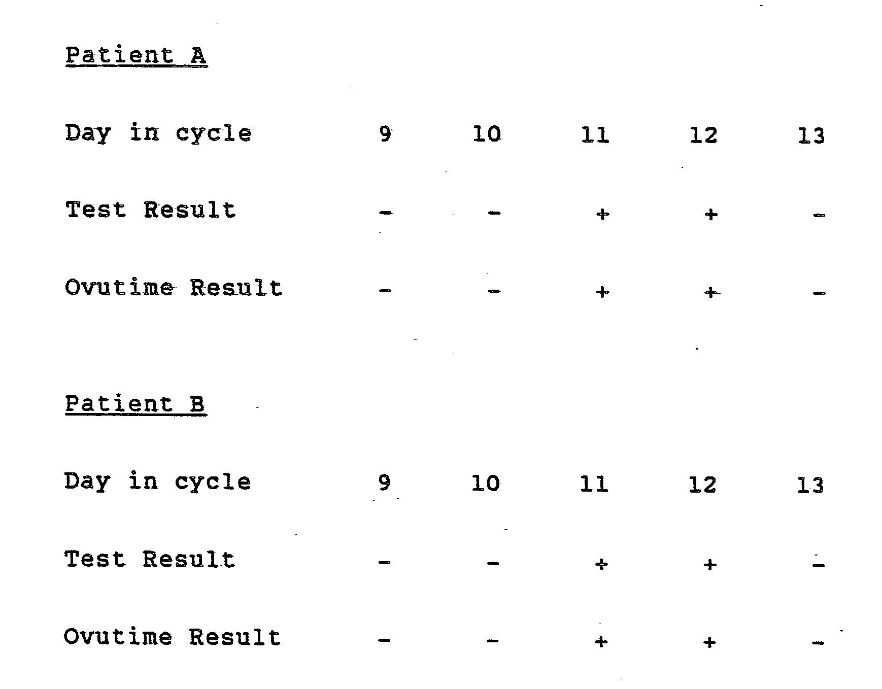
- Example 4 The procedure of Example 4 was followed utilizing urine samples from two women having 28 day menstrual cycles. The samples were collected over a five day period and the test results were verified by Advanced Care's Ovutime TestTM (Ortho Pharmaceutical Corporation) and the following results observed:
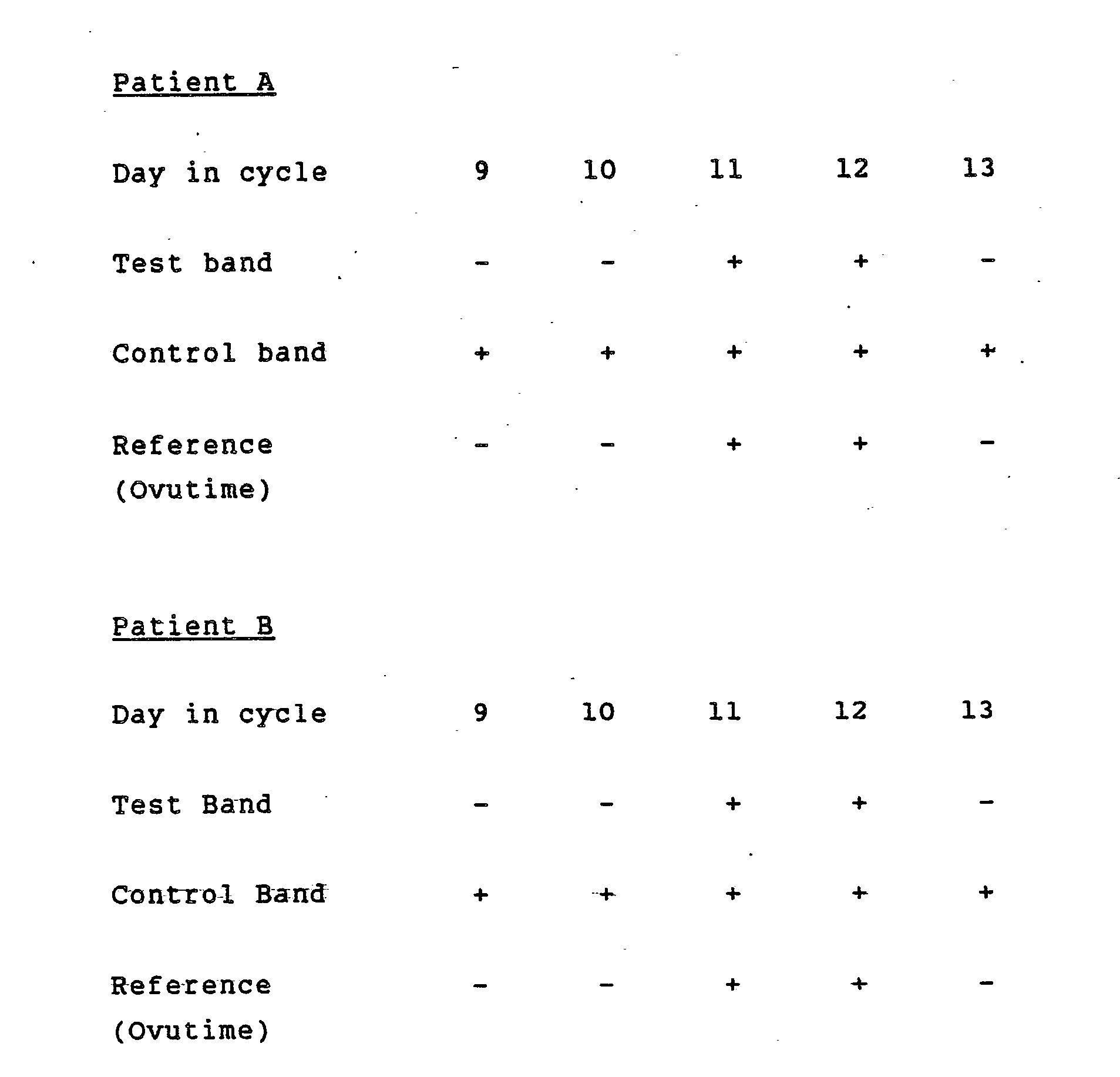
- a membrane strip was prepared pursuant to Example 4 except that the strip had two reagent bands.
- the lower band was coated with anti-LH antibody (forming the test band) and the upper band coated with LH (or hCG which reacts similarly due to cross-reactivity) serving as a control band.
- the colloidal gold labeled anti-LH antibody was prepared pursuant to Example 2. The procedure was as follows: 20 ⁇ ls of colloidal gold labeled anti-LH antibody was added to a test tube along with 100 ⁇ ls of urine sample and mixed. One end of the aforedescribed membrane strip was inserted into the mixture in the test tube and allowed to develop for five minutes. The results observed were as follows:
- Example 1 The same two women's urine sample A and B tested in Example 1 were tested conforming the same result. In addition, this test shows procedural test control band for all tests if it is done properly.
- Ovutime Reference TestTM was performed on the same samples and the results confirmed.
- BIODYNE membrane (pore size 3.0u) were placed in a monoclonal anti-hCG solution (1.2 ug/ml) buffered with 0.02M Phosphate buffered saline solution (PBSS). The membrane was incubated at room temperature for 30 minutes under constant agitation. The membrane was removed from the antibody solution and washed by filtration with a PBSS-0.1% Tween 20 wash solution. The washed membranes were placed in a blocking solution of 0.5% dry milk in PBSS and incubated at 37°C for one hour. The membranes were washed by filtration with PBSS-0.1% Tween wash solution, dried at 37°C and stored in a moisture excluding container containing desiccant at 2-30°C.
- PBSS monoclonal anti-hCG solution
- PBSS-0.1% Tween 20 wash solution 0.02M Phosphate buffered saline solution
- Monoclonal anti-hCG antibody was labeled with colloidal gold. These antibodies were then saturated with hCG producing a label-antibody-analyte complex.
- the previously prepared membranes were cut into strips 50 mm x 6 mm. 500 ul urine sample was mixed with 100 ul of marker. The strips were placed into sample solution (to a depth of 5 mm) and the fluid allowed to migrate to top of strip. The distance the colloidal dye migrated from the surface of the sample solution was visually observed. The results observed were as follows:
- Example 7 The procedures of Example 7 were repeated except that FITC was used to label the monoclonal anti-hCG antibody and results were observed by illuminating the strips with FITC. The following results were obtained:
- a membrane of 90 x 5 mm was prepared substantially pursuant to the procedures set forth in Example 3 except that anti-theophylline antibody was coated in 11 bands.
- Colloidal gold-BSA-theophylline was prepared pursuant to Example 2 (e.g. theophylline, chemically coupled with amide bonds to BSA, was substituted for the monoclonal antibody) and the following procedures performed: 20 ⁇ ls of the colloidal gold labeled reagent was added to a test tube along with 130 ⁇ ls of serum sample and mixed. One end of the membrane strip was inserted into the mixture and developed for 10 minutes and then read.
- Theophylline standards were prepared in concentration ranges from 0 ⁇ gs/ml to 40 ⁇ gs/ml and a control chart (standard concentration versus band height) was prepared from the standard run. The following results were observed.
- a membrane strip was prepared pursuant to Example 4 except that the strip had two reagent bands.
- the lower band was coated with anti-hCG antibody (forming the test band) and the upper band coated with hCG serving as a control/calibration band.
- the colloidal gold labeled anti-hCG antibody was prepared pursuant to Example 2. The procedure was as follows:
- Example 11 Whole Blood Pregnancy Test With Built-In Control
- a membrane strip (90 x 10 mm) was prepared pursuant to Example 10 and tested accordingly with whole blood samples. The procedure was as follows:
- a membrane strip was prepared pursuant to Example 6 except that the strip had two reagent bands.
- the lower band was coated with rubella antigen (Immuno Search, Toms River, NJ), and the upper band coated with human IgG serving as a control/calibration band.
- the colloidal gold labeled mouse anti-human antibody was prepared pursuant to Example 2. The procedure was as follows:
- test results were in complete agreement (100%) with an ELISA Rubella test.
- Another aspect of the present invention concerns the flexibility to build into the membrane strip a feature for ensuring that the liquid reagents and the biologically active molecules on the membrane strip are active, and that the assay steps have been performed properly including the addition of aqueous sample.
- This is particularly useful for immunoglobulin tests and is more specifically described in copending European patent application No.87305028.0 (claiming priority from USSN 872 358 ORD 66) filed contemporaneously herewith.
- the membrane strip would incorporate a reaction zone having an immobilized component capable of specifically reacting with a sample component which is ubiquitous in all similar samples from different sources. Thereafter, the labeled component is ideally chosen so that it can react both with the immunoglobulin or other ligand to be determined and the ubiquitous sample component. It will thus be seen that an internalized control is performed as an integral part of the assay.
Landscapes
- Health & Medical Sciences (AREA)
- Life Sciences & Earth Sciences (AREA)
- Immunology (AREA)
- Engineering & Computer Science (AREA)
- Molecular Biology (AREA)
- Chemical & Material Sciences (AREA)
- Biomedical Technology (AREA)
- Urology & Nephrology (AREA)
- Hematology (AREA)
- Biotechnology (AREA)
- Analytical Chemistry (AREA)
- Cell Biology (AREA)
- Pathology (AREA)
- Food Science & Technology (AREA)
- Medicinal Chemistry (AREA)
- Physics & Mathematics (AREA)
- Microbiology (AREA)
- Biochemistry (AREA)
- General Health & Medical Sciences (AREA)
- General Physics & Mathematics (AREA)
- Endocrinology (AREA)
- Investigating Or Analysing Biological Materials (AREA)
- Medicines Containing Antibodies Or Antigens For Use As Internal Diagnostic Agents (AREA)
- Investigating Or Analyzing Non-Biological Materials By The Use Of Chemical Means (AREA)
- Investigating Or Analysing Materials By The Use Of Chemical Reactions (AREA)
Abstract
Description
- This invention relates to the field of immunoassays and more particularly provides a new immunoassay using colloidal gold and membranes for detecting molecules, including small molecules such as those containing a single epitopic site, in urine or other body fluids.
- Many human health conditions can be ascertained by the use of immunologically-based immunoassays. Such assays rely upon the specificity of reaction between immunoglobulins whether monoclonal or polyclonal-based, and their respective binding partners which may be haptens, antigens or other analytes all of which may hereafter be collectively and interchangeably referred to as ligands and ligand binding partners or biologically active molecules generally. Immunoassays are procedures which utilize the specific binding ability between ligands and ligand binding partners in determining the presence or absence of ligands or their binding partners in a fluid sample. Many such procedures are known and include sandwich asays, competitive binding assays and the like. While the instant invention utilizes the basic precepts of such assays, it is not deemed necessary to review in detail such conventionally known procedures.
- Horisberger and Rosset, J. Histochem., Cytochem., 25:295-305 [1977], described an agglutination assay for mannan using colloidal gold as a label. While such colloidal gold agglutination assays have recently been commercialized, they have disadvantages associated with their speed, difficulty in determining subjective end points by color and the like.
- It is an object of the present invention to employ colloidal gold as a label while avoiding disadvantages associated with Horisberger's agglutination assays.
- In U.S. Patent No. 4,313,734, Leuvering describes a new immunoassay procedure involving the use of colloidal metal substances. While those procedures describe the use of a new label, i.e. colloidal metal, all were dependent upon the use of spectrophotometers or atomic flameless spectrophotometers for identifying the presence of the colloidal metal after it had been chemically leached from the site of immunological reaction. As a result, Leuvering's assays were dependent upon cumbersome procedures and expensive instrumentation in order to derive relatively insensitive results. It is not surprising therefore that the leaching procedures described by Leuvering have not found their way into the commercial marketplace which demands fast, sensitive and economical procedures capable of being performed without such dependence upon expensive instrumentation.
- It is one aspect of the instant invention to utilize a colloidal gold label such as described by Leuvering but to utilize the label in an entirely new procedure which overcomes all of the deficiencies of the Leuvering procedures.
- European Patent Application 0 158,746 assigned to Janssen describes blot overlay assay methods which rely upon diffusion to the surface. While providing a dramatic improvement over Leuvering, the described methods still suffer from excessive time requirements to obtain minimally acceptable sensitivity. Janssen also described new techniques for improving the sensitivity or visualizability of assay results by innovative silver enhancement procedures.
- It is an object of the present invention to provide yet additional improvements over the Janssen methods in both sensitivity and speed of assay.
- In U.S. Patent No. 3,888,629, Bagshawe described an immunoassay procedure utilizing radioisotopes with a cartridge including a reaction surface in contact with an absorbent material for drawing fluids through the membrane. While the Bagshawe device claimed high sensitivity, it presented a less desirable solution inasmuch as it relied upon isotopic labels. Such labels require sophisticated detection equipment for determining their presence upon the reaction surface. Further, and as is well-known, radioisotopes pose substantial health and procedural difficulties making such procedures generally less desirable.
- It is another aspect of the present invention to improve upon the mechanical aspects of the Bagshawe device while avoiding the disadvantages associated with isotopic procedures generally.
- U.S. Patent Nos. 4,235,601 and 4,361,537 to Deutsch et al. describe related test devices for determining a particular characteristic of a sample such as for determining the presence of a substance in a fluid sample. The Deutsch et al. device employs a strip element having a plurality of zones for receiving a fluid sample or containing various reagent components. One end of the strip element is then immersed into a developing fluid which flows upward through the capillary passages thereby coming into contact with the sample and conveying the sample in a continuing upward direction through other zones containing various reagents. One such zone might for instance contain a labeled reagent (e.g. zone 14 in FIG. 1) and another zone (e.g. zone 15 in FIG. 1) containing an immobilizing element such as an antibody for retaining the label in that location if the sample applied to the first zone 13 contained the element for which the label and immobilized antibody were specific. Thus, all immunologically reactive components are stored on the strip element, the only liquid reagent being the developing fluid which was described as comprising an aqueous solution of sodium barbitol, hydrochloric acid, thimerosal and disodium ethylenediamine tetraacetate.
- It is another aspect of the present invention to utilize an approach similar to Deutsch et al. but to avoid the necessity of using complicated developing solutions as taught by Deutsch et al. while employing superior label systems.
- EPA 164,194 describes a similar system using a strip element, however, the point of novelty in that invention appears to be solely directed to the employment of lasers to cut out the strip elements thereby ensuring a uniformly level advancing fluid front.
- It is still another aspect of the present invention to provide new immunoassay methods which are substantially insensitive to methods of strip manufacture and thus do not require the methods taught by EPA 164,194.
- EPA 143,574 teaches a strip element immunoassay system which employs a binding agent to aggregate RBCs at the air/liquid interface on the bibulous element. The assay relies upon conventional ELISA techniques and goes so far as to state on page 22 that individual labels will not be sufficient to produce the desired sensitivity thereby requiring an enzyme label, each of which can generate a plurality of detectable molecules.
- It is still yet another aspect of the present invention to provide immunoassays which utilize similar strip elements but which do not require enzyme labels and which can still be read visually.
- It is yet another aspect of the present invention to provide assay procedures which may be interpreted entirely visually without reliance upon instrumentation but which may also be used with instrumentation if higher sensitivity is desired or in alternative quantitation modes.
- It is yet a further aspect of the present invention to provide assays which may be produced economically, performed expeditiously, and derive the required sensitivity necessary to perform satisfactorily in all medical environments in both the stat and batch modes.
- In keeping with the aspects and goals of the present invention, there is provided a new immunoassay procedure for determining the presence of antibodies or ligands, such as drugs of abuse in fluid samples such as urine, cerebropinal fluid, saliva, whole blood, blood components or other aqueous body discharges or solubilized discharges. The assay is performed with a matrix surface having a tortuous path therethrough and having attached either a ligand or the ligand binding partner depending on whether a competitive or sandwich type assay is to be performed. The matrix surface or membrane is preferably bibulous in nature thereby providing capillary passages for the flow of fluids therethrough thereby bringing the fluid into contact with the ligand or ligand binding partner immobilized within the membrane at one or more locations. Most preferably, the ligand containing sample and a colloidal gold labeled biologically active molecule are pre-mixed and then brought into contact with one end of the bibulous strip. The mixture is then drawn through the matrix by capillary action, allowing the localization of colloidal gold in areas having an immobilized binding partner while washing away unbound colloidal gold in the process. With immunoglobulin tests, it is most preferred to sequentially add sample and colloidal gold labeled reagent with intermediate wash steps. Surprisingly, colloidal gold remaining in the matrix results in the presence of a very strong, visually detectable colored spot.
- As a result of the instant invention, fewer liquid reagents are required than in conventional assays, e.g. one reagent with an optional wash fluid as opposed to four or more reagents required with ELISA procedures. No instruments are required in order to read the results but may be used optionally to measure the presence of colloidal gold based on its ability to scatter or absorb light. And, the entire procedure including all handling steps and a single incubation step can be performed generally in less time than comparable assays, while still readily achieving great sensitivity. In the case of LH, a sensitivity of 20 mIU/ml urine or better is routinely obtained with a two-three drop sample and a total test performance time of approximately five minutes, inclusive of incubation and handling times. Another advantage provided is the elimination of incubation related errors since there are no requisite wash steps with assays for antigens and haptens. Further, completion of the test results in a stable endpoint.
- The inventors hereof have surprisingly discovered that the unobvious combination of a strip matrix or membrane surface and immunoassays employing colloidal gold as a label, can result in extremely rapid and sensitive assays providing results which may be visually detected. Such an assay is especially useful for large and small molecules, and for antibodies but is not so limited. Such assays have not heretofore been possible. This is particularly true since conventional surface assays just don't provide enough colloidal gold to be visually detectable and prior methods have required ELISA techniques or instruments to detect fluorescent labels. Surprisingly, a membrane surface provides many "layers" which, rather unexpectantly, provide sufficient concentration of colloidal gold to be visually detectable. This observation has figured prominantly in a colloidal gold membrane assay (COGMA) described in copending European patent application No.87305026.6 claiming priority from USSN 872 355 - ORD 63) filed contemporaneously herewith. Unlike that assay, the instant assay generally achieves higher sensitivity per ml of sample because all of the sample flows past areas of immunological component in the test strip rather than just through certain sections as in COGMA. As a related advantage, less colloidal gold labeled component is required in order to achieve comparable sensitivity.
- The instant assay is most efficaciously performed utilizing a tortuous or bibulous membrane strip which permits the capillary flow of fluids therethrough and to which immunologically active components may be attached. By using such a tortuous membrane, reaction kinetics normally associated with solution phase reactions become possible despite the fact that one of the immunological reactions takes place at a solid phase surface. Further, such materials allow for flexibility with respect to orientation since the direction of flow is not critical. While a number of types of filter membranes may be employed including for example activated Whatman™ 31 ET chromatography paper, activated nylon 66 (e.g. Biodyne™ from Pall), and activated PVDF (polyvinylidine fluoride such as Immobilon™ from Millipore), the most preferred material is the Whatman material for ease of handling.
- Using LH (leutinizing hormone) as an example of the utilization of the present invention in a sandwich assay format, one would attach LH specific antibody to the membrane. While the membrane is preferably a strip having dimensions such as 5 x 90 mm, the instant invention permits great freedom regarding physical construction. The anti-LH antibody may be attached either continuously throughout the membrane strip or in equally spaced zones along the membrane strip.
- The sample, such as a urine, containing the suspected LH is mixed with a liquid reaqent comprising a colloidal gold labeled (directly or indirectly through an intermediate antibody or other linkage) anti-LH antibody which is specific for a different epitopic site than is the membrane immobilized antibody.
- The mixture of sample and reagent is then ideally applied to one end of the exposed membrane and allowed to migrate through the strip. While the instant assay permits extraordinary variation in the volume, it has been noted that increased volumes, limited only by the absorbent capacity of the membrane and the amount of reagent, may be advantageously used to improve the assay's sensitivity.
- Continuing with the LH example, LH present in female urine (generally associated with the onset of ovulation) is captured by labeled antibody during the mixture step and then becomes attached within the membrane to the anti-LH antibodies immobilized therein. The derivation of such antibodies, either of polyclonal or monoclonal nature, is an art well-known and need not be described in detail here. From a specificity and manufacturing viewpoint, the most preferred antibodies will be monoclonal antibodies derived through the methods of Kohler and Millstein first described in 1975 and published extensively elsewhere.
- Unattached colloidal gold labeled reagent as well as the remaining portions of the sample including the fluid of the sample and non-LH ligands are drawn throughout the remaining portion of the membrane. As a result one will be able to observe the colloidal gold (a bluish-red color) for a distance from the end to which sample was applied (e.g. the end immersed in the sample-reagent mixture). If the immobilized antibody was applied continuously throughout the strip, then some portion of the strip will 30 show continuous color. The length of the membrane showing color will be proportional to the amount of LH present in the sample. Similarly, different zones will show "steps" of color and the number of steps or zones of color can be correlated to LH concentration. Such zones are preferred in that they act as concentrations of the labeled component thereby allowing lower concentrations of labeled components to be used while retaining, or even augmenting the contrast between the zones and the background areas on the strip element. As a related advantage, wash steps are obviated since the labeled component is diluted by the sample, all of which flows by the reaction zone. More LH in the sample will result in a larger distance or a greater number of colored zones as well as greater intensity of color in the colored zones.
- Since in concentrations greater than normal background levels (e.g. equal to or less than 10 mIU/ml urine) LH elevated should be monitored over a number of days (e.g. 6-9) during the mid-menstrual cycle, a preferred commercial product or kit will include an equal number (6-9) of membranes plus one or more reagent bottles containing the colloidal gold labeled reagent and ideally also instructions. The kit may be expanded to nine tests for those women suffering irregular menstrual cycles. Optionally, an extra positive control (LH either labeled directly with colloidal gold or for mixture with colloidal gold labeled antibody reagent) with test strip may also be provided. Alternatively, an internal control could be provided by having an additional zone or bard of LH on the membrane strip. Optionally, the LH (or equivalent ligand) can be calibrated to known concentrations in order to provide a standard color comparative zone. Such internal comparative zones are additional novel aspects of the present invention. While one may now utilize an optional wash step to remove unbound material, the most preferred embodiment based on procedural simplicity advantageously saves time and dispenses with such wash step as being unnecessary in order to obtain the clinically relevant sensitivities.
- Experimentation has shown a substantial variety of colloidal gold sizes as measured by the optical density ratio of 540 nm over 600 nm may be utilized including from about 1.7 (large particles) to 3.0 (small particles) with a preferred ratio within the range of 2 to 2.5. While 1.7 is associated with the size of the largest particle tested, there is no reason to believe larger particles, albeit more difficult to make, could not be used satisfactorily. Colloidal gold particles in the preferred size or optical ratio range may be visually observed as a reddish blue color when in solution. Preferred methods of colloidal gold formation and their attachment to immunoglobulins are described later.
- While the best mode contemplates direct labeling of the ligand or ligand binding partner with colloidal gold, it is also contempIated that equivalent indirect labeling may be employed and even preferred in certain circumstances. Such indirect labeling would typically utilize a third immunoglobulin which is itself labeled with the colloidal gold and is specifically reactive with the second immunoglobulin, in turn specific for the ligand to be detected. Such methods are particularly useful for antibody tests using anti-globulin labeled colloidal gold. Again, indirect labeling procedures are subjects readily understood by skilled practitioners in the art.
- While it is most preferred to allow the membrane strip to develop for a period of time, e.g. five minutes, following contact with the ligand containing sample - colloidal gold labeled reagent mixture, no special incubation conditions are required, room temperature being perfectly adequate. Results may then be directly observed, at any convenient time thereafter. It will be readily understood that immunoglobulin tests will preferably have sequential additional of the sample, intermediate wash and colloidal gold labeled reagent to the strip membrane.
- It will be noted that as a result of the instant invention, fewer liquid reagents are required in order to perform an assay (indeed, most assays will require only one reagent having a biologically active component), and fewer steps need be performed. As a result, the typical reaction may often be run in less than five minutes for ligands or ten minutes for antibodies while still obtaining levels of sensitivity comparable to the best conventional assays but, unlike those other assays, providing visually detectable, unequivocal results with astounding speed.
- Other advantages are provided by the instant invention. Neither the reagents, nor the membrane containing a biologically active molecule need be at room temperature when the assay is performed. This is in stark contrast with conventional ELISA procedures which are highly temperature sensitive.
- The invention further avoids the disadvantages associated with ELISA techniques including the characteristic criticality associated with timing of steps, pH, and temperature of solutions. In contrast, the instant invention avoids such limitations.
- Perhaps the most surprising aspect of the instant invention is the fact that a positive reaction may be visually detected at all. While this aspect of the invention is as yet still not fully understood by the inventors hereof, it is believed that the multi-layered nature of the membrane coupled with the light scattering properties of the colloidal gold results in accumulation, if not amplification of the visual effect of the presence of colloidal gold.
- Other non-antibody molecules can be tested in a fashion similar to LH. For example, one may detect small molecules having a single epitopic site by competitive assay procedures. Such small molecules include drugs of abuse, digoxin, theophylline and many others whose presence has clinical relevance. In the case of theophylline (THEO) in a blood sample, the test would be performed as follows: specific anti-THEO antibodies are attached covalently on the membrane strip in narrow bands equally spaced along the length of the strip. One end of the membrane strip, thus capable of binding theophylline immunologically, is inserted into a tube containing a premixed suspension of blood sampe and colloidal gold labeled THEO. The unlabeled THEO from the sample will thus compete with the colloidal gold labeled THEO for antibody binding sites on the paper. As a result, the height of the colored visual signal is directly proportional to the theophylline concentration in the sample.
- Alternatively, one may attach theophylline directly onto the membrane strip in narrow bands (crosswise) along the entire length of the strip. Alternatively, in both this and the foregoing example, the biologically active molecule may be attached continuously along the length of the membrane strip rather than in zones. One end of the membrane strip, capable of binding anti-theophylline antibody immunologically, is inserted into a tube containing a premixed suspension of the blood sample containing unlabeled theophylline, and colloidal gold labeled anti-theophylline antibody. Again, the height of the visual signal is directly proportional to the theophylline concentration in the sample.
- In a number of instances, the presence of antibodies is used to determine exposure to disease causative organisms. Such tests include for instance Rubella IgG, Rubella IgM, Toxoplasmosis IgG and IgM, Cytomeglovirus IgG and IgM, HTLV III, Anti-EBNA, Mononucleosis, Anti-Core, HAA, Herpes, and Anti-Streptolysin O. Generally, antibody tests will be accomplished in substantially the same manner as the small molecule test in that the ligand or antigen itself will be immobilized on the membrane for reaction with the antibody in the patient sample. The colloidal gold labeled reagent may then be suitably selected as an anti-human IgG or IgM as appropriate. The height of the color will then be proportional to the amount of antibody present in the sample.
- The instant invention may also be employed to detect the presence of infectious disease agents including microorganisms such as Streptococcus, Chlamydia, Gonococcus or viral organisms such as hepatitis and the like.
- Most preferred embodiments of the instant invention will have the capacity to perform multiple tests using the same sample. In such embodiments, a plurality of zones will be used, each with different specificity for sample ligands or antibodies. Contemplated is the simultaneous performance of a test for antibodies present in blood samples, specific for hepatitis core, HTLV-III (AIDS), CMV and the like. The TORCH series (toxoplasmosis, rubella, cytomeglovirus and herpes) is a prime example. In such instances, the control zone(s), if used, should preferably be placed at the top (e.g. furthest from sample addition) in order to ensure that all prior zones have worked properly and have not been neutralized in the absence of gold. These and other aspects of the present invention will become clear upon study of the accompanying examples.
- All glassware surfaces which came into contact with colloidal gold sold were siliconized utilizing 1% Silwet 720 (Union Carbide) by soaking 10 minutes and then rinsing with Distilled Water (D.W.). 1 liter filtered distilled water was heated to 100°C. for at least 10 minutes. 10 ml of 1% gold chloride (Aldrich) in Milli-Q filtered D.W. was added to the reactor and mixed for one minute. 10 ml of 34 mM sodium citrate or 10 ml of 64 mM sodium malate or malic acid at pH 4.2 was added to the reactor to act as a reducing agent and rapid mixing continued for 20 minutes. A color change indicated successful formation of a gold sol. The heat source was removed and the reactor cooled to 15-30°C. 1 ml of 1% PEG 20,000 was added and pH of the sol and the reactor was adjusted to 7.1 ± .1 pH with the addition of 0.1 molar K₂CO₃. The colloidal gold O.D. may be measured at 540 and at 600 nm. In this form, the gold sol is suitable for coupling to antibody. Colloidal gold thusly produced has a size of about 40 nm to about 50 nm as determined by the OD 540/600 ratio, such size being dependent on the chemical reducing conditions.
- Antibody was dialyzed into 0.01 M HEPES buffer solution at pH 7.1 using 12,000-14,000 molecular weight cut off dialysis tubing and 300 ml solution/ml of antibody for 18 hours and then filtered through a 0.45 µm SWINEX™ filter. Protein concentration of antibody may be obtained by measuring the antibody's absorption at 280 nm and diluted to a concentration of 3-4 mg/ml with 0.01 HEPES pH 7.1 to obtain optimal antibody--colloidal adsorption coupling as determined by the antibody to be labeled. With the sol still present in the reactor from Example 1, the thusly filtered and diluted antibody was added while the sol was stirred at room temperature. Stirring continued for approximately 30 minutes whereupon (0.1 x sol volume) mls of buffer (D) comprising 0.01 M HEPES; 0.3 M D-MANITOL; 0.05% PEG 20,000; 0.1% BSA--RIA grade 0.05% sodium azide was added. Stirring continued at room temperature for 30 minutes. The solution was then transferred to a high speed centrifuge, centrifuged and the supernatent removed. The sol was then washed four times utilizing the aforedescribed buffer D and resuspended to 1/10 the starting sol concentration, filtered through a 0.45 µm membrane and stored in polypropylene at 2-8°C.
- Mix equivalent weight amount of BSA (100 mg BSA per 100 mg SOL) into SOL generated from Example 1 with pH preadjusted to 6.0. Stir for 2 hours at room temperature. Filter through 0.22 micron membrane filter to remove large particles. Measure OD 540 and 600 nm and calculate OD ratio; OD 540/OD 600 of acceptable material should be 2.50 ± 0.30. Take 200 ml of thusly prepared BSA sol, centrifuge (25,000 G for 30 minutes) and wash once with distilled water. Mix the washed particles with 1% wt/vol. glutaraldehyde and stir for 2 hours. Centrifuge (25,000 G for 30 minutes) and wash three times with distilled water. Mix the activated and washed particles with 2 mg of purified appropriate monoclonal IgG in pH 7.4 phosphate buffer and stir overnight at 2-8°C. Add 2.5 mg sodium borohydride to stabilize the coupling. Stir 30 minutes, and quench with 5 ml pH 8.0 glycine BSA buffer. Centrifuge (25,000 G for 30 minutes) and wash three times with phosphate buffered saline (pH 8.0). Resuspend and adjust dye concentration with final pH 8 buffer (50 mM triethanolamine, 100 mM NaCl, 0.2% BSA, 0.1% NaN₃) to about 10 O.D. at 540 nm. Filter through 0.22 micron filter and store at 2-8°C.
- Whatman 31ET paper (90 x 152 mm) was immersed in pyridine solution (Baker) containing 0.2 M 1,1ʹ-carbonyldiimidazole and incubated at room temperature for 60 minutes. The membrane strip was then washed with tetrahydrofuran (Baker). The membrane was placed on a glass plate (TLC plate) and secured with tape for ease of handling. Using a LINOMAT™ applicator. 2 µ1/inch of mouse anti-theophylline monoclonal antibody was applied horizontally at evenly spaced locations along the length of the strip. The membrane was removed from the plate and incubated at room temperature for 30 minutes. Blocking solution comprising 0.5% w/v casein Type 1, 0.1% w/v TWEEN™ and PBSS was applied over the membrane and incubated for 90 minutes at 37°C. The membrane was washed with wash buffer comprising 0.1% w/v TWEEN in PBSS at pH 7.3 for 15 minutes at room temperature. The membrane strip was immersed in 0.5% w/v polyvinyl alcohol solution (DuPont) and incubated for 30 minutes. Thereafter the membrane was dried at 37°C. and stored in a moisture excluding container such as an aluminum or plastic bag containing desiccant at 2-30°C. until use. For antigen coated membrane (in theophylline examples) theophylline-BSA conjugate was applied in substitution for the antibody.
- 20 µls of colloidal gold labeled anti-LH antibody was added to two drops (100 µls) of urine sample and mixed in a test tube. A membrane strip prepared pursuant to Example 3 having anti-LH antibody applied thereto had one end dipped into the test tube. The following results using an LH standard prepared in concentration ranges from 0 mIU per ml to 200 mIU per ml was tested and the following results observed:

- The procedure of Example 4 was followed utilizing urine samples from two women having 28 day menstrual cycles. The samples were collected over a five day period and the test results were verified by Advanced Care's Ovutime Test™ (Ortho Pharmaceutical Corporation) and the following results observed:
- A membrane strip was prepared pursuant to Example 4 except that the strip had two reagent bands. The lower band was coated with anti-LH antibody (forming the test band) and the upper band coated with LH (or hCG which reacts similarly due to cross-reactivity) serving as a control band. The colloidal gold labeled anti-LH antibody was prepared pursuant to Example 2. The procedure was as follows: 20 µls of colloidal gold labeled anti-LH antibody was added to a test tube along with 100 µls of urine sample and mixed. One end of the aforedescribed membrane strip was inserted into the mixture in the test tube and allowed to develop for five minutes. The results observed were as follows:
- The same two women's urine sample A and B tested in Example 1 were tested conforming the same result. In addition, this test shows procedural test control band for all tests if it is done properly.
- The foregoing procedure was repeated with the colloidal gold labeled antibody lyophilized in the test tube. Three drops (100 µls) of urine was added to the test tube and mixed and thereafter one end of the membrane strip was inserted and developed for five minutes. The observed results were identical.
- The test was repeated with clinical samples:

- Ovutime Reference Test™ was performed on the same samples and the results confirmed.
- BIODYNE membrane (pore size 3.0u) were placed in a monoclonal anti-hCG solution (1.2 ug/ml) buffered with 0.02M Phosphate buffered saline solution (PBSS). The membrane was incubated at room temperature for 30 minutes under constant agitation. The membrane was removed from the antibody solution and washed by filtration with a PBSS-0.1% Tween 20 wash solution. The washed membranes were placed in a blocking solution of 0.5% dry milk in PBSS and incubated at 37°C for one hour. The membranes were washed by filtration with PBSS-0.1% Tween wash solution, dried at 37°C and stored in a moisture excluding container containing desiccant at 2-30°C. Monoclonal anti-hCG antibody was labeled with colloidal gold. These antibodies were then saturated with hCG producing a label-antibody-analyte complex. The previously prepared membranes were cut into strips 50 mm x 6 mm. 500 ul urine sample was mixed with 100 ul of marker. The strips were placed into sample solution (to a depth of 5 mm) and the fluid allowed to migrate to top of strip. The distance the colloidal dye migrated from the surface of the sample solution was visually observed. The results observed were as follows:

- The procedures of Example 7 were repeated except that FITC was used to label the monoclonal anti-hCG antibody and results were observed by illuminating the strips with FITC. The following results were obtained:

- A membrane of 90 x 5 mm was prepared substantially pursuant to the procedures set forth in Example 3 except that anti-theophylline antibody was coated in 11 bands. Colloidal gold-BSA-theophylline was prepared pursuant to Example 2 (e.g. theophylline, chemically coupled with amide bonds to BSA, was substituted for the monoclonal antibody) and the following procedures performed: 20 µls of the colloidal gold labeled reagent was added to a test tube along with 130 µls of serum sample and mixed. One end of the membrane strip was inserted into the mixture and developed for 10 minutes and then read. Theophylline standards were prepared in concentration ranges from 0 µgs/ml to 40 µgs/ml and a control chart (standard concentration versus band height) was prepared from the standard run. The following results were observed.

- It will be noted that the presence of multiple bands assists in providing a clear contrast between the signal and the background thereby enhancing contrast and facilitating interpretation of results. The test results were not time dependent and remained observable at any time following development. Further, the ability to prepare control charts allows the quantitation of theophylline concentration in those instances when qualitative results (e.g. above a predetermined height) is insufficient.
- A membrane strip was prepared pursuant to Example 4 except that the strip had two reagent bands. The lower band was coated with anti-hCG antibody (forming the test band) and the upper band coated with hCG serving as a control/calibration band. The colloidal gold labeled anti-hCG antibody was prepared pursuant to Example 2. The procedure was as follows:
- 40 µls of colloidal gold labeled anti-hCG antibody was added to a test tube along with 100 µls of urine sample and mixed. One end of the aforedescribed membrane strip was inserted into the mixture in the test tube and allowed to develop for five minutes. The results observed were as follows:

- The test results were in complete agreement (100%) with Hybritech's TANDEM ICON™ hCG Test.
- A membrane strip (90 x 10 mm) was prepared pursuant to Example 10 and tested accordingly with whole blood samples. The procedure was as follows:
- 10 µls of whole blood sample was spotted on the membrane at the position between the test band and the bottom end. The membrane strip was then inserted into a test tube containing a mixture of 40 µls of colloidal gold labeled anti-hCG antibody and 160 µls of buffer (0.1 M PBS plus 5% BSA, 0.05% Tween-20). After ten minutes, the results were observed as follows:

- The test results were in complete agreement (100%) with Hybritech's TANDEM ICON™ hCG Test.
- A membrane strip was prepared pursuant to Example 6 except that the strip had two reagent bands. The lower band was coated with rubella antigen (Immuno Search, Toms River, NJ), and the upper band coated with human IgG serving as a control/calibration band. The colloidal gold labeled mouse anti-human antibody was prepared pursuant to Example 2. The procedure was as follows:
- 10 µls of serum sample was diluted with 25 µls of Buffer A (0.1M PBS plus 5% BSA, 0.05% Tween-20) in a test tube. One end of the aforedescribed membrane strip was inserted into the test tube and allowed to develop for one minute. Another 25 µls of Buffer A was added to the test tube and the strip was further developed for two minutes. Finally, the strip was transferred to a second test tube containing a mixture of 30 µls of colloidal gold labeled mouse anti-human antibody and 100 µls of Buffer A. The strip was allowed to develop for seven minutes and the results observed were as follows:
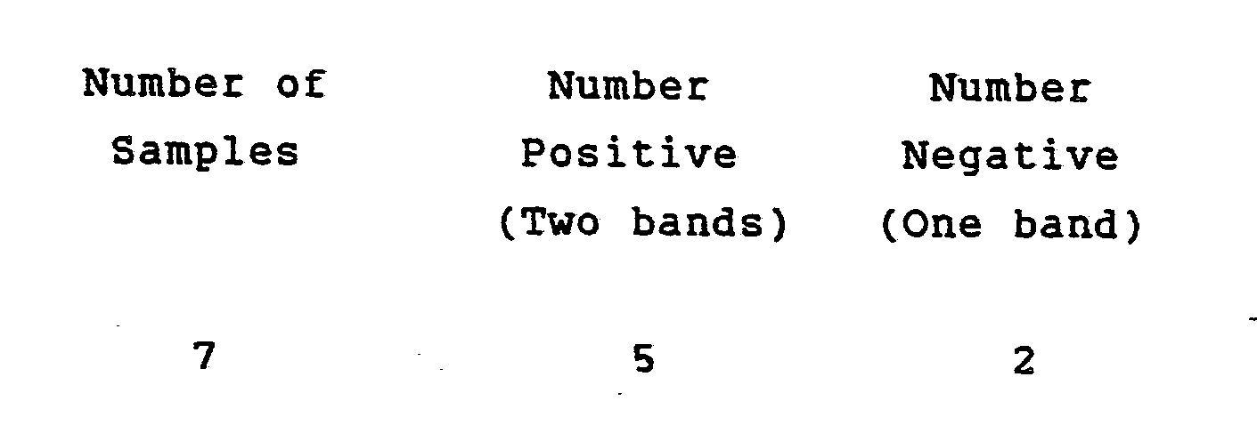
- The test results were in complete agreement (100%) with an ELISA Rubella test.
- Another aspect of the present invention concerns the flexibility to build into the membrane strip a feature for ensuring that the liquid reagents and the biologically active molecules on the membrane strip are active, and that the assay steps have been performed properly including the addition of aqueous sample. This is particularly useful for immunoglobulin tests and is more specifically described in copending European patent application No.87305028.0 (claiming priority from USSN 872 358 ORD 66) filed contemporaneously herewith. In summary, the membrane strip would incorporate a reaction zone having an immobilized component capable of specifically reacting with a sample component which is ubiquitous in all similar samples from different sources. Thereafter, the labeled component is ideally chosen so that it can react both with the immunoglobulin or other ligand to be determined and the ubiquitous sample component. It will thus be seen that an internalized control is performed as an integral part of the assay.
- From the foregoing, it will be readily perceived by one skilled in the art that numerous alterations may be made with regard to the selection of immunologically active components, the membranes, and the liquid reagent while still employing the essence of the invention comprising the combination of a colloidal gold labeled reagent in an immunoassay performed in a matrix membrane by flowing materials through the membrane to obtain a visually detectable result.
Claims (16)
in step b) is a ligand, labelled directly or indirectly with colloidal gold, which competes with the ligand in the sample for the ligand binding partner is used; and
the reagent and the sample are mixed prior to their simultaneous application the one end of the strip membrane.
the ligand binding partner in step a) is an antigen having the same immunological reactivity as said sample antigen;
the ligand binding partner in step b) is an antibody, labelled directly or indirectly with colloidal gold, which is specific for said sample antigen; and
the reagent and the sample are mixed prior to their simultaneous application to the one end of the strip membrane.
the ligand is an antibody;
the ligand binding partner in step a) is an antigen for which the antibody is specific; and
the ligand binding partner in the reagent is an immunoglobulin, labelled directly or indirectly with colloidal gold, which is specific for said antibody.
the ligand is an antigen having at least two epitopic sites;
the ligand binding partner in step a) is an antibody which is specific for a first epitopic site on the antigen; and
the ligand binding partner in step b) is an antibody, labelled directly or indirectly with colloidal gold, which is specific for a second epitopic site on the antigen.
the ligand is an antigen having at least two epitopic sites;
the ligand binding partner in step a) is an antigen having the same immunological reactivity as said sample antigen;
the ligand binding partner in step b) is an antibody, labelled directly or indirectly with colloidal gold, which is specific for the antigens; and
the reagent and the sample are mixed prior to their simultaneous application to the one end of the strip membrane.
Priority Applications (1)
| Application Number | Priority Date | Filing Date | Title |
|---|---|---|---|
| AT87305027T ATE79956T1 (en) | 1986-06-09 | 1987-06-08 | IMMUNOASSAY USING COLLOIDAL GOLD. |
Applications Claiming Priority (2)
| Application Number | Priority Date | Filing Date | Title |
|---|---|---|---|
| US87235786A | 1986-06-09 | 1986-06-09 | |
| US872357 | 1986-06-09 |
Publications (3)
| Publication Number | Publication Date |
|---|---|
| EP0250137A2 true EP0250137A2 (en) | 1987-12-23 |
| EP0250137A3 EP0250137A3 (en) | 1988-09-07 |
| EP0250137B1 EP0250137B1 (en) | 1992-08-26 |
Family
ID=25359420
Family Applications (1)
| Application Number | Title | Priority Date | Filing Date |
|---|---|---|---|
| EP87305027A Expired - Lifetime EP0250137B1 (en) | 1986-06-09 | 1987-06-08 | Colloidal gold immunoassay |
Country Status (8)
| Country | Link |
|---|---|
| EP (1) | EP0250137B1 (en) |
| JP (1) | JP2537873B2 (en) |
| AR (1) | AR241965A1 (en) |
| AT (1) | ATE79956T1 (en) |
| AU (1) | AU602694B2 (en) |
| CA (1) | CA1288337C (en) |
| DE (1) | DE3781335T2 (en) |
| MX (1) | MX173476B (en) |
Cited By (44)
| Publication number | Priority date | Publication date | Assignee | Title |
|---|---|---|---|---|
| GB2204398A (en) * | 1987-04-27 | 1988-11-09 | Unilever Plc | Assays |
| WO1990013031A1 (en) * | 1989-04-26 | 1990-11-01 | Arthur Randell Bradwell | Radial immunodiffusion and like techniques |
| EP0299428A3 (en) * | 1987-07-13 | 1991-07-31 | Abbott Laboratories | Process for immunochromatography with colloidal particles |
| EP0428412A3 (en) * | 1989-11-16 | 1992-02-26 | Ortho Diagnostic Systems Inc. | Method of producing a metal sol reagent containing colloidal metal particles of a preselected size |
| US5252496A (en) * | 1989-12-18 | 1993-10-12 | Princeton Biomeditech Corporation | Carbon black immunochemical label |
| EP0615128A3 (en) * | 1993-03-12 | 1995-06-28 | Lofarma Farma Lab | Equipment and test to determine an antigen or an antibody in a sample. |
| US5556756A (en) * | 1988-01-13 | 1996-09-17 | Nycomed As | Gold sol of less than 5 nm test method and reagent kit therefor |
| US5563040A (en) * | 1994-03-21 | 1996-10-08 | The United States Of America As Represented By The Secretary Of Agriculture | Method and apparatus for immunological diagnosis of fungal decay in wood |
| US5571726A (en) * | 1986-06-09 | 1996-11-05 | Ortho Diagnostic Systems, Inc. | Kit containing glutaraldehyde coated colloidal metal particles of a preselected size |
| US5620845A (en) * | 1988-06-06 | 1997-04-15 | Ampcor, Inc. | Immunoassay diagnostic kit |
| US5656448A (en) * | 1990-01-10 | 1997-08-12 | Princeton Biomeditech Corporation | Dipstick immunoassay device |
| US5712170A (en) * | 1992-12-29 | 1998-01-27 | Oy Medix Biochemica Ab | Test strip, its production and use |
| US5759774A (en) * | 1988-05-18 | 1998-06-02 | Cobe Laboratories, Inc. | Method of detecting circulating antibody types using dried or lyophilized cells |
| US5851777A (en) * | 1996-02-05 | 1998-12-22 | Dade Behring Inc. | Homogeneous sol-sol assay |
| EP0903584A1 (en) * | 1997-09-18 | 1999-03-24 | Matsushita Electric Industrial Co., Ltd. | Immunochromatography-assisted device |
| US5980746A (en) * | 1996-11-14 | 1999-11-09 | Pall Corporation | Membrane and methods of preparing and using same |
| US6372514B1 (en) | 1998-09-18 | 2002-04-16 | Syntron Bioresearch, Inc. | Even fluid front for liquid sample on test strip device |
| WO2002095409A1 (en) * | 2001-04-30 | 2002-11-28 | Erling Sundrehagen | Quantitative non-instrumental immunoassay and device using coloured particles |
| EP1069433A3 (en) * | 1999-07-12 | 2004-01-28 | Fuji Photo Film Co., Ltd. | Agglutination assay method in porous medium layer |
| US6767714B2 (en) | 1995-05-02 | 2004-07-27 | Armkel, Llc | Diagnostic detection device and method |
| WO2004109285A1 (en) * | 2003-06-03 | 2004-12-16 | Bayer Healthcare Llc | Native analyte as reference in lateral flow assays |
| US7270959B2 (en) | 2001-07-25 | 2007-09-18 | Oakville Hong Kong Company Limited | Specimen collection container |
| US7300633B2 (en) | 2001-07-25 | 2007-11-27 | Oakville Hong Kong Company Limited | Specimen collection container |
| DE112006003813T5 (en) | 2006-03-20 | 2009-01-22 | Inverness Medical Switzerland Gmbh | Water-soluble conjugates for electrochemical detection |
| US7560272B2 (en) | 2003-01-04 | 2009-07-14 | Inverness Medical Switzerland Gmbh | Specimen collection and assay container |
| US7666614B2 (en) | 1997-07-25 | 2010-02-23 | Genzyme Corporation | Method of use of one step immunochromatographic device for Streptococcus A antigen |
| EP1493029B1 (en) * | 2002-04-10 | 2010-03-31 | Response Biomedical Corporation | Sensitive immunochromatographic assay |
| US7875433B2 (en) | 2007-02-26 | 2011-01-25 | Response Biomedical Corporation | Comparative multiple analyte assay |
| US8101429B2 (en) | 2003-06-03 | 2012-01-24 | Siemens Healthcare Diagnostics Inc. | Native analyte as a reference in lateral flow assays |
| WO2012099897A1 (en) | 2011-01-18 | 2012-07-26 | Symbolics, Llc | Lateral flow assays using two dimensional features |
| WO2014012077A1 (en) | 2012-07-13 | 2014-01-16 | Genisphere, Llc | Lateral flow assays using dna dendrimers |
| WO2014015076A1 (en) | 2012-07-18 | 2014-01-23 | Symbolics, Llc | Lateral flow assays using two dimensional features |
| US8664001B2 (en) | 2004-06-15 | 2014-03-04 | Ani Biotech Oy | Immunochemical filter device and methods for use thereof |
| WO2015038978A1 (en) | 2013-09-13 | 2015-03-19 | Symbolics, Llc | Lateral flow assays using two dimensional test and control signal readout patterns |
| WO2016164365A1 (en) | 2015-04-06 | 2016-10-13 | Bludiagnostics, Inc. | A test device for detecting an analyte in a saliva sample and method of use |
| WO2018009143A1 (en) * | 2016-07-07 | 2018-01-11 | Cell Id Pte Ltd | Haemoglobin test kit |
| WO2018039047A1 (en) | 2016-08-23 | 2018-03-01 | Qoolabs, Inc. | Lateral flow assay for assessing recombinant protein expression or reporter gene expression |
| US10076314B2 (en) | 2009-02-16 | 2018-09-18 | Express Diagnostics Int'l, Inc. | Device for assaying analytes in bodily fluids |
| US10670596B2 (en) | 2013-02-26 | 2020-06-02 | Astute Medical, Inc. | Lateral flow assay with test strip retainer |
| CN113311176A (en) * | 2021-04-14 | 2021-08-27 | 昆明天沃生物科技有限公司 | Method for detecting pregnancy and gestation number of sows |
| CN115902199A (en) * | 2022-12-30 | 2023-04-04 | 圣湘生物科技股份有限公司 | A kind of processing method and application of colloidal gold label |
| CN117571988A (en) * | 2024-01-09 | 2024-02-20 | 山东康华生物医疗科技股份有限公司 | A blood screening multi-line test strip, kit and preparation method and application thereof |
| US12195531B2 (en) | 2013-03-21 | 2025-01-14 | Code Biotherapeutics, Inc. | Cellular delivery of DNA intercalating agents |
| US12338435B2 (en) | 2016-02-19 | 2025-06-24 | Code Biotherapeutics, Inc. | Nucleic acid carriers and therapeutic methods of use |
Families Citing this family (17)
| Publication number | Priority date | Publication date | Assignee | Title |
|---|---|---|---|---|
| US5622871A (en) | 1987-04-27 | 1997-04-22 | Unilever Patent Holdings B.V. | Capillary immunoassay and device therefor comprising mobilizable particulate labelled reagents |
| US5030558A (en) * | 1986-11-07 | 1991-07-09 | Syntex (U.S.A.) Inc. | Qualitative immunochromatographic method and device |
| US5202267A (en) * | 1988-04-04 | 1993-04-13 | Hygeia Sciences, Inc. | Sol capture immunoassay kit and procedure |
| AU2684488A (en) | 1988-06-27 | 1990-01-04 | Carter-Wallace, Inc. | Test device and method for colored particle immunoassay |
| US6352862B1 (en) | 1989-02-17 | 2002-03-05 | Unilever Patent Holdings B.V. | Analytical test device for imuno assays and methods of using same |
| JP3126383B2 (en) * | 1991-03-28 | 2001-01-22 | 株式会社アズウェル | Simple analyzer |
| US5998220A (en) | 1991-05-29 | 1999-12-07 | Beckman Coulter, Inc. | Opposable-element assay devices, kits, and methods employing them |
| US5468648A (en) | 1991-05-29 | 1995-11-21 | Smithkline Diagnostics, Inc. | Interrupted-flow assay device |
| US5877028A (en) | 1991-05-29 | 1999-03-02 | Smithkline Diagnostics, Inc. | Immunochromatographic assay device |
| US5607863A (en) | 1991-05-29 | 1997-03-04 | Smithkline Diagnostics, Inc. | Barrier-controlled assay device |
| US5869345A (en) | 1991-05-29 | 1999-02-09 | Smithkline Diagnostics, Inc. | Opposable-element assay device employing conductive barrier |
| US6168956B1 (en) | 1991-05-29 | 2001-01-02 | Beckman Coulter, Inc. | Multiple component chromatographic assay device |
| US5879951A (en) | 1997-01-29 | 1999-03-09 | Smithkline Diagnostics, Inc. | Opposable-element assay device employing unidirectional flow |
| US5939252A (en) | 1997-05-09 | 1999-08-17 | Lennon; Donald J. | Detachable-element assay device |
| US6140136A (en) * | 1998-09-18 | 2000-10-31 | Syntron Bioresearch, Inc. | Analytical test device and method of use |
| DE10102065C2 (en) * | 2001-01-17 | 2003-04-17 | Sartorius Gmbh | Diagnostic membrane and method for producing a diagnostic membrane |
| US7517495B2 (en) | 2003-08-25 | 2009-04-14 | Inverness Medical Switzerland Gmbh | Biological specimen collection and analysis system |
Family Cites Families (9)
| Publication number | Priority date | Publication date | Assignee | Title |
|---|---|---|---|---|
| NL7807532A (en) * | 1978-07-13 | 1980-01-15 | Akzo Nv | METAL IMMUNO TEST. |
| US4235601A (en) * | 1979-01-12 | 1980-11-25 | Thyroid Diagnostics, Inc. | Test device and method for its use |
| US4366241A (en) * | 1980-08-07 | 1982-12-28 | Syva Company | Concentrating zone method in heterogeneous immunoassays |
| GB2111201A (en) * | 1981-12-10 | 1983-06-29 | Celltech Ltd | Immunoassay of human luteinising hormone |
| IL68506A (en) * | 1982-05-04 | 1988-06-30 | Syva Co | Simultaneous calibration heterogeneous immunoassay method and device and kit for use therein |
| GB8331514D0 (en) * | 1983-11-25 | 1984-01-04 | Janssen Pharmaceutica Nv | Visualization method |
| EP0167834A3 (en) * | 1984-06-08 | 1988-03-09 | Yeda Research And Development Company, Ltd. | Metal-containing polyaldehyde microspheres |
| GB8415998D0 (en) * | 1984-06-22 | 1984-07-25 | Janssen Pharmaceutica Nv | Staining method |
| AU7385387A (en) * | 1986-06-09 | 1987-12-10 | Ortho Diagnostic Systems Inc. | Immunoassay using detection of colloidal gold |
-
1987
- 1987-06-04 AU AU73854/87A patent/AU602694B2/en not_active Ceased
- 1987-06-05 CA CA000538983A patent/CA1288337C/en not_active Expired - Lifetime
- 1987-06-08 JP JP62141620A patent/JP2537873B2/en not_active Expired - Lifetime
- 1987-06-08 AR AR87307805A patent/AR241965A1/en active
- 1987-06-08 DE DE8787305027T patent/DE3781335T2/en not_active Expired - Lifetime
- 1987-06-08 EP EP87305027A patent/EP0250137B1/en not_active Expired - Lifetime
- 1987-06-08 AT AT87305027T patent/ATE79956T1/en not_active IP Right Cessation
- 1987-06-09 MX MX006829A patent/MX173476B/en unknown
Cited By (73)
| Publication number | Priority date | Publication date | Assignee | Title |
|---|---|---|---|---|
| US5571726A (en) * | 1986-06-09 | 1996-11-05 | Ortho Diagnostic Systems, Inc. | Kit containing glutaraldehyde coated colloidal metal particles of a preselected size |
| US5602040A (en) * | 1987-04-27 | 1997-02-11 | Unilever Patent Holdings B.V. | Assays |
| GB2204398B (en) * | 1987-04-27 | 1991-08-28 | Unilever Plc | Analytical test device for use in assays |
| EP0560411B1 (en) * | 1987-04-27 | 2000-07-26 | Unilever N.V. | Specific binding assays |
| GB2204398A (en) * | 1987-04-27 | 1988-11-09 | Unilever Plc | Assays |
| US6534320B2 (en) | 1987-07-13 | 2003-03-18 | Abbott Laboratories | Process for immunochromatography with colloidal particles |
| EP0299428A3 (en) * | 1987-07-13 | 1991-07-31 | Abbott Laboratories | Process for immunochromatography with colloidal particles |
| US5578577A (en) * | 1987-07-13 | 1996-11-26 | Abbott Laboratories | Method for storing labile proteins |
| US5120643A (en) * | 1987-07-13 | 1992-06-09 | Abbott Laboratories | Process for immunochromatography with colloidal particles |
| US5616467A (en) * | 1988-01-13 | 1997-04-01 | Nycomed As | Method and kit for analyte detection employing gold-sol bound antibodies |
| US5556756A (en) * | 1988-01-13 | 1996-09-17 | Nycomed As | Gold sol of less than 5 nm test method and reagent kit therefor |
| US5759774A (en) * | 1988-05-18 | 1998-06-02 | Cobe Laboratories, Inc. | Method of detecting circulating antibody types using dried or lyophilized cells |
| US5620845A (en) * | 1988-06-06 | 1997-04-15 | Ampcor, Inc. | Immunoassay diagnostic kit |
| US5429951A (en) * | 1989-04-26 | 1995-07-04 | Bradwell; Arthur R. | Radial immunodiffusion and like techniques |
| GB2247314B (en) * | 1989-04-26 | 1992-08-05 | Arthur Randell Bradwell | Method and apparatus for detecting the presence of complementary matrix forming agents |
| GB2247314A (en) * | 1989-04-26 | 1992-02-26 | Arthur Randell Bradwell | Radial immunodiffusion and like techniques |
| EP0396342A1 (en) * | 1989-04-26 | 1990-11-07 | BRADWELL, Arthur Randell | Radial immunodiffusion and like techniques |
| WO1990013031A1 (en) * | 1989-04-26 | 1990-11-01 | Arthur Randell Bradwell | Radial immunodiffusion and like techniques |
| GR900100804A (en) * | 1989-11-16 | 1992-04-17 | Ortho Diagnostic Systems Inc | Method of producing a metal for reagent containingmethod of producing a metal for reagent containing colloidal metal particles of a preselected size colloidal metal particles of a preselected size |
| EP0428412A3 (en) * | 1989-11-16 | 1992-02-26 | Ortho Diagnostic Systems Inc. | Method of producing a metal sol reagent containing colloidal metal particles of a preselected size |
| US6541277B1 (en) | 1989-12-18 | 2003-04-01 | Princeton Biomeditech Corporation | Immunoassay devices and materials |
| US5728587A (en) * | 1989-12-18 | 1998-03-17 | Pmb-Selfcare, Llc | Immunoassay devices and materials |
| US5252496A (en) * | 1989-12-18 | 1993-10-12 | Princeton Biomeditech Corporation | Carbon black immunochemical label |
| US6506612B2 (en) | 1989-12-18 | 2003-01-14 | Princeton Biomeditech Corporation | Immunoassay devices and materials |
| US6027943A (en) * | 1989-12-18 | 2000-02-22 | Princeton Biomeditech Corporation | Immunoassay devices and materials |
| US5559041A (en) * | 1989-12-18 | 1996-09-24 | Princeton Biomeditech Corporation | Immunoassay devices and materials |
| US6737277B1 (en) | 1989-12-18 | 2004-05-18 | Pmb-Selfcare Llc | Immunoassay devices and materials |
| US5656448A (en) * | 1990-01-10 | 1997-08-12 | Princeton Biomeditech Corporation | Dipstick immunoassay device |
| US5712170A (en) * | 1992-12-29 | 1998-01-27 | Oy Medix Biochemica Ab | Test strip, its production and use |
| US5965458A (en) * | 1992-12-29 | 1999-10-12 | Oy Medix Biochemica A.B. | Test strip, its production and use |
| EP0615128A3 (en) * | 1993-03-12 | 1995-06-28 | Lofarma Farma Lab | Equipment and test to determine an antigen or an antibody in a sample. |
| US5563040A (en) * | 1994-03-21 | 1996-10-08 | The United States Of America As Represented By The Secretary Of Agriculture | Method and apparatus for immunological diagnosis of fungal decay in wood |
| US6767714B2 (en) | 1995-05-02 | 2004-07-27 | Armkel, Llc | Diagnostic detection device and method |
| US7045342B2 (en) | 1995-05-02 | 2006-05-16 | Church & Dwight Co., Inc. | Diagnostic detection device and method |
| US5851777A (en) * | 1996-02-05 | 1998-12-22 | Dade Behring Inc. | Homogeneous sol-sol assay |
| US5980746A (en) * | 1996-11-14 | 1999-11-09 | Pall Corporation | Membrane and methods of preparing and using same |
| US7666614B2 (en) | 1997-07-25 | 2010-02-23 | Genzyme Corporation | Method of use of one step immunochromatographic device for Streptococcus A antigen |
| EP0903584A1 (en) * | 1997-09-18 | 1999-03-24 | Matsushita Electric Industrial Co., Ltd. | Immunochromatography-assisted device |
| US6194225B1 (en) | 1997-09-18 | 2001-02-27 | Matsushita Electric Industrial Co., Ltd. | Immunochromatography-assisted device |
| US6372514B1 (en) | 1998-09-18 | 2002-04-16 | Syntron Bioresearch, Inc. | Even fluid front for liquid sample on test strip device |
| EP1069433A3 (en) * | 1999-07-12 | 2004-01-28 | Fuji Photo Film Co., Ltd. | Agglutination assay method in porous medium layer |
| WO2002095409A1 (en) * | 2001-04-30 | 2002-11-28 | Erling Sundrehagen | Quantitative non-instrumental immunoassay and device using coloured particles |
| US7270959B2 (en) | 2001-07-25 | 2007-09-18 | Oakville Hong Kong Company Limited | Specimen collection container |
| US7300633B2 (en) | 2001-07-25 | 2007-11-27 | Oakville Hong Kong Company Limited | Specimen collection container |
| EP1493029B1 (en) * | 2002-04-10 | 2010-03-31 | Response Biomedical Corporation | Sensitive immunochromatographic assay |
| US7691595B2 (en) | 2002-04-10 | 2010-04-06 | Response Biomedical Corporation | Sensitive immunochromatographic assay |
| US7560272B2 (en) | 2003-01-04 | 2009-07-14 | Inverness Medical Switzerland Gmbh | Specimen collection and assay container |
| US8101429B2 (en) | 2003-06-03 | 2012-01-24 | Siemens Healthcare Diagnostics Inc. | Native analyte as a reference in lateral flow assays |
| WO2004109285A1 (en) * | 2003-06-03 | 2004-12-16 | Bayer Healthcare Llc | Native analyte as reference in lateral flow assays |
| US8664001B2 (en) | 2004-06-15 | 2014-03-04 | Ani Biotech Oy | Immunochemical filter device and methods for use thereof |
| DE112006003813T5 (en) | 2006-03-20 | 2009-01-22 | Inverness Medical Switzerland Gmbh | Water-soluble conjugates for electrochemical detection |
| US7875433B2 (en) | 2007-02-26 | 2011-01-25 | Response Biomedical Corporation | Comparative multiple analyte assay |
| US10076314B2 (en) | 2009-02-16 | 2018-09-18 | Express Diagnostics Int'l, Inc. | Device for assaying analytes in bodily fluids |
| WO2012099897A1 (en) | 2011-01-18 | 2012-07-26 | Symbolics, Llc | Lateral flow assays using two dimensional features |
| US11016090B2 (en) | 2011-01-18 | 2021-05-25 | Symbolics, Llc | Lateral flow assays using two dimensional features |
| EP3187876A1 (en) | 2011-01-18 | 2017-07-05 | Symbolics, LLC | Lateral flow assays using two dimensional features |
| US9874576B2 (en) | 2011-01-18 | 2018-01-23 | Symbolics, Llc | Lateral flow assays using two dimensional features |
| US9851366B2 (en) | 2011-01-18 | 2017-12-26 | Symbolics, Llc | Lateral flow assays using two dimensional features |
| WO2014012077A1 (en) | 2012-07-13 | 2014-01-16 | Genisphere, Llc | Lateral flow assays using dna dendrimers |
| US9651549B2 (en) | 2012-07-13 | 2017-05-16 | Genisphere, Llc | Lateral flow assays using DNA dendrimers |
| WO2014015076A1 (en) | 2012-07-18 | 2014-01-23 | Symbolics, Llc | Lateral flow assays using two dimensional features |
| US9874556B2 (en) | 2012-07-18 | 2018-01-23 | Symbolics, Llc | Lateral flow assays using two dimensional features |
| US10670596B2 (en) | 2013-02-26 | 2020-06-02 | Astute Medical, Inc. | Lateral flow assay with test strip retainer |
| US12195531B2 (en) | 2013-03-21 | 2025-01-14 | Code Biotherapeutics, Inc. | Cellular delivery of DNA intercalating agents |
| WO2015038978A1 (en) | 2013-09-13 | 2015-03-19 | Symbolics, Llc | Lateral flow assays using two dimensional test and control signal readout patterns |
| WO2016164365A1 (en) | 2015-04-06 | 2016-10-13 | Bludiagnostics, Inc. | A test device for detecting an analyte in a saliva sample and method of use |
| US12338435B2 (en) | 2016-02-19 | 2025-06-24 | Code Biotherapeutics, Inc. | Nucleic acid carriers and therapeutic methods of use |
| CN109564228A (en) * | 2016-07-07 | 2019-04-02 | 源鉴定私人有限公司 | Hemoglobin detection kit |
| WO2018009143A1 (en) * | 2016-07-07 | 2018-01-11 | Cell Id Pte Ltd | Haemoglobin test kit |
| WO2018039047A1 (en) | 2016-08-23 | 2018-03-01 | Qoolabs, Inc. | Lateral flow assay for assessing recombinant protein expression or reporter gene expression |
| CN113311176A (en) * | 2021-04-14 | 2021-08-27 | 昆明天沃生物科技有限公司 | Method for detecting pregnancy and gestation number of sows |
| CN115902199A (en) * | 2022-12-30 | 2023-04-04 | 圣湘生物科技股份有限公司 | A kind of processing method and application of colloidal gold label |
| CN117571988A (en) * | 2024-01-09 | 2024-02-20 | 山东康华生物医疗科技股份有限公司 | A blood screening multi-line test strip, kit and preparation method and application thereof |
Also Published As
| Publication number | Publication date |
|---|---|
| AU7385487A (en) | 1987-12-10 |
| EP0250137B1 (en) | 1992-08-26 |
| JP2537873B2 (en) | 1996-09-25 |
| DE3781335T2 (en) | 1992-12-17 |
| DE3781335D1 (en) | 1992-10-01 |
| ATE79956T1 (en) | 1992-09-15 |
| JPS6325553A (en) | 1988-02-03 |
| EP0250137A3 (en) | 1988-09-07 |
| AU602694B2 (en) | 1990-10-25 |
| MX173476B (en) | 1994-03-08 |
| CA1288337C (en) | 1991-09-03 |
| AR241965A1 (en) | 1993-01-29 |
Similar Documents
| Publication | Publication Date | Title |
|---|---|---|
| EP0250137B1 (en) | Colloidal gold immunoassay | |
| FI92110B (en) | Simultaneous determn. of several analytes | |
| EP0466914B2 (en) | Immunochromatographic method | |
| JP2999238B2 (en) | Chromatography strip binding assay device | |
| EP0349215B1 (en) | Method and cell for detection | |
| AU770689B2 (en) | Analytical test device and method of use | |
| US4469787A (en) | Immunoassay involving soluble complex of second antibody and labeled binding protein | |
| EP0138826B1 (en) | Detection of human chorionic gonadotropin | |
| TW203120B (en) | ||
| EP0335244B1 (en) | Solid-phase analytical device and method for using same | |
| EP0987550B1 (en) | Analytical test device and method of use | |
| JP3358737B2 (en) | Assays with improved dose response curves | |
| JP4846573B2 (en) | Lateral flow assay device and method with natural analyte as reference | |
| EP0480497B1 (en) | Device for performing a rapid single manual assay | |
| US4639419A (en) | Immunological color change test involving two differently colored reagent spots | |
| EP0258963A2 (en) | Colloidal gold immunoassay | |
| EP0462376A2 (en) | Conjugate recovery binding assays | |
| WO1998036278A1 (en) | Multiple-site antibody capture immunoassays and kits | |
| WO1997031268A1 (en) | Chromatographic strip having detection and control zones oriented parallel to the direction of flow | |
| US20070172963A1 (en) | Native analyte as a reference in lateral flow assays | |
| JP3493544B2 (en) | Antibody assay device | |
| US3955923A (en) | Serologic reaction method | |
| Monroe | Enzyme Channelling Immunoassay (ECIA): A Unique and Rapid Quantitative Technique | |
| JP2532788C (en) |
Legal Events
| Date | Code | Title | Description |
|---|---|---|---|
| PUAI | Public reference made under article 153(3) epc to a published international application that has entered the european phase |
Free format text: ORIGINAL CODE: 0009012 |
|
| AK | Designated contracting states |
Kind code of ref document: A2 Designated state(s): AT BE CH DE FR GB IT LI LU NL SE |
|
| PUAL | Search report despatched |
Free format text: ORIGINAL CODE: 0009013 |
|
| AK | Designated contracting states |
Kind code of ref document: A3 Designated state(s): AT BE CH DE FR GB IT LI LU NL SE |
|
| 17P | Request for examination filed |
Effective date: 19890220 |
|
| 17Q | First examination report despatched |
Effective date: 19900717 |
|
| GRAA | (expected) grant |
Free format text: ORIGINAL CODE: 0009210 |
|
| AK | Designated contracting states |
Kind code of ref document: B1 Designated state(s): AT BE CH DE FR GB IT LI LU NL SE |
|
| REF | Corresponds to: |
Ref document number: 79956 Country of ref document: AT Date of ref document: 19920915 Kind code of ref document: T |
|
| ITF | It: translation for a ep patent filed | ||
| REF | Corresponds to: |
Ref document number: 3781335 Country of ref document: DE Date of ref document: 19921001 |
|
| ET | Fr: translation filed | ||
| PLBE | No opposition filed within time limit |
Free format text: ORIGINAL CODE: 0009261 |
|
| STAA | Information on the status of an ep patent application or granted ep patent |
Free format text: STATUS: NO OPPOSITION FILED WITHIN TIME LIMIT |
|
| 26N | No opposition filed | ||
| EPTA | Lu: last paid annual fee | ||
| EAL | Se: european patent in force in sweden |
Ref document number: 87305027.2 |
|
| PGFP | Annual fee paid to national office [announced via postgrant information from national office to epo] |
Ref country code: SE Payment date: 19970618 Year of fee payment: 11 |
|
| PG25 | Lapsed in a contracting state [announced via postgrant information from national office to epo] |
Ref country code: SE Free format text: LAPSE BECAUSE OF NON-PAYMENT OF DUE FEES Effective date: 19980609 |
|
| EUG | Se: european patent has lapsed |
Ref document number: 87305027.2 |
|
| REG | Reference to a national code |
Ref country code: GB Ref legal event code: IF02 |
|
| PGFP | Annual fee paid to national office [announced via postgrant information from national office to epo] |
Ref country code: AT Payment date: 20050613 Year of fee payment: 19 |
|
| PGFP | Annual fee paid to national office [announced via postgrant information from national office to epo] |
Ref country code: LU Payment date: 20050622 Year of fee payment: 19 |
|
| PGFP | Annual fee paid to national office [announced via postgrant information from national office to epo] |
Ref country code: BE Payment date: 20050817 Year of fee payment: 19 |
|
| PGFP | Annual fee paid to national office [announced via postgrant information from national office to epo] |
Ref country code: DE Payment date: 20060601 Year of fee payment: 20 |
|
| PGFP | Annual fee paid to national office [announced via postgrant information from national office to epo] |
Ref country code: NL Payment date: 20060604 Year of fee payment: 20 |
|
| PGFP | Annual fee paid to national office [announced via postgrant information from national office to epo] |
Ref country code: GB Payment date: 20060607 Year of fee payment: 20 |
|
| PG25 | Lapsed in a contracting state [announced via postgrant information from national office to epo] |
Ref country code: AT Free format text: LAPSE BECAUSE OF NON-PAYMENT OF DUE FEES Effective date: 20060608 |
|
| PGFP | Annual fee paid to national office [announced via postgrant information from national office to epo] |
Ref country code: FR Payment date: 20060608 Year of fee payment: 20 |
|
| PGFP | Annual fee paid to national office [announced via postgrant information from national office to epo] |
Ref country code: CH Payment date: 20060614 Year of fee payment: 20 |
|
| PG25 | Lapsed in a contracting state [announced via postgrant information from national office to epo] |
Ref country code: BE Free format text: LAPSE BECAUSE OF NON-PAYMENT OF DUE FEES Effective date: 20060630 |
|
| PGFP | Annual fee paid to national office [announced via postgrant information from national office to epo] |
Ref country code: IT Payment date: 20060630 Year of fee payment: 20 |
|
| PG25 | Lapsed in a contracting state [announced via postgrant information from national office to epo] |
Ref country code: NL Free format text: LAPSE BECAUSE OF EXPIRATION OF PROTECTION Effective date: 20070608 |
|
| REG | Reference to a national code |
Ref country code: GB Ref legal event code: PE20 |
|
| REG | Reference to a national code |
Ref country code: CH Ref legal event code: PL |
|
| NLV7 | Nl: ceased due to reaching the maximum lifetime of a patent |
Effective date: 20070608 |
|
| PG25 | Lapsed in a contracting state [announced via postgrant information from national office to epo] |
Ref country code: GB Free format text: LAPSE BECAUSE OF EXPIRATION OF PROTECTION Effective date: 20070607 |
|
| BERE | Be: lapsed |
Owner name: *ORTHO DIAGNOSTIC SYSTEMS INC. Effective date: 20060630 |
|
| PG25 | Lapsed in a contracting state [announced via postgrant information from national office to epo] |
Ref country code: LU Free format text: LAPSE BECAUSE OF NON-PAYMENT OF DUE FEES Effective date: 20060608 |