EP0242466A1 - Osteogenic use of partially purified bone-inducing factor - Google Patents
Osteogenic use of partially purified bone-inducing factor Download PDFInfo
- Publication number
- EP0242466A1 EP0242466A1 EP86303011A EP86303011A EP0242466A1 EP 0242466 A1 EP0242466 A1 EP 0242466A1 EP 86303011 A EP86303011 A EP 86303011A EP 86303011 A EP86303011 A EP 86303011A EP 0242466 A1 EP0242466 A1 EP 0242466A1
- Authority
- EP
- European Patent Office
- Prior art keywords
- bone
- factor
- fraction
- partially purified
- proteins
- Prior art date
- Legal status (The legal status is an assumption and is not a legal conclusion. Google has not performed a legal analysis and makes no representation as to the accuracy of the status listed.)
- Granted
Links
Images

Classifications
-
- C—CHEMISTRY; METALLURGY
- C07—ORGANIC CHEMISTRY
- C07K—PEPTIDES
- C07K14/00—Peptides having more than 20 amino acids; Gastrins; Somatostatins; Melanotropins; Derivatives thereof
- C07K14/435—Peptides having more than 20 amino acids; Gastrins; Somatostatins; Melanotropins; Derivatives thereof from animals; from humans
- C07K14/475—Growth factors; Growth regulators
- C07K14/51—Bone morphogenetic factor; Osteogenins; Osteogenic factor; Bone-inducing factor
-
- A—HUMAN NECESSITIES
- A61—MEDICAL OR VETERINARY SCIENCE; HYGIENE
- A61L—METHODS OR APPARATUS FOR STERILISING MATERIALS OR OBJECTS IN GENERAL; DISINFECTION, STERILISATION OR DEODORISATION OF AIR; CHEMICAL ASPECTS OF BANDAGES, DRESSINGS, ABSORBENT PADS OR SURGICAL ARTICLES; MATERIALS FOR BANDAGES, DRESSINGS, ABSORBENT PADS OR SURGICAL ARTICLES
- A61L27/00—Materials for grafts or prostheses or for coating grafts or prostheses
- A61L27/14—Macromolecular materials
- A61L27/22—Polypeptides or derivatives thereof, e.g. degradation products
- A61L27/227—Other specific proteins or polypeptides not covered by A61L27/222, A61L27/225 or A61L27/24
-
- A—HUMAN NECESSITIES
- A61—MEDICAL OR VETERINARY SCIENCE; HYGIENE
- A61K—PREPARATIONS FOR MEDICAL, DENTAL OR TOILETRY PURPOSES
- A61K38/00—Medicinal preparations containing peptides
Definitions
- the present invention relates to protein chemistry and osteoplasty. More particularly, it relates to a partially purified proteinaceous factor that promotes rapid bone growth.
- U.S. 4,434,094 reported the partial purification of what is evidently a bone generation-stimulating, bone-derived protein by extraction with chaotropic agents, fractionation on anion and cation exchange columns, and recovery of the activity from a fraction adsorbed to CMC at pH 4.8.
- This new protein fraction was termed "osteogenic factor" (OF) and was characterized as having a molecular weight below about 30,000 daltons and as tracking the purification process described.
- OEF osteoogenic factor
- Two proteins were purified to homogeneity from this protein. Those two proteins eluted from CMC resin at about a 150-200 mM NaCl gradient and have molular weights of about 26,000 daltons. These proteins are described in commonly owned European Patent Application 85.304348.6, published 22 January 1986. While these proteins are believed to be involved in osteogenesis, they exhibit no osteogenic activity by themselves.
- the present application concerns a bone inducing factor present in the above-described CMC-bound fraction that elutes from the resin in the portion of the NaCl gradient below about 150 mM.
- the invention relates to methods for obtaining a partially purified bone inducing factor from demineralized bone (DMB), the factor prepared thereby, implant materials containing the factor, and methods for inducing bone formation in mammals using the factor.
- DMB demineralized bone
- the process for preparing the partially purified bone-inducing factor comprises:
- the partially purified bone-inducing factor is characterized in that it is prepared by the above-described process.
- the osteogenic implant material is characterized in that its active ingredient is the above-described partially purified bone-inducing factor.
- the method of inducing bone formation at a predetermined site in a living mammal comprises implanting an osteogenic material at said site and is characterized in that the material is the above-described partially purified bone-inducing factor.
- the native sources of the bone-inducing factor of the claimed invention are bone, dentin, osterosarcomas or chondrosarcomas.
- bone inductive proteins from human, monkey, bovine and rat are nonspecies specific in their ability to produce endochondral bone in xenogeneic implants (Sampath, T.
- the terms “substantially equivalent” and “substantially homologous” as used herein are intended to mean factors regardless of species, that have the same amino acid composition or sequence, as the case may be, of the factor described in the example and factors of similar but different amino acid composition or sequence, which difference(s) does not affect nonspecies specific endochondral bone-inducing activity adversely. Accordingly, such factors may be derived from cells or tissue of diverse mammalian origin. The source of factor prepared by purification from native sources is advantageously porcine or bovine long bone because of its ready availability.
- a variety of initial preparation procedures are possible, but basically the bone is first cleaned using mechanical or abrasive techniques, fragmented, and further washed with, for example, dilute aqueous acid preferably at low temperature, and then defatted by extraction with a lipophilic solvent such as ether or ethyl acetate. The bone is then demineralized by removal of the calcium phosphates in their various forms, usually by extraction with stronger acid.
- a demineralized bone is the starting material for the preparation of the claimed osteogenic factor.
- the initial extraction is designed to remove the nonfibrous (e.g., noncollagenous) proteins from the demineralized bone.
- chaotropic agents such as guanidine hydrochloride (at least about 4 molar), urea (8 molar) plus salt, or sodium dodecylsulfate (at least about 1% by volume) or such other chaotropic agents as are known in the art (Termine, et al, J Biol Chem (1980) 255 :9760-9772; and Sajera and Hascall, J Biol Chem (1969) 244 :77-87 and 2384-2396).
- the extraction is preferably carried out at reduced temperatures in the presence of a protease inhibitor to reduce the likelihood of digestion or denaturation of the extracted protein.
- protease inhibitors examples include phenylmethylsulfonylfluoride (PMSF) sodium azide, N-ethyl maleimide (NEM), benzamidine, and 6-amino hexanoic acid.
- PMSF phenylmethylsulfonylfluoride
- NEM N-ethyl maleimide
- benzamidine 6-amino hexanoic acid.
- the pH of the medium depends upon the extractant selected.
- the process of extraction generally takes on the order of about 4 hr to 1 day.
- the extractant may be removed by suitable means such as dialysis against water, preceded by concentration by ultrafiltration if desired. Salts can also be removed by controlled electrophoresis, or by molecular sieving, or by any other means known in the art. It is also preferred to maintain a low temperature during this process so as to minimize denaturation of the proteins.
- the extractant chaotropic agent need not be removed, but rather the solution need only be concentrated, for example, by ultrafiltration.
- the extract, dissolved or redissolved in chaotropic agent, is subjected to gel filtration to obtain fractions of molecular weight below about 30,000 daltons, thus resulting in a major enhancement of purity.
- Gel sizing is done using standard techniques, preferably on a Sephacryl column at room (10°C-25°C) temperature.
- the low molecular weight fraction is then subjected to ion exchange chromatography using CMC at approximately pH 4.5-5.2, preferably about 4.8, in the presence of a nonionic chaotropic agent such as 6 M urea.
- CMC ion exchange chromatography
- Other cation exchangers may be used, including those derived from polyacrylamide and cross-linked dextran; however cellulosic cation exchangers are preferred.
- the solution must be freed of competing ions before application to the column.
- the factor is adsorbed on the column and is eluted in an increasing salt concentration gradient in the range of about 10 mM to about 150 mM.
- the 10 mM-150 mM NaCl fraction from the cation exchange column may be subjected to RP-HPLC or nondenaturing gel electrophoresis for further purification.
- Fresh bovine metatarsal bone was obtained fresh from the slaughterhouse and transported on dry ice.
- the bones were cleaned of marrow and non-bone tissues, broken in fragments smaller than 1 cm diameter, and pulverized in a mill at 4°C.
- the pulverized bone was washed twice with 9.4 liters of double distilled water per kg of bone for about 15 min each, and then washed overnight in 0.01 N HCl at 4°C.
- Washed bone was defatted using 3 X 3 volumes ethanol, followed by 3 X 3 volumes diethylether, each washed for 20 min, and all at room temperature.
- the resulting defatted bone powder was then demineralized in 0.5 N HCl (25 l/kg defatted bone) at 4°C.
- the acid was decanted and the resulting DMB washed until the wash pH was greater than 4, and the DMB dried on a suction filter.
- the DMB as prepared in ⁇ A was extracted with 3.3 l of 4 M guanidine-HCl, 10 mM ethylenediaminetetraacetic acid (EDTA), pH 6.8, 1 mM PMSF, 10 mM NEM per kg for 16 hr, the suspension suction filtered and the nonsoluble material extracted again for 4 hr.
- the soluble fractions were combined and concentrated at least 5-fold by ultrafiltration using an Amicon ultrafiltration (10K) unit, and the concentrate dialyzed against 6 changes of 35 volumes cold deionized water over a period of 4 days, and then lyophilized. All of the procedures of this paragraph were conducted at 4°C except the lyophilization which was conducted under standard lyophilization conditions.
- Fraction F2 from ⁇ C was dissolved in 6 M urea, 10 mM NaCl, 1 mM NEM, 50 mM sodium acetate, pH 4.8 and centrifuged at 10,000 rpm for 5 min. The supernatant was fractionated on a CM52 (a commercially available CMC) column equilibrated in the same buffer. Bound proteins were eluted from the column using a 10 mM to 400 mM NaCl gradient in the same buffer, and a total volume of 350 ml at a flow rate of 27 ml/hr. Three major fractions, designated CM-1, CM-2 and CM-3, were collected as shown in Figure 2. Each fraction was dialyzed against 6 changes of 110 volumes of deionized water for 4 days and lyophilized. All of the foregoing operations were conducted at room temperature except dialysis (4°C).
- CM-2 and CM-3 from ⁇ D were each dissolved in 0.1% trifluoroacetic acid (TFA) and aliquots of the solution loaded onto a Vydac C18 RP-HPLC column (4.6 mm ID X 25 cm) and washed with 0.1% TFA for 5 min at 1 ml/min.
- the eluting solvent was a 0%-60% acetonitrile gradient in 0.1% TFA at a rate of 2%/min. Two peaks were obtained--peak A at about 29.5 min and peak B at about 31.3 min.
- the presence of the desired proteins in the fractions during purification was confirmed using an in vitro assay for the production of proteoglycans (PG), the identity of which was confirmed by enzyme-linked immunosorbent assay (ELISA).
- the assay is an agarose gel culture model using leg muscle cells isolated from rat fetuses. It assesses the ability of the samples to induce the production of cartilage specific PGs.
- the correlation between in vitro cartilage induction and in vivo bone formation has been shown by Seyedin, S., et al, J Cell Biol (1983) 97 :1950-1953.
- the cell culture was prepared by removing muscle tissue aseptically from the upper limbs of nineteen-day-old Sprague Dawley rat fetuses, mincing the tissue, and culturing it in Eagle's Minimum Essential Medium (MEM) with 10% fetal bovine serum (FBS) and 50 units penicillin, 50 g streptomycin per ml. Cellular outgrowth usually reached confluency within one week, whereupon cells were trypsinized, split 1:2 and used for experimentation within the first three passages.
- MEM Eagle's Minimum Essential Medium
- FBS fetal bovine serum
- the cells were placed in agarose gel cultures either with control medium or with samples to be tested.
- the procedure was basically that of Benya, et al, Cell (1982) 30 :215. Briefly, the cell monolayers were harvested by trypsinization, counted on a hemocytometer, and resuspended at two times the final cell concentration in the medium with or without the protein fraction to be tested.
- the control medium was either Hams F12, Dulbecco's Minimum Essential Medium (DMEM) or CMRL 1066 (Gibco) each containing 10% FBS and antibiotics.
- test protein fractions in 0.01 N HCl were diluted directly to the desired concentration of test protein diluted with an equal volume with 1% low melting agarose (Bio-Rad, #162-0017) in F-12, and 0.2 ml of the dilution was plated on 17 mm wells coated with 0.15 ml of 1% high melting (Bio-Rad, #162-0100) agarose.
- the resulting cultures were incubated at 37°C for 5 min, chilled at 4°C for 10 min, and then overlayed with 1 ml of the corresponding medium (control or test protein).
- the cells were then cultured in a humidified atmosphere of 5% CO2, 95% air and fed every 3-4 days thereafter by a complete change with control medium. After 7 days the cultures were frozen and stored at -80°C before assay.
- the cultures were assayed by thawing at 4°C, homogenizing in 4 M guanidine-HCl with 50 mM Na acetate, 13 mM EDTA, 6 mM NEM, and 3 mM PMSF at pH 5.8, and extracting by shaking overnight at 4°C.
- the supernatant fraction from centrifugation at 25,000 X g for 40 min at 4°C was dialyzed overnight at 4°C against 50 volumes 0.2 M NaCl, 50 mM Tris, pH 7.4.
- the supernatant was assayed for proteoglycans by ELISA as described by Renard, et al, Anal Biochem (1980) 104 :205, and in U.S. 4,434,094.
- the proteins prepared in the above described manner were combined with a 9:1 weight ratio mixture of bone collagen powder (BCP, lyophilized solids from ⁇ C) and collagen in solution (CIS, available commercially from Collagen Corporation, Palo Alto, California under the trademark ZYGENâ) containing 10% native soluble bovine skin collagen by weight in ratios according to their in vitro chondrogenic activities.
- BCP bone collagen powder
- CIS collagen in solution
- ZYGENâ collagen in solution
- CMC-B-1 Only for CIF-A the dosage was doubled and CMC-B-1 was reconstituted in two different dosages.
- the protein content of CMC-B-1 was determined by Biuret assay and the one of all the other samples was measured by integration of the HPLC peaks compared to the peak area of a known bovine serum albumin standard at 220 nm.
- the HPLC fractions were directly added to the CIS solution before mixing with BCP.
- a control sample consisting of the BCP/CIS carrier was prepared under the same conditions.
- the osteoinductive abilities of samples were assayed by their ability to induce endochondral bone formation in the subcutaneous tissue of young male Sprague-Dawley rats.
- the samples were wetted with two volumes of sterile double distilled water (v/w), thoroughly mixed, packed in a 1 cc syringe, cut and weighed. All the samples were implanted on the ventral thoracic region, one on each side of the animal. Explants were removed after 14 and 28 days and evaluated biochemically and histologically.
- Explants which had been removed after 14 and 28 days were subjected to histological assessment by fixing in 10% neutral formalin for 26 hr, and then processing for paraffin embedding.
- Four-six micron sections were taken from the imbedded tissues and were subsequently stained with either hematoxylin-eosin (general cytology), with safronin-O (proteoglycans) and Gomori trichrome (collagen).
- the 14 day explants were split in half, the wet weight determined and frozen at -80°C till processed.
- the samples were first extracted and assayed for alkaline phosphatase activity and subsequently extracted and assayed for cartilage-specific proteoglycans.
- the right side 28 day explants were extracted and assayed first for alkaline phosphatase and then for calcium. The extraction and assay procedures are described below.
- Cartilage proteoglycan was assayed by an ELISA technique.
- the explants were weighed immediately after removal and frozen at -70°C until extraction.
- the explants were cut into slices, and homogenized in ice cold extraction buffer in a Tekmar Tissuemizer for two 30 sec bursts at maximum setting.
- the extraction buffer was 6 M guanidine hydrochloride, 75 mM sodium acetate or 4 M guanidine hydrochloride, 50 mM acetate both containing 20 mM EDTA, 1 mM PMSF and 10 mM NEM at pH 5.8.
- Buffer was used in a 10:1 volume to the weight of the explant extracted, and the samples were incubated overnight (20 hr) at 4°C. The samples were then centrifuged at 12,000 rpm for 1 hr at 4°C, and the supernatants dialyzed overnight at 4°C against 50 volumes of 50 mM Tris, 200 mM NaCl, pH 7.4. The dialyzate was subjected to ELISA performed as described by Renard, et al, Arch Biochem Biophys (1980) 207 :399 and by Seyedin, S., et al, J Cell Biol (1983) 97 :1950 using polystyrene microplates (Flow Laboratories, McClean, Virginia).
- the antisera and the proteoglycan standard were prepared from Swarm rat chondrosarcoma tissue as described by Seyedin, S., et al, (supra). Horseradish peroxidase conjugated goat anti-rabbit IgG was used as the second antibody, samples were assayed in different solutions in PBS, 0.05% Tween 20, 1 mg/ml BSA and quantitated by use of the inhibition ELISA described by Shuures, et al, Clin Chem (1977) 81 :1.
- the formation of bone was also assessed by determination of calcium.
- the explants were cut in small pieces and suspended in 1:10 (m/v) of 0.5 N HCl to dissolve the ions. The samples were incubated for another 5 days at room temperature and centrifuged at 12,000 rpm for 40 min. The calcium concentration of the supernatant was determined by atomic adsorption (Trace Analysis Laboratory, Hayward, California).
- alkaline phosphatase AP
- the explants were cut in small pieces and homogenized in 10 volumes (1/10) of ice cold 1.5 M NaCl, 3 mM NaHCO3, pH 7.5.
- the homogenized samples were then centrifuged at 12,000 rpm for 50 min at 4°C, and an aliquot of the supernatant diluted 1:10 in cold distilled water.
- the method of Huggins, et al, J Exp Med (1961) 114 :761 was used to assess alkaline phosphatase using polystyrene plates.
- the claimed osteogenic material may be used as the active ingredient of osteogenic implant compositions for repairing, replacing, or augmenting bone tissue in living mammals, including man.
- Osteogenically effective amounts of the material will normally be formulated with pharmacologically and physiologically acceptable solid carriers such as BCP for implantation.
- the weight ratio of active protein to carrier will typically be in the range of 1:50 to 1:1000.
- the implants may be placed at a predetermined site in the patient by conventional surgical techniques.
- the claimed factor may also be useful for treating bone deficiencies, such as osteoporosis and osteopetrosis, systemically.
- the proteins will be formulated in therapeutically effective amounts with injectable carriers and administered parenterally to the patient.
Abstract
Description
- The present invention relates to protein chemistry and osteoplasty. More particularly, it relates to a partially purified proteinaceous factor that promotes rapid bone growth.
- It has been established that bone contains materials which can stimulate the formation of new bone when placed in contact with living systems. (Urist, M. R., Clin Orthop (1968) 56:37; Science (1965) 150:893; Reddi, A. H., et al, Proc Natl Acad Sci (USA) (1972) 69:1601.) Attempts have been made to purify whatever factors are responsible for this activity. A "bone morphogenic protein" (BMP) was extracted from demineralized bone using urea or guanidine hydrochloride and reprecipitated according to the disclosures in U.S. Patent Nos. 4,294,753 and 4,455,256 to Urist. Urist subsequently reported (Urist, M. R., Clin Orthop Rel Res (1982) 162:219) that ion exchange purification of this crude protein mixture yielded an activity which was unadsorbed to carboxymethyl cellulose (CMC) at pH 4.8. Urist's most recent reports (Science (1983) 220:680-685 and Proc Natl Acad Science (USA) (1984) 81:371-375) describe BMPs having molecular weights of 17,500 and 18,500 daltons.
- U.S. 4,434,094 reported the partial purification of what is evidently a bone generation-stimulating, bone-derived protein by extraction with chaotropic agents, fractionation on anion and cation exchange columns, and recovery of the activity from a fraction adsorbed to CMC at pH 4.8. This new protein fraction was termed "osteogenic factor" (OF) and was characterized as having a molecular weight below about 30,000 daltons and as tracking the purification process described. Two proteins were purified to homogeneity from this protein. Those two proteins eluted from CMC resin at about a 150-200 mM NaCl gradient and have molular weights of about 26,000 daltons. These proteins are described in commonly owned European Patent Application 85.304348.6, published 22 January 1986. While these proteins are believed to be involved in osteogenesis, they exhibit no osteogenic activity by themselves.
- The present application concerns a bone inducing factor present in the above-described CMC-bound fraction that elutes from the resin in the portion of the NaCl gradient below about 150 mM.
- The invention relates to methods for obtaining a partially purified bone inducing factor from demineralized bone (DMB), the factor prepared thereby, implant materials containing the factor, and methods for inducing bone formation in mammals using the factor.
- The process for preparing the partially purified bone-inducing factor comprises:
- (a) extracting demineralized bone with a chaotropic (dissociative) extractant that solubilizes nonfibrous proteins;
- (b) subjecting the extract from step (a) to gel filtration to recover a fraction containing proteins of molecular weight 10,000-30,000 daltons;
- (c) adsorbing the fraction from step (b) onto a carboxymethyl cellulose cation exchanger at approximately pH 4.5-5.5; and
- (d) eluting an active protein fraction from the cation exchanger with a sodium chloride gradient characterized in that this active fraction is eluted in the portion of the gradient from about 10 mM to about 150 mM and in the presence of a nonionic chaotropic agent.
- The partially purified bone-inducing factor is characterized in that it is prepared by the above-described process.
- The osteogenic implant material is characterized in that its active ingredient is the above-described partially purified bone-inducing factor.
- The method of inducing bone formation at a predetermined site in a living mammal comprises implanting an osteogenic material at said site and is characterized in that the material is the above-described partially purified bone-inducing factor.
- In the drawings:
- Figure 1 is a graph of the optical densities (absorbances) (280 nm) of the gel filtration fractions of the example (¶C);
- Figure 2 is a graph of the optical densities (280 nm) of eluate fractions from the preparative ion exchange chromatography of the example (¶D);
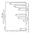
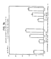
- Figures 3(a) and 3(b) are graphs showing the specific chondrogenic activity as determined per ¶F of certain proteins described in the example;
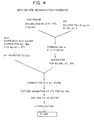
- Figure 4 is a schematic diagram outlining the reconstitution procedure described in ¶G of the example, and
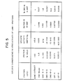
- Figure 5 is a table that provides details of the reconstituted samples described in ¶G and their specific chondrogenic activity as determined by the assay of ¶F.
- The native sources of the bone-inducing factor of the claimed invention are bone, dentin, osterosarcomas or chondrosarcomas. In view of the showing that bone inductive proteins from human, monkey, bovine and rat are nonspecies specific in their ability to produce endochondral bone in xenogeneic implants (Sampath, T. K., et al, Proc Natl Acad Sci (USA) (1983) 80:6591) it is believed that the factor of the claimed invention has been highly conserved among mammalian species (i.e., factors from different mammalian species will have substantially homologous amino acid sequences that vary, if at all, in one or more amino acid residue additions, deletions, or substitutions that do not affect the nonspecies specific bone inducing activity of the molecule adversely). In this regard the terms "substantially equivalent" and "substantially homologous" as used herein are intended to mean factors regardless of species, that have the same amino acid composition or sequence, as the case may be, of the factor described in the example and factors of similar but different amino acid composition or sequence, which difference(s) does not affect nonspecies specific endochondral bone-inducing activity adversely. Accordingly, such factors may be derived from cells or tissue of diverse mammalian origin. The source of factor prepared by purification from native sources is advantageously porcine or bovine long bone because of its ready availability.
- A variety of initial preparation procedures are possible, but basically the bone is first cleaned using mechanical or abrasive techniques, fragmented, and further washed with, for example, dilute aqueous acid preferably at low temperature, and then defatted by extraction with a lipophilic solvent such as ether or ethyl acetate. The bone is then demineralized by removal of the calcium phosphates in their various forms, usually by extraction with stronger acid. These techniques are understood in the art, and are disclosed, for example, in U.S. 4,434,094. The resulting preparation, a demineralized bone, is the starting material for the preparation of the claimed osteogenic factor.
- The initial extraction is designed to remove the nonfibrous (e.g., noncollagenous) proteins from the demineralized bone. This can be done with the use of chaotropic agents such as guanidine hydrochloride (at least about 4 molar), urea (8 molar) plus salt, or sodium dodecylsulfate (at least about 1% by volume) or such other chaotropic agents as are known in the art (Termine, et al, J Biol Chem (1980) 255:9760-9772; and Sajera and Hascall, J Biol Chem (1969) 244:77-87 and 2384-2396). The extraction is preferably carried out at reduced temperatures in the presence of a protease inhibitor to reduce the likelihood of digestion or denaturation of the extracted protein. Examples of protease inhibitors that may be included are phenylmethylsulfonylfluoride (PMSF) sodium azide, N-ethyl maleimide (NEM), benzamidine, and 6-amino hexanoic acid. The pH of the medium depends upon the extractant selected. The process of extraction generally takes on the order of about 4 hr to 1 day.
- After extraction, the extractant may be removed by suitable means such as dialysis against water, preceded by concentration by ultrafiltration if desired. Salts can also be removed by controlled electrophoresis, or by molecular sieving, or by any other means known in the art. It is also preferred to maintain a low temperature during this process so as to minimize denaturation of the proteins. Alternatively, the extractant chaotropic agent need not be removed, but rather the solution need only be concentrated, for example, by ultrafiltration.
- The extract, dissolved or redissolved in chaotropic agent, is subjected to gel filtration to obtain fractions of molecular weight below about 30,000 daltons, thus resulting in a major enhancement of purity. Gel sizing is done using standard techniques, preferably on a Sephacryl column at room (10°C-25°C) temperature.
- The low molecular weight fraction is then subjected to ion exchange chromatography using CMC at approximately pH 4.5-5.2, preferably about 4.8, in the presence of a nonionic chaotropic agent such as 6 M urea. Other cation exchangers may be used, including those derived from polyacrylamide and cross-linked dextran; however cellulosic cation exchangers are preferred. Of course, as in any ion exchange procedure, the solution must be freed of competing ions before application to the column. The factor is adsorbed on the column and is eluted in an increasing salt concentration gradient in the range of about 10 mM to about 150 mM.
- The 10 mM-150 mM NaCl fraction from the cation exchange column may be subjected to RP-HPLC or nondenaturing gel electrophoresis for further purification.
- The presence of the factor in the 10 mM-150 mM NaCl fraction is confirmed using an in vivo bone-induction assay described in detail below.
- The following example is intended to illustrate the process for purification as applied to a particular sample. It is not intended to limit the invention.
- Fresh bovine metatarsal bone was obtained fresh from the slaughterhouse and transported on dry ice. The bones were cleaned of marrow and non-bone tissues, broken in fragments smaller than 1 cm diameter, and pulverized in a mill at 4°C. The pulverized bone was washed twice with 9.4 liters of double distilled water per kg of bone for about 15 min each, and then washed overnight in 0.01 N HCl at 4°C. Washed bone was defatted using 3
X 3 volumes ethanol, followed by 3 X 3 volumes diethylether, each washed for 20 min, and all at room temperature. The resulting defatted bone powder was then demineralized in 0.5 N HCl (25 l/kg defatted bone) at 4°C. The acid was decanted and the resulting DMB washed until the wash pH was greater than 4, and the DMB dried on a suction filter. - The DMB as prepared in ¶A was extracted with 3.3 l of 4 M guanidine-HCl, 10 mM ethylenediaminetetraacetic acid (EDTA), pH 6.8, 1 mM PMSF, 10 mM NEM per kg for 16 hr, the suspension suction filtered and the nonsoluble material extracted again for 4 hr. The soluble fractions were combined and concentrated at least 5-fold by ultrafiltration using an Amicon ultrafiltration (10K) unit, and the concentrate dialyzed against 6 changes of 35 volumes cold deionized water over a period of 4 days, and then lyophilized. All of the procedures of this paragraph were conducted at 4°C except the lyophilization which was conducted under standard lyophilization conditions.
- The extract from ¶B, redissolved in 4 M guanidine-HCl, was fractionated on a Sephacryl S-200 column equilibrated in 4 M guanidine-HCl, 0.02% sodium azide, 10 mM EDTA, pH 6.8. Fractions were assayed by their absorbance at 280 nm and the fractions were combined as shown in Figure 1. Fraction F2 of Figure 1, constituting a low molecular weight (LMW, 10,000-30,000 daltons) protein fraction possessing the greatest activity was dialyzed against 6 changes of 180 volumes of deionized water and lyophilized. All operations except lyophilization and dialysis (4°C) were conducted at room temperature.
- Fraction F2 from ¶C was dissolved in 6 M urea, 10 mM NaCl, 1 mM NEM, 50 mM sodium acetate, pH 4.8 and centrifuged at 10,000 rpm for 5 min. The supernatant was fractionated on a CM52 (a commercially available CMC) column equilibrated in the same buffer. Bound proteins were eluted from the column using a 10 mM to 400 mM NaCl gradient in the same buffer, and a total volume of 350 ml at a flow rate of 27 ml/hr. Three major fractions, designated CM-1, CM-2 and CM-3, were collected as shown in Figure 2. Each fraction was dialyzed against 6 changes of 110 volumes of deionized water for 4 days and lyophilized. All of the foregoing operations were conducted at room temperature except dialysis (4°C).
- The lyophilized fractions CM-2 and CM-3 from ¶D were each dissolved in 0.1% trifluoroacetic acid (TFA) and aliquots of the solution loaded onto a Vydac C18 RP-HPLC column (4.6
mm ID X 25 cm) and washed with 0.1% TFA for 5 min at 1 ml/min. The eluting solvent was a 0%-60% acetonitrile gradient in 0.1% TFA at a rate of 2%/min. Two peaks were obtained--peak A at about 29.5 min and peak B at about 31.3 min. - The presence of the desired proteins in the fractions during purification was confirmed using an in vitro assay for the production of proteoglycans (PG), the identity of which was confirmed by enzyme-linked immunosorbent assay (ELISA). The assay is an agarose gel culture model using leg muscle cells isolated from rat fetuses. It assesses the ability of the samples to induce the production of cartilage specific PGs. The correlation between in vitro cartilage induction and in vivo bone formation has been shown by Seyedin, S., et al, J Cell Biol (1983) 97:1950-1953.
- The cell culture was prepared by removing muscle tissue aseptically from the upper limbs of nineteen-day-old Sprague Dawley rat fetuses, mincing the tissue, and culturing it in Eagle's Minimum Essential Medium (MEM) with 10% fetal bovine serum (FBS) and 50 units penicillin, 50 g streptomycin per ml. Cellular outgrowth usually reached confluency within one week, whereupon cells were trypsinized, split 1:2 and used for experimentation within the first three passages.
- The cells were placed in agarose gel cultures either with control medium or with samples to be tested. The procedure was basically that of Benya, et al, Cell (1982) 30:215. Briefly, the cell monolayers were harvested by trypsinization, counted on a hemocytometer, and resuspended at two times the final cell concentration in the medium with or without the protein fraction to be tested. The control medium was either Hams F12, Dulbecco's Minimum Essential Medium (DMEM) or CMRL 1066 (Gibco) each containing 10% FBS and antibiotics. The test protein fractions in 0.01 N HCl were diluted directly to the desired concentration of test protein diluted with an equal volume with 1% low melting agarose (Bio-Rad, #162-0017) in F-12, and 0.2 ml of the dilution was plated on 17 mm wells coated with 0.15 ml of 1% high melting (Bio-Rad, #162-0100) agarose. The resulting cultures were incubated at 37°C for 5 min, chilled at 4°C for 10 min, and then overlayed with 1 ml of the corresponding medium (control or test protein). The cells were then cultured in a humidified atmosphere of 5% CO₂, 95% air and fed every 3-4 days thereafter by a complete change with control medium. After 7 days the cultures were frozen and stored at -80°C before assay.
- The cultures were assayed by thawing at 4°C, homogenizing in 4 M guanidine-HCl with 50 mM Na acetate, 13 mM EDTA, 6 mM NEM, and 3 mM PMSF at pH 5.8, and extracting by shaking overnight at 4°C. The supernatant fraction from centrifugation at 25,000 X g for 40 min at 4°C was dialyzed overnight at 4°C against 50 volumes 0.2 M NaCl, 50 mM Tris, pH 7.4. The supernatant was assayed for proteoglycans by ELISA as described by Renard, et al, Anal Biochem (1980) 104:205, and in U.S. 4,434,094.
- Briefly, for the ELISA, antiserum to cartilage PGs was raised in rabbits using standard techniques which showed no cross-reactivity with hyaluronic acid or PGs extracted from rat bone. Purified proteoglycan (Seyedin. S., et al, supra) from Swarm rat chondrosarcoma tissue was used as standard antigen. The dialyzed samples were diluted 1:1 (v/v) in phosphate-buffered saline (PBS) with 0.05
% Tween - The results of the ELISA of the three CM-bound fractions from ¶D (designated CMC-B-1, CMC-B-2, and CMC-B-3) and the protein of peak A (designated CIF-A) from ¶E are shown in Figures 3(a) and 3(b).
- For reconstitution, the proteins prepared in the above described manner were combined with a 9:1 weight ratio mixture of bone collagen powder (BCP, lyophilized solids from ¶C) and collagen in solution (CIS, available commercially from Collagen Corporation, Palo Alto, California under the trademark ZYGENâ) containing 10% native soluble bovine skin collagen by weight in ratios according to their in vitro chondrogenic activities. The procedure is depicted schematically in Figure 4 and details of the compositions of the reconstituted (R) materials are reported in the Table shown in Figure 5. According to this method all the reconstituted samples contained approximately an equal number of units of chondrogenic activity (about 1000 units/100 mg R-DBP). Only for CIF-A the dosage was doubled and CMC-B-1 was reconstituted in two different dosages. The protein content of CMC-B-1 was determined by Biuret assay and the one of all the other samples was measured by integration of the HPLC peaks compared to the peak area of a known bovine serum albumin standard at 220 nm. In case of pure CIF-A and CIF-B the HPLC fractions were directly added to the CIS solution before mixing with BCP. Also, a control sample consisting of the BCP/CIS carrier was prepared under the same conditions.
- The osteoinductive abilities of samples were assayed by their ability to induce endochondral bone formation in the subcutaneous tissue of young male Sprague-Dawley rats. The samples were wetted with two volumes of sterile double distilled water (v/w), thoroughly mixed, packed in a 1 cc syringe, cut and weighed. All the samples were implanted on the ventral thoracic region, one on each side of the animal. Explants were removed after 14 and 28 days and evaluated biochemically and histologically.
- Explants which had been removed after 14 and 28 days were subjected to histological assessment by fixing in 10% neutral formalin for 26 hr, and then processing for paraffin embedding. Four-six micron sections were taken from the imbedded tissues and were subsequently stained with either hematoxylin-eosin (general cytology), with safronin-O (proteoglycans) and Gomori trichrome (collagen).
- The 14 day explants were split in half, the wet weight determined and frozen at -80°C till processed. The samples were first extracted and assayed for alkaline phosphatase activity and subsequently extracted and assayed for cartilage-specific proteoglycans. The right side 28 day explants were extracted and assayed first for alkaline phosphatase and then for calcium. The extraction and assay procedures are described below.
- Cartilage proteoglycan was assayed by an ELISA technique. The explants were weighed immediately after removal and frozen at -70°C until extraction. For the extraction, the explants were cut into slices, and homogenized in ice cold extraction buffer in a Tekmar Tissuemizer for two 30 sec bursts at maximum setting. The extraction buffer was 6 M guanidine hydrochloride, 75 mM sodium acetate or 4 M guanidine hydrochloride, 50 mM acetate both containing 20 mM EDTA, 1 mM PMSF and 10 mM NEM at pH 5.8. Buffer was used in a 10:1 volume to the weight of the explant extracted, and the samples were incubated overnight (20 hr) at 4°C. The samples were then centrifuged at 12,000 rpm for 1 hr at 4°C, and the supernatants dialyzed overnight at 4°C against 50 volumes of 50 mM Tris, 200 mM NaCl, pH 7.4. The dialyzate was subjected to ELISA performed as described by Renard, et al, Arch Biochem Biophys (1980) 207:399 and by Seyedin, S., et al, J Cell Biol (1983) 97:1950 using polystyrene microplates (Flow Laboratories, McClean, Virginia). The antisera and the proteoglycan standard were prepared from Swarm rat chondrosarcoma tissue as described by Seyedin, S., et al, (supra). Horseradish peroxidase conjugated goat anti-rabbit IgG was used as the second antibody, samples were assayed in different solutions in PBS, 0.05
% Tween - The formation of bone was also assessed by determination of calcium. The explants were cut in small pieces and suspended in 1:10 (m/v) of 0.5 N HCl to dissolve the ions. The samples were incubated for another 5 days at room temperature and centrifuged at 12,000 rpm for 40 min. The calcium concentration of the supernatant was determined by atomic adsorption (Trace Analysis Laboratory, Hayward, California).
- To determine alkaline phosphatase (AP), the explants were cut in small pieces and homogenized in 10 volumes (1/10) of ice cold 1.5 M NaCl, 3 mM NaHCO₃, pH 7.5. The homogenized samples were then centrifuged at 12,000 rpm for 50 min at 4°C, and an aliquot of the supernatant diluted 1:10 in cold distilled water. The method of Huggins, et al, J Exp Med (1961) 114:761 was used to assess alkaline phosphatase using polystyrene plates.
- Partial results are summarized in the table below.

- The above data show that CMC-B-1 proteins enhance osteoinduction relative to CMC-bound (the total bound fraction) as reflected by a higher rate, quantity, and quality of bone formation. These studies indicate that endochondral bone formation is affected by the purity of the bone-inducing material and the distribution of proteins therein. These studies further suggest that the two proteins identified in European Patent Application 85.304848.6 may play roles in the differentiation of cells involved in bone formation and affect rate and relative amounts of cartilage and bone formation.
- As indicated, histological and biochemical data of the 14 and 28 days explanted materials demonstrated that reconstituted total CMC-bound (total proteins bound by CMC) and CMC-B-1 induced cartilage and bone formation in all implants. At 14 days cartilage formation was very high with the R-CMC-bound implants and uniformly distributed over the whole implant. Some new bone was formed peripherally. In contrast, R-CMC-B-1 showed only little cartilage and already lots of bone at 14 days. At 28 days R-CMC-bound explants still contained cartilage. (The amount of cartilage and bone appeared to be about the same.) R-CMC-B-1 meanwhile, contained only traces of cartilage and well-developed bone with fatty marrow cavities. All of the latter explants appeared to be larger than usually found. Histological observations were confirmed by biochemical data. Both materials showed high levels of alkaline phosphatase activity. The calcium content at 28 days was very high in both materials (approximately 32 mg Ca/g wet tissue for R-CMC-bound and approximately 39 mg Ca/g wet tissue for R-CMC-B-1).
- The claimed osteogenic material may be used as the active ingredient of osteogenic implant compositions for repairing, replacing, or augmenting bone tissue in living mammals, including man. Osteogenically effective amounts of the material will normally be formulated with pharmacologically and physiologically acceptable solid carriers such as BCP for implantation. The weight ratio of active protein to carrier will typically be in the range of 1:50 to 1:1000. The implants may be placed at a predetermined site in the patient by conventional surgical techniques.
- The claimed factor may also be useful for treating bone deficiencies, such as osteoporosis and osteopetrosis, systemically. For such treatment the proteins will be formulated in therapeutically effective amounts with injectable carriers and administered parenterally to the patient.
Claims (4)
Priority Applications (2)
| Application Number | Priority Date | Filing Date | Title |
|---|---|---|---|
| DE8686303011T DE3682899D1 (en) | 1984-07-16 | 1986-04-22 | PARTLY PURIFIED BONE INDUCING FACTOR FOR USE IN ORTEOGENESIS. |
| AT86303011T ATE70277T1 (en) | 1984-07-16 | 1986-04-22 | PARTIALLY PURIFIED BONE-INDUCING FACTOR FOR APPLICATION IN ORTEOGENESIS. |
Applications Claiming Priority (2)
| Application Number | Priority Date | Filing Date | Title |
|---|---|---|---|
| US06/630,928 US4586315A (en) | 1983-07-14 | 1984-07-16 | Apparatus for introducing stacks of paper layers into cartons |
| US06/705,479 US4627982A (en) | 1984-07-16 | 1985-02-26 | Partially purified bone-inducing factor |
Publications (2)
| Publication Number | Publication Date |
|---|---|
| EP0242466A1 true EP0242466A1 (en) | 1987-10-28 |
| EP0242466B1 EP0242466B1 (en) | 1991-12-11 |
Family
ID=27091267
Family Applications (1)
| Application Number | Title | Priority Date | Filing Date |
|---|---|---|---|
| EP86303011A Expired EP0242466B1 (en) | 1984-07-16 | 1986-04-22 | Osteogenic use of partially purified bone-inducing factor |
Country Status (4)
| Country | Link |
|---|---|
| US (1) | US4627982A (en) |
| EP (1) | EP0242466B1 (en) |
| AT (1) | ATE70277T1 (en) |
| DE (1) | DE3682899D1 (en) |
Cited By (4)
| Publication number | Priority date | Publication date | Assignee | Title |
|---|---|---|---|---|
| EP0336760A2 (en) * | 1988-04-06 | 1989-10-11 | Celtrix Pharmaceuticals, Inc. | Bone-inducing protein |
| EP0349048A2 (en) * | 1988-06-27 | 1990-01-03 | Yissum Research Development Company Of The Hebrew University Of Jerusalem | Osteogenic growth factors derived from regenerating bone marrow |
| EP0321277A3 (en) * | 1987-12-16 | 1990-04-25 | Collagen Corporation | An injectable composition for inductive bone repair |
| US9757330B2 (en) | 2013-10-18 | 2017-09-12 | Industrial Technology Research Institute | Recipe for in-situ gel, and implant, drug delivery system formed thereby |
Families Citing this family (77)
| Publication number | Priority date | Publication date | Assignee | Title |
|---|---|---|---|---|
| US4804744A (en) * | 1984-01-04 | 1989-02-14 | International Genetic Engineering, Inc. | Osteogenic factors |
| USRE35694E (en) * | 1984-07-16 | 1997-12-16 | Celtrix Pharmaceuticals, Inc. | Polypeptide cartilage-inducing factors found in bone |
| US4888366A (en) | 1984-10-24 | 1989-12-19 | Collagen Corporation | Inductive collagen-based bone repair preparations |
| US5284763A (en) * | 1985-03-22 | 1994-02-08 | Genentech, Inc. | Nucleic acid encoding TGF-β and its uses |
| US4886747A (en) * | 1985-03-22 | 1989-12-12 | Genentech, Inc. | Nucleic acid encoding TGF-β and its uses |
| US4992226A (en) * | 1985-03-28 | 1991-02-12 | Collagen Corporation | Method of making molds with xenogeneic collagen/mineral preparations for bone repair |
| US5246457A (en) * | 1985-03-28 | 1993-09-21 | Collagen Corporation | Xenogeneic collagen/mineral preparations in bone repair |
| EP0205337A3 (en) * | 1985-06-11 | 1988-10-12 | COLLAGEN CORPORATION (a Delaware corporation) | Chondrocyte antigens and monoclonal antibodies |
| US5529982A (en) * | 1985-08-06 | 1996-06-25 | Celtrix Pharmaceuticals, Inc. | Inducing granulocyte production or B cell production in peripheral blood by TGF-β |
| US5904718A (en) * | 1986-03-27 | 1999-05-18 | Biocoll Laboratories, Inc. | Delayed drug delivery system |
| US5366875A (en) * | 1986-07-01 | 1994-11-22 | Genetics Institute, Inc. | Methods for producing BMP-7 proteins |
| US5106748A (en) * | 1986-07-01 | 1992-04-21 | Genetics Institute, Inc. | Dna sequences encoding 5 proteins |
| US4877864A (en) * | 1987-03-26 | 1989-10-31 | Genetics Institute, Inc. | Osteoinductive factors |
| US5543394A (en) * | 1986-07-01 | 1996-08-06 | Genetics Institute, Inc. | Bone morphogenetic protein 5(BMP-5) compositions |
| US5939388A (en) * | 1986-07-01 | 1999-08-17 | Rosen; Vicki A. | Methods of administering BMP-5 compositions |
| US5013649A (en) * | 1986-07-01 | 1991-05-07 | Genetics Institute, Inc. | DNA sequences encoding osteoinductive products |
| US5459047A (en) | 1986-07-01 | 1995-10-17 | Genetics Institute, Inc. | BMP-6 proteins |
| US6150328A (en) | 1986-07-01 | 2000-11-21 | Genetics Institute, Inc. | BMP products |
| US5187076A (en) * | 1986-07-01 | 1993-02-16 | Genetics Institute, Inc. | DNA sequences encoding BMP-6 proteins |
| US6432919B1 (en) | 1986-07-01 | 2002-08-13 | Genetics Institute, Inc. | Bone morphogenetic protein-3 and compositions |
| DE3775363D1 (en) * | 1986-10-22 | 1992-01-30 | Biotechnolog Forschung Gmbh | GROWTH-STIMULATING MATERIAL, MANUFACTURING PROCESS AND THERAPEUTIC COMPOSITION. |
| US4743259A (en) * | 1986-10-29 | 1988-05-10 | The University Of Virginia Alumni Patents Foundation | Use of demineralized bone matrix in the repair of segmental defects |
| US4931548A (en) * | 1987-01-30 | 1990-06-05 | Techne Corporation | Heterodimer form of transforming growth factor-beta |
| US5221734A (en) * | 1987-10-01 | 1993-06-22 | Ciba-Geigy Corporation | Process for preparing a polypeptide growth factor for milk |
| US4935497A (en) * | 1988-09-06 | 1990-06-19 | Northwestern University | Dentin chondrogenic inductive agent |
| US5284756A (en) * | 1988-10-11 | 1994-02-08 | Lynn Grinna | Heterodimeric osteogenic factor |
| US5106626A (en) * | 1988-10-11 | 1992-04-21 | International Genetic Engineering, Inc. | Osteogenic factors |
| EP0366029B1 (en) * | 1988-10-25 | 1994-09-07 | Takao Yamamuro | Bone repairing material and artificial bone fixing agent |
| PT94241A (en) * | 1989-06-02 | 1991-02-08 | Chiron Corp | METHOD FOR THE PREPARATION OF OESEA CALCIFICATION FACT AND OF PHARMACEUTICAL COMPOSITIONS CONTAINING THEM |
| CA2020729A1 (en) * | 1989-07-19 | 1991-01-20 | Michael C. Kiefer | Bone morphogenetic protein |
| JPH05500503A (en) * | 1989-08-21 | 1993-02-04 | セルトリックス ファーマシューティカルズ、インコーポレイテッド | bone specific protein |
| US7378392B1 (en) | 1990-05-16 | 2008-05-27 | Genetics Institute, Llc | Bone and cartilage inductive proteins |
| US5688678A (en) * | 1990-05-16 | 1997-11-18 | Genetics Institute, Inc. | DNA encoding and methods for producing BMP-8 proteins |
| US5206023A (en) * | 1991-01-31 | 1993-04-27 | Robert F. Shaw | Method and compositions for the treatment and repair of defects or lesions in cartilage |
| US5169837A (en) * | 1991-03-28 | 1992-12-08 | Allelix Biopharmaceuticals Inc. | Isolated osteogenic factor |
| US5563124A (en) * | 1991-04-22 | 1996-10-08 | Intermedics Orthopedics/ Denver, Inc. | Osteogenic product and process |
| US5290763A (en) * | 1991-04-22 | 1994-03-01 | Intermedics Orthopedics/Denver, Inc. | Osteoinductive protein mixtures and purification processes |
| ZA923086B (en) * | 1991-04-29 | 1993-10-28 | South African Medical Research | A delivery system for biologicaly active growth or morphogenetic factors and a method for preparing such delivery system |
| US5693615A (en) * | 1991-06-05 | 1997-12-02 | The Procter & Gamble Company | Therapeutic compositions for osteoinduction |
| US6287816B1 (en) * | 1991-06-25 | 2001-09-11 | Genetics Institute, Inc. | BMP-9 compositions |
| KR100255415B1 (en) * | 1991-06-25 | 2000-05-01 | 브루스 엠. 에이센 | Bone morphogenetic protein-9 compositions |
| US5270300A (en) * | 1991-09-06 | 1993-12-14 | Robert Francis Shaw | Methods and compositions for the treatment and repair of defects or lesions in cartilage or bone |
| ATE238417T1 (en) * | 1991-11-04 | 2003-05-15 | Inst Genetics Llc | RECOMBINANT BONE MORPHOGENETIC PROTEIN HETERODIMERS, COMPOSITIONS AND METHODS OF USE |
| US5322933A (en) * | 1992-05-07 | 1994-06-21 | The United States Of America As Represented By The Secretary Of The Department Of Health And Human Services | Crystal structure of TGF-β-2 |
| US5928635A (en) * | 1994-12-07 | 1999-07-27 | Schmidt; Karlheinz | Process for producing active agent complexes |
| US5637480A (en) * | 1993-05-12 | 1997-06-10 | Genetics Institute, Inc. | DNA molecules encoding bone morphogenetic protein-10 |
| US5531791A (en) * | 1993-07-23 | 1996-07-02 | Bioscience Consultants | Composition for repair of defects in osseous tissues, method of making, and prosthesis |
| US6291206B1 (en) | 1993-09-17 | 2001-09-18 | Genetics Institute, Inc. | BMP receptor proteins |
| US6027919A (en) * | 1993-12-07 | 2000-02-22 | Genetics Institute, Inc. | BMP-12 and BMP-13 proteins and DNA encoding them |
| CA2176942C (en) | 1993-12-07 | 2011-11-01 | Anthony J. Celeste | Bmp-12, bmp-13 and tendon-inducing compositions thereof |
| US5707962A (en) * | 1994-09-28 | 1998-01-13 | Gensci Regeneration Sciences Inc. | Compositions with enhanced osteogenic potential, method for making the same and therapeutic uses thereof |
| US6180606B1 (en) | 1994-09-28 | 2001-01-30 | Gensci Orthobiologics, Inc. | Compositions with enhanced osteogenic potential, methods for making the same and uses thereof |
| JP3881707B2 (en) * | 1995-07-20 | 2007-02-14 | 学校法人松本歯科大学 | Method for producing osteogenesis promoter and method for producing osteogenic composition using osteogenesis promoter |
| US6902584B2 (en) | 1995-10-16 | 2005-06-07 | Depuy Spine, Inc. | Bone grafting matrix |
| US5776193A (en) * | 1995-10-16 | 1998-07-07 | Orquest, Inc. | Bone grafting matrix |
| ES2225870T3 (en) * | 1996-02-29 | 2005-03-16 | Bioactive Bone Substitute Oy, Ab | OSTEOGENIC DEVICE AND ITS PREPARATION PROCEDURE. |
| US6492508B1 (en) * | 1996-06-03 | 2002-12-10 | United States Surgical Corp. A Division Of Tyco Healthcare Group | Nucleic acids encoding extracellular matrix proteins |
| US6927287B1 (en) * | 1996-06-03 | 2005-08-09 | United States Surgical Corporation | Nucleic acid encoding extracellular matrix protein or fragment thereof |
| US6034062A (en) * | 1997-03-13 | 2000-03-07 | Genetics Institute, Inc. | Bone morphogenetic protein (BMP)-9 compositions and their uses |
| EP1007673B1 (en) | 1997-07-30 | 2008-12-17 | Emory University | Novel bone mineralization proteins, dna, vectors, expression systems |
| US7923250B2 (en) | 1997-07-30 | 2011-04-12 | Warsaw Orthopedic, Inc. | Methods of expressing LIM mineralization protein in non-osseous cells |
| DE69714035T2 (en) | 1997-08-14 | 2003-03-06 | Sulzer Innotec Ag | Composition and device for repairing cartilage tissue in vivo consisting of nanocapsules with osteoinductive and / or chondroinductive factors |
| US6727224B1 (en) | 1999-02-01 | 2004-04-27 | Genetics Institute, Llc. | Methods and compositions for healing and repair of articular cartilage |
| DE19906096A1 (en) | 1999-02-13 | 2000-08-17 | Walter Sebald | Protein with a heparin-binding epitope |
| WO2001028602A1 (en) | 1999-10-15 | 2001-04-26 | Genetics Institute, Inc. | Formulations of hyaluronic acid for delivery of osteogenic proteins |
| WO2001057082A2 (en) * | 2000-02-03 | 2001-08-09 | Regeneration Technologies, Inc. | Extraction of growth factors from tissue |
| US6492327B2 (en) | 2000-12-19 | 2002-12-10 | Sulzer Biologics Inc. | Isolation of purified TGF- β1 and TGF -β2 from bone tissue |
| CA2449008A1 (en) | 2001-06-01 | 2002-12-12 | Wyeth | Compositions and methods for systemic administration of sequences encoding bone morphogenetic proteins |
| TWI267378B (en) | 2001-06-08 | 2006-12-01 | Wyeth Corp | Calcium phosphate delivery vehicles for osteoinductive proteins |
| US7132110B2 (en) * | 2001-08-30 | 2006-11-07 | Isotis Orthobiologics, Inc. | Tissue repair compositions and methods for their manufacture and use |
| AU2003256300A1 (en) | 2002-06-24 | 2004-01-06 | New York Society For The Ruptured And Crippled Maintaining The Hospital For Special Surgery | Complexed-acidic-phospholipid-collagen composites for bone induction |
| US7241874B2 (en) * | 2002-06-26 | 2007-07-10 | Zimmer Ortho Biologics, Inc. | Rapid isolation of osteoinductive protein mixtures from mammalian bone tissue |
| US7622562B2 (en) | 2002-06-26 | 2009-11-24 | Zimmer Orthobiologics, Inc. | Rapid isolation of osteoinductive protein mixtures from mammalian bone tissue |
| EP1675608B1 (en) | 2003-09-12 | 2007-03-21 | Wyeth | Injectable calcium phosphate solid rods for delivery of osteogenic proteins |
| US9011930B2 (en) * | 2006-05-01 | 2015-04-21 | Zycal Bioceuticals Healthcare Company, Inc. | Nutritional supplement and use thereof |
| US20090136929A1 (en) * | 2007-11-28 | 2009-05-28 | Zimmer Orthobiologics, Inc. | Novel in vitro method of quantifying demineralized bone osteoinductivity |
| US9700584B2 (en) * | 2007-12-21 | 2017-07-11 | Rti Surgical, Inc. | Osteoinductive putties and methods of making and using such putties |
Citations (6)
| Publication number | Priority date | Publication date | Assignee | Title |
|---|---|---|---|---|
| US4455256A (en) * | 1981-05-05 | 1984-06-19 | The Regents Of The University Of California | Bone morphogenetic protein |
| EP0121976A2 (en) * | 1983-04-12 | 1984-10-17 | Collagen Corporation | Partially purified osteogenic factor and process for preparing same from demineralized bone or an osteosarcoma |
| EP0128041A2 (en) * | 1983-06-06 | 1984-12-12 | David Jeston Baylink | Polypeptides exhibiting skeletal growth factor activity |
| EP0148155A2 (en) * | 1984-01-04 | 1985-07-10 | International Genetic Engineering, Inc. (Ingene) | Osteogenic factors |
| WO1985004173A1 (en) * | 1984-03-20 | 1985-09-26 | Caplan Arnold I | Bone protein purification process |
| EP0169016A2 (en) * | 1984-07-16 | 1986-01-22 | Celtrix Pharmaceuticals, Inc. | Polypeptide cartilage-inducing factors found in bone |
Family Cites Families (7)
| Publication number | Priority date | Publication date | Assignee | Title |
|---|---|---|---|---|
| US3458397A (en) * | 1966-12-08 | 1969-07-29 | Squibb & Sons Inc | Process for producing osteogenic material |
| US4488911A (en) * | 1975-10-22 | 1984-12-18 | Luck Edward E | Non-antigenic collagen and articles of manufacture |
| US4294753A (en) * | 1980-08-04 | 1981-10-13 | The Regents Of The University Of California | Bone morphogenetic protein process |
| US4394370A (en) * | 1981-09-21 | 1983-07-19 | Jefferies Steven R | Bone graft material for osseous defects and method of making same |
| US4440750A (en) * | 1982-02-12 | 1984-04-03 | Collagen Corporation | Osteogenic composition and method |
| US4485097A (en) * | 1982-05-26 | 1984-11-27 | Massachusetts Institute Of Technology | Bone-equivalent and method for preparation thereof |
| US4563350A (en) * | 1984-10-24 | 1986-01-07 | Collagen Corporation | Inductive collagen based bone repair preparations |
-
1985
- 1985-02-26 US US06/705,479 patent/US4627982A/en not_active Expired - Lifetime
-
1986
- 1986-04-22 EP EP86303011A patent/EP0242466B1/en not_active Expired
- 1986-04-22 AT AT86303011T patent/ATE70277T1/en active
- 1986-04-22 DE DE8686303011T patent/DE3682899D1/en not_active Expired - Lifetime
Patent Citations (6)
| Publication number | Priority date | Publication date | Assignee | Title |
|---|---|---|---|---|
| US4455256A (en) * | 1981-05-05 | 1984-06-19 | The Regents Of The University Of California | Bone morphogenetic protein |
| EP0121976A2 (en) * | 1983-04-12 | 1984-10-17 | Collagen Corporation | Partially purified osteogenic factor and process for preparing same from demineralized bone or an osteosarcoma |
| EP0128041A2 (en) * | 1983-06-06 | 1984-12-12 | David Jeston Baylink | Polypeptides exhibiting skeletal growth factor activity |
| EP0148155A2 (en) * | 1984-01-04 | 1985-07-10 | International Genetic Engineering, Inc. (Ingene) | Osteogenic factors |
| WO1985004173A1 (en) * | 1984-03-20 | 1985-09-26 | Caplan Arnold I | Bone protein purification process |
| EP0169016A2 (en) * | 1984-07-16 | 1986-01-22 | Celtrix Pharmaceuticals, Inc. | Polypeptide cartilage-inducing factors found in bone |
Cited By (6)
| Publication number | Priority date | Publication date | Assignee | Title |
|---|---|---|---|---|
| EP0321277A3 (en) * | 1987-12-16 | 1990-04-25 | Collagen Corporation | An injectable composition for inductive bone repair |
| EP0336760A2 (en) * | 1988-04-06 | 1989-10-11 | Celtrix Pharmaceuticals, Inc. | Bone-inducing protein |
| EP0336760A3 (en) * | 1988-04-06 | 1991-03-13 | Celtrix Pharmaceuticals, Inc. | Bone-inducing protein |
| EP0349048A2 (en) * | 1988-06-27 | 1990-01-03 | Yissum Research Development Company Of The Hebrew University Of Jerusalem | Osteogenic growth factors derived from regenerating bone marrow |
| EP0349048A3 (en) * | 1988-06-27 | 1990-07-11 | Yissum Research Development Company Of The Hebrew University Of Jerusalem | Osteogenic growth factors derived from regenerating bone marrow |
| US9757330B2 (en) | 2013-10-18 | 2017-09-12 | Industrial Technology Research Institute | Recipe for in-situ gel, and implant, drug delivery system formed thereby |
Also Published As
| Publication number | Publication date |
|---|---|
| US4627982A (en) | 1986-12-09 |
| EP0242466B1 (en) | 1991-12-11 |
| DE3682899D1 (en) | 1992-01-23 |
| ATE70277T1 (en) | 1991-12-15 |
Similar Documents
| Publication | Publication Date | Title |
|---|---|---|
| EP0242466B1 (en) | Osteogenic use of partially purified bone-inducing factor | |
| AU594949B2 (en) | Partially purified bone-inducing factor | |
| US4843063A (en) | Polypeptide cartilage-inducing factors found in bone | |
| US4563350A (en) | Inductive collagen based bone repair preparations | |
| US5001169A (en) | Inductive collagen-based bone repair preparations | |
| Sampath et al. | Isolation of osteogenin, an extracellular matrix-associated, bone-inductive protein, by heparin affinity chromatography. | |
| US5290763A (en) | Osteoinductive protein mixtures and purification processes | |
| Sampath et al. | Homology of bone-inductive proteins from human, monkey, bovine, and rat extracellular matrix. | |
| US4806523A (en) | Method of treating inflammation | |
| JP2522568B2 (en) | Bone formation device | |
| US7078221B2 (en) | Nucleic acid molecules encoding osteogenic proteins | |
| US5496552A (en) | Osteogenic devices | |
| US7186811B2 (en) | Osteogenic device and a method for preparing the device | |
| Sampath et al. | Distribution of bone inductive proteins in mineralized and demineralized extracellular matrix | |
| EP1539812B1 (en) | Osteoinductive biomaterials | |
| Sampath et al. | Extracellular matrix proteins involved in bone induction are vitamin D dependent | |
| USRE35694E (en) | Polypeptide cartilage-inducing factors found in bone | |
| USRE34090E (en) | Polypeptide cartilage-inducing factors found in bone | |
| CA1266435A (en) | Partially purified bone-inducing factor | |
| JPS62255429A (en) | Partially purified bone induction factor | |
| ZA200500427B (en) | Osteoinductive biomaterials |
Legal Events
| Date | Code | Title | Description |
|---|---|---|---|
| PUAI | Public reference made under article 153(3) epc to a published international application that has entered the european phase |
Free format text: ORIGINAL CODE: 0009012 |
|
| AK | Designated contracting states |
Kind code of ref document: A1 Designated state(s): AT BE CH DE FR GB IT LI LU NL SE |
|
| 17P | Request for examination filed |
Effective date: 19880203 |
|
| RAP1 | Party data changed (applicant data changed or rights of an application transferred) |
Owner name: COLLAGEN CORPORATION (A DELAWARE CORPORATION) |
|
| 17Q | First examination report despatched |
Effective date: 19900208 |
|
| RAP1 | Party data changed (applicant data changed or rights of an application transferred) |
Owner name: CELTRIX LABORATORIES, INC. |
|
| GRAA | (expected) grant |
Free format text: ORIGINAL CODE: 0009210 |
|
| AK | Designated contracting states |
Kind code of ref document: B1 Designated state(s): AT BE CH DE FR GB IT LI LU NL SE |
|
| PG25 | Lapsed in a contracting state [announced via postgrant information from national office to epo] |
Ref country code: IT Free format text: LAPSE BECAUSE OF FAILURE TO SUBMIT A TRANSLATION OF THE DESCRIPTION OR TO PAY THE FEE WITHIN THE PRE;WARNING: LAPSES OF ITALIAN PATENTS WITH EFFECTIVE DATE BEFORE 2007 MAY HAVE OCCURRED AT ANY TIME BEFORE 2007. THE CORRECT EFFECTIVE DATE MAY BE DIFFERENT FROM THE ONE RECORDED.SCRIBED TIME-LIMIT Effective date: 19911211 Ref country code: NL Effective date: 19911211 Ref country code: SE Effective date: 19911211 Ref country code: AT Effective date: 19911211 Ref country code: BE Effective date: 19911211 |
|
| REF | Corresponds to: |
Ref document number: 70277 Country of ref document: AT Date of ref document: 19911215 Kind code of ref document: T |
|
| REF | Corresponds to: |
Ref document number: 3682899 Country of ref document: DE Date of ref document: 19920123 |
|
| PG25 | Lapsed in a contracting state [announced via postgrant information from national office to epo] |
Ref country code: GB Effective date: 19920422 |
|
| EN | Fr: translation not filed | ||
| PG25 | Lapsed in a contracting state [announced via postgrant information from national office to epo] |
Ref country code: CH Effective date: 19920430 Ref country code: FR Effective date: 19920430 Ref country code: LU Free format text: LAPSE BECAUSE OF NON-PAYMENT OF DUE FEES Effective date: 19920430 Ref country code: LI Effective date: 19920430 |
|
| NLV1 | Nl: lapsed or annulled due to failure to fulfill the requirements of art. 29p and 29m of the patents act | ||
| PLBE | No opposition filed within time limit |
Free format text: ORIGINAL CODE: 0009261 |
|
| STAA | Information on the status of an ep patent application or granted ep patent |
Free format text: STATUS: NO OPPOSITION FILED WITHIN TIME LIMIT |
|
| 26N | No opposition filed | ||
| GBPC | Gb: european patent ceased through non-payment of renewal fee | ||
| REG | Reference to a national code |
Ref country code: CH Ref legal event code: PL |
|
| PG25 | Lapsed in a contracting state [announced via postgrant information from national office to epo] |
Ref country code: DE Effective date: 19930101 |
|
| REG | Reference to a national code |
Ref country code: FR Ref legal event code: ST |