EP0217527B1 - Monoclonal antibodies binding determinants of gram negative bacteria - Google Patents
Monoclonal antibodies binding determinants of gram negative bacteria Download PDFInfo
- Publication number
- EP0217527B1 EP0217527B1 EP86306420A EP86306420A EP0217527B1 EP 0217527 B1 EP0217527 B1 EP 0217527B1 EP 86306420 A EP86306420 A EP 86306420A EP 86306420 A EP86306420 A EP 86306420A EP 0217527 B1 EP0217527 B1 EP 0217527B1
- Authority
- EP
- European Patent Office
- Prior art keywords
- xmmen
- gram negative
- antibody
- gram
- mice
- Prior art date
- Legal status (The legal status is an assumption and is not a legal conclusion. Google has not performed a legal analysis and makes no representation as to the accuracy of the status listed.)
- Expired - Lifetime
Links
Classifications
-
- C—CHEMISTRY; METALLURGY
- C12—BIOCHEMISTRY; BEER; SPIRITS; WINE; VINEGAR; MICROBIOLOGY; ENZYMOLOGY; MUTATION OR GENETIC ENGINEERING
- C12N—MICROORGANISMS OR ENZYMES; COMPOSITIONS THEREOF; PROPAGATING, PRESERVING, OR MAINTAINING MICROORGANISMS; MUTATION OR GENETIC ENGINEERING; CULTURE MEDIA
- C12N5/00—Undifferentiated human, animal or plant cells, e.g. cell lines; Tissues; Cultivation or maintenance thereof; Culture media therefor
- C12N5/10—Cells modified by introduction of foreign genetic material
- C12N5/12—Fused cells, e.g. hybridomas
-
- G—PHYSICS
- G01—MEASURING; TESTING
- G01N—INVESTIGATING OR ANALYSING MATERIALS BY DETERMINING THEIR CHEMICAL OR PHYSICAL PROPERTIES
- G01N33/00—Investigating or analysing materials by specific methods not covered by groups G01N1/00 - G01N31/00
- G01N33/48—Biological material, e.g. blood, urine; Haemocytometers
- G01N33/50—Chemical analysis of biological material, e.g. blood, urine; Testing involving biospecific ligand binding methods; Immunological testing
- G01N33/53—Immunoassay; Biospecific binding assay; Materials therefor
- G01N33/569—Immunoassay; Biospecific binding assay; Materials therefor for microorganisms, e.g. protozoa, bacteria, viruses
- G01N33/56911—Bacteria
- G01N33/56916—Enterobacteria, e.g. shigella, salmonella, klebsiella, serratia
-
- C—CHEMISTRY; METALLURGY
- C07—ORGANIC CHEMISTRY
- C07K—PEPTIDES
- C07K16/00—Immunoglobulins [IG], e.g. monoclonal or polyclonal antibodies
- C07K16/12—Immunoglobulins [IG], e.g. monoclonal or polyclonal antibodies against material from bacteria
- C07K16/1203—Gram-negative bacteria
-
- C—CHEMISTRY; METALLURGY
- C07—ORGANIC CHEMISTRY
- C07K—PEPTIDES
- C07K16/00—Immunoglobulins [IG], e.g. monoclonal or polyclonal antibodies
- C07K16/12—Immunoglobulins [IG], e.g. monoclonal or polyclonal antibodies against material from bacteria
- C07K16/1203—Gram-negative bacteria
- C07K16/1214—Pseudomonadaceae (F)
-
- C—CHEMISTRY; METALLURGY
- C07—ORGANIC CHEMISTRY
- C07K—PEPTIDES
- C07K16/00—Immunoglobulins [IG], e.g. monoclonal or polyclonal antibodies
- C07K16/18—Immunoglobulins [IG], e.g. monoclonal or polyclonal antibodies against material from animals or humans
-
- A—HUMAN NECESSITIES
- A61—MEDICAL OR VETERINARY SCIENCE; HYGIENE
- A61K—PREPARATIONS FOR MEDICAL, DENTAL OR TOILETRY PURPOSES
- A61K38/00—Medicinal preparations containing peptides
-
- C—CHEMISTRY; METALLURGY
- C07—ORGANIC CHEMISTRY
- C07K—PEPTIDES
- C07K2317/00—Immunoglobulins specific features
- C07K2317/70—Immunoglobulins specific features characterized by effect upon binding to a cell or to an antigen
- C07K2317/77—Internalization into the cell
-
- G—PHYSICS
- G01—MEASURING; TESTING
- G01N—INVESTIGATING OR ANALYSING MATERIALS BY DETERMINING THEIR CHEMICAL OR PHYSICAL PROPERTIES
- G01N2333/00—Assays involving biological materials from specific organisms or of a specific nature
- G01N2333/195—Assays involving biological materials from specific organisms or of a specific nature from bacteria
- G01N2333/21—Assays involving biological materials from specific organisms or of a specific nature from bacteria from Pseudomonadaceae (F)
-
- G—PHYSICS
- G01—MEASURING; TESTING
- G01N—INVESTIGATING OR ANALYSING MATERIALS BY DETERMINING THEIR CHEMICAL OR PHYSICAL PROPERTIES
- G01N2400/00—Assays, e.g. immunoassays or enzyme assays, involving carbohydrates
- G01N2400/10—Polysaccharides, i.e. having more than five saccharide radicals attached to each other by glycosidic linkages; Derivatives thereof, e.g. ethers, esters
- G01N2400/50—Lipopolysaccharides; LPS
-
- Y—GENERAL TAGGING OF NEW TECHNOLOGICAL DEVELOPMENTS; GENERAL TAGGING OF CROSS-SECTIONAL TECHNOLOGIES SPANNING OVER SEVERAL SECTIONS OF THE IPC; TECHNICAL SUBJECTS COVERED BY FORMER USPC CROSS-REFERENCE ART COLLECTIONS [XRACs] AND DIGESTS
- Y10—TECHNICAL SUBJECTS COVERED BY FORMER USPC
- Y10S—TECHNICAL SUBJECTS COVERED BY FORMER USPC CROSS-REFERENCE ART COLLECTIONS [XRACs] AND DIGESTS
- Y10S530/00—Chemistry: natural resins or derivatives; peptides or proteins; lignins or reaction products thereof
- Y10S530/808—Materials and products related to genetic engineering or hybrid or fused cell technology, e.g. hybridoma, monoclonal products
- Y10S530/809—Fused cells, e.g. hybridoma
-
- Y—GENERAL TAGGING OF NEW TECHNOLOGICAL DEVELOPMENTS; GENERAL TAGGING OF CROSS-SECTIONAL TECHNOLOGIES SPANNING OVER SEVERAL SECTIONS OF THE IPC; TECHNICAL SUBJECTS COVERED BY FORMER USPC CROSS-REFERENCE ART COLLECTIONS [XRACs] AND DIGESTS
- Y10—TECHNICAL SUBJECTS COVERED BY FORMER USPC
- Y10S—TECHNICAL SUBJECTS COVERED BY FORMER USPC CROSS-REFERENCE ART COLLECTIONS [XRACs] AND DIGESTS
- Y10S530/00—Chemistry: natural resins or derivatives; peptides or proteins; lignins or reaction products thereof
- Y10S530/82—Proteins from microorganisms
- Y10S530/825—Bacteria
-
- Y—GENERAL TAGGING OF NEW TECHNOLOGICAL DEVELOPMENTS; GENERAL TAGGING OF CROSS-SECTIONAL TECHNOLOGIES SPANNING OVER SEVERAL SECTIONS OF THE IPC; TECHNICAL SUBJECTS COVERED BY FORMER USPC CROSS-REFERENCE ART COLLECTIONS [XRACs] AND DIGESTS
- Y10—TECHNICAL SUBJECTS COVERED BY FORMER USPC
- Y10S—TECHNICAL SUBJECTS COVERED BY FORMER USPC CROSS-REFERENCE ART COLLECTIONS [XRACs] AND DIGESTS
- Y10S530/00—Chemistry: natural resins or derivatives; peptides or proteins; lignins or reaction products thereof
- Y10S530/863—Chemistry: natural resins or derivatives; peptides or proteins; lignins or reaction products thereof involving IgM
- Y10S530/864—Monoclonal
Definitions
- the present invention relates generally to infectious diseases and, more particularly, to the prevention, diagnosis and treatment of infections caused by gram negative bacteria.
- Bacterial sepsis and related septic shock are frequently lethal conditions caused by infections which can result from certain types of surgery, abdominal trauma and immune suppression related to cancer, transplantation therapy or other disease states. It is estimated that over 700,000 patients become susceptible to septic shock-causing bacterial infections each year in the United States alone. Of these, 160,000 actually develop septic shock, resulting in 50,000 deaths annually.
- Gram negative bacterial infections comprise the most serious infectious disease problem seen in modern hospitals. Two decades ago, most sepsis contracted in hospitals was attributable to more acute gram positive bacterial pathogens such as Staphylococcus and Streptococcus . By contrast, the recent incidence of infection due to gram negative bacteria, such as Escherichia coli and Pseudomonas aeruginosa , has increased.
- Gram negative bacteria now account for some 200,000 cases of hospital-acquired infections yearly in the United States, with an overall mortality rate in the range of 20% to 60%. The majority of these hospital-acquired infections are due to such gram negative bacilli as E. coli (most common pathogen isolated from patients with gram negative sepsis), followed in frequency by Klebsiella pneumoniae and P. aeruginosa .
- Gram negative sepsis is a disease syndrome resulting from the systemic invasion of gram negative rods and subsequent endotoxemia.
- the severity of the disease ranges from a transient, self-limiting episode of bacteremia to a fulminant, life-threatening illness often complicated by organ failure and shock.
- the disease is often the result of invasion from a localized infection site, or may result from trauma, wounds, ulcerations or gastrointestinal obstructions.
- the symptoms of gram negative sepsis include fever, chills, pulmonary failure and septic shock (severe hypotension).
- Gram negative infections are particularly common among patients receiving anti-cancer chemotherapy and immunosuppressive treatment. Infections in such immuno-compromised hosts characteristically exhibit resistance to many antibiotics, or develop resistance over the long course of the infection, making conventional treatment difficult.
- the ever increasing use of cytotoxic and immunosuppressive therapy and the natural selection for drug resistant bacteria by the extensive use of antibiotics have contributed to gram negative bacteria evolving into pathogens of major clinical significance.
- This core structure contains material identified as "lipid A” that is felt to be responsible for all of the biologic properties of "endotoxin,” including pyrogenicity, activation of the complement and clotting systems, hypotension and death in experimental animals.
- This core or LPS structure is therefore significant for at least two reasons; its association with endotoxicity and its conservation in gram negative bacterial genera.
- Immunotherapy involves the administration of immunoglobulins (antibodies or active fragments thereof) to bolster the host's native defenses against the toxic effects of the bacteria, for example, by enhancing opsonization and phagocytosis of the infecting bacterial cells, or by neutralization of the biological effects of LPS.
- Antibodies, or active fragments thereof, that bind with the core structure or lipid A, i.e., LPS, could have a broad reactivity with a number of gram negative endotoxins.
- Antibodies directed against epitopes or antigenic determinants on the O-specific side chains of smooth gram negative bacteria have limited utility for use in immunotherapy. This is because they are effective against only those strains of bacteria having complementary or cross-reactive antigenic determinants. Such strain-specific antibodies are of only limited utility. While the core oligosaccharide and lipid A of all strains are thought to share antigenic determinants, the few previous attempts to produce and utilize monoclonal antibodies reactive with these regions in Pseudomonas have been largely unsuccessful.
- Immunoglobulins that bind most of the clinically significant gram negative pathogens are essential to the success of immunotherapy.
- P. aeruginosa organisms which account for 5% to 15% of bloodstream infections, have at least 16 different serotypes (0-antigenic types).
- Klebsiella organisms have more than 80 capsular types, and E. coli organisms, which are far more common, have more than 130 serotypes.
- Patients with bacteremia often do not have a confirmed specific diagnosis as to the type of bacterial infection until bacteriologic results are available, which may take several days. Therapy often must be started based on an empirical diagnosis in order to prevent a patient's condition from rapidly deteriorating during the critical first 24 to 48 hours of illness.
- MoAb monoclonal antibodies
- epitope or antigenic determinant present on all important pathogenic strains of gram negative bacteria, thus permitting effective diagnosis, prophylaxis, control of bacterial infection and neutralization of associated endotoxemia attributable to gram negative bacterial genera. It would also be beneficial to have available MoAbs which are cross-reactive with gram positive bacteria useful in the diagnosis, treatment and prevention of bacterial infections generally.
- EP O 101 039 A2 published February 22, 1984, discloses a monoclonal antibody to Pseudomonas aeruginosa and methods for its use in diagnosis and therapy
- WO 84/04458 published November 22, 1984, discloses MoAbs reactive with endotoxin core
- WO 85/01659 published April 25, 1985, discloses MoAbs against endotoxin of gram negative bacteria
- EP O 163 493 published April 12, 1985, discloses human MoAbs against gram negative bacteria and specific for serotypic determinants of lipopolysaccharide useful for treating or preventing P .
- the present invention provides novel hybridoma cell lines which produce monoclonal antibodies (MoAbs) that bind epitopes found on lipopolysaccharide most commonly associated with the endotoxin core of gram negative bacteria.
- MoAbs monoclonal antibodies
- a particularly preferred monoclonal antibody is that obtained from hybridoma cell line XMMEN-OE5 having ATCC Accession No. HB 9081.
- One aspect of the invention provides the use of a gram-negative bacterial endotoxin core specific monoclonal antibody, or an active fragment thereof, in the preparation of a medicament for the therapeutic treatment of a subject having gram-negative bacterial infection, or in the preparation of a medicament for the prophylactic treatment of a subject at risk from gram-negative bacterial infection, wherein said antibody is of the IgM isotype and is the monoclonal antibody from hybridoma cell line XMMEN-OE5(ATCC HB 9081) or the gram-negative bacterial endotoxin core epitope binding equivalent thereof.
- the invention provides the above monoclonal antibodies, and active fragments thereof, for pharmaceutical use.
- the present invention involves certain hybrid cells, and their functional equivalents, capable of producing monoclonal antibodies (MoAbs) which bind one or more epitopes present on lipopolysaccharide (LPS) commonly associated with gram negative bacteria.
- MoAbs monoclonal antibodies
- LPS lipopolysaccharide
- the invention further provides methods for employing such compounds in the treatment and prevention of bacterial infections.
- Hybridoma formation and monoclonal antibody production may be effected by many different techniques which are well-known in the art. Basically, the process involves first obtaining immune cells, such as those from the spleen of a mammal, which have been previously stimulated with an antigen either in vivo or in vitro . These cells are then fused to cells, such as myeloma cells or transformed cells, which are capable of replicating indefinitely in cell culture, thereby producing an immortal, immunoglobulin-secreting cell line. The resulting fused cells, or hybridomas, are cultured and the resulting colonies screened for the production of the desired monoclonal antibodies.
- Mammalian lymphocytes are immunized by in vivo immunization of the animal or in vitro contact with whole cells or cell extracts of gram negative bacteria or free lipopolysaccharide. Such immunization are repeated as necessary at intervals of up to a few weeks so as to obtain a sufficient titer of antibodies.
- the cells or cell extracts are carried in appropriate solutions or adjuvants. Following the last antigen boost, the animals are sacrificed and spleen cells removed.
- Fusion with mammalian myeloma cells or other fusion partners capable of replicating indefinitely in cell culture is effected by standard and well-known techniques (e.g., Milstein and Köhler, Eur. J. Immunol . (1976) 6 :511, the disclosures of which are hereby incorporated by reference), for example by using polyethylene glycol (PEG) or other fusing agent.
- PEG polyethylene glycol
- This immortal cell line which is preferably murine, but may also be derived from cells of other mammalian, species, including but not limited to rats and humans, is selected to be deficient in enzymes necessary for the utilization of certain nutrients, to be capable of rapid growth and to have good fusion capability. Many such cell lines are known to those skilled in the art, the others are regularly described.
- Enzyme deficiencies may include, for example, thymidine kinase (TK) or hypoxanthine-guanine phosphoribosyl transferase (HGPRT). These deficiencies allow selection for fused cells according to their ability to grow on, for example, hypoxanthine-aminopterinthymidine medium (HAT).
- HAT hypoxanthine-aminopterinthymidine medium
- the immortal fusion partners utilized are derived from a line which does not secrete immunoglobulin.
- Feeder cells such as irradiated thymocytes or other cells, may be used to increase the viability of the cells.
- Hybridoma culture supernatants from the individual wells are assayed for antibody binding to purified lipopolysaccharide or whole gram negative bacteria, or by other suitable detection methods known in the art, such as enzyme-linked immunoassay (EIA) and immunodot assay.
- EIA enzyme-linked immunoassay
- immunodot assay for the former, culture supernatants are placed in reaction wells which had been coated with lipopolysaccharide. After incubation, the reaction wells are washed, and remaining antibody bound to the antigen is detected through a labelled antibody reactive with the anti-LPS antibody.
- Appropriate labels include radioisotopes, luminescent substrates such as fluorescing agents and components of enzymatic labels.
- the Immunodot method is also utilized to screen for clones expressing anti-LPS antibodies (Towbin, et al ., Immunol. Method (1984) 72 :313, the disclosures of which are hereby incorporated by reference).
- Purified LPS is applied to cellulose nitrate membrane as "dots" and allowed to dry. After blocking in a gelatin solution, the membranes are sequentially immersed in culture supernatant, an anti-mouse Ig-peroxidase conjugate solution and a 4-chloro-1-naphthal solution, with phosphate-buffered saline (PBS) washes in between.
- PBS phosphate-buffered saline
- mice Large quantities of monoclonal antibodies from secreting hybridomas are produced by injecting the clones into the peritoneal cavity of mice and harvesting the ascites fluid therefrom.
- the mice preferably primed with pristane or other tumor-promoter and immunosuppressed chemically or by irradiation, may be of various strains, such as New Zealand Black or Balb/c strains.
- the ascites fluid is harvested from the mice and the monoclonal antibody purified therefrom, for example, by CM Sepharose column or other chromatographic means. High titers of antibodies may be so recovered.
- the hybridomas may be cultured in vitro of ways, utilizing either perfusion cultures or suspension cultures, both in batch or continuous culture processes, and monoclonal antibodies recovered from the culture medium or supernatant.
- the monoclonal antibodies so produced have a number of diagnostic and therapeutic uses. They are used as in vitro diagnostic agents to test for the presence of gram negative bacteria or bacteria generally in mammals by subjecting body fluids of tissues or other human-derived substances or fluids to standard immunoassay protocols. Additionally, extracts of inanimate objects of which contamination by bacteria would be detrimental, such as medical devices, foodstuffs or water, may also be tested. Such assays may be of a radioimmunoassay, EIA or chemiluminescent format. In one such assay, body fluid is contacted to antibodies of the present invention and a labelled second antibody used to detect the presence of bacteria to which the antibodies are bound.
- a competitive immunoassay or a "sandwich” type assay can be employed.
- histochemical methods are well-known in the art; protocols are found, for example, in Methods in Immunodiagnosis , 2nd edition, Rose and Bigazzi, eds., John Wiley and Sons, 1980, which is incorporated by reference, and in Campbell et al ., Methods of Immunology , W.A. Benjamin, Inc., 1964.
- monoclonal antibodies of the present invention can be used for the in vivo detection of localized areas of bacterial infection, for example, abscesses or cysts in the soft tissue and osteomyelitis in bone.
- labelled antibody is administered to a mammal suspected of having a bacterial infection.
- the antibody selectively binds to the bacteria present in the mammal, thereby concentrating the label in the area of infection.
- Labels appropriate for such use include radioisotopes, such as 125Iodine, 131Iodine, 99Technetium and 111Indium, which can then be detected using standard radiographic techniques.
- the monoclonal antibodies can be labelled with paramagnetic contrast agents, and detected by nuclear magnetic resonance methods.
- the labelled antibodies thus produce a detectable image of the bacterial abscess.
- these monoclonal antibodies may be employed in a method for detecting and quantifying microbial endotoxins in body fluids, secretions, and extracts as well as in drugs, diagnostic agents or liquid intermediates produced in the manufacture of diagnostic and therapeutic agents.
- the monoclonal anti-LPS antibodies hereof can be used prophylactically in patients at risk for gram negative bacterial infection.
- Administration of effective amounts of these monoclonal antibodies serves to enhance the body's potential ability to defend against the particular organism, thereby lessening the risk of subsequent infection.
- the monoclonal antibodies of the present invention may also be used therapeutically to treat potentially lethal bacterial infections and septic shock.
- the antibodies are administered either intravenously or intramuscularly in a physiologically acceptable solution, either alone or in combination with antibiotics. Although to do so may affect the binding characteristics of the present monoclonal antibodies, they may be lyophilyzed for storage and shipment and reconstituted prior to administration.
- the monoclonal antibodies have special affinity for lipopolysaccharide and they provide selective treatment for life-threatening symptoms of endotoxemia, such as septic shock associated with gram negative infections, which are otherwise often unresponsive to antibiotic treatment.
- endotoxemia such as septic shock associated with gram negative infections
- septic shock associated with gram negative infections which are otherwise often unresponsive to antibiotic treatment.
- opsonization and phagocytosis of some bacteria presumably by binding to the bacterial cell wall.
- the monoclonal antibodies thus aid in combatting the toxic effects of the bacterial infections.
- the monoclonal antibodies and other necessary reagents and appropriate devices and accessories may be provided in kit form so as to be readily available and easily used.
- mice (Charles Rivers, Wilmington, MA) were immunized with 1 x 10⁸ boiled cells of either E. coli J5 - Salmonella minnesota R595.
- the LPS of J5, a mutant of strain E. coli 0111, and the LPS of R595, an Re mutant of S . minnesota both lack outer O-specific side chains and consist only of the core LPS (lipid A and core oligosaccharide).
- the mice were boosted with an intraperitoneal injection of 1 x 10⁸ boiled cells lacking O-specific side chains at one month intervals.
- the present example focuses on XMMEN-OE5 produced from E .
- 5 x 10⁷ spleen cells were fused with an equal number of P63Ag8.653, a nonsecreting mouse myeloma cell line of Balb/c origin (American Type Culture Collection, Rockville, MD), using polyethylene glycol 4000 (Merck and Co., Inc., Rahway, NJ).
- Hybrid cells were placed into 96-well culture plates (Costar, Cambridge, MA #3596) on medium which has been pre-incubated with a feeder layer of normal Balb/c thymocytes (1 x 10⁵ cells per well, one day before fusion). Cells were cultured at 37°C, in a 10% CO2 atmosphere, in the following medium for the first two weeks.
- Dulbecco's modified Eagle's Medium, with glutamine, and glucose at 4.5 g/l (Gibco, Santa Clara, CA, #320-1965), Fetal Bovine Serum (10%) (Microbiological Associates, Walkerville, MD), sodium pyruvate (1 mM) (Gibco, Santa Clara, CA, #320-1360), penicillin (50 ⁇ /ml) -- Streptomycin (50 ⁇ /ml) (Gibco, Santa Clara, CA, #600-5070), and hypoxanthine-aminopterin-thymidine (HAT) which was prepared by using hypoxanthine (10 mM) thymidine (1.6 mM) combined with 0.04 mM aminopterin (Sigma Chemical Co., St. Louis, MO).
- Medium for regular maintenance was identical to the above, except that no aminopterin was included in the medium (HT medium).
- XMMEN-OE5 Three clones, designated XMMEN-OE5, XMMEN-LY1 and XMMEN-LY2 were found to stably secrete monoclonal antibody; the monoclonal antibody was determined to be of immunoglobulin class IgM by radial immunodiffusion and EIA using standard methods.
- Hybridomas XMMEN-OE5 and XMMEN-LY1 are presently on deposit with the American Type Culture Collection (A.T.C.C.), 12301 Parklawn Dr., Rockville, MD 20852, USA. The deposit was made on April 24, 1986, and given A.T.C.C. Accession Nos. HB 9081 and HB 9082 , respectively.
- mice (Charles River) were used to culture the hybridomas intraperitoneally. Approximately 3 x 10⁶ hybridoma cells were injected intraperitoneally (i.p.) into mice that had been pretreated as follows: injected i.p. one week earlier with 0.5 ml of pristane (Aldrich Chemical Co., Milwaukee, WI). The resultant ascites fluid, collected 11-15 days after injection of the hybridomas, contained on average 2 mg/ml of the antibody, as determined by radial-immunodiffusion (Meloy, Radial Immunodiffusion, Springfield, VA, Plate #J-304).
- the antibody in ascites fluid was purified by fast flow ion exchange chromatography using a CM Sepharose column (Pharmacia, Inc., Piscataway, NJ) by methods well-known to those skilled in the art.
- tissue culture medium with high glucose content (DMEM, 0.45% glucose, 2 mM glutamine, 2mM pyruvate) the cell line was grown in vitro in a 1.5 l conventional chemostat.
- DMEM tissue culture medium with high glucose content
- MoAb concentration was 25 ⁇ g/ml
- volumetric productivity was 0.63 ⁇ g MoAb per hour
- specific productivity 30 ⁇ g MoAb/106 cells/day.
- E. coli J5 lipopolysaccharide (List Biologicals, Campbell, CA) was diluted to 20 ug/ml in 0.01 PBS (pH 7.4). 100 ul of this solution was added to each well of EIA reaction plates (Gilford Diagnostics, Oberlin, OH), and allowed to incubate overnight at room temperature (RT). On the following day, the plates were washed 2X with PBS, then blocked by adding 150 ul/well of 2% bovine serum albumin (Sigma Chemical Co., St. Louis, MO), and allowing this solution to incubate overnight at RT. On the next day, the plates were washed 2X with PBS and either used immediately, or stored at 4°C.
- Antigens were applied to 0.20 um cellulose nitrate membrane (Sartorius) as 1 ul "dots" and allowed to air dry for five minutes. (Concentrations used were: for purified LPS, 100 ug/ml in PBS, for boiled cell preparations, approximately 1 x 10⁸ cells/ml.) The antigen spotted membranes were then blocked with a 1% solution (wt/vol PBS) of gelatin (Difco, Detroit, MI), for 30 minutes at room temperature (RT). Following a rapid (1 minute each, 2X) wash, XMMEN-OE5 antibody (2-5 ug/ml in PBS) was incubated with the membranes for 30 minutes at RT.
- the membranes were washed rapidly 3X with PBS, then incubated with goat anti-mouse IgM-peroxidase conjugated (Cappel, Malvern, PA) for 30 minutes at RT. After washing the membranes 3-4X with PBS, substrate (4 chloro-1-napthol, Sigma Chemical Co., St. Louis, MO) was incubated with the membranes at RT. Positive reactions usually appeared as purple dots within 2-5 minutes, and were visually graded as 4+ (very strong) to 1+ (weak). The results are summarized in Table 2.
- the bacteria were neither modified nor incorporated into a mucin challenge.
- organisms were grown for 16-18 hours in trypticase soy agar plates. Bacterial colonies were then harvested by gentle washing using a cotton tipped applicator, centrifuged, resuspended, and finally washed three more times.
- a stock solution of bacteria was prepared from a suspension that corresponded to an optical density of MacFarland-1. A series of 10-fold dilutions in saline were inoculated onto agar plates to quantitate the organisms in suspension.
- CD-1 mice female, approx. 22 g were injected on day one with 0.4 mg of column purified anti-core endotoxin monoclonal antibody, either XMMEN-LY1 or XMMEN-LY2.
- Control animals received saline (0.2 ml) on day 2, these same mice were injected with an approximate LD100 dose of bacteria (i.p.). Survivors were recorded at 48 hours post-infection.
- mice treated with antibody survived the LD50 endotoxin challenge versus 7/17 (41%) control mice, whereas 5/17 (29%) mice treated with antibody survived the LD100 endotoxin challenge versus 3/17 (18%) control mice.
- mice were injected with LD100 amounts of live, gram negative bacteria. Two hours after these inoculations, antibiotics or saline controls were injected intramuscularly. Because the four bacterial strains differed in their susceptibility to antibiotics, the following calculated doses of antibiotics were used (in mg/mouse): Four hours after inoculation with a gram negative organisms and two hours after treatment with antibiotics, mice received intravenous injections of either XMMEN-OE5 antibody or saline control. For each of the four gram negative organisms listed above, all five groups of mice received the following series of treatments. Assessments of lethality were made 48 hours after the initial inoculation.
- Monoclonal antibodies which react selectively with an antigenic determinant common to strains of infecting bacteria were utilized to determine the location and extent of a localized bacterial infection, especially those caused by gram negative bacteria, by methods well-known in the art, for example, Larson, et al ., Journal of Clinical Investigation (1983) 72 :2101, which is incorporated by reference.
- Monoclonal antibodies are preferably radiolabelled by radioiodination or by other radiolabelling techniques well-known in the art, such as chelation using a chelating agent such as diethylenetriaminepenta-acetic acid (DTPA); or are otherwise labelled, such as with agents having paramagnetic properties, with chemiluminescent substrates, or with components of an enzymatic reaction.
- DTPA diethylenetriaminepenta-acetic acid
- the radiolabelled monoclonal antibodies are purified and formulated for pharmaceutical use.
- the appropriate dose is in the range of about 100 ⁇ g to 50 mg.
- Time is permitted for the antibodies to migrate to regions of the body having concentrations of cells with antigenic determinants reactive therewith.
- Concentrations of radioisotopes in certain tissues are determined or may be mapped either by techniques of whole body imaging which are well-known in the art (See, for example, Rainsbury, et al ., Lancet October 22, 1983, 934 which is incorporated by reference), or by evaluating biopsied tissue or extracted body fluid using a scintillation counter. Where non-radioactive labels are used, other appropriate monitoring means are employed, such as a detector of nuclear magnetic resonance or a spectrophotometer. Areas of high radiation levels are indicative of the presence of localized bacterial infection.
- Hosts determined to have a bacterial infection are treated with monoclonal antibodies reactive with an antigenic determinant common to all strains of the bacterium.
- the monoclonal antibodies are administered venously, intramuscularly, intraperitoneally, or the like, in a physiologically acceptable carrier solution, such as phosphate buffered saline.
- the dosage is determined by the body weight of the host, it preferably being in the range of about 0.1 mg/kg to about 40 mg/kg body weight, and usually about 1 mg/kg to about 10 mg/kg of host body weight.
- the dosage is established by evaluating the extent of the remaining infection, as by quantitatively standardized EIA radioimaging or other methods. Treatment is repeated at intervals as necessary, to effect enhancement of the hosts' ability to recover from the infection.
- Monoclonal antibodies of the present invention are utilized in an immunoassay using standard and well-known methods (for example, Methods in Immunodiagnosis , 2nd Edition, Rose and Bigazzi, eds., John Wiley and Sons, 1980).
- Such assay may be, for example, of direct format (labelled first antibody reactive with the antigen), an indirect format (a labelled second antibody reactive with the first antibody), a competitive format (for example, addition of labelled antigen), or a sandwich format (both labelled and unlabelled antibody), as well as other formats well-known in the art.
- a tissue extract from a patient suspected of having a bacterial infection is applied to an insoluble matrix or solid substrate, such as cellulose strips, agarose or other particles so as to produce a bacteria-substrate complex.
- the substrate may have attached thereto an antibody so as to effect attachment of bacteria from a solution onto the substrate.
- the substrate is then washed, preferably with PBS, to remove unbound materials.
- the MoAbs of the present invention are employed in a method for the quantification of microbial endotoxins in body fluids, secretions and extracts or in drugs, diagnostic agents or liquid intermediates produced in the manufacture of diagnostic and therapeutic agents.
- the activated surface may be exposed to various concentrations of the test sample for varying periods of time ranging from one hour to 24 hours and at various incubation temperatures ranging from 4° to 37°C. Removal of unreacted or depleted test sample is accomplished by simple washing procedures with standard biochemical solutions such as buffered saline.
- Detection and quantification of endotoxin levels in the test solution is accomplished by the addition of a detection probe or binding partner, a second MoAb which belongs to the IgG subclass and possesses a binding specificity for a specific and unique determinant as endotoxin which is different from that bound by the IgM MoAb and which is not significantly affected in this system by the IgM absorbed to the solid surface.
- a detection probe or binding partner a second MoAb which belongs to the IgG subclass and possesses a binding specificity for a specific and unique determinant as endotoxin which is different from that bound by the IgM MoAb and which is not significantly affected in this system by the IgM absorbed to the solid surface.
- Quantification is accomplished by relating the amount of the detection probe bound to the test sample to the amount of the detection probe bound to a defined quantity of an endotoxin reference standard. Assessment of the amount of the detection probe bound to the reference standard and the test sample is accomplished in a variety of ways such as: (a) radiolabeling of the probe and determination of radioactivity bound, (b) enzymatic labeling of the probe and measurement of enzymatic activity associated with specific amounts of the probe bound to the reference standard and test sample, (c) direct coupling to the probe of certain specific wavelengths emitting substances such as fluorochromes and quantification and comparison of these emissions by the standard reference and test samples. Quantification is also accomplished by labeling the detection probe with a substance such as biotin which allows the system to incorporate a standard second highly specific indicator such as avidin labeled with a radioisotope, enzyme, or fluorochrome.
- a second specific indicator consisting of radiolabled, enzyme labeled, or fluorochrome labeled antibody specifically reactive with mouse Ig may be used to quantify the amount of endotoxin.
- An advantage to initial adsorption of the IgM monoclonal antibody to the solid surface derives from the high binding avidity associated with antibodies belonging to the IgM class. This diminishes the level of endotoxin which might be lost from the primary binding liquid during the assay process.
- the present invention provides pan-reactive MoAbs useful in the diagnosis, treatment and prevention of bacterial infection.
- the MoAbs of the invention have particular utility in blocking gram negative endotoxin, thus reducing its detrimental effects in hosts infected with gram negative bacteria.
Landscapes
- Health & Medical Sciences (AREA)
- Chemical & Material Sciences (AREA)
- Life Sciences & Earth Sciences (AREA)
- Organic Chemistry (AREA)
- Immunology (AREA)
- Molecular Biology (AREA)
- Engineering & Computer Science (AREA)
- General Health & Medical Sciences (AREA)
- Genetics & Genomics (AREA)
- Biochemistry (AREA)
- Medicinal Chemistry (AREA)
- Biomedical Technology (AREA)
- Proteomics, Peptides & Aminoacids (AREA)
- Biophysics (AREA)
- Biotechnology (AREA)
- Urology & Nephrology (AREA)
- Hematology (AREA)
- Microbiology (AREA)
- Cell Biology (AREA)
- Analytical Chemistry (AREA)
- Bioinformatics & Cheminformatics (AREA)
- Food Science & Technology (AREA)
- Physics & Mathematics (AREA)
- Virology (AREA)
- General Physics & Mathematics (AREA)
- Pathology (AREA)
- Tropical Medicine & Parasitology (AREA)
- Wood Science & Technology (AREA)
- Zoology (AREA)
- General Engineering & Computer Science (AREA)
- Preparation Of Compounds By Using Micro-Organisms (AREA)
- Medicines Containing Antibodies Or Antigens For Use As Internal Diagnostic Agents (AREA)
- Micro-Organisms Or Cultivation Processes Thereof (AREA)
- Peptides Or Proteins (AREA)
Description
- The present invention relates generally to infectious diseases and, more particularly, to the prevention, diagnosis and treatment of infections caused by gram negative bacteria.
- Bacterial sepsis and related septic shock are frequently lethal conditions caused by infections which can result from certain types of surgery, abdominal trauma and immune suppression related to cancer, transplantation therapy or other disease states. It is estimated that over 700,000 patients become susceptible to septic shock-causing bacterial infections each year in the United States alone. Of these, 160,000 actually develop septic shock, resulting in 50,000 deaths annually.
- Gram negative bacterial infections comprise the most serious infectious disease problem seen in modern hospitals. Two decades ago, most sepsis contracted in hospitals was attributable to more acute gram positive bacterial pathogens such as Staphylococcus and Streptococcus. By contrast, the recent incidence of infection due to gram negative bacteria, such as Escherichia coli and Pseudomonas aeruginosa, has increased.
- Gram negative bacteria now account for some 200,000 cases of hospital-acquired infections yearly in the United States, with an overall mortality rate in the range of 20% to 60%. The majority of these hospital-acquired infections are due to such gram negative bacilli as E. coli (most common pathogen isolated from patients with gram negative sepsis), followed in frequency by Klebsiella pneumoniae and P. aeruginosa.
- Gram negative sepsis is a disease syndrome resulting from the systemic invasion of gram negative rods and subsequent endotoxemia. The severity of the disease ranges from a transient, self-limiting episode of bacteremia to a fulminant, life-threatening illness often complicated by organ failure and shock. The disease is often the result of invasion from a localized infection site, or may result from trauma, wounds, ulcerations or gastrointestinal obstructions. The symptoms of gram negative sepsis include fever, chills, pulmonary failure and septic shock (severe hypotension).
- Gram negative infections are particularly common among patients receiving anti-cancer chemotherapy and immunosuppressive treatment. Infections in such immuno-compromised hosts characteristically exhibit resistance to many antibiotics, or develop resistance over the long course of the infection, making conventional treatment difficult. The ever increasing use of cytotoxic and immunosuppressive therapy and the natural selection for drug resistant bacteria by the extensive use of antibiotics have contributed to gram negative bacteria evolving into pathogens of major clinical significance.
- Fortunately, more than a decade ago, investigators in the United States and Germany demonstrated that gram negative endotoxins of many different bacterial genera have a "common core structure". In other words, while many infectious gram negative organisms contain individual capsule and surface polysaccharides, there is a core lipopolysaccharide (LPS) structure that is widely shared among the diverse gram negative bacterial genera and their endotoxins.
- This core structure contains material identified as "lipid A" that is felt to be responsible for all of the biologic properties of "endotoxin," including pyrogenicity, activation of the complement and clotting systems, hypotension and death in experimental animals. This core or LPS structure is therefore significant for at least two reasons; its association with endotoxicity and its conservation in gram negative bacterial genera.
- Because antibiotic treatment remains largely suboptimal against gram negative sepsis, particularly that associated with P. aeruginosa bacterial infection, (antibiotics are only effective in treating the bacteria and not in reducing the effects of microbial endotoxins) attention has increasingly focused on immunologic methods to prevent and control such infections. Immunotherapy involves the administration of immunoglobulins (antibodies or active fragments thereof) to bolster the host's native defenses against the toxic effects of the bacteria, for example, by enhancing opsonization and phagocytosis of the infecting bacterial cells, or by neutralization of the biological effects of LPS. Antibodies, or active fragments thereof, that bind with the core structure or lipid A, i.e., LPS, could have a broad reactivity with a number of gram negative endotoxins.
- Antibodies directed against epitopes or antigenic determinants on the O-specific side chains of smooth gram negative bacteria have limited utility for use in immunotherapy. This is because they are effective against only those strains of bacteria having complementary or cross-reactive antigenic determinants. Such strain-specific antibodies are of only limited utility. While the core oligosaccharide and lipid A of all strains are thought to share antigenic determinants, the few previous attempts to produce and utilize monoclonal antibodies reactive with these regions in Pseudomonas have been largely unsuccessful.
- Immunoglobulins that bind most of the clinically significant gram negative pathogens are essential to the success of immunotherapy. P. aeruginosa organisms, which account for 5% to 15% of bloodstream infections, have at least 16 different serotypes (0-antigenic types). Klebsiella organisms have more than 80 capsular types, and E. coli organisms, which are far more common, have more than 130 serotypes.
- Patients with bacteremia often do not have a confirmed specific diagnosis as to the type of bacterial infection until bacteriologic results are available, which may take several days. Therapy often must be started based on an empirical diagnosis in order to prevent a patient's condition from rapidly deteriorating during the critical first 24 to 48 hours of illness.
- There therefore exists a longstanding need for the production of monoclonal antibodies (MoAb), or active fragments thereof, reactive with an epitope or antigenic determinant present on all important pathogenic strains of gram negative bacteria, thus permitting effective diagnosis, prophylaxis, control of bacterial infection and neutralization of associated endotoxemia attributable to gram negative bacterial genera. It would also be beneficial to have available MoAbs which are cross-reactive with gram positive bacteria useful in the diagnosis, treatment and prevention of bacterial infections generally.
- Bacterial infections have received widespread treatment in the scientific and patent literature. Much of this treatment has focused on sepsis due to gram negative bacterial endotoxin. The following is a list of relevant articles and published applications and a brief description of each:
EP O 101 039 A2, published February 22, 1984, discloses a monoclonal antibody to Pseudomonas aeruginosa and methods for its use in diagnosis and therapy;
WO 84/04458, published November 22, 1984, discloses MoAbs reactive with endotoxin core;
WO 85/01659, published April 25, 1985, discloses MoAbs against endotoxin of gram negative bacteria;
EP O 163 493, published April 12, 1985, discloses human MoAbs against gram negative bacteria and specific for serotypic determinants of lipopolysaccharide useful for treating or preventing P. aeruginosa infection;
Feingold, et al., Arch. Int. Med. (1965) 116:326-28, described the use of polyclonal antisera derived from human patients recovering from gram negative infection to effectively treat gram negative sepsis in a human patient;
Abe, et al., Jpn. J. Exp. Med. (1975) 45:355-59, describe the use of polyclonal antisera produced in response to immunization of mice with P. aeruginosa endotoxin;
Apicella, et al., Infect. Immun. (1981) 34:751-56, report the analysis of lipopolysaccharide from Neisseria gonorrhorae and N. meningitidis using monoclonal antibodies;
Zeigler, et al., N. Eng. J. Med. (1982) 307:1225-30, report the results of a double-blind trial wherein gram negative bacteremic human patients were treated with human antiserum prepared by vaccinating healthy donors with heat-killed E. coli J5 mutant;
Hancock, et al., Infect. Immun. (1982) 37:166-71, describe MoAbs specific for Pseudomonas aeruginosa outer membrane antigens;
Hiernaux, et al., Eur. J. Immunology (1982) 12:797-803 describe MoAbs specific for E. coli 0113 lipopolysaccharide (LPS);
Machie, et al., J. Immunol. (1982) 129:829-32, describe MoAbs which bind gram negative bacteria of different genera;
Young, et al., Clin. Res. (1982) 30:522A, describe MoAbs prepared using S. minnesota RS-95 LPS as the immunogen;
Pollack, et al., J. Clin, Invest. (1983) 72:1874-81, report enhanced survival of P. aeruginosa septicemia associated with high levels of circulating antibody to E. coli endotoxin core;
Sawada, et al., J. Infect. Dis. (1984) 150:570-76, report protection in mice against infection with P. aeruginosa by passive transfer of MoAbs to lipopolysaccharides and outer membrane proteins;
Three relevant abstracts were included in Abstracts of the 24th Interscience Conference on Antimicrobial Agents and Chemotherapy (1984) 106: Black and Cannon, "Monoclonal antibody to common pathogenic neisseria antigen (H8Ag) protects against meningococcemia (ME) in mice"; Williams, et al., "Panreactive monoclonal antibody (MCA) to Porin Protein F of Pseudomonas aeruginosa (PA): Passive immunotherapy in mice"; and Kim, et al., "Studies of the protective mechanism of monoclonal antibodies against E. coli";
Mutharia, et al., Infect. Immun. (Sept. 1984) 45:631-36, describe MoAbs cross-reactive with gram negative bacteria of different genera believed to bind lipid A and not reactive with gram positive bacteria;
Nelles and Niswander, Infect. Immun. (Dec. 1984) 46:677-81, describe two mouse monoclonal antibodies reactive with lipopolysaccharide derived from the J5 mutant of E. coli 0111:B4 which binds lipopolysaccharide from both smooth and rough phenotype, gram negative bacteria;
Young, et al., Clin. Res. (1984) 32:518A, describe a MoAb directed against the "core" of glycolipid of enterobacterial endotoxin;
Young, L.S., Clin. Res. (1984), reports the functional activity of MoAbs against lipopolysaccharide antigens of gram negative bacilli;
Young, L.S., Principles and Practice of Infectious Disease (1985) John Wiley and Sons, N.Y., NY, pp. 452-75, provides an overview of gram negative sepsis;
Sadoff, et al., Antibiot. Chemother. (1985) 36:134-46, describe the characterization of mouse monoclonal antibodies directed against P. aeruginosa lipopolysaccharides;
Teng, et al., Proc. Nat. Acad. Sci. USA (March 1985) 82:1790-94, report the protection of mice against gram negative bacteremia and endotoxemia with human monoclonal IgM antibodies. The MoAbs showed no significant protection from gram positive bacterial infection;
Giglliotti and Shenep, J. Infect. Dis. (June 1985) 151:1005-11, describe MoAbs that bind LPS from E. coli rough mutant J5 but do not bind intact smooth strains of E. coli 0111:B4 or K1:07;
Peters, et al., Infect. Immun. (Nov. 1985) 50:459-66, describe MoAbs to enterobacterial common antigen and E. coli lipopolysaccharide outer core and demonstrate a shared antigenic determinant believed to be at least in part 4-linked α-N-acetylglucosamine;
Dunn, et al., Surgery (August 1985) 98:283-90, report the production of a strain specific binding MoAb using E. coli smooth strain 0111:B4 as the immunogen and the production of a gram negative bacteria cross-reactive MoAb using E. coli rough mutant J5 as the immunogen. These MoAbs were not reactive with gram positive bacteria;
Dunn, et al., Arch. Surg. (Jan 1986) 121:58-62, report on the immunotherapy of gram negative sepsis employing a single murine monoclonal IgG antibody, demonstrating reactivity with a variety of gram negative organisms, promotion of phagocytosis and providing protection during experimental gram negative sepsis. Also, the MoAb showed no reactivity with gram positive bacteria tested;
Miner, et al., Infect Immune. (April 1986) 52: 56-62, describe the characterization of murine MoAbs to E. coli J5. - The present invention provides novel hybridoma cell lines which produce monoclonal antibodies (MoAbs) that bind epitopes found on lipopolysaccharide most commonly associated with the endotoxin core of gram negative bacteria.
- A particularly preferred monoclonal antibody is that obtained from hybridoma cell line XMMEN-OE5 having ATCC Accession No. HB 9081.
- One aspect of the invention provides the use of a gram-negative bacterial endotoxin core specific monoclonal antibody, or an active fragment thereof, in the preparation of a medicament for the therapeutic treatment of a subject having gram-negative bacterial infection, or in the preparation of a medicament for the prophylactic treatment of a subject at risk from gram-negative bacterial infection, wherein said antibody is of the IgM isotype and is the monoclonal antibody from hybridoma cell line XMMEN-OE5(ATCC HB 9081) or the gram-negative bacterial endotoxin core epitope binding equivalent thereof.
- More generally, the invention provides the above monoclonal antibodies, and active fragments thereof, for pharmaceutical use.
- The present invention involves certain hybrid cells, and their functional equivalents, capable of producing monoclonal antibodies (MoAbs) which bind one or more epitopes present on lipopolysaccharide (LPS) commonly associated with gram negative bacteria. The invention further provides methods for employing such compounds in the treatment and prevention of bacterial infections.
- Hybridoma formation and monoclonal antibody production may be effected by many different techniques which are well-known in the art. Basically, the process involves first obtaining immune cells, such as those from the spleen of a mammal, which have been previously stimulated with an antigen either in vivo or in vitro. These cells are then fused to cells, such as myeloma cells or transformed cells, which are capable of replicating indefinitely in cell culture, thereby producing an immortal, immunoglobulin-secreting cell line. The resulting fused cells, or hybridomas, are cultured and the resulting colonies screened for the production of the desired monoclonal antibodies. Colonies producing such antibodies are cloned, and grown either in vivo or in vitro so as to produce large quantities of antibody (for description of the theoretical basis and practical methodology of fusing such cells, see Köhler and Milstein, Nature (1975) 256:495, the disclosures of which are hereby incorporated by reference. While such methods are described in further detail hereinafter, it will be appreciated by those skilled in the art that modifications and additions to the techniques may be made without departing from the scope of the present invention.
- Mammalian lymphocytes are immunized by in vivo immunization of the animal or in vitro contact with whole cells or cell extracts of gram negative bacteria or free lipopolysaccharide. Such immunization are repeated as necessary at intervals of up to a few weeks so as to obtain a sufficient titer of antibodies. The cells or cell extracts are carried in appropriate solutions or adjuvants. Following the last antigen boost, the animals are sacrificed and spleen cells removed.
- Fusion with mammalian myeloma cells or other fusion partners capable of replicating indefinitely in cell culture is effected by standard and well-known techniques (e.g., Milstein and Köhler, Eur. J. Immunol. (1976) 6:511, the disclosures of which are hereby incorporated by reference), for example by using polyethylene glycol (PEG) or other fusing agent. This immortal cell line, which is preferably murine, but may also be derived from cells of other mammalian, species, including but not limited to rats and humans, is selected to be deficient in enzymes necessary for the utilization of certain nutrients, to be capable of rapid growth and to have good fusion capability. Many such cell lines are known to those skilled in the art, the others are regularly described. Enzyme deficiencies may include, for example, thymidine kinase (TK) or hypoxanthine-guanine phosphoribosyl transferase (HGPRT). These deficiencies allow selection for fused cells according to their ability to grow on, for example, hypoxanthine-aminopterinthymidine medium (HAT). Preferably, the immortal fusion partners utilized are derived from a line which does not secrete immunoglobulin.
- Individual fused cells are grown in individual tissue culture wells. Feeder cells, such as irradiated thymocytes or other cells, may be used to increase the viability of the cells. Hybridoma culture supernatants from the individual wells are assayed for antibody binding to purified lipopolysaccharide or whole gram negative bacteria, or by other suitable detection methods known in the art, such as enzyme-linked immunoassay (EIA) and immunodot assay. For the former, culture supernatants are placed in reaction wells which had been coated with lipopolysaccharide. After incubation, the reaction wells are washed, and remaining antibody bound to the antigen is detected through a labelled antibody reactive with the anti-LPS antibody. Appropriate labels include radioisotopes, luminescent substrates such as fluorescing agents and components of enzymatic labels.
- The Immunodot method is also utilized to screen for clones expressing anti-LPS antibodies (Towbin, et al., Immunol. Method (1984) 72:313, the disclosures of which are hereby incorporated by reference). Purified LPS is applied to cellulose nitrate membrane as "dots" and allowed to dry. After blocking in a gelatin solution, the membranes are sequentially immersed in culture supernatant, an anti-mouse Ig-peroxidase conjugate solution and a 4-chloro-1-naphthal solution, with phosphate-buffered saline (PBS) washes in between. Clones expressing reactive immunoglobulin appear as colored dots. Other screening systems known to those in the art may be utilized.
- Large quantities of monoclonal antibodies from secreting hybridomas are produced by injecting the clones into the peritoneal cavity of mice and harvesting the ascites fluid therefrom. The mice, preferably primed with pristane or other tumor-promoter and immunosuppressed chemically or by irradiation, may be of various strains, such as New Zealand Black or Balb/c strains. The ascites fluid is harvested from the mice and the monoclonal antibody purified therefrom, for example, by CM Sepharose column or other chromatographic means. High titers of antibodies may be so recovered. Alternatively, the hybridomas may be cultured in vitro of ways, utilizing either perfusion cultures or suspension cultures, both in batch or continuous culture processes, and monoclonal antibodies recovered from the culture medium or supernatant.
- The monoclonal antibodies so produced have a number of diagnostic and therapeutic uses. They are used as in vitro diagnostic agents to test for the presence of gram negative bacteria or bacteria generally in mammals by subjecting body fluids of tissues or other human-derived substances or fluids to standard immunoassay protocols. Additionally, extracts of inanimate objects of which contamination by bacteria would be detrimental, such as medical devices, foodstuffs or water, may also be tested. Such assays may be of a radioimmunoassay, EIA or chemiluminescent format. In one such assay, body fluid is contacted to antibodies of the present invention and a labelled second antibody used to detect the presence of bacteria to which the antibodies are bound. Alternatively, a competitive immunoassay or a "sandwich" type assay can be employed. Such histochemical methods are well-known in the art; protocols are found, for example, in Methods in Immunodiagnosis, 2nd edition, Rose and Bigazzi, eds., John Wiley and Sons, 1980, which is incorporated by reference, and in Campbell et al., Methods of Immunology, W.A. Benjamin, Inc., 1964.
- Further, monoclonal antibodies of the present invention can be used for the in vivo detection of localized areas of bacterial infection, for example, abscesses or cysts in the soft tissue and osteomyelitis in bone. For example, labelled antibody is administered to a mammal suspected of having a bacterial infection. The antibody selectively binds to the bacteria present in the mammal, thereby concentrating the label in the area of infection. Labels appropriate for such use include radioisotopes, such as ¹²⁵Iodine, ¹³¹Iodine, ⁹⁹Technetium and ¹¹¹Indium, which can then be detected using standard radiographic techniques. Alternatively, the monoclonal antibodies can be labelled with paramagnetic contrast agents, and detected by nuclear magnetic resonance methods. The labelled antibodies thus produce a detectable image of the bacterial abscess. Similarly, these monoclonal antibodies may be employed in a method for detecting and quantifying microbial endotoxins in body fluids, secretions, and extracts as well as in drugs, diagnostic agents or liquid intermediates produced in the manufacture of diagnostic and therapeutic agents.
- More particularly, however, the monoclonal anti-LPS antibodies hereof can be used prophylactically in patients at risk for gram negative bacterial infection. Administration of effective amounts of these monoclonal antibodies serves to enhance the body's potential ability to defend against the particular organism, thereby lessening the risk of subsequent infection.
- The monoclonal antibodies of the present invention may also be used therapeutically to treat potentially lethal bacterial infections and septic shock. The antibodies are administered either intravenously or intramuscularly in a physiologically acceptable solution, either alone or in combination with antibiotics. Although to do so may affect the binding characteristics of the present monoclonal antibodies, they may be lyophilyzed for storage and shipment and reconstituted prior to administration.
- The monoclonal antibodies have special affinity for lipopolysaccharide and they provide selective treatment for life-threatening symptoms of endotoxemia, such as septic shock associated with gram negative infections, which are otherwise often unresponsive to antibiotic treatment. Among the effects of treatment with these monoclonal antibodies are the facilitation of opsonization and phagocytosis of some bacteria, presumably by binding to the bacterial cell wall. The monoclonal antibodies thus aid in combatting the toxic effects of the bacterial infections.
- While the foregoing has focused on human applications of the present invention, the inherent utility of the invention in veterinary applications will be obvious to one skilled in the art.
- For all such diagnostic, prophylactic and therapeutic uses, the monoclonal antibodies and other necessary reagents and appropriate devices and accessories may be provided in kit form so as to be readily available and easily used.
- The following examples are offered by way of illustration and not limitation.
- Balb/c mice (Charles Rivers, Wilmington, MA) were immunized with 1 x 10⁸ boiled cells of either E. coli J5 - Salmonella minnesota R595. The LPS of J5, a mutant of strain E. coli 0111, and the LPS of R595, an Re mutant of S. minnesota, both lack outer O-specific side chains and consist only of the core LPS (lipid A and core oligosaccharide). After primary immunization, the mice were boosted with an intraperitoneal injection of 1 x 10⁸ boiled cells lacking O-specific side chains at one month intervals. (The present example focuses on XMMEN-OE5 produced from E. coli immunogen rather than XMMEN-LY1 and XMMEN-LY2, both produced from R595 immunogen. Data on prophylaxis against bacterial challenge in MoAbs produced by all three is provided, however.)
Four days following the last antigen boost, spleen cells from an immunized mouse were aseptically removed. Following procedures as outlined elsewhere (St. Groth, J. Immuno. Meth. (1980) 35:1, which is incorporated by reference), 5 x 10⁷ spleen cells were fused with an equal number of P63Ag8.653, a nonsecreting mouse myeloma cell line of Balb/c origin (American Type Culture Collection, Rockville, MD), using polyethylene glycol 4000 (Merck and Co., Inc., Rahway, NJ). Hybrid cells were placed into 96-well culture plates (Costar, Cambridge, MA #3596) on medium which has been pre-incubated with a feeder layer of normal Balb/c thymocytes (1 x 10⁵ cells per well, one day before fusion). Cells were cultured at 37°C, in a 10% CO₂ atmosphere, in the following medium for the first two weeks. Dulbecco's modified Eagle's Medium, with glutamine, and glucose at 4.5 g/l (Gibco, Santa Clara, CA, #320-1965), Fetal Bovine Serum (10%) (Microbiological Associates, Walkerville, MD), sodium pyruvate (1 mM) (Gibco, Santa Clara, CA, #320-1360), penicillin (50 µ/ml) -- Streptomycin (50 µ/ml) (Gibco, Santa Clara, CA, #600-5070), and hypoxanthine-aminopterin-thymidine (HAT) which was prepared by using hypoxanthine (10 mM) thymidine (1.6 mM) combined with 0.04 mM aminopterin (Sigma Chemical Co., St. Louis, MO). Medium for regular maintenance (past two-weeks fusion date) was identical to the above, except that no aminopterin was included in the medium (HT medium). - Between two to four weeks post-fusion, cultures of hybrid cells were tested for antibody binding to purified LPS by EIA and immunodot assay. Cultures that were positive on repeated testing were then cloned by limiting dilution techniques. Briefly, the cells were cultured at varying dilutions between 10-1 cells per well in 96-well tissue culture plates (Costar, Cambridge, MA, #3596). Wells that contained only one colony were identified by microscopic examination, then tested for anti-J5 or anti-R595 activity by EIA. Positive clones were expanded onto 24-well tissue cultures plates (Costar, Cambridge, MA #3524), recloned and retested by the same methods. Three clones, designated XMMEN-OE5, XMMEN-LY1 and XMMEN-LY2 were found to stably secrete monoclonal antibody; the monoclonal antibody was determined to be of immunoglobulin class IgM by radial immunodiffusion and EIA using standard methods. Hybridomas XMMEN-OE5 and XMMEN-LY1 are presently on deposit with the American Type Culture Collection (A.T.C.C.), 12301 Parklawn Dr., Rockville, MD 20852, USA. The deposit was made on April 24, 1986, and given A.T.C.C. Accession Nos. HB 9081 and HB 9082, respectively.
- Balb/c mice (Charles River) were used to culture the hybridomas intraperitoneally. Approximately 3 x 10⁶ hybridoma cells were injected intraperitoneally (i.p.) into mice that had been pretreated as follows: injected i.p. one week earlier with 0.5 ml of pristane (Aldrich Chemical Co., Milwaukee, WI). The resultant ascites fluid, collected 11-15 days after injection of the hybridomas, contained on average 2 mg/ml of the antibody, as determined by radial-immunodiffusion (Meloy, Radial Immunodiffusion, Springfield, VA, Plate #J-304).
- The antibody in ascites fluid was purified by fast flow ion exchange chromatography using a CM Sepharose column (Pharmacia, Inc., Piscataway, NJ) by methods well-known to those skilled in the art.
- Utilizing tissue culture medium with high glucose content (DMEM, 0.45% glucose, 2 mM glutamine, 2mM pyruvate) the cell line was grown in vitro in a 1.5 l conventional chemostat. At a dilution rate of 0.021/hour steady state conditions were maintained where cell concentrations were 5 to 6 x 10⁵/ml with 84% viability, MoAb concentration was 25 µg/ml, the volumetric productivity was 0.63 µg MoAb per hour and the specific productivity 30 µg MoAb/10⁶ cells/day.
- 1. Preparation of antigen-coated EIA plates: E. coli J5 lipopolysaccharide (List Biologicals, Campbell, CA) was diluted to 20 ug/ml in 0.01 PBS (pH 7.4). 100 ul of this solution was added to each well of EIA reaction plates (Gilford Diagnostics, Oberlin, OH), and allowed to incubate overnight at room temperature (RT). On the following day, the plates were washed 2X with PBS, then blocked by adding 150 ul/well of 2% bovine serum albumin (Sigma Chemical Co., St. Louis, MO), and allowing this solution to incubate overnight at RT. On the next day, the plates were washed 2X with PBS and either used immediately, or stored at 4°C.
- 2. Inhibition assay: Purified XMMEN-OE5 monoclonal antibody was diluted to 2 ug/ml in 0.01 M PBS, and mixed with graduated amounts of purified antigens (E. coli J5 lipopolysaccharide (LPS), Salmonella minnesota R 595 LPS, Salmonella minnesota R 595 Lipid A, and Pseudomonas aeruginosa Fisher type 1 LPS, all from List Biologicals, Campbell, CA). These antibody-antigen mixtures were incubated overnight at room temperature.
- On the following day, 100 ul of the various antibody-antigen mixtures were added to EIA wells coated with E. coli J5 LPS, and allowed to react for one hour at RT, then washed 2X with PBS. The next step was the addition of 100 ul/well of goat anti-mouse IgM-peroxidase conjugate (Cappel, Malvern, PA), which was also allowed to react for one hour at RT. Following a final 3X wash with PBS, enzyme substrate (ABTS, from Boehringer Mannheim, Indianapolis, IN) was added @ 100 ul/well and allowed to react for 45 minutes at RT. Enzyme reaction was stopped by the addition of 100 ul/well of 10 mM sodium azide. Absorbance was recorded at 405 nm, utilizing a Gilford Manual EIA reader. The results are summarized in Table 1.

- Antigens were applied to 0.20 um cellulose nitrate membrane (Sartorius) as 1 ul "dots" and allowed to air dry for five minutes. (Concentrations used were: for purified LPS, 100 ug/ml in PBS, for boiled cell preparations, approximately 1 x 10⁸ cells/ml.) The antigen spotted membranes were then blocked with a 1% solution (wt/vol PBS) of gelatin (Difco, Detroit, MI), for 30 minutes at room temperature (RT). Following a rapid (1 minute each, 2X) wash, XMMEN-OE5 antibody (2-5 ug/ml in PBS) was incubated with the membranes for 30 minutes at RT. The membranes were washed rapidly 3X with PBS, then incubated with goat anti-mouse IgM-peroxidase conjugated (Cappel, Malvern, PA) for 30 minutes at RT. After washing the membranes 3-4X with PBS, substrate (4 chloro-1-napthol, Sigma Chemical Co., St. Louis, MO) was incubated with the membranes at RT. Positive reactions usually appeared as purple dots within 2-5 minutes, and were visually graded as 4+ (very strong) to 1+ (weak). The results are summarized in Table 2.

- 100 ul of XMMEN-OE5 monoclonal antibody (0.5 mg/ml in PBS) was mixed with an equal volume of live bacteria cells (approx. 1 x 10⁹/ml) on a glass slide. Positive agglutination was scored visually within 15 minutes as strong (4+) to weak (1+). The results are summarized in Table 3.

- 1. Description of the In Vivo Model: For all of the in vivo efficacy studies, female CD-1 mice weighing an average of 20 grams at 4-6 weeks of age were obtained from Charles River Breeding Laboratories. XMMEN-OE5 antibody was injected i.p. in various doses at differing time intervals. The challenge organisms consisted of one of the following:
- Pseudomonas aeruginosa strain 3632 which is a Fisher serotype 2, serum resistant organism isolated from a patient at UCLA Medical Center.
- Escherichia coli sero-group 014K7 which is a well characterized serum sensitive strain obtained from Dr. Erwin Neter at Buffalo Children's Hospital. This strain is the most common enteric organism isolated from human gram negative infections.
- Escherichia coli 04K12 which is a serum resistant, encapsulated organism isolated from a patient at the UCLA Medical Center.
- Escherichia coli 085H9 which is a serum resistant organism obtained from the Centers for Disease Control in Atlanta, Georgia.
- For these studies, the bacteria were neither modified nor incorporated into a mucin challenge. To prepare the bacterial inoculum, organisms were grown for 16-18 hours in trypticase soy agar plates. Bacterial colonies were then harvested by gentle washing using a cotton tipped applicator, centrifuged, resuspended, and finally washed three more times. A stock solution of bacteria was prepared from a suspension that corresponded to an optical density of MacFarland-1. A series of 10-fold dilutions in saline were inoculated onto agar plates to quantitate the organisms in suspension.
- Aliquots (0.1 ml) of these dilutions were then inoculated into animals to determine lethality. It was possible to determine an approximate LD₁₀₀ --the lowest dose of organisms that, in individual challenge experiments, produced a uniformly lethal effect in mice. The LD₁₀₀ dose for each of the gram negative organisms was used in the following studies.
- 2. Prophylaxis Against Bacterial Challenge: In a series of antibody protection studies, XMMEN-OE5, XMMEN-LY1 and XMMEN-LY2 were administered to mice at intervals ranging from 4 to 18 hours prior to a challenge with an LD₁₀₀ dose of one of four gram negative organisms. The results of these studies, as shown in Tables 4 and 5, demonstrated that these antibodies provided significant protection against three of the four challenging organisms. A summary of each of these studies follows.
- 1) Treatment: XMMEN-OE5 400 ug (20 mg/kg) i.p. at both 48 hours and 24 hours prior to challenge.
Challenge: Pseudomonas aeruginosa strain 3632 injection i.p. at a dose of 1 x 10⁸ cells per mouse.
Results: At 48 hours, 4/4 mice that received XMMEN-OE5 survived versus 1/4 mice receiving a saline control. - 2) Treatment: XMMEN-OE5 800 ug (40 mg/kg) i.p. at two hours prior to challenge.
Challenge: E. coli strain 014K7 injected i.p. at a dose of 5 x 10⁸ cells/mouse.
Results: At 48 hours, 7/8 mice that received XMMEN-OE5 survived versus 0/4 animals that received a saline control. - 3) Treatment; XMMEN-OE5 400 ug (20 mg/kg) i.p. at both 48 hours and 24 hours prior to challenge.
Challenge: E. coli strain 085:H9 injected i.p. at a dose of 5 x 10⁷ cells per mouse.
Results: At 48 hours, 5/12 mice that received XMMEN/OE5 survived versus 4/12 mice that received saline control. - 4) Treatment: XMMEN-OE5 400 ug (20 mg/kg) i.p. four hours prior to challenge.
Challenge: E. coli strain 04:K12 injected i.p. at a dose of 5 x 10⁷ cells per mouse.
Results: At 48 hours, 5/5 mice that received XMMEN-OE5 survived versus 1/5 mice that received saline control. - In summary, these studies demonstrated significant protection against three of the four challenge organisms tested -- Pseudomonas aeruginosa strain 3632, E. coli strain 014K7 and E. coli strain 04K12 -- as a result of treatment with XMMEN-OE5 antibody. No significant protection was noted against E. coli strain 085H9 in these experiments.

- CD-1 mice (female, approx. 22 g) were injected on day one with 0.4 mg of column purified anti-core endotoxin monoclonal antibody, either XMMEN-LY1 or XMMEN-LY2. Control animals received saline (0.2 ml) on day 2, these same mice were injected with an approximate LD₁₀₀ dose of bacteria (i.p.). Survivors were recorded at 48 hours post-infection.

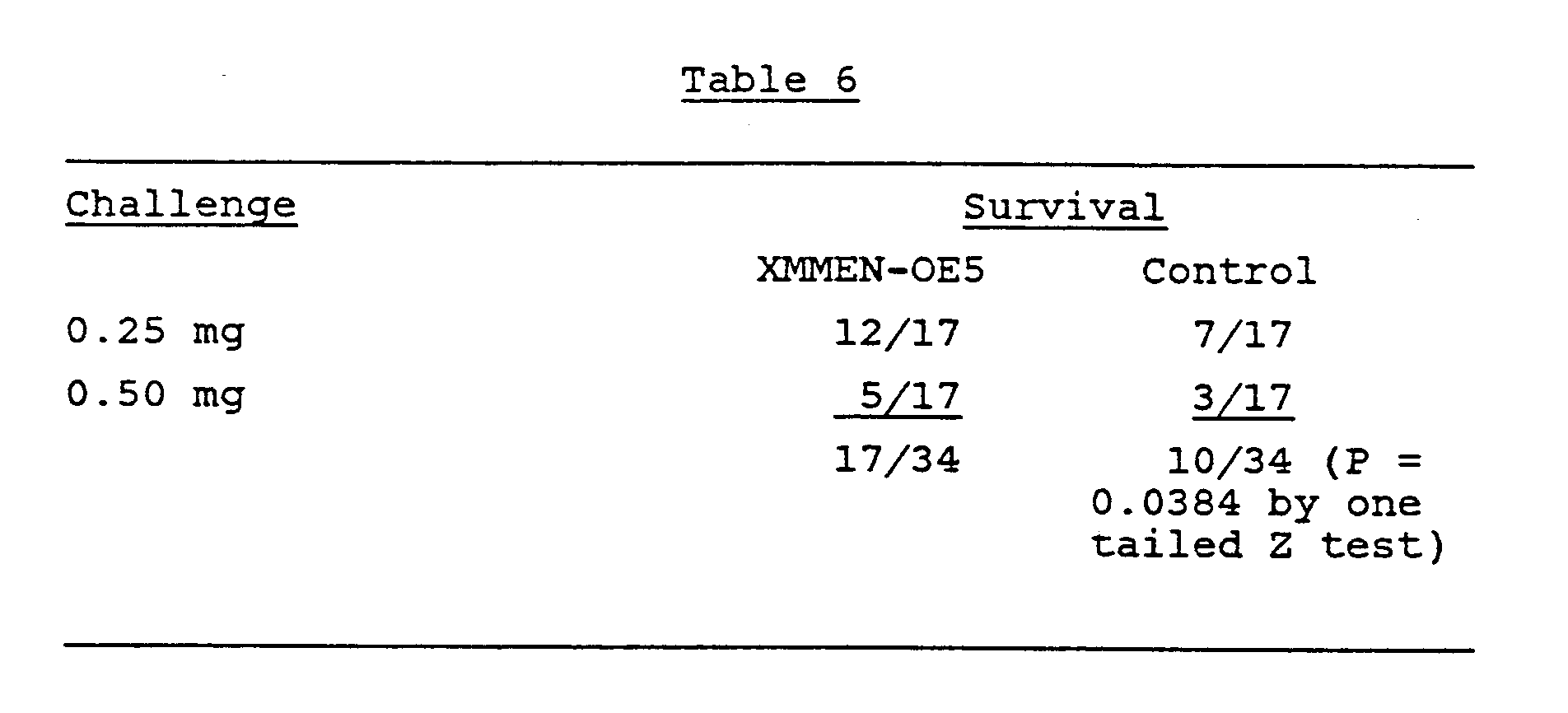
- 3. Prophylaxis Against Endotoxin Challenge: The ability of XMMEN-OE5 antibody to neutralize bacterial endotoxin was demonstrated in the same animal model described above. For the neutralization studies, purified endotoxin from the J5 E. coli mutant strain 0111 was obtained from List Laboratories, Campbell, Ca. An LD₅₀ value of endotoxin in mice was determined to be 0.25 mg/mouse and the LD₁₀₀ to be 0.50 mg/mouse. Twenty four hours prior to endotoxin challenge, mice received either 800 ug (40 mg/kg) of XMMEN-OE5 or saline control by IP injection. Results, as shown in Table 5, revealed that 12/17 (71%) mice treated with antibody survived the LD₅₀ endotoxin challenge versus 7/17 (41%) control mice, whereas 5/17 (29%) mice treated with antibody survived the LD₁₀₀ endotoxin challenge versus 3/17 (18%) control mice. These observations indicate that in a passive protection study, XMMEN-OE5 significantly increased survival after an LD₅₀ endotoxin challenge. Furthermore, this protective effect appears to be dependent upon the amount of the endotoxin challenge administered. These results are illustrated below.
- 4. Adjuvant Therapy Studies: To assess the therapeutic efficacy of XMMEN-OE5 combined with standard antibiotic therapy administered to mice after the onset of infection, the following series of experiments were performed.
- In a preliminary study, female CD-1 mice were infected with Pseudomonas aeruginosa strain 3632 (1.6 x 10⁸ cells/mouse). Immediately after challenge, mice received intramuscular antibiotics either alone or in combination with antiendotoxin antibody. The antibiotic regimen consisted of amakacin 1.56 mg/mouse and cefoperazone 12.5 mg/mouse given as single doses. Three hours after challenge, mice received intravenous injecttions of 0.4 mg of XMMEN-OE5 (20 mg/kg) or saline control. Results of this study demonstrated that 11/15 mice receiving antibiotics plus antiendotoxin antibody survived versus 5/15 mice that received antibiotics alone (P = .02 by Fisher's exact test).
- Therapy studies were expanded to incorporate additional gram negative infections. As in the previous study, female CD-1 mice were injected with LD₁₀₀ amounts of live, gram negative bacteria. Two hours after these inoculations, antibiotics or saline controls were injected intramuscularly. Because the four bacterial strains differed in their susceptibility to antibiotics, the following calculated doses of antibiotics were used (in mg/mouse):

Four hours after inoculation with a gram negative organisms and two hours after treatment with antibiotics, mice received intravenous injections of either XMMEN-OE5 antibody or saline control. For each of the four gram negative organisms listed above, all five groups of mice received the following series of treatments. Assessments of lethality were made 48 hours after the initial inoculation. - Group 1; LD₁₀₀ of bacteria plus combination antibiotics plus 300 ug of XMMEN-OE5 antibody.
- Group 2: LD₁₀₀ of bacteria plus combination of antibiotics plus 75 ug of XMMEN-OE5 antibody.
- Group 3: LD₁₀₀ of bacteria plus saline plus 300 ug of XMMEN-OE5 antibody.
- Group 4: LD₁₀₀ of bacteria plus combination antibiotics plus saline.
- Group 5: LD₁₀₀ of bacteria plus saline plus saline.
- No significant toxicity occurred in any of the more than 2500 mice involved in efficacy studies, or in any of the mice, rats, guinea pigs, or nonhuman primates treated in the toxicology studies.
- Efficacy experiments used a lot of XMMEN-OE5 antibody with which EIA binding to endotoxin-coated wells was successfully demonstrated. These studies, summarized in Table 7, demonstrated increased survival of mice receiving the higher dose of XMMEN-OE5 plus antibiotics when compared to antibiotics alone.

- Of the four gram negative organisms tested, the least efficacy was observed against E. coli 014K7. On the other hand, the 300 ug dose of XMMEN-OE5 plus combination antibiotic therapy was associated with the highest survival rate when the challenge was either of the remaining three organisms -- Pseudomonas aeruginosa 3632, E. coli 08H59, or E. coli 04K12 (see Table 8).
- 5. Summary of Efficacy Experiments: In spite of a significant number of variables inherent in the experimental animal model used in these experiments, XMMEN-OE5 was shown to 1) provide significant protection against several gram negative organisms; 2) neutralize lethal doses of purified endotoxin in a dose dependent manner; and 3) to have moderate efficacy as an adjunct to conventional antibiotics in the treatment of established gram negative infections.

- Early data from the Phase I clinical trial of monoclonal antibodies produced by hybridoma XMMEN-OE5 are only in anecdotal form but are reported below.
- 1. The first patient, F.R., was a 60 year old male with suspected gram negative sepsis. He received 0.1 mg/kg (total dose 8.5 mg) of XMMEN-OE5 i.v. over one hour. He experienced no adverse effects. Blood cultures subsequently demonstrated that he did not have gram negative bacteremia, but instead had a raging fungemia due to Torulopsis. The fungal abscess was surgically drained and he remained in stable condition three weeks after the administration of antibody.
- 2. The second patient, T.G., was a 57 year old female with documented gram negative bacteremia and pyelonephritis secondary to ureteral obstruction. She underwent surgical nephrostomy placement and received 0.5 mg/kg (total dose 36 mg) of XMMEN-OE5 i.v. over 1½ hours. She experienced no adverse effects, rapidly defervesced, and remained afebrile 12 hours after infusion. The prompt resolution of her fever is possibly due to the administration of antibody.
- 3. The third patient, K.S., was a 60 year old female with severe coronary artery disease who was admitted for unstable angina. She underwent coronary artery bypass surgery which was complicated by a transient episode of acute renal failure. Post-operatively, she developed a fever of 102.8° and subsequently has Acinetobacter cultured from her blood. Antibiotics were started, and ten days surgery, she received XMMEN-OE5 0.5 mg/kg (total dose 42 mg) i.v. over 1½ hours. Following antibiotic and anti-endotoxin antibody treatment, blood cultures became negative and her condition steadily improved. She remained in stable condition one week after administration of antibody.
- 4. The fourth patient, S.D., was a 60 year old female with persistent gram negative bacteremia (Klebsiella) of unknown etiology. An extensive workup revealed no source of infection, but urinary abnormalities made pyelonephritis the most likely cause. On her third hospital day, despite antibiotic therapy, her blood cultures again grew Klebsiella organisms. She experienced spiking fevers up to 104.5°. An emergency CT scan of her body was performed with the hope of locating an abscess which could be drained in an attempt to improve her clinical condition. No abscess or other site of infection was found. She was then treated with XMMEN-OE5 monoclonal antibody 2 mg/kg (total dose 157 mg) i.v. over 2½ hours. Twelve hours after infusion, her temperature was below 100°, and her clinical condition had improved markedly. Blood cultures were repeated and continued to grow gram negative organisms. Therefore, despite continued gram negative sepsis, her clinical condition improved markedly following the administration of monoclonal antibody. Her antibiotic regimen was then changed to a different combination of drugs, and her blood cultures subsequently became negative. She remained in improved condition 4 days after antibody therapy.
- The following table provides a summary of the binding characteristics of the MoAbs exemplified above. This table characterizes the MoAbs by their known antigens and not epitopes which are, for the most part, as yet uncharacterized.
Table 9 Summary of MoAb Binding ANTIGENS MoAb Rough LPS Smooth LPS Whole cell XMMEN-OE5 (IgM) inner-core Rc, Rd, Re, lipid-A NONE strong reaction: rough E. coli, P. aeruginosa moderate reaction: some smooth E. coli, P. aeruginosa XMMEN-LY1 (IgM) inner-core Rc, Rd, Re NONE S. minnesota R595 XMMEN-LY2 (IgM) lipid-A - Monoclonal antibodies which react selectively with an antigenic determinant common to strains of infecting bacteria were utilized to determine the location and extent of a localized bacterial infection, especially those caused by gram negative bacteria, by methods well-known in the art, for example, Larson, et al., Journal of Clinical Investigation (1983) 72:2101, which is incorporated by reference. Monoclonal antibodies are preferably radiolabelled by radioiodination or by other radiolabelling techniques well-known in the art, such as chelation using a chelating agent such as diethylenetriaminepenta-acetic acid (DTPA); or are otherwise labelled, such as with agents having paramagnetic properties, with chemiluminescent substrates, or with components of an enzymatic reaction. The radiolabelled monoclonal antibodies are purified and formulated for pharmaceutical use. A solution of the labelled monoclonal antibodies in a carrier, for example in phosphate buffered saline, is injected intravenously into a host. The appropriate dose is in the range of about 100 µg to 50 mg. Time is permitted for the antibodies to migrate to regions of the body having concentrations of cells with antigenic determinants reactive therewith. Concentrations of radioisotopes in certain tissues are determined or may be mapped either by techniques of whole body imaging which are well-known in the art (See, for example, Rainsbury, et al., Lancet October 22, 1983, 934 which is incorporated by reference), or by evaluating biopsied tissue or extracted body fluid using a scintillation counter. Where non-radioactive labels are used, other appropriate monitoring means are employed, such as a detector of nuclear magnetic resonance or a spectrophotometer. Areas of high radiation levels are indicative of the presence of localized bacterial infection.
- Hosts determined to have a bacterial infection are treated with monoclonal antibodies reactive with an antigenic determinant common to all strains of the bacterium. The monoclonal antibodies are administered venously, intramuscularly, intraperitoneally, or the like, in a physiologically acceptable carrier solution, such as phosphate buffered saline. The dosage is determined by the body weight of the host, it preferably being in the range of about 0.1 mg/kg to about 40 mg/kg body weight, and usually about 1 mg/kg to about 10 mg/kg of host body weight. Alternatively, the dosage is established by evaluating the extent of the remaining infection, as by quantitatively standardized EIA radioimaging or other methods. Treatment is repeated at intervals as necessary, to effect enhancement of the hosts' ability to recover from the infection.
- Monoclonal antibodies of the present invention, or their functional equivalents, are utilized in an immunoassay using standard and well-known methods (for example, Methods in Immunodiagnosis, 2nd Edition, Rose and Bigazzi, eds., John Wiley and Sons, 1980). Such assay may be, for example, of direct format (labelled first antibody reactive with the antigen), an indirect format (a labelled second antibody reactive with the first antibody), a competitive format (for example, addition of labelled antigen), or a sandwich format (both labelled and unlabelled antibody), as well as other formats well-known in the art.
- In one such embodiment, a tissue extract from a patient suspected of having a bacterial infection is applied to an insoluble matrix or solid substrate, such as cellulose strips, agarose or other particles so as to produce a bacteria-substrate complex. Alternatively, the substrate may have attached thereto an antibody so as to effect attachment of bacteria from a solution onto the substrate. The substrate is then washed, preferably with PBS, to remove unbound materials.
- The MoAbs of the present invention are employed in a method for the quantification of microbial endotoxins in body fluids, secretions and extracts or in drugs, diagnostic agents or liquid intermediates produced in the manufacture of diagnostic and therapeutic agents.
- An IgM MoAb which possesses a binding specificity for a specific and unique determinant on endotoxin, such as XMMEN-OE5, is adsorbed to a solid surface. Removal of excess unadsorbed material is followed by blockage of potential additional nonspecific binding sites on the solid surface by an appropriate blocking reagent such as albumin. Unbound albumin is removed by simple washing procedures. The activated surface may be exposed to various concentrations of the test sample for varying periods of time ranging from one hour to 24 hours and at various incubation temperatures ranging from 4° to 37°C. Removal of unreacted or depleted test sample is accomplished by simple washing procedures with standard biochemical solutions such as buffered saline. Detection and quantification of endotoxin levels in the test solution is accomplished by the addition of a detection probe or binding partner, a second MoAb which belongs to the IgG subclass and possesses a binding specificity for a specific and unique determinant as endotoxin which is different from that bound by the IgM MoAb and which is not significantly affected in this system by the IgM absorbed to the solid surface.
- Quantification is accomplished by relating the amount of the detection probe bound to the test sample to the amount of the detection probe bound to a defined quantity of an endotoxin reference standard. Assessment of the amount of the detection probe bound to the reference standard and the test sample is accomplished in a variety of ways such as: (a) radiolabeling of the probe and determination of radioactivity bound, (b) enzymatic labeling of the probe and measurement of enzymatic activity associated with specific amounts of the probe bound to the reference standard and test sample, (c) direct coupling to the probe of certain specific wavelengths emitting substances such as fluorochromes and quantification and comparison of these emissions by the standard reference and test samples. Quantification is also accomplished by labeling the detection probe with a substance such as biotin which allows the system to incorporate a standard second highly specific indicator such as avidin labeled with a radioisotope, enzyme, or fluorochrome.
- In another embodiment of this system a second specific indicator consisting of radiolabled, enzyme labeled, or fluorochrome labeled antibody specifically reactive with mouse Ig may be used to quantify the amount of endotoxin.
- An advantage to initial adsorption of the IgM monoclonal antibody to the solid surface derives from the high binding avidity associated with antibodies belonging to the IgM class. This diminishes the level of endotoxin which might be lost from the primary binding liquid during the assay process.
- The present invention provides pan-reactive MoAbs useful in the diagnosis, treatment and prevention of bacterial infection. The MoAbs of the invention have particular utility in blocking gram negative endotoxin, thus reducing its detrimental effects in hosts infected with gram negative bacteria.
- Although the present invention has been described in some detail by way of illustration and example for purposes of clarity of understanding, it will be obvious that certain changes and modifications may be practiced.
Claims (6)
- Use of a gram-negative bacterial endotoxin core specific monoclonal antibody, or an active fragment thereof, in the preparation of a medicament for the therapeutic treatment of a subject having gram-negative bacterial infection, wherein said antibody is of the IgM isotype and is the monoclonal antibody from hybridoma cell line XMMEN-OE5(ATCC HB 9081) or the gram-negative bacterial endotoxin core epitope binding equivalent thereof.
- Use of a gram-negative bacterial endotoxin core specific monoclonal antibody, or an active fragment thereof, in the preparation of a medicament for the prophylactic treatment of a subject at risk from gram-negative bacterial infection, wherein said antibody is of the IgM isotype and is the monoclonal antibody from hybridoma cell line XMMEN-OE5(ATCC HB 9081) or the gram-negative bacterial endotoxin core epitope binding equivalent thereof.
- Hybridoma cell line XMMEN-OE5 having ATCC Accession No. HB 9081.
- A monoclonal antibody derived from the cell line according to claim 3, or active fragment thereof, or the gram-negative bacterial endotoxin core epitope binding equivalent thereof.
- A monoclonal antibody, or active fragment thereof, according to claim 4 for pharmaceutical use.
- The use according to claim 1 wherein the antibody is present in the medicament in an amount sufficient to provide a dose in the range 0.1 to 2 mg/kg body weight.
Priority Applications (1)
| Application Number | Priority Date | Filing Date | Title |
|---|---|---|---|
| AT86306420T ATE81358T1 (en) | 1985-09-27 | 1986-08-19 | MONOCLONAL ANTIBODIES THAT BIND DETERMINANTS OF GRAM-NEGATIVE BACTERIA. |
Applications Claiming Priority (4)
| Application Number | Priority Date | Filing Date | Title |
|---|---|---|---|
| US781242 | 1985-09-27 | ||
| US06/781,242 US4777136A (en) | 1985-09-27 | 1985-09-27 | Monoclonal antibody reactive with Pseudomonas Aeruginosa |
| US855878 | 1986-04-24 | ||
| US06/855,878 US4918163A (en) | 1985-09-27 | 1986-04-24 | Monoclonal antibodies specific for lipid-A determinants of gram negative bacteria |
Related Child Applications (1)
| Application Number | Title | Priority Date | Filing Date |
|---|---|---|---|
| EP92104180.2 Division-Into | 1992-03-11 |
Publications (3)
| Publication Number | Publication Date |
|---|---|
| EP0217527A2 EP0217527A2 (en) | 1987-04-08 |
| EP0217527A3 EP0217527A3 (en) | 1989-02-08 |
| EP0217527B1 true EP0217527B1 (en) | 1992-10-07 |
Family
ID=27119830
Family Applications (1)
| Application Number | Title | Priority Date | Filing Date |
|---|---|---|---|
| EP86306420A Expired - Lifetime EP0217527B1 (en) | 1985-09-27 | 1986-08-19 | Monoclonal antibodies binding determinants of gram negative bacteria |
Country Status (11)
| Country | Link |
|---|---|
| US (2) | US4918163A (en) |
| EP (1) | EP0217527B1 (en) |
| JP (1) | JP2539797B2 (en) |
| KR (1) | KR910002427B1 (en) |
| AU (1) | AU606581B2 (en) |
| DE (1) | DE3686931T2 (en) |
| ES (1) | ES2004340A6 (en) |
| IE (1) | IE59685B1 (en) |
| IL (1) | IL79719A (en) |
| NZ (1) | NZ217283A (en) |
| PH (1) | PH26525A (en) |
Cited By (1)
| Publication number | Priority date | Publication date | Assignee | Title |
|---|---|---|---|---|
| US6790661B1 (en) | 1999-07-16 | 2004-09-14 | Verax Biomedical, Inc. | System for detecting bacteria in blood, blood products, and fluids of tissues |
Families Citing this family (73)
| Publication number | Priority date | Publication date | Assignee | Title |
|---|---|---|---|---|
| US5179018A (en) * | 1983-10-14 | 1993-01-12 | Centocor, Inc. | Mamalian monoclonal antibodies against endotoxin of gram-negative bacteria |
| IL81370A (en) * | 1986-02-07 | 1991-06-30 | Genetic Systems Corp | Pharmaceutical and diagnostic compositions comprising a plurality of human monoclonal antibodies for the treatment of bacterial diseases,methods for the preparation of the compositions and antibodies and kits containing said compositions |
| GB2192185B (en) * | 1986-07-03 | 1991-01-16 | Genetic Systems Corp | Monoclonal antibodies to pseudomonas aeruginosa flagella |
| JP2725041B2 (en) * | 1986-12-15 | 1998-03-09 | 三井東圧化学株式会社 | Human monoclonal antibody and its preventive and therapeutic agent for infectious diseases |
| US5521085A (en) * | 1986-12-15 | 1996-05-28 | Mitsui Toatsu Chemicals, Inc. | Transformed cell lines producing human monoclonal antibodies specific for Pseudomonas aeruginosa serotypes |
| WO1988008135A1 (en) * | 1987-04-10 | 1988-10-20 | Xoma Corporation | Human monoclonal antibodies binding determinants of gram negative bacteria |
| GB2218703B (en) * | 1988-05-10 | 1992-10-28 | Sumitomo Chemical Co | Human monoclonal antibody to p.aeruginosa: its production and use |
| US5888519A (en) * | 1988-06-02 | 1999-03-30 | The United States Of America As Represented By The Secretary Of The Army | Encapsulated high-concentration lipid a compositions as immunogenic agents to produce human antibodies to prevent or treat gram-negative bacterial infections |
| US5594113A (en) * | 1988-06-23 | 1997-01-14 | Associates Of Cape Cod, Inc. | Endotoxin binding and neutralizing protein and uses thereof |
| ATE118096T1 (en) * | 1988-12-22 | 1995-02-15 | Baxter Diagnostics Inc | LIPOHAPTENS AND USE OF CARRIER-BONDED LIPOHAPTENS IN THE IMMUNE TEST. |
| DE3843784A1 (en) * | 1988-12-24 | 1990-06-28 | Battelle Institut E V | Method for the detection of Gram-negative microorganisms, and DNA probes and antibodies suitable for this purpose |
| JPH02283294A (en) * | 1989-04-24 | 1990-11-20 | Sumitomo Chem Co Ltd | Human monoclonal antibody |
| JP2744130B2 (en) * | 1989-08-01 | 1998-04-28 | スクリップス クリニック アンド リサーチ ファウンデーション | Composition for treating sepsis |
| US6075121A (en) * | 1990-05-15 | 2000-06-13 | Chiron Corporation | Modified peptide and peptide libraries with protease resistance, derivatives thereof and methods of producing and screening such |
| US5198339A (en) * | 1990-07-13 | 1993-03-30 | Board Of Regents, The University Of Texas System | Method for detection of gram-negative bacterial lipopolysaccharides in biological fluids |
| CA2094588A1 (en) * | 1990-10-22 | 1992-04-23 | Carl R. Alving | Lipid a composition as immunogenic agents to prevent or treat gram-negative bacterial infections |
| US5434075A (en) * | 1992-03-20 | 1995-07-18 | Board Of Trustees Of The University Of Illinois | Monoclonal antibody to a human MDR1 multidrug resistance gene product, and uses |
| US5994088A (en) | 1991-03-08 | 1999-11-30 | Board Of Trustees Of The University Of Illinois | Methods and reagents for preparing and using immunological agents specific for P-glycoprotein |
| US5773280A (en) * | 1992-03-20 | 1998-06-30 | The Board Of Trustees Of The University Of Illinois | Monoclonal antibody to a human MDR1 multidrug resistance gene product, and uses |
| US5233024A (en) * | 1991-04-09 | 1993-08-03 | The Brigham & Women's Hospital | Anti-idiotypic monoclonal antibodies for mucoid pseudomonas aeruginosa, their preparation and use |
| AU2006892A (en) * | 1991-05-16 | 1992-12-30 | Associates Of Cape Cod, Inc. | Endotoxin binding and neutralizing protein and uses thereof |
| US5279956A (en) * | 1991-06-24 | 1994-01-18 | The Scripps Research Institute | Activated protein C polypeptides and anti-peptide antibodies, diagnostic methods and systems for inhibiting activated protein C |
| US5370872A (en) * | 1991-08-12 | 1994-12-06 | Swiss Serum And Vaccine Institute Berne | Escherichia coliO-polysaccharide-protein conjugate vaccine |
| AU660325B2 (en) * | 1991-10-11 | 1995-06-22 | Eisai Co. Ltd. | Anti-endotoxin compounds and related molecules and methods |
| US5530113A (en) * | 1991-10-11 | 1996-06-25 | Eisai Co., Ltd. | Anti-endotoxin compounds |
| WO1994014846A1 (en) * | 1992-12-18 | 1994-07-07 | Molecular Rx, Inc. | Assay and treatment for demyelinating diseases such as multiple sclerosis |
| US6660267B1 (en) | 1992-12-21 | 2003-12-09 | Promega Corporation | Prevention and treatment of sepsis |
| JPH08504824A (en) * | 1992-12-21 | 1996-05-28 | オフィディアン ファーマシューティカルズ,インコーポレーテッド | Prevention and treatment of sepsis |
| US5545721A (en) * | 1992-12-21 | 1996-08-13 | Ophidian Pharmaceuticals, Inc. | Conjugates for the prevention and treatment of sepsis |
| DE69420137T2 (en) | 1993-06-03 | 1999-12-23 | Therapeutic Antibodies, Inc. | PRODUCTION OF ANTIBODY FRAGMENTS |
| US5648343A (en) * | 1994-02-28 | 1997-07-15 | The University Of Georgia Research Foundation | Method for treating LPS-mediated disorders |
| DK0710118T3 (en) * | 1994-04-20 | 1999-08-09 | Us Army | Vaccine against infections with gram-negative bacteria |
| US5807694A (en) * | 1995-09-07 | 1998-09-15 | Economic Innovation And Technology Council, University Of Manitoba | Detection of salmonella enteritidis and other pathogenic microorganisms and monoclonal antibody useful therefor |
| US6962702B2 (en) | 1998-06-22 | 2005-11-08 | Immunomedics Inc. | Production and use of novel peptide-based agents for use with bi-specific antibodies |
| US8383081B2 (en) | 1999-05-10 | 2013-02-26 | Immunomedics, Inc. | Anti-CD74 immunoconjugates and methods of use |
| US8119101B2 (en) | 1999-05-10 | 2012-02-21 | The Ohio State University | Anti-CD74 immunoconjugates and methods of use |
| US7829064B2 (en) | 1999-05-10 | 2010-11-09 | Immunomedics, Inc. | Anti-CD74 immunoconjugates and methods |
| AU2008202582C1 (en) * | 1999-07-16 | 2012-04-05 | Verax Biomedical, Inc. | Method and kit immuno-detecting bacteria in blood and tissues |
| ATE408836T1 (en) * | 1999-07-16 | 2008-10-15 | Verax Biomedical Inc | METHOD AND KIT FOR THE IMMUNOLOGICAL DETECTION OF BACTERIA IN BLOOD AND TISSUE |
| JP4472351B2 (en) | 2002-03-01 | 2010-06-02 | イミューノメディクス、インコーポレイテッド | Internalizing anti-CD74 antibody and method of use thereof |
| US9770517B2 (en) | 2002-03-01 | 2017-09-26 | Immunomedics, Inc. | Anti-Trop-2 antibody-drug conjugates and uses thereof |
| DE10247430A1 (en) * | 2002-10-11 | 2004-04-29 | Fresenius Hemocare Gmbh | Method for determining the level of endotoxins in liquids |
| US8420086B2 (en) | 2002-12-13 | 2013-04-16 | Immunomedics, Inc. | Camptothecin conjugates of anti-CD22 antibodies for treatment of B cell diseases |
| US8883160B2 (en) * | 2004-02-13 | 2014-11-11 | Ibc Pharmaceuticals, Inc. | Dock-and-lock (DNL) complexes for therapeutic and diagnostic use |
| US9550838B2 (en) | 2004-02-13 | 2017-01-24 | Ibc Pharmaceuticals, Inc. | Dock-and-lock (DNL) complexes for therapeutic and diagnostic use |
| US9707302B2 (en) | 2013-07-23 | 2017-07-18 | Immunomedics, Inc. | Combining anti-HLA-DR or anti-Trop-2 antibodies with microtubule inhibitors, PARP inhibitors, bruton kinase inhibitors or phosphoinositide 3-kinase inhibitors significantly improves therapeutic outcome in cancer |
| US10058621B2 (en) | 2015-06-25 | 2018-08-28 | Immunomedics, Inc. | Combination therapy with anti-HLA-DR antibodies and kinase inhibitors in hematopoietic cancers |
| US8475794B2 (en) | 2005-04-06 | 2013-07-02 | Ibc Pharmaceuticals, Inc. | Combination therapy with anti-CD74 antibodies provides enhanced toxicity to malignancies, Autoimmune disease and other diseases |
| US8349332B2 (en) | 2005-04-06 | 2013-01-08 | Ibc Pharmaceuticals, Inc. | Multiple signaling pathways induced by hexavalent, monospecific and bispecific antibodies for enhanced toxicity to B-cell lymphomas and other diseases |
| US20070254322A1 (en) * | 2006-05-01 | 2007-11-01 | Romaschin Alexander D | Complement-based analyte assay |
| US9040250B2 (en) * | 2006-09-07 | 2015-05-26 | The United States Of America, As Represented By The Secretary Of The Navy | Binding interaction of proanthocyanidins with bacteria and bacterial components |
| WO2011085354A1 (en) * | 2010-01-11 | 2011-07-14 | Center For Molecular Medicine And Immunology | Enhanced cytotoxicity of anti-cd74 and anti-hla-dr antibodies with interferon-gamma |
| EP2788020A4 (en) | 2011-12-05 | 2015-04-29 | Immunomedics Inc | Therapeutic use of anti-cd22 antibodies for inducing trogocytosis |
| US9757458B2 (en) | 2011-12-05 | 2017-09-12 | Immunomedics, Inc. | Crosslinking of CD22 by epratuzumab triggers BCR signaling and caspase-dependent apoptosis in hematopoietic cancer cells |
| US9492566B2 (en) | 2012-12-13 | 2016-11-15 | Immunomedics, Inc. | Antibody-drug conjugates and uses thereof |
| US9931417B2 (en) | 2012-12-13 | 2018-04-03 | Immunomedics, Inc. | Antibody-SN-38 immunoconjugates with a CL2A linker |
| AU2013360335B2 (en) | 2012-12-13 | 2017-12-07 | Immunomedics, Inc. | Dosages of immunoconjugates of antibodies and SN-38 for improved efficacy and decreased toxicity |
| US10137196B2 (en) | 2012-12-13 | 2018-11-27 | Immunomedics, Inc. | Dosages of immunoconjugates of antibodies and SN-38 for improved efficacy and decreased toxicity |
| US9107960B2 (en) | 2012-12-13 | 2015-08-18 | Immunimedics, Inc. | Antibody-SN-38 immunoconjugates with a CL2A linker |
| US20240148873A1 (en) | 2012-12-13 | 2024-05-09 | Immunomedics, Inc. | Dosages of immunoconjugates of antibodies and sn-38 for improved efficacy and decreased toxicity |
| US10744129B2 (en) | 2012-12-13 | 2020-08-18 | Immunomedics, Inc. | Therapy of small-cell lung cancer (SCLC) with a topoisomerase-I inhibiting antibody-drug conjugate (ADC) targeting Trop-2 |
| US12310958B2 (en) | 2012-12-13 | 2025-05-27 | Immunomedics, Inc. | Antibody-drug conjugates and uses thereof |
| US10413539B2 (en) | 2012-12-13 | 2019-09-17 | Immunomedics, Inc. | Therapy for metastatic urothelial cancer with the antibody-drug conjugate, sacituzumab govitecan (IMMU-132) |
| US10206918B2 (en) | 2012-12-13 | 2019-02-19 | Immunomedics, Inc. | Efficacy of anti-HLA-DR antiboddy drug conjugate IMMU-140 (hL243-CL2A-SN-38) in HLA-DR positive cancers |
| US11253606B2 (en) | 2013-07-23 | 2022-02-22 | Immunomedics, Inc. | Combining anti-HLA-DR or anti-Trop-2 antibodies with microtubule inhibitors, PARP inhibitors, Bruton kinase inhibitors or phosphoinositide 3-kinase inhibitors significantly improves therapeutic outcome in cancer |
| US9797907B2 (en) | 2015-04-22 | 2017-10-24 | Immunomedics, Inc. | Isolation, detection, diagnosis and/or characterization of circulating Trop-2-positive cancer cells |
| US10195175B2 (en) | 2015-06-25 | 2019-02-05 | Immunomedics, Inc. | Synergistic effect of anti-Trop-2 antibody-drug conjugate in combination therapy for triple-negative breast cancer when used with microtubule inhibitors or PARP inhibitors |
| US10570194B2 (en) | 2015-08-06 | 2020-02-25 | Grifols Worldwide Operations Limited | Method for treating infectious diseases using a composition comprising plasma-derived immunoglobulin M (IgM) |
| US9913903B2 (en) | 2015-08-06 | 2018-03-13 | Grifols Worldwide Operations Limited | Method for the treatment or prevention of infection-related immune conditions using a composition comprising IgM |
| WO2017139623A1 (en) | 2016-02-10 | 2017-08-17 | Immunomedics, Inc. | Combination of abcg2 inhibitors with sacituzumab govitecan (immu-132) overcomes resistance to sn-38 in trop-2 expressing cancers |
| EP3448260A4 (en) | 2016-04-27 | 2019-10-09 | Immunomedics, Inc. | EFFICACY OF ANTI-TROP-2-SN-38 MEDICINAL-ANTIBODY CONJUGATES FOR THE TREATMENT OF RECYCLANT / REFRACTORY TUMORS TO CONTROL POINT INHIBITORS |
| US10918734B2 (en) | 2017-03-27 | 2021-02-16 | Immunomedics, Inc. | Treatment of high Trop-2 expressing triple negative breast cancer (TNBC) with sacituzumab govitecan (IMMU-132) overcomes homologous recombination repair (HRR) rescue mediated by Rad51 |
| CN110352201A (en) | 2017-04-03 | 2019-10-18 | 免疫医疗公司 | The subcutaneous administration of antibody drug conjugate for cancer therapy |
Family Cites Families (26)
| Publication number | Priority date | Publication date | Assignee | Title |
|---|---|---|---|---|
| US4647447A (en) * | 1981-07-24 | 1987-03-03 | Schering Aktiengesellschaft | Diagnostic media |
| US4443549A (en) * | 1981-10-19 | 1984-04-17 | Molecular Genetics, Inc. | Production of monoclonal antibodies against bacterial adhesins |
| US4428931A (en) * | 1982-03-15 | 1984-01-31 | Merck & Co., Inc. | Bacterial toxoids and gram-negative immune globulin therefrom |
| US4464465A (en) * | 1982-04-05 | 1984-08-07 | Genetic Systems Corporation | Cell-driven viral transfer in eukaryotes |
| FI69639C (en) * | 1982-07-02 | 1986-03-10 | Orion Yhtymae Oy | PREPARAT FOER ANVAENDNING VID KLAMYDIA-DIAGNOSTIK |
| JPS5929622A (en) * | 1982-08-10 | 1984-02-16 | Meiji Seika Kaisha Ltd | Monoclonal antibody, preparation and use thereof |
| US4689299A (en) * | 1982-09-30 | 1987-08-25 | University Of Rochester | Human monoclonal antibodies against bacterial toxins |
| US4525453A (en) * | 1982-10-26 | 1985-06-25 | Eastman Kodak Company | Process for rapidly differentiating between gram-negative and gram-positive microorganisms |
| US4574116A (en) * | 1983-01-13 | 1986-03-04 | The Board Of Trustees Of The Leland Stanford Jr. University | Methods and cell lines for immortalization and monoclonal antibody production by antigen-stimulated B-lymphocytes |
| DE3303649A1 (en) * | 1983-02-03 | 1984-08-09 | Wacker-Chemie GmbH, 8000 München | EXCLUDING WATER STORAGE CROSS-LINKABLE DIMENSIONS WHEN THE WATER ACCESSES AT ROOM TEMPERATURE |
| US5057598A (en) * | 1983-05-06 | 1991-10-15 | Centocor, Inc. | Monoclonal antibodies reactive with endotoxin core |
| DE3484773D1 (en) * | 1983-05-06 | 1991-08-08 | Velos Group | REACTIVE MONOCLONAL ANTIBODIES WITH ENDOTOXIN CORE. |
| US4587121A (en) * | 1983-06-14 | 1986-05-06 | Miles Laboratories, Inc. | High titer Pseudomonas immune serum globulin |
| US4525465A (en) * | 1983-10-07 | 1985-06-25 | Nippon Kayaku Kabushiki Kaisha | Water-insoluble biospecific absorbent containing argininal derivative |
| US5179018A (en) * | 1983-10-14 | 1993-01-12 | Centocor, Inc. | Mamalian monoclonal antibodies against endotoxin of gram-negative bacteria |
| US4617264A (en) * | 1983-11-04 | 1986-10-14 | Syntex (U.S.A.) Inc. | Pretreatment method and composition |
| US4683196A (en) * | 1983-12-12 | 1987-07-28 | Meru, Inc. | Method and materials for the identification of lipopolysaccharide producing microorganisms |
| EP0168422A1 (en) * | 1983-12-12 | 1986-01-22 | Meru, Inc. | Method and materials for the identification of lipopolysaccharide producing microorganisms |
| US4596769A (en) * | 1984-03-05 | 1986-06-24 | Temple University | Monoclonal antibodies to peptidoglycan and methods of preparing same |
| US4834975A (en) * | 1984-05-25 | 1989-05-30 | Genetics Corporation | Human monoclonal antibodies to serotypic lipopolysaccharide determinants on gram-negative bacteria and their production |
| US4646465A (en) * | 1984-06-22 | 1987-03-03 | Suiseki Fujimoto | Method of inoculating mushroom basidiospores seed basidiospore bed for inoculation, culture container for seed basidiospore bed, and boring apparatus for host wood for inoculation |
| FI853395A7 (en) * | 1984-09-05 | 1986-03-06 | Cetus Corp | Monoclonal antibodies that block gram-negative bacterial endotoxin. |
| JP2691708B2 (en) * | 1984-09-26 | 1997-12-17 | 住友製薬株式会社 | Human monoclonal antibody and method for producing the same |
| JPS61130300A (en) * | 1984-11-27 | 1986-06-18 | ザ ボ−ド オブ トラステイ−ズ オブ ザ レランド スタンフオ−ド ジユニア ユニバ−シテイ | Human monoclonal antibody |
| AU597877B2 (en) * | 1984-12-26 | 1990-06-14 | Teijin Limited | Anti-pseudomonas aeruginosa human monoclonal antibody |
| CA2300446C (en) * | 2000-03-10 | 2009-05-19 | Stan Mcclean | Beverage brewing apparatus having automatic and semi-automatic brewing modes |
-
1986
- 1986-04-24 US US06/855,878 patent/US4918163A/en not_active Expired - Fee Related
- 1986-08-14 IL IL79719A patent/IL79719A/en not_active IP Right Cessation
- 1986-08-19 DE DE8686306420T patent/DE3686931T2/en not_active Expired - Fee Related
- 1986-08-19 EP EP86306420A patent/EP0217527B1/en not_active Expired - Lifetime
- 1986-08-20 NZ NZ217283A patent/NZ217283A/en unknown
- 1986-09-25 PH PH34297A patent/PH26525A/en unknown
- 1986-09-26 IE IE254686A patent/IE59685B1/en not_active IP Right Cessation
- 1986-09-26 ES ES8602198A patent/ES2004340A6/en not_active Expired
- 1986-09-27 KR KR1019860008128A patent/KR910002427B1/en not_active Expired
- 1986-09-27 JP JP61229481A patent/JP2539797B2/en not_active Expired - Lifetime
- 1986-09-29 AU AU63236/86A patent/AU606581B2/en not_active Ceased
-
1994
- 1994-07-01 US US08/277,934 patent/US5484591A/en not_active Expired - Fee Related
Cited By (1)
| Publication number | Priority date | Publication date | Assignee | Title |
|---|---|---|---|---|
| US6790661B1 (en) | 1999-07-16 | 2004-09-14 | Verax Biomedical, Inc. | System for detecting bacteria in blood, blood products, and fluids of tissues |
Also Published As
| Publication number | Publication date |
|---|---|
| AU606581B2 (en) | 1991-02-14 |
| JP2539797B2 (en) | 1996-10-02 |
| IL79719A0 (en) | 1986-11-30 |
| PH26525A (en) | 1992-08-07 |
| EP0217527A2 (en) | 1987-04-08 |
| ES2004340A6 (en) | 1989-01-01 |
| EP0217527A3 (en) | 1989-02-08 |
| US4918163B1 (en) | 1993-08-17 |
| IE59685B1 (en) | 1994-03-23 |
| IL79719A (en) | 1991-06-10 |
| US4918163A (en) | 1990-04-17 |
| KR910002427B1 (en) | 1991-04-22 |
| JPS62228295A (en) | 1987-10-07 |
| AU6323686A (en) | 1987-04-02 |
| KR870003195A (en) | 1987-04-15 |
| DE3686931T2 (en) | 1993-04-29 |
| DE3686931D1 (en) | 1992-11-12 |
| NZ217283A (en) | 1991-07-26 |
| US5484591A (en) | 1996-01-16 |
| IE862546L (en) | 1987-03-27 |
Similar Documents
| Publication | Publication Date | Title |
|---|---|---|
| US5484591A (en) | Method for treating gram negative bacterial infections in humans | |
| US5179018A (en) | Mamalian monoclonal antibodies against endotoxin of gram-negative bacteria | |
| FI89278B (en) | METHOD FOR FRAMSTAELLNING AV MONOCLONAL HUMAN ANTICROPP MOT SEROTYPICAL LIPOPOLYSACKARIDDETERMINANTER PAO GRAMNEGATIVE BAKTERIER | |
| CA2132953C (en) | Monospecific polyclonal antibodies to shiga-like toxins | |
| Bogard Jr et al. | Isolation and characterization of murine monoclonal antibodies specific for gram-negative bacterial lipopolysaccharide: association of cross-genus reactivity with lipid A specificity | |
| Baumgartner | Immunotherapy with antibodies to core lipopolysaccharide: a critical appraisal | |
| JPH05304990A (en) | Monoclonal antibody to mycoplasma pneumoniae | |
| Tsang et al. | A murine monoclonal antibody specific for the outer core oligosaccharide of Salmonella lipopolysaccharide | |
| JPS62500172A (en) | Monoclonal antibodies and their uses | |
| WO1986002364A1 (en) | Monoclonal antibodies and their use | |
| Sjögren et al. | Etiologic diagnosis of pneumonia by antigen detection: crossreactions between pneumococcal C-polysaccharide and oral microorganisms | |
| US4777136A (en) | Monoclonal antibody reactive with Pseudomonas Aeruginosa | |
| Coughlin et al. | Immunoprotective murine monoclonal antibodies specific for the outer-core polysaccharide and for the O-antigen of Escherichia coli 0111: B4 lipopolysaccharide (LPS). | |
| WO1988008135A1 (en) | Human monoclonal antibodies binding determinants of gram negative bacteria | |
| US5034515A (en) | Staphylococcal fibronectin receptor, monoclonal antibodies thereto and methods of use | |
| Yamada et al. | Serodiagnosis by passive hemagglutination test and verotoxin enzyme-linked immunosorbent assay of toxin-producing Escherichia coli infections in patients with hemolytic-uremic syndrome | |
| US5698198A (en) | Method for treating gram negative bacterial infections in humans | |
| EP0494085A1 (en) | Monoclonal antibodies binding determinants of gram negative bacteria | |
| EP0188603A1 (en) | A salmonella-specific analyses reagent | |
| Brauner et al. | Antibody responses to Escherichia coli J5 lipopolysaccharide and to Salmonella porin in patients with bacteremia | |
| JPH04211393A (en) | Human monoclonal antibody and therapeutic agent containing the same as active ingredient for infectious disease caused by pseudomonas aeruginosa | |
| EP0326148A1 (en) | Human monoclonal antibody specific for Pseudomonas aeruginosa and hybrid cell lines | |
| Nys et al. | A direct enzyme-linked immunosorbent assay (ELISA) for antibodies to enterobacterial Re core glycolipid and lipid A: results in healthy subjects and in patients infected by gram-negative bacteria | |
| EP0193431A1 (en) | Monoclonal antibodies against chlamydial genus specific lipopolysaccharide | |
| Ward et al. | Monoclonal antibodies to salmonella lipopolysaccharide: functional analysis of anti-lipid A antibodies |
Legal Events
| Date | Code | Title | Description |
|---|---|---|---|
| PUAI | Public reference made under article 153(3) epc to a published international application that has entered the european phase |
Free format text: ORIGINAL CODE: 0009012 |
|
| AK | Designated contracting states |
Kind code of ref document: A2 Designated state(s): AT BE CH DE FR GB IT LI LU NL SE |
|
| PUAL | Search report despatched |
Free format text: ORIGINAL CODE: 0009013 |
|
| AK | Designated contracting states |
Kind code of ref document: A3 Designated state(s): AT BE CH DE FR GB IT LI LU NL SE |
|
| 17P | Request for examination filed |
Effective date: 19890724 |
|
| 17Q | First examination report despatched |
Effective date: 19910227 |
|
| GRAA | (expected) grant |
Free format text: ORIGINAL CODE: 0009210 |
|
| AK | Designated contracting states |
Kind code of ref document: B1 Designated state(s): AT BE CH DE FR GB IT LI LU NL SE |
|
| REF | Corresponds to: |
Ref document number: 81358 Country of ref document: AT Date of ref document: 19921015 Kind code of ref document: T |
|
| XX | Miscellaneous (additional remarks) |
Free format text: TEILANMELDUNG 92104180.2 EINGEREICHT AM 19/08/86. |
|
| REF | Corresponds to: |
Ref document number: 3686931 Country of ref document: DE Date of ref document: 19921112 |
|
| ITF | It: translation for a ep patent filed | ||
| ET | Fr: translation filed | ||
| PLBE | No opposition filed within time limit |
Free format text: ORIGINAL CODE: 0009261 |
|
| STAA | Information on the status of an ep patent application or granted ep patent |
Free format text: STATUS: NO OPPOSITION FILED WITHIN TIME LIMIT |
|
| PG25 | Lapsed in a contracting state [announced via postgrant information from national office to epo] |
Ref country code: SE Effective date: 19930820 |
|
| 26N | No opposition filed | ||
| BERE | Be: lapsed |
Owner name: THE REGENTS OF THE UNIVERSITY OF CALIFORNIA Effective date: 19930831 |
|
| PG25 | Lapsed in a contracting state [announced via postgrant information from national office to epo] |
Ref country code: NL Effective date: 19940301 |
|
| NLV4 | Nl: lapsed or anulled due to non-payment of the annual fee | ||
| GBPC | Gb: european patent ceased through non-payment of renewal fee |
Effective date: 19930819 |
|
| REG | Reference to a national code |
Ref country code: CH Ref legal event code: PL |
|
| REG | Reference to a national code |
Ref country code: FR Ref legal event code: ST |
|
| NLXE | Nl: other communications concerning ep-patents (part 3 heading xe) |
Free format text: A REQUEST FOR RESTORATION TO THE PRIOR STATE HAS BEEN FILED ON 940511 |
|
| REG | Reference to a national code |
Ref country code: CH Ref legal event code: AEN |
|
| REG | Reference to a national code |
Ref country code: FR Ref legal event code: R1 |
|
| REG | Reference to a national code |
Ref country code: GB Ref legal event code: 728C |
|
| EPTA | Lu: last paid annual fee | ||
| REG | Reference to a national code |
Ref country code: GB Ref legal event code: 728A |
|
| REG | Reference to a national code |
Ref country code: FR Ref legal event code: DS |
|
| BERR | Be: reestablished | ||
| EUG | Se: european patent has lapsed |
Ref document number: 86306420.0 Effective date: 19940310 |
|
| NLXE | Nl: other communications concerning ep-patents (part 3 heading xe) |
Free format text: THE REQUEST FOR RESTORATION TO THE PRIOR STATE HAS BEEN REJECTED |
|
| PGFP | Annual fee paid to national office [announced via postgrant information from national office to epo] |
Ref country code: SE Payment date: 19970722 Year of fee payment: 12 |
|
| PGFP | Annual fee paid to national office [announced via postgrant information from national office to epo] |
Ref country code: FR Payment date: 19990802 Year of fee payment: 14 Ref country code: DE Payment date: 19990802 Year of fee payment: 14 |
|
| PGFP | Annual fee paid to national office [announced via postgrant information from national office to epo] |
Ref country code: GB Payment date: 19990803 Year of fee payment: 14 Ref country code: AT Payment date: 19990803 Year of fee payment: 14 |
|
| PGFP | Annual fee paid to national office [announced via postgrant information from national office to epo] |
Ref country code: CH Payment date: 19990805 Year of fee payment: 14 |
|
| PGFP | Annual fee paid to national office [announced via postgrant information from national office to epo] |
Ref country code: BE Payment date: 19990819 Year of fee payment: 14 |
|
| PGFP | Annual fee paid to national office [announced via postgrant information from national office to epo] |
Ref country code: LU Payment date: 19990820 Year of fee payment: 14 |
|
| PG25 | Lapsed in a contracting state [announced via postgrant information from national office to epo] |
Ref country code: LU Free format text: LAPSE BECAUSE OF NON-PAYMENT OF DUE FEES Effective date: 20000819 Ref country code: GB Free format text: LAPSE BECAUSE OF NON-PAYMENT OF DUE FEES Effective date: 20000819 Ref country code: AT Free format text: LAPSE BECAUSE OF NON-PAYMENT OF DUE FEES Effective date: 20000819 |
|
| PG25 | Lapsed in a contracting state [announced via postgrant information from national office to epo] |
Ref country code: LI Free format text: LAPSE BECAUSE OF NON-PAYMENT OF DUE FEES Effective date: 20000831 Ref country code: CH Free format text: LAPSE BECAUSE OF NON-PAYMENT OF DUE FEES Effective date: 20000831 Ref country code: BE Free format text: LAPSE BECAUSE OF NON-PAYMENT OF DUE FEES Effective date: 20000831 |
|
| BERE | Be: lapsed |
Owner name: THE REGENTS OF THE UNIVERSITY OF CALIFORNIA Effective date: 20000831 |
|
| GBPC | Gb: european patent ceased through non-payment of renewal fee |
Effective date: 20000819 |
|
| REG | Reference to a national code |
Ref country code: CH Ref legal event code: PL |
|
| PG25 | Lapsed in a contracting state [announced via postgrant information from national office to epo] |
Ref country code: FR Free format text: LAPSE BECAUSE OF NON-PAYMENT OF DUE FEES Effective date: 20010430 |
|
| PG25 | Lapsed in a contracting state [announced via postgrant information from national office to epo] |
Ref country code: DE Free format text: LAPSE BECAUSE OF NON-PAYMENT OF DUE FEES Effective date: 20010501 |
|
| REG | Reference to a national code |
Ref country code: FR Ref legal event code: ST |
|
| PG25 | Lapsed in a contracting state [announced via postgrant information from national office to epo] |
Ref country code: IT Free format text: LAPSE BECAUSE OF NON-PAYMENT OF DUE FEES;WARNING: LAPSES OF ITALIAN PATENTS WITH EFFECTIVE DATE BEFORE 2007 MAY HAVE OCCURRED AT ANY TIME BEFORE 2007. THE CORRECT EFFECTIVE DATE MAY BE DIFFERENT FROM THE ONE RECORDED. Effective date: 20050819 |