EP0167852B1 - Human interferon-gamma - Google Patents
Human interferon-gamma Download PDFInfo
- Publication number
- EP0167852B1 EP0167852B1 EP85107142A EP85107142A EP0167852B1 EP 0167852 B1 EP0167852 B1 EP 0167852B1 EP 85107142 A EP85107142 A EP 85107142A EP 85107142 A EP85107142 A EP 85107142A EP 0167852 B1 EP0167852 B1 EP 0167852B1
- Authority
- EP
- European Patent Office
- Prior art keywords
- huifn
- dna sequence
- cells
- animal cell
- animal
- Prior art date
- Legal status (The legal status is an assumption and is not a legal conclusion. Google has not performed a legal analysis and makes no representation as to the accuracy of the status listed.)
- Expired
Links
Images





Classifications
-
- C—CHEMISTRY; METALLURGY
- C07—ORGANIC CHEMISTRY
- C07K—PEPTIDES
- C07K14/00—Peptides having more than 20 amino acids; Gastrins; Somatostatins; Melanotropins; Derivatives thereof
- C07K14/435—Peptides having more than 20 amino acids; Gastrins; Somatostatins; Melanotropins; Derivatives thereof from animals; from humans
- C07K14/52—Cytokines; Lymphokines; Interferons
- C07K14/555—Interferons [IFN]
- C07K14/57—IFN-gamma
Definitions
- the present invention relates to the chromosomal DNA sequence coding for human interferon- ⁇ (hereinafter referred to as HuIFN- ⁇ ) which is ligated to a promoter sequence that enables the IFN- ⁇ to be expressed in animal cell cultures, to cell cultures transformed with said DNA sequence, and to a method for the production of HuIFN- ⁇ using said transformed cell cultures.
- HuIFN- ⁇ human interferon- ⁇
- IFN Interferon
- IFN enhances the activity of cytotoxic T-cells [Lindahl,P.et al. (1972): Proc.Natl.Acad.Sci., 69 , 721], and activates macrophages to change them to ones with antitumor activities [Le,J.et al. (1983): J.Immunol., 131 ,2821].
- HuIFNs are classified into three groups, which are called IFN- ⁇ , IFN- ⁇ , and IFN- ⁇ ,respectively[Stewart II,W.E. et al. (1980): Nature, 286 ,110].
- IFN- ⁇ has the ability to inhibit cell proliferation at much lower concentrations than IFN- ⁇ and IFN- ⁇ [Rubin,B.Y.et al.(1980): Proc.Natl.Acad.Sci. USA, 77 , 5928)]. It also activates cells, including natural killer cells, killer T-cells, K-cells and macrophages which participate in what is called immunological surveillance mechanism of cancer. Therefore great expectations are directed to the clinical application of IFN- ⁇ .
- HuIFN- ⁇ is known to be induced in human lymphocytes stimulated by phytohemaagglutinin,staphylococcal enterotoxin A,concanavalin A,or galactose oxidase (Wheelock,E.F. (1965): Science, 149 ,310; Langford,M.P. et al. (1979): Infect.Immun., 26 , 36; de Lay,M.etal. (1980): Eur.J.Immunol., 10 ,877; Dianzani,F. et al.(1979): Infect.Immun., 25 ,879].
- IFN- ⁇ produced by a microbial protein synthesis system is substantially different from the natural HuIFN- ⁇ , and therefore, in cases of prolonged or frequent use of such an IFN- ⁇ , problems as the reduction of activities of the drug by antigen-antibody reaction and allergic responses such as shock reactions have to be taken into account
- IFN- ⁇ produced in Escherichia coli is different from natural HuIFN- ⁇ in antigen-antibody. reactions with monoclonal antibodies, and the difference of reactivity is not due to the absence of sugar chains[Le et al. (1984): J. Immunol., 132 , 1300].
- HuIFN- ⁇ was also produced by cloned T-cell lines [Nathan, H. et al. (1981): Nature, 292 , 842], T-cell hybridomas [Le et al. (1982): Proc. Natl. Acad. Sci. USA, 79 , 7857], and T-cells which were transformed by adult T-cell leukemia virus [Sugamura, K. et al. (1983): J. Immunol., 131 , 1611]. Since these cells are supposed to contain human leukemia virus as provirus, or to release virus particles, their use in the production of IFN- ⁇ is problematic with respect to biohazards [Sugamura, K. et al. (1983): ibid.].
- HuIFN- ⁇ was also attempted using animal cell cultures in which the simian virus 40 (hereinafter referred to as SV40) promoter sequence had been ligated to the IFN- ⁇ DNA sequence
- SV40 simian virus 40
- mRNA stable messenger RNA
- the splicing is supposed to be a process necessary for the accumulation of stable mRNA or the transfer of mRNA from nucleus to cytoplasm. Therefore, it is supposed that the addition of viral introns to the intron-free cDNA sequence of HuIFN- ⁇ leads to the intracellular accumulation of mRNA in the producing methods referred to above.
- the HuIFN- ⁇ gene is composed of four exons, three introns, and two sequences adjacent to the 5'-side and the 3'-side [Gray, P.W. et al. (1982): Nature, 298 , 859; Taya, Y.
- mRNA which is transcribed from the original, intron-bearing, chromosomal DNA sequence of HuIFN- ⁇ , and is submitted to splicing is more stable and therefore is accumulated at higher concentration in the cytoplasm compared with mRNA which is transcribed from the cDNA sequence.
- a regulatory region which controls the expression of HuIFN- ⁇ is present at the 5'-side of the HuIFN- ⁇ gene sequence. This region which contains the DNA sequence to which the RNA polymerase binds and which initiates the synthesis of mRNA, is called promoter region.
- the HuIFN- ⁇ gene sequence which contains said promoter region is introduced into animal cells as it is, the production of HuIFN- ⁇ is extremely low. This finding indicates that in order to obtain an effective production the 5'-promoter region of the HuIFN- ⁇ gene which is in a repressed state in most animal cell cultures has to be replaced by other promoter regions which are able to initiate the synthesis of mRNA in animal cell cultures.
- HuIFN- ⁇ is a glycoprotein.
- the structure of the sugar chain of HuIFN- ⁇ is still unclear in many respects. For example, it is unknown whether there are differences between sugar chains of HuIFN- ⁇ produced by cells of organisms other than human and HuIFN- ⁇ produced in human cells in their structure and antigenic properties.
- the HuIFN- ⁇ produced by human cells however is thought to be the same as the natural one, and to be higher in safety than those produced by cells of organisms other than human. The latter may cause problems such as allergic and shock reactions during the long-term therapeutic administration, since their preparations are possibly contaminated with components such as protein or the like or secretions from the parent organisms.
- preparations of the IFN- ⁇ produced by human cells never substantially contain any substance other than the human components, that is, substances intrinsically existing in the human blood. For these reasons, a high safety is expected for products derived from human cells.
- the cell For establishing human cell cultures producing HuIFN- ⁇ , the cell can be tranformed by an HuIFN- ⁇ expression vector. Such transformed cells, however, are killed by the self-produced HuIFN- ⁇ since this generally has a strong toxicity against human cells. Therefore, it is extremely difficult to isolate transformed strains which can be subcultured. Thus, whereas transformed strains which are able to be subcultured and to produce HuIFN- ⁇ can be easily obtained from hamster cells, transformed strains which are able to be subcultured are difficult to obtain from human cell cultures.
- the Present inventors isolated chromosomal DNA coding for HuIFN- ⁇ , and prepared plasmids in which the HuIFN- ⁇ gene is ligated to a promoter sequence that functions in animal cells, for example, SV40 early promoter, SV40 late promoter, or thymidine kinase promoter of herpes simplex virus. Further these plasmids were attached to a selective marker gene, for example, Ecogpt. The resulting plasmids, for example, pSVeSmaI ⁇ , pSV2pTK ⁇ , pSV2L ⁇ and pSV3L ⁇ were introduced into various animal cells according to the calcium phosphate method or the cell fusion method.
- the inventors succeeded in separating HuIFN- ⁇ -producing strains by transformation of cell lines derived ,from blood cells. These are proliferable as separate cells which do not substantially stick to vessel walls during cultivation and hardly yield cell floculations. These HuIFN- ⁇ -producing cell lines can be readily submitted to suspension culture processes for carrying out a large scale culture.
- the present invention provides a means for the large scale production of highly safe HuIFN- ⁇ .
- Figure 1 displays a restriction map of the 8.6Kb BamHI fragment which contains the HuIFN- ⁇ gene.
- Figure 2 shows the plasmid pBR ⁇ 8.6-1.
- Figure 3 shows the plasmids pSV2 ⁇ 8.6-1 and pSV2 ⁇ 8.6-2.
- Figures 4(a) and 4(b) schematically show the construction of an expression vector, pSVeSmaI ⁇ , capable of producing HuIFN- ⁇ in mammalian cell cultures.
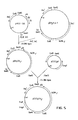
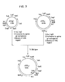
- Figs. 5-7 schematically show the construction of the expression vectors pSV2pTK ⁇ , pSV2L ⁇ and pSV3L ⁇ , respectively, which are capable of producing HuIFN- ⁇ in mammalian cell cultures.
- Figures 8(a) and 8(b) are schematic outlines of a process for preparing the expression-vector pSVeBPV ⁇ .
- the gene region which codes for HuIFN- ⁇ is contained within a DNA fragment of about 8.6kb, as shown in Fig. 1.
- Exons of HuIFN- ⁇ are present at four places separately. These exons on the DNA sequence are referred to as the first to fourth exon in order from the 5'-side, respectively (each indicated by a thick line in Fig. 1).
- the fourth exon has the signal AATAAA for polyadenylation of mRNA, following the termination codon.
- the first exon codes for a polypeptide which is composed of 38 amino acid residues. Twenty-three (23) N-terminal amino acids, which arise from the protein-synthesis-initiating amino acid, methionine,form a signal peptide, which is present at the N-terminus of many secretory proteins. The signal peptide is cleaved off during secretion from the cell.
- the chromosomal DNA sequence which codes for HuIFN- ⁇ is the DNA sequence which contains the 4 exons and 3 introns shown in Fig. 1.
- the chromosomal DNA sequence which codes for HuIFN- ⁇ is cloned from the human DNA.
- the human DNA is prepared by the method of Blin et al. [Blin, N. et al.
- ⁇ phage vectors represented by Charon 28 plasmid vectors represented by pBR322, or cosmids represented by pHC79 are used.
- the gene manipulation technique is generally adopted with the use of the ⁇ phage vector with which large DNA fragments can be cloned with high efficiencies. That is, after cutting of human high-molecular weight DNA using an appropriate restriction enzyme, the DNA fragment is inserted instead of the substitutable region of ⁇ phage DNA for obtaining the recombinant phage DNA.
- infectious phage particles are prepared. Then the phage particles are plated together with the host Escherichia coli to obtain plaques of recombinant phages [Enquist, L. et al. (1979): Methods in Enzymology, 68 , 281; Horn, B. (1979): Methods in Enzymology, 68 , 299].
- the plaque hybridization technique is used with 32p-labelled cDNA or synthetic DNA as a probe [Woo, S.L.C.
- the recombinant phage which carries the HuIFN- ⁇ gene can be prepared in large amounts by recovering it from a plaque selected by the plaque hybridization, and by culturing it together with the host Escherichia coli.
- the recombinant phage DNA can be prepared according to the method described in Maniatis, T. et al. [(1982): Molecular Cloning, A Laboratory Mannual, Cold Spring Harbor Laboratory].
- the amino acid sequence of HuIFN- ⁇ can be derived from the base sequence of the exon parts of the cloned HuIFN- ⁇ gene.
- the base sequence in turn, can be determined according to the Maxam-Gilbert method [Maxam A.M. et al. (1980): Methods in Enzymology, 65 , 499].
- a DNA sequence composed of HuIFN- ⁇ sequence ligated to a functional promoter sequence, and a selectable marker gene is appropriate.
- selectable marker in animal cells, such genes as Ecogpt [Mulligan, R.C. et al. (1980): Science, 209 , 1422], neo [Southern, P.J. et al. (1982): J. Mol. Appl. Genet., 1 , 327], and dhfr [Wigler, M. et al. (1980): Proc. Natl. Acad. Sci. USA, 77 , 3567] are used.
- the SV40 early promoter As a promoter which functions in animal cell cultures, the SV40 early promoter is known. This promoter is contained by the Hind III-Pvu II fragment of SV40 DNA (about 350 base pairs in size) and has an activity as SV40 late gene promoter in the opposite orientation.
- the thymidine kinase promoter of type 1 of herpes simplex virus (HSV) is a constitutive promoter similarly to SV40 early promoter.
- the structure has been shown by Wagner et al. [Wagner, M.J. et al. (1981): Proc. Natl. Acad. Sci. USA, 79 , 1441.].
- the functional promoter region contains the initiation origin of mRNA synthesis, but not the methionine codon which is the first amino acid of the protein that is regulated by said promoter.
- any of these plasmids is characterized in that its DNA sequence comprises the origin of DNA replication (ori) of Escherichia coli and of the animal cell cultures, the HuIFN- ⁇ chromosomal DNA sequence ligated to a functional promoter, and a selectable marker gene in Escherichia coli (Ampicillin resistance gene )and in animal cell cultures (Ecogpt).
- DNA material for the calcium phosphate method microorganisms, as Escherichia coli, phages and DNA solutions can be used.
- microbial protoplasts containing the desired DNA sequence as a plasmid are used.
- both the HuIFN- ⁇ and the T antigen gene are integrated into the cells more effectively than by co-transfection of them. It may also be possible to obtain cell lines highly expressing HuIFN- ⁇ using various cell strains. Furthermore, it is also possible to use SV40 transformants as a host.
- HuIFN- ⁇ For the production of HuIFN- ⁇ by cells into which a DNA sequence containing the chromosomal HuIFN- ⁇ gene ligated to a functional promoter, is introduced, it is necessary that said DNA sequence is fitted for the RNA synthesis system inherent in the cells to be used, the maturation system of RNA, the protein synthesis system, the maturation system of protein, as well as for functions as secretion and the like.
- RNA synthesis system inherent in the cells to be used
- the maturation system of RNA the protein synthesis system
- maturation system of protein as well as for functions as secretion and the like.
- capping at the 5'-end, splicing at normal sites, and polyadenylation of the 3'-end of the mRNA take place.
- the inventors investigated different animal cell strains available from the American type culture collection (ATCC).
- the cells were derived from hamster, monkey, mouse, rat or human, including CHO (hamster ovary cells), HeLa (human cerevicitis tumor cells), FL (human amnion cells), WISH (human amnion cells), Chimp liver (chimpanzee liver cells), WI-26VA4 (human lung cells transformed by SV40), and Vero (kidney cells of African green monkey).
- HuIFN- ⁇ -resistant cell lines or HuIFN- ⁇ -resistant variants out of many cell cultures derived from human subcultivation of culture cells in a medium containing HuIFN- ⁇ by one unit to several ten thousand units is useful.
- HuIFN- ⁇ -sensitive culture cells derived from human are denatured, deformed, and finally killed by the toxicity of HuIFN- ⁇ during the subcultivation, the HuIFN- ⁇ -resistant cell lines are not affected, but continue to grow. The latter therefore are easily selectable.
- HuIFN- ⁇ -sensitive lines become significantly sensitive to a double-stranded RNA as poly I:C.
- the HuIFN- ⁇ -resistant cell lines are distinguishable from these HuIFN- ⁇ -sensitive cell lines.
- Established cell lines of mammals are derived from blood cells as lymphoblasts, myeloblasts, monocytes and erythroblasts which are derived from their stem cells. Additionally cell lines were established from cells which are further differentiated or derived from said blastocytes.
- the established cells derived from blood cells which have been proliferated in a common medium display spherical morphology and are suitable for suspension cultures. Some of these cells, namely those producing globulin are advantageously used as hosts for the production of HuIFN- ⁇ , because they have high ability to synthesize and secrete proteins.
- IFN The activity of IFN was determined with the CPE-inhibiting method with the combined use of FL cells and vascular stomatitis virus or Sindovis virus [Philip, C. et al. (1981): Methods in Enzymology, 78 , 387].
- the dialyzate was treated with 100 ⁇ g/ml ribonuclease for 3.5 hours at 37°C, again extracted with phenol, and dialysed against 20mM tris ⁇ HCl (pH 8.0), 1mM EDTA, and 10mM NaCl to obtain about 33 mg of high molecular human DNA.
- the human DNA was digested with the restriction enzyme BamHl, and then BamHl DNA fragments of about 8 to 9 kilobases (hereinafter abbreviated to kb) were prepared with sucrose density-gradient centrifugation.
- Lambda( ⁇ )-phage vector charon 28A DNA (Bethesda Research Laboratories, Gaithersburg, USA) was digested with BamHl, and the fractions containing the left-arm and right-arm of Charon 28 were collected with sucrose density-gradient centrifugation, and recovered by ethanol precipitation: The DNA fragments with both arms of Charon 28 and the human BamHl fragments of 8 to 9 kb were ligated together using T4 DNA ligase. Then, the ligated DNA was submitted to an in vitro packaging technique according to the Enquist-Sternberg method [Enquist, L. and N.
- DNA of phage clone S8-11 containing HuIFN- ⁇ gene was digested with the restriction enzyme BamHl, and ligated to a BamHl site of plasmid pBR322 which had been treated with bacterial alkaline phosphatase [Bolivar, F. et al. (1977): Gene, 2 , 951 and transformed to Escherichia coli C600 ⁇ m ⁇ .
- a transformant containing a plasmid into which the HuIFN- ⁇ gene of about 8.6 kb had been inserted into the BamHl site of pBR322 was selected out as transformants having ampicillin-resistant and tetracycline-sensitive characters This plasmid was designated as pBR ⁇ 8.6-1.
- the structure of pBR ⁇ 8.6-1 is shown in Fig. 2.
- the plasmid pBR ⁇ 8.6-1 was digested with BamHl, DNA fragments of about 8.6 kb were prepared by agarose gel electrophoresis and ligated to the plasmid pSV2gpt (ATCC 37145) which had been treated with bacterial alkaline phosphatase with the use of T4DNA ligase and transformed to Escherichia coli C600 ⁇ m ⁇ . Strains bearing the plasmids pSV2 ⁇ 8.6-1 and pSV2 ⁇ 8.6-2, whose structure are shown in Fig. 3, were selected from among other transformants.
- plasmids pSV2 ⁇ 8.6-1 and pSV2 ⁇ 8.6-2 are plasmids into which the HuIFN- ⁇ gene-bearing DNA fragment has been inserted clockwise and counterclockwise into the BamHl sites of pSV2gpt, respectively.
- Respective plasmid DNA was prepared by cesium chloride density-equilibrium centrifugation. Plasmid DNA was prepared as needed with the use of Escherichia coli GM33 dam ⁇ as a host.
- the b ase sequence in the HuIFN- ⁇ gene of plasmid pBR ⁇ 8.6-1 was determined according to the Maxam-Gilbert method [Maxam, A.M. and W. Gilber (1980): Methods in Enzymology, 65 , 499].
- the base sequences around four (4) exons coding for HuIFN- ⁇ were determined as follows:
- Plasmid pSVeSmaI ⁇ which has the sequence of SV40 promoter region and ligated chromosomal DNA sequence of HuIFN- ⁇ , is prepared according to the procedures shown in Figs. 4-(a) and 4-(b), with the use of pBR8.6-1, pSV2gpt and pSV3gpt (ATCC37144) [Mulligan, R.C. and P. Berg (1980): Science, 209 , 1422] as starting materials.
- pSV3gpt (ATCC37144) was digested with Hind III, and the largest DNA fragment was made cyclic with T4 DNA ligase to make pH1.
- the Pvu II site of pH1 was changed into the Sal I site with the use of Sal I linker to make pH2.
- the Hind III site of pH2 was changed to the Sma I site with the use of Hind III - Sma I adaptor to make pHSmaI.
- psv2gpt was digested with Bam HI, The termini were filled to make blunt ends with DNA polymerase I (klenow).
- a Sal I site was introduced with the use of Sal I linker and T4DNA ligase to make PSI.
- the PSI was digested with Sal I and EcoRI, and an ampicillin-resistance gene-bearing DNA fragment was ligated to the Sal I-EcoRI fragment of pHSmaI having SV40 promoter region to make pSVeSmaI.
- pBR ⁇ 8.6-1 was digested with Mst II, and, after the termini were made blunt with DNA polymerase (Klenow), digested with BamHI to obtain HuIFN- ⁇ sequence-bearing DNA fragment.
- the DNA fragment was introduced into pSVeSmaI which was digested with SmaI and BamHI, to make pSVeSmaI ⁇ .
- Plasmid pSV2pTK ⁇ which has the sequence of thymidine kinase promoter region of herpes simplex virus type I and the HuIFN- ⁇ gene, is prepared according to the procedures shown in Fig. 5, with the use of pBR ⁇ 8.6-1, pHSV106 (Bethesda Research Laboratories, Gaithersburg, USA) [Mcknight, S.L. and E.R. Gabis (1980): Nucleic Acids Res., 8 , 5931], and pSVgpt as starting materials.
- Sal I linker and Sma I adaptor used were d(pGGTCGACC) and d(pAGCTCCCGGG), respectively.
- Employed DNA polymerase I was Klenow fragment.
- HuIFN- ⁇ -expression vector, pSV2L ⁇ was prepared according to the following procedures.
- Plasmid pSV2gpt [Mulligan, R.C. and P. Berg (1980): Science, 209 , 1422] having both ampicillin-resistance gene and guanine phosphoribosyl transferase gene (Ecogpt) of Escherichia coli, was digested with the restriction enzyme Pvu II.
- Bcl I linker (CTGATCAG) which had been phosphorylated with T4 polynucleotide kinase, was ligated to the termini with the use of T4 ligase.
- the DNA fragment was recovered by 0.8% agarose gel electrophoresis.
- the recovered DNA fragment was circularized with the use of T4 DNA ligase to obtain pSV2gptBclI by transformation of E. coli GM33 dam ⁇ .
- the pBR 8.6-1 described in Example 2 was digested with the use of Bam HI and Bcl I.
- HuIFN- ⁇ gene-bearing DNA fragment of 5.5 kb was recovered, and inserted into the Bcl I site of pSV2gptBclI to obtain pSV2L ⁇ .
- the preparation of pSV2L ⁇ is shown in Fig. 6.
- the plasmid pSV2L ⁇ described in Example 5 was digested with restriction enzymes Pvu I and Bgl II, and a HuIFN- ⁇ gene-containing DNA fragment of about 7.8 kb was recovered by 0.8% agarose gel electrophoresis. The recovered fragment was ligated to another DNA fragment which contained SV40 T antigen of pSV3gpt [Mulligan, R.C. and P. Berg (1980): Science, 209 , 1422] that had been digested with Pvu I and Bal II, and was circularized to pSV3L ⁇ . The preparation of pSV3L ⁇ is shown in Fig. 7.
- the plasmid pSVeSmaI ⁇ described in Example 4 was digested with the restriction enzymes Sal I and Bam HI to obtain a DNA fragment of about 6.0 kb which contained HuIFN- ⁇ gene bearing SV40 early promotor.
- pdBPV-1 ATCC 37134
- pdBPV-1 ATCC 37134
- the obtained fragment of about 2.4 kb was ligated to the above fragment of about 6.0 kb to make pML2d ⁇ .
- the bovine papilloma virus genome-bearing DNA fragment of about 7.9 kb was cut out of pdBPV-1, and indroduced into the Bam HI site of pML2d ⁇ to obtain plasmid pSVeBPV ⁇ of about 16.3 kb.
- the preparation of pSVeBPV ⁇ shown in Figs. 8-a and 8-b.
- the plasmid was introduced into varied animal cell cultures in accordance with procedures of Wigler et al. [Wigler et al. (1977): Cell, 11 , 223].
- the co-precipitate of the plasmid with calcium phosphate was added to cells (3x10⁵ cells/3.6-ml medium/6-cm round dish) which had been preliminarily grown in Eagle's MEM medium containing 10% fetal calf serum (hereinafter abbreviated as FCS). Cultivation was continued for 48 hours in the medium which was renewed 15 hours after the initiation. IFN activity involved in the respective medium was determined.
- Mycophenolic acid-resistant CHO cells CHO-33, were isolated by the procedures mentioned in Example 9, and cultured in an MEM medium to obtain 120 ml of culture liquid. Then, 1.0 g of Controlled Pore Glass CPG350 (Electronucleonics Corp.; mesh size 120/200) was added to 60 ml of the obtained culture liquid, agitated for 3 hours at 4°C, packed into a column, washed with 150mM-phosphate-0.15M NaCl buffer (pH 7.4), and eluted by the same buffer which, in addition, contained 50% ethylene glycol. The active fraction was diluted 10-fold with 20mM phosphate buffer, passed through a 5-ml ConA-Sepharose Column to get IFN adsorbed.
- Controlled Pore Glass CPG350 Electrode
- the adsorbed IFN was eluted with 20mM phosphate buffer containing 0.15M NaCl and 0.15M ⁇ -methyl-D-mannoside.
- the active fractions were collected, dialyzed against 0.2M ammonium acetate (pH 6.0) and 0.15M NaCl, concentrated by polyethylene glycol 20000, and passed through Biogel P-100 (2.6 x 60cm) which had been equilibrated with the same buffer to obtain an IFN sample.
- the obtained IFN sample lost its activity on treating with acid of pH 2.0 or with 0.1% SDS.
- the promoting effect was observed only for human cells.
- cells were grown in monolayer on a 24-hole multidish, treated with IFN at 300 units/ml for 20 hrs, and then cultured for 24 hours in an MEM medium containing 10 ⁇ g/ml poly I:C. While CHO and BHK (hamster cells) underwent no changes, cells of FL, WISH and Hela were observed to become round and exfoliated.
- the plasmid was introduced into animal cell cultures in accordance with the procedures of Wigler et al. [Wigler et al. (1977): Cell, 11 , 223].
- the co-precipitate of the plasmid with calcium phosphate was added to cells (3x10⁵ cells/3.6 ml medium/6cm-diameter dish) which had been preliminarily grown in an Eagle's MEM medium containing 10% newborn calf serum. The medium was renewed 15 hours after the initiation, and still incubated for 48 hours. Then, IFN activity involved in the medium was determined.
- the expression of IFN activity was observed in the cell strains of CHO-Kl, Vero or Wl-26VA4 which contained pSV2L ⁇ or pSV3L ⁇ , while no activity was observed in the cell strains which contained only calf thymus DNA. Further, the concurrent introduction of pSV2L ⁇ and pSV3gpt, containing the SV40 T antigen-coding gene, exhibited a 8 to 15 times higher activity than the single introduction of pSV2L ⁇ . WI-26 VA4 continuously expresses SV40 T antigen, and exhibites higher expression than CHO-Kl.
- HuIFN- ⁇ -yielding CHO cells obtained in Example 12 were cultured for 4 days in a 5% FCS-containing MEM medium, and 60 ml of the medium was recovered therefrom.
- a 5% FCS-containing MEM medium a 5% FCS-containing MEM medium
- 60 ml of the medium was recovered therefrom.
- 0.5 g of Controlled Pore Glass (Electronucleonics Corp.) CPG350 (meshsize 120/200) was added, agitated for 3 hours at 4°C, filled into a column, washed with 150mM phosphate buffer (pH 7.4) and 0.15M NaCl, and eluted with the same buffer containing 50% ethylene glycol. Then the obtained active fractions were diluted 10-fold with 20mM phosphate buffer. Thus, about 20 000 units of IFN was purified and recovered.
- FL cells were cultured on the whole bottom surface of 75 cm2 of a flask. Then the culture was renewed by an MEM medium which contained 100 units/ml HuIFN- ⁇ , and 10 ⁇ g/ml poly I:C, and still incubated for 48-hours. Although most of FL cells were exfoliated from the bottom, the medium was replaced by another MEM medium containing 100 units/ml HuIFN- ⁇ , and still incubated for about one month. Colonies of HuIFN- ⁇ -resistant FL cells were developed, proliferated, and established.
- the co-precipitate of 1,44 ⁇ g plasmid with calcium phosphate was added to the above cells (3 ⁇ 105 cells/3.6 ml medium/6cm-diameter disk)which had been preliminarily grown in an Eagle's MEM medium containing 10% newborn calf serum. After 15 hours, the medium was renewed, and after 48-hour incubation, the medium was replaced by another medium which contained 25 ⁇ g/ml mycophenolic acid, 250 ⁇ g/ml xanthine, 0.1 ⁇ g/ml aminopterin, 25 ⁇ g/ml adenine, and 5 ⁇ g/ml thymidine, and after further 2-week incubation the numbers of developed transformant colonies were counted.
- transformants were obtained from all used strains for pSV2gpt, while few transformant colonies were obtained from HuINF- ⁇ -sensitive strains for HuIFN- ⁇ gene-containing pSVeSmaI ⁇ or pSV2pTK ⁇ , although many were from HuIFN- ⁇ -resistant WI-26 VA4 and HuIFN- ⁇ -resistant FL cells. Plasmid Numbers of transformant colonies formed HeLa FL WISH WI-26VA4 HuIFN-resistant FL pSV2gpt 120 52 28 36 40 pSVeSmaI ⁇ 1 0 3 28 37 pSV2pTK ⁇ 1 1 0 25 44
- the colonies of WI-26 VA4 transformant obtained in Example 17 using pSveSmaI ⁇ or pSV2pTK ⁇ were separated, and proliferated in an MEM medium containing 5% FCS. The transformant was proliferated on the whole bottom surface of the 24-well multiplate. After the medium was renewed, and incubated for 24 hours, the IFN activity in the culture liquid was determined. The IFN activities of 32.3 and 524 units/24 hr/106 cells were observed for transformants by pSVeSmaI ⁇ and pSV2pTK ⁇ , respectively.
- WI-26 VA4 strain was transformed in a similar way to that in Example 17. The obtained results are as follows.
- the colonies of HuIFN- ⁇ -resistant FL transformant obtained in Example 17 using pSV2pTK ⁇ were separated, and proliferated in an MEM medium containing 5% FCS.
- the transformant was proliferated on the whole bottom surface of the 24-well multiplate. After the medium was renewed, and incubated for 24 hours, the IFN activity in the culture liquid was determined. The development of the activity of 3 units/24 hr/106 cells was observed.
- the pSVeSmaI ⁇ described in Example 4 was introduced into blood cell-derived cell lines obtained from ATCC or others according to the method of Oi, et al. [Oi, V.T. et al. (1980): Proc. Natl. Acad. Sci. USA, 80 , 825].
- Plasmid pSVeSmaI ⁇ -bearing Escherichia coli K-12 strain C600 ⁇ m ⁇ was cultured with an L medium at 37°C until the absorbance at 600 nm reached 0.6 to 0.8. After the addition of chloramphenicol up to 125 ⁇ g/ml and the further culturing for 12 to 16 hours, the bacteria were centrifuged and collected. Then, cold 0.05M tris.
- HCl (pH 8.0) which contained sucrose by 20% per 250 ml medium, was added to disperse the bacteria. After the addition of 0.25 ml of 5 mg/ml lysozyme solution, and 5-minute standing on ice, 0.5 ml of 0.25M EDTA (pH 8.0) was added, and the liquid was allowed to stand again for 5 minutes on ice. Then, after 0.5 ml of 0.05M Tris ⁇ HCl (pH 8.0) was added again, the liquid was incubated for 10 to 15 minutes at 37°C to protoplastize the bacteria. The protoplast was diluted with 10 ml of DMEM medium containing 10% sucrose, and allowed to stand for 10 minutes at room temperature.
- the cells were cultured in DMEM medium containing 10% FCS to 106 cells/ml, added to by protoplast suspension by 5 ml per 2 x 10⁶ cells, and centrifuged at about 500xg at room temperature. After removing the supernatant, the precipitate was added to by 2 ml of 50% polyethylene glycol solution (pH 8.0) in DMEM medium, gently stirred, and centrifuged for 3 minutes at 500xg. The cells were washed with 7 ml of DMEM medium, dispersed into DMEM medium containing 10% FCS, and cultured at a cell concentration of 2 x 10⁵ cells/well ⁇ ml with a 24-well multidish plate.
- Each of the mycophenolic acid-resistant cells was cultured on the whole bottom surface of a 24-well multidish, and further cultured with 5% FCS-containing or FCS-free DMEM medium for 48 hours, and the IFN activity in the medium was determined. As shown below, the isolated cell lines proved to produce IFN regardless of the presence or absence of serum. Similarly, both MPC 11 and EL4 transformed lines produced IFN.
Landscapes
- Chemical & Material Sciences (AREA)
- Health & Medical Sciences (AREA)
- Life Sciences & Earth Sciences (AREA)
- Organic Chemistry (AREA)
- General Health & Medical Sciences (AREA)
- Gastroenterology & Hepatology (AREA)
- Biochemistry (AREA)
- Biophysics (AREA)
- Zoology (AREA)
- Genetics & Genomics (AREA)
- Medicinal Chemistry (AREA)
- Molecular Biology (AREA)
- Proteomics, Peptides & Aminoacids (AREA)
- Toxicology (AREA)
- Preparation Of Compounds By Using Micro-Organisms (AREA)
- Micro-Organisms Or Cultivation Processes Thereof (AREA)
Description
- The present invention relates to the chromosomal DNA sequence coding for human interferon-γ (hereinafter referred to as HuIFN-γ) which is ligated to a promoter sequence that enables the IFN-γ to be expressed in animal cell cultures, to cell cultures transformed with said DNA sequence, and to a method for the production of HuIFN-γ using said transformed cell cultures.
- Interferon (hereinafter referred to as IFN) was, at first, discovered as a virus-inhibiting substance, but thereafter was shown to be a substance that bears various biological and immunological activities. From fairly long ago, IFN was known to be effective in inhibiting cell proliferation [Rubin, B.Y. et al (1980): Proc. Natl. Acad. Sci. USA, 77, 5928]. Further with the recent advances in immunological science, it was found that IFN can activate natural killer cells and other cells having antibody-dependent cell cytotoxicity which are supposed to take part in what is called immunological surveillance mechanism of cancer, so as to enhance the antitumor activity of those cells [Catalona, W.J. et al. (1981): Nature, 291, 77]. It has been also shown that IFN enhances the activity of cytotoxic T-cells [Lindahl,P.et al. (1972): Proc.Natl.Acad.Sci.,69, 721], and activates macrophages to change them to ones with antitumor activities [Le,J.et al. (1983): J.Immunol.,131,2821]. These findings indicate that IFN potentially is an antitumor agent.
- According to differences in physiological,biochemical and immunological properties of the proteins, or the producing cells and in the induction mechanisms, HuIFNs are classified into three groups, which are called IFN-α, IFN-β, and IFN-γ,respectively[Stewart II,W.E. et al. (1980): Nature,286,110].
- Among others, IFN-γ has the ability to inhibit cell proliferation at much lower concentrations than IFN-α and IFN-β [Rubin,B.Y.et al.(1980): Proc.Natl.Acad.Sci. USA,77, 5928)]. It also activates cells,including natural killer cells, killer T-cells, K-cells and macrophages which participate in what is called immunological surveillance mechanism of cancer. Therefore great expectations are directed to the clinical application of IFN-γ.
- HuIFN-γ is known to be induced in human lymphocytes stimulated by phytohemaagglutinin,staphylococcal enterotoxin A,concanavalin A,or galactose oxidase (Wheelock,E.F. (1965): Science,149,310; Langford,M.P. et al. (1979): Infect.Immun., 26, 36; de Lay,M.etal. (1980): Eur.J.Immunol.,10,877; Dianzani,F. et al.(1979): Infect.Immun.,25,879]. Nevertheless,since in these production methods large amounts of fresh human lymphocytes are needed, the mass production of HuIFN-γ as a therapeutic drug by such means is rather difficult. Recently the cloning of cDNA has made possible the production of HUIFN-γ-like proteins by Escherichia coli [Gray, P.W. et al. (1982): Nature, 295, 503]. However, since the mechanisms of protein synthesis in animal cells are different from those of microorganisms, IFN-γ produced by microorganisms is often different from those naturally produced with respect to the terminal amino groups of protein. Furthermore, IFN-γ produced by microorganisms has no combined sugar chains, while the natural HuIFN-γ has them. Thus IFN-γ produced by a microbial protein synthesis system is substantially different from the natural HuIFN-γ, and therefore, in cases of prolonged or frequent use of such an IFN-γ, problems as the reduction of activities of the drug by antigen-antibody reaction and allergic responses such as shock reactions have to be taken into account According to Le et al., IFN-γ produced in Escherichia coli is different from natural HuIFN-γ in antigen-antibody. reactions with monoclonal antibodies, and the difference of reactivity is not due to the absence of sugar chains[Le et al. (1984): J. Immunol., 132, 1300].
- HuIFN-γ was also produced by cloned T-cell lines [Nathan, H. et al. (1981): Nature, 292, 842], T-cell hybridomas [Le et al. (1982): Proc. Natl. Acad. Sci. USA, 79, 7857], and T-cells which were transformed by adult T-cell leukemia virus [Sugamura, K. et al. (1983): J. Immunol., 131, 1611]. Since these cells are supposed to contain human leukemia virus as provirus, or to release virus particles, their use in the production of IFN-γ is problematic with respect to biohazards [Sugamura, K. et al. (1983): ibid.].
- On the other hand, production of HuIFN-γ was also attempted using animal cell cultures in which the simian virus 40 (hereinafter referred to as SV40) promoter sequence had been ligated to the IFN-γDNA sequence [Gray, P.W. et al. (1982): Nature, 295, 503; Haynes, J. et al. (1983): Nucleic Acids Res., 11, 687; Scahill, S,J, et al (1983); Proc. Natl. Acad. Sci. USA, 80, 4654; Devos, R. et al. (1982); Nucleic Acids Res., 10, 2487' European Patent Application No. 0077670; European Patent Application No. 0088540]. But DNA used in these production methods was cDNA.
- It has been known that many proteins of higher organisms are encoded by chromosomal DNA sequences in the form of splitted sequences. The DNA sequences which encode mature messenger RNA are called exons, and the intervening sequences are introns. Although there are still unclear points as to the biological significance and function of intron, it has been known that far less ovalbumin [Wickens, M.P. et al (1980): Nature, 285, 628], or viral protein [Lai, C.J. et al. (1979): Proc. Natl. Acad. Sci. USA. 76, 71] is produced in animal cell cultures, into which an intron- free ovalbumin gene or viral gene is introduced, than in animal cell cultures transformed with intron-containing sequences. Further, it is known that the addition of introns of the β-globin gene to an intron-free SV40 gene causes the accumulation of stable messenger RNA (hereinafter referred to as mRNA) [Homer, D.H. et al. (1979): Cell, 18, 1299).
- Removing intron sequences from the primary RNA transcribed from a gene is called splicing. The splicing is supposed to be a process necessary for the accumulation of stable mRNA or the transfer of mRNA from nucleus to cytoplasm. Therefore, it is supposed that the addition of viral introns to the intron-free cDNA sequence of HuIFN-γ leads to the intracellular accumulation of mRNA in the producing methods referred to above. As shown in Fig. 1, the HuIFN-γ gene is composed of four exons, three introns, and two sequences adjacent to the 5'-side and the 3'-side [Gray, P.W. et al. (1982): Nature, 298, 859; Taya, Y. et al. (1982): EMBO J., 1, 953). The present inventors presumed that mRNA which is transcribed from the original, intron-bearing, chromosomal DNA sequence of HuIFN-γ, and is submitted to splicing, is more stable and therefore is accumulated at higher concentration in the cytoplasm compared with mRNA which is transcribed from the cDNA sequence. A regulatory region which controls the expression of HuIFN-γ is present at the 5'-side of the HuIFN-γ gene sequence. This region which contains the DNA sequence to which the RNA polymerase binds and which initiates the synthesis of mRNA, is called promoter region. If the HuIFN-γ gene sequence which contains said promoter region is introduced into animal cells as it is, the production of HuIFN-γ is extremely low. This finding indicates that in order to obtain an effective production the 5'-promoter region of the HuIFN-γ gene which is in a repressed state in most animal cell cultures has to be replaced by other promoter regions which are able to initiate the synthesis of mRNA in animal cell cultures.
- Whereas, in general, splicing of introns at correct sites is essential for the expression of normal and functional proteins, an unusual splicing was found when the insulin gene and the promoter region of SV40 were combined and introduced into COS cells [Laub, O. et al. (1983): J. Biol. Chem., 258, 6043]. As for expression of amylase, it is known that tissue-specific splicings take place and that by two different splicing processes, both salivary and hepatic amylases are synthesized from the same gene [Young, R.A. et al. (1981): Cell, 23, 451]. Also for adenovirus [Chow, L.T. (1977): Cell, 12, 1], a plural number of mRNAs and proteins were proved to be synthesized through different splicing processes from the same gene. Accordingly, some normal splicing processes are essential for producing the HuIFN-γ by introducing introncontaining HuIFN-γ genes into animal cell cultures.
- HuIFN-γ is a glycoprotein. The structure of the sugar chain of HuIFN-γ is still unclear in many respects. For example, it is unknown whether there are differences between sugar chains of HuIFN-γ produced by cells of organisms other than human and HuIFN-γ produced in human cells in their structure and antigenic properties. The HuIFN-γ produced by human cells however is thought to be the same as the natural one, and to be higher in safety than those produced by cells of organisms other than human. The latter may cause problems such as allergic and shock reactions during the long-term therapeutic administration, since their preparations are possibly contaminated with components such as protein or the like or secretions from the parent organisms. In contrast, preparations of the IFN-γ produced by human cells never substantially contain any substance other than the human components, that is, substances intrinsically existing in the human blood. For these reasons, a high safety is expected for products derived from human cells.
- For establishing human cell cultures producing HuIFN-γ, the cell can be tranformed by an HuIFN-γ expression vector. Such transformed cells, however, are killed by the self-produced HuIFN-γ since this generally has a strong toxicity against human cells. Therefore, it is extremely difficult to isolate transformed strains which can be subcultured. Thus, whereas transformed strains which are able to be subcultured and to produce HuIFN-γ can be easily obtained from hamster cells, transformed strains which are able to be subcultured are difficult to obtain from human cell cultures.
- Most cells have a tendency to stick to vessel walls. Even if cultured under mechanical agitation of the culture medium, cells having such a tendency do not proliferate to separate off-springs, but yield cell floculations. The floculations are uneven in size, and result in an uneven cell density in the culture medium. In addition, because of difficulties in controlling the agitation conditions when the agitation is mild, cells exhibit a greater tendency to stick. If, on the other hand, the agitation is too strong the cells may be damaged.
- Based on the above background, the Present inventors isolated chromosomal DNA coding for HuIFN-γ, and prepared plasmids in which the HuIFN-γ gene is ligated to a promoter sequence that functions in animal cells, for example, SV40 early promoter, SV40 late promoter, or thymidine kinase promoter of herpes simplex virus. Further these plasmids were attached to a selective marker gene, for example, Ecogpt. The resulting plasmids, for example, pSVeSmaIγ, pSV2pTKγ, pSV2Lγ and pSV3Lγ were introduced into various animal cells according to the calcium phosphate method or the cell fusion method. It was found that in the obtained transformed cell lines the introduced human chromosomal DNA sequences were normally transcribed normally spliced and translated. Sugar chain-bearing active HuIFN-γ was secreted. Furthermore, the inventors for the first time obtained subculturable transformed cell lines using HuIFN-γ-resistant cell cultures derived from humans, or HuIFN-γ-resistant mutants. It is thought that, basically, subculturable HuIFN-γ-producing cell lines can be derived from any sort of cell cultures derived from humans, and that, consequently natural-type HuIFN-γ is high in safety.
- In addition, the inventors succeeded in separating HuIFN-γ-producing strains by transformation of cell lines derived ,from blood cells. These are proliferable as separate cells which do not substantially stick to vessel walls during cultivation and hardly yield cell floculations. These HuIFN-γ-producing cell lines can be readily submitted to suspension culture processes for carrying out a large scale culture. The present invention provides a means for the large scale production of highly safe HuIFN-γ.
- Figure 1 displays a restriction map of the 8.6Kb BamHI fragment which contains the HuIFN-γ gene.
- Figure 2 shows the plasmid pBRγ8.6-1.
- Figure 3 shows the plasmids pSV2γ8.6-1 and pSV2γ8.6-2.
- Figures 4(a) and 4(b) schematically show the construction of an expression vector, pSVeSmaIγ, capable of producing HuIFN-γ in mammalian cell cultures.
- Figs. 5-7 schematically show the construction of the expression vectors pSV2pTKγ, pSV2Lγ and pSV3Lγ, respectively, which are capable of producing HuIFN-γ in mammalian cell cultures.
- Figures 8(a) and 8(b) are schematic outlines of a process for preparing the expression-vector pSVeBPVγ.
- When human chromosomal DNA is cleaved with the restriction enzyme BamHI, the gene region which codes for HuIFN-γ is contained within a DNA fragment of about 8.6kb, as shown in Fig. 1. Exons of HuIFN-γ are present at four places separately. These exons on the DNA sequence are referred to as the first to fourth exon in order from the 5'-side, respectively (each indicated by a thick line in Fig. 1). On the sequences of the 5'-side adjacent to the first exon, there is a regulatory site which controls the expression of HuIFN-γ. The fourth exon has the signal AATAAA for polyadenylation of mRNA, following the termination codon. The first exon codes for a polypeptide which is composed of 38 amino acid residues. Twenty-three (23) N-terminal amino acids, which arise from the protein-synthesis-initiating amino acid, methionine,form a signal peptide, which is present at the N-terminus of many secretory proteins. The signal peptide is cleaved off during secretion from the cell. The chromosomal DNA sequence which codes for HuIFN-γ is the DNA sequence which contains the 4 exons and 3 introns shown in Fig. 1. The chromosomal DNA sequence which codes for HuIFN-γ is cloned from the human DNA. The human DNA is prepared by the method of Blin et al. [Blin, N. et al. (1976): Nucleic Acids. Res., 3, 2303] from human leukocytes or tissue, for example. As a vector for cloning the HuIFN-γ gene, λ phage vectors represented by Charon 28, plasmid vectors represented by pBR322, or cosmids represented by pHC79 are used. However, the gene manipulation technique is generally adopted with the use of the λ phage vector with which large DNA fragments can be cloned with high efficiencies. That is, after cutting of human high-molecular weight DNA using an appropriate restriction enzyme, the DNA fragment is inserted instead of the substitutable region of λ phage DNA for obtaining the recombinant phage DNA. Next, using the in vitro packaging technique, infectious phage particles are prepared. Then the phage particles are plated together with the host Escherichia coli to obtain plaques of recombinant phages [Enquist, L. et al. (1979): Methods in Enzymology, 68, 281; Horn, B. (1979): Methods in Enzymology, 68, 299]. For detection of the plaques of the recombinant phage which carries DNA fragments coding for HuIFN-γ, the plaque hybridization technique is used with 32p-labelled cDNA or synthetic DNA as a probe [Woo, S.L.C. (1979): Methods in Enzymology, 68, 389; Szostak, J.W. et al. (1979): Methods in Enzymology, 68, 419]. Furthermore, the recombinant phage which carries the HuIFN-γ gene can be prepared in large amounts by recovering it from a plaque selected by the plaque hybridization, and by culturing it together with the host Escherichia coli. The recombinant phage DNA can be prepared according to the method described in Maniatis, T. et al. [(1982): Molecular Cloning, A Laboratory Mannual, Cold Spring Harbor Laboratory].
- The amino acid sequence of HuIFN-γ can be derived from the base sequence of the exon parts of the cloned HuIFN-γ gene. The base sequence, in turn, can be determined according to the Maxam-Gilbert method [Maxam A.M. et al. (1980): Methods in Enzymology, 65, 499].
- For the purpose of selective proliferation of the cells in which the desired gene is introduced and stably expressed, a DNA sequence composed of HuIFN-γ sequence ligated to a functional promoter sequence, and a selectable marker gene, is appropriate. As the selectable marker in animal cells, such genes as Ecogpt [Mulligan, R.C. et al. (1980): Science, 209, 1422], neo [Southern, P.J. et al. (1982): J. Mol. Appl. Genet., 1, 327], and dhfr [Wigler, M. et al. (1980): Proc. Natl. Acad. Sci. USA, 77, 3567] are used.
- As a promoter which functions in animal cell cultures, the SV40 early promoter is known. This promoter is contained by the Hind III-Pvu II fragment of SV40 DNA (about 350 base pairs in size) and has an activity as SV40 late gene promoter in the opposite orientation.
- The thymidine kinase promoter of
type 1 of herpes simplex virus (HSV) is a constitutive promoter similarly to SV40 early promoter. The structure has been shown by Wagner et al. [Wagner, M.J. et al. (1981): Proc. Natl. Acad. Sci. USA, 79, 1441.]. The functional promoter region contains the initiation origin of mRNA synthesis, but not the methionine codon which is the first amino acid of the protein that is regulated by said promoter. - For preparing a large amount of the DNA ( HuIFN-γexpression vector )sequence in which the sequence of the functional promoter region is ligated to the HuIFN-γ chromosomal DNA sequence,it is desirable that such a DNA is a plasmid or a phage DNA which can be replicated in Escherichia coli.
- PlasmidspSVeSmaIγ, pSV2pTKγ, and the like which are described in examples 4 to 8 meet the above conditions. In other words,any of these plasmids is characterized in that its DNA sequence comprises the origin of DNA replication (ori) of Escherichia coli and of the animal cell cultures, the HuIFN-γ chromosomal DNA sequence ligated to a functional promoter, and a selectable marker gene in Escherichia coli (Ampicillin resistance gene )and in animal cell cultures (Ecogpt).
- For the introduction of DNA into animal cell cultures, such methods as the calcium phosphate method [Wigler,M.et al. (1977): Cell,11,223],microinjection method [Anderson,w.f. et al.(1980):Proc.Natl.Acad.Sci.USA, 77,5399], lyposome method,DEAE-dextran method or cell fusion method [Schoffner,W. et al. (1980) :Proc.Natl.Acad.Sci.USA,77,2163],are used, although these methods differ from each other in the transfection efficiency. As DNA material for the calcium phosphate method, microorganisms, as Escherichia coli, phages and DNA solutions can be used. In the cell fusion method, microbial protoplasts containing the desired DNA sequence as a plasmid are used.
- It is known that the mRNA synthesis from the SV40 late promoter is increased in the presence of the SV40 T antigen (Keller, J.M. et al. (1984): Cell, 36, 381). It was found that the yield of HuIFN-γ was enhanced when cells were co-transfected with a DNA sequence which codes for the SV40 T antigen and the HuIFN-γ gene linked to the SV40 late promoter. When cells are co-transfected with a DNA sequence coding for SV40 T antigen, T antigen bearing plasmids such as pSV3gpt can be used. Further, when cells are transfected with the DNA sequence comprising the HuIFN-γ gene and the T antigen gene, both the HuIFN-γ and the T antigen gene are integrated into the cells more effectively than by co-transfection of them. It may also be possible to obtain cell lines highly expressing HuIFN-γ using various cell strains. Furthermore, it is also possible to use SV40 transformants as a host.
- For the production of HuIFN-γ by cells into which a DNA sequence containing the chromosomal HuIFN-γ gene ligated to a functional promoter, is introduced, it is necessary that said DNA sequence is fitted for the RNA synthesis system inherent in the cells to be used, the maturation system of RNA, the protein synthesis system, the maturation system of protein, as well as for functions as secretion and the like. In addition for the synthesis of mRMA from the introduced DNA, it is necessary that capping at the 5'-end, splicing at normal sites, and polyadenylation of the 3'-end of the mRNA take place. Furthermore, for expression of active IFN, it is necessary that higher order structures of the IFN peptide to be synthesized are formed and maintained, that the signal peptide is precisely cleaved off and that the resulting peptide is secreted from the cell. The inventors investigated different animal cell strains available from the American type culture collection (ATCC). The cells were derived from hamster, monkey, mouse, rat or human, including CHO (hamster ovary cells), HeLa (human cerevicitis tumor cells), FL (human amnion cells), WISH (human amnion cells), Chimp liver (chimpanzee liver cells), WI-26VA4 (human lung cells transformed by SV40), and Vero (kidney cells of African green monkey). It is also possible to use other cells, hybridomas, normal and variant cells at least derived from vertebrates, and cells transformed by virus for the production of HuIFN-γ according to the present invention. Further improvements of safety of products from the cell lines transformed by SV40 are expected in comparison with the cell lines with an unaccountable malignancy provided that proper means are taken. As a transformant by SV40, WI-26 VA4 is known.
- For selecting HuIFN-γ-resistant cell lines or HuIFN-γ-resistant variants out of many cell cultures derived from human subcultivation of culture cells in a medium containing HuIFN-γ by one unit to several ten thousand units is useful. Whereas HuIFN-γ-sensitive culture cells derived from human are denatured, deformed, and finally killed by the toxicity of HuIFN-γ during the subcultivation, the HuIFN-γ-resistant cell lines are not affected, but continue to grow. The latter therefore are easily selectable. In addition, HuIFN-γ-sensitive lines become significantly sensitive to a double-stranded RNA as poly I:C. When being treated with HuIFN-γ the cells are denatured markedly and killed when being cultured in a medium containing poly I:C. Thus the HuIFN-γ-resistant cell lines are distinguishable from these HuIFN-γ-sensitive cell lines.
- Established cell lines of mammals are derived from blood cells as lymphoblasts, myeloblasts, monocytes and erythroblasts which are derived from their stem cells. Additionally cell lines were established from cells which are further differentiated or derived from said blastocytes. The established cells derived from blood cells which have been proliferated in a common medium display spherical morphology and are suitable for suspension cultures. Some of these cells, namely those producing globulin are advantageously used as hosts for the production of HuIFN-γ, because they have high ability to synthesize and secrete proteins.
- The inventors investigated different animal cell cultures derived from blood cells, which were obtained from the ATCC, including MOPC-31C (plastocytoma_, MPC 11 (myeloma), MB III (lymphosarcoma), RBL-1 (leakemic basophilic granulocyte), RPMI 7666 (lymphoblastoid), MOLT-3 (acute lymphoblastic leukemia), L1210, CCRF-CEM, or EL4, U937, and X63-Ag8.653. They can produce HuIFN-γ active in established cells, hybridomas, normal and variant cells at least derived from mammalian blood cells, and cells transformed by virus, provided that the manufacturing methods described in the specifications of the invention are employed. It was found that cells which were obtained to produce HuIFN-γ by the introduction of HuIFN-γ chromosomal DNA sequence ligated to a functional promoter by, for example, the calcium phosphate method or the cell fusion method, can produce HuIFN-γ not only in a medium containing serum, but also in quite serum-free medium. The use of serum-free medium for production of HuIFN-γ makes the recovery and purification of HuIFN-γ from the medium easier.
- Well-known methods are applicable for the recovery and purification of HuIFN-γ from medium. For example, IFN can be recovered by adsorption to controlled pore glass (Elecronucleonics Corp.), washing with PBS (0.15M NaCl-0.15M phosphoric acid buffer; pH=7.4), elution with PBS containing polyethylene glycol by 50%, further adsorption to ConA-Sepharose, elution with phosphoric acid buffer containing 0.15M NaCl and 0.15M α-methyl-D-mannoside, and finally by passing it through Biogel-P-100.
- The activity of IFN was determined with the CPE-inhibiting method with the combined use of FL cells and vascular stomatitis virus or sindovis virus [Philip, C. et al. (1981): Methods in Enzymology, 78, 387].
- Various experiments which were relevant to the invention and described in the Examples given below, were carried out in accordance with "the Japanese Guideline on Recombinant DNA" prescribed by the Prime Minister. Detailed manipulations of phages, plasmids, DNA, various enzymes and Escherichia coli in Examples were performed by reference to the following jounals or books:
- 1. Proteins, Nucleic Acids and Enzymes, Vol. 26, No.4 (Extra Edition: Gene Manipulation) (1981). Kyroritsu Publishing Co.
- 2. Y. Takagi: Experimental Methods of Gene Manipulation, 1981. Kodansha Ltd.
- 3. Y. Takagi: Manual of Gene Manipulation, 1982. Kodansha Ltd.
- 4. T. Maniatis et al.: Molecular Cloning, A Laboratory Manual, 1982. Cold Spring Harbor Laboratory
- 5. L. Grossman et al.: Methods in Enzymology, Vol.65, 1980. Academic Press
- 6. R. Wu: Methods in Enzymology, Vol.68, 1979. Academic Press
- Blood samples were taken from several healthy adults, and, after collecting the buffy coat, hemolysed by adding about ten-fold volume of 0.83% NH₄Cℓ. The sample was washed with Eagle's MEM medium to obtain leucocytes. Ten billion (10¹⁰) leucocytes were shaken with 50 ml of a solution containing 0.5M EDTA, 0.5% sarcosyl, and 100 µg/ml protease K for 3 hours at 50°C to be dissolved. The obtained solution was extracted with phenol three times, and the aqueous layer was dialysed against 20mM tris. HCℓ (pH 8.0), 10mM EDTA, and 10mM NaCℓ. The dialyzate was treated with 100 µg/ml ribonuclease for 3.5 hours at 37°C, again extracted with phenol, and dialysed against 20mM tris · HCℓ (pH 8.0), 1mM EDTA, and 10mM NaCℓ to obtain about 33 mg of high molecular human DNA. The human DNA was digested with the restriction enzyme BamHl, and then BamHl DNA fragments of about 8 to 9 kilobases (hereinafter abbreviated to kb) were prepared with sucrose density-gradient centrifugation. Lambda(λ)-phage vector charon 28A DNA (Bethesda Research Laboratories, Gaithersburg, USA) was digested with BamHl, and the fractions containing the left-arm and right-arm of Charon 28 were collected with sucrose density-gradient centrifugation, and recovered by ethanol precipitation: The DNA fragments with both arms of Charon 28 and the human BamHl fragments of 8 to 9 kb were ligated together using T4 DNA ligase. Then, the ligated DNA was submitted to an in vitro packaging technique according to the Enquist-Sternberg method [Enquist, L. and N. Sternberg (1979): Methods in Enzymol., 28, 281] to form plaques of recombinant phages with the use of Escherichia coli LE392 as a host. Next, HuIFN-γ gene-bearing recombinant phage clones were selected out according to the plaque hybridization technique [Benton, W.D. and R.W. Davis (1977): Sience, 196, 180]. Oligonucleotides CTTGGCTGTTAC, CCTGGCAGTAAC, and GCTCTTCGACCTCG were synthesized by the phosphotriester method [Miyoshi, K. et al. (1980): Nucleic Acids Res., 8, 5507], labelled with [γ-³²P]ATP and T4 polynucleotide kinase, and used as probes.
- Four phage clones, S8-11 S18-6, S19-5 and S20-1 to be hybridized with all of used three synthetic DNA probes were obtained out of about two million recombinant phages. DNA was prepared from the respective phage clones, and digested with the restriction enzyme BamHl. As a result, DNA fragments of about 8.6 kb proved to be contained by each phage clone. Furthermore, by the restriction enzyme analysis, and the analysis of DNA sequences mentioned later, the selected four recombinant phage clones were identified as those containing HuIFN-γ BamHl fragments of 8.6 kb.
- DNA of phage clone S8-11 containing HuIFN-γ gene was digested with the restriction enzyme BamHl, and ligated to a BamHl site of plasmid pBR322 which had been treated with bacterial alkaline phosphatase [Bolivar, F. et al. (1977): Gene, 2, 951 and transformed to Escherichia coli C600γ⁻m⁻. A transformant containing a plasmid into which the HuIFN-γ gene of about 8.6 kb had been inserted into the BamHl site of pBR322 (ATCC31344) was selected out as transformants having ampicillin-resistant and tetracycline-sensitive characters This plasmid was designated as pBRγ8.6-1. The structure of pBRγ8.6-1 is shown in Fig. 2.
- The plasmid pBRγ8.6-1 was digested with BamHl, DNA fragments of about 8.6 kb were prepared by agarose gel electrophoresis and ligated to the plasmid pSV2gpt (ATCC 37145) which had been treated with bacterial alkaline phosphatase with the use of T4DNA ligase and transformed to Escherichia coli C600γ⁻m⁻. Strains bearing the plasmids pSV2γ8.6-1 and pSV2γ8.6-2, whose structure are shown in Fig. 3, were selected from among other transformants.
- plasmids pSV2γ8.6-1 and pSV2γ8.6-2 are plasmids into which the HuIFN-γ gene-bearing DNA fragment has been inserted clockwise and counterclockwise into the BamHl sites of pSV2gpt, respectively. Respective plasmid DNA was prepared by cesium chloride density-equilibrium centrifugation. Plasmid DNA was prepared as needed with the use of Escherichia coli GM33 dam⁻ as a host.
- The base sequence in the HuIFN-γ gene of plasmid pBRγ8.6-1 was determined according to the Maxam-Gilbert method [Maxam, A.M. and W. Gilber (1980): Methods in Enzymology, 65, 499]. The base sequences around four (4) exons coding for HuIFN-γ were determined as follows:


- Plasmid pSVeSmaIγ which has the sequence of SV40 promoter region and ligated chromosomal DNA sequence of HuIFN-γ, is prepared according to the procedures shown in Figs. 4-(a) and 4-(b), with the use of pBR8.6-1, pSV2gpt and pSV3gpt (ATCC37144) [Mulligan, R.C. and P. Berg (1980): Science, 209, 1422] as starting materials.
- First, pSV3gpt (ATCC37144) was digested with Hind III, and the largest DNA fragment was made cyclic with T4 DNA ligase to make pH1. Next, the Pvu II site of pH1 was changed into the Sal I site with the use of Sal I linker to make pH2. Further, the Hind III site of pH2 was changed to the Sma I site with the use of Hind III - Sma I adaptor to make pHSmaI. psv2gpt was digested with Bam HI, The termini were filled to make blunt ends with DNA polymerase I (klenow). A Sal I site was introduced with the use of Sal I linker and T4DNA ligase to make PSI. The PSI was digested with Sal I and EcoRI, and an ampicillin-resistance gene-bearing DNA fragment was ligated to the Sal I-EcoRI fragment of pHSmaI having SV40 promoter region to make pSVeSmaI. Next, pBRγ8.6-1 was digested with Mst II, and, after the termini were made blunt with DNA polymerase (Klenow), digested with BamHI to obtain HuIFN-γ sequence-bearing DNA fragment. The DNA fragment was introduced into pSVeSmaI which was digested with SmaI and BamHI, to make pSVeSmaIγ.
- Plasmid pSV2pTKγ which has the sequence of thymidine kinase promoter region of herpes simplex virus type I and the HuIFN-γ gene, is prepared according to the procedures shown in Fig. 5, with the use of pBRγ8.6-1, pHSV106 (Bethesda Research Laboratories, Gaithersburg, USA) [Mcknight, S.L. and E.R. Gabis (1980): Nucleic Acids Res., 8, 5931], and pSVgpt as starting materials.
- Sal I linker and Sma I adaptor used were d(pGGTCGACC) and d(pAGCTCCCGGG), respectively. Employed DNA polymerase I was Klenow fragment.
- A HuIFN-γ-expression vector, pSV2Lγ was prepared according to the following procedures.
- Plasmid pSV2gpt [Mulligan, R.C. and P. Berg (1980): Science, 209, 1422] having both ampicillin-resistance gene and guanine phosphoribosyl transferase gene (Ecogpt) of Escherichia coli, was digested with the restriction enzyme Pvu II. Bcl I linker (CTGATCAG) which had been phosphorylated with T4 polynucleotide kinase, was ligated to the termini with the use of T4 ligase.
- After treatment with the restriction enzyme Bcl I, the DNA fragment was recovered by 0.8% agarose gel electrophoresis. The recovered DNA fragment was circularized with the use of T4 DNA ligase to obtain pSV2gptBclI by transformation of E. coli GM33 dam⁻. The pBR 8.6-1 described in Example 2 was digested with the use of Bam HI and Bcl I. HuIFN-γ gene-bearing DNA fragment of 5.5 kb was recovered, and inserted into the Bcl I site of pSV2gptBclI to obtain pSV2Lγ. The preparation of pSV2Lγ is shown in Fig. 6.
- The plasmid pSV2Lγ described in Example 5 was digested with restriction enzymes Pvu I and Bgl II, and a HuIFN-γ gene-containing DNA fragment of about 7.8 kb was recovered by 0.8% agarose gel electrophoresis. The recovered fragment was ligated to another DNA fragment which contained SV40 T antigen of pSV3gpt [Mulligan, R.C. and P. Berg (1980): Science, 209, 1422] that had been digested with Pvu I and Bal II, and was circularized to pSV3Lγ. The preparation of pSV3Lγ is shown in Fig. 7.
- The plasmid pSVeSmaIγ described in Example 4 was digested with the restriction enzymes Sal I and Bam HI to obtain a DNA fragment of about 6.0 kb which contained HuIFN-γ gene bearing SV40 early promotor. On the other hand, pdBPV-1 (ATCC 37134) [Sarver, N. et al. (1982): Proc. Natl. Acad. Sci. USA, 79, 7147] which was composed of both bovine papilloma virus genome and pML2d, was digested with Sal I and Bam HI to obtain an ampicillin-resistance gene-containing DNA fragment of about 2.4 kb. The obtained fragment of about 2.4 kb was ligated to the above fragment of about 6.0 kb to make pML2dγ. Next, the bovine papilloma virus genome-bearing DNA fragment of about 7.9 kb was cut out of pdBPV-1, and indroduced into the Bam HI site of pML2dγ to obtain plasmid pSVeBPVγ of about 16.3 kb. The preparation of pSVeBPVγ shown in Figs. 8-a and 8-b.
- For the purpose to investigate the expression of HuIFN-γ gene of pSVeSmaIγ or pSVpTKγ, the plasmid was introduced into varied animal cell cultures in accordance with procedures of Wigler et al. [Wigler et al. (1977): Cell, 11, 223]. The co-precipitate of the plasmid with calcium phosphate was added to cells (3x10⁵ cells/3.6-ml medium/6-cm round dish) which had been preliminarily grown in Eagle's MEM medium containing 10% fetal calf serum (hereinafter abbreviated as FCS). Cultivation was continued for 48 hours in the medium which was renewed 15 hours after the initiation. IFN activity involved in the respective medium was determined. As shown in the table below, expression was observed in every cell culture which contained pSVeSmaI or pSV2pTKγ, while little expression was observed in cell cultures which contained pSV2 8.6-1, pBRγ8.6-1, pSV5gpt or calf thymus DNA.

- The medium of CHO cells into which pSVeSmaIγ or pSV2pTKγ had been introduced in Example 8, was replaced by an MEM medium which contains 10% FCS, 25 µg/ml mycophenolic acid, and 250 µg/ml xanthine. Then, mycophenolic acid-resistant strains were separated therefrom, respectively. Each mycophenolic acid-resistant strain was grown confluently in 24-well multidish, then cultured for 24 hours with 5% FCS-containing MEM medium, and with quite FCS-free MEM medium, respectively, and the IFN activities of the respective media were determined. Isolated cell lines produced IFN regardless of the existence of serum or not as follows:

- Mycophenolic acid-resistant CHO cells, CHO-33, were isolated by the procedures mentioned in Example 9, and cultured in an MEM medium to obtain 120 ml of culture liquid. Then, 1.0 g of Controlled Pore Glass CPG350 (Electronucleonics Corp.; mesh size 120/200) was added to 60 ml of the obtained culture liquid, agitated for 3 hours at 4°C, packed into a column, washed with 150mM-phosphate-0.15M NaCℓ buffer (pH 7.4), and eluted by the same buffer which, in addition, contained 50% ethylene glycol. The active fraction was diluted 10-fold with 20mM phosphate buffer, passed through a 5-ml ConA-Sepharose Column to get IFN adsorbed. Then the adsorbed IFN was eluted with 20mM phosphate buffer containing 0.15M NaCℓ and 0.15M α-methyl-D-mannoside. The active fractions were collected, dialyzed against 0.2M ammonium acetate (pH 6.0) and 0.15M NaCℓ, concentrated by polyethylene glycol 20000, and passed through Biogel P-100 (2.6 x 60cm) which had been equilibrated with the same buffer to obtain an IFN sample.
- The obtained IFN sample lost its activity on treating with acid of pH 2.0 or with 0.1% SDS. In testing the sensitivity-promoting effect of the purified IFN on the cytotoxicity of poly I:C, the promoting effect was observed only for human cells. In fact, cells were grown in monolayer on a 24-hole multidish, treated with IFN at 300 units/ml for 20 hrs, and then cultured for 24 hours in an MEM medium containing 10 µg/ml poly I:C. While CHO and BHK (hamster cells) underwent no changes, cells of FL, WISH and Hela were observed to become round and exfoliated.
- For the purpose to investigate the expression of HuIFN-γ gene contained in pSV2Lγ and pSV3Lγ, the plasmid was introduced into animal cell cultures in accordance with the procedures of Wigler et al. [Wigler et al. (1977): Cell, 11, 223]. The co-precipitate of the plasmid with calcium phosphate was added to cells (3x10⁵ cells/3.6 ml medium/6cm-diameter dish) which had been preliminarily grown in an Eagle's MEM medium containing 10% newborn calf serum. The medium was renewed 15 hours after the initiation, and still incubated for 48 hours. Then, IFN activity involved in the medium was determined. As shown below, the expression of IFN activity was observed in the cell strains of CHO-Kl, Vero or Wl-26VA4 which contained pSV2Lγ or pSV3Lγ, while no activity was observed in the cell strains which contained only calf thymus DNA.

Further, the concurrent introduction of pSV2Lγ and pSV3gpt, containing the SV40 T antigen-coding gene, exhibited a 8 to 15 times higher activity than the single introduction of pSV2Lγ. WI-26 VA4 continuously expresses SV40 T antigen, and exhibites higher expression than CHO-Kl. - The medium of CHO-Kl cells into which pSV3Lγ had been introduced in Example 11, was replaced by MEM medium which contained 10% FCS, 25 µg/ml mycophenolic acid, and 250 µg/ml xanthine. Then, mycophenolic acid-resistant cells were isolated therefrom, respectively. Mycophenolic acid-resistant cells were grown confluently in 24-well multidish, then cultured for 24 hours with 5% FCS-containing MEM medium, and with quite FCS-free MEM medium, respectively, and the IFN activities involved in respective media were determined. Five (5) cells out of separated 35 cells secreted 188 units/ml. or more IFN into the medium including the one secreting more than 1500 units/ml. By cultivating the latter cell in a FCS-free medium, 150 units/ml IFN-containing culture medium was obtained.
- HuIFN-γ-yielding CHO cells obtained in Example 12 were cultured for 4 days in a 5% FCS-containing MEM medium, and 60 ml of the medium was recovered therefrom. To the recovered culture medium, 0.5 g of Controlled Pore Glass (Electronucleonics Corp.) CPG350 (meshsize 120/200) was added, agitated for 3 hours at 4°C, filled into a column, washed with 150mM phosphate buffer (pH 7.4) and 0.15M NaCℓ, and eluted with the same buffer containing 50% ethylene glycol. Then the obtained active fractions were diluted 10-fold with 20mM phosphate buffer. Thus, about 20 000 units of IFN was purified and recovered.
- First,7.2µg of PSVeSPV was introduced into CHO-kl cells and mouse cells C127I in accordance with the method of Wigler et al. [Wigler et al.(1977):Cell,11,223]. The co-precipitate of the plasmid with calcium phosphate was added to cells(3 to 10x10⁵ cells/3.6ml medium/6cm-diameter dish) which had been preliminarily incubated in a Dulbecco modified Eagle's MEM medium (hereinafter referred to as DMEM medium) containing 10% FCS. After 15 hours the medium was renewed,and after 48 hours an aliquot of the medium was recovered,and its IFN activity was determined as 24units/ml for CHO-Kl,and 14units/ml for C-127.
- In another way,a strain transformed through 2-week cultivation was sub-planted to a 96-well multidish at a rate of 200 µl medium/well,and cultured for 3 days.As a result, transformed strains which had activities of 750 units/ml for CHO-Kl,and 1500 units/ml for c-127,respectively,were obtained.
- Cell cultures of human origin such as HeLa,FL,WISH, and WI-26 VA-4, were cultured for 24 hours using a 24-well multidish plate in an MEM medium which contained 100 units/ml HuIFN-γ and 5% FCS. After removing the culture liquid, the cultured cells were washed with PBS one time,added to with MEM buffer containig 10 µg/ml poly I:C,and further cultivated at 37°C. After 16 to 24 hours,it was observed that cells of HeLa, FL and WISH were got round and exfoliated from the plate bottom,but cells of WI-26 VA4 were not. This finding suggests that WI-26 VA4 cells are resistant to HuIFN-γ.
- FL cells were cultured on the whole bottom surface of 75 cm² of a flask. Then the culture was renewed by an MEM medium which contained 100 units/ml HuIFN-γ, and 10 µg/ml poly I:C, and still incubated for 48-hours. Although most of FL cells were exfoliated from the bottom, the medium was replaced by another MEM medium containing 100 units/ml HuIFN-γ, and still incubated for about one month. Colonies of HuIFN-γ-resistant FL cells were developed, proliferated, and established.
- The pSVeSmaIγ or pSV2pTKγ which had been prepared so as to express HuIFN-γ in Example 4, or the pSV2gpt which had no HuIFN-γ gene, was introduced into the HuIFN-γ-sensitive HeLa, FL or WISH cells, or the HuIFN-γ-resistant WI-26VA4 cells, and the HuIFN-γ-resistant FL cells in accordance to the calcium phosphate method [Wigler et al. (1977): Cell, 11, 223]. The co-precipitate of 1,44 µg plasmid with calcium phosphate was added to the above cells (3×10⁵ cells/3.6 ml medium/6cm-diameter disk)which had been preliminarily grown in an Eagle's MEM medium containing 10% newborn calf serum. After 15 hours, the medium was renewed, and after 48-hour incubation, the medium was replaced by another medium which contained 25 µg/ml mycophenolic acid, 250 µg/ml xanthine, 0.1 µg/ml aminopterin, 25 µg/ml adenine, and 5 µg/ml thymidine, and after further 2-week incubation the numbers of developed transformant colonies were counted. As shown below, transformants were obtained from all used strains for pSV2gpt, while few transformant colonies were obtained from HuINF-γ-sensitive strains for HuIFN-γ gene-containing pSVeSmaIγ or pSV2pTKγ, although many were from HuIFN-γ-resistant WI-26 VA4 and HuIFN-γ-resistant FL cells.
Plasmid Numbers of transformant colonies formed HeLa FL WISH WI-26VA4 HuIFN-resistant FL pSV2gpt 120 52 28 36 40 pSVeSmaIγ 1 0 3 28 37 pSV2pTKγ 1 1 0 25 44 - The colonies of WI-26 VA4 transformant obtained in Example 17 using pSveSmaIγ or pSV2pTKγ were separated, and proliferated in an MEM medium containing 5% FCS. The transformant was proliferated on the whole bottom surface of the 24-well multiplate. After the medium was renewed, and incubated for 24 hours, the IFN activity in the culture liquid was determined. The IFN activities of 32.3 and 524 units/24 hr/10⁶ cells were observed for transformants by pSVeSmaIγ and pSV2pTKγ, respectively. In addition, using 1.44 µg of pSV2γ8.6-1, pSV2pTKγ or pSVeSmaIγ, WI-26 VA4 strain was transformed in a similar way to that in Example 17. The obtained results are as follows.

- The colonies of HuIFN-γ-resistant FL transformant obtained in Example 17 using pSV2pTKγ were separated, and proliferated in an MEM medium containing 5% FCS. The transformant was proliferated on the whole bottom surface of the 24-well multiplate. After the medium was renewed, and incubated for 24 hours, the IFN activity in the culture liquid was determined. The development of the activity of 3 units/24 hr/10⁶ cells was observed.
- The pSVeSmaIγ described in Example 4 was introduced into blood cell-derived cell lines obtained from ATCC or others according to the method of Oi, et al. [Oi, V.T. et al. (1980): Proc. Natl. Acad. Sci. USA, 80, 825]. Plasmid pSVeSmaIγ-bearing Escherichia coli K-12 strain C600γ⁻m⁻ was cultured with an L medium at 37°C until the absorbance at 600 nm reached 0.6 to 0.8. After the addition of chloramphenicol up to 125 µg/ml and the further culturing for 12 to 16 hours, the bacteria were centrifuged and collected. Then, cold 0.05M tris. HCl (pH 8.0) which contained sucrose by 20% per 250 ml medium, was added to disperse the bacteria. After the addition of 0.25 ml of 5 mg/ml lysozyme solution, and 5-minute standing on ice, 0.5 ml of 0.25M EDTA (pH 8.0) was added, and the liquid was allowed to stand again for 5 minutes on ice. Then, after 0.5 ml of 0.05M Tris·HCℓ (pH 8.0) was added again, the liquid was incubated for 10 to 15 minutes at 37°C to protoplastize the bacteria. The protoplast was diluted with 10 ml of DMEM medium containing 10% sucrose, and allowed to stand for 10 minutes at room temperature. The cells were cultured in DMEM medium containing 10% FCS to 10⁶ cells/ml, added to by protoplast suspension by 5 ml per 2 x 10⁶ cells, and centrifuged at about 500xg at room temperature. After removing the supernatant, the precipitate was added to by 2 ml of 50% polyethylene glycol solution (pH 8.0) in DMEM medium, gently stirred, and centrifuged for 3 minutes at 500xg. The cells were washed with 7 ml of DMEM medium, dispersed into DMEM medium containing 10% FCS, and cultured at a cell concentration of 2 x 10⁵ cells/well·ml with a 24-well multidish plate. After a 24-hour cultivation, the culture supernatant was taken, and its IFN activity was determined. As shown below, pSVeSmaIγ-introduced cells exhibited IFN activity.
Production of IFN by blood cell-derived cell lines Cell line IFN activity(units/ml/48hrs) MOPC-31C (CCL-130) 160 MPC 11 (CCL-167) 30 MB III (CCL-32) 3 RBL-1 (CRL-1378) 3 RPMI7666 (CCL-114) 3 MOLT-3 (CRL-1552) 3 L1210 (CCL-219) 60 CCRF-CEM (CCL-119) 12 *EL-4 60 U937 (CRL-1593) 12 X63-Ag8 (CRL-1580) 60 *EL-4: Farrar, J.J. et al.: (1980) J. Imm. 125, 2555. Note: The CCL and CRL numbers refer to ATCC depositions. - The medium of MOPC-31C cells into which pSVeSmaIy had been introduced by the protoplast fusion method in Example 20, was replaced by a DMEM medium which contained 10% FCS, 25 µg/ml mycophenolic acid, 250 µg/ml xanthine, 5 µg/ml thymidine, 0.1 µg/ml aminopterin, and 25 µg/ml adenine, and further cultured for 3 to 4 weeks, and mycophenolic acid-resistant cell colonies were isolated therefrom. Each of the mycophenolic acid-resistant cells Was cultured on the whole bottom surface of a 24-well multidish, and further cultured with 5% FCS-containing or FCS-free DMEM medium for 48 hours, and the IFN activity in the medium was determined. As shown below, the isolated cell lines proved to produce IFN regardless of the presence or absence of serum. Similarly, both MPC 11 and EL4 transformed lines produced IFN.

Claims (24)
- A DNA sequence containing(a) a chromosomal DNA sequence coding for human interferon-γ (HuIFN-γ) ligated to a DNA sequence of the promoter region of a gene that functions in an animal cell; and(b) a selectable marker gene for an animal cell.
- The DNA sequence according to claim 1, wherein said promoter region is a promoter region of a gene displaying constitutive expression.
- The DNA sequence according to claim 2, wherein said promoter region is the early promoter region of simian virus 40 (SV40), or the promoter region of the thymidine kinase gene of herpes simplex virus (HSV-1).
- The DNA sequence according to claim 2, wherein said promoter region is the late promoter region of SV40.
- The DNA sequence according to claim 4, wherein said promoter region is present in said DNA sequence in combination with a T antigen gene sequence of SV40.
- The DNA sequence according to any one of claims 1 to 5, additionally containing:(c) a selectable marker in a microorganism; and(d) an origin for the replication of said DNA sequence in a microorganism.
- The DNA sequence according to any one of claims 1 to 6 which is pSVeSmaIγ, having the restriction map shown in Figure 4b, pBRpTKγ, having the restriction map shown in Figure 5, pSV2pTKγ, having the restriction map shown in Figure 5, pSV2Lγ, having the restriction map shown in Figure 7, PSV3Lγ, having the restriction map shown in Figure 7.
- An animal cell transformed with a DNA sequence according to any one of claims 1 to 7.
- The animal cell according to claim 8, which is of vertebrate origin.
- The animal cell according to claim 8 or 9, which is of mammalian origin.
- The animal cell according to any one of claims 8 to 10 which is of human origin.
- The animal cell according to claim 11, which is HuIFN-γ-resistant.
- The animal cell according to claim 11, which is a HuIFN-γ-resistant mutant.
- The animal cell according to any one of claims 8 to 13, which is transformed with SV40.
- The animal cell according to any one of claims 8 to 14, which is derived from a blood cell-derived cell line.
- The animal cell according to claim 15, which is derived from a lymphatic cell line.
- The animal cell according to claim 15, which is derived from a globulin-producing cell line.
- The animal cell according to any one of claims 8 to 17, which is a CHO, HeLa, FL, WISH, WI-26 VA4, MOPC-31C, MPC 11, EL4, MBIII, RPMI 7666, MOLT-3, RBL-1, or L1210 cell, or a derivative thereof.
- A method for preparing HuIFN-γ, comprising the steps of (i) cultivating an animal cell according to any one of claims 8 to 18, and (ii) recovering the produced HuIFN-γ.
- The method according to claim 19, wherein said cultivation in step (i) is effected in a culture medium and wherein said HuIFN-γ is recovered from the culture medium.
- The method according to claim 19, wherein said method comprises the steps of (i) proliferating said animal cell in animal bodies, and (ii) recovering HuIFN-γ from said animal bodies.
- The method according to any one of claims 19 and 20, wherein said methods comprise the steps of (i) proliferating said animal cell in animal bodies, (ii) recovering said animal cells from said animal bodies, and (iii) cultivating said animal cell in a culture medium.
- The method according to any one of claims 20 to 22, wherein said culture medium is a serum-free culture medium.
- A microorganism containing a DNA sequence according to any one of claims 1 to 7.
Applications Claiming Priority (6)
| Application Number | Priority Date | Filing Date | Title |
|---|---|---|---|
| JP119648/84 | 1984-06-11 | ||
| JP59119648A JPS60262594A (en) | 1984-06-11 | 1984-06-11 | Human interferon-gamma |
| JP91618/85 | 1985-04-27 | ||
| JP91619/85 | 1985-04-27 | ||
| JP60091618A JPS61249394A (en) | 1985-04-27 | 1985-04-27 | Interferon-gamma (gamma) |
| JP60091619A JPS61249385A (en) | 1985-04-27 | 1985-04-27 | Established cell derived from transformed blood corpuscle and production of human interferon-gamma using same |
Publications (3)
| Publication Number | Publication Date |
|---|---|
| EP0167852A2 EP0167852A2 (en) | 1986-01-15 |
| EP0167852A3 EP0167852A3 (en) | 1988-01-07 |
| EP0167852B1 true EP0167852B1 (en) | 1992-09-02 |
Family
ID=27306799
Family Applications (1)
| Application Number | Title | Priority Date | Filing Date |
|---|---|---|---|
| EP85107142A Expired EP0167852B1 (en) | 1984-06-11 | 1985-06-11 | Human interferon-gamma |
Country Status (4)
| Country | Link |
|---|---|
| US (1) | US4970161A (en) |
| EP (1) | EP0167852B1 (en) |
| CA (1) | CA1302320C (en) |
| DE (1) | DE3586574T2 (en) |
Families Citing this family (6)
| Publication number | Priority date | Publication date | Assignee | Title |
|---|---|---|---|---|
| EP0255320A3 (en) * | 1986-07-28 | 1989-09-06 | Genzyme Corporation | Production of proteins in myeloma cells |
| DK1656952T3 (en) | 1998-10-16 | 2014-01-20 | Biogen Idec Inc | Polyalkylene glycol conjugates of interferon beta-1A and applications thereof |
| CN103755800B (en) | 2004-02-02 | 2016-02-24 | Ambrx公司 | Modified human interferon polypeptides and its purposes |
| KR20100019467A (en) * | 2007-05-02 | 2010-02-18 | 암브룩스, 인코포레이티드 | Modified interferon beta polypeptides and their uses |
| NZ602170A (en) | 2008-02-08 | 2014-03-28 | Ambrx Inc | Modified leptin polypeptides and their uses |
| WO2012125809A1 (en) | 2011-03-15 | 2012-09-20 | Biogen Idec Inc. | Method for reducing flu-like symptoms associated with intramuscular administration of interferon using a fast titration escalating dosing regimen |
Family Cites Families (6)
| Publication number | Priority date | Publication date | Assignee | Title |
|---|---|---|---|---|
| IL65475A0 (en) * | 1981-04-17 | 1982-07-30 | Meloy Lab | Production of immune interferon and its mrna |
| AU561343B2 (en) * | 1981-10-19 | 1987-05-07 | Genentech Inc. | Human immune interferon by recombinant dna |
| DD206791A1 (en) * | 1981-12-28 | 1984-02-08 | Adw Ddr | PROCESS FOR PREPARING HYBRID EXPRESSION VECTORS |
| ZA831094B (en) * | 1982-02-22 | 1983-11-30 | Biogen Nv | Dna sequences,recombinant dna molecules and processes for producing human immune interferon-like polypeptides |
| US4855238A (en) * | 1983-12-16 | 1989-08-08 | Genentech, Inc. | Recombinant gamma interferons having enhanced stability and methods therefor |
| IL71691A (en) * | 1984-04-27 | 1991-04-15 | Yeda Res & Dev | Production of interferon-ypsilon |
-
1985
- 1985-05-30 CA CA000482839A patent/CA1302320C/en not_active Expired - Lifetime
- 1985-06-11 DE DE8585107142T patent/DE3586574T2/en not_active Expired - Fee Related
- 1985-06-11 EP EP85107142A patent/EP0167852B1/en not_active Expired
-
1988
- 1988-01-19 US US07/144,988 patent/US4970161A/en not_active Expired - Fee Related
Also Published As
| Publication number | Publication date |
|---|---|
| CA1302320C (en) | 1992-06-02 |
| US4970161A (en) | 1990-11-13 |
| DE3586574D1 (en) | 1992-10-08 |
| DE3586574T2 (en) | 1993-01-07 |
| EP0167852A2 (en) | 1986-01-15 |
| EP0167852A3 (en) | 1988-01-07 |
Similar Documents
| Publication | Publication Date | Title |
|---|---|---|
| EP0077670B1 (en) | Human immune interferon | |
| EP0159714B1 (en) | Production of interferon gamma | |
| US5096705A (en) | Human immune interferon | |
| CA2137120A1 (en) | Recombinant dna and transformant containing the same | |
| JP2749838B2 (en) | Interleukin-7 | |
| PL155410B1 (en) | Method of obtaining lymphotoxins | |
| JPS6140221A (en) | Tumor nectrotic factor | |
| US5582824A (en) | Recombinant DES-CYS-TYR-CYS human immune interferon | |
| IE61773B1 (en) | Novel lymphokine related peptides | |
| JP4637913B2 (en) | Human interferon beta mutant | |
| CN112142859A (en) | Human interleukin 10-Fc fusion protein and coding gene and application thereof | |
| EP0357067B1 (en) | Recombinant natural killer cell activator | |
| US5405952A (en) | DNA sequence encoding nonglycosylated analogs of human colony stimulating factors | |
| EP1756155A2 (en) | PURIFIED INTERLEUKIN-15/Fc FUSION PROTEIN AND PREPARATION THEREOF | |
| EP0167852A2 (en) | Human interferon-gamma | |
| AU620537B2 (en) | Human interleukin-4 muteins | |
| Cannon-Carlson et al. | Expression, purification, and characterization of recombinant human interleukin-13 from NS-0 cells | |
| KR100541850B1 (en) | Human Interferon-beta Variants and Methods for Making the Same | |
| JPS6188875A (en) | Human interferon-gamma | |
| JPS61249394A (en) | Interferon-gamma (gamma) | |
| KR100512018B1 (en) | Production of human mutant proteins in human cells by homologous recombination | |
| JPS60262594A (en) | Human interferon-gamma | |
| NZ214261A (en) | Human immune interferon: production by recombinant dna technology | |
| JPS61249385A (en) | Established cell derived from transformed blood corpuscle and production of human interferon-gamma using same |
Legal Events
| Date | Code | Title | Description |
|---|---|---|---|
| PUAI | Public reference made under article 153(3) epc to a published international application that has entered the european phase |
Free format text: ORIGINAL CODE: 0009012 |
|
| AK | Designated contracting states |
Designated state(s): BE CH DE FR GB IT LI NL SE |
|
| PUAL | Search report despatched |
Free format text: ORIGINAL CODE: 0009013 |
|
| AK | Designated contracting states |
Kind code of ref document: A3 Designated state(s): BE CH DE FR GB IT LI NL SE |
|
| 17P | Request for examination filed |
Effective date: 19880309 |
|
| 17Q | First examination report despatched |
Effective date: 19900330 |
|
| GRAA | (expected) grant |
Free format text: ORIGINAL CODE: 0009210 |
|
| AK | Designated contracting states |
Kind code of ref document: B1 Designated state(s): BE CH DE FR GB IT LI NL SE |
|
| ET | Fr: translation filed | ||
| REF | Corresponds to: |
Ref document number: 3586574 Country of ref document: DE Date of ref document: 19921008 |
|
| ITF | It: translation for a ep patent filed | ||
| PLBE | No opposition filed within time limit |
Free format text: ORIGINAL CODE: 0009261 |
|
| STAA | Information on the status of an ep patent application or granted ep patent |
Free format text: STATUS: NO OPPOSITION FILED WITHIN TIME LIMIT |
|
| 26N | No opposition filed | ||
| PGFP | Annual fee paid to national office [announced via postgrant information from national office to epo] |
Ref country code: GB Payment date: 19940523 Year of fee payment: 10 |
|
| PGFP | Annual fee paid to national office [announced via postgrant information from national office to epo] |
Ref country code: FR Payment date: 19940615 Year of fee payment: 10 |
|
| PGFP | Annual fee paid to national office [announced via postgrant information from national office to epo] |
Ref country code: SE Payment date: 19940621 Year of fee payment: 10 |
|
| PGFP | Annual fee paid to national office [announced via postgrant information from national office to epo] |
Ref country code: NL Payment date: 19940630 Year of fee payment: 10 |
|
| PGFP | Annual fee paid to national office [announced via postgrant information from national office to epo] |
Ref country code: BE Payment date: 19940706 Year of fee payment: 10 |
|
| PGFP | Annual fee paid to national office [announced via postgrant information from national office to epo] |
Ref country code: CH Payment date: 19940721 Year of fee payment: 10 |
|
| PGFP | Annual fee paid to national office [announced via postgrant information from national office to epo] |
Ref country code: DE Payment date: 19940830 Year of fee payment: 10 |
|
| EAL | Se: european patent in force in sweden |
Ref document number: 85107142.3 |
|
| PG25 | Lapsed in a contracting state [announced via postgrant information from national office to epo] |
Ref country code: GB Effective date: 19950611 |
|
| PG25 | Lapsed in a contracting state [announced via postgrant information from national office to epo] |
Ref country code: SE Effective date: 19950612 |
|
| PG25 | Lapsed in a contracting state [announced via postgrant information from national office to epo] |
Ref country code: LI Effective date: 19950630 Ref country code: CH Effective date: 19950630 Ref country code: BE Effective date: 19950630 |
|
| BERE | Be: lapsed |
Owner name: KANEGAFUCHI KAGAKU KOGYO K.K. Effective date: 19950630 |
|
| PG25 | Lapsed in a contracting state [announced via postgrant information from national office to epo] |
Ref country code: NL Effective date: 19960101 |
|
| GBPC | Gb: european patent ceased through non-payment of renewal fee |
Effective date: 19950611 |
|
| PG25 | Lapsed in a contracting state [announced via postgrant information from national office to epo] |
Ref country code: FR Effective date: 19960229 |
|
| REG | Reference to a national code |
Ref country code: CH Ref legal event code: PL |
|
| NLV4 | Nl: lapsed or anulled due to non-payment of the annual fee |
Effective date: 19960101 |
|
| PG25 | Lapsed in a contracting state [announced via postgrant information from national office to epo] |
Ref country code: DE Effective date: 19960301 |
|
| EUG | Se: european patent has lapsed |
Ref document number: 85107142.3 |
|
| REG | Reference to a national code |
Ref country code: FR Ref legal event code: ST |