EP0166583B1 - Compositions for enzyme immunoassay of prostaglandins - Google Patents
Compositions for enzyme immunoassay of prostaglandins Download PDFInfo
- Publication number
- EP0166583B1 EP0166583B1 EP85304434A EP85304434A EP0166583B1 EP 0166583 B1 EP0166583 B1 EP 0166583B1 EP 85304434 A EP85304434 A EP 85304434A EP 85304434 A EP85304434 A EP 85304434A EP 0166583 B1 EP0166583 B1 EP 0166583B1
- Authority
- EP
- European Patent Office
- Prior art keywords
- reagent
- antibody
- animal
- prostaglandin
- enzyme
- Prior art date
- Legal status (The legal status is an assumption and is not a legal conclusion. Google has not performed a legal analysis and makes no representation as to the accuracy of the status listed.)
- Expired - Lifetime
Links
- 239000000203 mixture Substances 0.000 title claims abstract description 53
- 102000004190 Enzymes Human genes 0.000 title claims abstract description 40
- 108090000790 Enzymes Proteins 0.000 title claims abstract description 40
- 150000003180 prostaglandins Chemical class 0.000 title claims abstract description 28
- 238000003018 immunoassay Methods 0.000 title claims abstract description 13
- 229940094443 oxytocics prostaglandins Drugs 0.000 title abstract description 10
- 238000000034 method Methods 0.000 claims abstract description 134
- 239000003153 chemical reaction reagent Substances 0.000 claims abstract description 87
- 241001465754 Metazoa Species 0.000 claims abstract description 28
- 230000003053 immunization Effects 0.000 claims abstract description 24
- 102000004169 proteins and genes Human genes 0.000 claims abstract description 16
- 108090000623 proteins and genes Proteins 0.000 claims abstract description 16
- 239000000427 antigen Substances 0.000 claims abstract description 12
- 102000036639 antigens Human genes 0.000 claims abstract description 12
- 108091007433 antigens Proteins 0.000 claims abstract description 12
- 238000003556 assay Methods 0.000 claims abstract description 9
- 229940027941 immunoglobulin g Drugs 0.000 claims abstract description 9
- 108010074605 gamma-Globulins Proteins 0.000 claims abstract description 7
- 238000006243 chemical reaction Methods 0.000 claims description 43
- GWNVDXQDILPJIG-NXOLIXFESA-N leukotriene C4 Chemical compound CCCCC\C=C/C\C=C/C=C/C=C/[C@H]([C@@H](O)CCCC(O)=O)SC[C@@H](C(=O)NCC(O)=O)NC(=O)CC[C@H](N)C(O)=O GWNVDXQDILPJIG-NXOLIXFESA-N 0.000 claims description 23
- 230000000694 effects Effects 0.000 claims description 20
- 150000001875 compounds Chemical class 0.000 claims description 18
- 239000007790 solid phase Substances 0.000 claims description 13
- 102000005936 beta-Galactosidase Human genes 0.000 claims description 10
- 108010005774 beta-Galactosidase Proteins 0.000 claims description 10
- 239000002207 metabolite Substances 0.000 claims description 10
- 239000007853 buffer solution Substances 0.000 claims description 9
- 230000002860 competitive effect Effects 0.000 claims description 6
- RZWIIPASKMUIAC-VQTJNVASSA-N thromboxane Chemical compound CCCCCCCC[C@H]1OCCC[C@@H]1CCCCCCC RZWIIPASKMUIAC-VQTJNVASSA-N 0.000 claims description 6
- 108091003079 Bovine Serum Albumin Proteins 0.000 claims description 5
- 229940098773 bovine serum albumin Drugs 0.000 claims description 5
- 230000002485 urinary effect Effects 0.000 claims description 4
- XEYBRNLFEZDVAW-ARSRFYASSA-N dinoprostone Chemical compound CCCCC[C@H](O)\C=C\[C@H]1[C@H](O)CC(=O)[C@@H]1C\C=C/CCCC(O)=O XEYBRNLFEZDVAW-ARSRFYASSA-N 0.000 claims description 3
- VNYSSYRCGWBHLG-AMOLWHMGSA-N leukotriene B4 Chemical compound CCCCC\C=C/C[C@@H](O)\C=C\C=C\C=C/[C@@H](O)CCCC(O)=O VNYSSYRCGWBHLG-AMOLWHMGSA-N 0.000 claims description 2
- YEESKJGWJFYOOK-IJHYULJSSA-N leukotriene D4 Chemical compound CCCCC\C=C/C\C=C/C=C/C=C/[C@H]([C@@H](O)CCCC(O)=O)SC[C@H](N)C(=O)NCC(O)=O YEESKJGWJFYOOK-IJHYULJSSA-N 0.000 claims description 2
- 238000002156 mixing Methods 0.000 claims description 2
- FMNQRUKVXAQEAZ-JNRFBPFXSA-N (5z,8s,9r,10e,12s)-9,12-dihydroxy-8-[(1s)-1-hydroxy-3-oxopropyl]heptadeca-5,10-dienoic acid Chemical compound CCCCC[C@H](O)\C=C\[C@@H](O)[C@H]([C@@H](O)CC=O)C\C=C/CCCC(O)=O FMNQRUKVXAQEAZ-JNRFBPFXSA-N 0.000 claims 3
- BGKHCLZFGPIKKU-UHFFFAOYSA-N (13E,15S)-15-hydroxy-9-oxo-prosta-10,13-dienoic acid Natural products CCCCCC(O)C=CC1C=CC(=O)C1CCCCCCC(O)=O BGKHCLZFGPIKKU-UHFFFAOYSA-N 0.000 claims 1
- GWNVDXQDILPJIG-SHSCPDMUSA-N Leukotriene C4 Natural products CCCCCC=C/CC=C/C=C/C=C/C(SCC(NC(=O)CCC(N)C(=O)O)C(=O)NCC(=O)O)C(O)CCCC(=O)O GWNVDXQDILPJIG-SHSCPDMUSA-N 0.000 claims 1
- 102000005840 alpha-Galactosidase Human genes 0.000 claims 1
- 108010030291 alpha-Galactosidase Proteins 0.000 claims 1
- OTZRAYGBFWZKMX-FRFVZSDQSA-N leukotriene E4 Chemical class CCCCC\C=C/C\C=C/C=C/C=C/[C@@H](SC[C@H](N)C(O)=O)[C@@H](O)CCCC(O)=O OTZRAYGBFWZKMX-FRFVZSDQSA-N 0.000 claims 1
- 238000005259 measurement Methods 0.000 abstract description 11
- 238000001514 detection method Methods 0.000 abstract description 10
- 150000002617 leukotrienes Chemical class 0.000 abstract description 5
- 239000003463 adsorbent Substances 0.000 abstract 1
- 150000003595 thromboxanes Chemical class 0.000 abstract 1
- 239000000243 solution Substances 0.000 description 57
- XKRFYHLGVUSROY-UHFFFAOYSA-N Argon Chemical compound [Ar] XKRFYHLGVUSROY-UHFFFAOYSA-N 0.000 description 30
- 229940088598 enzyme Drugs 0.000 description 27
- 238000002360 preparation method Methods 0.000 description 23
- XNRNNGPBEPRNAR-JQBLCGNGSA-N thromboxane B2 Chemical compound CCCCC[C@H](O)\C=C\[C@H]1OC(O)C[C@H](O)[C@@H]1C\C=C/CCCC(O)=O XNRNNGPBEPRNAR-JQBLCGNGSA-N 0.000 description 20
- 239000000126 substance Substances 0.000 description 18
- 229910052786 argon Inorganic materials 0.000 description 15
- 239000011541 reaction mixture Substances 0.000 description 14
- RTZKZFJDLAIYFH-UHFFFAOYSA-N Diethyl ether Chemical compound CCOCC RTZKZFJDLAIYFH-UHFFFAOYSA-N 0.000 description 12
- 239000008363 phosphate buffer Substances 0.000 description 12
- 239000007789 gas Substances 0.000 description 11
- 238000011088 calibration curve Methods 0.000 description 10
- 239000000758 substrate Substances 0.000 description 10
- 210000002700 urine Anatomy 0.000 description 10
- -1 PGC3 Chemical compound 0.000 description 9
- 238000002649 immunization Methods 0.000 description 8
- 239000007791 liquid phase Substances 0.000 description 7
- 210000002966 serum Anatomy 0.000 description 7
- XLYOFNOQVPJJNP-UHFFFAOYSA-N water Substances O XLYOFNOQVPJJNP-UHFFFAOYSA-N 0.000 description 7
- OKKJLVBELUTLKV-UHFFFAOYSA-N Methanol Chemical compound OC OKKJLVBELUTLKV-UHFFFAOYSA-N 0.000 description 6
- 239000002671 adjuvant Substances 0.000 description 6
- 239000012298 atmosphere Substances 0.000 description 6
- 239000000872 buffer Substances 0.000 description 6
- 238000006911 enzymatic reaction Methods 0.000 description 6
- 239000006228 supernatant Substances 0.000 description 6
- 241000283973 Oryctolagus cuniculus Species 0.000 description 5
- 125000003277 amino group Chemical group 0.000 description 5
- 239000011324 bead Substances 0.000 description 5
- 125000003178 carboxy group Chemical group [H]OC(*)=O 0.000 description 5
- 238000005119 centrifugation Methods 0.000 description 5
- 230000021615 conjugation Effects 0.000 description 5
- 238000002290 gas chromatography-mass spectrometry Methods 0.000 description 5
- 238000012360 testing method Methods 0.000 description 5
- VILFTWLXLYIEMV-UHFFFAOYSA-N 1,5-difluoro-2,4-dinitrobenzene Chemical compound [O-][N+](=O)C1=CC([N+]([O-])=O)=C(F)C=C1F VILFTWLXLYIEMV-UHFFFAOYSA-N 0.000 description 4
- CSCPPACGZOOCGX-UHFFFAOYSA-N Acetone Chemical compound CC(C)=O CSCPPACGZOOCGX-UHFFFAOYSA-N 0.000 description 4
- 206010003445 Ascites Diseases 0.000 description 4
- IJGRMHOSHXDMSA-UHFFFAOYSA-N Atomic nitrogen Chemical compound N#N IJGRMHOSHXDMSA-UHFFFAOYSA-N 0.000 description 4
- VEXZGXHMUGYJMC-UHFFFAOYSA-N Hydrochloric acid Chemical compound Cl VEXZGXHMUGYJMC-UHFFFAOYSA-N 0.000 description 4
- PXIPVTKHYLBLMZ-UHFFFAOYSA-N Sodium azide Chemical compound [Na+].[N-]=[N+]=[N-] PXIPVTKHYLBLMZ-UHFFFAOYSA-N 0.000 description 4
- FAPWRFPIFSIZLT-UHFFFAOYSA-M Sodium chloride Chemical compound [Na+].[Cl-] FAPWRFPIFSIZLT-UHFFFAOYSA-M 0.000 description 4
- WYURNTSHIVDZCO-UHFFFAOYSA-N Tetrahydrofuran Chemical compound C1CCOC1 WYURNTSHIVDZCO-UHFFFAOYSA-N 0.000 description 4
- 241000473945 Theria <moth genus> Species 0.000 description 4
- 230000003187 abdominal effect Effects 0.000 description 4
- 210000004369 blood Anatomy 0.000 description 4
- 239000008280 blood Substances 0.000 description 4
- 230000001268 conjugating effect Effects 0.000 description 4
- 230000007547 defect Effects 0.000 description 4
- 239000000839 emulsion Substances 0.000 description 4
- 230000036046 immunoreaction Effects 0.000 description 4
- 238000013035 low temperature curing Methods 0.000 description 4
- 238000010532 solid phase synthesis reaction Methods 0.000 description 4
- 241000894007 species Species 0.000 description 4
- RYHBNJHYFVUHQT-UHFFFAOYSA-N 1,4-Dioxane Chemical compound C1COCCO1 RYHBNJHYFVUHQT-UHFFFAOYSA-N 0.000 description 3
- LMDZBCPBFSXMTL-UHFFFAOYSA-N 1-ethyl-3-(3-dimethylaminopropyl)carbodiimide Chemical compound CCN=C=NCCCN(C)C LMDZBCPBFSXMTL-UHFFFAOYSA-N 0.000 description 3
- 239000004793 Polystyrene Substances 0.000 description 3
- 229920005654 Sephadex Polymers 0.000 description 3
- 239000012507 Sephadex™ Substances 0.000 description 3
- HEMHJVSKTPXQMS-UHFFFAOYSA-M Sodium hydroxide Chemical compound [OH-].[Na+] HEMHJVSKTPXQMS-UHFFFAOYSA-M 0.000 description 3
- 150000008065 acid anhydrides Chemical class 0.000 description 3
- 238000000605 extraction Methods 0.000 description 3
- 239000011521 glass Substances 0.000 description 3
- 229920002223 polystyrene Polymers 0.000 description 3
- 230000035945 sensitivity Effects 0.000 description 3
- 125000006850 spacer group Chemical group 0.000 description 3
- PXGPLTODNUVGFL-BRIYLRKRSA-N (E,Z)-(1R,2R,3R,5S)-7-(3,5-Dihydroxy-2-((3S)-(3-hydroxy-1-octenyl))cyclopentyl)-5-heptenoic acid Chemical compound CCCCC[C@H](O)C=C[C@H]1[C@H](O)C[C@H](O)[C@@H]1CC=CCCCC(O)=O PXGPLTODNUVGFL-BRIYLRKRSA-N 0.000 description 2
- DNKGWNLXBRCUCF-NLOSNHEGSA-N 2,3-dinor-6-oxoprostaglandin F1alpha Chemical compound CCCCC[C@H](O)\C=C\[C@H]1[C@H](O)C[C@H](O)[C@@H]1CC(=O)CCC(O)=O DNKGWNLXBRCUCF-NLOSNHEGSA-N 0.000 description 2
- YOETUEMZNOLGDB-UHFFFAOYSA-N 2-methylpropyl carbonochloridate Chemical compound CC(C)COC(Cl)=O YOETUEMZNOLGDB-UHFFFAOYSA-N 0.000 description 2
- LQILVUYCDHSGEU-UHFFFAOYSA-N 4-[(2,5-dioxopyrrol-1-yl)methyl]cyclohexane-1-carboxylic acid Chemical compound C1CC(C(=O)O)CCC1CN1C(=O)C=CC1=O LQILVUYCDHSGEU-UHFFFAOYSA-N 0.000 description 2
- HSHNITRMYYLLCV-UHFFFAOYSA-N 4-methylumbelliferone Chemical compound C1=C(O)C=CC2=C1OC(=O)C=C2C HSHNITRMYYLLCV-UHFFFAOYSA-N 0.000 description 2
- 150000000640 6-keto-prostaglandin F1α derivatives Chemical class 0.000 description 2
- 108010088751 Albumins Proteins 0.000 description 2
- 102000009027 Albumins Human genes 0.000 description 2
- 241000283707 Capra Species 0.000 description 2
- 108090000371 Esterases Proteins 0.000 description 2
- DHMQDGOQFOQNFH-UHFFFAOYSA-N Glycine Chemical compound NCC(O)=O DHMQDGOQFOQNFH-UHFFFAOYSA-N 0.000 description 2
- TWRXJAOTZQYOKJ-UHFFFAOYSA-L Magnesium chloride Chemical compound [Mg+2].[Cl-].[Cl-] TWRXJAOTZQYOKJ-UHFFFAOYSA-L 0.000 description 2
- 241001494479 Pecora Species 0.000 description 2
- RJKFOVLPORLFTN-LEKSSAKUSA-N Progesterone Chemical compound C1CC2=CC(=O)CC[C@]2(C)[C@@H]2[C@@H]1[C@@H]1CC[C@H](C(=O)C)[C@@]1(C)CC2 RJKFOVLPORLFTN-LEKSSAKUSA-N 0.000 description 2
- 241000700159 Rattus Species 0.000 description 2
- UIIMBOGNXHQVGW-UHFFFAOYSA-M Sodium bicarbonate Chemical compound [Na+].OC([O-])=O UIIMBOGNXHQVGW-UHFFFAOYSA-M 0.000 description 2
- 238000010521 absorption reaction Methods 0.000 description 2
- OIPILFWXSMYKGL-UHFFFAOYSA-N acetylcholine Chemical compound CC(=O)OCC[N+](C)(C)C OIPILFWXSMYKGL-UHFFFAOYSA-N 0.000 description 2
- 229960004373 acetylcholine Drugs 0.000 description 2
- 238000004440 column chromatography Methods 0.000 description 2
- 239000000470 constituent Substances 0.000 description 2
- 239000000356 contaminant Substances 0.000 description 2
- 238000007796 conventional method Methods 0.000 description 2
- 201000010099 disease Diseases 0.000 description 2
- 208000037265 diseases, disorders, signs and symptoms Diseases 0.000 description 2
- 238000006386 neutralization reaction Methods 0.000 description 2
- 229910052757 nitrogen Inorganic materials 0.000 description 2
- 239000002504 physiological saline solution Substances 0.000 description 2
- 230000002285 radioactive effect Effects 0.000 description 2
- 239000011780 sodium chloride Substances 0.000 description 2
- 239000002904 solvent Substances 0.000 description 2
- YLQBMQCUIZJEEH-UHFFFAOYSA-N tetrahydrofuran Natural products C=1C=COC=1 YLQBMQCUIZJEEH-UHFFFAOYSA-N 0.000 description 2
- 238000005406 washing Methods 0.000 description 2
- WYTZZXDRDKSJID-UHFFFAOYSA-N (3-aminopropyl)triethoxysilane Chemical compound CCO[Si](OCC)(OCC)CCCN WYTZZXDRDKSJID-UHFFFAOYSA-N 0.000 description 1
- AGCPZMJBXSCWQY-UHFFFAOYSA-N 1,1,2,3,4-pentachlorobutane Chemical compound ClCC(Cl)C(Cl)C(Cl)Cl AGCPZMJBXSCWQY-UHFFFAOYSA-N 0.000 description 1
- RJHNVFKNIJQTQF-LMIBIYGPSA-N 2,3-Dinor-TXB2 Chemical compound CCCCC[C@H](O)\C=C\[C@H]1OC(O)C[C@H](O)[C@@H]1C\C=C/CC(O)=O RJHNVFKNIJQTQF-LMIBIYGPSA-N 0.000 description 1
- FPQQSJJWHUJYPU-UHFFFAOYSA-N 3-(dimethylamino)propyliminomethylidene-ethylazanium;chloride Chemical compound Cl.CCN=C=NCCCN(C)C FPQQSJJWHUJYPU-UHFFFAOYSA-N 0.000 description 1
- UJTRCPVECIHPBG-UHFFFAOYSA-N 3-cyclohexylpyrrole-2,5-dione Chemical class O=C1NC(=O)C(C2CCCCC2)=C1 UJTRCPVECIHPBG-UHFFFAOYSA-N 0.000 description 1
- YUDPTGPSBJVHCN-DZQJYWQESA-N 4-methylumbelliferyl beta-D-galactoside Chemical compound C1=CC=2C(C)=CC(=O)OC=2C=C1O[C@@H]1O[C@H](CO)[C@H](O)[C@H](O)[C@H]1O YUDPTGPSBJVHCN-DZQJYWQESA-N 0.000 description 1
- 102100031126 6-phosphogluconolactonase Human genes 0.000 description 1
- 108010029731 6-phosphogluconolactonase Proteins 0.000 description 1
- 229920000936 Agarose Polymers 0.000 description 1
- 108020004774 Alkaline Phosphatase Proteins 0.000 description 1
- 102000002260 Alkaline Phosphatase Human genes 0.000 description 1
- YZXBAPSDXZZRGB-DOFZRALJSA-M Arachidonate Chemical compound CCCCC\C=C/C\C=C/C\C=C/C\C=C/CCCC([O-])=O YZXBAPSDXZZRGB-DOFZRALJSA-M 0.000 description 1
- BTBUEUYNUDRHOZ-UHFFFAOYSA-N Borate Chemical compound [O-]B([O-])[O-] BTBUEUYNUDRHOZ-UHFFFAOYSA-N 0.000 description 1
- 0 CCCCC[C@](C=CC(C(CC=CCCC*)C(C1)I)OC1O)IO Chemical compound CCCCC[C@](C=CC(C(CC=CCCC*)C(C1)I)OC1O)IO 0.000 description 1
- 241000282472 Canis lupus familiaris Species 0.000 description 1
- 108700000434 Cannabis sativa edestin Proteins 0.000 description 1
- 241000700198 Cavia Species 0.000 description 1
- FBPFZTCFMRRESA-KVTDHHQDSA-N D-Mannitol Chemical compound OC[C@@H](O)[C@@H](O)[C@H](O)[C@H](O)CO FBPFZTCFMRRESA-KVTDHHQDSA-N 0.000 description 1
- GUBGYTABKSRVRQ-WFVLMXAXSA-N DEAE-cellulose Chemical compound OC1C(O)C(O)C(CO)O[C@H]1O[C@@H]1C(CO)OC(O)C(O)C1O GUBGYTABKSRVRQ-WFVLMXAXSA-N 0.000 description 1
- 229920002307 Dextran Polymers 0.000 description 1
- 241000277305 Electrophorus electricus Species 0.000 description 1
- 241000282326 Felis catus Species 0.000 description 1
- 108010010803 Gelatin Proteins 0.000 description 1
- 108010044091 Globulins Proteins 0.000 description 1
- 102000006395 Globulins Human genes 0.000 description 1
- 108010073178 Glucan 1,4-alpha-Glucosidase Proteins 0.000 description 1
- 102100022624 Glucoamylase Human genes 0.000 description 1
- 108010015776 Glucose oxidase Proteins 0.000 description 1
- 239000004366 Glucose oxidase Substances 0.000 description 1
- 108010018962 Glucosephosphate Dehydrogenase Proteins 0.000 description 1
- SXRSQZLOMIGNAQ-UHFFFAOYSA-N Glutaraldehyde Chemical compound O=CCCCC=O SXRSQZLOMIGNAQ-UHFFFAOYSA-N 0.000 description 1
- 239000004471 Glycine Substances 0.000 description 1
- 241000581652 Hagenia abyssinica Species 0.000 description 1
- NGTXCORNXNELNU-BOIFFFMUSA-N LTB3 Chemical compound CCCCCCCC[C@H](O)\C=C\C=C/C=C/[C@H](O)CCCC(O)=O NGTXCORNXNELNU-BOIFFFMUSA-N 0.000 description 1
- 102000013460 Malate Dehydrogenase Human genes 0.000 description 1
- 108010026217 Malate Dehydrogenase Proteins 0.000 description 1
- 102000016943 Muramidase Human genes 0.000 description 1
- 108010014251 Muramidase Proteins 0.000 description 1
- 241000699670 Mus sp. Species 0.000 description 1
- 108010062010 N-Acetylmuramoyl-L-alanine Amidase Proteins 0.000 description 1
- 241000283977 Oryctolagus Species 0.000 description 1
- 108010057150 Peplomycin Proteins 0.000 description 1
- 102000003992 Peroxidases Human genes 0.000 description 1
- 229920000954 Polyglycolide Polymers 0.000 description 1
- 241000700157 Rattus norvegicus Species 0.000 description 1
- WNROFYMDJYEPJX-UHFFFAOYSA-K aluminium hydroxide Chemical compound [OH-].[OH-].[OH-].[Al+3] WNROFYMDJYEPJX-UHFFFAOYSA-K 0.000 description 1
- 229910021502 aluminium hydroxide Inorganic materials 0.000 description 1
- 238000004458 analytical method Methods 0.000 description 1
- 230000003466 anti-cipated effect Effects 0.000 description 1
- 239000002246 antineoplastic agent Substances 0.000 description 1
- 239000007864 aqueous solution Substances 0.000 description 1
- 229940114078 arachidonate Drugs 0.000 description 1
- 239000013060 biological fluid Substances 0.000 description 1
- 210000000601 blood cell Anatomy 0.000 description 1
- 150000001718 carbodiimides Chemical class 0.000 description 1
- 210000001715 carotid artery Anatomy 0.000 description 1
- 229920002678 cellulose Polymers 0.000 description 1
- 239000001913 cellulose Substances 0.000 description 1
- 238000007385 chemical modification Methods 0.000 description 1
- 239000003795 chemical substances by application Substances 0.000 description 1
- 125000000422 delta-lactone group Chemical group 0.000 description 1
- 239000003599 detergent Substances 0.000 description 1
- 239000012153 distilled water Substances 0.000 description 1
- 229940079593 drug Drugs 0.000 description 1
- 239000003814 drug Substances 0.000 description 1
- 239000003480 eluent Substances 0.000 description 1
- 230000001804 emulsifying effect Effects 0.000 description 1
- 150000002148 esters Chemical class 0.000 description 1
- 230000005284 excitation Effects 0.000 description 1
- 239000000835 fiber Substances 0.000 description 1
- 238000004108 freeze drying Methods 0.000 description 1
- 125000000524 functional group Chemical group 0.000 description 1
- 229920000159 gelatin Polymers 0.000 description 1
- 239000008273 gelatin Substances 0.000 description 1
- 235000019322 gelatine Nutrition 0.000 description 1
- 235000011852 gelatine desserts Nutrition 0.000 description 1
- 229940116332 glucose oxidase Drugs 0.000 description 1
- 235000019420 glucose oxidase Nutrition 0.000 description 1
- 150000004676 glycans Chemical class 0.000 description 1
- 108060003552 hemocyanin Proteins 0.000 description 1
- 125000002887 hydroxy group Chemical group [H]O* 0.000 description 1
- 238000011835 investigation Methods 0.000 description 1
- 238000002372 labelling Methods 0.000 description 1
- AIJDQMYBRDJHHT-ABAXQOPJSA-N leukotriene C3 Chemical compound CCCCCCCC\C=C/C=C/C=C/[C@@H](SC[C@H](NC(=O)CC[C@H](N)C(O)=O)C(=O)NCC(O)=O)[C@@H](O)CCCC(O)=O AIJDQMYBRDJHHT-ABAXQOPJSA-N 0.000 description 1
- SBFPHBDUKAKWJL-OGQVOSTCSA-N leukotriene D3 Chemical compound CCCCCCCC\C=C/C=C/C=C/[C@@H](SC[C@H](N)C(=O)NCC(O)=O)[C@@H](O)CCCC(O)=O SBFPHBDUKAKWJL-OGQVOSTCSA-N 0.000 description 1
- KRTWHKZMWCZCIK-VRZYSOTLSA-N leukotriene E3 Chemical compound CCCCCCCC\C=C/C=C/C=C/[C@@H](SC[C@H](N)C(O)=O)[C@@H](O)CCCC(O)=O KRTWHKZMWCZCIK-VRZYSOTLSA-N 0.000 description 1
- OTZRAYGBFWZKMX-JUDRUQEKSA-N leukotriene E4 Chemical compound CCCCCC=CCC=C\C=C\C=C\[C@@H](SC[C@H](N)C(O)=O)[C@@H](O)CCCC(O)=O OTZRAYGBFWZKMX-JUDRUQEKSA-N 0.000 description 1
- 229960000274 lysozyme Drugs 0.000 description 1
- 239000004325 lysozyme Substances 0.000 description 1
- 235000010335 lysozyme Nutrition 0.000 description 1
- 229910001629 magnesium chloride Inorganic materials 0.000 description 1
- 235000010355 mannitol Nutrition 0.000 description 1
- 239000000463 material Substances 0.000 description 1
- PBMIETCUUSQZCG-UHFFFAOYSA-N n'-cyclohexylmethanediimine Chemical compound N=C=NC1CCCCC1 PBMIETCUUSQZCG-UHFFFAOYSA-N 0.000 description 1
- QIMGFXOHTOXMQP-GFAGFCTOSA-N peplomycin Chemical compound N([C@H](C(=O)N[C@H](C)[C@@H](O)[C@H](C)C(=O)N[C@@H]([C@H](O)C)C(=O)NCCC=1SC=C(N=1)C=1SC=C(N=1)C(=O)NCCCN[C@@H](C)C=1C=CC=CC=1)[C@@H](O[C@H]1[C@H]([C@@H](O)[C@H](O)[C@H](CO)O1)O[C@@H]1[C@H]([C@@H](OC(N)=O)[C@H](O)[C@@H](CO)O1)O)C=1NC=NC=1)C(=O)C1=NC([C@H](CC(N)=O)NC[C@H](N)C(N)=O)=NC(N)=C1C QIMGFXOHTOXMQP-GFAGFCTOSA-N 0.000 description 1
- 229950003180 peplomycin Drugs 0.000 description 1
- 229940072417 peroxidase Drugs 0.000 description 1
- 108040007629 peroxidase activity proteins Proteins 0.000 description 1
- NMHMNPHRMNGLLB-UHFFFAOYSA-N phloretic acid Chemical compound OC(=O)CCC1=CC=C(O)C=C1 NMHMNPHRMNGLLB-UHFFFAOYSA-N 0.000 description 1
- 229920002401 polyacrylamide Polymers 0.000 description 1
- 229920001282 polysaccharide Polymers 0.000 description 1
- 239000005017 polysaccharide Substances 0.000 description 1
- 239000002244 precipitate Substances 0.000 description 1
- 230000001376 precipitating effect Effects 0.000 description 1
- 238000002203 pretreatment Methods 0.000 description 1
- 229960003387 progesterone Drugs 0.000 description 1
- 239000000186 progesterone Substances 0.000 description 1
- 235000010409 propane-1,2-diol alginate Nutrition 0.000 description 1
- BGKHCLZFGPIKKU-LDDQNKHRSA-N prostaglandin A1 Chemical compound CCCCC[C@H](O)\C=C\[C@H]1C=CC(=O)[C@@H]1CCCCCCC(O)=O BGKHCLZFGPIKKU-LDDQNKHRSA-N 0.000 description 1
- MYHXHCUNDDAEOZ-FOSBLDSVSA-N prostaglandin A2 Chemical compound CCCCC[C@H](O)\C=C\[C@H]1C=CC(=O)[C@@H]1C\C=C/CCCC(O)=O MYHXHCUNDDAEOZ-FOSBLDSVSA-N 0.000 description 1
- KSIRMUMXJFWKAC-FHJHOUOTSA-N prostaglandin A3 Chemical compound CC\C=C/C[C@H](O)\C=C\[C@H]1C=CC(=O)[C@@H]1C\C=C/CCCC(O)=O KSIRMUMXJFWKAC-FHJHOUOTSA-N 0.000 description 1
- PRFXRIUZNKLRHM-HKVRTXJWSA-N prostaglandin B2 Chemical compound CCCCC[C@H](O)\C=C\C1=C(C\C=C/CCCC(O)=O)C(=O)CC1 PRFXRIUZNKLRHM-HKVRTXJWSA-N 0.000 description 1
- DQRGQQAJYRBDRP-UNBCGXALSA-N prostaglandin B3 Chemical compound CC\C=C/C[C@H](O)\C=C\C1=C(C\C=C/CCCC(O)=O)C(=O)CC1 DQRGQQAJYRBDRP-UNBCGXALSA-N 0.000 description 1
- PUIBPGHAXSCVRF-QHFGJBOXSA-N prostaglandin C1 Chemical compound CCCCC[C@H](O)\C=C\C1=CCC(=O)[C@@H]1CCCCCCC(O)=O PUIBPGHAXSCVRF-QHFGJBOXSA-N 0.000 description 1
- CMBOTAQMTNMTBD-KLASNZEFSA-N prostaglandin C2 Chemical compound CCCCC[C@H](O)\C=C\C1=CCC(=O)[C@@H]1C\C=C/CCCC(O)=O CMBOTAQMTNMTBD-KLASNZEFSA-N 0.000 description 1
- CIMMACURCPXICP-PNQRDDRVSA-N prostaglandin D1 Chemical compound CCCCC[C@H](O)\C=C\[C@@H]1[C@@H](CCCCCCC(O)=O)[C@@H](O)CC1=O CIMMACURCPXICP-PNQRDDRVSA-N 0.000 description 1
- BHMBVRSPMRCCGG-OUTUXVNYSA-N prostaglandin D2 Chemical compound CCCCC[C@H](O)\C=C\[C@@H]1[C@@H](C\C=C/CCCC(O)=O)[C@@H](O)CC1=O BHMBVRSPMRCCGG-OUTUXVNYSA-N 0.000 description 1
- ANOICLBSJIMQTA-WXGBOJPQSA-N prostaglandin D3 Chemical compound CC\C=C/C[C@H](O)\C=C\[C@@H]1[C@@H](C\C=C/CCCC(O)=O)[C@@H](O)CC1=O ANOICLBSJIMQTA-WXGBOJPQSA-N 0.000 description 1
- CBOMORHDRONZRN-QLOYDKTKSA-N prostaglandin E3 Chemical compound CC\C=C/C[C@H](O)\C=C\[C@H]1[C@H](O)CC(=O)[C@@H]1C\C=C/CCCC(O)=O CBOMORHDRONZRN-QLOYDKTKSA-N 0.000 description 1
- DZUXGQBLFALXCR-CDIPTNKSSA-N prostaglandin F1alpha Chemical compound CCCCC[C@H](O)\C=C\[C@H]1[C@H](O)C[C@H](O)[C@@H]1CCCCCCC(O)=O DZUXGQBLFALXCR-CDIPTNKSSA-N 0.000 description 1
- 150000003168 prostaglandin F1α derivatives Chemical class 0.000 description 1
- WGJJROVFWIXTPA-OALUTQOASA-N prostanoic acid Chemical class CCCCCCCC[C@H]1CCC[C@@H]1CCCCCCC(O)=O WGJJROVFWIXTPA-OALUTQOASA-N 0.000 description 1
- 230000002797 proteolythic effect Effects 0.000 description 1
- 238000003127 radioimmunoassay Methods 0.000 description 1
- 238000011160 research Methods 0.000 description 1
- 238000012827 research and development Methods 0.000 description 1
- 238000000926 separation method Methods 0.000 description 1
- 239000011734 sodium Substances 0.000 description 1
- 235000017557 sodium bicarbonate Nutrition 0.000 description 1
- 229910000030 sodium bicarbonate Inorganic materials 0.000 description 1
- 238000001179 sorption measurement Methods 0.000 description 1
- 238000010561 standard procedure Methods 0.000 description 1
- 239000000057 synthetic resin Substances 0.000 description 1
- 229920003002 synthetic resin Polymers 0.000 description 1
- 238000010257 thawing Methods 0.000 description 1
- 125000003396 thiol group Chemical group [H]S* 0.000 description 1
- JSDWWNLTJCCSAV-VZBVYBAISA-N thromboxane B1 Chemical compound CCCCC[C@H](O)\C=C\[C@H]1OC(O)C[C@H](O)[C@@H]1CCCCCCC(O)=O JSDWWNLTJCCSAV-VZBVYBAISA-N 0.000 description 1
- OYPPJMLKAYYWHH-NXJDUNGTSA-N thromboxane B3 Chemical compound CC\C=C/C[C@H](O)\C=C\[C@H]1OC(O)C[C@H](O)[C@@H]1C\C=C/CCCC(O)=O OYPPJMLKAYYWHH-NXJDUNGTSA-N 0.000 description 1
- IMFACGCPASFAPR-UHFFFAOYSA-N tributylamine Chemical compound CCCCN(CCCC)CCCC IMFACGCPASFAPR-UHFFFAOYSA-N 0.000 description 1
Images




Classifications
-
- G—PHYSICS
- G01—MEASURING; TESTING
- G01N—INVESTIGATING OR ANALYSING MATERIALS BY DETERMINING THEIR CHEMICAL OR PHYSICAL PROPERTIES
- G01N33/00—Investigating or analysing materials by specific methods not covered by groups G01N1/00 - G01N31/00
- G01N33/48—Biological material, e.g. blood, urine; Haemocytometers
- G01N33/50—Chemical analysis of biological material, e.g. blood, urine; Testing involving biospecific ligand binding methods; Immunological testing
- G01N33/88—Chemical analysis of biological material, e.g. blood, urine; Testing involving biospecific ligand binding methods; Immunological testing involving prostaglandins or their receptors
Definitions
- the present invention relates to compositions for the enzyme immunoassay (EIA) of prostaglandins (abbreviated as PGs hereafter) and methods for the determination of PGs by using them. More particularly, the present invention relates to compositions for EIA by means of the Double Antibody Solid Phase method (abbreviated as DASP method hereafter) using PGs as the sample, and methods for determination of PGs by using them.
- EIA enzyme immunoassay
- DASP method Double Antibody Solid Phase method
- EIA includes two methods, ie the Solid Phase Antibody method (or simply called “Solid Phase method” and the Double Antibody Liquid Phase method (or simply called “DA method”) (cf. Abstracts on General Meeting of the Japanese Biochemical society in 1983, page 797 (1983) and “Enzyme Immunoassay” the 2nd Edition, edited by Eizi Ishikawa et al, published by Igakushoin (1982) (which is abbreviated as “literature A” hereafter, page 403). Chemical Abstracts 99 (1983), No 16648x (and. Adv. Prostaglandin, Thromboxane, Leukotriene Res.
- TXB 2 discloses an enzyme immunoassay TXB 2 and 6-keto-PGF ia using S-galactosidase a label.
- the assay for TXB 2 is said to have, as some of its components, TXB 2 -S-galactosidase conjugate, rabbit anti-TXB 2 serum and goat anti-rabbit IgG antiserum.
- GC-MS is appreciated as an excellent standard method and RIA is a good method in which relatively many samples can be measured sensitively at the same time, both methods being well known.
- PGs in a sample can be determined by subjecting PGs in the sample and enzyme-labelled PGs to competitive immuno-reaction for an antibody linked to a support, and then measuring the enzyme activity.
- PGs in a sample can be determined by subjecting PGs in the sample and enzyme-labelled PGs to competitive immuno-reaction for a first antibody, precipitating by a second antibody, and then measuring the enzyme activity in the precipitates.
- EIA by means of the DASP method is also known in the art (cf. "literature A", page 35).
- a substance in a sample can be determined by subjecting the substance in the sample and an enzyme-labelkled substance to be measured to competitive immuno-reaction for a first antibody, binding to a second antibody linked to a support, separating the binding substance thus obtained, and then measuring the enzyme activity of the enzyme-labelled actigen bound to the second antibody.
- EIA by means of the DASP method is applicable to the determination of various substances to be measured other than PGs, for example:
- the first antibody which is prepared using expensive and valuable PGs is required in quantity because the first antibody should be linked to a support, and, therefore, the method is not economical. Further, it has the defect that the method is liable to be affected by contaminants in the sample because of competitive reaction between labelled antigen and unlabelled antigen using the first antibody linked to a support, and, therefore, that the assay is insufficiently precise.
- EIA by means of DASP method should be applied to the determination of various substance to be measured other than PGs (cf. item of the DASP method in the above Prior Arts passage. It is a fact that the range and limitation of detection is affected by the type of substance to be measured. It, therefore, cannot be anticipated at all whether or not the range and limitation of detection and assay precision having practical value can be obtained when the method is applied to the determination of PGs.
- an object of the present invention is to provide new compositions for the enzyme immunoassay of a prostaglandin by means of the Double Antibody Solid Phase method.
- Another main object of the present invention is to provide methods for determination of prostaglandins by using the said compositions.
- compositions for EIA of PGs which comprises at least the following reagents:
- Reagent (A), (B) and (C) hereinbefore described are essential constituents.
- the present compositions may, if necessary, comprise standard PGs for preparing a calibration curve, a set of reagents required for measuring the activity of labelled enzymes(e.g. substrate, solvent for the substrate and stopper of enzymatic reaction), buffer solutions as subsidiary constituents.
- PGs as used in the present invention refers to derivatives of prostanoic acid having a PC skeleton and to any PGs such that an antibody can be formed when immunizing an animal with the conjugate of a functional group in PGs (e.g. a carboxyl group or an amino group) and a protein.
- PGs e.g. a carboxyl group or an amino group
- Any PG which forms part of the so- called Arachidonate Cascade and which exists in a stable form may be included.
- Examples of such PGs are PG compounds, thromboxane (abbreviated as TX hereinafter) compounds and leukotriene (abbreviated as LX hereinafter) compounds, e.g.
- All compounds exemplified herein are, or are considered to be, biological substances or their metabolites.
- derivatives which are prepared by chemical modification of the biological substance described hereinbefore, and their metabolites may be included.
- Preferred examples of PGs used in the present invention are PGAs, PGEs, PGFs, PGls, TXBs, LTBs, LTCs, LTDs, LTEs and their metabolites, and more preferable examples of PGs are PGEs, PGFs, TXBs, LTCs and their metabolites, e.g. PGF-MUM, PGE-MUM, 6-keto-PGF 1 ⁇ , 2,3-dinor-6-keto-PGF 1 ⁇ and 2,3-dinor-TXB 2 .
- the first antibody of Reagent (A) may be prepared by subjecting a carboxyl group, an amino group or a characterized group newly introduced, of PGs to a conjugation reaction with a characterized group of a protein such as an amino group, a mercapto group, or a hydroxy group, emulsifying the resulting conjugate of PG and protein with an appropriate adjuvant, administering the emulsion to the first animal to immunize, and then collecting serum.
- suitable proteins are albumin, globulin, cycloglobulin, hemocyanin and, edestin preferably albumin.
- PGs with a protein
- a suitable solvent e.g. phosphate buffer
- the reaction may be carried out in a suitable solvent (e.g. phosphate buffer), when PGs can be directly conjugated with a protein, by employing a method using a carbodiimide or a method using an acid anhydride, preferably by employing the method using cyclohexylcarbodiimide or using 1-ethyl-3-(3-dimethylaminopropyl)carbodiimide as a conjugating agent, or when PGs can not be directly conjugated with a protein, by employing the method using a spacer known in the field, e.g.
- the resulting conjugate may be isolated and purified by column chromatography. Any adjuvants known in this field can be used, for example, complete or incomplete Freund's adjuvant or aluminium hydroxide.
- the method for immunization is also known. That is, the immunization is carried out by administering an emulsion of the conjugate of PGs and protein, with the above adjuvant to a suitable animal (e.g. rats, mice, guinea-pigs, rabbits, cats, dogs, sheep and goats) at an appropriate interval for several months.
- a suitable animal e.g. rats, mice, guinea-pigs, rabbits, cats, dogs, sheep and goats
- serum from the animal may be collected by known methods to obtain the desirable first antibody (Rinshokensa, vol. 26(7), page 777 (1982)) (abbreviated as the literature "B" hereafter).
- PGF-MUM has two carboxyl groups. Therefore, an antibody having high specificity to PGF-MUM may be obtained by protecting the carboxyl group in a-chain in form or ⁇ -lactone and then conjugating with a protein. An antibody having high specificity to PGE-MUM may be obtained by converting PGE-MUM into the bicyclo compound thereof by alkaline treatment, and then conjugating with a protein.
- an antibody having high specificity and selectivity cannot be obtained by the mixed acid anhydride method because of three carboxyl groups in the structure of LTC 4 -(cf. structural formula as shown in Example 4 hereinafter). Giving attention to only one amino group in the molecular, an antibody having high selectivity can be obtained by conjugating the amino group with a protein using a spacer.
- the antigen of Reagent (B) may be prepared by labelling PGs with an enzyme. That is, the binding of PGs to an enzyme may be carried out by using the conjugation-reaction of PGs with a protein as hereinbefore described in processes for the preparation of Reagent (A). It, preferably, may be carried out by a method using a mixed acid anhydride using isobutylchloroformate or by a method using 4-(N-maleimidomethyl)cyclohexane-1-carboxylic acid N-hydroxysuccineimide ester as a spacer. PGs to be used herein may be those to be measured or any other PGs.
- Enzymes to be used herein may be any enzymes which are generally used in EIA except for acetylcholine esterase derived from electrophones electricus, for example, malate dehydrogenase, glucose-6-phosphate dehydrogenase, glucose oxidase, peroxidase, acetylcholine esterase from species other than electrophorus electricus, alkaline phosphatase, glucoamylase, lysozyme, /3-D-galactosidase, preferably 6-D-galactosidase (cf. the literature "A", page 67).
- the second antibody of Reagent (C) may be prepared by administering -y-globulin or immunoglobulin G of the same species of animal as used for immunization in preparing the first antibody of Reagent (A), to another species of animal, and then immunizing by generally known methods (cf. the literature "B").
- the antibody for the serum of another species of animal is on the market, and may be used.
- the linkage of the second antibody to a support may be carried out by generally known methods, that is, by physical adsorption or by chemical bond.
- Any support materials generally used for EIA may be used in the present invention, for example, a polysaccharide such as agarose, dextran and cellulose, a synthetic resin such as polystyrene, glass or polyacrylamide. Any shapes easily separable may be taken for support of the Present invention, for example, small spheres, small test tubes, tubes, fibers, microplates. Polystyrene balls or glass beads are preferred (cf. "Koteika Koso", edited by Ichiro Chihata, published by Kohdansha (1975) in detail).
- the standard PGs for preparing a calibration curve, a set of reagents required for measuring the activity of the labelled enzyme and buffer solutions, which may be contained in the compositions of the present invention, may be prepared by methods known per se.
- the present invention furthermore, relates to methods for determination of PGs by using the composition of this invention.
- the methods for determination may be carried out as follows:
- Both methods may be preferably used. It is necessary to use either method depending on the substances to be measured.
- PGF-MUM, PGE-MUM or TXB 2 is measured, the method (ii) is preferred and when LTC 4 is measured, the method (i) is preferred.
- the first reaction in the method (i), and the first and second reaction in the method (ii) may be carried out at a temperature from 25 C to 37° C for one to four hours.
- the sensitivity of determination is not influenced in the range of temperature and time of the reaction hereinbefore described and stable measurement is possible.
- the second reaction in the method (i) and the third reaction in the method (ii) may be carried out at 4 C overnight.
- the method for measuring the enzyme activity nay be carried out by methods known per se (cf. the literature "A”, page 21).
- PGF-MUM has the following structural formula:
- the ⁇ -lactone compound of PGF-MUM has the following structural formula and may be easily prepared chemically.
- the conjugation ratio of PGF-MUM for BSA was calculated by adding 3 H-PGF-MUM to reaction system and was 10 mol PGF-MUM/mol BSA.
- 0.5 ml of complete Freund's adjuvant was added to a solution of one mg of the conjugate in 0.5 ml of physiological saline solution and emulsified.
- the emulsion thus obtained was administered intradermally to dorsal skin and paws of male Japanese white rabbits to immunize.
- the immunization was carried out six times at two-week intervals and blood was collected from the carotid artery after two weeks from the latest immunization. Blood thus obtained was allowed to stand for 30 minutes at room temperature and the serum was collected by centrifugation to obtain the first antibody.
- the first antibody thus obtained was stored at -20 C after lyophilization.
- IgG immunoglobulin G fraction of the second antibody
- IgG i.e. immunoglobulin G fraction of the second antibody
- DEAE-cellulose column chromatography and Na 2 S0 4 fraction from sheep anti rabbit ⁇ -globulin antiserum prepared by Daiichi Radioisotope) (Cf. Men-eki Jikken Sohsa-hou, edited by the Japanese Immunology Association, vol. IV, page 1137 (1975)).
- 3000 polystyrene balls were soaked in 500 ml of 0.05M phosphate buffer (pH 7.4) containing 1 mg/ml of the second antibody IgG and were allowed to stand for two hours at room temperature and then overnight at 4 C for absorption to obtain desired DASP ball.
- a kit comprises the following reagents:
- Frozen urine was thawed and centrifuged for ten minutes (3000 rpm). Its supernatant was used as an urine sample.
- 0.5 ml of the first antibody was added to each 20 ⁇ l of the urine sample or standard PGF-MUM having various concentrations (reagent (b) in the kit) and the solution was reacted for one hour at 37° C (defined as the first reaction).
- DASP balls (reagent (e) in the kit) were added to the first reaction mixture (one DASP ball/sample) and reacted for two hours at 37° C (defined as the second reaction).
- 0.1 ml of FMO was added to the second reaction mixture containing DASP ball and the mixture was reacted overnight at 4*C (defined as the third reaction).
- PGF-MUM in urine can be measured in the range of 10 to 5000 pg by using the composition of the present invention.
- PGE-MUM has the following structural formula:
- PGE-MUM may be easily converted into the stable bicyclo compound of PGE-MUM having the following structural formula by alkaline treatment.
- the first antibody was prepared by using 4 mg of the bicyclo compound of PGE-MUM and 6 mg of BSA.
- the conjugation ratio of PGE-MUM for BSA was 10 mol bicyclo compound of PGE-MUM/mol BSA.
- EMO was prepared by using 100 ⁇ g of the bicyclo compound of PGE-MUM and 0.4 mg of e-D-galactosidase.
- DASP ball was prepared by the same procedure as described in Example 1-(3).
- a kit comprises the following reagents:
- 0.5 ml of the first antibody (reagent (g) in the kit) was added to each 0.1 ml of the urine sample or standard PGE-MUM having various concentration (reagent (f) in the kit) and the mixture was reacted for one hour at 37° C (defined as the first reaction).
- DASP balls (reagent (i) in the kit) were added to the first reaction mixture (one DASP ball/sample) and reacted for two hours at 37° C (defined as the second reaction).
- 0.1 ml off EMO (reagent (h) in the kit) was added to the second reaction mixture containing DASP ball and the mixture was reacted overnight at 4°C (defined as the third reaction).
- the bicyclo compound of PGE-MUM in urine can be measured in the range of 0.5 to 2000 pg by using the composition of the present invention.
- Process for the preparation off the composition for measuring TXB 2 and the measurement of TXB 2 by using such composition TXB 2 has the following structural formula:
- the first antibody was prepared by using 4 mg of TXB 2 and 8 mg of BSA.
- the conjugation ratio of TXB 2 for BSA was 9.4 mol TXB 2 /mol BSA.
- TBG was prepared by using 250 ⁇ g of TXB 2 and 0.4 mg of ⁇ -D-galactosidase.
- the beads thus obtained were soaked in 100 ml of 0.25M phosphate buffer (pH 7.5) containing 13 mg of the second antibody IgG (prepared in Example 1-(3)), and were allowed to stand overnight at 4°C for absorption to obtain desired DASP ball.
- a kit comprises the following reagents:
- 0.5 ml of the first antibody (reagent (c) in the kit) was added to each 0.1 ml of the sample or standard TXB 2 having various concentration (reagent (b) in the kit) and the mixture was reacted for one hour at 37° C (defined as the first reaction).
- DASP balls (reagent (e) in the kit) were added to the first reaction mixture (one DASP ball/sample) and reacted for two hours at 37° C (defined as the second reaction).
- 0.1 ml of TBG (reagent (d) in the kit) was added to the second reaction mixture containing DASP ball and the mixture was reacted overnight at 4 ⁇ C (defined as the third reaction).
- TXB 2 in abdominal ascites can be measured in the range of 2 to 1000 pg by using the composition of the present invention.
- the conjugation ratio of L TC4 for BSA was 8.14 mol LTC 4 /mol BSA.
- One ml of incomplete Freund's adjuvant was added to a solution of one mg of the conjugate thus obtained in 0.5 ml of physiological saline solution and emulsified under an atmosphere of argon.
- the obtained emulsion was administered subcutaneously to the dorsal neck of rabbits at 15 points to immunize.
- the immunization was carried out four times at three-week intervals and blood was collected after 10 days from the latest immunization and then the serum was collected by centrifugation to obtain the first antibody.
- DASP ball was prepared by the same procedure as described in Example 1-(3).
- a kit comprises the following reagents:
- LTC 4 . in abdominal ascites can be measured in the range of 10 to 5000 pg by using the composition of the present invention.
- trace amounts of the substance in samples can be measured by more simplified procedure by using the compositions for measurement of the present invention. Furthermore, it has now been found that the method of the present invention is surprisingly excellent in the range and limitation of detection, and assay precision.
- the method of the present invention can be compared with conventional methods in the range and limitation of detection, and assay precision as shown in the following Table 1.
- the determination by the method of the present invention has the following characteristics.
Landscapes
- Life Sciences & Earth Sciences (AREA)
- Health & Medical Sciences (AREA)
- Engineering & Computer Science (AREA)
- Molecular Biology (AREA)
- Biomedical Technology (AREA)
- Chemical & Material Sciences (AREA)
- Hematology (AREA)
- Immunology (AREA)
- Urology & Nephrology (AREA)
- Food Science & Technology (AREA)
- Biochemistry (AREA)
- Cell Biology (AREA)
- Biotechnology (AREA)
- Medicinal Chemistry (AREA)
- Physics & Mathematics (AREA)
- Analytical Chemistry (AREA)
- Microbiology (AREA)
- General Health & Medical Sciences (AREA)
- General Physics & Mathematics (AREA)
- Pathology (AREA)
- Measuring Or Testing Involving Enzymes Or Micro-Organisms (AREA)
- Investigating Or Analysing Biological Materials (AREA)
- Preparation Of Compounds By Using Micro-Organisms (AREA)
- Peptides Or Proteins (AREA)
Abstract
Description
- The present invention relates to compositions for the enzyme immunoassay (EIA) of prostaglandins (abbreviated as PGs hereafter) and methods for the determination of PGs by using them. More particularly, the present invention relates to compositions for EIA by means of the Double Antibody Solid Phase method (abbreviated as DASP method hereafter) using PGs as the sample, and methods for determination of PGs by using them.
- Now, PGs in biological fluids, especially in blood or urine in various diseases have been measured and the relation between diseases and PGs has been discussed (cf. The Japanese Journal of Clinical Medicine, 38, 1139 (1980)). Consequently, methods for the determination of PGs have been improved in order to measure trace amounts of PGs more precisely by more simplified procedures. At present, methods for the determination of PGs are generally classified as follows:
- (1) Gas Chromatography-Mass spectrometry method (GC-MS),
- (2) Radioimmunoassay using radioactive label (RIA) and
- (3) EIA.
- Further, EIA includes two methods, ie the Solid Phase Antibody method (or simply called "Solid Phase method" and the Double Antibody Liquid Phase method (or simply called "DA method") (cf. Abstracts on General Meeting of the Japanese Biochemical society in 1983, page 797 (1983) and "Enzyme Immunoassay" the 2nd Edition, edited by Eizi Ishikawa et al, published by Igakushoin (1982) (which is abbreviated as "literature A" hereafter, page 403). Chemical Abstracts 99 (1983), No 16648x (and. Adv. Prostaglandin, Thromboxane, Leukotriene Res. (1983), 11, 181-4) discloses an enzyme immunoassay TXB2 and 6-keto-PGFia using S-galactosidase a label. The assay for TXB2 is said to have, as some of its components, TXB2-S-galactosidase conjugate, rabbit anti-TXB2 serum and goat anti-rabbit IgG antiserum.
- Chemical Abstracts (1983) 99, No 16650s (and Adv. Prostaglandin, Thromboxane, Leukotriene Res. 1983, 191-6) discloses an RIA and an EIA for PGE-MUM, wherein the EIA is by a DA method using anti-I serum raised in rabbits.
- GC-MS is appreciated as an excellent standard method and RIA is a good method in which relatively many samples can be measured sensitively at the same time, both methods being well known.
- In EIA by means of the Solid Phase Antibody method, PGs in a sample can be determined by subjecting PGs in the sample and enzyme-labelled PGs to competitive immuno-reaction for an antibody linked to a support, and then measuring the enzyme activity.
- In EIA by means of the Double Antibody Liquid Phase method, PGs in a sample can be determined by subjecting PGs in the sample and enzyme-labelled PGs to competitive immuno-reaction for a first antibody, precipitating by a second antibody, and then measuring the enzyme activity in the precipitates.
- On the other hand, EIA by means of the DASP method is also known in the art (cf. "literature A", page 35). In the method, a substance in a sample can be determined by subjecting the substance in the sample and an enzyme-labelkled substance to be measured to competitive immuno-reaction for a first antibody, binding to a second antibody linked to a support, separating the binding substance thus obtained, and then measuring the enzyme activity of the enzyme-labelled actigen bound to the second antibody.
- It is known that EIA by means of the DASP method is applicable to the determination of various substances to be measured other than PGs, for example:
- (1) the determination of progesterone (cf. Japanese Patent Kokai No 51-91325, British Patent No 1431974, Derwent No 30143X/17),
- (2) the determination of an anti-cancer agent (cf. Japanese Patent Kokai No 57-148253, Derwent No 89338E/42),
- (3) the determination of crenbuterol (cf. Japanese Patent Kokai No 57-192867, Derwent No 03206K/02 and
- (4) the determination of Mytomicin C (cf. Japanese Patent Kokai No. 58-167961, not described in Derwent Abstract.)
- Heretofore, there has been no teaching to employ EIA by means of DASP method to the determination of PGs.
- All conventional methods for determination of PGs, however, have great defects and there is no method for determination by which trace of PGs can be measured highly sensitively by a more simplified procedure.
- That is, in GC-MS, pretreatment which requires complicated and high technique is essential to clinical application, for example, the extraction of PGs from a sample and the conversion of them into their derivatives suitable for measuring. Therefore, a large quantity of sample is required and many samples can not be measured at the same time. Further, expensive equipment for analysis is required and it is impractical.
- In RIA, it is necessary that radioactive isotopes should be treated by specialists in a specially controlled laboratory, and, furthermore, the method has the defect that it is difficult to maintain stable measuring system because the quality of the radio-labelled reagents changes with time.
- In EIA by means of the Solid Phase Antibody method, the first antibody which is prepared using expensive and valuable PGs is required in quantity because the first antibody should be linked to a support, and, therefore, the method is not economical. Further, it has the defect that the method is liable to be affected by contaminants in the sample because of competitive reaction between labelled antigen and unlabelled antigen using the first antibody linked to a support, and, therefore, that the assay is insufficiently precise.
- In EIA by means of the Double Antibody Liquid Phase method, a large quantity of the second antibody is required in order to separate "bound" from "free" and complicated procedures such as centrifugation for separation are required. Furthermore, it has the defect that the precision is insufficient.
- There are some examples that EIA by means of DASP method should be applied to the determination of various substance to be measured other than PGs (cf. item of the DASP method in the above Prior Arts passage. It is a fact that the range and limitation of detection is affected by the type of substance to be measured. It, therefore, cannot be anticipated at all whether or not the range and limitation of detection and assay precision having practical value can be obtained when the method is applied to the determination of PGs.
- Research and development have been carried out in order to establish a method for determination by which trace amounts of the substance (PGs) in a sample can be measured with high sensitivity by more simplified procedures. As a result of energetic investigation of the possibility of PGs being determined by EIA by means of the DASP method, the determination of PGs by the said method has been successful for the first time and it is found to be of practical value. Unexpectedly, it has now been found that EIA by means of the DASP method is surprisingly excellent in the limitation and range of detection and assay precision.
- Accordingly, an object of the present invention is to provide new compositions for the enzyme immunoassay of a prostaglandin by means of the Double Antibody Solid Phase method.
- Another main object of the present invention is to provide methods for determination of prostaglandins by using the said compositions.
- In the drawings:
- Figure 1 is a calibration curve of PGF-MUM,
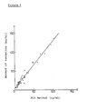
- Figure 2 is a graph showing the correlation between data measured in Example 1 and that by RIA,
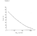
- Figure 3 is a calibration curve of PGE-MUM,
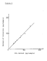
- Figure 4 is a graph showing the correlation between data measured in Example 2 and that by RIA.
- Figure 5 is a calibration curve of TXB2,
- Figure 6 is a graph showing the correlation between data measured in Example 3 and that by RIA.
- Figure 7 is a calibration curve of L TC4, and
- Figure 8 is a graph showing the correlation between data measured in Example 4 and that by RIA.
- Accordingly, the present invention is concerned with compositions for EIA of PGs which comprises at least the following reagents:
- Reagent (A): a first antibody which has been obtained by immunizing a first animal with a prostaglandin conjugated to a protein,
- Reagent (B): an antigen consisting of enzyme-labelled PGs, and
- Reagent (C): a second antibody, which has been obtained by immunizing a second animal with y-globulin or immunoglobulin G from the first animal, linked to a solid phase support,
and methods for determination of PGs by using them. - In the compositions of the present invention, Reagent (A), (B) and (C) hereinbefore described are essential constituents. The present compositions may, if necessary, comprise standard PGs for preparing a calibration curve, a set of reagents required for measuring the activity of labelled enzymes(e.g. substrate, solvent for the substrate and stopper of enzymatic reaction), buffer solutions as subsidiary constituents.
- The term PGs as used in the present invention, i.e. PGs which can be determined by using the compositions of the Present invention, refers to derivatives of prostanoic acid having a PC skeleton and to any PGs such that an antibody can be formed when immunizing an animal with the conjugate of a functional group in PGs (e.g. a carboxyl group or an amino group) and a protein. Any PG which forms part of the so- called Arachidonate Cascade and which exists in a stable form may be included. Examples of such PGs are PG compounds, thromboxane (abbreviated as TX hereinafter) compounds and leukotriene (abbreviated as LX hereinafter) compounds, e.g. PGA1, PGA2, PGA3, PCB1, PGB2, PGB3, PGC1, PGC2, PGC3, PGD1, PGD2, PGD3, PGEi, PGE2, PGE3, PGF1α, PGF2a, PGF3a, PGI1, PG12, PG13, TXB1, TXB2, TXB3, LTB3, LTB4, LTBs, LTC3, LTC4, LTCs, LTD3, LTD4, LTDS, LTE3, LTE4, LTEs, and their biological metabolites, for example, PGF-MUM (main urinary metabolite of PGF1α or PGF2a, having structural formula as shown in Example 1 hereinafter), PGE-MUM (main urinary metabolite of PGE, or PGE2, having the structural formula as shown in Example 2 hereinafter), 6-keto-PGF1α and 2,3-dinor-6-keto-PGF1α (metabolites of PG12), 2,3- dinor-TXB2 (metabolite of TXB2). All compounds exemplified herein are, or are considered to be, biological substances or their metabolites. Further, in PGs used in the present invention, derivatives which are prepared by chemical modification of the biological substance described hereinbefore, and their metabolites may be included. Preferred examples of PGs used in the present invention are PGAs, PGEs, PGFs, PGls, TXBs, LTBs, LTCs, LTDs, LTEs and their metabolites, and more preferable examples of PGs are PGEs, PGFs, TXBs, LTCs and their metabolites, e.g. PGF-MUM, PGE-MUM, 6-keto-PGF1α, 2,3-dinor-6-keto-PGF1α and 2,3-dinor-TXB2.
- The process for the preparation of reagents constituting the present invention is as follows.
- The first antibody of Reagent (A) may be prepared by subjecting a carboxyl group, an amino group or a characterized group newly introduced, of PGs to a conjugation reaction with a characterized group of a protein such as an amino group, a mercapto group, or a hydroxy group, emulsifying the resulting conjugate of PG and protein with an appropriate adjuvant, administering the emulsion to the first animal to immunize, and then collecting serum. Examples of suitable proteins are albumin, globulin, cycloglobulin, hemocyanin and, edestin preferably albumin. The conjugating-reaction of a hapten (i.e. PGs) with a protein is known, and, for example, is described in literature "A", at page 82 in particular. That is, the reaction may be carried out in a suitable solvent (e.g. phosphate buffer), when PGs can be directly conjugated with a protein, by employing a method using a carbodiimide or a method using an acid anhydride, preferably by employing the method using cyclohexylcarbodiimide or using 1-ethyl-3-(3-dimethylaminopropyl)carbodiimide as a conjugating agent, or when PGs can not be directly conjugated with a protein, by employing the method using a spacer known in the field, e.g. 1,5-difluoro-2,4-dinitrobenzene. After reaction, the resulting conjugate may be isolated and purified by column chromatography. Any adjuvants known in this field can be used, for example, complete or incomplete Freund's adjuvant or aluminium hydroxide.
- The method for immunization is also known. That is, the immunization is carried out by administering an emulsion of the conjugate of PGs and protein, with the above adjuvant to a suitable animal (e.g. rats, mice, guinea-pigs, rabbits, cats, dogs, sheep and goats) at an appropriate interval for several months. After immunization, serum from the animal may be collected by known methods to obtain the desirable first antibody (Rinshokensa, vol. 26(7), page 777 (1982)) (abbreviated as the literature "B" hereafter).
- Practically, many PGs having an analogous chemical structure other than the PGs to be measured also exist in the sample in many cases. Accordingly, it is necessary to obtain an antibody having a high specificity to the PGs to be measured. For example, PGF-MUM has two carboxyl groups. Therefore, an antibody having high specificity to PGF-MUM may be obtained by protecting the carboxyl group in a-chain in form or δ-lactone and then conjugating with a protein. An antibody having high specificity to PGE-MUM may be obtained by converting PGE-MUM into the bicyclo compound thereof by alkaline treatment, and then conjugating with a protein. In LTC4, an antibody having high specificity and selectivity cannot be obtained by the mixed acid anhydride method because of three carboxyl groups in the structure of LTC4 -(cf. structural formula as shown in Example 4 hereinafter). Giving attention to only one amino group in the molecular, an antibody having high selectivity can be obtained by conjugating the amino group with a protein using a spacer.
- The antigen of Reagent (B) may be prepared by labelling PGs with an enzyme. That is, the binding of PGs to an enzyme may be carried out by using the conjugation-reaction of PGs with a protein as hereinbefore described in processes for the preparation of Reagent (A). It, preferably, may be carried out by a method using a mixed acid anhydride using isobutylchloroformate or by a method using 4-(N-maleimidomethyl)cyclohexane-1-carboxylic acid N-hydroxysuccineimide ester as a spacer. PGs to be used herein may be those to be measured or any other PGs. Enzymes to be used herein may be any enzymes which are generally used in EIA except for acetylcholine esterase derived from electrophones electricus, for example, malate dehydrogenase, glucose-6-phosphate dehydrogenase, glucose oxidase, peroxidase, acetylcholine esterase from species other than electrophorus electricus, alkaline phosphatase, glucoamylase, lysozyme, /3-D-galactosidase, preferably 6-D-galactosidase (cf. the literature "A", page 67). In order to enhance the specificity of enzyme-labelled PGs, techniques as hereinbefore described in processes for the preparation of Reagent (A) may be carried out. The sensitivity of determination may, if necessary, be enhanced by changing the quantity of PGs to be bound to an enzyme.
- The second antibody of Reagent (C) may be prepared by administering -y-globulin or immunoglobulin G of the same species of animal as used for immunization in preparing the first antibody of Reagent (A), to another species of animal, and then immunizing by generally known methods (cf. the literature "B"). The antibody for the serum of another species of animal is on the market, and may be used. The linkage of the second antibody to a support may be carried out by generally known methods, that is, by physical adsorption or by chemical bond. Any support materials generally used for EIA may be used in the present invention, for example, a polysaccharide such as agarose, dextran and cellulose, a synthetic resin such as polystyrene, glass or polyacrylamide. Any shapes easily separable may be taken for support of the Present invention, for example, small spheres, small test tubes, tubes, fibers, microplates. Polystyrene balls or glass beads are preferred (cf. "Koteika Koso", edited by Ichiro Chihata, published by Kohdansha (1975) in detail).
- The standard PGs for preparing a calibration curve, a set of reagents required for measuring the activity of the labelled enzyme and buffer solutions, which may be contained in the compositions of the present invention, may be prepared by methods known per se.
- The present invention, furthermore, relates to methods for determination of PGs by using the composition of this invention. The methods for determination may be carried out as follows:
- (i) by mixing Reagent (A), Reagent (B) and PGs in a sample in order to effect a competitive antigen-antibody reaction (defined as the first reaction), and adding Reagent (C) to the reaction mixture to bind the antigen-antibody complex to the second antibody adsorbed on the support (defined as the second reaction), and then measuring the enzyme activity of the enzyme-labelled PGs, or
- (ii) by subjecting Reagent (A) and PGs in a sample to an antigen-antibody reaction (defined as the first reaction), and adding Reagent (C) to the antigen-antibody complex thus obtained to bind the complex to the second antibody adsorbed on the support (defined as the second reaction), and adding Reagent (B) thereto to bind competitively enzyme-labelled antigen (defined as the third reaction), and then measuring the enzyme activity of enzyme-labelled PGs.
- Both methods may be preferably used. It is necessary to use either method depending on the substances to be measured. When PGF-MUM, PGE-MUM or TXB2 is measured, the method (ii) is preferred and when LTC4 is measured, the method (i) is preferred.
- The first reaction in the method (i), and the first and second reaction in the method (ii) may be carried out at a temperature from 25 C to 37° C for one to four hours. The sensitivity of determination is not influenced in the range of temperature and time of the reaction hereinbefore described and stable measurement is possible. The second reaction in the method (i) and the third reaction in the method (ii) may be carried out at 4 C overnight.
- The method for measuring the enzyme activity nay be carried out by methods known per se (cf. the literature "A", page 21).
- The following Examples illustrate, but do not limit, the present invention in detail.
- PGF-MUM has the following structural formula:

- The δ-lactone compound of PGF-MUM has the following structural formula and may be easily prepared chemically.

- 4 mg of δ-lactone compound of PGF-MUM and 8 mg of bovine serum albumin (abbreviated as BSA hereafter) were dissolved in each 4 ml of 0.01 M phosphate buffer (pH 7.3) and both solutions were mixed. After adding thereto 4 mg of 1-ethyl-3-(3-dimethylaminopropyl) carbodiimide hydrochloride, the mixture was adjusted to pH 5.5 by adding hydrochloric acid and reacted at 4° C for 24 hours under an atmosphere of nitrogen. The reaction mixture thus obtained was dialyzed at 4°C for 24 hours against 0.01 M phosphate buffer (pH 7.3) containing 0.15M sodium chloride and then was lyophilized to obtain 8.4 mg of conjugate. The conjugation ratio of PGF-MUM for BSA was calculated by adding 3H-PGF-MUM to reaction system and was 10 mol PGF-MUM/mol BSA. 0.5 ml of complete Freund's adjuvant was added to a solution of one mg of the conjugate in 0.5 ml of physiological saline solution and emulsified. The emulsion thus obtained was administered intradermally to dorsal skin and paws of male Japanese white rabbits to immunize. The immunization was carried out six times at two-week intervals and blood was collected from the carotid artery after two weeks from the latest immunization. Blood thus obtained was allowed to stand for 30 minutes at room temperature and the serum was collected by centrifugation to obtain the first antibody. The first antibody thus obtained was stored at -20 C after lyophilization.
- A mixture of 2.5 µl of tri-n-butylamine in 25 µl of dioxane, 1.3 µl of isobutylchloroformate in 25 µl of dioxane and 2 mg of δ-lactone compound of PGF-MUM in 50 µl of dioxane was reacted at 10°C for 30 minutes. The reaction mixture was added dropwise at 10° C over 10 minutes to a solution of 0.4 mg of β-D-galactosidase (manufactured by Boehringer Mannheim) in 0.7 ml of 0.5% sodium bicarbonate. The solution thus obtained was allowed to stand at 4° C for two hours under an atmosphere of nitrogen and purified by Sephadex G-25@ column (manufactured by Pharmacia) (1.0 x 20 cm) to obtain FMO.
- IgG (i.e. immunoglobulin G) fraction of the second antibody was prepared by employing DEAE-cellulose column chromatography and Na2S04 fraction from sheep anti rabbit γ-globulin antiserum (prepared by Daiichi Radioisotope) (Cf. Men-eki Jikken Sohsa-hou, edited by the Japanese Immunology Association, vol. IV, page 1137 (1975)). 3000 polystyrene balls were soaked in 500 ml of 0.05M phosphate buffer (pH 7.4) containing 1 mg/ml of the second antibody IgG and were allowed to stand for two hours at room temperature and then overnight at 4 C for absorption to obtain desired DASP ball.
- A kit comprises the following reagents:
- (a) Buffer Solution:
- 250 ml of 30 mM phosphate buffer (pH 6.8) containing 0.3M sodium chloride, 3 mM magnesium chloride, 0.3% BSA and 0.3% of sodium azide. The solution should be diluted with water three times prior to use (defined as "Buff-A" hereafter).
- (b) Standard PGF-MUM:
- A solution of 500 ng of PGF-MUM dissolved in one ml of Buff-A solution from which BSA is removed. (C) The First Antibody:
- 60 ml of a solution that the first antibody prepared in (1) hereinbefore described was diluted with Buf-A solution 400,000 times.
- (d) FMO:
- 12 ml of Buff-A solution containing FMO prepared in (2) hereinbefore described in concentration of 60 ng/ml.
- (e) DASP Ball:
- 100 DASP ball prepared in (3) hereinbefore described, soaked in 100 ml of Buff-A solution.
- (f) Substrate of Enzyme:
- Two vials in which 4-methylumbelliferyl-β-D-galactoside was dissolved in 12 ml of 10 mM phosphate buffer (pH 7.0) containing 0.5% mannite, being in concentration of 0.5 mM, and then the obtained solution was lyophilyzed. The contents should be diluted with 30 ml off Buff-A solution to be in concentration of 0.2 mM prior to use.
- (g) Stopper of Enzymatic Reaction:
- 30 ml of 1 M glycine buffer (pH 10.5). The solution should be diluted with water ten times prior to use.
- Frozen urine was thawed and centrifuged for ten minutes (3000 rpm). Its supernatant was used as an urine sample.
- 0.5 ml of the first antibody (reagent (c) in the kit) was added to each 20 µl of the urine sample or standard PGF-MUM having various concentrations (reagent (b) in the kit) and the solution was reacted for one hour at 37° C (defined as the first reaction). DASP balls (reagent (e) in the kit) were added to the first reaction mixture (one DASP ball/sample) and reacted for two hours at 37° C (defined as the second reaction). 0.1 ml of FMO (reagent (d) in the kit) was added to the second reaction mixture containing DASP ball and the mixture was reacted overnight at 4*C (defined as the third reaction). After reaction, the supernatant was discarded and the remaining DASP ball was washed twice with 2.5 ml of Buff-A solution (reagent (a) in the kit) and then the ball was reacted in 0.5 ml off the substrate of enzyme (reagent (f) in the kit) for 30 to 60 minutes at 37° C in order to measure the activity off β-D-galactosidase bound to DASP ball. The reaction was stopped by adding 2.5 ml of the stopper of enzymatic reaction (reagent (g) in the kit). Fluorescence intensity of the produced 4-methylumbelliferone was measured. The wavelengths fixed for excitation and emission was 360 and 450 nm, respectively. The calibration curve is shown in Figure 1.
- It was found that PGF-MUM in urine can be measured in the range of 10 to 5000 pg by using the composition of the present invention.
- The correlation between the concentration of PGF-MUM measured in Example 1 (Y) (50 samples) and that measured by the RIA method (X) (described in Prostaglandins, vol. 10, page 549 - 555 (1975)) is shown in Figure 2.

- Correlation coefficient: -y = 0.9606 These values shows that the correlation between the two methods is excellent.
- The concentration of PGF-MUM in urine of healthy human, measured by using the composition of Example 1 was in the range from 3.1 to 60.0 ng/ml (mean value: 24.1 ± 13.2 ng/ml, n = 21).
- PGE-MUM has the following structural formula:

- PGE-MUM may be easily converted into the stable bicyclo compound of PGE-MUM having the following structural formula by alkaline treatment.

- By the same procedure as described in Example 1-(1), the first antibody was prepared by using 4 mg of the bicyclo compound of PGE-MUM and 6 mg of BSA. The conjugation ratio of PGE-MUM for BSA was 10 mol bicyclo compound of PGE-MUM/mol BSA.
- By the same procedure as described in Example 1-(2), EMO was prepared by using 100 µg of the bicyclo compound of PGE-MUM and 0.4 mg of e-D-galactosidase.
- DASP ball was prepared by the same procedure as described in Example 1-(3).
- A kit comprises the following reagents:
- (a) Buffer solution-1:
- 250 ml of the buffer solution prepared in Example 1-(4)-(a). The solution should be diluted with water three times prior to use (defined as "Buf-A" hereafter).
- (b) Buffer solution-2:
- 500 ml of 50 mM phosphate buffer (pH 7.0) containing 0.1% sodium azide.
- (c) Buffer solution-3:
- 100 ml of Buff-A solution containing 0.05% gelatin.
- (d) Solution for conversion:
- 10 ml of 4M(4N) sodium hydroxide solution.
- (e) Solution for neutralization:
- 10 ml of 4M(4N) hydrochloric acid
- (f) Standard PGE-MUM:
- A solution of 500 ng of the bicyclo compound of PGE-MUM dissolved in one ml of Buff-A solution from which BSA is removed.
- (g) The First Antibody:
- 60 ml of a solution that the first antibody prepared in (1) hereinbefore described was diluted with Buf-A solution 300,000 times.
- (h) EMO:
- 12 ml of Buff-A solution containing EMO prepared in (2) hereinbefore described, in concentration of 500 ng/ml.
- (i) DASP Ball:
- DASP ball prepared by the same procedure as described in Example 1-(4)-(e).
- (j) Substrate of Enzyme:
- Substrate of enzyme prepared by the same procedure as described in Example 1-(4)-(f).
- (k) Stopper of Enzymatic Reaction:
- Stopper prepared by the same procedure as described in Example 1-(4)-(g).
- As pre-treatment, to 0.1 ml of the supernatant obtained by thawing the freezed urine and then by centrifuging, was added 0.1 ml of the solution for conversion (reagent (d) in the kit) and the obtained solution was reacted for one hour at room temperature. To the reaction solution was added 0.1 ml of the solution for neutralization (reagent (e) in the kit) and the obtained solution was diluted with 5 ml of the buffer solution-2 (reagent (b) in the kit) to obtain an urine sample.
- 0.5 ml of the first antibody (reagent (g) in the kit) was added to each 0.1 ml of the urine sample or standard PGE-MUM having various concentration (reagent (f) in the kit) and the mixture was reacted for one hour at 37° C (defined as the first reaction). DASP balls (reagent (i) in the kit) were added to the first reaction mixture (one DASP ball/sample) and reacted for two hours at 37° C (defined as the second reaction). 0.1 ml off EMO (reagent (h) in the kit) was added to the second reaction mixture containing DASP ball and the mixture was reacted overnight at 4°C (defined as the third reaction). After reaction, the supernatant was discarded and the remaining DASP ball was washed twice with 2.5 ml of Buff-A solution (reagent (a) in the kit) and then the activity of jS-D-gaiactosidase bound to DASP ball was measured by the same procedure as described in Example 1-(5). The calibration curve is shown in Figure 3.
- It was found that the bicyclo compound of PGE-MUM in urine can be measured in the range of 0.5 to 2000 pg by using the composition of the present invention.
- The correlation between the concentration of the bicyclo compound of PGE-MUM measured in Example 2 (Y) (39 samples) and that measured by the RIA method (X) (described in Advance in Prostaglandins, Thromboxane, and Leukotriene Research, vol. 11, page 191 - 196 (1983)) is shown in Figure 4.

- Correlation coefficient: y = 0.9841 These values shows that the correlation between the two methods is excellent.
-

- By the same procedure as described in Example 1-(1), the first antibody was prepared by using 4 mg of TXB2 and 8 mg of BSA. The conjugation ratio of TXB2 for BSA was 9.4 mol TXB2/mol BSA.
- By the same procedure as described in Example 1-(2), TBG was prepared by using 250 µg of TXB2 and 0.4 mg of β-D-galactosidase.
- After about 1000 glass beads were soaked in the detergent containing proteolytic .enzyme overnight for washing enough, they were dried for 5 hours at 500 C. The beads thus obtained were soaked in 100 ml of acetone containing 2% 3-aminopropyltriethoxysilane, allowed to stand for 24 hours at 45 C, and then washed with acetone several times and dried at room temperature. The beads thus obtained were soaked in 1% aqueous solution of glutaraldehyde allowed to stand for one hour at room temperature, and then washed enough with water and 0.25M phosphate buffer (pH 7.5), successively. The beads thus obtained were soaked in 100 ml of 0.25M phosphate buffer (pH 7.5) containing 13 mg of the second antibody IgG (prepared in Example 1-(3)), and were allowed to stand overnight at 4°C for absorption to obtain desired DASP ball.
- A kit comprises the following reagents:
- (a) Buffer solution:
- 250 ml of the buffer solution prepared in Example 1-(4)-(a). The solution should be diluted with water three times prior to use (defined as "Buf-A" hereafter).
- (b) Standard TXB2:
- A solution off 500 ng of TXB2 dissolved in one ml of Buff-A solution from which BSA is removed.
- (c) the First Antibody:
- 60 ml of a solution that the first antibody prepared in (1) hereinbefore described was diluted with Buf-A solution 200,000 times.
- (d) TBG:
- 12 ml of Buf-A solution containing TBG prepared in (2) hereinbefore described, in concentration of 120 ng/ml.
- (E) DASP Ball:
- 100 DASP ball prepared in (3) hereinbefore described, soaked in 100 ml of Buf-A solution.
- (f) Substrate of Enzyme:
- Substrate of enzyme prepared by the same procedure as described in Example 1-(4)-(f).
- (g) Stopper of Enzymatic Reaction:
- Stopper prepared by the same procedure as described in Example 1-(4)-(g).
- Abdominal ascites of rats from which blood cell was removed by centrifugation, were diluted with Buf-A solution (reagent (a) in the kit) 10 times to be used as an sample.
- 0.5 ml of the first antibody (reagent (c) in the kit) was added to each 0.1 ml of the sample or standard TXB2 having various concentration (reagent (b) in the kit) and the mixture was reacted for one hour at 37° C (defined as the first reaction). DASP balls (reagent (e) in the kit) were added to the first reaction mixture (one DASP ball/sample) and reacted for two hours at 37° C (defined as the second reaction). 0.1 ml of TBG (reagent (d) in the kit) was added to the second reaction mixture containing DASP ball and the mixture was reacted overnight at 4` C (defined as the third reaction). After reaction, the supernatant was discarded and the remaining DASP ball was washed twice with 2.5 ml of Buf-A solution (reagent (a) in the kit) and then the activity of β-D-galactosidase bound to DASP ball was measured by the same procedure as described in Example 1-15). The calibration curve is shown in Figure 5.
- It was found that TXB2 in abdominal ascites can be measured in the range of 2 to 1000 pg by using the composition of the present invention.
- The correlation between the concentration of TXB2 measured in Example 3 (Y) (8 samples) and that measured by the RIA method (X) is shown in Figure 6.
- Y = 0.996X + 0.430
- Correlation coefficient: -y = 0.9947 These value shows that the correlation between two methods is excellent.
-

- A solution of 10 mg of 1,5-difluoro-2,4-dinitrobenzene (DFDNB) in 0.7 ml of methanol was added to a solution of 2.5 mg of LTC4 in 0.5 ml of 0.1 M phosphate buffer (pH 7.2, degassed and replaced by argon gas) and the mixture was stirred for 30 minutes at 24° C. After reaction, methanol was removed by using argon gas and unreacted DFDNB was removed from the reaction mixture by the extraction with diethyl ether (0.5 ml x 3 times). After diethyl ether remaining in the solution was removed by using argon gas, a solution of 10 mg of BSA in one ml of 0.2M borate buffer (pH 8.5, degassed and replaced by argon gas) was added thereto, and the solution thus obtained was allowed to stand for two days at room temperature under an atmosphere of argon with shading the light. After reaction, the reaction mixture was purified by Sephadex G-25@ column (manufactured by Pharmacia) (1.0 x 50 cm) with shading the light by using distilled water (degassed and replaced by argon gas) as eluent to obtain a protein fraction. The obtained fraction was lyophilized to obtain 9.8 mg of conjugate. The conjugation ratio of L TC4 for BSA was 8.14 mol LTC4/mol BSA. One ml of incomplete Freund's adjuvant was added to a solution of one mg of the conjugate thus obtained in 0.5 ml of physiological saline solution and emulsified under an atmosphere of argon. The obtained emulsion was administered subcutaneously to the dorsal neck of rabbits at 15 points to immunize. The immunization was carried out four times at three-week intervals and blood was collected after 10 days from the latest immunization and then the serum was collected by centrifugation to obtain the first antibody.
- A solution of one mg of 4-(N-maleimidomethyl)-cyclohexane-1-carboxylic acid N-hydroxysuccinimide ester (MCAE) in 0.1 ml of tetrahydrofuran was added to a solution of 100 ng of LTC4 in one ml of 0.075M phosphate buffer (pH 7.2, degassed and replaced by argon gas) and the mixture was reacted for 30 minutes at 24° C under an atmosphere of argon. After reaction, tetrahydrofuran was removed by using argon gas and unreacted MCAE was removed from the reaction mixture by the extraction with diethyl ether (1 ml x 3 times). Diethyl ether remaining in the aqueous layer was completely removed by using argon gas to obtain cyclohexylmaleimide derivative of LTC4. The derivative thus obtained was added dropwise to a solution of 1.5 mg of 6-D-galactosidase in one ml of 0.075M phosphate buffer (pH 7.2, degassed and replaced by argon gas) under an atmosphere of argon. After addition the mixture was stirred for 30 minutes at 24° C and the reaction mixture thus obtained was purified by Sephadex G-25@ column (manufactured by Pharmacia) (1.0 x 50 cm) to obtain LCG.
- DASP ball was prepared by the same procedure as described in Example 1-(3).
- A kit comprises the following reagents:
- (a) Buffer solution:
- 250 ml of the buffer solution prepared in Example 1-(4)-(a). The solution should be diluted with water three times prior to use (defined as "Buf-A" hereafter).
- (b) Standard LTC4:
- A solution of 500 ng of LTC4 dissolved in one ml of Buf-A solution (degassed and replaced by argon gas) from which BSA is removed.
- (c) the First Antibody:
- 60 ml of a solution that the first antibody prepared in (1) hereinbefore described was diluted with Buf-A solution 15,000 times.
- (d) LCG:
- 12 ml of Buf-A solution (degassed and replaced by argon gas) containing 1800 ng of LCG prepared in
- (2) hereinbefore described.
- (e) DASP Ball:
- DASP ball prepared by the same procedure as described in Example 1-(4)-(e).
- (f) Substrate of Enzyme:
- Substrate of enzyme prepared by the same procedure as described in Example 1-(4)-(f).
- (g) Stopper of Enzymatic Reaction:
- Stopper prepared by the same procedure as described in Example 1-(4)-(g).
- Abdominal ascites of rats pretreated by the same procedure as described in Example 3-(5) were used as a sample.
- 0.5 ml of the first antibody (reagent (c) in the kit) and 0.1 ml of LCG (reagent (d) in the kit) were added to each 0.1 ml of the sample or standard LTC4 having various concentration (reagent (b) in the kit). The mixture was reacted for one hour at 25 C (defined as the first reaction). DASP balls (reagent (e) in the kit) were added to the first reaction mixture (one DASP ball/sample) and reacted overnight at 4°C. After reaction, the supernatant was discarded and the remaining DASP ball was washed twice with 2.5 ml of Buf-A solution (reagent (a) in the kit) and then the activity of β-D-galactosidase bound to DASP ball was measured by the same procedure as described in Example 1-(5). The calibration curve is shown in Figure 7.
- It was found that LTC4. in abdominal ascites can be measured in the range of 10 to 5000 pg by using the composition of the present invention.
- The correlation between the concentration of LTC4 measured in Example 4 (Y) (12 samples) and that measured by the RIA method (X) is shown in Figure 8.

- Correlation coefficient: y = 0.979 These values shows that the correlation between the two methods is excellent.
- As can be understood from the Examples, trace amounts of the substance in samples can be measured by more simplified procedure by using the compositions for measurement of the present invention. Furthermore, it has now been found that the method of the present invention is surprisingly excellent in the range and limitation of detection, and assay precision.
- The method of the present invention can be compared with conventional methods in the range and limitation of detection, and assay precision as shown in the following Table 1.

- As understood from the Table, the determination by the method of the present invention:
- (i) is superior to that by RIA by a factor of 2 to 8 in the limitation of detection,
- (ii) is superior to that by the Double Antibody Liquid Phase method by a factor 2 to 20 in assay precision, and
- (iii) has the almost same level of the range and limitation of detection as the determination by the DASP method using crenbuterol as a sample, and moreover is highly superior to that by the DASP method
- using pepleomycin or mytomicin C as a sample in the range and limitation of detection. Some examples
- that the DASP method may be applied to the determination of some substances to be measured other
- than those shown in the Table, are already known. However, it is a fact that the range and limitation of detection should be greatly affected by the type of substance to be measured.
- Apart from the above, the determination by the method of the present invention has the following characteristics.
- (1) The pretreatment is simpler as compared with GC-MS, and many samples can be measured at the same time.
- (2) The treatment by specialists in a specially controlled laboratory,as required in RIA, is unnecessary, and the enzyme-labelled reagent is stable for a long time.
- (3) The first antibody is not required in as great a quantity as that required in the Solid Phase Antibody method and, therefore, it is economical. Further, the present method is hardly affected by contaminants in the sample.
- (4) The present method has wider applicability than both the Solid Phase Antibody method and the Double Antibody Liquid Phase method because the second antibody for the first antibody should be linked to a solid phase support. That is simply by changing the type of first antibody, compositions for measuring various PGs may be obtained by using in common the second antibody linked to a solid phase support, i.e. Reagent (C).
- (5) A large quantity of the second antibody is not required as compared with the Double Antibody Liquid Phase method,and it is easy to separate "bound" from "free" (No complicated procedures such as centrifugation are required.)
- (6) The Double Antibody Solid Phase method is superior to the Double Antibody Liquid Phase method in reproducibility, and it is possible to develop the autoanalytic system. For example, procedures such as the washing and movement of balls can be easily carried out by burying a magnet in the ball. Furthermore, when the second antibody is linked to each well of a microplate, it is possible to effect immuno-reaction in the said wells and to measure the enzyme activity automatically per well.
Claims (11)
Priority Applications (1)
| Application Number | Priority Date | Filing Date | Title |
|---|---|---|---|
| AT85304434T ATE67603T1 (en) | 1984-06-28 | 1985-06-20 | COMPOSITIONS FOR THE IMMUNOENZYMATIC ASSAY OF PROSTAGLANDINS. |
Applications Claiming Priority (2)
| Application Number | Priority Date | Filing Date | Title |
|---|---|---|---|
| JP131898/84 | 1984-06-28 | ||
| JP59131898A JPS6111664A (en) | 1984-06-28 | 1984-06-28 | Reagent composition for enzyme immunomeasurement of prostaglandin and measuring method of prostaglandin using said composition |
Publications (3)
| Publication Number | Publication Date |
|---|---|
| EP0166583A2 EP0166583A2 (en) | 1986-01-02 |
| EP0166583A3 EP0166583A3 (en) | 1987-08-05 |
| EP0166583B1 true EP0166583B1 (en) | 1991-09-18 |
Family
ID=15068736
Family Applications (1)
| Application Number | Title | Priority Date | Filing Date |
|---|---|---|---|
| EP85304434A Expired - Lifetime EP0166583B1 (en) | 1984-06-28 | 1985-06-20 | Compositions for enzyme immunoassay of prostaglandins |
Country Status (4)
| Country | Link |
|---|---|
| EP (1) | EP0166583B1 (en) |
| JP (1) | JPS6111664A (en) |
| AT (1) | ATE67603T1 (en) |
| DE (1) | DE3584114D1 (en) |
Cited By (1)
| Publication number | Priority date | Publication date | Assignee | Title |
|---|---|---|---|---|
| US11169162B2 (en) * | 2016-03-14 | 2021-11-09 | Fujirebio Inc. | Kit and method for measuring prostaglandin E-major urinary metabolite |
Families Citing this family (9)
| Publication number | Priority date | Publication date | Assignee | Title |
|---|---|---|---|---|
| JPH02191544A (en) * | 1988-03-31 | 1990-07-27 | Sankyo Co Ltd | Immobilized antibody column |
| ES2060757T3 (en) * | 1988-03-31 | 1994-12-01 | Sankyo Co | METHOD FOR THE DETERMINATION OF ARAQUIDONIC ACID DERIVATIVES AND EQUIPMENT FOR THE SAME. |
| AU5009993A (en) * | 1992-08-11 | 1994-03-15 | Cayman Chemical Company, Inc. | Isoprostane-protein conjugates |
| US5916757A (en) * | 1994-05-31 | 1999-06-29 | Johnson & Johnson Clinical Diagnostics, Inc. | Specific binding assay using enzyme inhibitor and anti-inhibitor antibodies |
| US5589344A (en) * | 1994-06-15 | 1996-12-31 | Johnson & Johnson Clinical Diagnostics, Inc. | Test kit and method for competitive specific binding assay |
| JP2638521B2 (en) * | 1994-11-24 | 1997-08-06 | 有限会社ビー・エス・アール | Determination of liver disease by measurement of serum bile acids by ELISA |
| EP1870708B1 (en) * | 2005-04-14 | 2014-05-07 | Fujirebio Inc. | Method for determining the stage of ulcerative colitis by determining PGE-MUM levels and reagent kit therefore |
| EP3179250B1 (en) * | 2014-08-06 | 2020-02-12 | The University of Tokyo | Method and kit for testing for food allergy |
| GB202018541D0 (en) * | 2020-11-25 | 2021-01-06 | Aberystwyth Univ | Biomarkers for microbacterium avium paratuberculosis |
Family Cites Families (9)
| Publication number | Priority date | Publication date | Assignee | Title |
|---|---|---|---|---|
| JPS5242850B2 (en) * | 1972-10-12 | 1977-10-27 | ||
| JPS5235033B2 (en) | 1972-11-24 | 1977-09-07 | ||
| FR2288312A1 (en) | 1974-10-14 | 1976-05-14 | Pasteur Institut | PROGESTERONE IMMUNOENZYMATIC ASSAY PROCESS |
| IL51667A (en) * | 1977-03-16 | 1979-10-31 | Miles Yeda Ltd | Immunoassay for the determination of a hapten |
| NZ190452A (en) * | 1978-05-19 | 1981-04-24 | Becton Dickinson Co | Solid phase assay receptor contacted with analyte and then with labeled analyte |
| JPS57148253A (en) | 1981-03-09 | 1982-09-13 | Dainippon Pharmaceut Co Ltd | Quantitative kit for cancer resisting agent |
| JPS57192867A (en) | 1981-05-25 | 1982-11-27 | Teijin Ltd | Enzyme immunoassay method of clenbuterol and its analog |
| JPS58167961A (en) | 1982-03-29 | 1983-10-04 | Dainippon Pharmaceut Co Ltd | Kit and method for quantitative analysis of anti-cancer agent mitomycin c |
| FR2550799B1 (en) * | 1983-08-17 | 1986-02-21 | Commissariat Energie Atomique | COMPOUND MARKED BY AN ENZYME, ITS PREPARATION METHOD AND ITS USE IN ENZYMOIMMUNOLOGY |
-
1984
- 1984-06-28 JP JP59131898A patent/JPS6111664A/en active Pending
-
1985
- 1985-06-20 AT AT85304434T patent/ATE67603T1/en not_active IP Right Cessation
- 1985-06-20 DE DE8585304434T patent/DE3584114D1/en not_active Expired - Lifetime
- 1985-06-20 EP EP85304434A patent/EP0166583B1/en not_active Expired - Lifetime
Non-Patent Citations (1)
| Title |
|---|
| PATENT ABSTRACTS OF JAPAN, vol. 10, no. 157 (P-464)[2213], 06 June 1986 * |
Cited By (1)
| Publication number | Priority date | Publication date | Assignee | Title |
|---|---|---|---|---|
| US11169162B2 (en) * | 2016-03-14 | 2021-11-09 | Fujirebio Inc. | Kit and method for measuring prostaglandin E-major urinary metabolite |
Also Published As
| Publication number | Publication date |
|---|---|
| JPS6111664A (en) | 1986-01-20 |
| EP0166583A3 (en) | 1987-08-05 |
| DE3584114D1 (en) | 1991-10-24 |
| EP0166583A2 (en) | 1986-01-02 |
| ATE67603T1 (en) | 1991-10-15 |
Similar Documents
| Publication | Publication Date | Title |
|---|---|---|
| US4629692A (en) | Immunoassay for nonenzymatically glucosylated proteins and protein fragments an index of glycemia | |
| EP0166623A2 (en) | Antibody lectin sandwich assay | |
| CA2008360A1 (en) | Myocardial infarction immunoassay | |
| CA1256025A (en) | Immuno-chemical measurement process for haptens and proteins | |
| EP0149602A1 (en) | Immunometric assay using polyclonal and monoclonal antibodies and a kit for use therein | |
| JP2517187B2 (en) | Analyte variant analysis | |
| EP0166583A2 (en) | Compositions for enzyme immunoassay of prostaglandins | |
| CA1285477C (en) | Immunochemical method for assaying stable glycosylated hemoglobin | |
| EP0304292B1 (en) | Antibody to propeptide of procollagen type III, and assay method using it | |
| EP0639272B1 (en) | Method for the determination of the amount of a thyroid hormone ligand in a biological fluid and kit for carrying out such a method | |
| JPH0664060B2 (en) | Analyte detection method and detection agent | |
| US5047330A (en) | Compound labelled by the acetyl cholinesterase of Electrophorus electricus, its preparation process and its use as a tracer or marker in enzymoimmunological determinations | |
| JPH07325083A (en) | Method for measuring ratio of specific sugar chain of glycoprotein | |
| EP0088974A2 (en) | Homogeneous immunoassay with labelled monoclonal anti-analyte | |
| US5010017A (en) | Process for the immunological quantitative determination of T3 and/or T4 thyroid hormones, using thyroglobulin | |
| DK161168B (en) | IMMUNOMETRIC PROCEDURE FOR DETERMINING HAPTENES AND ANALYTICAL METHOD FOR USING THE PROCEDURE | |
| CA1337926C (en) | Method for measuring human insulin | |
| EP0158291A2 (en) | A method for purifying carcinoembryonic antigen and a method for producing a carcinoembryonic antigen-reactive monoclonal antibody | |
| US4786591A (en) | Process for determining the binding capacity of thyroxin-binding globulin | |
| JPS5925183B2 (en) | Method for measuring elastase-1 | |
| EP0188608A1 (en) | Two site enzyme labeled cross reaction immunometric sandwich assay method | |
| JP3015121B2 (en) | Monoclonal antibody against non-A1c glycated hemoglobin | |
| JPH0326787B2 (en) | ||
| JP2657672B2 (en) | Ultrasensitive specific antibody assay | |
| JPH0587809A (en) | Measurement of saccharification ratio of specific protein |
Legal Events
| Date | Code | Title | Description |
|---|---|---|---|
| PUAI | Public reference made under article 153(3) epc to a published international application that has entered the european phase |
Free format text: ORIGINAL CODE: 0009012 |
|
| AK | Designated contracting states |
Designated state(s): AT BE CH DE FR GB IT LI LU NL SE |
|
| PUAL | Search report despatched |
Free format text: ORIGINAL CODE: 0009013 |
|
| AK | Designated contracting states |
Kind code of ref document: A3 Designated state(s): AT BE CH DE FR GB IT LI LU NL SE |
|
| 17P | Request for examination filed |
Effective date: 19880201 |
|
| 17Q | First examination report despatched |
Effective date: 19890817 |
|
| GRAA | (expected) grant |
Free format text: ORIGINAL CODE: 0009210 |
|
| AK | Designated contracting states |
Kind code of ref document: B1 Designated state(s): AT BE CH DE FR GB IT LI LU NL SE |
|
| REF | Corresponds to: |
Ref document number: 67603 Country of ref document: AT Date of ref document: 19911015 Kind code of ref document: T |
|
| REF | Corresponds to: |
Ref document number: 3584114 Country of ref document: DE Date of ref document: 19911024 |
|
| ITF | It: translation for a ep patent filed | ||
| ET | Fr: translation filed | ||
| PLBE | No opposition filed within time limit |
Free format text: ORIGINAL CODE: 0009261 |
|
| STAA | Information on the status of an ep patent application or granted ep patent |
Free format text: STATUS: NO OPPOSITION FILED WITHIN TIME LIMIT |
|
| 26N | No opposition filed | ||
| EPTA | Lu: last paid annual fee | ||
| EAL | Se: european patent in force in sweden |
Ref document number: 85304434.5 |
|
| PGFP | Annual fee paid to national office [announced via postgrant information from national office to epo] |
Ref country code: FR Payment date: 19970610 Year of fee payment: 13 |
|
| PGFP | Annual fee paid to national office [announced via postgrant information from national office to epo] |
Ref country code: GB Payment date: 19970611 Year of fee payment: 13 Ref country code: AT Payment date: 19970611 Year of fee payment: 13 |
|
| PGFP | Annual fee paid to national office [announced via postgrant information from national office to epo] |
Ref country code: SE Payment date: 19970618 Year of fee payment: 13 |
|
| PGFP | Annual fee paid to national office [announced via postgrant information from national office to epo] |
Ref country code: NL Payment date: 19970630 Year of fee payment: 13 Ref country code: DE Payment date: 19970630 Year of fee payment: 13 Ref country code: CH Payment date: 19970630 Year of fee payment: 13 |
|
| PGFP | Annual fee paid to national office [announced via postgrant information from national office to epo] |
Ref country code: LU Payment date: 19970708 Year of fee payment: 13 |
|
| PGFP | Annual fee paid to national office [announced via postgrant information from national office to epo] |
Ref country code: BE Payment date: 19970812 Year of fee payment: 13 |
|
| PG25 | Lapsed in a contracting state [announced via postgrant information from national office to epo] |
Ref country code: LU Free format text: LAPSE BECAUSE OF NON-PAYMENT OF DUE FEES Effective date: 19980620 Ref country code: GB Free format text: LAPSE BECAUSE OF NON-PAYMENT OF DUE FEES Effective date: 19980620 Ref country code: AT Free format text: LAPSE BECAUSE OF NON-PAYMENT OF DUE FEES Effective date: 19980620 |
|
| PG25 | Lapsed in a contracting state [announced via postgrant information from national office to epo] |
Ref country code: SE Free format text: LAPSE BECAUSE OF NON-PAYMENT OF DUE FEES Effective date: 19980621 |
|
| PG25 | Lapsed in a contracting state [announced via postgrant information from national office to epo] |
Ref country code: LI Free format text: LAPSE BECAUSE OF NON-PAYMENT OF DUE FEES Effective date: 19980630 Ref country code: CH Free format text: LAPSE BECAUSE OF NON-PAYMENT OF DUE FEES Effective date: 19980630 Ref country code: BE Free format text: LAPSE BECAUSE OF NON-PAYMENT OF DUE FEES Effective date: 19980630 |
|
| BERE | Be: lapsed |
Owner name: ONO PHARMACEUTICAL CO. LTD Effective date: 19980630 |
|
| PG25 | Lapsed in a contracting state [announced via postgrant information from national office to epo] |
Ref country code: NL Free format text: LAPSE BECAUSE OF NON-PAYMENT OF DUE FEES Effective date: 19990101 |
|
| GBPC | Gb: european patent ceased through non-payment of renewal fee |
Effective date: 19980620 |
|
| REG | Reference to a national code |
Ref country code: CH Ref legal event code: PL |
|
| PG25 | Lapsed in a contracting state [announced via postgrant information from national office to epo] |
Ref country code: FR Free format text: LAPSE BECAUSE OF NON-PAYMENT OF DUE FEES Effective date: 19990226 |
|
| EUG | Se: european patent has lapsed |
Ref document number: 85304434.5 |
|
| NLV4 | Nl: lapsed or anulled due to non-payment of the annual fee |
Effective date: 19990101 |
|
| PG25 | Lapsed in a contracting state [announced via postgrant information from national office to epo] |
Ref country code: DE Free format text: LAPSE BECAUSE OF NON-PAYMENT OF DUE FEES Effective date: 19990401 |
|
| REG | Reference to a national code |
Ref country code: FR Ref legal event code: ST |