EP0156578B1 - A process for preparing hybridoma cells which produce tumour specific monoclonal antibodies - Google Patents
A process for preparing hybridoma cells which produce tumour specific monoclonal antibodies Download PDFInfo
- Publication number
- EP0156578B1 EP0156578B1 EP85301678A EP85301678A EP0156578B1 EP 0156578 B1 EP0156578 B1 EP 0156578B1 EP 85301678 A EP85301678 A EP 85301678A EP 85301678 A EP85301678 A EP 85301678A EP 0156578 B1 EP0156578 B1 EP 0156578B1
- Authority
- EP
- European Patent Office
- Prior art keywords
- cells
- tumour
- antigens
- animal
- normal
- Prior art date
- Legal status (The legal status is an assumption and is not a legal conclusion. Google has not performed a legal analysis and makes no representation as to the accuracy of the status listed.)
- Expired
Links
Classifications
-
- C—CHEMISTRY; METALLURGY
- C07—ORGANIC CHEMISTRY
- C07K—PEPTIDES
- C07K16/00—Immunoglobulins [IG], e.g. monoclonal or polyclonal antibodies
- C07K16/18—Immunoglobulins [IG], e.g. monoclonal or polyclonal antibodies against material from animals or humans
- C07K16/28—Immunoglobulins [IG], e.g. monoclonal or polyclonal antibodies against material from animals or humans against receptors, cell surface antigens or cell surface determinants
- C07K16/30—Immunoglobulins [IG], e.g. monoclonal or polyclonal antibodies against material from animals or humans against receptors, cell surface antigens or cell surface determinants from tumour cells
- C07K16/3023—Lung
-
- C—CHEMISTRY; METALLURGY
- C07—ORGANIC CHEMISTRY
- C07K—PEPTIDES
- C07K16/00—Immunoglobulins [IG], e.g. monoclonal or polyclonal antibodies
- C07K16/18—Immunoglobulins [IG], e.g. monoclonal or polyclonal antibodies against material from animals or humans
- C07K16/28—Immunoglobulins [IG], e.g. monoclonal or polyclonal antibodies against material from animals or humans against receptors, cell surface antigens or cell surface determinants
- C07K16/30—Immunoglobulins [IG], e.g. monoclonal or polyclonal antibodies against material from animals or humans against receptors, cell surface antigens or cell surface determinants from tumour cells
- C07K16/3046—Stomach, Intestines
-
- Y—GENERAL TAGGING OF NEW TECHNOLOGICAL DEVELOPMENTS; GENERAL TAGGING OF CROSS-SECTIONAL TECHNOLOGIES SPANNING OVER SEVERAL SECTIONS OF THE IPC; TECHNICAL SUBJECTS COVERED BY FORMER USPC CROSS-REFERENCE ART COLLECTIONS [XRACs] AND DIGESTS
- Y10—TECHNICAL SUBJECTS COVERED BY FORMER USPC
- Y10S—TECHNICAL SUBJECTS COVERED BY FORMER USPC CROSS-REFERENCE ART COLLECTIONS [XRACs] AND DIGESTS
- Y10S424/00—Drug, bio-affecting and body treating compositions
- Y10S424/806—Drug, bio-affecting and body treating compositions involving IgM
- Y10S424/807—Monoclonal
-
- Y—GENERAL TAGGING OF NEW TECHNOLOGICAL DEVELOPMENTS; GENERAL TAGGING OF CROSS-SECTIONAL TECHNOLOGIES SPANNING OVER SEVERAL SECTIONS OF THE IPC; TECHNICAL SUBJECTS COVERED BY FORMER USPC CROSS-REFERENCE ART COLLECTIONS [XRACs] AND DIGESTS
- Y10—TECHNICAL SUBJECTS COVERED BY FORMER USPC
- Y10S—TECHNICAL SUBJECTS COVERED BY FORMER USPC CROSS-REFERENCE ART COLLECTIONS [XRACs] AND DIGESTS
- Y10S530/00—Chemistry: natural resins or derivatives; peptides or proteins; lignins or reaction products thereof
- Y10S530/806—Antigenic peptides or proteins
-
- Y—GENERAL TAGGING OF NEW TECHNOLOGICAL DEVELOPMENTS; GENERAL TAGGING OF CROSS-SECTIONAL TECHNOLOGIES SPANNING OVER SEVERAL SECTIONS OF THE IPC; TECHNICAL SUBJECTS COVERED BY FORMER USPC CROSS-REFERENCE ART COLLECTIONS [XRACs] AND DIGESTS
- Y10—TECHNICAL SUBJECTS COVERED BY FORMER USPC
- Y10S—TECHNICAL SUBJECTS COVERED BY FORMER USPC CROSS-REFERENCE ART COLLECTIONS [XRACs] AND DIGESTS
- Y10S530/00—Chemistry: natural resins or derivatives; peptides or proteins; lignins or reaction products thereof
- Y10S530/808—Materials and products related to genetic engineering or hybrid or fused cell technology, e.g. hybridoma, monoclonal products
-
- Y—GENERAL TAGGING OF NEW TECHNOLOGICAL DEVELOPMENTS; GENERAL TAGGING OF CROSS-SECTIONAL TECHNOLOGIES SPANNING OVER SEVERAL SECTIONS OF THE IPC; TECHNICAL SUBJECTS COVERED BY FORMER USPC CROSS-REFERENCE ART COLLECTIONS [XRACs] AND DIGESTS
- Y10—TECHNICAL SUBJECTS COVERED BY FORMER USPC
- Y10S—TECHNICAL SUBJECTS COVERED BY FORMER USPC CROSS-REFERENCE ART COLLECTIONS [XRACs] AND DIGESTS
- Y10S530/00—Chemistry: natural resins or derivatives; peptides or proteins; lignins or reaction products thereof
- Y10S530/827—Proteins from mammals or birds
- Y10S530/828—Cancer
-
- Y—GENERAL TAGGING OF NEW TECHNOLOGICAL DEVELOPMENTS; GENERAL TAGGING OF CROSS-SECTIONAL TECHNOLOGIES SPANNING OVER SEVERAL SECTIONS OF THE IPC; TECHNICAL SUBJECTS COVERED BY FORMER USPC CROSS-REFERENCE ART COLLECTIONS [XRACs] AND DIGESTS
- Y10—TECHNICAL SUBJECTS COVERED BY FORMER USPC
- Y10S—TECHNICAL SUBJECTS COVERED BY FORMER USPC CROSS-REFERENCE ART COLLECTIONS [XRACs] AND DIGESTS
- Y10S530/00—Chemistry: natural resins or derivatives; peptides or proteins; lignins or reaction products thereof
- Y10S530/863—Chemistry: natural resins or derivatives; peptides or proteins; lignins or reaction products thereof involving IgM
- Y10S530/864—Monoclonal
Definitions
- the present invention relates to a process for preparing hybridoma cells which produce monoclonal antibodies capable of binding to tumour cells of one or more types, but substantially not to normal cells.
- Such monclonal antibodies are hereinafter referred to as tumour-specific monoclonal antibodies.
- monoclonal antibodies specific for antigens which are components of human tumour cells, but not normal human cells Such monoclonal antibodies may be used for the diagnosis and treatment of appropriate human tumours and for analysis of human tumour antigens.
- antisera have been prepared which are reactive with human tumour cells by immunizing various animals with human tumour antigens of the same cells, generally the total antigens of the tumour cells or the membrane antigens of the same cells. If, however, an animal is immunized with whole tumour cells or membrane fragments of tumour cells, the antiserum which can be obtained from the immunized animal is capable of reacting with normal cells as well as tumour cells, since the majority of the immunizing antigens are common to both tumour cells and normal cells. Tumour-specific antibodies cannot be readily isolated from such an antiserum.
- tumour-specific monoclonal antibodies by using antibody-forming cells isolated from animals immunized with tumour antigens (e.g. fragments of neoplasteic tissue, isolated tumour cells or membrane fragments of tumour cells), which have previously been made immunologically tolerant (or immunologically unresponsive) to normal cell antigens, comprising the total antigens, or at least a proportion of the total antigens, of the normal cells corresponding to the tumour cells chosen as the source of the immunizing antigens.
- tumour antigens e.g. fragments of neoplasteic tissue, isolated tumour cells or membrane fragments of tumour cells
- normal cell antigens comprising the total antigens, or at least a proportion of the total antigens, of the normal cells corresponding to the tumour cells chosen as the source of the immunizing antigens.
- the normal cell antigens to which the animal chosen for immunization is made immunologically tolerant are hereinafter collectively referred to as the tolerogen).
- Hybridoma cells prepared by a process according to the present invention can be cultured in vitro or administered intraperitoneally to a syngeneic animal (i.e. cultured in vivo) for the purpose of obtaining in good yield the tumour-specific monoclonal antibody synthesized by the cells.
- the monoclonal antibody is isolated from the culture medium or ascites fluid respectively using conventional methods.
- tumour tissue fragments appropriate isolated whole tumour cells, an appropriate broken tumour cell preparation or membrane fragments thereof can be used for immunization.
- tumour antigens originating from human tumours, especially human lung, stomach, large intestine, pancreatic, liver, endometrium, cervical, kidney, bladder and cerebral tumours.
- the animal chosen for immunization will generally be a mouse or rat.
- the tolerogen it is preferred to use (a) one member selected from isolated cells, broken cells, tissue fragments and membrane fragments originating from a normal organ of the same type as that from which the tumour antigens chosen for the subsequent immunization are derived, or (b) one member selected from isolated cells, broken cells, tissue fragments and membrane fragments originating from various normal organs comprising appropriate antigens. It is possible for the tolerogen to be one member selected from isolated cells, broken cells, tissue fragments and membrane fragments originating from a normal organ of a different type than the organ from which the tumour antigens used for immunization are derived. If desired, more than one appropriate individual may be used for the preparation of the tolerogen. Induction of immunological tolerance
- Immunological tolerance to an antigen may usually be induced in an adult animal by administration of the antigen in a larger amount than normally used for immunization. Immunological tolerance to certain antigens can also be induced in adult animals by repeated administration of the antigen in a much smaller amount than normally used for immunization. In the case of a neonatal animal (e.g. 24 hours after birth), it is generally possible to induce immunological tolerance to an antigen by administration of a dose of the antigen lower than the normal adult immunization dose.
- the preferred dose of tolerogen depends both on the nature of the tolerogen and the type of animal chosen for treatment with the tolerogen. If a tolerogen of normal cells or membrane fragments of normal cells is employed, and the animal to be treated is a mouse, the preferred dose will generally be as indicated below.
- More effective and more stable immunological tolerance can be induced in a neonatal animal than an adult animal.
- tumour antigens may be effected, for example, by administration of one member selected from tumour tissue fragments, isolated tumour cells, broken tumour cells and membrane fragments of tumour cells, together with a suitable adjuvant for example, Freund's complete adjuvant, aluminium hydroxide gel or pertussis adjuvant.
- a suitable adjuvant for example, Freund's complete adjuvant, aluminium hydroxide gel or pertussis adjuvant.
- the administration may be effected by subcutaneous, intravenous or abdominal injection.
- tumour tissue fragments For immunization, tumour tissue fragments, isolated tumour cells, broken tumour cells and membrane fragments of tumour cells may be prepared as follows.
- a suitable tissue either immediately after dissection or after freezing and defrosting, is cut into small pieces (e.g. 1-2 mmxl-2 mm).
- the small fragments are implanted under the skin of the animal.
- a suitable tissue is cut into small pieces and homogenized.
- the homogenized cells are suspended in a suitable solution, for example, a PBS solution, and the cell suspension is administered to the animal chosen for immunization by, for example, subcutaneous or intraperitoneal injection (suitable immunization dose for mice: 106-10' cells).
- Suitable cells are crushed or ground and administered to the animal chosen for immunization by, for example, subcutaneous or intraperitoneal injection.
- a suitable broken cell preparation is centrifuged at 10,000 g and the resulting supernatant is further centrifuged at 100,000 g.
- the precipitated fraction is suspended in a suitable solution, for example, a PBS solution and the resulting suspension (the membrane fragment preparation) is administered to the animal chosen for immunization by, for example, subcutaneous or intraperitoneal injection.
- a suitable solution for example, a PBS solution
- the resulting suspension (the membrane fragment preparation) is administered to the animal chosen for immunization by, for example, subcutaneous or intraperitoneal injection.
- the chosen tumour antigens may be administered 2-5 times with an interval of 1-2 weeks.
- immunization is preferably carried out at 3-10 weeks after birth, for example, 8 weeks after birth.
- 3-7 days after every immunization blood may be collected from the orbital plexus vein and the reactivity of the antiserum to various isolated normal cells, normal tissues, isolated tumour cells, tumour tissues and membrane fragment preparations derived from normal and tumour cells investigated by an enzyme-linked immunosorbent assay method.
- a suitable enzyme-linked immunosorbent assay method is hereinafter described [see also "Koso Meneki Sokutei-ho", published by Igaku Shoin, 1976].
- a membrane fragment preparation of normal or tumour cells (containing 10-1000 ⁇ g/ml protein) is poured into wells of an Elisa plate with 96 wells (Flow Laboratories, U.S.A.) such that the amount per well is 100-200 pl.
- the plate is allowed to stand at 4°C over 1-2 nights, and bovine serum albumin (BSA) is coated on the bottom surface of each well.
- BSA bovine serum albumin
- each well is well-washed with deionized water or a PBS solution (disodium phosphate 1.83 g; monopotassium phosphate 0.21 g; sodium chloride 7.65 g; deionized water 1 I; pH 7.2).
- each well is thoroughly washed with deionized water or a PBS solution, and the antiserum to be tested, diluted with a BSA-PBS solution, is added to each well (100 ⁇ l/well). The plate is then allowed to stand at 4°C overnight. After washing once with deionized water and 6 times with 2M NaCI solution, an appropriate secondary IgG or F (ab') 2 -urease combined product, e.g.
- CSL Combinationwealth Serum Laboratories, Australia
- the plate is washed 3 times with deionized water, and a urease substrate solution (CSL) is then poured into each well (100 ⁇ l/well). After 10-30 minutes at room temperature, the reaction is discontinued by adding 1% methylthiolate (20 ⁇ l per well) and the antibody titre is determined by measuring the optical density at 600 nm.
- CSL urease substrate solution
- a Falcon 3072 plate is used for culturing the target cells. After addition of a 0.25% glutaraldehyde-PBS solution, the plate is allowed to stand at room temperature for 1-2 hours. After washing each well with a PBS solution, a 1 % BSA-PBS solution (100-200 pl/well) is added to the plate which is then allowed to stand for a further 2 hours. The plate is then thoroughly washed with deionized water or a PBS solution and following the addition of antiserum diluted with a BSA-PBS solution (100 ⁇ l/well), the antibody titre is determined as in (a).
- Antisera from animals immunized with tumour antigens which have previously been treated with normal cell antigens so as to induce immunological tolerance may be compared with antisera from animals not treated with the tolerogen, but identically immunized.
- Suitable animals for the preparation of antibody-forming cells are tolerogen-treated and immunized animals from which antisera are isolated with high reactivity to tumour tissue fragments, isolated tumour cells or membranes of tumour cells comprising tumour antigens used for immunization, but considerably weaker reactivity than control antisera to normal cell antigens.
- tumour antigens for example, abdominal injection.
- tumour tissue fragments or isolated tumour cells 2-5x10 6 cells, membrane fragments, 20-400 ⁇ g.
- the preferred antibody-forming cells are spleen cells which may be isolated as follows.
- Removed spleen tissue is cut into small pieces in MEM medium (Nissui Seiyaku K.K., Japan).
- MEM medium Nisui Seiyaku K.K., Japan.
- the cells are loosened by using a pincette and centrifuged (1200 r.p.m./5 min) to remove supernatant.
- the remaining material is treated with tris-ammonium chloride buffer solution (pH 7.65) for 1-2 minutes to remove the red cells.
- the spleen cells obtained by washing the material with MEM medium 3 times may be used for fusion.
- Preferred myeloma cells for the purpose of the present invention include various cells lines of mouse origin, for example, myeloma cells originating from 8 - azaguanine - resistant Balb/c mice, such as P3-X63-Ag8-U1 (P3-U1) [Current Topics in Microbiology and Immunology-1; Euro. J. Immunology, 6, 511-519 (1976)], SP2/0-Ag 14 (SP-2) [Nature, 276,269-270 (1978)], P3-X63-Ag8.653 (653) [J. Immunology, 123, 1548-1550 (1979)] and P3-X63-Ag8 (X63) [Nature, 256, 495-497 (1975)].
- These cell lines may be subcultured using 8-azaguanine medium, prepared by adding 8-azaguanine (15 pg/ml) to RPMI-1640 normal medium containing glutamine (1.5 mM), 2-mercaptoethanol (5x10 -5 M), gentamycin (10 pg/ml) and foetal calf serum (FCS; 10% CSL, Australia).
- 8-azaguanine medium prepared by adding 8-azaguanine (15 pg/ml) to RPMI-1640 normal medium containing glutamine (1.5 mM), 2-mercaptoethanol (5x10 -5 M), gentamycin (10 pg/ml) and foetal calf serum (FCS; 10% CSL, Australia).
- FCS foetal calf serum
- the preferred method for cell fusion and selection of hybridoma cells is as follows.
- the isolated antibody-forming cells and the chosen myeloma cells are well-washed with MEM medium or a PBS solution and mixed together at a ratio of 5-10:1.
- the mixture is centrifuged (1200 r.p.m./5 min) to remove supernatant and the precipitated cells are well loosened.
- a solution of polyethylene glycol 1000 (PEG 1000; 2 g), MEM (2 ml) and dimethylsulfoxide (0.7 ml) is added at 37°C with stirring (0.2-1 ml per 10 8 antibody-producing cells).
- MEM medium 1-2 ml
- the mixture is centrifuged (900 r.p.m./5 min) to remove supernatant and precipitated cells are loosened.
- a normal medium 100 ml
- the cells are suspended using a MoHr pipette.
- 1 ml of cell suspension is added to each well of a culture plate with 24 wells for culturing at 37°C for 24 hours, using an incubator containing 5% C0 2 gas.
- 1 ml of HAT medium prepared by adding to a normal medium hypoxanthine (10- 4 M), thymidine (1.5x10- 5 M) and aminopterin (4x10 -7 M)] is then added to each well for culturing for a further 24 hours. Following this, for 2 days, every 24 hours 1 ml of supernatant is removed from each well and replaced by the same amount of new HAT medium. Culturing is continued at 37°C for 10-14 days in a C0 2 incubator. The wells with growing colonies of hybridoma cells are selected.
- a HT medium prepared by adding to a normal medium hypoxanthine (10- 4 M) and thymidine (1.5xlO- 5 M)]. This is repeated every 24 hours for 2 days. After culturing for 3-4 days using HT medium, a portion of the supernatant is collected from each culture so as to determine the titre of anti-tumour antibodies by the enzyme immunosorbent assay method hereinbefore described. At the same time, the reactivities of the supernatants with normal cell antigens are also determined by a similar method.
- tumour tissue fragments, tumour cells or membrane fragments of tumour cells comprising the tumour antigens used for immunization
- cloning of the hybridoma cells is repeated twice by the limiting dilution method.
- hybridoma cells which secrete the monoclonal antibody are preferably administered intraperitoneally to a suitable animal.
- a mouse is intraperitoneally injected with 0.5 ml of 2,6,10,14-tetramethyipentadecane (pristane) and bred for 2 weeks to obtain a pristane-treated mouse of 8-10 weeks.
- pristane 2,6,10,14-tetramethyipentadecane
- 2-4x10 6 suitable hybridoma cells, capable of forming a tumour-specific monoclonal antibody are administered to the mouse by intraperitoneal injection and 10-21 days later the ascites fluid is collected.
- Suitable methods for the isolation of monoclonal antibodies of the IgG or IgM class from ascities fluid are as follows:
- the ascities fluid is centrifuged (3000 r.p.m./5 min) to remove the solid impurities.
- the supernatant is then subjected twice to salting-out using respectively 50% and 40% ammonium sulphate.
- the desired monoclonal antibody is recovered by chromatography using, for example, DEAE Sepharose or Protein A Sepharose (Pharmacia Fine Chemicals AB, Sweden).
- the ascities fluid is centrifuged (3000 r.p.m./5 min) to remove the solid impurities and the supernatant is then dialyzed against, for example, deionized water or 0.01 M phosphate buffer solution (pH 6.0) for 2-4 hours.
- the precipitates subsequently collected by centrifugation are dissolved in 3-5% sodium chloride solution which is then dialyzed against, for example, 0.85% sodium chloride solution for 1-2 days.
- the desired monoclonal antibody is finally recovered by chromatography using, for example, DEAE Sepharose or Protein A Sepharose.
- the isotypes of the monoclonal antibodies may be investigated by Ouchterlony's method (the double diffusion method) with reference to "Meneki-gaku Jikken-ho Nyumon, Seibutsu Kagaku Jikken-ho 15", published by Gakkai Shuppan Centre, Tokyo, p. 74 (1981).
- immunoglobulin protein may be carried out using the Folin's method with reference to the absorbance at 280 nm [1.4 (OD 280 ) ⁇ immunoglobulin 1 mg/ml]. '
- Reactivities may be determined, for example, using an enzyme-linked immunosorbent assay method, a fluorescence antibody method or an immuno-histological method. Binding to individual antigens, for example, carcinoembryonic antigen (CEA) may also be investigated.
- CEA carcinoembryonic antigen
- Tumour-specific monoclonal antibodies or antigen-binding fragments of such antibodies may be used for in vitro or in vivo diagnosis of tumours with appropriate tumour-specific antigens (for example, diagnosis of tumours by histological examination and examination of serums) and for treatment of tumours.
- appropriate tumour-specific antigens for example, diagnosis of tumours by histological examination and examination of serums
- tumour-specific monoclonal antibodies or antigen-binding fragments of such antibodies may be linked with tracer-labels (e.g. radio labels, fluorescent agents).
- tracer-labels e.g. radio labels, fluorescent agents.
- Tumour-specific monoclonal antibodies and antigen-binding fragments of tumour-specific monoclonal antibodies may also be linked to anti-tumour agents to form products commonly referred to as immunotoxins, which may be used in the treatment of tumours.
- tumour-specific monoclonal antibodies and antigen-binding fragments of such antibodies is in the purification and characterization of the tumour antigens which they bind.
- Affinity columns comprising a tumour specific monoclonal antibody or an antigen-binding fragment of such an antibody may, for example, be prepared for this purpose.
- hybridomas which secrete tumour-specific monoclonal antibodies.
- the hybridomas of one set were prepared using spleen cells isolated from neonatal Balb/c mice, which were first treated so as to induce immunological tolerance with a broken cell preparation derived from normal human lung and then immunized with a broken cell preparation derived from either human lung squamous cell carcinoma or human lung adenocarcinoma.
- the hybridomas of the second set were similarly prepared using spleen cells isolated from neonatal Balb/c mice which were first treated so as to induce immunological tolerance with a broken cell preparation derived from normal human stomach and then immunized with a broken cell preparation derived from either human stomach squamous cell carcinoma or human stomach adenocarcinoma.
- the known binding characteristics of th IgM tumour-specific monoclonal antibodies of these hybridomas are summarized in Tables 4 and 5.
- the two hybridomas of the first set designated SLC-1 and ALC-1 were deposited at the Institute for Fermentation, Osaka, Japan on 5 March 1985 under the accession numbers IFO 50044 and IFO 50045, respectively. It can be seen from the results given in Table 4 that the monoclonal antibodies produced by SLC-1 and ALC-1 are especially useful for diagnosis and treatment of human lung squamous cell carcinoma and human lung adenocarcinoma.
- Neonatal Balb/c mice 24 hours after birth, were administered with CCD-8Lu (ATCC CCL-201) lung cells of human origin (8x10 5 cells/mouse) or a broken-cell preparation derived from normal human lung (2-8 mg, calculated as protein/mouse).
- the administration was effected by intravenous, abdominal and/or subcutaneous injections. 8 weeks later, each mouse was abdominally injected with one member selected from A549 adenocarcinoma cells originating from human lung (ATCC CCL-185) (2-5x10 6 cells), a broken cell preparation derived from human lung squamous cell carcinoma (100 pg, calculated as protein) and a broken cell preparation derived from adenocarcinoma of human lung (100 ⁇ g, calculated as protein).
- the chosen immunogen was administered together with aluminium hydroxide gel (2 mg) and pertussis adjuvant (1 x10 9 cells). After an interval of 1-2 weeks, a dose of the same immunogen was abdominally administered to each animal [2-5 x 10⁶ A549 adenocarcinoma cells or 100 pg (calculated as protein) of a broken lung tumour cell preparation]. 3 ⁇ 4 days after this immunization, blood was collected from the orbital plexus vein of each animal to prepare antiserum. 8-week old Balb/c, not treated so as to induce immunological tolerance with CCD-8Lu cells or a broken lung cell preparation, were identically immunized. Antisera from these mice were compared with the antisera obtained from the tolerogen-treated mice.
- Tables 1-3 show the reactivities of particular antisera with particular cells, tissue fragments or broken cell preparations.
- the antibody titres were determined by the enzyme immunosorbent assay method hereinbefore described.
- Table 1 indicates the reactivities of two different antisera with CCD-8Lu cells and A549 adenocarcinoma cells.
- One antiserum was obtained from a mouse immunized with A549 adenocarcinoma cells after treatment with CCD-8Lu cells so as to induce immunological tolerance.
- the second antiserum was obtained from an identically immunized mouse, not treated with a tolerogen.
- Table 2 indicates the reactivities of two different antisera with broken cell preparations derived from normal lung and squamous cell carcinoma of human lung.
- One antiserum was obtained from a mouse immunised with a broken cell preparation derived from squamous cell carcinoma of human lung after treatment with a broken cell preparation of normal lung so as to induce immunological tolerance.
- the second antiserum was obtained from an identically immunized mouse, not treated with a tolerogen.
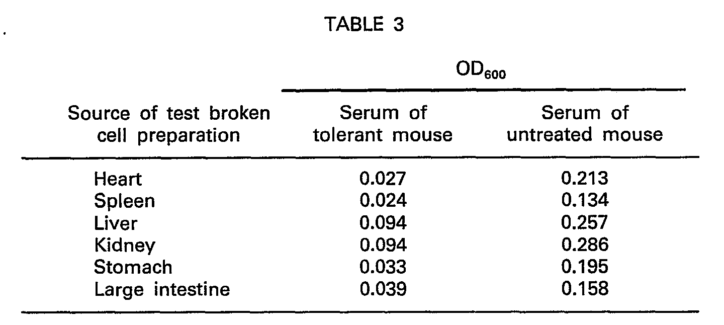
- Table 3 indicates the reactivities of two different antisera with broken cell preparations derived from various normal human organs.
- One antiserum was obtained from a mouse first treated so as to induce immunological tolerance with a broken cell preparation derived from normal human lung and then immunized with a broken lung tumour cell preparation.
- the second antiserum was obtained from an identically immunized mouse, not treated with a tolerogen.
- mice When a mouse was treated so as to induce immunological tolerance with a broken cell preparation derived from either a human stomach tumour or a human lung tumour and was then given an immunizing dose of either a broken cell preparation derived from a human stomach tumour or a human lung tumour, it was noted that the resultant mouse antiserum exhibited no reactivity with whole cells, tissue fragments or a broken cell preparation derived from a human stomach tumour or little reactivity compared with an antiserum from a mouse identically immunized, but not treated with a tolerogen.
- the control antiserum was strongly reactive with tissue fragments, and isolated whole cells or broken cells derived from a human stomach tumour.
- mice treated with a tolerogen from which antisera were obtained which exhibited no or little reactivity compared with control antisera with normal human cell antigens, but which exhibited strong reactivity with human squamous cell carcinoma and/or human lung adenocarcinoma were used for the preparation of spleen cells.
- the spleen of each selected animal was collected 3-4 days after the final immunization.
- the spleen cells obtained by step (1) and the myeloma cells obtained by step (2) were mixed together at a ratio of 5:1.
- Cell fusion was effected in the manner hereinbefore described.
- the fused cells were cultured at 37°C for 14 days using a HAT medium in a 5% C0 2 atmosphere.
- Growing hybridoma cells were selected and further cultured as hereinbefore described.
- the anti-human tumour activities of the culture supernatants were determined.
- reactivities of the supernatants with normal cell antigens were determined.
- hybridoma cell clones were obtained, which produce monoclonal antibodies capable of binding to human lung squamous cell carcinoma and/or human lung adenocarcinoma, but not normal tissues (SLC 1-6 and ALC 1-4).
- the hybridoma cells obtained by step (3) were abdominally administered to pristane-treated mice (Balb/c; female; 8 weeks after birth) in an amount of 4 X 10 6 cells per animal. 10-21 days later, ascities fluid was collected from the mice and centrifuged (3000 r.p.m./5 min) to remove solid impurities. The remaining solution was dialyzed against deionized water or a 0.01 M PBS solution (pH 6.5) for 2 hours. The residual solution was centrifuged (10,000 r.p.m./15 min) to remove. supernatant. The remaining material was dissolved in a 3% sodium chloride solution and was dialyzed against a 0.85% sodium chloride solution. The residual solution was used as a partially purified monoclonal antibody (IgM class) solution.
- IgM class monoclonal antibody
- the IgM class monoclonal antibodies of hybridoma cells of the cell lines SLC 1-6 and ALC 1-4 were tested for binding with a variety of tissues and cells and also carcinoembryonic antigen, using a conventional fluorescence method.
- a conventional fluorescence method See e.g. Immunofluorescence Technology in selected Theoretical and Clinical Aspects, ed. G. Wick, K. N. Trail and K. Schauenstein, Elsevier Biomedical Press). The results are summarized in Table 4.
- SLC-2 and ALC-2 are especially useful for diagnosis and treatment of lung cancer
- SLC-1 and ALC-1 are useful for diagnosis and treatment of lung squamous cell carcinoma and lung adenocarcinoma, respectively.
- Example 2 To obtain hybridoma cell lines which produce monoclonal antibodies capable of binding to human stomach squamous cell carcinoma and/or human adenocarcinoma, but not normal human tissues, a similar procedure to that described in Example 1 was carried out. A broken cell preparation derived from human stomach was administered to neonatal mice so as to induce immunological tolerance and the same mice were then immunized with a broken cell preparation derived from either squamous cell carcinoma of human stomach or human adenocarcinoma of human stomach. The IgM class monoclonal antibodies of 7 hybridoma cell lines which were selected were tested for binding with a variety of tissues and cells and also carcinoembryonic antigen as in Example 1. The results are summarized in Table 5.
Landscapes
- Health & Medical Sciences (AREA)
- Chemical & Material Sciences (AREA)
- Organic Chemistry (AREA)
- Immunology (AREA)
- Life Sciences & Earth Sciences (AREA)
- Biophysics (AREA)
- Biochemistry (AREA)
- General Health & Medical Sciences (AREA)
- Genetics & Genomics (AREA)
- Medicinal Chemistry (AREA)
- Molecular Biology (AREA)
- Proteomics, Peptides & Aminoacids (AREA)
- Cell Biology (AREA)
- Preparation Of Compounds By Using Micro-Organisms (AREA)
- Micro-Organisms Or Cultivation Processes Thereof (AREA)
- Medicines Containing Antibodies Or Antigens For Use As Internal Diagnostic Agents (AREA)
Description
- The present invention relates to a process for preparing hybridoma cells which produce monoclonal antibodies capable of binding to tumour cells of one or more types, but substantially not to normal cells. Such monclonal antibodies are hereinafter referred to as tumour-specific monoclonal antibodies.
- It is particularly desired to obtain monoclonal antibodies specific for antigens which are components of human tumour cells, but not normal human cells. Such monoclonal antibodies may be used for the diagnosis and treatment of appropriate human tumours and for analysis of human tumour antigens.
- Previously, antisera have been prepared which are reactive with human tumour cells by immunizing various animals with human tumour antigens of the same cells, generally the total antigens of the tumour cells or the membrane antigens of the same cells. If, however, an animal is immunized with whole tumour cells or membrane fragments of tumour cells, the antiserum which can be obtained from the immunized animal is capable of reacting with normal cells as well as tumour cells, since the majority of the immunizing antigens are common to both tumour cells and normal cells. Tumour-specific antibodies cannot be readily isolated from such an antiserum.
- Following the report on the preparation of monoclonal antibody-secreting hybridoma cells by Köhler and Milstein in 1975 [Nature, 256, 495-497 (1975)], many researchers proposed to produce hybridoma cells which secrete tumour-specific monoclonal antibodies by fusing antibody-forming cells derived from animals immunized with tumour antigens and non-immunoglobulin secreting myeloma cells. For examples, see EPA-154550 and EPA-90343. When such cell fusions are carried out, the vast majority of resulting hybridoma cells secrete monoclonal antibodies which bind to antigen determinants common to both normal and tumour cells and hybridoma cells which secrete tumour-specific monoclonal antibodies must be isolated by careful selection. When tumour-specific monoclonal antibody-producing hybridoma cells have been thus prepared and cloned, the production yields of practically useable clones have often been found to be very low. There is thus a need for a more advantageous process for preparing hybridoma cells which produce tumour-specific monoclonal antibodies. Middleton et al. (Fed. Pro. (USA)V39 N3 1980 abs. 3464) suggested tolerance induction in T-cell selection.
- We have found that it is possible to obtain more readily high-yielding hybridoma cell lines which secrete tumour-specific monoclonal antibodies by using antibody-forming cells isolated from animals immunized with tumour antigens (e.g. fragments of neoplasteic tissue, isolated tumour cells or membrane fragments of tumour cells), which have previously been made immunologically tolerant (or immunologically unresponsive) to normal cell antigens, comprising the total antigens, or at least a proportion of the total antigens, of the normal cells corresponding to the tumour cells chosen as the source of the immunizing antigens. By preparing hybridoma cells in such a manner using human tumour antigens for immunization, we have obtained particularly useful tumour-specific monoclonal antibodies capable of binding to human tumour cells of one or more types.
- According to one aspect of the present invention, we therefore provide a process for preparing hybridoma cells which produce a tumour-specific monoclonal antibody by immunizing a suitable animal with tumour antigens, fusing antibody-forming cells isolated from the immunized animal with non-immunoglobulin secreting myeloma cells and selecting hybridoma cells which secrete a desired monoclonal antibody capable of binding to a tumour-specific antigen, characterized in
that the animal chosen for immunization is first made immunologically tolerant to normal cell antigens by the administration of at least a proportion of the total antigens of the normal cells corresponding to the tumour cells chosen as the source of the immunizing antigens. (The normal cell antigens to which the animal chosen for immunization is made immunologically tolerant are hereinafter collectively referred to as the tolerogen). - Hybridoma cells prepared by a process according to the present invention can be cultured in vitro or administered intraperitoneally to a syngeneic animal (i.e. cultured in vivo) for the purpose of obtaining in good yield the tumour-specific monoclonal antibody synthesized by the cells. The monoclonal antibody is isolated from the culture medium or ascites fluid respectively using conventional methods.
- Conveniently, appropriate tumour tissue fragments, appropriate isolated whole tumour cells, an appropriate broken tumour cell preparation or membrane fragments thereof can be used for immunization. Particularly preferred for immunization are tumour antigens originating from human tumours, especially human lung, stomach, large intestine, pancreatic, liver, endometrium, cervical, kidney, bladder and cerebral tumours. The animal chosen for immunization will generally be a mouse or rat.
- As the tolerogen, it is preferred to use (a) one member selected from isolated cells, broken cells, tissue fragments and membrane fragments originating from a normal organ of the same type as that from which the tumour antigens chosen for the subsequent immunization are derived, or (b) one member selected from isolated cells, broken cells, tissue fragments and membrane fragments originating from various normal organs comprising appropriate antigens. It is possible for the tolerogen to be one member selected from isolated cells, broken cells, tissue fragments and membrane fragments originating from a normal organ of a different type than the organ from which the tumour antigens used for immunization are derived. If desired, more than one appropriate individual may be used for the preparation of the tolerogen. Induction of immunological tolerance
- Immunological tolerance to an antigen may usually be induced in an adult animal by administration of the antigen in a larger amount than normally used for immunization. Immunological tolerance to certain antigens can also be induced in adult animals by repeated administration of the antigen in a much smaller amount than normally used for immunization. In the case of a neonatal animal (e.g. 24 hours after birth), it is generally possible to induce immunological tolerance to an antigen by administration of a dose of the antigen lower than the normal adult immunization dose.
- In a hybridoma cell preparation process according to the present invention, the preferred dose of tolerogen depends both on the nature of the tolerogen and the type of animal chosen for treatment with the tolerogen. If a tolerogen of normal cells or membrane fragments of normal cells is employed, and the animal to be treated is a mouse, the preferred dose will generally be as indicated below.
- (a) Adult animal-larger dose than usual for immunization: 10-100x the usual dose for immunization, [for example, 5x10⁵―107 cells, 1-20 mg of membrane fragments]
- (b) Adult animal-smaller dose than usual for immunization: 1/10-1/100x the usual dose for immunization [for example, 102―5x10³ cells, 0.1 pg-10 Ilg of membrane fragments]
- (c) dose for newborn animal (e.g. 24 hours after birth): more than 105 cells, more than 100 Ilg of membrane fragments.
- More effective and more stable immunological tolerance can be induced in a neonatal animal than an adult animal. In order to prepare a hybridoma cell by a process according to the present invention, it is thus generally preferable to administer the chosen tolerogen in an appropriate amount to a neonatal animal, for example, an animal born 24 hours previously.
- Following induction of tolerance to normal cell antigens in the chosen animal, immunization of the same animal (the tolerant animal) with tumour antigens may be effected, for example, by administration of one member selected from tumour tissue fragments, isolated tumour cells, broken tumour cells and membrane fragments of tumour cells, together with a suitable adjuvant for example, Freund's complete adjuvant, aluminium hydroxide gel or pertussis adjuvant. The administration may be effected by subcutaneous, intravenous or abdominal injection.
- For immunization, tumour tissue fragments, isolated tumour cells, broken tumour cells and membrane fragments of tumour cells may be prepared as follows.
- A suitable tissue, either immediately after dissection or after freezing and defrosting, is cut into small pieces (e.g. 1-2 mmxl-2 mm). The small fragments are implanted under the skin of the animal.
- A suitable tissue is cut into small pieces and homogenized. The homogenized cells are suspended in a suitable solution, for example, a PBS solution, and the cell suspension is administered to the animal chosen for immunization by, for example, subcutaneous or intraperitoneal injection (suitable immunization dose for mice: 106-10' cells).
- Suitable cells are crushed or ground and administered to the animal chosen for immunization by, for example, subcutaneous or intraperitoneal injection.
- A suitable broken cell preparation is centrifuged at 10,000 g and the resulting supernatant is further centrifuged at 100,000 g. The precipitated fraction is suspended in a suitable solution, for example, a PBS solution and the resulting suspension (the membrane fragment preparation) is administered to the animal chosen for immunization by, for example, subcutaneous or intraperitoneal injection. (Suitable immunization dose for mice: 10-500 ug, calculated as protein).
- After the primary immunization, the chosen tumour antigens may be administered 2-5 times with an interval of 1-2 weeks. In the case of a neonatal tolerant animal, immunization is preferably carried out at 3-10 weeks after birth, for example, 8 weeks after birth. 3-7 days after every immunization, blood may be collected from the orbital plexus vein and the reactivity of the antiserum to various isolated normal cells, normal tissues, isolated tumour cells, tumour tissues and membrane fragment preparations derived from normal and tumour cells investigated by an enzyme-linked immunosorbent assay method. A suitable enzyme-linked immunosorbent assay method is hereinafter described [see also "Koso Meneki Sokutei-ho", published by Igaku Shoin, 1976].
- A membrane fragment preparation of normal or tumour cells (containing 10-1000 µg/ml protein) is poured into wells of an Elisa plate with 96 wells (Flow Laboratories, U.S.A.) such that the amount per well is 100-200 pl. The plate is allowed to stand at 4°C over 1-2 nights, and bovine serum albumin (BSA) is coated on the bottom surface of each well. After removal of the supernatant, each well is well-washed with deionized water or a PBS solution (disodium phosphate 1.83 g; monopotassium phosphate 0.21 g; sodium chloride 7.65 g; deionized water 1 I; pH 7.2). Then, 100-200 µl of a 1 % BSA-PBS solution is added to each well and the plate is allowed to stand at 4°C over 1-2 nights. (This treatment with BSA prevents interference of the assay by non-specific binding). After removal of the BSA-PBS solution, each well is thoroughly washed with deionized water or a PBS solution, and the antiserum to be tested, diluted with a BSA-PBS solution, is added to each well (100 µl/well). The plate is then allowed to stand at 4°C overnight. After washing once with deionized water and 6 times with 2M NaCI solution, an appropriate secondary IgG or F (ab')2-urease combined product, e.g. in the case of a mouse antiserum an anti-mouse immunoglobulin IgG or F (ab')2-urease combined product, [Commonwealth Serum Laboratories, Australia, hereinafter referred to as CSL] (x100; 100 pl/well) is added to each well and the plate is allowed to stand at room temperature for 2 hours.
- The plate is washed 3 times with deionized water, and a urease substrate solution (CSL) is then poured into each well (100 µl/well). After 10-30 minutes at room temperature, the reaction is discontinued by adding 1% methylthiolate (20 µl per well) and the antibody titre is determined by measuring the optical density at 600 nm.
- A Falcon 3072 plate is used for culturing the target cells. After addition of a 0.25% glutaraldehyde-PBS solution, the plate is allowed to stand at room temperature for 1-2 hours. After washing each well with a PBS solution, a 1 % BSA-PBS solution (100-200 pl/well) is added to the plate which is then allowed to stand for a further 2 hours. The plate is then thoroughly washed with deionized water or a PBS solution and following the addition of antiserum diluted with a BSA-PBS solution (100 µl/well), the antibody titre is determined as in (a).
- Antisera from animals immunized with tumour antigens which have previously been treated with normal cell antigens so as to induce immunological tolerance may be compared with antisera from animals not treated with the tolerogen, but identically immunized. Suitable animals for the preparation of antibody-forming cells are tolerogen-treated and immunized animals from which antisera are isolated with high reactivity to tumour tissue fragments, isolated tumour cells or membranes of tumour cells comprising tumour antigens used for immunization, but considerably weaker reactivity than control antisera to normal cell antigens.
- 3―4 days before the isolation of antibody-forming cells from, for example, spleen, lymph nodes or peripheral blood, it is preferable to give a booster dose of tumour antigens by, for example, abdominal injection. (Suitable dose for mice: tumour tissue fragments or isolated tumour cells, 2-5x10⁶ cells, membrane fragments, 20-400 µg).
- The preferred antibody-forming cells are spleen cells which may be isolated as follows.
- Removed spleen tissue is cut into small pieces in MEM medium (Nissui Seiyaku K.K., Japan). The cells are loosened by using a pincette and centrifuged (1200 r.p.m./5 min) to remove supernatant. The remaining material is treated with tris-ammonium chloride buffer solution (pH 7.65) for 1-2 minutes to remove the red cells. The spleen cells obtained by washing the material with MEM medium 3 times may be used for fusion.
- Preferred myeloma cells for the purpose of the present invention include various cells lines of mouse origin, for example, myeloma cells originating from 8 - azaguanine - resistant Balb/c mice, such as P3-X63-Ag8-U1 (P3-U1) [Current Topics in Microbiology and Immunology-1; Euro. J. Immunology, 6, 511-519 (1976)], SP2/0-Ag 14 (SP-2) [Nature, 276,269-270 (1978)], P3-X63-Ag8.653 (653) [J. Immunology, 123, 1548-1550 (1979)] and P3-X63-Ag8 (X63) [Nature, 256, 495-497 (1975)]. These cell lines may be subcultured using 8-azaguanine medium, prepared by adding 8-azaguanine (15 pg/ml) to RPMI-1640 normal medium containing glutamine (1.5 mM), 2-mercaptoethanol (5x10⁻⁵M), gentamycin (10 pg/ml) and foetal calf serum (FCS; 10% CSL, Australia). For 3-4 days prior to carrying out cell fusion, it is preferable to further subculture the chosen myeloma cells using a normal medium.
- The preferred method for cell fusion and selection of hybridoma cells is as follows.
- The isolated antibody-forming cells and the chosen myeloma cells are well-washed with MEM medium or a PBS solution and mixed together at a ratio of 5-10:1. The mixture is centrifuged (1200 r.p.m./5 min) to remove supernatant and the precipitated cells are well loosened. To the cells, a solution of polyethylene glycol 1000 (PEG 1000; 2 g), MEM (2 ml) and dimethylsulfoxide (0.7 ml) is added at 37°C with stirring (0.2-1 ml per 108 antibody-producing cells). MEM medium (1-2 ml) is added to the mixture several times at intervals of 1-2 minutes and then further MEM medium is added to make up the total volume to 50 ml. The mixture is centrifuged (900 r.p.m./5 min) to remove supernatant and precipitated cells are loosened. After adding a normal medium (100 ml), the cells are suspended using a MoHr pipette.
- 1 ml of cell suspension is added to each well of a culture plate with 24 wells for culturing at 37°C for 24 hours, using an incubator containing 5% C02 gas. 1 ml of HAT medium [prepared by adding to a normal medium hypoxanthine (10-4 M), thymidine (1.5x10⁻⁵ M) and aminopterin (4x10⁻⁷ M)] is then added to each well for culturing for a further 24 hours. Following this, for 2 days, every 24 hours 1 ml of supernatant is removed from each well and replaced by the same amount of new HAT medium. Culturing is continued at 37°C for 10-14 days in a C02 incubator. The wells with growing colonies of hybridoma cells are selected. From each of these wells, 1 ml of supernatant is removed and replaced by an equal amount of a HT medium [prepared by adding to a normal medium hypoxanthine (10-4 M) and thymidine (1.5xlO-5 M)]. This is repeated every 24 hours for 2 days. After culturing for 3-4 days using HT medium, a portion of the supernatant is collected from each culture so as to determine the titre of anti-tumour antibodies by the enzyme immunosorbent assay method hereinbefore described. At the same time, the reactivities of the supernatants with normal cell antigens are also determined by a similar method.
- In the case of wells for which negative results are obtained with normal cell antigens, but positive results are obtained with tumour tissue fragments, tumour cells or membrane fragments of tumour cells comprising the tumour antigens used for immunization, cloning of the hybridoma cells is repeated twice by the limiting dilution method.
- From the resultant hybridoma cells, those exhibiting a stable and high antibody titre with respect to tumour-specific antigens are selected.
- In order to prepare a desired tumour-specific monoclonal antibody in good yield, hybridoma cells which secrete the monoclonal antibody are preferably administered intraperitoneally to a suitable animal.
- For example, a mouse is intraperitoneally injected with 0.5 ml of 2,6,10,14-tetramethyipentadecane (pristane) and bred for 2 weeks to obtain a pristane-treated mouse of 8-10 weeks. 2-4x10⁶ suitable hybridoma cells, capable of forming a tumour-specific monoclonal antibody, are administered to the mouse by intraperitoneal injection and 10-21 days later the ascites fluid is collected.
- Suitable methods for the isolation of monoclonal antibodies of the IgG or IgM class from ascities fluid are as follows:
- The ascities fluid is centrifuged (3000 r.p.m./5 min) to remove the solid impurities. The supernatant is then subjected twice to salting-out using respectively 50% and 40% ammonium sulphate. After dialysis against a PBS solution (pH 7.2) for 1-2 days, the desired monoclonal antibody is recovered by chromatography using, for example, DEAE Sepharose or Protein A Sepharose (Pharmacia Fine Chemicals AB, Sweden).
- The ascities fluid is centrifuged (3000 r.p.m./5 min) to remove the solid impurities and the supernatant is then dialyzed against, for example, deionized water or 0.01 M phosphate buffer solution (pH 6.0) for 2-4 hours. The precipitates subsequently collected by centrifugation are dissolved in 3-5% sodium chloride solution which is then dialyzed against, for example, 0.85% sodium chloride solution for 1-2 days. The desired monoclonal antibody is finally recovered by chromatography using, for example, DEAE Sepharose or Protein A Sepharose.
- The isotypes of the monoclonal antibodies may be investigated by Ouchterlony's method (the double diffusion method) with reference to "Meneki-gaku Jikken-ho Nyumon, Seibutsu Kagaku Jikken-ho 15", published by Gakkai Shuppan Centre, Tokyo, p. 74 (1981).
- The determination of immunoglobulin protein may be carried out using the Folin's method with reference to the absorbance at 280 nm [1.4 (OD280)≏immunoglobulin 1 mg/ml]. '
- It is desirable to investigate the specificities of prepared monoclonal antibodies with reference to their reactivities with a wide range of cells, tissue fragment preparations and membrane fragment preparations derived from both normal and tumour tissues. Reactivities may be determined, for example, using an enzyme-linked immunosorbent assay method, a fluorescence antibody method or an immuno-histological method. Binding to individual antigens, for example, carcinoembryonic antigen (CEA) may also be investigated.
- Tumour-specific monoclonal antibodies or antigen-binding fragments of such antibodies may be used for in vitro or in vivo diagnosis of tumours with appropriate tumour-specific antigens (for example, diagnosis of tumours by histological examination and examination of serums) and for treatment of tumours. For the diagnosis of tumours, tumour-specific monoclonal antibodies or antigen-binding fragments of such antibodies may be linked with tracer-labels (e.g. radio labels, fluorescent agents). Tumour-specific monoclonal antibodies and antigen-binding fragments of tumour-specific monoclonal antibodies may also be linked to anti-tumour agents to form products commonly referred to as immunotoxins, which may be used in the treatment of tumours. A further possible use of tumour-specific monoclonal antibodies and antigen-binding fragments of such antibodies is in the purification and characterization of the tumour antigens which they bind. Affinity columns comprising a tumour specific monoclonal antibody or an antigen-binding fragment of such an antibody may, for example, be prepared for this purpose.
- Using a hybridoma cell preparation process according to the present invention, we have obtained two particularly useful sets of hybridomas which secrete tumour-specific monoclonal antibodies. The hybridomas of one set were prepared using spleen cells isolated from neonatal Balb/c mice, which were first treated so as to induce immunological tolerance with a broken cell preparation derived from normal human lung and then immunized with a broken cell preparation derived from either human lung squamous cell carcinoma or human lung adenocarcinoma. The hybridomas of the second set were similarly prepared using spleen cells isolated from neonatal Balb/c mice which were first treated so as to induce immunological tolerance with a broken cell preparation derived from normal human stomach and then immunized with a broken cell preparation derived from either human stomach squamous cell carcinoma or human stomach adenocarcinoma. The known binding characteristics of th IgM tumour-specific monoclonal antibodies of these hybridomas are summarized in Tables 4 and 5.
- The two hybridomas of the first set designated SLC-1 and ALC-1 were deposited at the Institute for Fermentation, Osaka, Japan on 5 March 1985 under the accession numbers IFO 50044 and IFO 50045, respectively. It can be seen from the results given in Table 4 that the monoclonal antibodies produced by SLC-1 and ALC-1 are especially useful for diagnosis and treatment of human lung squamous cell carcinoma and human lung adenocarcinoma.
- Further details of the processes whereby the hybridomas SLC-1 and ALC-1 were prepared are given in the following non-limiting examples.
- Neonatal Balb/c mice, 24 hours after birth, were administered with CCD-8Lu (ATCC CCL-201) lung cells of human origin (8x10⁵ cells/mouse) or a broken-cell preparation derived from normal human lung (2-8 mg, calculated as protein/mouse). The administration was effected by intravenous, abdominal and/or subcutaneous injections. 8 weeks later, each mouse was abdominally injected with one member selected from A549 adenocarcinoma cells originating from human lung (ATCC CCL-185) (2-5x10⁶ cells), a broken cell preparation derived from human lung squamous cell carcinoma (100 pg, calculated as protein) and a broken cell preparation derived from adenocarcinoma of human lung (100 µg, calculated as protein). The chosen immunogen was administered together with aluminium hydroxide gel (2 mg) and pertussis adjuvant (1 x10⁹ cells). After an interval of 1-2 weeks, a dose of the same immunogen was abdominally administered to each animal [2-5 x 10⁶ A549 adenocarcinoma cells or 100 pg (calculated as protein) of a broken lung tumour cell preparation]. 3―4 days after this immunization, blood was collected from the orbital plexus vein of each animal to prepare antiserum. 8-week old Balb/c, not treated so as to induce immunological tolerance with CCD-8Lu cells or a broken lung cell preparation, were identically immunized. Antisera from these mice were compared with the antisera obtained from the tolerogen-treated mice.
- The following Tables 1-3 show the reactivities of particular antisera with particular cells, tissue fragments or broken cell preparations. The antibody titres were determined by the enzyme immunosorbent assay method hereinbefore described.
- Table 1 indicates the reactivities of two different antisera with CCD-8Lu cells and A549 adenocarcinoma cells. One antiserum was obtained from a mouse immunized with A549 adenocarcinoma cells after treatment with CCD-8Lu cells so as to induce immunological tolerance. The second antiserum was obtained from an identically immunized mouse, not treated with a tolerogen.

- Table 2 indicates the reactivities of two different antisera with broken cell preparations derived from normal lung and squamous cell carcinoma of human lung. One antiserum was obtained from a mouse immunised with a broken cell preparation derived from squamous cell carcinoma of human lung after treatment with a broken cell preparation of normal lung so as to induce immunological tolerance. The second antiserum was obtained from an identically immunized mouse, not treated with a tolerogen.

- Table 3 indicates the reactivities of two different antisera with broken cell preparations derived from various normal human organs. One antiserum was obtained from a mouse first treated so as to induce immunological tolerance with a broken cell preparation derived from normal human lung and then immunized with a broken lung tumour cell preparation. The second antiserum was obtained from an identically immunized mouse, not treated with a tolerogen.
- When a mouse was treated so as to induce immunological tolerance with a broken cell preparation derived from either a human stomach tumour or a human lung tumour and was then given an immunizing dose of either a broken cell preparation derived from a human stomach tumour or a human lung tumour, it was noted that the resultant mouse antiserum exhibited no reactivity with whole cells, tissue fragments or a broken cell preparation derived from a human stomach tumour or little reactivity compared with an antiserum from a mouse identically immunized, but not treated with a tolerogen. The control antiserum was strongly reactive with tissue fragments, and isolated whole cells or broken cells derived from a human stomach tumour.
- Mice treated with a tolerogen from which antisera were obtained which exhibited no or little reactivity compared with control antisera with normal human cell antigens, but which exhibited strong reactivity with human squamous cell carcinoma and/or human lung adenocarcinoma were used for the preparation of spleen cells. The spleen of each selected animal was collected 3-4 days after the final immunization.
- Myeloma cells of the cell line P3-U1, originating from a 8-azaguanine-resistant mouse, were cultured using a normal medium. The resultant cells (more than 2x10⁷ cells) were used for hybridization.
- The spleen cells obtained by step (1) and the myeloma cells obtained by step (2) were mixed together at a ratio of 5:1. Cell fusion was effected in the manner hereinbefore described. The fused cells were cultured at 37°C for 14 days using a HAT medium in a 5% C02 atmosphere. Growing hybridoma cells were selected and further cultured as hereinbefore described. The anti-human tumour activities of the culture supernatants were determined. At the same time, reactivities of the supernatants with normal cell antigens were determined. By means of the limiting dilution method, hybridoma cell clones were obtained, which produce monoclonal antibodies capable of binding to human lung squamous cell carcinoma and/or human lung adenocarcinoma, but not normal tissues (SLC 1-6 and ALC 1-4).
- The hybridoma cells obtained by step (3) were abdominally administered to pristane-treated mice (Balb/c; female; 8 weeks after birth) in an amount of 4X10⁶ cells per animal. 10-21 days later, ascities fluid was collected from the mice and centrifuged (3000 r.p.m./5 min) to remove solid impurities. The remaining solution was dialyzed against deionized water or a 0.01 M PBS solution (pH 6.5) for 2 hours. The residual solution was centrifuged (10,000 r.p.m./15 min) to remove. supernatant. The remaining material was dissolved in a 3% sodium chloride solution and was dialyzed against a 0.85% sodium chloride solution. The residual solution was used as a partially purified monoclonal antibody (IgM class) solution.
- The IgM class monoclonal antibodies of hybridoma cells of the cell lines SLC 1-6 and ALC 1-4, were tested for binding with a variety of tissues and cells and also carcinoembryonic antigen, using a conventional fluorescence method. (See e.g. Immunofluorescence Technology in selected Theoretical and Clinical Aspects, ed. G. Wick, K. N. Trail and K. Schauenstein, Elsevier Biomedical Press). The results are summarized in Table 4.
-
- A: broken cell preparation derived from squamous cell carcinoma of human lung
- B: broken cell preparation derived from adenocarcinoma of human lung
- CEA: carcinoembryonic antigen
- Site: site of antigen
- S: membrane
- I: interior of the cell
- Class: class of antibody

- From Table 4, it is apparent that these monoclonal antibodies may be used for diagnosis and treatment of tumours, since they are not reactive with human normal cells and tissues, but specifically reactive with tumour cells and tissues. In particular, SLC-2 and ALC-2 are especially useful for diagnosis and treatment of lung cancer, and SLC-1 and ALC-1 are useful for diagnosis and treatment of lung squamous cell carcinoma and lung adenocarcinoma, respectively.
- To obtain hybridoma cell lines which produce monoclonal antibodies capable of binding to human stomach squamous cell carcinoma and/or human adenocarcinoma, but not normal human tissues, a similar procedure to that described in Example 1 was carried out. A broken cell preparation derived from human stomach was administered to neonatal mice so as to induce immunological tolerance and the same mice were then immunized with a broken cell preparation derived from either squamous cell carcinoma of human stomach or human adenocarcinoma of human stomach. The IgM class monoclonal antibodies of 7 hybridoma cell lines which were selected were tested for binding with a variety of tissues and cells and also carcinoembryonic antigen as in Example 1. The results are summarized in Table 5.

Claims (14)
that the animal chosen for immunization is first made immunologically tolerant to normal cell antigens by the administration of at least a proportion of the total antigens of the normal cells corresponding to the tumour cells chosen as the source of the immunizing antigens.
Applications Claiming Priority (2)
| Application Number | Priority Date | Filing Date | Title |
|---|---|---|---|
| JP46683/84 | 1984-03-12 | ||
| JP59046683A JPS60190721A (en) | 1984-03-12 | 1984-03-12 | Preparation of monoclonal antibody having antitumor specificity |
Publications (3)
| Publication Number | Publication Date |
|---|---|
| EP0156578A2 EP0156578A2 (en) | 1985-10-02 |
| EP0156578A3 EP0156578A3 (en) | 1987-11-25 |
| EP0156578B1 true EP0156578B1 (en) | 1990-03-07 |
Family
ID=12754172
Family Applications (1)
| Application Number | Title | Priority Date | Filing Date |
|---|---|---|---|
| EP85301678A Expired EP0156578B1 (en) | 1984-03-12 | 1985-03-11 | A process for preparing hybridoma cells which produce tumour specific monoclonal antibodies |
Country Status (5)
| Country | Link |
|---|---|
| US (1) | US4892934A (en) |
| EP (1) | EP0156578B1 (en) |
| JP (1) | JPS60190721A (en) |
| CA (1) | CA1312024C (en) |
| DE (2) | DE3576359D1 (en) |
Families Citing this family (12)
| Publication number | Priority date | Publication date | Assignee | Title |
|---|---|---|---|---|
| US5552291A (en) * | 1985-10-09 | 1996-09-03 | Kyowa, Hakko Kogyo Co., Ltd. | Anti-human pulmonary adenocarcinoma specific monoclonal antibody |
| JPS6283898A (en) * | 1985-10-09 | 1987-04-17 | Kyowa Hakko Kogyo Co Ltd | Antihuman pulmonary adenocarcinoma specific monoclonal antibody |
| US4816402A (en) * | 1986-01-07 | 1989-03-28 | Northwestern University | Murine hybridoma and diagnostic antibody produced thereby |
| JPS62207298A (en) * | 1986-03-06 | 1987-09-11 | Kyowa Hakko Kogyo Co Ltd | Anti-human gastric carcinoma monoclonal antibody |
| JPS6319561A (en) * | 1986-07-11 | 1988-01-27 | Kyowa Hakko Kogyo Co Ltd | Anti-human lung cancer monoclonal antibody |
| JPH0673470B2 (en) * | 1986-07-15 | 1994-09-21 | 協和醗酵工業株式会社 | Anti-human gastric cancer monoclonal antibody AMC-462 |
| JPS63157995A (en) * | 1986-12-18 | 1988-06-30 | Kyowa Hakko Kogyo Co Ltd | Anti-human colon carcinoma monoclonal antibody acc-574 |
| US5580740A (en) * | 1988-03-28 | 1996-12-03 | Kyowa Hakko Kogyo Co., Ltd. | Antihuman pulmonary adenocarcinoma monoclonal antibody |
| JPH01277498A (en) * | 1988-04-28 | 1989-11-07 | Kyowa Hakko Kogyo Co Ltd | Anti-human gastric cancer monoclonal antibody amc-84 |
| AU6102494A (en) * | 1993-02-05 | 1994-08-29 | Epigen, Inc. | Human carcinoma antigen (hca), hca antibodies, hca immunoassays, methods of imaging and therapy |
| US8071322B2 (en) * | 2002-11-12 | 2011-12-06 | Epitomics, Inc. | Method for identifying differentially expressed proteins |
| US20080008699A1 (en) * | 2005-10-28 | 2008-01-10 | Jianxun Li | Method for proteomic analysis utilizing immune recognition and cumulative subtraction |
Family Cites Families (7)
| Publication number | Priority date | Publication date | Assignee | Title |
|---|---|---|---|---|
| US4172124A (en) * | 1978-04-28 | 1979-10-23 | The Wistar Institute | Method of producing tumor antibodies |
| US4427653A (en) * | 1981-01-26 | 1984-01-24 | President And Fellows Of Harvard College | Method of making monoclonal antibodies |
| US4650756A (en) * | 1981-08-31 | 1987-03-17 | Sloan Kettering Institute For Cancer Research | Monoclonal antibodies to cell surface antigens of human renal cancer |
| DE3211356A1 (en) * | 1982-03-27 | 1983-09-29 | Behringwerke Ag, 3550 Marburg | CELL HYBRID, METHOD FOR PRODUCING IT AND ITS USE |
| US4513088A (en) * | 1983-03-30 | 1985-04-23 | The Board Of Trustees Of The Leland Stanford Junior University | Assay for monoclonal antibody against surface Ig of a human B cell tumor |
| US4666845A (en) * | 1983-12-16 | 1987-05-19 | Sloan-Kettering Institute | Monoclonal antibodies to ovarian, cervical and uterine human cancers and method of diagnosis |
| GR850552B (en) * | 1984-03-07 | 1985-07-05 | Du Pont |
-
1984
- 1984-03-12 JP JP59046683A patent/JPS60190721A/en active Pending
-
1985
- 1985-03-11 EP EP85301678A patent/EP0156578B1/en not_active Expired
- 1985-03-11 DE DE8585301678T patent/DE3576359D1/en not_active Expired - Lifetime
- 1985-03-11 CA CA000476216A patent/CA1312024C/en not_active Expired - Lifetime
- 1985-03-11 DE DE198585301678T patent/DE156578T1/en active Pending
-
1988
- 1988-09-14 US US07/244,601 patent/US4892934A/en not_active Expired - Fee Related
Also Published As
| Publication number | Publication date |
|---|---|
| DE156578T1 (en) | 1986-05-22 |
| EP0156578A3 (en) | 1987-11-25 |
| CA1312024C (en) | 1992-12-29 |
| US4892934A (en) | 1990-01-09 |
| EP0156578A2 (en) | 1985-10-02 |
| JPS60190721A (en) | 1985-09-28 |
| DE3576359D1 (en) | 1990-04-12 |
Similar Documents
| Publication | Publication Date | Title |
|---|---|---|
| US4892935A (en) | Anti-human pulmonary carcinoma monoclonal antibody | |
| EP0586002B1 (en) | Monoclonal antibodies recognizing the epidermal growth factor receptor, cells and methods for their production and compositions containing them | |
| FI91421C (en) | Method for producing monoclonal antibody reacting with human tumor necrosis factor (TNF) and hybrid cell line producing antibody | |
| US4683200A (en) | Monoclonal antibody to human cancer antigen and method for producing same | |
| US4892934A (en) | Process for preparing hybridoma cells which produce tumor-specific monoclonal antibodies | |
| EP0396505A2 (en) | Monoclonal antibodies specific for an immunoglobulin isotype | |
| US4898932A (en) | Monoclonal antibodies reactive with activated and oncogenic ras p21 proteins | |
| EP0155172A2 (en) | Anti-human lung cancer monoclonal antibodies | |
| EP0218257B1 (en) | Anti-human pulmonary adenocarcinoma specific monoclonal antibody | |
| AU598447B2 (en) | Monoclonal antibody to human adenocarcinoma cells, and its preparation and use | |
| EP0253646B1 (en) | Anti-human gastric cancer monoclonal antibody | |
| CA1238285A (en) | Monoclonal antibody specific for a mammary tumor cytoplasmis antigen | |
| EP0428485B1 (en) | Novel antiidiotypic monoclonal antibodies | |
| EP0581108A2 (en) | Bispecific antibody reactive with sialyl-Lea antigen and CD3 | |
| EP0131415A2 (en) | Monoclonal antibodies, processes for their preparation and methods for their use | |
| EP0445750A1 (en) | Anti-cancer-related human galactosyltransferase monoclonal antibody | |
| US4894327A (en) | Anti-human mesothelial cell monoclonal antibody | |
| EP0654532B1 (en) | Antimucoglycoprotein monoclonal antibody | |
| EP0328690B1 (en) | Method for diagnosing chronic articular rheumatism | |
| EP0285143B1 (en) | Antihuman pulmonary adenocarcinoma monoclonal antibody | |
| RU2105062C1 (en) | ANTI-iGM-ANTIBODY-BASE CONJUGATE (VARIANTS) AND METHOD OF DECREASE OF IGM-ANTIBODY SECRETION BY LYMPHOCYTES (VARIANTS) | |
| EP0207170A1 (en) | Process for preparing human cancer-specific monoclonal antibody | |
| EP0749981B1 (en) | Monoclonal antibody specific for pd-ecgf / thymidine phosphorylase | |
| US4990454A (en) | YH206 cell line and monoclonal antibody produced by it | |
| CA2116753C (en) | Monoclonal antibodies recognizing the epidermal growth factor-receptor, cells and methods for producing them and compositions containing them |
Legal Events
| Date | Code | Title | Description |
|---|---|---|---|
| PUAI | Public reference made under article 153(3) epc to a published international application that has entered the european phase |
Free format text: ORIGINAL CODE: 0009012 |
|
| AK | Designated contracting states |
Designated state(s): DE FR GB |
|
| EL | Fr: translation of claims filed | ||
| DET | De: translation of patent claims | ||
| PUAL | Search report despatched |
Free format text: ORIGINAL CODE: 0009013 |
|
| AK | Designated contracting states |
Kind code of ref document: A3 Designated state(s): DE FR GB |
|
| 17P | Request for examination filed |
Effective date: 19880307 |
|
| 17Q | First examination report despatched |
Effective date: 19880511 |
|
| GRAA | (expected) grant |
Free format text: ORIGINAL CODE: 0009210 |
|
| AK | Designated contracting states |
Kind code of ref document: B1 Designated state(s): DE FR GB |
|
| ET | Fr: translation filed | ||
| REF | Corresponds to: |
Ref document number: 3576359 Country of ref document: DE Date of ref document: 19900412 |
|
| PLBE | No opposition filed within time limit |
Free format text: ORIGINAL CODE: 0009261 |
|
| STAA | Information on the status of an ep patent application or granted ep patent |
Free format text: STATUS: NO OPPOSITION FILED WITHIN TIME LIMIT |
|
| 26N | No opposition filed | ||
| PGFP | Annual fee paid to national office [announced via postgrant information from national office to epo] |
Ref country code: FR Payment date: 19980226 Year of fee payment: 14 |
|
| PGFP | Annual fee paid to national office [announced via postgrant information from national office to epo] |
Ref country code: GB Payment date: 19980304 Year of fee payment: 14 |
|
| PGFP | Annual fee paid to national office [announced via postgrant information from national office to epo] |
Ref country code: DE Payment date: 19980327 Year of fee payment: 14 |
|
| PG25 | Lapsed in a contracting state [announced via postgrant information from national office to epo] |
Ref country code: GB Free format text: LAPSE BECAUSE OF NON-PAYMENT OF DUE FEES Effective date: 19990311 |
|
| GBPC | Gb: european patent ceased through non-payment of renewal fee |
Effective date: 19990311 |
|
| PG25 | Lapsed in a contracting state [announced via postgrant information from national office to epo] |
Ref country code: FR Free format text: LAPSE BECAUSE OF NON-PAYMENT OF DUE FEES Effective date: 19991130 |
|
| REG | Reference to a national code |
Ref country code: FR Ref legal event code: ST |
|
| PG25 | Lapsed in a contracting state [announced via postgrant information from national office to epo] |
Ref country code: DE Free format text: LAPSE BECAUSE OF NON-PAYMENT OF DUE FEES Effective date: 20000101 |