EP0154587B1 - Fragments of dna which encode peptides capable of inducing in vivo the synthesis of anti-hepatitis a virus antibodies - Google Patents
Fragments of dna which encode peptides capable of inducing in vivo the synthesis of anti-hepatitis a virus antibodies Download PDFInfo
- Publication number
- EP0154587B1 EP0154587B1 EP85400369A EP85400369A EP0154587B1 EP 0154587 B1 EP0154587 B1 EP 0154587B1 EP 85400369 A EP85400369 A EP 85400369A EP 85400369 A EP85400369 A EP 85400369A EP 0154587 B1 EP0154587 B1 EP 0154587B1
- Authority
- EP
- European Patent Office
- Prior art keywords
- hav
- dna
- cdna
- proteins
- expression
- Prior art date
- Legal status (The legal status is an assumption and is not a legal conclusion. Google has not performed a legal analysis and makes no representation as to the accuracy of the status listed.)
- Expired - Lifetime
Links
Images
Classifications
-
- C—CHEMISTRY; METALLURGY
- C07—ORGANIC CHEMISTRY
- C07K—PEPTIDES
- C07K14/00—Peptides having more than 20 amino acids; Gastrins; Somatostatins; Melanotropins; Derivatives thereof
- C07K14/005—Peptides having more than 20 amino acids; Gastrins; Somatostatins; Melanotropins; Derivatives thereof from viruses
-
- A—HUMAN NECESSITIES
- A61—MEDICAL OR VETERINARY SCIENCE; HYGIENE
- A61K—PREPARATIONS FOR MEDICAL, DENTAL OR TOILETRY PURPOSES
- A61K39/00—Medicinal preparations containing antigens or antibodies
-
- C—CHEMISTRY; METALLURGY
- C12—BIOCHEMISTRY; BEER; SPIRITS; WINE; VINEGAR; MICROBIOLOGY; ENZYMOLOGY; MUTATION OR GENETIC ENGINEERING
- C12N—MICROORGANISMS OR ENZYMES; COMPOSITIONS THEREOF; PROPAGATING, PRESERVING, OR MAINTAINING MICROORGANISMS; MUTATION OR GENETIC ENGINEERING; CULTURE MEDIA
- C12N2770/00—MICROORGANISMS OR ENZYMES; COMPOSITIONS THEREOF; PROPAGATING, PRESERVING, OR MAINTAINING MICROORGANISMS; MUTATION OR GENETIC ENGINEERING; CULTURE MEDIA ssRNA viruses positive-sense
- C12N2770/00011—Details
- C12N2770/32011—Picornaviridae
- C12N2770/32411—Hepatovirus, i.e. hepatitis A virus
- C12N2770/32422—New viral proteins or individual genes, new structural or functional aspects of known viral proteins or genes
-
- Y—GENERAL TAGGING OF NEW TECHNOLOGICAL DEVELOPMENTS; GENERAL TAGGING OF CROSS-SECTIONAL TECHNOLOGIES SPANNING OVER SEVERAL SECTIONS OF THE IPC; TECHNICAL SUBJECTS COVERED BY FORMER USPC CROSS-REFERENCE ART COLLECTIONS [XRACs] AND DIGESTS
- Y02—TECHNOLOGIES OR APPLICATIONS FOR MITIGATION OR ADAPTATION AGAINST CLIMATE CHANGE
- Y02A—TECHNOLOGIES FOR ADAPTATION TO CLIMATE CHANGE
- Y02A50/00—TECHNOLOGIES FOR ADAPTATION TO CLIMATE CHANGE in human health protection, e.g. against extreme weather
- Y02A50/30—Against vector-borne diseases, e.g. mosquito-borne, fly-borne, tick-borne or waterborne diseases whose impact is exacerbated by climate change
Definitions
- Hepatitis A is a liver disease which, although not commonly fatal, can induce long periods of debilitating illness. The disease is commonly spread by direct contact with an infected individual or by hepatitis A virus (HAV) contaminated drinking water and/or food.
- HAV hepatitis A virus
- HAV hepatitis A virus
- One of the major drawbacks to examining the protective antigenicity of HAV proteins has been the lack of sufficient quantities of HAV and its polypeptide components.
- the virus is made in very small quantities in cell culture, has a limited animal host range, and is difficult to purify from infected cell cultures and animal tissues.
- Recently a patent application claiming the VP-1 structural protein of HAV prepared by isolation from the virus was filed by the assignee of this application and is presently copending as U.S.S.N. 541,836, filed October 14, 1983 (See EP-A-0138 704).
- VP-1 is recognized in our laboratories as being the main structural protein of HAV, and accordingly the most important for vaccine use.
- the appropriate dosage forms and regions are in this application, which is incorporated by reference.
- the Von de Helm work describes preparation of cDNA and cloning of that cDNA into plasmids, and subsequent expression of HAV antigen, but no nucleotide sequence of the DNA or amino acid sequence of the corresponding proteins is described, nor is the identity of any of the viral proteins expressed known.
- the weak antigenic responses of the viral proteins obtained by Von de Helm et al. tend to indicate that most portions of the HAV genomic encoding the important antigenic proteins of HAV has not been successfully identified or used in the cloning experiments.
- Ticehurst does report a partial nucleotide sequence from the 3' terminus of HAV, but does not appear to have identified that part of the sequence which encodes for the antigenic proteins. From our experiments, we believe that Ticehurst has not sequenced the antigenic portion; that the Ticehurst sequence work is far outside the antigenic region.
- Another object is to provide transformed hosts containing a vector containing the VP-1, VP-2, VP-3 or VP-4 genes, or parts thereof and being capable of expressing the peptide encoded by said genes or parts thereof.
- Fragments of DNA which code for immunogenic peptides VP-1, VP-2, VP-3 and VP-4 capable of inducing in vivo the formation of anti-Hepatitis A virus antibodies are provided and their nucleotide sequence determined.
- the invention concerns fragments of DNA which code for the antigenic determinants on peptides normally encoded by the RNA of HAV or by corresponding double-stranded cDNA; these antigenic determinants being essential for the antigenic properties of the products of natural viral RNA expression.
- RNA was extracted from purified hepatitis A virus particles, annealed to a dT-tailed plasmid, and the complementary cDNA synthesized. This single stranded DNA was used as a template for synthesis of its complementary strand using DNA polymerase. The double stranded cDNA thus formed corresponded to at least a portion of the HAV antigenic protein genes. It was modified to provide "sticky ends" and placed into an appropriate vector; suitable hosts include prokaryotic and eukaryotic organisms. These hosts were exposed to the resulting vector and those which stably incorporated the vector were identified and isolated.
- Vector DNA was extracted from these hosts and this material was characterized. At least 5 fragments of cloned HAV DNA were sequenced to determine the nucleotide sequence of the region encoding the antigenic peptides, VP-1, VP-2, VP-3 and VP-4. The corresponding amino acid sequence encoded thereby was inferred.
- nucleotide sequences encoding the amino acid sequences of these peptides makes possible their synthesis as well as subunits and homologues and analogues thereof thus allowing determination of the relationship between structure and function.
- the peptides which as noted above, do not form part of this invention, can be used in pharmaceutical compositions to make vaccines.
- the HAV peptides of the present invention may be prepared from their constituent amino acids by standard methods of protein synthesis, e.g., Schroeder et al., "The Peptides”, Vol. I, Academic Press, 1965, or Bodanszky et al ., “Peptide Synthesis”, Interscience Publishers 1966, or McOmie (ed.), "Protective Groups in Organic Chemistry", Plenum Press 1973, the disclosures of which are hereby incorporated by reference.
- the peptides of HAV also may be prepared by recombinant DNA techniques by, for example, the isolation or preparation of appropriate DNA sequences and incorporation of these sequences into vectors in a suitable host and expression of the desired peptide therefrom.
- recombinant DNA techniques is described in many published articles, for example, Maniatis et al ., Molecular Cloning, A Laboratory Manual, Cold Spring Harbor, New York 1982, the disclosure of which is hereby incorporated by reference. Modification of the nucleotides coding for the peptides of the present invention according to known techniques permits the preparation via recombinant DNA techniques of peptides having altered amino acid sequences of the peptides of the present invention.
- amino acids may be coded for by more than one triplet nucleotide sequence. It is to be understood that the disclosed nucleotide sequence is to include other codons coding for the same amino acid, e.g., the codons CGA and AGA each code for arginine.
- Suitable hosts for expression of the HAV peptides include prokaryotic organisms such as E. coli and B. subtilis, and eukaryotic organisms such as Saccharomyces cerevisiae and Chinese hamster ovary cells. It is also to be understood that these proteins can be expressed directly in a mammalian species by means of appropriate expression vectors such as vaccinia, varicella zoster, adeno or herpes simplex viruses.
- the initial 3' cDNA clones were prepared using the procedure of H. Okayama and P. Berg (Molecular and Cellular Biology 2 : 161-170 (1982)).
- the viral RNA was annealed to dT-tailed plasmid (derived from pSV 0.71-0.86) and cDNA synthesized by reverse transcriptase using the dT-tailed linker (derived from pSV 0.19-0.32).
- the linker as a primer
- the second cDNA strand was synthesized by DNA polymerase after removal of the RNA by RNase H.
- the cDNA was ligated and transformed into E. coli .
- the resulting clones were selected by resistance to ampicillin and screened by hybridization to a 32P-labeled HAV cDNA prepared from the viral RNA.
- the largest viral insert obtained was approximately 2.3Kb in size and was called clone ⁇ 18. Restriction enzyme analysis of this clone demonstrated the presence of two Pvu II sites near the 5' end of the cloned insert.
- the cloned DNA was restricted with Pvu II and the 280 bp 5' proximal fragment was purified by agarose gel electrophoresis, visualized by ethidium bromide staining and electroeluted in dialysis tubing for use as a primer for further cDNA cloning.
- the clones were labeled with [32P- ⁇ ] dCTP by nick translation and hybridized to uninfected and HAV-infected LLC-MK2 cellular RNA bound to nitrocellulose membrane filters.
- the cDNA clones hybridized only to the HAY-infected cell RNA.
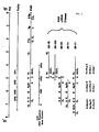
- the cloned cDNAs were analyzed by restriction enzyme digestion and by cross-hybridization of the clones to one another in order to locate the relative positions of five clones, T28-18, T28-123, T28-94, T28-71 and T28-77, as shown in Fig. 1. From this analysis one can deduce that over 90% of the viral genome has been cloned in overlapping cDNA clones starting at the 3' end of the genome.
- the restriction enzyme map of the HAV genome is that line marked "HAV" in Fig. 1.
- the location of the structural proteins on the polio genome is also shown in Fig. 1 as an indication of the probable predicted position on the HAY genome, since polio and HAV are related viruses.
- DNA sequencing has been performed by two methods.
- a region of 3000 contiguous bases have been sequenced. This region contains the sequences which encode the two structural proteins VP-1 and VP-3 of HAV; and the other proteins VP-2, and VP-4.
- the proteins VP-1 and VP-3 are the major antigenic proteins; VP-2 and VP-4 are thought to be less valuable as antigens.
- amino acid sequence of a mixture of cyanogen bromide cleavage fragments generated from purified VP-3 protein (covered in copending USSN 541,836, supra) has been compared to the translation of the DNA sequence to more precisely locate the region which encodes the VP-3.
- amino acid sequence of a mixture of cyanogen bromide cleavage fragments generated from purified VP-1 protein (covered in copending USSN 541,836, supra) has been compared to the translation of the DNA sequence to more precisely locate the region which encodes the VP-1 protein.
- the restriction map for the HAV genome indicates the location of the following 4 restriction sites: the Hinc II cleavage site at base 1643 of the DNA sequence; the Hind III cleavage site at base 1744; the Bam HI cleavage site at base 2049; and the Pvu II cleavage site at base 2722.
- These genome map positions are in the 5' to 3' direction on the genome:
- the nucleotide sequence of the region encoding the structural proteins, together with the reading frame for the protein is as follows:
- the sequences encoding for the structural proteins begin at base 403.
- the VP-3 protein is encoded starting approximately at map position 1149.
- the VP-I protein is encoded starting approximately at map position 1882.
- the key sub-unit sequences within VP-1 designated Sequences I, II, III, IV, and V, start, respectively at 1882, 1963, 1999, 2146, 2347.
- An effective vaccine can contain any one or more of these subunits, or of any other effective peptide subunit encoded within the genome sequence.
- nucleotide sequence encoding for an active peptide or peptide fragment can be synthesized and cloned into an expression system in order to produce the particular peptide or subunit desired.
- nucleotide sequences can be either made by total synthesis, that is, by linking bases according to known techniques, or by direct isolation from the viral RNA or cDNA. If the start of the nucleotide sequence desired does not correspond to the particular peptide, known techniques which enzymatically add or subtract the particular bases to the nucleotide chain can be employed to precisely make the nucleotide sequence.
- nucleotide sequences which are valuable as encoding antigenic proteins are the sequences from base 1749 to base 2722; from base 1487 to base 2980 and from base 1644 to base 2722, all contained within the above sequence.
- the sequence from base 1749 to base 2722 is especially valuable as a vector for producing antigen protein, see the following examples.
- a DNA clone containing the region of sequences encoding VP-3 and VP-1 in a single insert was first constructed.
- the DNA of clone 28-94 was cleaved with EcoRI and Xba I restriction enzymes and the 5.76 kb band of DNA containing the 5' portion of the viral structural protein genes was purified by agarose gel electrophoresis and electroelution.
- the 2.3 kb EcoRI-XbaI band containing the 3'portion of the viral structural genes of clone 28-77 was purified.
- These two fragments were ligated together to re-form pBR322 vector molecules containing the entire non-rearranged viral structural gene sequences.
- the ligated mixture was transformed into E. coli strain RR-1, thereby generating clone 57-5 with a plasmid containing an insert of intact VP-3 to VP-1 information.
- HAV-specified proteins containing antigenic reactivity to anti-HAV antibodies can be accomplished in bacterial cells, yeast, or higher eucaryotic cells. Some of the possible methods to obtain expression of the cDNA clones into proteins or polypeptide products are described below.
- fragments of HAV cDNA are inserted downstream from the bacterial lac promoter making a fusion product with the N-terminal amino acids of the bacterial ⁇ -galactosidase followed by HAV polypeptides.
- the fragments of HAV cDNA used having ends generated by Bgl II or Bam H1 or Hind III or Pst I or Taq I or Sau 3A or Nco I are ligated into the appropriate sites of the known expression plasmids pUC 7, 8, or 9 or pCQV2.
- the expression plasmid used with each HAV DNA fragment is selected to give an open reading frame for translation after insertion of the DNA.
- the recombinant expression plasmids are transformed into E. coli and appropriate clones are selected, grown to an optical density of 0.8 at a wavelength of 550, and lysed.
- the cellular extract is then used for detection of HAV-specific antigenic peptides.
- the fragments of HAV cDNA are inserted downstream of the Herpes simplex viral thymidine kinase gene promoter in a plasmid, cloned in E. coli and the resulting cloned recombinant plasmid is transfected into mouse L cells, mouse 3T3 cells, or other eukaryotic cells in culture.
- the fragments of HAV cDNA used having ends generated by Bgl II or Bam H1 or Hind III or Bal 31 enzymes are ligated behind the thymidine kinase ATG translational start codon at the Rsa I site using appropriate linker molecules of DNA to make the connection in frame with respect to translation.
- the eucaryotic cells transfected by the recombinant expression plasmids are selected by cotransfection with the entire Herpes thymidine kinase gen and subsequent growth in HAT selection medium. The colonies of selected cells are grown and cellular extracts prepared for detection of HAV-specific antigenic
- the cells expressing HAV proteins or polypeptides are labeled by growth in the presence of 35S-labeled methionine or 14C-labeled leucine. Extracts of the cells are then prepared and reacted with anti-HAV antiserum or anti-HAV monoclonal antibodies.
- the antigen-antibody complexes are collected by precipitation with formalin fixed Staph A, dissociated by boiling in SDS-EDTA, and separated by SDS-polyacrylamide gel electrophoresis. The proteins are then visualized by autoradiography.
- the expressed HAV proteins or polypeptides are detected by a competition radioimmune assay.
- Purified HAV virion particles are labeled in vitro with 125I and precipitated with anti-HAV antiserum or anti-HAV monoclonal antibodies. This precipitation is performed in the presence of increasing amounts of unlabeled extracts prepared from the expression cells.
- the antigen-antibody complexes are collected on filters and counted in a gamma-counter. The presence of expressed HAV products in the expression cells is shown by a decrease in counts as the amount of cellular extract is increased in the assay.
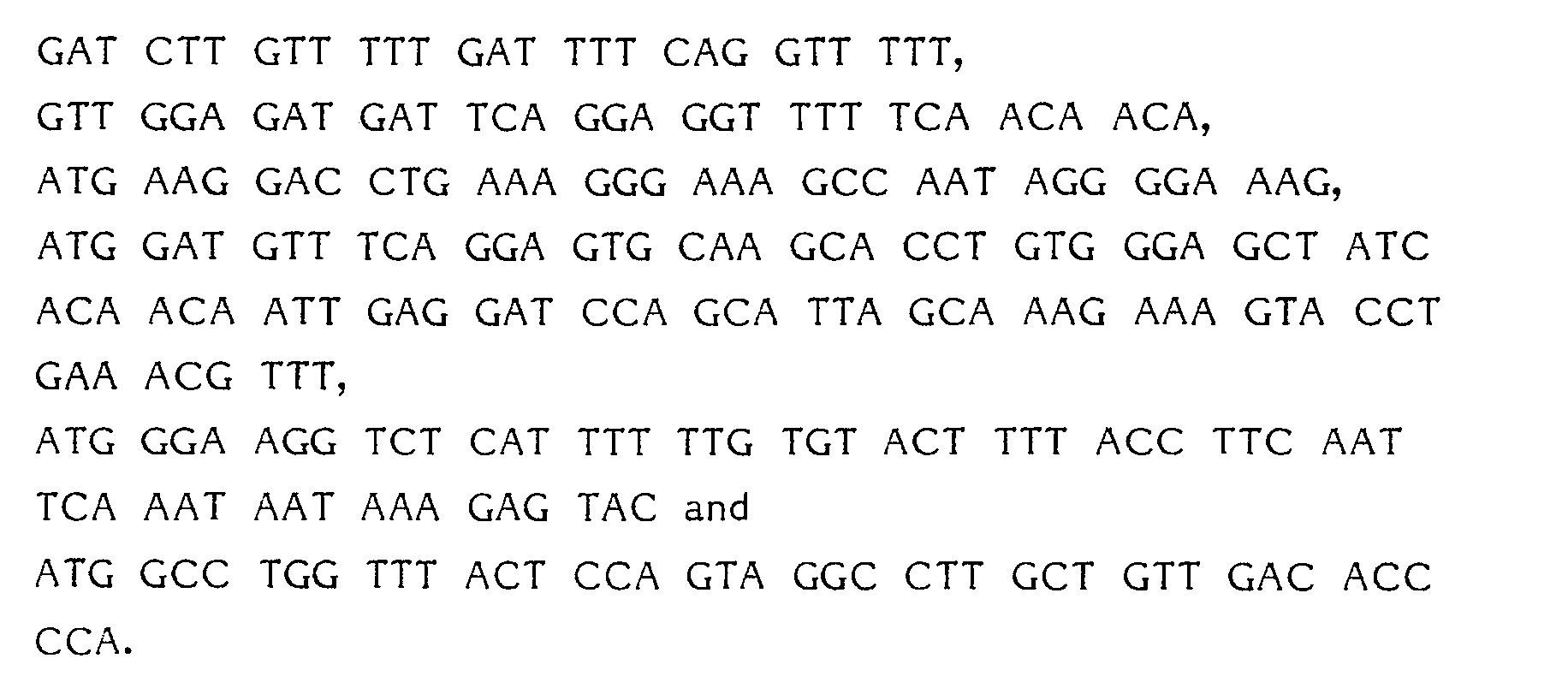
- Plasmid HAV57-11 contains the following nucleic acid sequences: Plasmid HAV-6 contains the nucleic acid sequences identified by numbers 5 and 6 above, but not those sequences identified by numbers 1, 2, 3 and 4.
- Expression plasmids pHAV-6 and pHAV57-11 were grown in L-broth to log phase and induced by heat shock at 42°C.
- the pCQV2 expression vector carries a temperature sensitive ⁇ repressor which suppresses synthesis of proteins inserted at the ⁇ rightward promoter ( ⁇ P R ). Heating at 42°C inactivated the repressor and allowed the expression of genes located downstream from ⁇ P R .
- 1 ml of cells were pelleted before induction and 30 minutes after induction, lysed by boiling with SDS mercaptoethanol and the proteins separated by discontinuous gel electrophoresis.
- the proteins were transferred either to nitrocellulose (BA-85) paper or to Millipore filters (HAHY grade) by electroblotting.
- the proteins that were transferred were probed with two polyclonal antihepatitis A antisera (antiHAV) designated DB-2 and C-149 raised by injection of SDS-heat denatured virus into rabbits.
- antiHAV polyclo
- HAV-6 HAV-6
- Heat inducibility a novel band absent in the original vector pCQV2 when probed with post-immune serum.
- Plasmid HAV-6 showed a unique band at ⁇ 30 and 20 k daltons when probed with DB-2 serum and a band at 30 kd when probed with C149 post immune serum. Since pHAV-6 contains an insert entirely within VP-1 coding region, expression of VP-1 segments in E. coli was achieved.
- Plasmid HAV57-11 showed six prominent bands when probed with DB-2 serum. They are, in order of decreasing size, ⁇ 50, 35, 23, 20, 16, and 14 k daltons. Presumably, the smaller peptides represent degradation products of the ⁇ 50 kd protein. The profile is simpler when the probe used was C149. The 50 kd band was clearly visible as well as two weak bands around 30 k daltons. The small molecular weight protein bands were presumably degradation products. Since pHAV57-11 contains a DNA insert which encodes 133 amino acids of VP-3 representing the C-terminus as well as most of VP-1 sequences excluding ⁇ 30 amino acids from the C-terminal end expression of VP-1 segments and VP-3 segments in E. coli was achieved.
- Pre- and postimmune serum from a marmoset injected with native virus were used to screen the protein blots.
- Preimmune serum failed to bind to any novel induced protein in both pHAV-6 and pHAV57-11.
- Postimmune serum on the other hand reacted with a protein ⁇ 50 kd in size produced in induced pHAV57-11 cells and another ⁇ 18k daltons in size.
- the detection of ⁇ 50 kd protein in pHAV57-11 suggests that at least some of the same determinants or epitopes seen on the native virus are also available for interaction with antibody in the 50 kd protein.
- ⁇ 133 amino acid residues of VP-3 sequences at the N-terminus of VP-1 presumably allows the VP-1 protein to assume a tertiary structure which exposes at least some of the determinants normally exposed on native virus.
- the 50 kd protein and possibly the ⁇ 18 kd protein made in E. coli are potential immunogenes which may be capable of eliciting normal immune response.
Landscapes
- Chemical & Material Sciences (AREA)
- Organic Chemistry (AREA)
- Health & Medical Sciences (AREA)
- Life Sciences & Earth Sciences (AREA)
- Molecular Biology (AREA)
- Biochemistry (AREA)
- Biophysics (AREA)
- Virology (AREA)
- General Health & Medical Sciences (AREA)
- Medicinal Chemistry (AREA)
- Genetics & Genomics (AREA)
- Proteomics, Peptides & Aminoacids (AREA)
- Gastroenterology & Hepatology (AREA)
- Preparation Of Compounds By Using Micro-Organisms (AREA)
- Peptides Or Proteins (AREA)
- Medicines Containing Antibodies Or Antigens For Use As Internal Diagnostic Agents (AREA)
- Micro-Organisms Or Cultivation Processes Thereof (AREA)
Description
- Hepatitis A is a liver disease which, although not commonly fatal, can induce long periods of debilitating illness. The disease is commonly spread by direct contact with an infected individual or by hepatitis A virus (HAV) contaminated drinking water and/or food.
- The prior art does not identify the protein or proteins of HAV (hepatitis A virus) which induce neutralizing antibodies to this virus. One of the major drawbacks to examining the protective antigenicity of HAV proteins has been the lack of sufficient quantities of HAV and its polypeptide components. The virus is made in very small quantities in cell culture, has a limited animal host range, and is difficult to purify from infected cell cultures and animal tissues. Recently a patent application claiming the VP-1 structural protein of HAV prepared by isolation from the virus was filed by the assignee of this application and is presently copending as U.S.S.N. 541,836, filed October 14, 1983 (See EP-A-0138 704). VP-1 is recognized in our laboratories as being the main structural protein of HAV, and accordingly the most important for vaccine use. The appropriate dosage forms and regions are in this application, which is incorporated by reference.
- Cloning of the genomic material of HAV and potential analysis of its sequence has recently been reported: Von Der Helm et al., J. Virological Methods 3 1981, 37-43; see also EPO 0061740, priority March 28, 1981 published October 6, 1982 disclosing the same work; and Ticehurst et al., Proc. National Academy Science USAVol. 80, pgs 5885-5889. October 1983.
- The Von de Helm work describes preparation of cDNA and cloning of that cDNA into plasmids, and subsequent expression of HAV antigen, but no nucleotide sequence of the DNA or amino acid sequence of the corresponding proteins is described, nor is the identity of any of the viral proteins expressed known. In fact, the weak antigenic responses of the viral proteins obtained by Von de Helm et al. tend to indicate that most portions of the HAV genomic encoding the important antigenic proteins of HAV has not been successfully identified or used in the cloning experiments.
- Ticehurst does report a partial nucleotide sequence from the 3' terminus of HAV, but does not appear to have identified that part of the sequence which encodes for the antigenic proteins. From our experiments, we believe that Ticehurst has not sequenced the antigenic portion; that the Ticehurst sequence work is far outside the antigenic region.
- It is an object of the present invention to provide a method for cloning the nucleotide sequences of HAV encoding the major antigenic proteins of HAV, including VP-1, VP-2, VP-3 and VP-4, especially VP-1. Another object is to determine the nucleotide sequences which encode the antigenic proteins, including VP-1. Yet another object is to provide a method for producing VP-1, VP-2, VP-3, VP-4, or parts thereof, by expression of cloned DNA in an appropriate host. Still another object is to provide a vector containing the VP-1, VP-2, VP-3 or VP-4 genes, or parts thereof, but especially a vector containing the VP-1, or part of VP-1, gene. Another object is to provide transformed hosts containing a vector containing the VP-1, VP-2, VP-3 or VP-4 genes, or parts thereof and being capable of expressing the peptide encoded by said genes or parts thereof. These and other objects of the present invention will be apparent from the following description.
- Fragments of DNA which code for immunogenic peptides VP-1, VP-2, VP-3 and VP-4 capable of inducing in vivo the formation of anti-Hepatitis A virus antibodies are provided and their nucleotide sequence determined. In other terms, the invention concerns fragments of DNA which code for the antigenic determinants on peptides normally encoded by the RNA of HAV or by corresponding double-stranded cDNA; these antigenic determinants being essential for the antigenic properties of the products of natural viral RNA expression.
- RNA was extracted from purified hepatitis A virus particles, annealed to a dT-tailed plasmid, and the complementary cDNA synthesized. This single stranded DNA was used as a template for synthesis of its complementary strand using DNA polymerase. The double stranded cDNA thus formed corresponded to at least a portion of the HAV antigenic protein genes. It was modified to provide "sticky ends" and placed into an appropriate vector; suitable hosts include prokaryotic and eukaryotic organisms. These hosts were exposed to the resulting vector and those which stably incorporated the vector were identified and isolated.
- Vector DNA was extracted from these hosts and this material was characterized. At least 5 fragments of cloned HAV DNA were sequenced to determine the nucleotide sequence of the region encoding the antigenic peptides, VP-1, VP-2, VP-3 and VP-4. The corresponding amino acid sequence encoded thereby was inferred.
- Knowledge of the nucleotide sequences encoding the amino acid sequences of these peptides makes possible their synthesis as well as subunits and homologues and analogues thereof thus allowing determination of the relationship between structure and function. The peptides, which as noted above, do not form part of this invention, can be used in pharmaceutical compositions to make vaccines.
- The HAV peptides of the present invention may be prepared from their constituent amino acids by standard methods of protein synthesis, e.g., Schroeder et al., "The Peptides", Vol. I, Academic Press, 1965, or Bodanszky et al., "Peptide Synthesis", Interscience Publishers 1966, or McOmie (ed.), "Protective Groups in Organic Chemistry", Plenum Press 1973, the disclosures of which are hereby incorporated by reference.
- The peptides of HAV also may be prepared by recombinant DNA techniques by, for example, the isolation or preparation of appropriate DNA sequences and incorporation of these sequences into vectors in a suitable host and expression of the desired peptide therefrom. The use of recombinant DNA techniques is described in many published articles, for example, Maniatis et al., Molecular Cloning, A Laboratory Manual, Cold Spring Harbor, New York 1982, the disclosure of which is hereby incorporated by reference. Modification of the nucleotides coding for the peptides of the present invention according to known techniques permits the preparation via recombinant DNA techniques of peptides having altered amino acid sequences of the peptides of the present invention. Certain of these amino acids may be coded for by more than one triplet nucleotide sequence. It is to be understood that the disclosed nucleotide sequence is to include other codons coding for the same amino acid, e.g., the codons CGA and AGA each code for arginine.
- Suitable hosts for expression of the HAV peptides include prokaryotic organisms such as E. coli and B. subtilis, and eukaryotic organisms such as Saccharomyces cerevisiae and Chinese hamster ovary cells. It is also to be understood that these proteins can be expressed directly in a mammalian species by means of appropriate expression vectors such as vaccinia, varicella zoster, adeno or herpes simplex viruses.
- The following examples illustrate the present invention.
-
- 1.1 Hepatitis A virus particles (strain CR326), purified from virus-infected LLC-MK2 cells (a monkey kidney cell line) by CsCl gradient centrifugation, were disrupted at 65°C with 1% SDS, 20 mM EDTA and phenol extraction using 0.1M tris pH 7.4 - saturated phenol.
- 1.2 The aqueous layer of the extraction was further extracted twice with equal volumes of CHCl₃; isoamyl alcohol (24:1) and precipitated with 0.2 M sodium acetate pH 5.5 and 2 volumes of EtOH at -20°C.
- 1.3 The EtOH precipitate was collected by centrifugation and dissolved in H₂O.
- The initial 3' cDNA clones were prepared using the procedure of H. Okayama and P. Berg (Molecular and Cellular Biology 2: 161-170 (1982)). In brief, the viral RNA was annealed to dT-tailed plasmid (derived from pSV 0.71-0.86) and cDNA synthesized by reverse transcriptase using the dT-tailed linker (derived from pSV 0.19-0.32). Using the linker as a primer, the second cDNA strand was synthesized by DNA polymerase after removal of the RNA by RNase H. The cDNA was ligated and transformed into E. coli. The resulting clones were selected by resistance to ampicillin and screened by hybridization to a ³²P-labeled HAV cDNA prepared from the viral RNA.
- The largest viral insert obtained was approximately 2.3Kb in size and was called clone α 18. Restriction enzyme analysis of this clone demonstrated the presence of two Pvu II sites near the 5' end of the cloned insert. The cloned DNA was restricted with Pvu II and the 280 bp 5' proximal fragment was purified by agarose gel electrophoresis, visualized by ethidium bromide staining and electroeluted in dialysis tubing for use as a primer for further cDNA cloning.
-
- 3.1 The 280 bp Pvu II restriction fragment of cDNA clone α 18 was denatured by boiling for 10 min. and quick-cooling in ice-water, and then used as a primer for HAV cDNA synthesis. The denatured primer was either annealed or hybridized to the viral RNA. For annealing, 160 ng of denatured primer was added to approximately 1 µg of purified HAV RNA in 20 µl of H₂O and heated to 70°C for 10 min. then 37°C for 15 min. For the hybridization, 140 ng of denatured primer was added to 1 µg of purified HAV RNA in 80% formamide, 0.4 M NaCl, 0.01 M PIPES pH 6.4, and 2 mM EDTA and incubated at 47°C for 3.5 hours. The formamide was removed by performing 4 sequential ethanol precipitations.
- 3.2 After annealing or hybridization of the primer to the RNA, the first strand of cDNA was synthesized using 50 mM Tris pH 8, 0.34 mM dCTP, 1 mM dGTP, dATP, dTTP, 10 mM 2-mercaptoethanol, 10 mM magnesium acetate, [³²P-α]dCTP (8 µ Ci/10 µl), RNasin (7 units/10 µl), and AMV reverse transcriptase (16 units/l0 µl) by incubating at 42°C for 30 min. The reaction mix was extracted twice with phenol, chloroform extracted, and precipitated with ethanol.
- 3.3 The second strand of cDNA was then synthesized using 20 mM Tris pH 7.4, 5 mM MgCl₂, 10 mM (NH₄)₂SO₄, 100 mM KCl, 100 µg/ml BSA, 0.04 mM dATP, dGTP, dCTP and dTTP, 9 units/ml RNase H, and 1750 units/ml DNA polymerase I by incubating at 12°C for 60 min. and then 22°C for 60 min. The reaction mix was extracted twice with phenol and chloroform and ethanol precipitated.
- 3.4 The double-stranded cDNA was dC-tailed using terminal deoxynucleotidyl transferase, annealed to pBR 322 DNA which was dG-tailed at the PstI site, and transformed into competent E. coli strain RR-1. The resulting clones were selected for tetracycline resistant, ampicillin-sensitive growth and screened for positive hybridization to a ³²P-labeled cDNA prepared from HAV RNA.
- To insure the cDNA clones were generated from Hepatitis A genetic information, the clones were labeled with [³²P-α] dCTP by nick translation and hybridized to uninfected and HAV-infected LLC-MK2 cellular RNA bound to nitrocellulose membrane filters. The cDNA clones hybridized only to the HAY-infected cell RNA.
- The cloned cDNAs were analyzed by restriction enzyme digestion and by cross-hybridization of the clones to one another in order to locate the relative positions of five clones, T28-18, T28-123, T28-94, T28-71 and T28-77, as shown in Fig. 1. From this analysis one can deduce that over 90% of the viral genome has been cloned in overlapping cDNA clones starting at the 3' end of the genome. The restriction enzyme map of the HAV genome is that line marked "HAV" in Fig. 1. The location of the structural proteins on the polio genome is also shown in Fig. 1 as an indication of the probable predicted position on the HAY genome, since polio and HAV are related viruses.
- DNA sequencing has been performed by two methods.
- Chemical DNA sequencing has been performed by the Maxam and Gilbert technique on clones T28-18, T28-123, T28-77, T28-94 and T28-71.
- Subclones of fragments of cDNA clone T28-77 were prepared in M13 phage and sequenced by the dideoxynucleotide termination procedure described by Sanger.
- In all, a region of 3000 contiguous bases have been sequenced. This region contains the sequences which encode the two structural proteins VP-1 and VP-3 of HAV; and the other proteins VP-2, and VP-4. The proteins VP-1 and VP-3 are the major antigenic proteins; VP-2 and VP-4 are thought to be less valuable as antigens.
- The amino acid sequence of a mixture of cyanogen bromide cleavage fragments generated from purified VP-3 protein (covered in copending USSN 541,836, supra) has been compared to the translation of the DNA sequence to more precisely locate the region which encodes the VP-3.
- The amino acid sequence of a mixture of cyanogen bromide cleavage fragments generated from purified VP-1 protein (covered in copending USSN 541,836, supra) has been compared to the translation of the DNA sequence to more precisely locate the region which encodes the VP-1 protein.
- Referring to Fig. 1, the restriction map for the HAV genome indicates the location of the following 4 restriction sites: the Hinc II cleavage site at base 1643 of the DNA sequence; the Hind III cleavage site at base 1744; the Bam HI cleavage site at base 2049; and the Pvu II cleavage site at base 2722. These genome map positions are in the 5' to 3' direction on the genome: The nucleotide sequence of the region encoding the structural proteins, together with the reading frame for the protein is as follows:


- Within this entire sequence is encoded the information necessary to make the antigenic proteins of HAV. The sequences encoding for the structural proteins begin at base 403. The VP-3 protein is encoded starting approximately at map position 1149. The VP-I protein is encoded starting approximately at map position 1882. The key sub-unit sequences within VP-1, designated Sequences I, II, III, IV, and V, start, respectively at 1882, 1963, 1999, 2146, 2347. An effective vaccine can contain any one or more of these subunits, or of any other effective peptide subunit encoded within the genome sequence.
- The value of the nucleotide sequence will be readily apparent to those skilled in the art: a particular nucleotide sequence encoding for an active peptide or peptide fragment can be synthesized and cloned into an expression system in order to produce the particular peptide or subunit desired. These nucleotide sequences can be either made by total synthesis, that is, by linking bases according to known techniques, or by direct isolation from the viral RNA or cDNA. If the start of the nucleotide sequence desired does not correspond to the particular peptide, known techniques which enzymatically add or subtract the particular bases to the nucleotide chain can be employed to precisely make the nucleotide sequence.
- Other nucleotide sequences which are valuable as encoding antigenic proteins are the sequences from base 1749 to base 2722; from base 1487 to base 2980 and from base 1644 to base 2722, all contained within the above sequence. The sequence from base 1749 to base 2722 is especially valuable as a vector for producing antigen protein, see the following examples.
- In order to begin studies on expression, a DNA clone containing the region of sequences encoding VP-3 and VP-1 in a single insert was first constructed. The DNA of clone 28-94 was cleaved with EcoRI and Xba I restriction enzymes and the 5.76 kb band of DNA containing the 5' portion of the viral structural protein genes was purified by agarose gel electrophoresis and electroelution. Similarly, the 2.3 kb EcoRI-XbaI band containing the 3'portion of the viral structural genes of clone 28-77 was purified. These two fragments were ligated together to re-form pBR322 vector molecules containing the entire non-rearranged viral structural gene sequences. The ligated mixture was transformed into E. coli strain RR-1, thereby generating clone 57-5 with a plasmid containing an insert of intact VP-3 to VP-1 information.
- The expression of HAV-specified proteins containing antigenic reactivity to anti-HAV antibodies can be accomplished in bacterial cells, yeast, or higher eucaryotic cells. Some of the possible methods to obtain expression of the cDNA clones into proteins or polypeptide products are described below.
- In the first example, fragments of HAV cDNA are inserted downstream from the bacterial lac promoter making a fusion product with the N-terminal amino acids of the bacterial β-galactosidase followed by HAV polypeptides. The fragments of HAV cDNA used having ends generated by Bgl II or Bam H1 or Hind III or Pst I or Taq I or Sau 3A or Nco I are ligated into the appropriate sites of the known expression plasmids pUC 7, 8, or 9 or pCQV2. The expression plasmid used with each HAV DNA fragment is selected to give an open reading frame for translation after insertion of the DNA. The recombinant expression plasmids are transformed into E. coli and appropriate clones are selected, grown to an optical density of 0.8 at a wavelength of 550, and lysed. The cellular extract is then used for detection of HAV-specific antigenic peptides.
- Alternatively, the fragments of HAV cDNA are inserted downstream of the Herpes simplex viral thymidine kinase gene promoter in a plasmid, cloned in E. coli and the resulting cloned recombinant plasmid is transfected into mouse L cells, mouse 3T3 cells, or other eukaryotic cells in culture. The fragments of HAV cDNA used having ends generated by Bgl II or Bam H1 or Hind III or Bal 31 enzymes are ligated behind the thymidine kinase ATG translational start codon at the Rsa I site using appropriate linker molecules of DNA to make the connection in frame with respect to translation. The eucaryotic cells transfected by the recombinant expression plasmids are selected by cotransfection with the entire Herpes thymidine kinase gen and subsequent growth in HAT selection medium. The colonies of selected cells are grown and cellular extracts prepared for detection of HAV-specific antigenic
- The cells expressing HAV proteins or polypeptides are labeled by growth in the presence of ³⁵S-labeled methionine or ¹⁴C-labeled leucine. Extracts of the cells are then prepared and reacted with anti-HAV antiserum or anti-HAV monoclonal antibodies. The antigen-antibody complexes are collected by precipitation with formalin fixed Staph A, dissociated by boiling in SDS-EDTA, and separated by SDS-polyacrylamide gel electrophoresis. The proteins are then visualized by autoradiography.
- Alternatively, the expressed HAV proteins or polypeptides are detected by a competition radioimmune assay. Purified HAV virion particles are labeled in vitro with ¹²⁵I and precipitated with anti-HAV antiserum or anti-HAV monoclonal antibodies. This precipitation is performed in the presence of increasing amounts of unlabeled extracts prepared from the expression cells. The antigen-antibody complexes are collected on filters and counted in a gamma-counter. The presence of expressed HAV products in the expression cells is shown by a decrease in counts as the amount of cellular extract is increased in the assay.
-
- (1) Approximately 0.5 µg of pCQV2 (Cary Queen) DNA was cleaved with BamHI and PvuII and treated with phosphatase and ligated to 2 µg of gel purified 675 bp long fragment derived from clone T28-123 by cleavage with BamHI and PvuII. The ligated mixture was transformed into HB101 cells and ampicillin resistant clones were screened for the presence of insert with purified nick translated 675 base pair fragment. One positive clone designated pHAV-6 was assayed for production of HAV specified proteins. The junction sequence between pCQV2 and the insert was confirmed by cleavage with BamHI and PvuII which released the appropriate fragment.
- (2) Clone 57-5 (prepared by coupling cDNA clone 28-94 with cDNA clone 28-77) was used get expression of almost the entire VP-1 gene and the C-terminal region of VP-3. DNA from plasmid 57-5 was cleaved with BglII and PvuII and 1.12 kbp fragment was purified by gel electrophoresis. Approximately 2 µg of the fragment was ligated to 0.5 µg of BamHI-PvuII cleaved pCQV2 DNA and the ligated mixture used to transform HB101 cells. Ampicillin resistant clones were screened by hybridization to nick translated BamHI-PvuII cleaved fragment. One of the clones which proved positive was designated pHAV-57-11 and was assayed for expression of VP3 and/or VP1.
- Both of these constructions allow read through from the initiating AUG codon within pCQV2 and continues beyond the end of the insert into the pCQV2 sequences for 50 amino acids before a termination codon is encountered. The predicted open reading frame of pHAV-6 is therefore 275 amino acid residues and that of pHAV57-11 is 464 amino acid residues. Plasmid HAV57-11 contains the following nucleic acid sequences:

Plasmid HAV-6 contains the nucleic acid sequences identified bynumbers 5 and 6 above, but not those sequences identified bynumbers - Expression plasmids pHAV-6 and pHAV57-11 were grown in L-broth to log phase and induced by heat shock at 42°C. The pCQV2 expression vector carries a temperature sensitive λ repressor which suppresses synthesis of proteins inserted at the λ rightward promoter (λPR). Heating at 42°C inactivated the repressor and allowed the expression of genes located downstream from λPR. 1 ml of cells were pelleted before induction and 30 minutes after induction, lysed by boiling with SDS mercaptoethanol and the proteins separated by discontinuous gel electrophoresis. The proteins were transferred either to nitrocellulose (BA-85) paper or to Millipore filters (HAHY grade) by electroblotting. The proteins that were transferred were probed with two polyclonal antihepatitis A antisera (antiHAV) designated DB-2 and C-149 raised by injection of SDS-heat denatured virus into rabbits.
- The antisera were in turn detected by I¹²⁵ labelled protein A. Two criteria were used to determine expression of HAV specified proteins: (1) Heat inducibility, and (2) Appearance of a novel band absent in the original vector pCQV2 when probed with post-immune serum. Plasmid HAV-6 showed a unique band at ∼ 30 and 20 k daltons when probed with DB-2 serum and a band at 30 kd when probed with C149 post immune serum. Since pHAV-6 contains an insert entirely within VP-1 coding region, expression of VP-1 segments in E. coli was achieved.
- Plasmid HAV57-11 showed six prominent bands when probed with DB-2 serum. They are, in order of decreasing size, ∼ 50, 35, 23, 20, 16, and 14 k daltons. Presumably, the smaller peptides represent degradation products of the ∼ 50 kd protein. The profile is simpler when the probe used was C149. The 50 kd band was clearly visible as well as two weak bands around 30 k daltons. The small molecular weight protein bands were presumably degradation products. Since pHAV57-11 contains a DNA insert which encodes 133 amino acids of VP-3 representing the C-terminus as well as most of VP-1 sequences excluding ∼ 30 amino acids from the C-terminal end expression of VP-1 segments and VP-3 segments in E. coli was achieved.
- Pre- and postimmune serum from a marmoset injected with native virus were used to screen the protein blots. Preimmune serum failed to bind to any novel induced protein in both pHAV-6 and pHAV57-11. Postimmune serum on the other hand reacted with a protein ∼ 50 kd in size produced in induced pHAV57-11 cells and another ∼ 18k daltons in size. The detection of ∼ 50 kd protein in pHAV57-11 suggests that at least some of the same determinants or epitopes seen on the native virus are also available for interaction with antibody in the 50 kd protein. The presence of ∼ 133 amino acid residues of VP-3 sequences at the N-terminus of VP-1 presumably allows the VP-1 protein to assume a tertiary structure which exposes at least some of the determinants normally exposed on native virus. Hence the 50 kd protein and possibly the ∼ 18 kd protein made in E. coli are potential immunogenes which may be capable of eliciting normal immune response.
Claims (5)
- Vectors containing all or part of the following nucleotide sequences and adapted to express in a suitable host the peptide coded for by said nucleotide sequence :

- The vectors of claim 1, wherein the host is a prokaryotic or eukaryotic organism.
- A suitable prokaryotic or eukaryotic host containing a vector according to claim 2, the host adapted to express at least part of the peptide coded for by said nucleotide sequence.
- Plasmid HAV 57-11 containing the nucleic acid sequences
- Plasmid HAV-6 containing the nucleic acid sequences

Applications Claiming Priority (2)
| Application Number | Priority Date | Filing Date | Title |
|---|---|---|---|
| US58581884A | 1984-03-02 | 1984-03-02 | |
| US585818 | 1984-03-02 |
Publications (3)
| Publication Number | Publication Date |
|---|---|
| EP0154587A2 EP0154587A2 (en) | 1985-09-11 |
| EP0154587A3 EP0154587A3 (en) | 1987-12-16 |
| EP0154587B1 true EP0154587B1 (en) | 1991-05-15 |
Family
ID=24343084
Family Applications (1)
| Application Number | Title | Priority Date | Filing Date |
|---|---|---|---|
| EP85400369A Expired - Lifetime EP0154587B1 (en) | 1984-03-02 | 1985-02-27 | Fragments of dna which encode peptides capable of inducing in vivo the synthesis of anti-hepatitis a virus antibodies |
Country Status (3)
| Country | Link |
|---|---|
| EP (1) | EP0154587B1 (en) |
| JP (1) | JPS60210989A (en) |
| DE (1) | DE3582812D1 (en) |
Families Citing this family (7)
| Publication number | Priority date | Publication date | Assignee | Title |
|---|---|---|---|---|
| JPS61500053A (en) * | 1983-09-30 | 1986-01-16 | マサチユ−セツツ インステイチユ−ト オブ テクノロジ− | Generation of cDNA representing hepatitis A virus sequence order |
| US4596674A (en) * | 1984-09-11 | 1986-06-24 | Merck & Co., Inc. | Immunogenic HAV peptides |
| CA1324094C (en) * | 1985-04-03 | 1993-11-09 | Dino Dina | Hepatitis a virus vaccines |
| SU1469856A1 (en) * | 1986-07-31 | 1990-09-30 | Институт биоорганической химии им.М.М.Шемякина | Method of constructing recombinant plasmide dna coding synthesis of polipeptide substances against hepatitis |
| GB9002387D0 (en) * | 1990-02-02 | 1990-04-04 | St Marys Hospit Med School | Improvements relating to the prevention of hepatitis |
| AU735733B2 (en) | 1996-04-19 | 2001-07-12 | Government Of The United States Of America, As Represented By The Secretary Of The Department Of Health And Human Services, The | Antigenically reactive regions of the hepatitis A virus polyprotein |
| US6942965B2 (en) * | 2001-10-12 | 2005-09-13 | Chiron Corporation | Hepatitis A virus nucleotide sequences, recombinant proteins and uses thereof |
Family Cites Families (3)
| Publication number | Priority date | Publication date | Assignee | Title |
|---|---|---|---|---|
| DE3112338C2 (en) * | 1981-03-28 | 1995-02-23 | Klaus Von Der Helm | Use of hybridization probes for the detection of HAV |
| JPS61500053A (en) * | 1983-09-30 | 1986-01-16 | マサチユ−セツツ インステイチユ−ト オブ テクノロジ− | Generation of cDNA representing hepatitis A virus sequence order |
| US4614793A (en) * | 1983-10-14 | 1986-09-30 | Merck & Co., Inc. | Hepatitis A--subunit antigen |
-
1985
- 1985-02-27 EP EP85400369A patent/EP0154587B1/en not_active Expired - Lifetime
- 1985-02-27 DE DE8585400369T patent/DE3582812D1/en not_active Expired - Lifetime
- 1985-03-02 JP JP60040344A patent/JPS60210989A/en active Pending
Also Published As
| Publication number | Publication date |
|---|---|
| EP0154587A2 (en) | 1985-09-11 |
| DE3582812D1 (en) | 1991-06-20 |
| EP0154587A3 (en) | 1987-12-16 |
| JPS60210989A (en) | 1985-10-23 |
Similar Documents
| Publication | Publication Date | Title |
|---|---|---|
| Carter et al. | Identification and sequence determination of the capsid protein gene of feline calicivirus | |
| EP0013828B2 (en) | Recombinant DNA, hosts transformed with it and processes for the preparation of polypeptides | |
| Errede et al. | STE12, a protein involved in cell-type-specific transcription and signal transduction in yeast, is part of protein-DNA complexes. | |
| US5206163A (en) | DNA encoding bovine diarrhea virus protein | |
| AU642942B2 (en) | Viral agent | |
| EP0192902B1 (en) | Dna sequences from varicella-zoster virus, vectors and hosts containing them, the corresponding polypeptides and the compositions containing them | |
| EP0208672B1 (en) | Vaccines and diagnostics derived from bovine diarrhea virus | |
| JPH07265085A (en) | Recombinant poxvirus | |
| IE52036B1 (en) | Non-passageable viruses | |
| WO1987005326A1 (en) | Processes for the production of hcmv glycoproteins, antibodies thereto and hcmv vaccines, and recombinant vectors therefor | |
| JP2511494B2 (en) | Method for producing Japanese encephalitis virus surface antigen protein | |
| JPH0690769A (en) | System for expression of baculovirus | |
| EP0040922A1 (en) | DNA sequences, recombinant DNA molecules and processes for producing polypeptides with the specificity of foot and mouth disease viral antigens | |
| JPH01501357A (en) | Vaccines for human respiratory viruses | |
| EP0182442B2 (en) | Recombinant DNA molecules and their method of production | |
| EP0154587A2 (en) | Fragments of DNA which encode peptides capable of inducing in vivo the synthesis of anti-hepatitis A virus antibodies | |
| Kowalski et al. | Heat-shock promoter-driven synthesis of secreted bovine herpesvirus glycoproteins in transfected cells | |
| US5061623A (en) | Peptides comprising an immunogenic site of poliovirus and dnas containing nucleotide sequences coding for these peptides | |
| GB2084583A (en) | Synthetic dna and process therefor | |
| WO1984002847A1 (en) | Methods and materials for development of parvovirus vaccine | |
| US5567582A (en) | Variants of cytomegalovirus gB | |
| US4743554A (en) | Recombinant DNA expression vector encoding for foot and mouth disease virus proteins | |
| JP2598596B2 (en) | Polypeptide derived from hepatitis C virus protein, test kit containing the polypeptide, and vaccine for preventing hepatitis C virus infection | |
| US6270955B1 (en) | Pharmaceutical compositions and methods for producing antibodies to hepatitis b virus and kits and methods for detecting antibodies to hepatitis b virus | |
| GB2079288A (en) | DNA Sequences, Recombinant DNA Molecules and Processes for Producing Polypeptides with the Specificity of Foot and Mouth Disease Viral Antigens |
Legal Events
| Date | Code | Title | Description |
|---|---|---|---|
| PUAI | Public reference made under article 153(3) epc to a published international application that has entered the european phase |
Free format text: ORIGINAL CODE: 0009012 |
|
| AK | Designated contracting states |
Designated state(s): CH DE FR GB IT LI NL |
|
| ITCL | It: translation for ep claims filed |
Representative=s name: SOCIETA' ITALIANA BREVETTI S.P.A. |
|
| PUAL | Search report despatched |
Free format text: ORIGINAL CODE: 0009013 |
|
| RHK1 | Main classification (correction) |
Ipc: C12N 15/00 |
|
| AK | Designated contracting states |
Kind code of ref document: A3 Designated state(s): CH DE FR GB IT LI NL |
|
| 17P | Request for examination filed |
Effective date: 19880613 |
|
| 17Q | First examination report despatched |
Effective date: 19881005 |
|
| GRAA | (expected) grant |
Free format text: ORIGINAL CODE: 0009210 |
|
| AK | Designated contracting states |
Kind code of ref document: B1 Designated state(s): CH DE FR GB IT LI NL |
|
| REF | Corresponds to: |
Ref document number: 3582812 Country of ref document: DE Date of ref document: 19910620 |
|
| ET | Fr: translation filed | ||
| ITF | It: translation for a ep patent filed | ||
| PGFP | Annual fee paid to national office [announced via postgrant information from national office to epo] |
Ref country code: FR Payment date: 19911223 Year of fee payment: 8 |
|
| PGFP | Annual fee paid to national office [announced via postgrant information from national office to epo] |
Ref country code: CH Payment date: 19920207 Year of fee payment: 8 |
|
| PGFP | Annual fee paid to national office [announced via postgrant information from national office to epo] |
Ref country code: GB Payment date: 19920226 Year of fee payment: 8 Ref country code: DE Payment date: 19920226 Year of fee payment: 8 |
|
| PLBI | Opposition filed |
Free format text: ORIGINAL CODE: 0009260 |
|
| PGFP | Annual fee paid to national office [announced via postgrant information from national office to epo] |
Ref country code: NL Payment date: 19920229 Year of fee payment: 8 |
|
| 26 | Opposition filed |
Opponent name: BEHRINGWERKE AKTIENGESELLSCHAFT Effective date: 19920213 |
|
| NLR1 | Nl: opposition has been filed with the epo |
Opponent name: BEHRINGWERKE AG. |
|
| PG25 | Lapsed in a contracting state [announced via postgrant information from national office to epo] |
Ref country code: GB Effective date: 19930227 |
|
| RDAG | Patent revoked |
Free format text: ORIGINAL CODE: 0009271 |
|
| STAA | Information on the status of an ep patent application or granted ep patent |
Free format text: STATUS: PATENT REVOKED |
|
| NLV4 | Nl: lapsed or anulled due to non-payment of the annual fee | ||
| 27W | Patent revoked |
Effective date: 19930611 |
|
| GBPC | Gb: european patent ceased through non-payment of renewal fee |
Effective date: 19930227 |
|
| REG | Reference to a national code |
Ref country code: CH Ref legal event code: PL |