EP0154272B1 - Production of human urokinase - Google Patents
Production of human urokinase Download PDFInfo
- Publication number
- EP0154272B1 EP0154272B1 EP19850102031 EP85102031A EP0154272B1 EP 0154272 B1 EP0154272 B1 EP 0154272B1 EP 19850102031 EP19850102031 EP 19850102031 EP 85102031 A EP85102031 A EP 85102031A EP 0154272 B1 EP0154272 B1 EP 0154272B1
- Authority
- EP
- European Patent Office
- Prior art keywords
- urokinase
- cdna
- dna
- sequence
- puk1
- Prior art date
- Legal status (The legal status is an assumption and is not a legal conclusion. Google has not performed a legal analysis and makes no representation as to the accuracy of the status listed.)
- Expired
Links
Images




Classifications
-
- C—CHEMISTRY; METALLURGY
- C12—BIOCHEMISTRY; BEER; SPIRITS; WINE; VINEGAR; MICROBIOLOGY; ENZYMOLOGY; MUTATION OR GENETIC ENGINEERING
- C12N—MICROORGANISMS OR ENZYMES; COMPOSITIONS THEREOF; PROPAGATING, PRESERVING, OR MAINTAINING MICROORGANISMS; MUTATION OR GENETIC ENGINEERING; CULTURE MEDIA
- C12N9/00—Enzymes; Proenzymes; Compositions thereof; Processes for preparing, activating, inhibiting, separating or purifying enzymes
- C12N9/14—Hydrolases (3)
- C12N9/48—Hydrolases (3) acting on peptide bonds (3.4)
- C12N9/50—Proteinases, e.g. Endopeptidases (3.4.21-3.4.25)
- C12N9/64—Proteinases, e.g. Endopeptidases (3.4.21-3.4.25) derived from animal tissue
- C12N9/6421—Proteinases, e.g. Endopeptidases (3.4.21-3.4.25) derived from animal tissue from mammals
- C12N9/6424—Serine endopeptidases (3.4.21)
- C12N9/6456—Plasminogen activators
- C12N9/6462—Plasminogen activators u-Plasminogen activator (3.4.21.73), i.e. urokinase
-
- C—CHEMISTRY; METALLURGY
- C12—BIOCHEMISTRY; BEER; SPIRITS; WINE; VINEGAR; MICROBIOLOGY; ENZYMOLOGY; MUTATION OR GENETIC ENGINEERING
- C12Y—ENZYMES
- C12Y304/00—Hydrolases acting on peptide bonds, i.e. peptidases (3.4)
- C12Y304/21—Serine endopeptidases (3.4.21)
- C12Y304/21073—Serine endopeptidases (3.4.21) u-Plasminogen activator (3.4.21.73), i.e. urokinase
Definitions
- This invention relates to a method of producing glycosylated single-chain pro-urokinase (hereinafter referred to as pro-UK). More particularly, it relates to a method of producing glycosylated pro-UK which comprises recovering mRNA from an established human kidney-derived cell line, preparing cDNA based on said mRNA, inserting the cDNA into a vector, introducing the resulting plasmid into an animal cell to thereby produce a transformant, and recovering glycosylated pro-UK from said animal cell.
- pro-UK glycosylated single-chain pro-urokinase
- Urokinase is a plasminogen activator obtainable from human urine and can produce excellent therapeutic effects in the treatment of various forms of thrombosis or embolic diseases as well as in the combined use with an anticancer agents.
- urokinase is produced mainly from human urine by some or other purification method (cf. Williams, J. R. B.: Br. J. Exp. Pathol., 32, 530).
- the present inventors studied in an attempt to develop a method of producing urokinase using recombinant DNA techniques. As a result, they obtained mRNA from an established human kidney-derived cell line, prepared the cDNA sequence shown in the following sequence (I) by using said mRNA as the template, cultivated animal cells transformed with a plasmid with said cDNA sequence inserted therein and obtained glycosylated pro-UK, and have now completed the present invention.
- the present invention relates to a method of producing glycosylated singly-chain pro-urokinase which comprises recovering mRNA from an establised human kidney-derived cell line, preparing cDNA based on said mRNA, inserting the cDNA into a vector, introducing the resulting plasmid into an animal cell to thereby produce a transformant, and recovering glycosylated pro-UK from said animal cell.
- the pro-UK according to the invention was produced in the following manner:
- cDNA was synthesized. Starting with 20 ⁇ g of mRNA and using reverse transcriptase and DNA polymerase I, there was obtained 3.3 ⁇ g of double-stranded cDNA (ds cDNA). After removal of the single-stranded portion by treatment with SI nuclease, the double stranded cDNA was subjected to sucrose density gradient centrifugation to thereby collect a large molecular ds cDNA fraction (750 ng).
- a 1/5 portion (150 ng) of this double-stranded cDNA was subjected to treatment for adding a poly(C) tail.
- a poly(G) tail was added to the Escherichia coli plasmid pBR322 prepared separately. Both the addition products were joined together by annealing. Escherichia coli was transformed with the pBR322 plasmid containing the ds cDNA inserted therein, giving about 70,000 transformants, over 90% of which showed ampicillin resistance indicative of the insertion of the cDNA at the Psti site of pBR322.
- pUK1, pUK2, pUK3 and pUK4 were found to contain 1900 bp, 170 bp, 1150 bp and 1300 bp cDNAs, respectively.
- Detailed restriction enzyme cleavage maps were prepared for pUK1, pUK2 and pUK3, whereby these cDNAs gave restriction maps overlapping one another (cf. Fig. 1).
- the partial DNA base sequence each of pUK1 and pUK4 as determined by the Maxam-Gilbert method was in agreement with the DNA base sequence deduced from the known amino acid sequence of urokinase.
- pUK1 had about 670 bp- long 3'-noncoding region-containing cDNA whereas pUK4 had a signal peptide and 5'-noncoding region-containing cDNA.
- pUK1 and pUK4 were joined together taking advantage of the Bglll site to thereby construct a plasmid containing the entire translation region for urokinase.
- the DNA base sequence of the entire urokinase translation region and its neighborhood was determined and, based on said sequence, the amino acid sequence of the signal peptide and translation region was further determined [hereinafter reffered to as sequence (II)].
- urokinase is constituted by a signal peptide composed of 20 amino acids and the subsequent 411 amino acids. Furthermore, the DNA base sequence of urokinase cDNA and other data revealed that a 5'-noncoding region comprising not less than 80 bases and a 3'-noncoding region comprising not less than 800 bases occur in urokinase mRNA [cf. Sequence (II)].
- the thus-obtained urokinase cDNA was inserted into a vector usable in a heterologous gene expression system in animal cells, followed by introduction into animal cells. Production of glycosylated pro-UK with molecular weight of 54,000 dalton in the transformant cells thus obtained was confirmed.
- RNA from human kidney-derived established cell line capable of producing urokinase these cells were first allowed to form a monolayer in a multistage plastic incubator and a Roux flask using a growth medium. Thereafter, the medium was changed for a maintenance medium and the cultivation was continued for further 10-15 hours. 2 to 5 x 10 9 cells at that stage were collected by trypsin treatment, followed by whole RNA extraction.
- DNA was first synthesized using DNA polymerase I and then the DNA chain was extended using reverse transcriptase.
- the ds cDNA obtained was then subjected to sucrose density gradient centrifugation (38,000 rpm, 4° C) and large molecular weight fractions were collected.
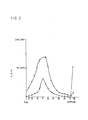
- the ultracentrifugation sedimentation pattern of urokinase ds cDNA is shown in Fig. 2 (indicated by • - • in the figure), together with the sedimentation pattern of globin ds cDNA as a marker (indicated by ⁇ - A in the figure).
- Urokinase ds cDNA shows a slight shift of the peak fraction to the bottom as compared with globin ds cDNA.
- Urokinase ds cDNA was also more abundant in large ds cDNA in fractions 11 to 15 and the neighboring fractions. Therefore, fractions 6-15 were collected and used as large ds cDNA. This large ds cDNA weighed 722 ng.
- the C-tail addition reaction was conducted on the assumption that urokinase ds cDNA is on an average 1,500 bp in size.
- the C-tail reaction should ideally be conducted such that a poly(C) tail comprising 15 to 20 dCs is added to the 3' OH terminal of ds cDNA.
- the inventors used 5 units of terminal transferase and carried out the reaction at 37° C. As a result, about 20 dCs were added as the C-tail.
- a poly(G) tail comprising about 15 dGs was added to the Pstl site of Escherichia coli plasmid pBR322 prepared separately. Equimolar amounts of both products were mixed and annealed under high ion strength conditions by gentle cooling from 70 C to 37° C.
- the Escherichia coli RRI was transformed with the annealed DNA, giving about 70,000 tetracycline- resistant strains.
- the transformants obtained were examined for ampicillin susceptibility by the replica method. More than 90% of the transformants, i.e. about 60,000 colonies, exhibited Ap s (Ampicillin susceptibility) and Tc r (Tetracycline-resistance), suggesting the insertion of cDNA.
- oligodeoxyribonucleotide mixture was synthesized based on the known amino acid sequence of Urinary Urokinase and transformant screening was conducted using this as a hybridization probe.
- probe The 14-base synthetic oligodeoxyribonucleotide mixture used as a probe (hereinafter referred to as probe) was custom-synthesized by Nippon Zeon Co., Ltd. (Tokyo).
- the nucleotide sequence of the probe used and the corresponding urokinase nucleotide sequence are shown in Table 4.
- the probe was drawn from the 73rd to 77th amino acids of human urine urokinase B chain. As is evident in Table 4, the probe is a mixture of 8 oligonucleotides.
- the 5' end of the probe was labelled with [Y- 32 P] ATP (Amersham, Specific activity 5,000 ci/mMol) and T4 polynucleotide kinase.
- the mole ratio of 32 P-ATP to 5'-OH end was 5:1 and the reaction system was as follows:
- the 32 P-labelled probe was purified by applying the reaction mixture prepared in i) on to a NACS-52 minicolumn (BRL).
- Formamide 500 ml
- Mixed-bed-resin 30 ml
- the hybridization process can be summarized as follows: Air drying, drying at 80° C under reduced pressure, and autoradiography at -70 C
- the hybridization was conducted at 37° C for 21 hours with occasional shaking. Thereafter, the filter was washed with WS II three times at room temperature and then three times at 37° C. The dried filter was placed on a filter paper (Whatman 3MM), and autoradiography was performed at -70° C for 2 days, followed by development. As a result, 65 colonies seemingly indicative of relatively high rate of hybridization were selected and again fixed on a filter for rescreening.
- the 65 colonies selected were arranged on an L-agar plate (with pBR322 as a negative control), and 2 filters of the same kind were prepared. Hybridization was conducted with the probe using the filters in pairs, the probe DNA concentration being 5.8 x 10 5 cpm/ml/oligonucleotide species.
- plasmid DNAs (named pUK1, 2, 3 and 4, respectively) were prepared and examined for the size of cDNA.
- pUK1 had the largest cDNA of about 1,900 bp.
- pUK3 and pUK4 were smaller than pUK1 and were about 1,300 bp and about 1,150 bp in size, respectively.
- pUK2 was the smallest cDNA about 170 bp in size. Since, in the restriction enzyme maps, pUK1, 3 and 4 showed overlapping with one another, they were considered to have cloned different regions of one and the same mRNA.
- restriction enzyme maps were prepared for comparison for the four plasmids selected in the above.
- restriction enzymes used were Accl, Alul, Aval, Avall, BamHl, Bcll, Bgll, Bglll, BstEll, Clal, EcoRl, Fnu4HI, Haelll, Hincll, Hindlll, Kpnl, Ncol, Mlul, Pstl, Pvull, Psul, Sacl, Sall, Sau961, Scal, Smal, Stul, Xbal and Xhol.
- the buffers used with these restriction enzymes are shown in Table 5.
- Rsal buffer was used; for Sacl, Kpnl buffer; for Stul, Sall and Xhol, BamHl buffer; for Ncol and Scal, Bglll buffer; and, for Mlul, Hindlll buffer.
- the size of cDNA inserted in pUk1 is 1,900 bp and the largest among pUK1 through 4.
- the size of pUK3 and that of pUK4 are 1,300 bp and 1,150 bp, respectively.
- the cDNA of pUK2 is a fairly small and 170 bp in size. Therefore, for the three plasmids except pUK2, the restriction enzyme map of each cDNA was prepared. Table 6 show the number of restriction sites for each enzyme was determined (Table 6) and thus the distance between restriction enzyme sites was determined for each enzyme. Upon cleavage with EcoRl, pUK1 and pUK4 gave fragments quite equal in size to each other (420 bp).
- the DNA base sequence was determined by the Maxam-Gilbert method. Comparison of the possible amino acid sequence deducible from the DNA base sequence obtained with the known amino acid sequence of human urinary urokinase revealed that cloned cDNAs is a sequence for signal peptide-containing pro-UK.
- Accl, BamHl, Bgll, EcoRl, Hpall, Pstl, Pvull, Sau3AI, Scal and Rsal were from Takara Shuzo and Bcll, Ncol and Taql from NEB.
- the DNA base sequence of each cDNA was determined by the Maxam-Gilbert sequencing method [Proc. Natl. Acad Sci. U.S.A., 74, 560 (1977); Methods in Enzymol., 65, 499 (1980)].
- the plasmid DNA was digested with an appropriate restriction enzyme and the 5'-end labelling was performed using [ ⁇ - 32 P]ATP and T4-polynucleotide Kinase (Fig. 5). The DNA fragments recovered were determined for its nucleotide sequence.
- Judgment as to whether the cDNA obtained was truly the urokinase cDNA was made by comparing the amino acid sequence deduced from the base sequence found with the known amino acid sequence of human urine urokinase [Hopper-Seyler's Z. physiol. Chem., 363, 1043 (1982)].
- the following sequence (III) shows the total amino acid sequence each of human urokinase A-chain and B-chain.
- pUK1 and pUK4 have restriction enzyme maps overlapping each other.
- pUK18 which was selected by colony hybridization using the Nick-translated Pstl fragment of pUK1
- Fig. 4 has a restriction enzyme map overlapping with the map of pUK1 (Fig. 4). Therefore, the cDNAs of these plasmids are considered to have cloned different regions of the same mRNA and accordingly can be combinedly regarded as a single cDNA having a total length of about 2,250 base pairs, as shown in Fig. 5. According to this way of thinking, the sequencing strategy illustrated in Fig. 5 was established and the base sequence was determined.
- Sequence(IV) shows the total base sequence, from the 5'-end to the 3'-noncoding region, of the cDNA coding for an amino acid sequence equivalent to that of urokinase, as determided on the basis of the results of sequencing of individual fragments.
- sequence(IV) shows the total base sequence, from the 5'-end to the 3'-noncoding region, of the cDNA coding for an amino acid sequence equivalent to that of urokinase, as determided on the basis of the results of sequencing of individual fragments.
- Sequence (II) there is a 5'-noncoding region covering at least 79 bases upstream from the protein synthesis start codon ATG (Met) and the first 20 amino acids beginning with Met (ATG) are not found in the amino acid sequence of human urine urokinase [Sequence (III)]. Therefore, it was found that said 20 amino acids constitute a signal peptide required for secretion of the urokinase molecule from the cell.
- the amino acid sequence beginning with the 21st amino acid, Ser agreed with that of human urine urokinase A-chain.
- the cDNA obtained codes for pre-pro UK having a signal peptide comprising 20 amino acids.
- Human urine urokinase which is composed of two chains, A-chain and B-chain, is presumably formed by activation cleavage into two chains, under the action of protease, such as plasmin for instance, of a pro-urokinase molecule synthesized in and secreted from the cell. This was confirmed by sequencing of the cDNA of pUK1.
- human urine urokinase is at first synthesized as a single-chain pro-urokinase composed of A-chain and B-chain coupled with other via Lys and that said pro-urokinase is cleaved at the Arg-Phe-Lys-Ile-Ile site with loss of Lys.
- Sequence(II) shows the terminal portion of the coding region of the cDNA provided by the present invention and part of the 3'-noncoding region thereof.
- the amino acid sequence agreeing with the carboxyl terminal of human urine urokinase B-chain, such as Gly-Leu-Ala-Leu is read out from this base sequence and, downstream therefrom, there occurs the protein synthesis termination codon TGA. Therefore, it was considered that the carboxyl terminal of human urine urokinase is identical with that of pro-UK according to the invention and that there is no polypeptide to be lost by processing.
- the restriction enzyme map suggests that the 3'-noncoding region should be huge. Although only part of the 3'-noncoding region has so far been sequenced, the 3'-noncoding region hitherto obtained in the study of pUK18 is about 850 bp in size. However, pUK18 does not contain the poly(A) sequence characteristic of the 3'-terminal of eukaryotic mRNA, so that the 3'-noncoding region is possibly much longer.
- pUK1 and pUK4 were digested with the restriction enzymes Hindlll and Bglll at 37° C for 1-2 hours according to the table in paragraph (10).
- a fragment of about 5.3 kb was isolated from pUK1, and a fragment of about 1.2 kb from pUK4.
- Both the fragments were ligated with each other by reaction in ligation buffer (20 mM Tris-HCI pH 7.5, 10 mM MgCl 2 , 10 mM dithiothreitol, 0.5 mM ATP) containing 10 units of T4 DNA ligase at 20 C for 2 hours.
- ligation buffer (20 mM Tris-HCI pH 7.5, 10 mM MgCl 2 , 10 mM dithiothreitol, 0.5 mM ATP) containing 10 units of T4 DNA ligase at 20 C for 2 hours.
- E stands for EcoRl, H for Hindlll, and B for BamHl
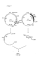
- pSV-GI (Fig. 8) was constructed using, as the starting plasmids, pcDVI and pLl (Fig. 7) created by Okayama and Berg [Molec. and Cell. Biol., 3, 280-289 (1983)].
- the pcDVI and pLl are hybrid plasmids of pBR322 DNA and SV40 DNA. They were obtained from PL Biochemicals (Pharmacia).
- pSV-GI was constructed by inserting a Hindlll-Pstl fragment of pLl into pcDVI at the Hindlll-Kpnl site thereof.
- reaction mixture No. 1 4 ⁇ g of pLl, 8 ⁇ l of 10 x T4 polymerase buffer, 4.0 ⁇ l each of 2 mM dNTP solution, d H 2 0
- 4 ⁇ l (10 U) of T4 polymerase was added to make the total volume 80 ⁇ l, and the reaction was allowed to proceed at 37° C for 5 minutes.
- pcDVI was digested with Hindlll and Kpnl, and a DNA fragment of about 2.7 kbp was isolated on 1 % agarose gel. A 100-ng portion of the fragment was ligated with the above-mentioned pLl- derived DNA fragment of about 600 bp. The ligation was carried out by adding 2 ⁇ l (5.6 U) of T4 ligase to 8 ⁇ l of reaction mixture No. 3 (150 ng of pLl fragment DNA, 100 ng of pcDVI fragment DNA, 1.0 ⁇ l of 10 x T4 ligase buffer, d H 2 0) and allowing the reaction to proceed at 16° C overnight. A 1/2 portion of the reaction mixture was used for performing transformation of E. coli HB101.
- plasmid DNAs were prepared by the mini-prep method and examined by digestion with various restriction enzymes. All the plasmid DNAs were found to contain the desired plasmid. Therefore, using one strain, plasmid DNAs were prepared in large amounts and named pSV-GI, the restriction enzyme map thereof was prepared (Fig. 8).
- the pSV-GI obtained could be judged to have SV-40 early enhancer/promoter element and late 5'-splciing junction arranged upstream and late poly(A) signal and terminater arranged downstream, with the Kpnl site as the cloning site and thus have elements involved in the functional mRNA synthesis in animal cells without any deficiency.
- pUK33 (containing urokinase cDNA 2.2 kbp in total length) obtained in (12) was partially digested with Pstl, and a 1.7 kbp fragment was isolated and inserted into Kpul site of the pSV-GI.
- 500 ⁇ l of reaction mixture No. 4 [100 ⁇ g of pUK33, 50 ⁇ l of 10 x Pstl buffer, 10 ⁇ l (60 U) of Pstl, d H 2 0] was maintained at 37° C. After 10, 15 and 20 minutes of reaction, the reaction mixture was sampled in about 160 ⁇ l portions.
- the sampled reaction mixtures were heat-treated at 65°C for 5 minutes, combined and subjected to 1% agarose gel clectrophoresis, whereby about 10 ⁇ g of the 1.7 kbp containing pre-UK cDNA with its 5'-and 3'-noncoding sequence fragment was recovered. About 5 ⁇ g of the recovered fragment of about 1.7 kbp was treated with T4 DNA polymerase to render the protruding 3'-end blunt, and subjected to ligation with Kpnl linker as m (13-a). The ligation product was digested with Kpnl and then further subjected to ligation with Kpnl-digested pSV-GI. The product was used for transformation of E. coli HB101, whereby pSV-GI-preUK (Fig. 9) was obtained.
- pSV-GI-preUK contains 5'-noncoding region of pro-UK cDNA, signal sequence and 3'-noncoding region as inserted downstream from SV-40 early promoter, and when introduced into appropriate recipient cells, the DNA can induce production of human pro-UK.
- pSV-Gl pSV-GI-Neo r
- a plasmid to be used as a dominant selective marker in transforming cells to be cultured was constructed in the following manner.
- the plasmid pNEO with the Tn5-derived Neo r gene clones therein was subjected to double digestion with BamHl and Hindlll, followed by recovery of DNA fragments by precipitation with ethanol.
- the DNAs recovered were rendered blunt-ended by treatment with E. coli DNA polymerase I large fragment (Klenow fragment) in the presence of all 4 dNTPs.
- a blunt-ended 1.5 kb DNA fragment was separated and then the Kpnl linker was ligated using T4 DNA ligase. After Kpnl linker ligation, the DNA fragment was completely digested with 60 U of Kpnl, and the desired Neo r fragment of about 1.5 kb with said linker joined thereto was recovered.
- a 150-ng portion of the DNA fragment thus recovered was ligated with 20 ng of pSV-GI cleaved with Kpnl in the above manner, and the ligation product was used for transformation of E. coli HB101. In this manner, pSV-GI-Neo r was obtained (Fig. 9).
- Chinese hamster ovary cell [CHO-KI, ATCC CCL61 (purchased from Flow Labo. Inc.)] was used as the recipient cells.
- Cell culture was conducted in Ham's F-12 (Gibco) containing 10% FCS (fetal calf serum) (Boehringer Mannheim). The cells were maintained by subculture in Falcon 3028 flasks. Subconfluent cells were collected 2 days after inoculation by trypsinization, suspended in the above-mentioned culture medium and inoculated into Falcon #3003 (100 mm) culture dishes at an inoculum size of about 5 x 10 5 cells/10ml/100mm. After 24 hours of incubation in a C0 2 incubator (5% CO 2 -95% air), the cells were subjected to transformation. The number of cells per dish on that occasion was about 7.0 x 10 5 .
- the transformant selection in eukaryotic cells was performed by cultivating the cells in G-418-containing medium (400 ug/ml) according to the method of Southern and Berg. [J. Molec, and Applied Genet., 1, 327-341 (1982)], with the Tn5-derived Neo r gene capable of giving G-418 resistance as the dominant selection marker.
- the G-418 used was from Gibco (Geneticin, Gibco, lot No. 75K6043 and 74N8040).
- G-418-resistant colonies were cloned and grown. On day 10, 83 such colonies were obtained. The colonies obtained were examined for production of urokinase with RPHA reagent for human urokinase (Green Cross Corp.) and by fibrin plate assay. Primary screening with RPHA reagent revealed that about 25% of the G-418-resistant colonies produced human urokinase-like substance.
- strains showing RPHA-aggregation were tested for plasminogen activation activity by fibrin plate assay, whereby the activity was observed in the culture supernatant. This activity was neutralized by antihuman urokinase antibody purified by urokinase antigen column chromatography.
- the strain CHO-KI-G-68 seemingly highly capable of producing urokinase-like substance as estimated in the screening was chosen and re-cloned by low density culture, and two strains C-68-53 and C-68-61 were obtained.
- the human urokinase-like substance synthesized and secreted was examined for its properties.
- the human urokinase-like protein secreted into the medium was examined for its properties.
- each clone was inoculated into 25 cm 2 culture flasks (Falcon #3013) at an inoculum size of 5 x 10 5 cells/10 ml/flask.
- the medium was replaced with 5 ml/flask of maintenance medium containing 1% of FCS.
- the plasminogen activator activity in the culture supernatant was measured.
- the molecular weight of human urokinase-like protein produced by said clones was determined.
- the culture supernatant after 24 hours of incubation in maintenance medium was subjected to 12.5% SDS-PAGE.
- the band was blotted onto a nitrocellulose filter electrophoretically in 25 mM Tris-192 mM glycine (pH 8.3)/20% methanol.
- the filter was then blocked in 20 mM Tris-HCI (pH 7.5)/500mM NaCl containing 3% of gelatin at room temperature for 60 minutes and subjected to treatment with rabbit antihuman urokinase IgG purified by means of a human urokinase antigen column and protein A Sepharose as a primary antibody.
- rabbit antihuman urokinase IgG purified by means of a human urokinase antigen column and protein A Sepharose as a primary antibody.
- the human Urokinase band was specificially identified by using immuno-BlotTM assay system (Bio-Rad).
- glycosylated single-chain pro-UK having a molecular weight of 54 K is synthesized and secreted, like in the case of said natural type, and part of it is converted secondarily to double-chain UK in the culture medium.
- the clones were cultivated until cells became confluent.
- the cells were washed twice with Dulbecco's PBS (phosphate-buffered Saline) (+) (Nissui) and then grown in 5 ml of proline-added, Met- free Eagle's MEM medium (product of Nissui) containing 1 % of FCS dialyzed against PBS(+), together with 200 aCi/ml of [3sS)-methionine, for 20 hours.
- Dulbecco's PBS phosphate-buffered Saline
- Met- free Eagle's MEM medium product of Nissui
- the [ 35 S]-labelled culture fluid was applied to an anti-human urokinase monoclonal antibody column (5 ml).
- the column was washed well with 0.04 M sodium phosphate buffer (pH 8.0)/ 0.03 M NaCI and then eluted with glycine-HCI (pH 7.5), whereby 35 S-labelled recombinant human urokinase was recovered.
- the eluate 35 S-labelled human urokinase was neutralized with 1 M Tris, dialyzed against 20 mM sodium phosphate buffer pH 7.4/150 mM NaCI and applied to a ConA-Sepharose 4B (Pharmacia) column.
Landscapes
- Chemical & Material Sciences (AREA)
- Health & Medical Sciences (AREA)
- Organic Chemistry (AREA)
- Life Sciences & Earth Sciences (AREA)
- Engineering & Computer Science (AREA)
- Bioinformatics & Cheminformatics (AREA)
- Zoology (AREA)
- Wood Science & Technology (AREA)
- Genetics & Genomics (AREA)
- Biochemistry (AREA)
- General Health & Medical Sciences (AREA)
- General Engineering & Computer Science (AREA)
- Biomedical Technology (AREA)
- Medicinal Chemistry (AREA)
- Molecular Biology (AREA)
- Biotechnology (AREA)
- Microbiology (AREA)
- Enzymes And Modification Thereof (AREA)
Description
- This invention relates to a method of producing glycosylated single-chain pro-urokinase (hereinafter referred to as pro-UK). More particularly, it relates to a method of producing glycosylated pro-UK which comprises recovering mRNA from an established human kidney-derived cell line, preparing cDNA based on said mRNA, inserting the cDNA into a vector, introducing the resulting plasmid into an animal cell to thereby produce a transformant, and recovering glycosylated pro-UK from said animal cell.
- Urokinase is a plasminogen activator obtainable from human urine and can produce excellent therapeutic effects in the treatment of various forms of thrombosis or embolic diseases as well as in the combined use with an anticancer agents. Currently, urokinase is produced mainly from human urine by some or other purification method (cf. Williams, J. R. B.: Br. J. Exp. Pathol., 32, 530).
- As a result of cell culture technology development in search of a substitute method, the method comprising isolating human kidney cells, subculturing the cells and recovering urokinase from the culture is becoming a second important method. However, recent improvements in the recombinant DNA technology have made it possible to produce desired proteins on a commercial scale by using microorganisms such as Escherichia coli, Saccharomyces cerevisiae and Bacillus subtilis or by using cultured mammalian cells [cf. Goeddel, D. V., Itakura, K., et al.: Proc. Natl. Acad. Sci. U.S.A., 76, 106 (1979) and Nagata, S., Taira, S. H. and Wissmarn, C.: Nature, 284,1316 (1980)].
- Under these circumstances, the present inventors studied in an attempt to develop a method of producing urokinase using recombinant DNA techniques. As a result, they obtained mRNA from an established human kidney-derived cell line, prepared the cDNA sequence shown in the following sequence (I) by using said mRNA as the template, cultivated animal cells transformed with a plasmid with said cDNA sequence inserted therein and obtained glycosylated pro-UK, and have now completed the present invention.
- Thus, the present invention relates to a method of producing glycosylated singly-chain pro-urokinase which comprises recovering mRNA from an establised human kidney-derived cell line, preparing cDNA based on said mRNA, inserting the cDNA into a vector, introducing the resulting plasmid into an animal cell to thereby produce a transformant, and recovering glycosylated pro-UK from said animal cell.


- The pro-UK according to the invention was produced in the following manner:
- Using a mRNA fraction (185 to 20S) containing urokinase mRNA as isolated and purified from an established, human kidney-derived cell line, cDNA was synthesized. Starting with 20 µg of mRNA and using reverse transcriptase and DNA polymerase I, there was obtained 3.3 µg of double-stranded cDNA (ds cDNA). After removal of the single-stranded portion by treatment with SI nuclease, the double stranded cDNA was subjected to sucrose density gradient centrifugation to thereby collect a large molecular ds cDNA fraction (750 ng). A 1/5 portion (150 ng) of this double-stranded cDNA was subjected to treatment for adding a poly(C) tail. A poly(G) tail was added to the Escherichia coli plasmid pBR322 prepared separately. Both the addition products were joined together by annealing. Escherichia coli was transformed with the pBR322 plasmid containing the ds cDNA inserted therein, giving about 70,000 transformants, over 90% of which showed ampicillin resistance indicative of the insertion of the cDNA at the Psti site of pBR322.
- Of these transformants, 10,000 colonies were subjected to screening for selecting urokinase cDNA. Based on the amino acid sequence of urokinase, a mixture of eight 14-mer oligodeoxyribonucleotides, namelywas synthetically prepared and used as a hybridization probe in screening for urokinase cDNA by colony hybridization. As a result, four positive colonies expected to contain urokinase cDNA were obtained. Plasmid DNAs (pUK1 through 4) were prepared from these four colonies and measured for the size of cDNA. pUK1, pUK2, pUK3 and pUK4 were found to contain 1900 bp, 170 bp, 1150 bp and 1300 bp cDNAs, respectively. Detailed restriction enzyme cleavage maps were prepared for pUK1, pUK2 and pUK3, whereby these cDNAs gave restriction maps overlapping one another (cf. Fig. 1). The partial DNA base sequence each of pUK1 and pUK4 as determined by the Maxam-Gilbert method was in agreement with the DNA base sequence deduced from the known amino acid sequence of urokinase. pUK1 had about 670 bp- long 3'-noncoding region-containing cDNA whereas pUK4 had a signal peptide and 5'-noncoding region-containing cDNA. These pUK1 and pUK4 were joined together taking advantage of the Bglll site to thereby construct a plasmid containing the entire translation region for urokinase. Using this plasmid, the DNA base sequence of the entire urokinase translation region and its neighborhood was determined and, based on said sequence, the amino acid sequence of the signal peptide and translation region was further determined [hereinafter reffered to as sequence (II)].


 It was thus revealed that urokinase is constituted by a signal peptide composed of 20 amino acids and the subsequent 411 amino acids. Furthermore, the DNA base sequence of urokinase cDNA and other data revealed that a 5'-noncoding region comprising not less than 80 bases and a 3'-noncoding region comprising not less than 800 bases occur in urokinase mRNA [cf. Sequence (II)].
- The thus-obtained urokinase cDNA was inserted into a vector usable in a heterologous gene expression system in animal cells, followed by introduction into animal cells. Production of glycosylated pro-UK with molecular weight of 54,000 dalton in the transformant cells thus obtained was confirmed.
- In the accompanying drawings,
- Fig. 1 shows the restriction enzyme map of cDNA;
- Fig. 2 shows the ultracentrifugation sedimentation pattern of cDNA;
- Fig. 3 shows the structure of pro-UK mRNA and the position of the synthetic DNA probe;
- Fig. 4 shows the restriction enzyme map each of pUK1, pUK4 and pUK18, pUK1 having cDNA of about 1,900 bp, pUK4 having cDNA of about 1,300 bp and pUK18 having cDNA of about 1,450 bp, their restriction enzyme maps overlapping with one another;
- Fig. 5 shows the restriction enzyme map of cDNA, wherein the restriction enzyme of cDNA is given in a unified form on the basis of Fig. 7; *, X and 0 indicating the position of [32P] labelling and the arrow indicating the direction of sequencing; *, X and 0 meaning that the sequencing was performed using pUK1, pUK4 and pUK18, respectively;
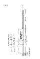
- Fig. 6 shows the flowchart for the construction of plasmid pUK33 by ligation of pUK1 with pUK4, wherein E, H and B stand for EcoRl, Hindlll and BamHl, respectively;
- Fig. 7 shows the flowchart for the ligation of pcDV1 with pL1 ;
- Fig. 8 shows the restriction enzyme map of pSVI-GI;
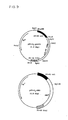
- Fig. 9 shows the restriction enzyme map of pSV-GI-preUK; and pSV-GI-NEO;
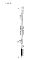
- Fig. 10 shows the structure of preUK cDNA and its neighborhood in pSV-GI-preUk, wherein NC, SS and SJ stand for noncoding region, signal sequence and splicing junction, respectively;
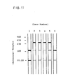
- Fig. 11 shows the results in SDS-PAGE for measurement of the molecular weight of urokinase-like protein in the culture supernatant.
-
lane 1,6 : Urinary urokinase - lane 2 : Culture supernatant of human kidney-derived cell line. (54k in molecular weight band was detected.)
-
lane
-
- For extraction of whole RNA from human kidney-derived established cell line capable of producing urokinase, these cells were first allowed to form a monolayer in a multistage plastic incubator and a Roux flask using a growth medium. Thereafter, the medium was changed for a maintenance medium and the cultivation was continued for further 10-15 hours. 2 to 5 x 10⁹ cells at that stage were collected by trypsin treatment, followed by whole RNA extraction.
- Using the method. disclosed in Japanese Tokkyo Kokai Koho 58-148899 (filed by Green Cross Corp.), mRNA having a size of 19-205 with which urokinase activity was noted upon injection into Xenopus laevis oocytes, was isolated and purified.
- The synthesis of cDNA was performed by the method of Maruin, P. W. et al.: J. Biol. Chem., 253, 2483-2495 (1978).
- (a) To the reaction mixture specified in Table 1, there was added 20 u.g of urokinase mRNA heated at 650 C for 5 minutes and rapidly cooled on ice. After stirring well in an Eppendorff tube, the mixture was allowed to stand on ice.

- (b) Single-stranded cDNA was synthesized by reaction at 46 C for 10-12 minutes.
- (c) The reaction was terminated by adding 20 µl of 0.5 M EDTA to the reaction mixture.
- (d) After extraction with 200 µl of phenol-chloroform, the mixture was centrifuged at 10,000 rpm for 3 minutes. The aqueous layer was separated. The lower phenol layer was reextracted with an equal amount of Sephadex G-100 buffer (10 mM Tris-HCI pH 8.0, 1 mM EDTA, 100 mM NaCI). Both the aqueous layers were combined and 60 µl of 80% glycerin was added to the resulting reaction mixture.
- (e) The reaction mixture was applied to a Sephadex G-100 column, followed by elution with G-100 buffer. The eluate was fractionated by 5 drops.
- (f) Each fraction was tested by means of a Cherenkov counter and radioactive peak portions were collected.
- (g) Thereto were added 1/10 volume of 3 M sodium acetate buffer, 10 µg of tRNA and 2 volumes of ethanol, and the mixture was cooled at -80° C for 20 minutes.
- (h) The precipitate was collected by centrifugation (15,000 rpm, 10 minutes) and dried under reduced pressure.
- (i) The precipitate was dissolved in 300 µl of 0.1 N aqueous NaOH, followed by incubation at 70° C for 20 minutes and cooling with water.
- (j) The solution was neutralized by adding about 30 µl of 1 N HCI. Thereafter, a 2 µl portion was sampled and measured for radioactivity.
- (k) 10 µg of tRAN was added, followed by precipitation with ethanol. The precipitate was collected by centrifugation and dried under reduced pressure.
- (I) The synthesis of single-stranded cDNA was thus completed. The yield of single-stranded cDNA was determined by recovering single-stranded cDNA-mRNA hybrid by G-100 column chromatography and degradative removal of mRNA by hot alkali treatment. As a result, 3.65 µg of urokinase-encoding single-stranded cDNA was obtained.
- For the synthesis of second strand DNA, DNA was first synthesized using DNA polymerase I and then the DNA chain was extended using reverse transcriptase.
- (a) 100 µl of single-stranded cDNA was suspended in 2 x reaction mixture (Table 2).

- (b) Incubation was conducted at 60 °C for 2 minutes, followed by centrifugation (15,000 rpm, 2.5 minutes). The supernatant was collected.
- (c) Following addition of 50 µl of water to the precipitate, the procedure of (b) was repeated. Both the supernatants were combined.
- (d) 50 units of DNA polymerase I Klenow fragment was added and the total amount was made 200 µl.
- (e) Incubation was performed at 15 °C overnight.
- (f) The reaction was terminated by adding 20 µl of 0.2 M EDTA (pH 8.0), then an equal volume (220 µl) of phenol-chloroform solution was added and the mixture was stirred and centrifuged. The aqueous layer was collected. The phenol layer was reextracted with 200 µl of TE buffer (10 mM Tris-HCI pH 8.0, 1 mM EDTA).
- (g) Both the aqueous layers were combined and 60 µl of 80% glycerin was added.
- (h) The resulting mixture was applied to a Sephadex G-100 column, followed by elution with G-100 buffer. The eluate was fractionated by 5 drops.
- (i) Each fraction was tested by means of a Cherenkov counter, and radioactivity peak fractions were collected. 20 µl aliquot was mixed with 10 ml of ACS II and the mixture was used for radioactivity measurement. To the remaining portion of the liquid, there were added 5 u.g of tRNA and 1/10 volumn of 3 M sodium acetate buffer, followed by precipitation with ethanol.
- (j) The precipitate was collected by centrifugation (15,000 rpm, 10 minutes) and dried under reduced pressure.
- Thereafter, synthetic extention of second strand DNA was conducted using reverse transcriptase.
-
- (a) The ds cDNA obtained was dissolved in 20 µl of water. To the solution was added 30 µl of the reaction mixture given in Table 3, followed by stirring.

- (b) This mixture was incubated at 42° C for 60 minutes.
- (c) The reaction was terminated by adding 5 µl of 0.2 M EDTA.
- (d) Deproteinization was performed by adding an equal volume (55 µl) of phenol-chloroform.
- (e) Thereafter, the same procedures [(g)-(j)] as in the synthesis of ds cDNA using polymerase I were performed, followed by digestion treatment of the single-stranded DNA portion with SI nuclease according to Shenk, T. E. et al. [cf. Proc. Natl. Acad. Sci. U.S.A., 72, 989 (1975)], as mentioned below.
-
- (a) The ds cDNA was suspended in 100 µl of buffer for SI nuclease [0.3 M NaCI, 30 mM sodium acetate (pH 4.5), 3 mM ZnCl2] and, after complete dissolution of ds cDNA by incubation at 37° C for 30 minutes, 0.5 units of SI nuclease (product of PL Biochemical) was added.
- (b) After 30 minutes of reaction at 37° C, 2 units of SI nuclease was further added and the reaction was allowed to proceed for further 15 minutes.
- (c) 20 µl of 0.2 M EDTA was added to terminate the reaction, followed by addition of 120 µl of phenol-chloroform for deproteinization.
- (d) Teh phenol layer was reextracted with TE buffer. Both the aqueous layers were combined and subjected to precipitation with ethanol to give ds cDNA.
- The ds cDNA obtained was then subjected to sucrose density gradient centrifugation (38,000 rpm, 4° C) and large molecular weight fractions were collected. The ultracentrifugation sedimentation pattern of urokinase ds cDNA is shown in Fig. 2 (indicated by • - • in the figure), together with the sedimentation pattern of globin ds cDNA as a marker (indicated by ▲ - A in the figure). Urokinase ds cDNA shows a slight shift of the peak fraction to the bottom as compared with globin ds cDNA. Urokinase ds cDNA was also more abundant in large ds cDNA in
fractions 11 to 15 and the neighboring fractions. Therefore, fractions 6-15 were collected and used as large ds cDNA. This large ds cDNA weighed 722 ng. - The C-tail addition reaction was conducted on the assumption that urokinase ds cDNA is on an average 1,500 bp in size. The C-tail reaction should ideally be conducted such that a poly(C) tail comprising 15 to 20 dCs is added to the 3' OH terminal of ds cDNA. The inventors used 5 units of terminal transferase and carried out the reaction at 37° C. As a result, about 20 dCs were added as the C-tail.
- In the same manner, a poly(G) tail comprising about 15 dGs was added to the Pstl site of Escherichia coli plasmid pBR322 prepared separately. Equimolar amounts of both products were mixed and annealed under high ion strength conditions by gentle cooling from 70 C to 37° C.
- The Escherichia coli RRI was transformed with the annealed DNA, giving about 70,000 tetracycline- resistant strains. The transformants obtained were examined for ampicillin susceptibility by the replica method. More than 90% of the transformants, i.e. about 60,000 colonies, exhibited Aps (Ampicillin susceptibility) and Tcr(Tetracycline-resistance), suggesting the insertion of cDNA.
- For cloning urokinase cDNA, an oligodeoxyribonucleotide mixture was synthesized based on the known amino acid sequence of Urinary Urokinase and transformant screening was conducted using this as a hybridization probe.
- The 14-base synthetic oligodeoxyribonucleotide mixture used as a probe (hereinafter referred to as probe) was custom-synthesized by Nippon Zeon Co., Ltd. (Tokyo).
- The nucleotide sequence of the probe used and the corresponding urokinase nucleotide sequence are shown in Table 4. The probe was drawn from the 73rd to 77th amino acids of human urine urokinase B chain. As is evident in Table 4, the probe is a mixture of 8 oligonucleotides.

- Note: In the middle line is shown all possible sequences of mRNA. In the lower line is shown the base sequence of the probe DNA complementary to said mRNA. The probe DNA is lacking in the third base in the codon for the 77th amino acid (Glu).
- The 5' end of the probe was labelled with [Y-32P] ATP (Amersham, Specific activity 5,000 ci/mMol) and T4 polynucleotide kinase. As regards the labelling conditions, the mole ratio of 32P-ATP to 5'-OH end was 5:1 and the reaction system was as follows:
- Reaction mixture:

- The reaction was carried out at 37° C for 60-120 minutes. After completion of the reaction, 1 µl of 0.5 M EDTA (pH 8.0), 35 µl of 1 M sodium chloride (in 10 mM Tris-HCI buffer and 1 mM EDTA) and 10.5 µl of 1 mg/ml tRNA solution and the final volume was made 35 µl, and the mixture was heat-treated at 70° C for 3 minutes and then applied to a NACS-52 minicolumn. ii) Purification of 32P-labelled probe
- The 32P-labelled probe was purified by applying the reaction mixture prepared in i) on to a NACS-52 minicolumn (BRL).
- The efficiency of labelling was almost constant irrespective of the kind of probe. Supposing that the recovery from the column was 80%, the specific activity of the probe DNA was estimated at about 1.0 x 10⁹ cpm/µg of DNA. (c) Colony hybridization
- Using the probe mentioned above, screening for urokinase cDNA was conducted by colony hybridization.
- Formamide (500 ml) and Mixed-bed-resin (30 ml) were mixed and the mixture was stirred at 4° C for 2 hours, suction-filtered and stored at -80 C.
- 50 x Denhardt's solution:
- Stored at -20 °C after bacterial filtration.
- 20 x SSPE:
- 3.6 M NaCl
- 0.2 M NaH2PO4 (adjusted to pH 7.4 with 10 N NaOH)
- 20 mM EDTA
- 20 x SSC
- Sonicated salmon sperm DNA
- Prehybridization washing solution (hereinafter, PHWS):
- 50 mM Tris-HCI (pH 8.0)
- 1 M NaCl
- 1 mM EDTA
- 0.1% SDS
- Prehybridization solution:
- 50% deionized formamide
- 5 x Denhardt's solution
- 5 x SSPE
- 0.1% SDS
- 100 µg/mt Sonicated DNA
- Washing solution I (hereinafter, WS I):
- 5 x SSC
- 1 x Denhardt's solution
- 0.1% SDS
- 100 µg/ml tRNA
- Washing solution II (hereinafter, WS II):
- 5 x SSC
- 0.1% SDS
- Hybridization solution (hereinafter, HS):
- 5 x SSC
- 1 x Denhardt's solution
- 0.1% SDS
- 100 µg/ml tRNA
- 32P-labelled probe
- The hybridization process can be summarized as follows:

- Using 3.0 x 10⁸ cpm of the 32P-labelled probe, 10,000 colonies (100 filters) were screened. The total amount of HS was 150 ml and therefore the probe concentration was 2.0 x 10⁶ cpm/ml.
- The hybridization was conducted at 37° C for 21 hours with occasional shaking. Thereafter, the filter was washed with WS II three times at room temperature and then three times at 37° C. The dried filter was placed on a filter paper (Whatman 3MM), and autoradiography was performed at -70° C for 2 days, followed by development. As a result, 65 colonies seemingly indicative of relatively high rate of hybridization were selected and again fixed on a filter for rescreening.
- The 65 colonies selected were arranged on an L-agar plate (with pBR322 as a negative control), and 2 filters of the same kind were prepared. Hybridization was conducted with the probe using the filters in pairs, the probe DNA concentration being 5.8 x 10⁵ cpm/ml/oligonucleotide species.
- After 21 hours of hybridization at 37° C, the filters were washed with WS II. One filter was dried at 37° C directly after washing, while the other was further washed three times at 42 °C for 3 minutes, followed by exposure of an X-ray film thereto.
- In this hybridization experiments, four colonies were found to be indicative of hybridization with the probe DNA. After washing at 42° C, these 4 colonies were still clearly recognized as positive colonies.
- For the 4 colonies selected, plasmid DNAs (named pUK1, 2, 3 and 4, respectively) were prepared and examined for the size of cDNA. Among the 4 plasmids pUK1 had the largest cDNA of about 1,900 bp. pUK3 and pUK4 were smaller than pUK1 and were about 1,300 bp and about 1,150 bp in size, respectively. pUK2 was the smallest cDNA about 170 bp in size. Since, in the restriction enzyme maps, pUK1, 3 and 4 showed overlapping with one another, they were considered to have cloned different regions of one and the same mRNA.
- The restriction enzyme maps were prepared for comparison for the four plasmids selected in the above.
- The restriction enzymes used were Accl, Alul, Aval, Avall, BamHl, Bcll, Bgll, Bglll, BstEll, Clal, EcoRl, Fnu4HI, Haelll, Hincll, Hindlll, Kpnl, Ncol, Mlul, Pstl, Pvull, Psul, Sacl, Sall, Sau961, Scal, Smal, Stul, Xbal and Xhol. The buffers used with these restriction enzymes are shown in Table 5.
- For Aval, Rsal buffer was used; for Sacl, Kpnl buffer; for Stul, Sall and Xhol, BamHl buffer; for Ncol and Scal, Bglll buffer; and, for Mlul, Hindlll buffer.
-

- As already mentioned hereinabove, the size of cDNA inserted in pUk1 is 1,900 bp and the largest among pUK1 through 4. The size of pUK3 and that of pUK4 are 1,300 bp and 1,150 bp, respectively. The cDNA of pUK2 is a fairly small and 170 bp in size. Therefore, for the three plasmids except pUK2, the restriction enzyme map of each cDNA was prepared. Table 6 show the number of restriction sites for each enzyme was determined (Table 6) and thus the distance between restriction enzyme sites was determined for each enzyme. Upon cleavage with EcoRl, pUK1 and pUK4 gave fragments quite equal in size to each other (420 bp). Upon double digestion with BamHl and EcoRl and with BamHl and Pstl, a fragment from pUK1 was entirely the same in size with one from pUK3. Therefore, taking the EcoRl site as a basis for pUK1 and pUK4 and the BamHl site as a standard for pUK1 and pUK3, other restriction enzyme sites of these plasmids were examined, whereby the restriction enzyme sites on cDNA of pUK1 were found to be almost in agreement in position with those on cDNA of pUK3 and of pUK4. On the supposition that the cDNA of pUK1, that of pUK3 and that of pUK4 are identical to one another, it was estimated that, in pUK1 and pUK4, the cDNA was inserted in pBR322 in the same direction and that, in pUK3, the cDNA was inserted in the opposite direction as compared with pUK1 and pUK4.
- Since the cDNAs inserted in pUK1, pUK3 and pUK4 could be regarded as one and the same in view of the restriction enzyme sites occurring therein, the possibility that these three cDNAs each might be the urokinase cDNA became very great. The grounds are that, in spite of very low probability of plasmids having a sequence identical with the base sequence of the synthetic 14-mer DNA used in screening, 3 out of the 4 colonies found to be positive in the screening had the same cDNA, rendering high the probability that the cDNA owned by these positive colonies might have the desired sequence for urokinase; and that the position of the DNA base sequence synthesized as a probe on urokinase mRNA (Fig. 3) agreed with the place of overlapping among pUK1, pUK3 and pUK4 (Fig. 1). The DNA base sequence of part of pUK1 and of pUK4 was determined. The corresponding amino acid sequence deduced was in agreement with the amino acid sequence of urokinase.

- For the purpose of directly clarifying whether the plasmids obtained by the procedure mentioned in the preceding paragraph did contain the urokinase cDNA, the DNA base sequence was determined by the Maxam-Gilbert method. Comparison of the possible amino acid sequence deducible from the DNA base sequence obtained with the known amino acid sequence of human urinary urokinase revealed that cloned cDNAs is a sequence for signal peptide-containing pro-UK.
- The above-mentioned pUK1 and pUK4 were used.
- Accl, BamHl, Bgll, EcoRl, Hpall, Pstl, Pvull, Sau3AI, Scal and Rsal were from Takara Shuzo and Bcll, Ncol and Taql from NEB.
- The restriction enzyme map of each plasmid with the cDNA inserted therein was prepared by the procedure mentioned under the preceding paragraph (10).
- The DNA base sequence of each cDNA was determined by the Maxam-Gilbert sequencing method [Proc. Natl. Acad Sci. U.S.A., 74, 560 (1977); Methods in Enzymol., 65, 499 (1980)].
- Thus, the plasmid DNA was digested with an appropriate restriction enzyme and the 5'-end labelling was performed using [γ-32P]ATP and T4-polynucleotide Kinase (Fig. 5). The DNA fragments recovered were determined for its nucleotide sequence.
- The [32P]-labelled DNA fragment was subjected to Maxam-Gilbert's limited chemical degradation [Proc. Natl. Acad. Sci. U.S.A., 74, 560 (1977); Methods in Enzymol., 65, 499 (1980)], followed by 8% and 20% 7M urea-polyacrylamide gel electrophoresis in the conventional manner and autoradiography at -70 C for 2-3 days.
- Judgment as to whether the cDNA obtained was truly the urokinase cDNA was made by comparing the amino acid sequence deduced from the base sequence found with the known amino acid sequence of human urine urokinase [Hopper-Seyler's Z. physiol. Chem., 363, 1043 (1982)]. The following sequence (III) shows the total amino acid sequence each of human urokinase A-chain and B-chain.


- pUK1 and pUK4 have restriction enzyme maps overlapping each other. On the other hand, pUK18, which was selected by colony hybridization using the Nick-translated Pstl fragment of pUK1, has a restriction enzyme map overlapping with the map of pUK1 (Fig. 4). Therefore, the cDNAs of these plasmids are considered to have cloned different regions of the same mRNA and accordingly can be combinedly regarded as a single cDNA having a total length of about 2,250 base pairs, as shown in Fig. 5. According to this way of thinking, the sequencing strategy illustrated in Fig. 5 was established and the base sequence was determined.
- Comparison of the DNA base sequence obtained by sequencing with the amino acid sequence of human urinary urokinase [Sequence(III)] revealed that pUK1, 4 and 18 each contained part of pro-UK cDNA.
- The following Sequence(IV) shows the total base sequence, from the 5'-end to the 3'-noncoding region, of the cDNA coding for an amino acid sequence equivalent to that of urokinase, as determided on the basis of the results of sequencing of individual fragments. Hereinafter, mention is made of the structure of the cDNA coding for an amino acid sequence equivalent to single-chain pro-urokinase as obtained in accordance with the present invention.


- As can be seen in Sequence (II), there is a 5'-noncoding region covering at least 79 bases upstream from the protein synthesis start codon ATG (Met) and the first 20 amino acids beginning with Met (ATG) are not found in the amino acid sequence of human urine urokinase [Sequence (III)]. Therefore, it was found that said 20 amino acids constitute a signal peptide required for secretion of the urokinase molecule from the cell. The amino acid sequence beginning with the 21st amino acid, Ser, agreed with that of human urine urokinase A-chain.
- In view of the above, it is evident that the cDNA obtained codes for pre-pro UK having a signal peptide comprising 20 amino acids.
- Human urine urokinase, which is composed of two chains, A-chain and B-chain, is presumably formed by activation cleavage into two chains, under the action of protease, such as plasmin for instance, of a pro-urokinase molecule synthesized in and secreted from the cell. This was confirmed by sequencing of the cDNA of pUK1.
- In sequence(II), it is seen that the carboxyl terminal amino acid sequence [the 157th amino acid the in Sequence (II)] of human urine urokinase A-chain is followed by Lys and further by the amino terminal amino acid sequence of B-chain, namely Ile-Ile-Gly-Gly-.....
- This indicates that human urine urokinase is at first synthesized as a single-chain pro-urokinase composed of A-chain and B-chain coupled with other via Lys and that said pro-urokinase is cleaved at the Arg-Phe-Lys-Ile-Ile site with loss of Lys.
- Sequence(II)shows the terminal portion of the coding region of the cDNA provided by the present invention and part of the 3'-noncoding region thereof. As shown in the Sequence(II), the amino acid sequence agreeing with the carboxyl terminal of human urine urokinase B-chain, such as Gly-Leu-Ala-Leu, is read out from this base sequence and, downstream therefrom, there occurs the protein synthesis termination codon TGA. Therefore, it was considered that the carboxyl terminal of human urine urokinase is identical with that of pro-UK according to the invention and that there is no polypeptide to be lost by processing.
- On the other hand, the restriction enzyme map suggests that the 3'-noncoding region should be huge. Although only part of the 3'-noncoding region has so far been sequenced, the 3'-noncoding region hitherto obtained in the study of pUK18 is about 850 bp in size. However, pUK18 does not contain the poly(A) sequence characteristic of the 3'-terminal of eukaryotic mRNA, so that the 3'-noncoding region is possibly much longer.
- pUK1 and pUK4 were digested with the restriction enzymes Hindlll and Bglll at 37° C for 1-2 hours according to the table in paragraph (10). A fragment of about 5.3 kb was isolated from pUK1, and a fragment of about 1.2 kb from pUK4. Both the fragments were ligated with each other by reaction in ligation buffer (20 mM Tris-HCI pH 7.5, 10 mM MgCl2, 10 mM dithiothreitol, 0.5 mM ATP) containing 10 units of T4 DNA ligase at 20 C for 2 hours. The above steps are shown in Fig. 6. (In Fig. 6, E stands for EcoRl, H for Hindlll, and B for BamHl). Resulted plasmid, named pUK33, containing about 2.2 kbp of pre-urokinase cDNA.
- pSV-GI (Fig. 8) was constructed using, as the starting plasmids, pcDVI and pLl (Fig. 7) created by Okayama and Berg [Molec. and Cell. Biol., 3, 280-289 (1983)]. The pcDVI and pLl are hybrid plasmids of pBR322 DNA and SV40 DNA. They were obtained from PL Biochemicals (Pharmacia). For facilitating the insertion of urokinase-encoding DNA into these plasmids, pSV-GI was constructed by inserting a Hindlll-Pstl fragment of pLl into pcDVI at the Hindlll-Kpnl site thereof.
- Thus, 4 µg of pLl was digested with Pstl, the protruding 3'-end was rendered blunt by treatment with T4 DNA polymerase. Namely, 76 µl of reaction mixture No. 1 (4 µg of pLl, 8 µl of 10 x T4 polymerase buffer, 4.0 µl each of 2 mM dNTP solution, d H20) was heated at 65 °C for 3 minutes and then quenched, 4 µl (10 U) of T4 polymerase was added to make the total volume 80 µl, and the reaction was allowed to proceed at 37° C for 5 minutes. Thereafter, 8 µl of 0.25 M EDTA (pH 8.0) and 80 µl of
d H 20 were added, the resulting mixture was subjected once to treatment with phenol-chroloform, and the aqueous layer was recovered. DNA was recovered by precipitation with ethanol, dried under reduced prssure, and subjected to Kpnl linker ligation. The ligation reaction was carried out at 16° C overnight in a reaction mixture No. 2 (4 µg of dried DNA, 2 µl (2 µg) of 5'-P Kpnl linker, 2.0 µl of 10 x ligase buffer, d H20) adding 2 µl (5 U) of T4 ligase. Thereafter, digestion was carried out with Kpnl and then with Hindlll, followed by 5% polyacrylamide gel electrophoresis (PAGE), whereby a DNA fragment of about 600 bp was recovered. - Separately, pcDVI was digested with Hindlll and Kpnl, and a DNA fragment of about 2.7 kbp was isolated on 1 % agarose gel. A 100-ng portion of the fragment was ligated with the above-mentioned pLl- derived DNA fragment of about 600 bp. The ligation was carried out by adding 2 µl (5.6 U) of T4 ligase to 8 µl of reaction mixture No. 3 (150 ng of pLl fragment DNA, 100 ng of pcDVI fragment DNA, 1.0 µl of 10 x T4 ligase buffer, d H20) and allowing the reaction to proceed at 16° C overnight. A 1/2 portion of the reaction mixture was used for performing transformation of E. coli HB101.
- Using 12 colonies out of the transformants obtained, plasmid DNAs were prepared by the mini-prep method and examined by digestion with various restriction enzymes. All the plasmid DNAs were found to contain the desired plasmid. Therefore, using one strain, plasmid DNAs were prepared in large amounts and named pSV-GI, the restriction enzyme map thereof was prepared (Fig. 8). The pSV-GI obtained could be judged to have SV-40 early enhancer/promoter element and late 5'-splciing junction arranged upstream and late poly(A) signal and terminater arranged downstream, with the Kpnl site as the cloning site and thus have elements involved in the functional mRNA synthesis in animal cells without any deficiency.
- pUK33 (containing urokinase cDNA 2.2 kbp in total length) obtained in (12) was partially digested with Pstl, and a 1.7 kbp fragment was isolated and inserted into Kpul site of the pSV-GI. Thus, 500 µl of reaction mixture No. 4 [100 µg of pUK33, 50 µl of 10 x Pstl buffer, 10 µl (60 U) of Pstl, d H20] was maintained at 37° C. After 10, 15 and 20 minutes of reaction, the reaction mixture was sampled in about 160 µl portions. The sampled reaction mixtures were heat-treated at 65°C for 5 minutes, combined and subjected to 1% agarose gel clectrophoresis, whereby about 10 µg of the 1.7 kbp containing pre-UK cDNA with its 5'-and 3'-noncoding sequence fragment was recovered. About 5 µg of the recovered fragment of about 1.7 kbp was treated with T4 DNA polymerase to render the protruding 3'-end blunt, and subjected to ligation with Kpnl linker as m (13-a). The ligation product was digested with Kpnl and then further subjected to ligation with Kpnl-digested pSV-GI. The product was used for transformation of E. coli HB101, whereby pSV-GI-preUK (Fig. 9) was obtained.
- As seen in Fig. 10 pSV-GI-preUK contains 5'-noncoding region of pro-UK cDNA, signal sequence and 3'-noncoding region as inserted downstream from SV-40 early promoter, and when introduced into appropriate recipient cells, the DNA can induce production of human pro-UK.
- Using the above pSV-Gl, pSV-GI-Neor, a plasmid to be used as a dominant selective marker in transforming cells to be cultured, was constructed in the following manner.
- The plasmid pNEO with the Tn5-derived Neor gene clones therein [Southern, P. J. and Berg, J.: Molec, and Applied Genet., 1, 327-341 (1982)] was subjected to double digestion with BamHl and Hindlll, followed by recovery of DNA fragments by precipitation with ethanol. The DNAs recovered were rendered blunt-ended by treatment with E. coli DNA polymerase I large fragment (Klenow fragment) in the presence of all 4 dNTPs. A blunt-ended 1.5 kb DNA fragment was separated and then the Kpnl linker was ligated using T4 DNA ligase. After Kpnl linker ligation, the DNA fragment was completely digested with 60 U of Kpnl, and the desired Neor fragment of about 1.5 kb with said linker joined thereto was recovered.
- A 150-ng portion of the DNA fragment thus recovered was ligated with 20 ng of pSV-GI cleaved with Kpnl in the above manner, and the ligation product was used for transformation of E. coli HB101. In this manner, pSV-GI-Neor was obtained (Fig. 9).
- Chinese hamster ovary cell [CHO-KI, ATCC CCL61 (purchased from Flow Labo. Inc.)] was used as the recipient cells. Cell culture was conducted in Ham's F-12 (Gibco) containing 10% FCS (fetal calf serum) (Boehringer Mannheim). The cells were maintained by subculture in Falcon 3028 flasks. Subconfluent cells were collected 2 days after inoculation by trypsinization, suspended in the above-mentioned culture medium and inoculated into Falcon #3003 (100 mm) culture dishes at an inoculum size of about 5 x 10⁵ cells/10ml/100mm. After 24 hours of incubation in a C02 incubator (5% CO2-95% air), the cells were subjected to transformation. The number of cells per dish on that occasion was about 7.0 x 10⁵.
- To the dished prepared in (d), there was added 1 ml per dish of a DNA CaP04 CO- precipitate containing 2.0 µg (as DNA)/ml of the plasmid DNA [pSV-GI-preUK and pSV-GI-Neo (mixing ration 100:1)] and 20 µg/ml of sonicated salmon sperm DNA as a carrier DNA, as prepared by the method of Osada et al. [Protein, Nucleic Acid and Enzyme, 28 (14), 1569-1581 (1983)]. About 18 hours after exposure to DNA, the medium was exchanged. The incubation was then continued further 48 hours or more, followed by selection using antibiotics G-418.
- Since the urokinase gene itself is not "selectable", the transformant selection in eukaryotic cells was performed by cultivating the cells in G-418-containing medium (400 ug/ml) according to the method of Southern and Berg. [J. Molec, and Applied Genet., 1, 327-341 (1982)], with the Tn5-derived Neor gene capable of giving G-418 resistance as the dominant selection marker. The G-418 used was from Gibco (Geneticin, Gibco, lot No. 75K6043 and 74N8040).
- While exchanging the medium every 4 days, G-418-resistant colonies were cloned and grown. On day 10, 83 such colonies were obtained. The colonies obtained were examined for production of urokinase with RPHA reagent for human urokinase (Green Cross Corp.) and by fibrin plate assay. Primary screening with RPHA reagent revealed that about 25% of the G-418-resistant colonies produced human urokinase-like substance.
- Fifteen strains showing RPHA-aggregation were tested for plasminogen activation activity by fibrin plate assay, whereby the activity was observed in the culture supernatant. This activity was neutralized by antihuman urokinase antibody purified by urokinase antigen column chromatography. The strain CHO-KI-G-68 seemingly highly capable of producing urokinase-like substance as estimated in the screening was chosen and re-cloned by low density culture, and two strains C-68-53 and C-68-61 were obtained.
- Using the culture supernatnat of these strains, the human urokinase-like substance synthesized and secreted was examined for its properties. The clones C-68-53 and C-68-61 secreted human urokinase-like protein into the medium in a stable and constitutive manner for more than 100 days.
- For said two clones, the human urokinase-like protein secreted into the medium was examined for its properties.
- First, the production of human urokinase-like protein by each of these two clones was determined. Thus, each clone was inoculated into 25 cm2 culture flasks (Falcon #3013) at an inoculum size of 5 x 10⁵ cells/10 ml/flask. When the cells became confluent after 3 days of incubation, the medium was replaced with 5 ml/flask of maintenance medium containing 1% of FCS. After 24 hours of incubation in maintenance medium, the plasminogen activator activity in the culture supernatant was measured. The activity measurement by the fibrin plate method using human urinary high molecular weight urokinase as a reference indicated a yield of about 200 IU/ml/day for both the clones. Such activity was specifically neutralized by antihuman urokinase monoclonal antibody.
- Next, the molecular weight of human urokinase-like protein produced by said clones was determined. Thus, the culture supernatant after 24 hours of incubation in maintenance medium was subjected to 12.5% SDS-PAGE. The band was blotted onto a nitrocellulose filter electrophoretically in 25 mM Tris-192 mM glycine (pH 8.3)/20% methanol. The filter was then blocked in 20 mM Tris-HCI (pH 7.5)/500mM NaCl containing 3% of gelatin at room temperature for 60 minutes and subjected to treatment with rabbit antihuman urokinase IgG purified by means of a human urokinase antigen column and protein A Sepharose as a primary antibody. Thus the human Urokinase band was specificially identified by using immuno-BlotTM assay system (Bio-Rad).
- When the samples were used after reduction treatment with 2-mercaptoethanol, the culture supernatants of both the clones gave three bands, a main band 54 K in molecular weight and subbands 34 K and 20 K in molecular weight. Simultaneous electrophoresis of human urine-derived two-chain high molecular UK (as a reference) and the culture supernatant of pro-UK-producing human kidney-derived cell live revealed that the 54 K, 34 K and 20 K bands from the culture supernatant from both C-68-53 and C-68-61 cells agreed in molecular weight and reactivity to anti-UK IgG with the human natural-type single-chain pro-UK (54 K) band and the 34 K and 20 K bands from double-chain high molecular UK, respectively, no distinction could be made. (Fig. 11)
- The above results indicated that, in both the clones, glycosylated single-chain pro-UK having a molecular weight of 54 K is synthesized and secreted, like in the case of said natural type, and part of it is converted secondarily to double-chain UK in the culture medium.
- Therefore, the presence or absence of the sugar chain in the human pro-UK synthesized and secreted by both the clones was then examined by column chromatography using ConA-Sepharose 4B (Pharmacia).
- Thus, the clones were cultivated until cells became confluent. The cells were washed twice with Dulbecco's PBS (phosphate-buffered Saline) (+) (Nissui) and then grown in 5 ml of proline-added, Met- free Eagle's MEM medium (product of Nissui) containing 1 % of FCS dialyzed against PBS(+), together with 200 aCi/ml of [3sS)-methionine, for 20 hours.
- The [35S]-labelled culture fluid was applied to an anti-human urokinase monoclonal antibody column (5 ml). The column was washed well with 0.04 M sodium phosphate buffer (pH 8.0)/ 0.03 M NaCI and then eluted with glycine-HCI (pH 7.5), whereby 35S-labelled recombinant human urokinase was recovered. The eluate 35S-labelled human urokinase was neutralized with 1 M Tris, dialyzed against 20 mM sodium phosphate buffer pH 7.4/150 mM NaCI and applied to a ConA-Sepharose 4B (Pharmacia) column. After washing with buffer, elution was performed with sodium phosphate buffer (pH 7.4) containing 0.1 M methyl-a-D-mannoside. Most of the radioactivity eluted from the anti-UK monoclonal antibody column was adsorbed on ConA-Sepharose and specifically eluted with mannoside.
- The above results indicate that the recombinant urokinase synthesized and secreted by both the clones is a glycoprotein purifiable by means of an anti-human urokinase monoclonal antibody column.
- The above results also indicate that the human pro-UK cDNA introduced into CHO-KI can be transcribed in a normal manner and the pro-UK is synthesized and secreted as a glycoprotein having a molecular weight equivalent to that of the natural-type pro-UK and having reactivity to monoclonal anti human Urokinase antibodies.
There was thus obtained 5.3 u.g of ds cDNA.
Claims (3)


Applications Claiming Priority (4)
| Application Number | Priority Date | Filing Date | Title |
|---|---|---|---|
| JP59037119A JPS60180591A (en) | 1984-02-27 | 1984-02-27 | Dna sequence, plasmid and host of human urokinase |
| JP37119/84 | 1984-02-27 | ||
| JP17969/85 | 1985-01-31 | ||
| JP1796985A JPS61177987A (en) | 1985-01-31 | 1985-01-31 | Production of human urokinase |
Publications (3)
| Publication Number | Publication Date |
|---|---|
| EP0154272A2 EP0154272A2 (en) | 1985-09-11 |
| EP0154272A3 EP0154272A3 (en) | 1988-01-20 |
| EP0154272B1 true EP0154272B1 (en) | 1992-01-08 |
Family
ID=26354568
Family Applications (1)
| Application Number | Title | Priority Date | Filing Date |
|---|---|---|---|
| EP19850102031 Expired EP0154272B1 (en) | 1984-02-27 | 1985-02-23 | Production of human urokinase |
Country Status (2)
| Country | Link |
|---|---|
| EP (1) | EP0154272B1 (en) |
| DE (1) | DE3585097D1 (en) |
Families Citing this family (10)
| Publication number | Priority date | Publication date | Assignee | Title |
|---|---|---|---|---|
| JPS62149625A (en) * | 1985-12-25 | 1987-07-03 | Green Cross Corp:The | Production of physiologically active substance |
| US5098840A (en) * | 1986-07-03 | 1992-03-24 | The Green Cross Corporation | Human prourokinase mutants |
| JPH0761266B2 (en) * | 1986-07-03 | 1995-07-05 | 株式会社ミドリ十字 | Mutant human prourokinase |
| JPS63105675A (en) * | 1986-10-23 | 1988-05-10 | Green Cross Corp:The | Production of human urokinase |
| ZA879286B (en) | 1986-12-16 | 1988-10-26 | Smith Kline Rit | New plasminogen activators |
| MY103358A (en) * | 1987-04-15 | 1993-06-30 | Novartis Ag | Process for the production of protiens. |
| EP0303028A1 (en) * | 1987-08-10 | 1989-02-15 | GRUPPO LEPETIT S.p.A. | Method for preparing single chain urokinase |
| JP2764264B2 (en) * | 1987-10-01 | 1998-06-11 | 株式会社ミドリ十字 | Fibrinolytic activity enhancer |
| JP2791418B2 (en) | 1987-12-02 | 1998-08-27 | 株式会社ミドリ十字 | Method for producing heterologous protein, recombinant DNA, transformant |
| US4920051A (en) * | 1988-02-03 | 1990-04-24 | Damon Biotech, Inc. | Recovery of urokinase compounds |
Family Cites Families (3)
| Publication number | Priority date | Publication date | Assignee | Title |
|---|---|---|---|---|
| US4370417A (en) * | 1980-04-03 | 1983-01-25 | Abbott Laboratories | Recombinant deoxyribonucleic acid which codes for plasminogen activator |
| GR78180B (en) * | 1982-04-15 | 1984-09-26 | Genentech Inc | |
| AR241654A1 (en) * | 1982-05-05 | 1992-10-30 | Genentech Inc | Human tissue plasminogen activator, pharmaceutical compositions containing it, processes for making it, and dna and transformed cell intermediates therefor |
-
1985
- 1985-02-23 DE DE8585102031T patent/DE3585097D1/en not_active Expired - Lifetime
- 1985-02-23 EP EP19850102031 patent/EP0154272B1/en not_active Expired
Also Published As
| Publication number | Publication date |
|---|---|
| EP0154272A3 (en) | 1988-01-20 |
| EP0154272A2 (en) | 1985-09-11 |
| DE3585097D1 (en) | 1992-02-20 |
Similar Documents
| Publication | Publication Date | Title |
|---|---|---|
| Holmes et al. | Cloning and expression of the gene for pro-urokinase in Escherichia coli | |
| EP0100561B1 (en) | Yeast hybrid vectors and their use for the production of polypeptides | |
| EP0162067B1 (en) | Production of factor viii and related products | |
| AU600463B2 (en) | Tissue plasminogen activator (tpa) analogs | |
| PL149278B1 (en) | Method of constructing a replicating vector for cloning | |
| WO1986006408A1 (en) | HIGH YIElD PRODUCTION OF ACTIVE FACTOR IX | |
| CA1339936C (en) | Animal interferons | |
| IE852394L (en) | Human uterine tissue plasminogen activator produced by¹recombinant DNA | |
| EP0154272A2 (en) | Production of human urokinase | |
| JP2641875B2 (en) | Hybrid protein | |
| CA1340867C (en) | Expression plasmids for improved production of heterologus protein in bacteria | |
| US5831023A (en) | Recombinant animal interferon polypeptides | |
| EP0211260B1 (en) | Chromosomal dna sequence, expression vector for human tissue plasminogen activating factor, cultured cells transfected with same and method of producing said activating factor | |
| US5955297A (en) | Expression plasmids for improved production of heterologous protein in bacteria | |
| EP0159123B1 (en) | Vectors for expressing bovine growth hormone derivatives | |
| EP0271003A2 (en) | Expression vectors | |
| US5827694A (en) | DNA encoding non-human animal interferons, vectors and hosts therefor, and recombinant production of IFN polypeptides | |
| EP0169006A2 (en) | Genetically modified micro-organisms | |
| WO1987005935A1 (en) | Methods and compositions for expression of competent eukaryotic gene products | |
| EP0232544A2 (en) | Process for producing physiologically active substances | |
| Hall et al. | The construction, identification and characterisation of plasmids containing human α-lactalbumin cDNA sequences | |
| EP0225177B1 (en) | Dna sequence coding for human tissue plasminogen activator | |
| EP0163603B1 (en) | A human t-cell growth factor | |
| CA2042005A1 (en) | Plasmids, transformed animal cells and process for producing foreign protein | |
| AU583018B2 (en) | Gibbon interleukin |
Legal Events
| Date | Code | Title | Description |
|---|---|---|---|
| PUAI | Public reference made under article 153(3) epc to a published international application that has entered the european phase |
Free format text: ORIGINAL CODE: 0009012 |
|
| AK | Designated contracting states |
Designated state(s): BE DE FR GB NL SE |
|
| PUAL | Search report despatched |
Free format text: ORIGINAL CODE: 0009013 |
|
| AK | Designated contracting states |
Kind code of ref document: A3 Designated state(s): BE DE FR GB NL SE |
|
| 17P | Request for examination filed |
Effective date: 19880609 |
|
| 17Q | First examination report despatched |
Effective date: 19900704 |
|
| GRAA | (expected) grant |
Free format text: ORIGINAL CODE: 0009210 |
|
| RAP1 | Party data changed (applicant data changed or rights of an application transferred) |
Owner name: GREEN CROSS CORPORATION |
|
| AK | Designated contracting states |
Kind code of ref document: B1 Designated state(s): BE DE FR GB NL SE |
|
| PGFP | Annual fee paid to national office [announced via postgrant information from national office to epo] |
Ref country code: SE Payment date: 19920210 Year of fee payment: 8 |
|
| PGFP | Annual fee paid to national office [announced via postgrant information from national office to epo] |
Ref country code: GB Payment date: 19920211 Year of fee payment: 8 |
|
| PGFP | Annual fee paid to national office [announced via postgrant information from national office to epo] |
Ref country code: BE Payment date: 19920217 Year of fee payment: 8 |
|
| REF | Corresponds to: |
Ref document number: 3585097 Country of ref document: DE Date of ref document: 19920220 |
|
| PGFP | Annual fee paid to national office [announced via postgrant information from national office to epo] |
Ref country code: FR Payment date: 19920226 Year of fee payment: 8 |
|
| PGFP | Annual fee paid to national office [announced via postgrant information from national office to epo] |
Ref country code: NL Payment date: 19920229 Year of fee payment: 8 |
|
| PGFP | Annual fee paid to national office [announced via postgrant information from national office to epo] |
Ref country code: DE Payment date: 19920323 Year of fee payment: 8 |
|
| ET | Fr: translation filed | ||
| PLBE | No opposition filed within time limit |
Free format text: ORIGINAL CODE: 0009261 |
|
| STAA | Information on the status of an ep patent application or granted ep patent |
Free format text: STATUS: NO OPPOSITION FILED WITHIN TIME LIMIT |
|
| 26N | No opposition filed | ||
| PG25 | Lapsed in a contracting state [announced via postgrant information from national office to epo] |
Ref country code: GB Effective date: 19930223 |
|
| PG25 | Lapsed in a contracting state [announced via postgrant information from national office to epo] |
Ref country code: SE Effective date: 19930224 |
|
| PG25 | Lapsed in a contracting state [announced via postgrant information from national office to epo] |
Ref country code: BE Effective date: 19930228 |
|
| BERE | Be: lapsed |
Owner name: GREEN CROSS CORP. Effective date: 19930228 |
|
| PG25 | Lapsed in a contracting state [announced via postgrant information from national office to epo] |
Ref country code: NL Effective date: 19930901 |
|
| NLV4 | Nl: lapsed or anulled due to non-payment of the annual fee | ||
| GBPC | Gb: european patent ceased through non-payment of renewal fee |
Effective date: 19930223 |
|
| PG25 | Lapsed in a contracting state [announced via postgrant information from national office to epo] |
Ref country code: FR Effective date: 19931029 |
|
| PG25 | Lapsed in a contracting state [announced via postgrant information from national office to epo] |
Ref country code: DE Effective date: 19931103 |
|
| REG | Reference to a national code |
Ref country code: FR Ref legal event code: ST |
|
| EUG | Se: european patent has lapsed |
Ref document number: 85102031.3 Effective date: 19930912 |