EP0153283B1 - A method of concentrating and detecting biomolecules and -cells - Google Patents
A method of concentrating and detecting biomolecules and -cells Download PDFInfo
- Publication number
- EP0153283B1 EP0153283B1 EP85850016A EP85850016A EP0153283B1 EP 0153283 B1 EP0153283 B1 EP 0153283B1 EP 85850016 A EP85850016 A EP 85850016A EP 85850016 A EP85850016 A EP 85850016A EP 0153283 B1 EP0153283 B1 EP 0153283B1
- Authority
- EP
- European Patent Office
- Prior art keywords
- substance
- detected
- flow
- tularensis
- affinity
- Prior art date
- Legal status (The legal status is an assumption and is not a legal conclusion. Google has not performed a legal analysis and makes no representation as to the accuracy of the status listed.)
- Expired - Lifetime
Links
Images


Classifications
-
- G—PHYSICS
- G01—MEASURING; TESTING
- G01N—INVESTIGATING OR ANALYSING MATERIALS BY DETERMINING THEIR CHEMICAL OR PHYSICAL PROPERTIES
- G01N33/00—Investigating or analysing materials by specific methods not covered by groups G01N1/00 - G01N31/00
- G01N33/48—Biological material, e.g. blood, urine; Haemocytometers
- G01N33/50—Chemical analysis of biological material, e.g. blood, urine; Testing involving biospecific ligand binding methods; Immunological testing
- G01N33/53—Immunoassay; Biospecific binding assay; Materials therefor
- G01N33/543—Immunoassay; Biospecific binding assay; Materials therefor with an insoluble carrier for immobilising immunochemicals
- G01N33/54306—Solid-phase reaction mechanisms
-
- G—PHYSICS
- G01—MEASURING; TESTING
- G01N—INVESTIGATING OR ANALYSING MATERIALS BY DETERMINING THEIR CHEMICAL OR PHYSICAL PROPERTIES
- G01N33/00—Investigating or analysing materials by specific methods not covered by groups G01N1/00 - G01N31/00
- G01N33/48—Biological material, e.g. blood, urine; Haemocytometers
- G01N33/50—Chemical analysis of biological material, e.g. blood, urine; Testing involving biospecific ligand binding methods; Immunological testing
- G01N33/53—Immunoassay; Biospecific binding assay; Materials therefor
- G01N33/543—Immunoassay; Biospecific binding assay; Materials therefor with an insoluble carrier for immobilising immunochemicals
- G01N33/54366—Apparatus specially adapted for solid-phase testing
-
- Y—GENERAL TAGGING OF NEW TECHNOLOGICAL DEVELOPMENTS; GENERAL TAGGING OF CROSS-SECTIONAL TECHNOLOGIES SPANNING OVER SEVERAL SECTIONS OF THE IPC; TECHNICAL SUBJECTS COVERED BY FORMER USPC CROSS-REFERENCE ART COLLECTIONS [XRACs] AND DIGESTS
- Y10—TECHNICAL SUBJECTS COVERED BY FORMER USPC
- Y10S—TECHNICAL SUBJECTS COVERED BY FORMER USPC CROSS-REFERENCE ART COLLECTIONS [XRACs] AND DIGESTS
- Y10S436/00—Chemistry: analytical and immunological testing
- Y10S436/807—Apparatus included in process claim, e.g. physical support structures
Definitions
- This invention relates to a method of concentrating and detecting biological material having affinity properties.
- the invention according to GB-A-1 460 631 is relating to a method for biochemical assay of a selected antigen wherein antibody specific to the antigen is immobilised to form an immunoadsorbent and a solution containing the antigen is flowed into and out of contact with the immunoadsorbent whereby at least part of said antigen is bound to the antibody of said immunoadsorbent and the remainder is not bound to said antibody.
- a suitable immunoadsorbent may be formed from solid surfaces such as those of beads of glass or water in soluble polymers, to which the mass of antibody is attached by co-valent bonds. A mass of such beads with attached antibody is supported on a screen in a tube or other hollow column through which the antigen bearing sample and other reagents are flowed sequentially.
- the system is composed of a sample turntable, reusable antibody chamber, various metering pumps for addition of samples, buffers and scintillation cocktails, scintillation counter, minicomputer, printer and CRT display.
- One major advantage is that the system offers both beta and gamma radiation detection capabilities and that the system uses the same antibody for each sample sincdthe antibody is regenerated in its free form at the end of each analysis.
- DE-A-3 145 007 relates to an immobilized biologically active substance of the group acetylcholin receptors and monoclonal antibodies preferably bound to the inner surface of a tube formed polyamid carrier and their use in affinity chromatography, especially immunosorption, and for diagnoses and/or therapy of autoimmuno diseases.
- the present invention relates to a method for separately concentrating biological and biochemical substances having affinity properties. This is achieved by letting, in a flow, a fluid sample of the substance to be detected and which substance has affinity to another substance, such as antigen-antibody, DNA-DNA, DNA-RNA, RNA-RNA, lectin-receptor, ligand-interactions as well as other receptors, e.g. phages and viruses, pass a preferably hydrophobic solid surface, such as the inner surface of a tube, at which surface the other substance to which the substance to be detected has affinity is attached, so that in relation to the sample volume momentarily in contact with said surface a many times larger fluid sample volume passes the surface and an enrichment of the substance to be detected is obtained.
- the substance to be detected adheres to the other substance to which it has affinity and forms a complex, which can be read off by different markers such as enzymes, radio activity or laser.
- the detection process according to this invention can be automatized and the sensitivity increased at least ten times as compared to previously known methods such as "enzyme-linked immunosorbent assay” (ELISA) with microplates or by radioimmunoanalysis (RIA) techniques, or immunofluorescence (Voller, A., Bartlett, A., and Bidwell, D. E., "Enzyme immunoassays with special reference to ELISA techniques", J. Clin. Pathol. 31, 507-520 (1978); Overby, L. R., and Mushahwar, J. K., "Radioimmune assays", p. 39-70, in M. W. Rytel (Ed.), Rapid diagnosis in infectious disease, CRC Press, Boca, Fla. (1979); Dahle, A. B., and Laake, M., "Diversity dynamics of marine bacteria studies by immunofluorescent staining on membrane filters", Appl. Environ. Microbiol. 43, 169-176 (1982).
- ELISA enzyme
- the concentration of bacteria and the amounts of the bound substances are other variables.
- the extent of bound substance is increased with increased specificity.
- the amount of flow must be tested each time and depends inter alia on the size of the molecules to be detected.
- the solid surface can be any solid surface which is immobilized, but is preferably hydrophobic and consists of polymeric substances such as glass, plastic material, metal and silicon. The binding to said surface is obtained
- the fluid sample can consist of water, water-based systems, e.g. buffer, body fluids, in cyclon media impacted air samples and in suitable buffer systems re-suspended solid samples containing the substance to be detected.
- water-based systems e.g. buffer, body fluids
- suitable buffer systems re-suspended solid samples containing the substance to be detected.
- the sample contains at least 10 3 bacteria. per milliliter suspended therein, but also as small amounts as 10 2 bacteria per milliliter could be detected.
- the concentration of the desired substance can be decreased if the volume of the sample is increased and yet give detectable results.
- a non-limiting example of a means used according to the present invention is shown in the enclosed schematical view of the concentration process by means of immunological technique ( Figure 1).
- a sample (1) containing, for example, antigen is pumped by a pump (2) into a duct (3) to the inner surfaces of which antibodies are bound.
- antigens are deposited on the antibodies and form a complex which is read off by different markers, e.g. a colour reaction with alkaline phosphatases.
- Hitherto used assay methods such as the above mentioned ELISA-methods have restrictions as regards the lowest amounts of detectable antigen (sensitivity).
- Different direct concentration increasing measures such as centrifugation and ultrafiltration have, as stated above, been used to increase the concentration and sensitivity without giving satisfactory results.
- Antibodies obtained from rabbits which had obtained antigens from Francisella tularensis were tested regarding their ability to recognize whole bacteria of F. tularensis. It was then found that the antibodies were bound to the surfaces of the bacteria and could be used as a diagnostic means.
- An assay system was developed based on said antibodies in which the marker antibodies were affinity purified against the antigens and labelled with alkaline phosphatase.
- the ELISA-methods as used proved to be unsatisfactory, as the lowest amounts of bacteria that can be detected by said methods are within a concentration of 10 5 bacteria per milliliter.
- a surface antigen was prepared as described by Sandstr6m, G., Tarnvik, A., Wolf-Watz, H., and Löfgren, S., "Preparation of antigen from Francisella tularensis for demonstration of antibodies by the ELISA", FOA Report C 40179-B 3, June 1983, Ume ⁇ , Sweden.
- Rabbits were immunized by F. tularensis antigen as described by Sandstr6m, G., and Wolf-Watz, H., "Rapid identification of Francisella tularensis in water", FOA Report C 40188-B 3, November 1983, Ume ⁇ , Sweden.
- F. tularensis LVS A live vaccine strain of F. tularensis (F. tularensis LVS) was provided by the U.S. Army Medical Research Institute of Infectious Diseases, Fort Detrick, Frederick, Md., USA.
- Bacteria of two strains of F. tularensis were grown on modified Thayer-Martin agar containing Gc-medium base (36 grams/liter; Difco Laboratories, Detroit, Mich., USA), hemoglobin (10 grams/liter; Difco) and IsoVitalex (10 milligrams/liter; BBL Microbiology Systems, Cockeysville, Md., USA) at 37°C in 5% C0 2 in air.
- the bacteria population was determined by counting live bacteria.
- Tween® 20 was added to each sample at a concentration of 0.05% (v/v).
- F. tularensis bacteria were suspended in amounts of 0, 10 1 , 10 2 , 10 3 , 104 and 10 5 /ml, respectively, or alternatively, cyclon medium was supplemented with 0.05% (v/v) Tween® 20 (Olsson, T., Stymne, S., and Thore, A, "Detetechnisch av bakterieaerosoler med luminescensanalys 1. Luminescensanalys av. Kunststoffprover", FOA Report C 40061-B 2 (1977), Ursvik, Sweden. Reference Example Antigen-antibody
- Microplate ELISA was performed substantially according to Voller, et al., "Enzyme immuno assays with special reference to ELISA techniques", J. Clin. Pathol., 31, 507-520 (1978). Rabbit antiserum against F. tularensis-antigen diluted 100:1 (ELISA-titer 1:5000) in 0.05 M sodium bicarbonate buffer pH 9.6 was coated on microplates (Flow Laboratories Svenska AB, Sweden). Bacterial suspensions were added to the microplates which were incubated for one hour at 37°C. In the next step, a solution of affinity-purified, alkaline phosphatase labelled rabbit antibodies against F. tularensis antigen was applied for one hour at 37°C. The reaction developed by the addition of the substrate to the enzyme and the absorbance at 405 nm was measured. The results thus obtained are shown in Figure 2 and Table 2.
- test samples were stationary in the tube while during other tests a flow was generated through the tube.
- E. coli HB101/pRHU845
- agglutinate A blood via mannose-resistant hemagglutination.
- Interaction occurs between a special protein structure of the bacteria to a Gal-Nac ⁇ (1 ⁇ 3) Gal 2 (1 ⁇ 4) Gal (3 (1-4) GLC-ceramide receptor on red blood corpuscles.
- a plastic tube (Tygon @ ) was coated with whole blood (A-blood).
- E. coli bacteria in concentrations of from 0 to 5 ⁇ 106 bacteria per milliliter were flooded through the tube system. Thereafter rabbit antibodies against E coli were added. Then anti-rabbit IgG labelled with alkaline phosphatase were added for one hour at 37°C and finally the absorbance at 405 nm was monitored by the addition of substrate to alkaline phosphatase. Using this method it was possible to detect E. coli at different densities.
- the level of detectable F. tularensis LVS was increased by using the fluid in flow.
- Suspensions containing 10 3 , 10 4 and 10 5 F. tularensis cells per milliter were passed through a Tygon O plastic tube coated with antibodies against F. tularensis.
- the flow rate of the test suspension was about 100 ml per 3 hours. According to this process the amount of F. tularensis LVS was enriched and the detectable level became lower than the detectable level obtained with microplate ELISA.
- the influence of Tygon ® plastic tube was examined.
- the ELISA assay was performed without any flow in the Tygon® plastic tube.
- the advantages of the fluid in flow were shown also in this test. A lower detectable level was obtained as compared to microplate ELISA.
- ELISA was performed in microplates.
- the microplates were pre-coated with immune serum against F. tularensis antigen at room temperature over night.
- the test samples were added after washing and allowed to react at 37°C for one hour.
- Affinity purified anti-F. tularensis antibodies labelled with alkaline phosphatase were subsequently added and the microplates were incubated for one hour at 37°C.
- Enzyme substrate was finally added and the reaction monitored after 30 minutes, using an automatic spectrophotometer. Stated values are mean values ⁇ standard deviation of 16 samples.
- the detection level for different concentrations of F. tularensis depended on the volume of the test sample which passed through the pre-coated Tygon ® tube. After 3 hours, 10 4 F. tularensis could be detected.
- test time i.e. the enrichment time, depends on the amount of bacteria in the sample to be tested.
Landscapes
- Health & Medical Sciences (AREA)
- Immunology (AREA)
- Life Sciences & Earth Sciences (AREA)
- Engineering & Computer Science (AREA)
- Chemical & Material Sciences (AREA)
- Molecular Biology (AREA)
- Biomedical Technology (AREA)
- Hematology (AREA)
- Urology & Nephrology (AREA)
- Physics & Mathematics (AREA)
- General Physics & Mathematics (AREA)
- Cell Biology (AREA)
- Food Science & Technology (AREA)
- Medicinal Chemistry (AREA)
- Biotechnology (AREA)
- Analytical Chemistry (AREA)
- Biochemistry (AREA)
- General Health & Medical Sciences (AREA)
- Microbiology (AREA)
- Pathology (AREA)
- Chemical Kinetics & Catalysis (AREA)
- Measuring Or Testing Involving Enzymes Or Micro-Organisms (AREA)
- Investigating Or Analysing Biological Materials (AREA)
- Peptides Or Proteins (AREA)
- Preparation Of Compounds By Using Micro-Organisms (AREA)
- Micro-Organisms Or Cultivation Processes Thereof (AREA)
- Apparatus Associated With Microorganisms And Enzymes (AREA)
- Medicines Containing Antibodies Or Antigens For Use As Internal Diagnostic Agents (AREA)
Abstract
Description
- This invention relates to a method of concentrating and detecting biological material having affinity properties.
- Previously known methods for assaying biological and biochemical materials have been unsatisfactory as it has often been difficult to concentrate said material for assay. Different techniques, such as spin-drying and ultrafiltration, to obtain an increased concentration of the biological and, biochemical material to be analysed have been used. However, it appeared that several molecules could not be concentrated in such a way and examples thereof are antigens, nucleic acids, hormones, enzymes and ligands. A serious drawback with said techniques is that also contaminating molecules are concentrated and interfere with the detection of the desired material.
- The invention according to GB-A-1 460 631 (Summa Corporation) is relating to a method for biochemical assay of a selected antigen wherein antibody specific to the antigen is immobilised to form an immunoadsorbent and a solution containing the antigen is flowed into and out of contact with the immunoadsorbent whereby at least part of said antigen is bound to the antibody of said immunoadsorbent and the remainder is not bound to said antibody. Said invention is predicated on the discoveries that (1) by forming the immunoadsorbent as immobilized antibody through which the antigen sample and other reagents may flow the procedure may be sequentially carried out with resort to the use of several manual steps and (2) the immunoadsorbent may be regenerated for reuse by the rinsing with a solvent or eluting solution having particular characteristics. A suitable immunoadsorbent may be formed from solid surfaces such as those of beads of glass or water in soluble polymers, to which the mass of antibody is attached by co-valent bonds. A mass of such beads with attached antibody is supported on a screen in a tube or other hollow column through which the antigen bearing sample and other reagents are flowed sequentially.
- The Becton Dickinson ARIA II system in Analytical Chemistry, vol. 49, No. 13, November 1977, pages 118 3A-119 OA, N. Godman; L. J. Bowie: "Automated Systems for Radio Immunoassay" was originally introduced in prototype form as the "Auto-Assay" system. The system was designed around a reusable antibody chamber and the first to use an automated flow-through concept in which samples, along with radiolabeled ligand, were pumped sequentially through a separation device (antibody chamber). At the end of each cycle the antibody chamber was regenerated and was ready for use with the next sample. The system is composed of a sample turntable, reusable antibody chamber, various metering pumps for addition of samples, buffers and scintillation cocktails, scintillation counter, minicomputer, printer and CRT display. One major advantage is that the system offers both beta and gamma radiation detection capabilities and that the system uses the same antibody for each sample sincdthe antibody is regenerated in its free form at the end of each analysis.
- DE-A-3 145 007 relates to an immobilized biologically active substance of the group acetylcholin receptors and monoclonal antibodies preferably bound to the inner surface of a tube formed polyamid carrier and their use in affinity chromatography, especially immunosorption, and for diagnoses and/or therapy of autoimmuno diseases.
- The present invention relates to a method for separately concentrating biological and biochemical substances having affinity properties. This is achieved by letting, in a flow, a fluid sample of the substance to be detected and which substance has affinity to another substance, such as antigen-antibody, DNA-DNA, DNA-RNA, RNA-RNA, lectin-receptor, ligand-interactions as well as other receptors, e.g. phages and viruses, pass a preferably hydrophobic solid surface, such as the inner surface of a tube, at which surface the other substance to which the substance to be detected has affinity is attached, so that in relation to the sample volume momentarily in contact with said surface a many times larger fluid sample volume passes the surface and an enrichment of the substance to be detected is obtained. During the passage of the flow, the substance to be detected adheres to the other substance to which it has affinity and forms a complex, which can be read off by different markers such as enzymes, radio activity or laser.
- The detection process according to this invention can be automatized and the sensitivity increased at least ten times as compared to previously known methods such as "enzyme-linked immunosorbent assay" (ELISA) with microplates or by radioimmunoanalysis (RIA) techniques, or immunofluorescence (Voller, A., Bartlett, A., and Bidwell, D. E., "Enzyme immunoassays with special reference to ELISA techniques", J. Clin. Pathol. 31, 507-520 (1978); Overby, L. R., and Mushahwar, J. K., "Radioimmune assays", p. 39-70, in M. W. Rytel (Ed.), Rapid diagnosis in infectious disease, CRC Press, Boca, Fla. (1979); Dahle, A. B., and Laake, M., "Diversity dynamics of marine bacteria studies by immunofluorescent staining on membrane filters", Appl. Environ. Microbiol. 43, 169-176 (1982).
- Depending on the flow ratio and the flow time, a considerable lower limit for the detectable level is obtained. The concentration of bacteria and the amounts of the bound substances are other variables. The extent of bound substance is increased with increased specificity.
- The amount of flow must be tested each time and depends inter alia on the size of the molecules to be detected.
- The solid surface can be any solid surface which is immobilized, but is preferably hydrophobic and consists of polymeric substances such as glass, plastic material, metal and silicon. The binding to said surface is obtained
- 1) by passive adsorption to the hydrophobic surface, or 2) by immobilizing the specific molecule, e.g. antibody, enzyme or antigen, to a solid phase by adsorption or covalent binding.
- The fluid sample can consist of water, water-based systems, e.g. buffer, body fluids, in cyclon media impacted air samples and in suitable buffer systems re-suspended solid samples containing the substance to be detected.
- In bacteria assay it has been shown to be advantageous if the sample contains at least 103 bacteria. per milliliter suspended therein, but also as small amounts as 102 bacteria per milliliter could be detected.
- The concentration of the desired substance can be decreased if the volume of the sample is increased and yet give detectable results.
- A non-limiting example of a means used according to the present invention is shown in the enclosed schematical view of the concentration process by means of immunological technique (Figure 1).
- A sample (1) containing, for example, antigen is pumped by a pump (2) into a duct (3) to the inner surfaces of which antibodies are bound. By the flow through the duct antigens are deposited on the antibodies and form a complex which is read off by different markers, e.g. a colour reaction with alkaline phosphatases.
- Hitherto used assay methods such as the above mentioned ELISA-methods have restrictions as regards the lowest amounts of detectable antigen (sensitivity). Different direct concentration increasing measures such as centrifugation and ultrafiltration have, as stated above, been used to increase the concentration and sensitivity without giving satisfactory results.
- Antibodies obtained from rabbits which had obtained antigens from Francisella tularensis (the causative agent of tularemia) were tested regarding their ability to recognize whole bacteria of F. tularensis. It was then found that the antibodies were bound to the surfaces of the bacteria and could be used as a diagnostic means. An assay system was developed based on said antibodies in which the marker antibodies were affinity purified against the antigens and labelled with alkaline phosphatase.
- When said diagnostic system was tested with whole bacteria of F. tularensis by so-called microplate-ELISA, it was found that the lowest amount of bacteria that significantly could be detected by said method was at a concentration of 105 bacteria per milliliter (Figure 2).
- Concentration by centrifugation or ultrafiltration gave the same result on analysis (i.e. no increase was obtained, not shown).
- Investigations of epidemics of tularemia have shown that F. tularensis can be present in and spread by water and air. A demand for assay of water and air containing minor amounts of bacteria has thus arisen. Another existing demand is also to assay very small samples, such as body fluids.
- As mentioned above, the ELISA-methods as used proved to be unsatisfactory, as the lowest amounts of bacteria that can be detected by said methods are within a concentration of 105 bacteria per milliliter.
- Therefore, tests were made using the affinity of F. tularensis to specified antibodies to increase the sensitivity of the test.
- A surface antigen was prepared as described by Sandstr6m, G., Tarnvik, A., Wolf-Watz, H., and Löfgren, S., "Preparation of antigen from Francisella tularensis for demonstration of antibodies by the ELISA", FOA Report C 40179-
B 3, June 1983, Umeå, Sweden. - A surface antigen (Sandstr6m et al., lnf. lmm. 45, 101―106 (1984)) was isolated which immunized on rabbit gave specific antibodies against F. tularensis (Sandstr6m, G., and Wolf-Watz, H., "Rapid identification of Francisella tularensis in water", FOA Report C 40188-
B 3, November 1983, Umeå, Sweden. - Rabbits were immunized by F. tularensis antigen as described by Sandstr6m, G., and Wolf-Watz, H., "Rapid identification of Francisella tularensis in water", FOA Report C 40188-
B 3, November 1983, Umeå, Sweden. - A live vaccine strain of F. tularensis (F. tularensis LVS) was provided by the U.S. Army Medical Research Institute of Infectious Diseases, Fort Detrick, Frederick, Md., USA. A wild strain of F. tularensis var. palaerctica (strain SBL R45) was provided by R. Möllby, The National Swedish Bacteriological Laboratory, Stockholm, Sweden.
- Bacteria of two strains of F. tularensis were grown on modified Thayer-Martin agar containing Gc-medium base (36 grams/liter; Difco Laboratories, Detroit, Mich., USA), hemoglobin (10 grams/liter; Difco) and IsoVitalex (10 milligrams/liter; BBL Microbiology Systems, Cockeysville, Md., USA) at 37°C in 5% C02 in air. The bacteria population was determined by counting live bacteria.
- Tap water was adjusted to pH 5.0 using 0.01 M hydrochloric acid. Tween® 20 was added to each sample at a concentration of 0.05% (v/v). F. tularensis bacteria were suspended in amounts of 0, 101, 102, 103, 104 and 105/ml, respectively, or alternatively, cyclon medium was supplemented with 0.05% (v/v) Tween® 20 (Olsson, T., Stymne, S., and Thore, A, "Detektion av bakterieaerosoler med
luminescensanalys 1. Luminescensanalys av. luftprover", FOA Report C 40061-B 2 (1977), Ursvik, Sweden. Reference Example Antigen-antibody - Microplate ELISA was performed substantially according to Voller, et al., "Enzyme immuno assays with special reference to ELISA techniques", J. Clin. Pathol., 31, 507-520 (1978). Rabbit antiserum against F. tularensis-antigen diluted 100:1 (ELISA-titer 1:5000) in 0.05 M sodium bicarbonate buffer pH 9.6 was coated on microplates (Flow Laboratories Svenska AB, Stockholm, Sweden). Bacterial suspensions were added to the microplates which were incubated for one hour at 37°C. In the next step, a solution of affinity-purified, alkaline phosphatase labelled rabbit antibodies against F. tularensis antigen was applied for one hour at 37°C. The reaction developed by the addition of the substrate to the enzyme and the absorbance at 405 nm was measured. The results thus obtained are shown in Figure 2 and Table 2.
- The process was performed in plastic tubes (Tygon°; Noax AB, Stockholm, Sweden) connected to a peristaltic pump system (Technicon Corp., Ardesly, USA). The volume of the tube was 40 µl/cm tube. For each
assay 10 cm of tube was connected. Antiserum in a stationary condition was coated overnight at room temperature. Test samples were passed through the tube at a flow rate of 1.9 ml/min (Figure 3). - In some experiments test samples were stationary in the tube while during other tests a flow was generated through the tube.
- After washing, stationary exposure to alkaline phosphatase-labelled antibodies was performed at 37°C for one hour. The tubes were disconnected from the pump system and cut from each side to a length of 5 cm, and substrate to alkaline phosphatase was added. After incubation at 37°C for 30 minutes the content of each tube (200 pl) was transferred to the well of a microplate and the absorbance at 405 nm was measured. The samples were tested in triplicate and the mean values were calculated.
- When the test samples were stationary in the duct, lower ELISA values were obtained in relation to the values obtained when the samples were flown through the duct (Figure 3).
- By using the process according to the present invention, an increased sensitivity was obtained. With an unlimited test sample it is possible to detect as few as 102 F. tularensis per milliliter (Figure 4). Viable counts of F. tularensis before and after the passage through the duct did not show any measurable decrease in bacteria.
- The possibilities of recirculating different volumes of test samples containing F. tularensis were tested. The smallest volume that for practical reasons could be recirculated was a 5 ml test sample. The sensitivity was increased at least 10 times (Figure 5), generally between 10-100 times, in relation to the time during which the test sample was recirculated (Figure 4). By circulating 104 F. tularensis bacteria suspended in 5, 15, and 50 ml of fluid test samples for 18 hours increased values were obtained depending on the amounts of fluid sample as tested (Table 1).
- When different volumes of the sample containing F. tularensis were flown through the duct coated with antibodies specific against F. tularensis, an enrichment in bacteria in the duct was obtained. When larger volumes were passed through the duct, a plateau level was obtained (Figure 3), probably due to the fact that all antigen deposition sites were already occupied and thus no further bacteria could be trapped.
- Similar results were obtained when F. tularensis were impacted in cyclon media supplemented with 0.05% (v/v) Tween° 20 (Olsson, T., Stymne, S., and Thore, A., "Detection av bacterieaerosoler
med luminescensanalys 1. Luminescensanalys av luftprover", FOA Report C 40061-B 2 (1977), Ursvik, Sweden. - E. coli (HB101/pRHU845) agglutinates A blood via mannose-resistant hemagglutination. Interaction occurs between a special protein structure of the bacteria to a Gal-Nac β (1→3) Gal 2 (1→4) Gal (3 (1-4) GLC-ceramide receptor on red blood corpuscles.
- A plastic tube (Tygon@) was coated with whole blood (A-blood). E. coli bacteria in concentrations of from 0 to 5 · 106 bacteria per milliliter were flooded through the tube system. Thereafter rabbit antibodies against E coli were added. Then anti-rabbit IgG labelled with alkaline phosphatase were added for one hour at 37°C and finally the absorbance at 405 nm was monitored by the addition of substrate to alkaline phosphatase. Using this method it was possible to detect E. coli at different densities.
-

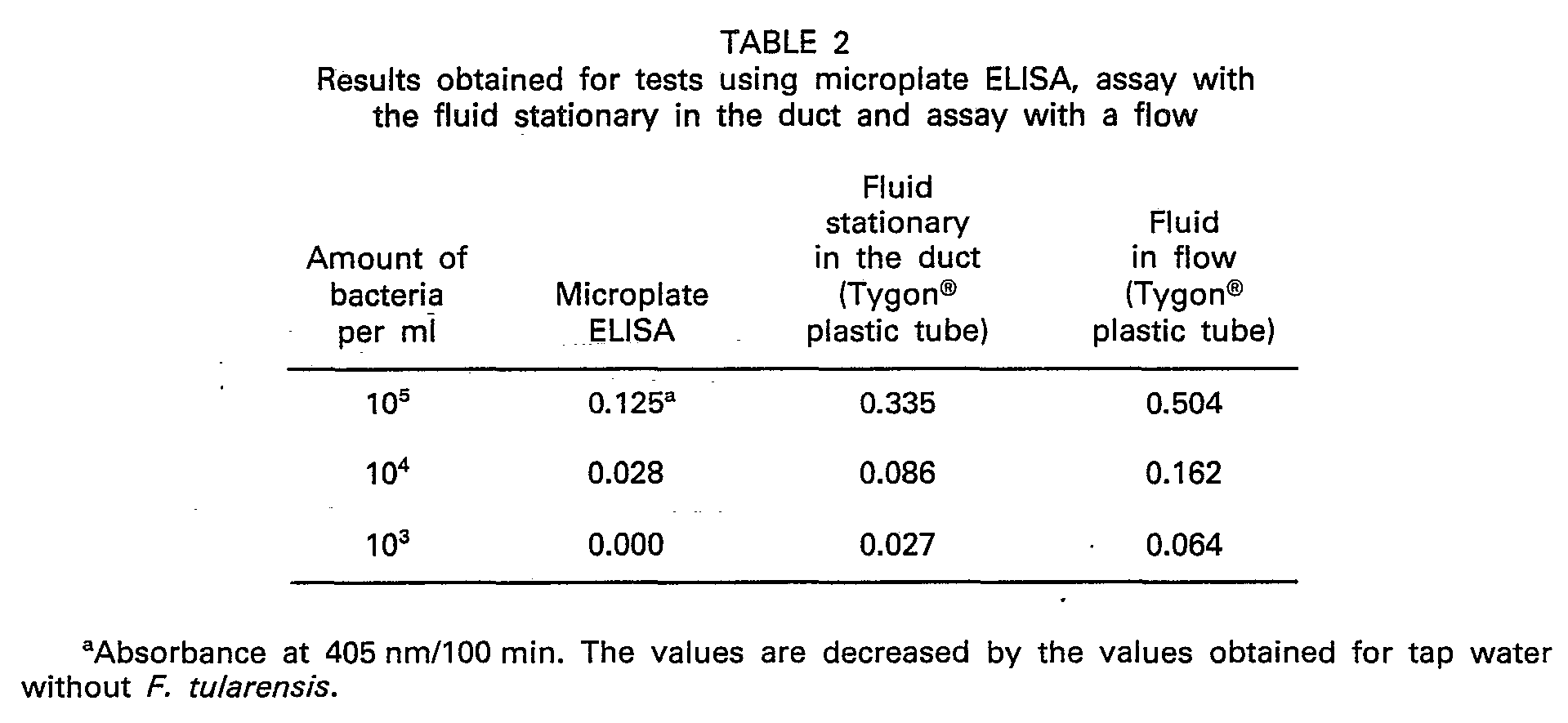
- The influence of Tygon® plastic tube was examined. The ELISA assay was performed without any flow in the Tygon® plastic tube. The advantages of the fluid in flow were shown also in this test. A lower detectable level was obtained as compared to microplate ELISA.
- Enrichment of the duct walls using a flow system. The flow rate was 1.9 ml/min and the
volume 40 µl/cm in the duct. The test sample was either flown straight through the duct or recirculated. The assay was performed in the same way as the microplate ELISA assay with respect to antigen and antibody. The length of the duct was 10 cm during the process, except for the last step when enzyme substrate was added. Before said addition the duct was cut from both ends to a length of 5 cm. - ELISA was performed in microplates. The microplates were pre-coated with immune serum against F. tularensis antigen at room temperature over night. The test samples were added after washing and allowed to react at 37°C for one hour. Affinity purified anti-F. tularensis antibodies labelled with alkaline phosphatase were subsequently added and the microplates were incubated for one hour at 37°C. Enzyme substrate was finally added and the reaction monitored after 30 minutes, using an automatic spectrophotometer. Stated values are mean values±standard deviation of 16 samples.
- Enrichment of F. tularensis at the duct wall using different volumes of water containing 105 bacteria per milliliter. The test time was 3 hours. Stated values are mean values±standard deviation of 3 samples.
- The detection level for different concentrations of F. tularensis depended on the volume of the test sample which passed through the pre-coated Tygon® tube. After 3 hours, 104 F. tularensis could be detected.
- To obtain a significant detecting level for as few as 102 bacteria per milliliter, about 4.5 I sample (a total of 4.5x10⁵ bacteria) was allowed to pass the Tygon° tube.
- Said test shows that antigen can be enriched at the inner wall of the Tygon® tube if said tube is coated with specific antibodies. It is also evident that the test time, i.e. the enrichment time, depends on the amount of bacteria in the sample to be tested.

- Mean values±standard deviation of 9-12 samples.
- The background values (tap water without F. tularensis) are substracted from the values as stated.
- Detection levels of F. tularensis when test samples are recirculated.

- The measurements were performed after 3, 6, 18 and 24 hours. Stated values are mean values±standard deviation of 10-15 samples.
- Absorbance at 405 nm/100 min at test with E. coli in concentrations between 0 and 5 · 106 per milliliter. The sample was recirculated for 3 hours. Volume=5 milliliter. Stated values are mean values of 3 samples.
Claims (6)
Priority Applications (1)
| Application Number | Priority Date | Filing Date | Title |
|---|---|---|---|
| AT85850016T ATE53912T1 (en) | 1984-01-25 | 1985-01-16 | METHODS FOR CONCENTRATION AND DETERMINATION OF BIOMOLECULES AND CELLS. |
Applications Claiming Priority (2)
| Application Number | Priority Date | Filing Date | Title |
|---|---|---|---|
| SE8400374 | 1984-01-24 | ||
| SE8400374A SE446229B (en) | 1984-01-25 | 1984-01-25 | SET AND DEVICE FOR DETECTING AN ANALYST IN A SAMPLE WHICH IS PASSED TO PASS A SURFACE TO ANY BUNDITS OF A SUBJECT WITH SPECIFIC AFFINITY TO THE ANALYST |
Publications (2)
| Publication Number | Publication Date |
|---|---|
| EP0153283A1 EP0153283A1 (en) | 1985-08-28 |
| EP0153283B1 true EP0153283B1 (en) | 1990-05-02 |
Family
ID=20354456
Family Applications (1)
| Application Number | Title | Priority Date | Filing Date |
|---|---|---|---|
| EP85850016A Expired - Lifetime EP0153283B1 (en) | 1984-01-25 | 1985-01-16 | A method of concentrating and detecting biomolecules and -cells |
Country Status (14)
| Country | Link |
|---|---|
| US (1) | US4822732A (en) |
| EP (1) | EP0153283B1 (en) |
| JP (1) | JPS61501226A (en) |
| AT (1) | ATE53912T1 (en) |
| AU (1) | AU589890B2 (en) |
| CA (1) | CA1254132A (en) |
| DE (1) | DE3577478D1 (en) |
| DK (1) | DK432485A (en) |
| ES (1) | ES8607555A1 (en) |
| FI (1) | FI82986C (en) |
| IL (1) | IL74144A (en) |
| NO (1) | NO166258C (en) |
| SE (1) | SE446229B (en) |
| WO (1) | WO1985003355A1 (en) |
Families Citing this family (8)
| Publication number | Priority date | Publication date | Assignee | Title |
|---|---|---|---|---|
| US5003988A (en) * | 1989-06-21 | 1991-04-02 | La Mina Ltd. | Modular multiple fluid sample preparation assembly |
| AU6847890A (en) * | 1990-01-19 | 1991-07-25 | Promega Corporation | Immunospecific and bioluminescent assay of cellular atp |
| CA2131728A1 (en) * | 1992-03-05 | 1993-09-16 | Alan Gaylard Shattock | Antigen detection apparatus |
| ATE123151T1 (en) * | 1992-10-21 | 1995-06-15 | Edith Dr Dr Huland | METHOD FOR THE QUANTITATIVE DETERMINATION OF DISEASE-SPECIFIC ANTIGENS AND DEVICE FOR IMPLEMENTING THE METHOD. |
| US5731157A (en) * | 1993-12-30 | 1998-03-24 | The Procter And Gamble Company | Two-site allergen immunoassay |
| US6194222B1 (en) * | 1998-01-05 | 2001-02-27 | Biosite Diagnostics, Inc. | Methods for monitoring the status of assays and immunoassays |
| US7713703B1 (en) | 2000-11-13 | 2010-05-11 | Biosite, Inc. | Methods for monitoring the status of assays and immunoassays |
| DE202004008808U1 (en) * | 2003-06-04 | 2004-09-09 | Inverness Medical Switzerland Gmbh | Instrument reading-out results of e.g. immuno assay, includes light sources and detectors to ensure velocity of fluid moving along porous lateral flow test strip lies within limits |
Family Cites Families (14)
| Publication number | Priority date | Publication date | Assignee | Title |
|---|---|---|---|---|
| GB157264A (en) * | 1915-03-11 | 1922-07-10 | Hermann Plauson | Improvements in the conversions of atmospheric electric energy |
| US3896217A (en) * | 1973-03-19 | 1975-07-22 | Summa Corp | Method and apparatus for radioimmunoassay with regeneration of immunoadsorbent |
| US3950134A (en) * | 1974-09-20 | 1976-04-13 | The Board Of Trustees Of The Leland Stanford Junior University | Assay machine and method |
| US4036946A (en) * | 1975-10-20 | 1977-07-19 | Marcos Kleinerman | Immunofluorometric method for measuring minute quantities of antigens, antibodies and other substances |
| GB1597345A (en) * | 1976-12-16 | 1981-09-03 | Millipore Corp | Diagnostic immunochemical test materials and procedure |
| FR2389134A1 (en) * | 1977-04-29 | 1978-11-24 | Seroa | Analytical use of capillary micro-tubes - as measuring and reaction vessel esp. for biochemical analysis |
| JPS56145222A (en) * | 1980-04-28 | 1981-11-11 | Toshiyuki Hamaoka | Improved antibody and its preparation |
| US4366241A (en) * | 1980-08-07 | 1982-12-28 | Syva Company | Concentrating zone method in heterogeneous immunoassays |
| FR2498331A1 (en) * | 1981-01-20 | 1982-07-23 | Kadouche Jean | Container for immunological tests e.g. antigen identification - having reagent molecules, e.g. antibodies, fixed to inner face of pipette cone point |
| SE430900B (en) * | 1981-06-04 | 1983-12-19 | Bo Gustav Mattiasson | PROCEDURE FOR DETERMINING BIOCHEMICAL DATA FOR MICRO ORGANISMS OR SINGLE ORGANISMS |
| NZ201901A (en) * | 1981-09-25 | 1986-11-12 | Commw Serum Lab Commission | An apparatus suitable for performing automated heterogeneous immunoassays in a plurality of samples |
| DE3145007A1 (en) * | 1981-11-12 | 1983-05-19 | Max-Planck-Gesellschaft zur Förderung der Wissenschaften e.V., 3400 Göttingen | IMMOBILIZED BIOLOGICALLY ACTIVE SUBSTANCES AND THEIR USE |
| US4693985A (en) * | 1984-08-21 | 1987-09-15 | Pall Corporation | Methods of concentrating ligands and active membranes used therefor |
| DE10154186C1 (en) * | 2001-11-05 | 2003-06-12 | Schukra Europa Gmbh | Device for adjusting a seat component |
-
1984
- 1984-01-25 SE SE8400374A patent/SE446229B/en not_active IP Right Cessation
-
1985
- 1985-01-16 AT AT85850016T patent/ATE53912T1/en not_active IP Right Cessation
- 1985-01-16 EP EP85850016A patent/EP0153283B1/en not_active Expired - Lifetime
- 1985-01-16 DE DE8585850016T patent/DE3577478D1/en not_active Expired - Fee Related
- 1985-01-23 WO PCT/SE1985/000026 patent/WO1985003355A1/en not_active Ceased
- 1985-01-23 AU AU38825/85A patent/AU589890B2/en not_active Ceased
- 1985-01-23 IL IL74144A patent/IL74144A/en unknown
- 1985-01-23 US US06/779,775 patent/US4822732A/en not_active Expired - Fee Related
- 1985-01-23 JP JP60500491A patent/JPS61501226A/en active Pending
- 1985-01-24 CA CA000472727A patent/CA1254132A/en not_active Expired
- 1985-01-24 ES ES539813A patent/ES8607555A1/en not_active Expired
- 1985-09-24 DK DK432485A patent/DK432485A/en not_active Application Discontinuation
- 1985-09-24 NO NO85853749A patent/NO166258C/en unknown
-
1986
- 1986-03-21 FI FI861205A patent/FI82986C/en not_active IP Right Cessation
Non-Patent Citations (1)
| Title |
|---|
| ANALYTICAL CHEMISTRY, vol. 49, no. 13, Nov 1977; N.GOCHMAN et al.: * |
Also Published As
| Publication number | Publication date |
|---|---|
| NO166258C (en) | 1991-06-19 |
| IL74144A (en) | 1989-08-15 |
| AU589890B2 (en) | 1989-10-26 |
| DE3577478D1 (en) | 1990-06-07 |
| JPS61501226A (en) | 1986-06-19 |
| ES8607555A1 (en) | 1986-05-16 |
| FI82986C (en) | 1991-05-10 |
| SE8400374D0 (en) | 1984-01-25 |
| NO853749L (en) | 1985-09-24 |
| FI82986B (en) | 1991-01-31 |
| DK432485D0 (en) | 1985-09-24 |
| SE8400374L (en) | 1985-07-26 |
| CA1254132A (en) | 1989-05-16 |
| SE446229B (en) | 1986-08-18 |
| WO1985003355A1 (en) | 1985-08-01 |
| FI861205L (en) | 1986-03-21 |
| ATE53912T1 (en) | 1990-06-15 |
| IL74144A0 (en) | 1985-04-30 |
| NO166258B (en) | 1991-03-11 |
| DK432485A (en) | 1985-09-24 |
| FI861205A0 (en) | 1986-03-21 |
| AU3882585A (en) | 1985-08-09 |
| EP0153283A1 (en) | 1985-08-28 |
| US4822732A (en) | 1989-04-18 |
| ES539813A0 (en) | 1986-05-16 |
Similar Documents
| Publication | Publication Date | Title |
|---|---|---|
| EP0239318B1 (en) | Assay employing binding pair members on particles and on a filter or membrane | |
| US5236826A (en) | Immunoassay for the detection or quantitation of an analyte | |
| US5279936A (en) | Method of separation employing magnetic particles and second medium | |
| EP0267317B1 (en) | Method for the detection of proteins and viruses | |
| SE443660B (en) | PROCEDURE AND REAGENT FOR IMMUNAL ANALYSIS WITH RF OR CLQ ADSORBED TO FIXED BEARERS | |
| CN105195243B (en) | The magnetic microparticle chemiluminescence micro-fluidic chip that a kind of myoglobins is quantitatively detected | |
| US5739001A (en) | Solid phase cell-based assay | |
| US6933109B2 (en) | Rapid particle detection | |
| AU3168799A (en) | Methods for assaying antibody and device for assaying antibody | |
| FI95752B (en) | Determination kit and method for immunological measurement of whole cells | |
| JPH06507475A (en) | Dilution buffer and its usage | |
| US20100285522A1 (en) | Versatile Multichannel Capillary Biosensor System | |
| US6933106B2 (en) | Platelet immunoglobulin bead suspension and flow cytometry | |
| AU589890B2 (en) | A method of concentrating and detecting biomolecules and -cells and a means therefore | |
| EP3508849B1 (en) | Antibody measurement method using antigen-carrying insoluble carrier particles on which antigen is immobilized by different methods, and reagent for antibody measurement | |
| US4307190A (en) | Immunoassay using ascitic fluid | |
| US4251360A (en) | Method and apparatus for the detection of a specific binding protein | |
| GB2104216A (en) | Process for detecting presence of auto blocking antibody | |
| EP0241140B1 (en) | Assay method with a multivalently labelled reagent, and means therefor | |
| EP0311604B1 (en) | Method and apparatus for qualitative and/or quantitative analysis of antigens, antibodies, mircroorganisms or other cells | |
| US6951716B2 (en) | Anti-platelet immunoglobulin bead positive control | |
| GB1573627A (en) | Process and device for the determination and detection of a reaction component | |
| EP4575501A1 (en) | Immunoassay method and reagent | |
| Mattiasson et al. | Partition Affinity Ligand Assay (PALA): Applications in the Analysis of Haptens, Macromolecules, and Cells | |
| Hayashi et al. | Semi-automated continuous-flow enzyme immunoassay for antiepileptic drugs in serum. |
Legal Events
| Date | Code | Title | Description |
|---|---|---|---|
| PUAI | Public reference made under article 153(3) epc to a published international application that has entered the european phase |
Free format text: ORIGINAL CODE: 0009012 |
|
| 17P | Request for examination filed |
Effective date: 19850213 |
|
| AK | Designated contracting states |
Designated state(s): AT BE CH DE FR GB IT LI LU NL SE |
|
| 17Q | First examination report despatched |
Effective date: 19880309 |
|
| RAP1 | Party data changed (applicant data changed or rights of an application transferred) |
Owner name: SYMBICOM AB |
|
| RAP1 | Party data changed (applicant data changed or rights of an application transferred) |
Owner name: SYMBICOM AB |
|
| GRAA | (expected) grant |
Free format text: ORIGINAL CODE: 0009210 |
|
| AK | Designated contracting states |
Kind code of ref document: B1 Designated state(s): AT BE CH DE FR GB IT LI LU NL SE |
|
| PG25 | Lapsed in a contracting state [announced via postgrant information from national office to epo] |
Ref country code: SE Effective date: 19900502 |
|
| REF | Corresponds to: |
Ref document number: 53912 Country of ref document: AT Date of ref document: 19900615 Kind code of ref document: T |
|
| REF | Corresponds to: |
Ref document number: 3577478 Country of ref document: DE Date of ref document: 19900607 |
|
| ITF | It: translation for a ep patent filed | ||
| ET | Fr: translation filed | ||
| ITTA | It: last paid annual fee | ||
| PLBE | No opposition filed within time limit |
Free format text: ORIGINAL CODE: 0009261 |
|
| STAA | Information on the status of an ep patent application or granted ep patent |
Free format text: STATUS: NO OPPOSITION FILED WITHIN TIME LIMIT |
|
| 26N | No opposition filed | ||
| PGFP | Annual fee paid to national office [announced via postgrant information from national office to epo] |
Ref country code: LU Payment date: 19911230 Year of fee payment: 8 |
|
| PGFP | Annual fee paid to national office [announced via postgrant information from national office to epo] |
Ref country code: GB Payment date: 19920110 Year of fee payment: 8 |
|
| PGFP | Annual fee paid to national office [announced via postgrant information from national office to epo] |
Ref country code: AT Payment date: 19920113 Year of fee payment: 8 |
|
| PGFP | Annual fee paid to national office [announced via postgrant information from national office to epo] |
Ref country code: CH Payment date: 19920115 Year of fee payment: 8 |
|
| PGFP | Annual fee paid to national office [announced via postgrant information from national office to epo] |
Ref country code: BE Payment date: 19920120 Year of fee payment: 8 |
|
| PGFP | Annual fee paid to national office [announced via postgrant information from national office to epo] |
Ref country code: FR Payment date: 19920121 Year of fee payment: 8 |
|
| PGFP | Annual fee paid to national office [announced via postgrant information from national office to epo] |
Ref country code: NL Payment date: 19920131 Year of fee payment: 8 |
|
| PGFP | Annual fee paid to national office [announced via postgrant information from national office to epo] |
Ref country code: DE Payment date: 19920228 Year of fee payment: 8 |
|
| EPTA | Lu: last paid annual fee | ||
| PG25 | Lapsed in a contracting state [announced via postgrant information from national office to epo] |
Ref country code: LU Free format text: LAPSE BECAUSE OF NON-PAYMENT OF DUE FEES Effective date: 19930116 Ref country code: GB Effective date: 19930116 Ref country code: AT Effective date: 19930116 |
|
| PG25 | Lapsed in a contracting state [announced via postgrant information from national office to epo] |
Ref country code: LI Effective date: 19930131 Ref country code: CH Effective date: 19930131 Ref country code: BE Effective date: 19930131 |
|
| BERE | Be: lapsed |
Owner name: SYMBICOM A.B. Effective date: 19930131 |
|
| PG25 | Lapsed in a contracting state [announced via postgrant information from national office to epo] |
Ref country code: NL Effective date: 19930801 |
|
| GBPC | Gb: european patent ceased through non-payment of renewal fee |
Effective date: 19930116 |
|
| NLV4 | Nl: lapsed or anulled due to non-payment of the annual fee | ||
| PG25 | Lapsed in a contracting state [announced via postgrant information from national office to epo] |
Ref country code: FR Effective date: 19930930 |
|
| REG | Reference to a national code |
Ref country code: CH Ref legal event code: PL |
|
| PG25 | Lapsed in a contracting state [announced via postgrant information from national office to epo] |
Ref country code: DE Effective date: 19931001 |
|
| REG | Reference to a national code |
Ref country code: FR Ref legal event code: ST |