EP0126617B1 - Macromolecule determination by phsysical development - Google Patents
Macromolecule determination by phsysical development Download PDFInfo
- Publication number
- EP0126617B1 EP0126617B1 EP84303296A EP84303296A EP0126617B1 EP 0126617 B1 EP0126617 B1 EP 0126617B1 EP 84303296 A EP84303296 A EP 84303296A EP 84303296 A EP84303296 A EP 84303296A EP 0126617 B1 EP0126617 B1 EP 0126617B1
- Authority
- EP
- European Patent Office
- Prior art keywords
- gel
- salt
- solution
- macromolecules
- protein
- Prior art date
- Legal status (The legal status is an assumption and is not a legal conclusion. Google has not performed a legal analysis and makes no representation as to the accuracy of the status listed.)
- Expired
Links
- 229920002521 macromolecule Polymers 0.000 title claims description 33
- 238000011161 development Methods 0.000 title description 20
- 239000000499 gel Substances 0.000 claims description 57
- 238000000034 method Methods 0.000 claims description 45
- KDLHZDBZIXYQEI-UHFFFAOYSA-N palladium Substances [Pd] KDLHZDBZIXYQEI-UHFFFAOYSA-N 0.000 claims description 41
- 102000004169 proteins and genes Human genes 0.000 claims description 30
- 108090000623 proteins and genes Proteins 0.000 claims description 30
- 235000018102 proteins Nutrition 0.000 claims description 26
- 125000003831 tetrazolyl group Chemical group 0.000 claims description 21
- 229910052763 palladium Inorganic materials 0.000 claims description 20
- -1 palladium tetramine salt Chemical class 0.000 claims description 19
- DBMJMQXJHONAFJ-UHFFFAOYSA-M Sodium laurylsulphate Chemical compound [Na+].CCCCCCCCCCCCOS([O-])(=O)=O DBMJMQXJHONAFJ-UHFFFAOYSA-M 0.000 claims description 18
- 238000001962 electrophoresis Methods 0.000 claims description 17
- 229920002401 polyacrylamide Polymers 0.000 claims description 15
- 229920001184 polypeptide Polymers 0.000 claims description 12
- 102000004196 processed proteins & peptides Human genes 0.000 claims description 12
- 108090000765 processed proteins & peptides Proteins 0.000 claims description 12
- RJTANRZEWTUVMA-UHFFFAOYSA-N boron;n-methylmethanamine Chemical compound [B].CNC RJTANRZEWTUVMA-UHFFFAOYSA-N 0.000 claims description 9
- 125000001425 triazolyl group Chemical group 0.000 claims description 8
- 229910052723 transition metal Inorganic materials 0.000 claims description 7
- RGHNJXZEOKUKBD-SQOUGZDYSA-N Gluconic acid Natural products OC[C@@H](O)[C@@H](O)[C@H](O)[C@@H](O)C(O)=O RGHNJXZEOKUKBD-SQOUGZDYSA-N 0.000 claims description 5
- ULXKXLZEOGLCRJ-UHFFFAOYSA-N 2-azaniumyl-3-ethylsulfanylpropanoate Chemical group CCSCC(N)C(O)=O ULXKXLZEOGLCRJ-UHFFFAOYSA-N 0.000 claims description 4
- RGHNJXZEOKUKBD-UHFFFAOYSA-N D-gluconic acid Natural products OCC(O)C(O)C(O)C(O)C(O)=O RGHNJXZEOKUKBD-UHFFFAOYSA-N 0.000 claims description 4
- 239000008139 complexing agent Substances 0.000 claims description 4
- 229940031005 ethyl cysteine Drugs 0.000 claims description 4
- 239000000174 gluconic acid Substances 0.000 claims description 4
- 235000012208 gluconic acid Nutrition 0.000 claims description 4
- 102000039446 nucleic acids Human genes 0.000 claims description 4
- 108020004707 nucleic acids Proteins 0.000 claims description 4
- 150000007523 nucleic acids Chemical class 0.000 claims description 4
- 150000003841 chloride salts Chemical group 0.000 claims 1
- 150000002148 esters Chemical class 0.000 claims 1
- 239000000243 solution Substances 0.000 description 54
- 239000000975 dye Substances 0.000 description 30
- 230000018109 developmental process Effects 0.000 description 19
- OKKJLVBELUTLKV-UHFFFAOYSA-N Methanol Chemical compound OC OKKJLVBELUTLKV-UHFFFAOYSA-N 0.000 description 15
- XLYOFNOQVPJJNP-UHFFFAOYSA-N water Substances O XLYOFNOQVPJJNP-UHFFFAOYSA-N 0.000 description 15
- 102100026189 Beta-galactosidase Human genes 0.000 description 11
- 108010005774 beta-Galactosidase Proteins 0.000 description 11
- 229910001385 heavy metal Inorganic materials 0.000 description 11
- 239000000203 mixture Substances 0.000 description 11
- 238000012800 visualization Methods 0.000 description 11
- 229910001868 water Inorganic materials 0.000 description 10
- 229910052751 metal Inorganic materials 0.000 description 9
- 239000002184 metal Substances 0.000 description 9
- 230000035945 sensitivity Effects 0.000 description 8
- 238000000926 separation method Methods 0.000 description 8
- GBMDVOWEEQVZKZ-UHFFFAOYSA-N methanol;hydrate Chemical compound O.OC GBMDVOWEEQVZKZ-UHFFFAOYSA-N 0.000 description 7
- PXHVJJICTQNCMI-UHFFFAOYSA-N Nickel Chemical compound [Ni] PXHVJJICTQNCMI-UHFFFAOYSA-N 0.000 description 6
- BQCADISMDOOEFD-UHFFFAOYSA-N Silver Chemical compound [Ag] BQCADISMDOOEFD-UHFFFAOYSA-N 0.000 description 6
- 239000003054 catalyst Substances 0.000 description 6
- 239000003638 chemical reducing agent Substances 0.000 description 6
- 238000001514 detection method Methods 0.000 description 6
- 229910052709 silver Inorganic materials 0.000 description 6
- 239000004332 silver Substances 0.000 description 6
- 238000010186 staining Methods 0.000 description 6
- QFVHZQCOUORWEI-UHFFFAOYSA-N 4-[(4-anilino-5-sulfonaphthalen-1-yl)diazenyl]-5-hydroxynaphthalene-2,7-disulfonic acid Chemical compound C=12C(O)=CC(S(O)(=O)=O)=CC2=CC(S(O)(=O)=O)=CC=1N=NC(C1=CC=CC(=C11)S(O)(=O)=O)=CC=C1NC1=CC=CC=C1 QFVHZQCOUORWEI-UHFFFAOYSA-N 0.000 description 5
- LFQSCWFLJHTTHZ-UHFFFAOYSA-N Ethanol Chemical compound CCO LFQSCWFLJHTTHZ-UHFFFAOYSA-N 0.000 description 5
- 102000008100 Human Serum Albumin Human genes 0.000 description 5
- 108091006905 Human Serum Albumin Proteins 0.000 description 5
- 239000003795 chemical substances by application Substances 0.000 description 5
- 239000011521 glass Substances 0.000 description 5
- VMGAPWLDMVPYIA-HIDZBRGKSA-N n'-amino-n-iminomethanimidamide Chemical compound N\N=C\N=N VMGAPWLDMVPYIA-HIDZBRGKSA-N 0.000 description 5
- BOLDJAUMGUJJKM-LSDHHAIUSA-N renifolin D Natural products CC(=C)[C@@H]1Cc2c(O)c(O)ccc2[C@H]1CC(=O)c3ccc(O)cc3O BOLDJAUMGUJJKM-LSDHHAIUSA-N 0.000 description 5
- 150000003839 salts Chemical class 0.000 description 5
- 239000002904 solvent Substances 0.000 description 5
- GGZHVNZHFYCSEV-UHFFFAOYSA-N 1-Phenyl-5-mercaptotetrazole Chemical compound SC1=NN=NN1C1=CC=CC=C1 GGZHVNZHFYCSEV-UHFFFAOYSA-N 0.000 description 4
- QTBSBXVTEAMEQO-UHFFFAOYSA-N Acetic acid Chemical compound CC(O)=O QTBSBXVTEAMEQO-UHFFFAOYSA-N 0.000 description 4
- 239000000872 buffer Substances 0.000 description 4
- 239000008367 deionised water Substances 0.000 description 4
- 229910021641 deionized water Inorganic materials 0.000 description 4
- 230000000694 effects Effects 0.000 description 4
- 238000007654 immersion Methods 0.000 description 4
- 238000011068 loading method Methods 0.000 description 4
- 230000006911 nucleation Effects 0.000 description 4
- 238000010899 nucleation Methods 0.000 description 4
- 239000012460 protein solution Substances 0.000 description 4
- 238000005406 washing Methods 0.000 description 4
- WEVYAHXRMPXWCK-UHFFFAOYSA-N Acetonitrile Chemical compound CC#N WEVYAHXRMPXWCK-UHFFFAOYSA-N 0.000 description 3
- WSFSSNUMVMOOMR-UHFFFAOYSA-N Formaldehyde Chemical compound O=C WSFSSNUMVMOOMR-UHFFFAOYSA-N 0.000 description 3
- PEDCQBHIVMGVHV-UHFFFAOYSA-N Glycerine Chemical compound OCC(O)CO PEDCQBHIVMGVHV-UHFFFAOYSA-N 0.000 description 3
- 108010044467 Isoenzymes Proteins 0.000 description 3
- KWYHDKDOAIKMQN-UHFFFAOYSA-N N,N,N',N'-tetramethylethylenediamine Chemical compound CN(C)CCN(C)C KWYHDKDOAIKMQN-UHFFFAOYSA-N 0.000 description 3
- 206010070834 Sensitisation Diseases 0.000 description 3
- HEMHJVSKTPXQMS-UHFFFAOYSA-M Sodium hydroxide Chemical compound [OH-].[Na+] HEMHJVSKTPXQMS-UHFFFAOYSA-M 0.000 description 3
- KRKNYBCHXYNGOX-UHFFFAOYSA-N citric acid Chemical compound OC(=O)CC(O)(C(O)=O)CC(O)=O KRKNYBCHXYNGOX-UHFFFAOYSA-N 0.000 description 3
- ORTQZVOHEJQUHG-UHFFFAOYSA-L copper(II) chloride Chemical compound Cl[Cu]Cl ORTQZVOHEJQUHG-UHFFFAOYSA-L 0.000 description 3
- 238000003745 diagnosis Methods 0.000 description 3
- 239000003999 initiator Substances 0.000 description 3
- 229910052759 nickel Inorganic materials 0.000 description 3
- 239000002243 precursor Substances 0.000 description 3
- 238000002360 preparation method Methods 0.000 description 3
- 230000008313 sensitization Effects 0.000 description 3
- 230000001235 sensitizing effect Effects 0.000 description 3
- GGCZERPQGJTIQP-UHFFFAOYSA-N sodium;9,10-dioxoanthracene-2-sulfonic acid Chemical compound [Na+].C1=CC=C2C(=O)C3=CC(S(=O)(=O)O)=CC=C3C(=O)C2=C1 GGCZERPQGJTIQP-UHFFFAOYSA-N 0.000 description 3
- 238000007794 visualization technique Methods 0.000 description 3
- PURYCGFBBYVQEQ-UHFFFAOYSA-N 1-(dimethylamino)ethanethiol Chemical compound CC(S)N(C)C PURYCGFBBYVQEQ-UHFFFAOYSA-N 0.000 description 2
- HRPVXLWXLXDGHG-UHFFFAOYSA-N Acrylamide Chemical group NC(=O)C=C HRPVXLWXLXDGHG-UHFFFAOYSA-N 0.000 description 2
- VVNCNSJFMMFHPL-VKHMYHEASA-N D-penicillamine Chemical compound CC(C)(S)[C@@H](N)C(O)=O VVNCNSJFMMFHPL-VKHMYHEASA-N 0.000 description 2
- XEEYBQQBJWHFJM-UHFFFAOYSA-N Iron Chemical compound [Fe] XEEYBQQBJWHFJM-UHFFFAOYSA-N 0.000 description 2
- 102000003855 L-lactate dehydrogenase Human genes 0.000 description 2
- 108700023483 L-lactate dehydrogenases Proteins 0.000 description 2
- OKIZCWYLBDKLSU-UHFFFAOYSA-M N,N,N-Trimethylmethanaminium chloride Chemical compound [Cl-].C[N+](C)(C)C OKIZCWYLBDKLSU-UHFFFAOYSA-M 0.000 description 2
- 229910002651 NO3 Inorganic materials 0.000 description 2
- NHNBFGGVMKEFGY-UHFFFAOYSA-N Nitrate Chemical compound [O-][N+]([O-])=O NHNBFGGVMKEFGY-UHFFFAOYSA-N 0.000 description 2
- ZLMJMSJWJFRBEC-UHFFFAOYSA-N Potassium Chemical compound [K] ZLMJMSJWJFRBEC-UHFFFAOYSA-N 0.000 description 2
- FEWJPZIEWOKRBE-UHFFFAOYSA-N Tartaric acid Natural products [H+].[H+].[O-]C(=O)C(O)C(O)C([O-])=O FEWJPZIEWOKRBE-UHFFFAOYSA-N 0.000 description 2
- 239000007983 Tris buffer Substances 0.000 description 2
- 230000002378 acidificating effect Effects 0.000 description 2
- 125000000217 alkyl group Chemical group 0.000 description 2
- 210000004381 amniotic fluid Anatomy 0.000 description 2
- 210000001124 body fluid Anatomy 0.000 description 2
- 239000010839 body fluid Substances 0.000 description 2
- UORVGPXVDQYIDP-UHFFFAOYSA-N borane Chemical compound B UORVGPXVDQYIDP-UHFFFAOYSA-N 0.000 description 2
- 230000003197 catalytic effect Effects 0.000 description 2
- PHFQLYPOURZARY-UHFFFAOYSA-N chromium trinitrate Chemical compound [Cr+3].[O-][N+]([O-])=O.[O-][N+]([O-])=O.[O-][N+]([O-])=O PHFQLYPOURZARY-UHFFFAOYSA-N 0.000 description 2
- UFMZWBIQTDUYBN-UHFFFAOYSA-N cobalt dinitrate Chemical compound [Co+2].[O-][N+]([O-])=O.[O-][N+]([O-])=O UFMZWBIQTDUYBN-UHFFFAOYSA-N 0.000 description 2
- 150000001875 compounds Chemical class 0.000 description 2
- NKLPQNGYXWVELD-UHFFFAOYSA-M coomassie brilliant blue Chemical compound [Na+].C1=CC(OCC)=CC=C1NC1=CC=C(C(=C2C=CC(C=C2)=[N+](CC)CC=2C=C(C=CC=2)S([O-])(=O)=O)C=2C=CC(=CC=2)N(CC)CC=2C=C(C=CC=2)S([O-])(=O)=O)C=C1 NKLPQNGYXWVELD-UHFFFAOYSA-M 0.000 description 2
- 230000008021 deposition Effects 0.000 description 2
- 235000019441 ethanol Nutrition 0.000 description 2
- 238000009472 formulation Methods 0.000 description 2
- 239000011159 matrix material Substances 0.000 description 2
- 150000002739 metals Chemical class 0.000 description 2
- 239000002667 nucleating agent Substances 0.000 description 2
- PIBWKRNGBLPSSY-UHFFFAOYSA-L palladium(II) chloride Chemical compound Cl[Pd]Cl PIBWKRNGBLPSSY-UHFFFAOYSA-L 0.000 description 2
- 125000001997 phenyl group Chemical group [H]C1=C([H])C([H])=C(*)C([H])=C1[H] 0.000 description 2
- 229920003023 plastic Polymers 0.000 description 2
- BASFCYQUMIYNBI-UHFFFAOYSA-N platinum Chemical compound [Pt] BASFCYQUMIYNBI-UHFFFAOYSA-N 0.000 description 2
- 229910052700 potassium Inorganic materials 0.000 description 2
- 239000011591 potassium Substances 0.000 description 2
- 230000008569 process Effects 0.000 description 2
- 230000002285 radioactive effect Effects 0.000 description 2
- 238000000163 radioactive labelling Methods 0.000 description 2
- 238000002791 soaking Methods 0.000 description 2
- 239000011550 stock solution Substances 0.000 description 2
- 235000002906 tartaric acid Nutrition 0.000 description 2
- 239000011975 tartaric acid Substances 0.000 description 2
- 238000012360 testing method Methods 0.000 description 2
- AGGKEGLBGGJEBZ-UHFFFAOYSA-N tetramethylenedisulfotetramine Chemical compound C1N(S2(=O)=O)CN3S(=O)(=O)N1CN2C3 AGGKEGLBGGJEBZ-UHFFFAOYSA-N 0.000 description 2
- DGVVWUTYPXICAM-UHFFFAOYSA-N β‐Mercaptoethanol Chemical compound OCCS DGVVWUTYPXICAM-UHFFFAOYSA-N 0.000 description 2
- KPZGRMZPZLOPBS-UHFFFAOYSA-N 1,3-dichloro-2,2-bis(chloromethyl)propane Chemical compound ClCC(CCl)(CCl)CCl KPZGRMZPZLOPBS-UHFFFAOYSA-N 0.000 description 1
- JKFYKCYQEWQPTM-UHFFFAOYSA-N 2-azaniumyl-2-(4-fluorophenyl)acetate Chemical compound OC(=O)C(N)C1=CC=C(F)C=C1 JKFYKCYQEWQPTM-UHFFFAOYSA-N 0.000 description 1
- HKJKONMZMPUGHJ-UHFFFAOYSA-N 4-amino-5-hydroxy-3-[(4-nitrophenyl)diazenyl]-6-phenyldiazenylnaphthalene-2,7-disulfonic acid Chemical compound OS(=O)(=O)C1=CC2=CC(S(O)(=O)=O)=C(N=NC=3C=CC=CC=3)C(O)=C2C(N)=C1N=NC1=CC=C([N+]([O-])=O)C=C1 HKJKONMZMPUGHJ-UHFFFAOYSA-N 0.000 description 1
- QTBSBXVTEAMEQO-UHFFFAOYSA-M Acetate Chemical compound CC([O-])=O QTBSBXVTEAMEQO-UHFFFAOYSA-M 0.000 description 1
- 102000009027 Albumins Human genes 0.000 description 1
- 108010088751 Albumins Proteins 0.000 description 1
- VHUUQVKOLVNVRT-UHFFFAOYSA-N Ammonium hydroxide Chemical compound [NH4+].[OH-] VHUUQVKOLVNVRT-UHFFFAOYSA-N 0.000 description 1
- 208000002109 Argyria Diseases 0.000 description 1
- 102100035882 Catalase Human genes 0.000 description 1
- 108010053835 Catalase Proteins 0.000 description 1
- VYZAMTAEIAYCRO-UHFFFAOYSA-N Chromium Chemical compound [Cr] VYZAMTAEIAYCRO-UHFFFAOYSA-N 0.000 description 1
- 229910021555 Chromium Chloride Inorganic materials 0.000 description 1
- 229910021584 Cobalt(II) iodide Inorganic materials 0.000 description 1
- 108010062580 Concanavalin A Proteins 0.000 description 1
- RYGMFSIKBFXOCR-UHFFFAOYSA-N Copper Chemical compound [Cu] RYGMFSIKBFXOCR-UHFFFAOYSA-N 0.000 description 1
- FEWJPZIEWOKRBE-JCYAYHJZSA-N Dextrotartaric acid Chemical compound OC(=O)[C@H](O)[C@@H](O)C(O)=O FEWJPZIEWOKRBE-JCYAYHJZSA-N 0.000 description 1
- KCXVZYZYPLLWCC-UHFFFAOYSA-N EDTA Chemical compound OC(=O)CN(CC(O)=O)CCN(CC(O)=O)CC(O)=O KCXVZYZYPLLWCC-UHFFFAOYSA-N 0.000 description 1
- RZSYLLSAWYUBPE-UHFFFAOYSA-L Fast green FCF Chemical compound [Na+].[Na+].C=1C=C(C(=C2C=CC(C=C2)=[N+](CC)CC=2C=C(C=CC=2)S([O-])(=O)=O)C=2C(=CC(O)=CC=2)S([O-])(=O)=O)C=CC=1N(CC)CC1=CC=CC(S([O-])(=O)=O)=C1 RZSYLLSAWYUBPE-UHFFFAOYSA-L 0.000 description 1
- CWYNVVGOOAEACU-UHFFFAOYSA-N Fe2+ Chemical compound [Fe+2] CWYNVVGOOAEACU-UHFFFAOYSA-N 0.000 description 1
- 102000003886 Glycoproteins Human genes 0.000 description 1
- 108090000288 Glycoproteins Proteins 0.000 description 1
- 208000026350 Inborn Genetic disease Diseases 0.000 description 1
- 229910021575 Iron(II) bromide Inorganic materials 0.000 description 1
- 229910002666 PdCl2 Inorganic materials 0.000 description 1
- BELBBZDIHDAJOR-UHFFFAOYSA-N Phenolsulfonephthalein Chemical compound C1=CC(O)=CC=C1C1(C=2C=CC(O)=CC=2)C2=CC=CC=C2S(=O)(=O)O1 BELBBZDIHDAJOR-UHFFFAOYSA-N 0.000 description 1
- 229910021607 Silver chloride Inorganic materials 0.000 description 1
- 229910021612 Silver iodide Inorganic materials 0.000 description 1
- QAOWNCQODCNURD-UHFFFAOYSA-L Sulfate Chemical compound [O-]S([O-])(=O)=O QAOWNCQODCNURD-UHFFFAOYSA-L 0.000 description 1
- 238000005411 Van der Waals force Methods 0.000 description 1
- 230000005856 abnormality Effects 0.000 description 1
- 238000002835 absorbance Methods 0.000 description 1
- MCDLETWIOVSGJT-UHFFFAOYSA-N acetic acid;iron Chemical compound [Fe].CC(O)=O.CC(O)=O MCDLETWIOVSGJT-UHFFFAOYSA-N 0.000 description 1
- 239000002253 acid Substances 0.000 description 1
- 239000000908 ammonium hydroxide Substances 0.000 description 1
- 238000004458 analytical method Methods 0.000 description 1
- 150000001450 anions Chemical class 0.000 description 1
- 239000007864 aqueous solution Substances 0.000 description 1
- 238000000376 autoradiography Methods 0.000 description 1
- 230000008901 benefit Effects 0.000 description 1
- 230000015572 biosynthetic process Effects 0.000 description 1
- 210000004369 blood Anatomy 0.000 description 1
- 239000008280 blood Substances 0.000 description 1
- 238000009835 boiling Methods 0.000 description 1
- 229910000085 borane Inorganic materials 0.000 description 1
- UDSAIICHUKSCKT-UHFFFAOYSA-N bromophenol blue Chemical compound C1=C(Br)C(O)=C(Br)C=C1C1(C=2C=C(Br)C(O)=C(Br)C=2)C2=CC=CC=C2S(=O)(=O)O1 UDSAIICHUKSCKT-UHFFFAOYSA-N 0.000 description 1
- 239000007853 buffer solution Substances 0.000 description 1
- 125000004432 carbon atom Chemical group C* 0.000 description 1
- 125000002091 cationic group Chemical group 0.000 description 1
- 230000001413 cellular effect Effects 0.000 description 1
- 230000019522 cellular metabolic process Effects 0.000 description 1
- 208000015114 central nervous system disease Diseases 0.000 description 1
- 210000001175 cerebrospinal fluid Anatomy 0.000 description 1
- 238000012512 characterization method Methods 0.000 description 1
- 230000009920 chelation Effects 0.000 description 1
- 239000003153 chemical reaction reagent Substances 0.000 description 1
- 125000001309 chloro group Chemical group Cl* 0.000 description 1
- 229910052804 chromium Inorganic materials 0.000 description 1
- 229940107218 chromium Drugs 0.000 description 1
- 239000011651 chromium Substances 0.000 description 1
- QSWDMMVNRMROPK-UHFFFAOYSA-K chromium(3+) trichloride Chemical compound [Cl-].[Cl-].[Cl-].[Cr+3] QSWDMMVNRMROPK-UHFFFAOYSA-K 0.000 description 1
- WYYQVWLEPYFFLP-UHFFFAOYSA-K chromium(3+);triacetate Chemical compound [Cr+3].CC([O-])=O.CC([O-])=O.CC([O-])=O WYYQVWLEPYFFLP-UHFFFAOYSA-K 0.000 description 1
- PPUZYFWVBLIDMP-UHFFFAOYSA-K chromium(3+);triiodide Chemical compound I[Cr](I)I PPUZYFWVBLIDMP-UHFFFAOYSA-K 0.000 description 1
- GRWVQDDAKZFPFI-UHFFFAOYSA-H chromium(III) sulfate Chemical compound [Cr+3].[Cr+3].[O-]S([O-])(=O)=O.[O-]S([O-])(=O)=O.[O-]S([O-])(=O)=O GRWVQDDAKZFPFI-UHFFFAOYSA-H 0.000 description 1
- XZQOHYZUWTWZBL-UHFFFAOYSA-L chromium(ii) bromide Chemical compound [Cr+2].[Br-].[Br-] XZQOHYZUWTWZBL-UHFFFAOYSA-L 0.000 description 1
- 235000015165 citric acid Nutrition 0.000 description 1
- 229910017052 cobalt Inorganic materials 0.000 description 1
- 239000010941 cobalt Substances 0.000 description 1
- GUTLYIVDDKVIGB-UHFFFAOYSA-N cobalt atom Chemical compound [Co] GUTLYIVDDKVIGB-UHFFFAOYSA-N 0.000 description 1
- GVPFVAHMJGGAJG-UHFFFAOYSA-L cobalt dichloride Chemical compound [Cl-].[Cl-].[Co+2] GVPFVAHMJGGAJG-UHFFFAOYSA-L 0.000 description 1
- KTVIXTQDYHMGHF-UHFFFAOYSA-L cobalt(2+) sulfate Chemical compound [Co+2].[O-]S([O-])(=O)=O KTVIXTQDYHMGHF-UHFFFAOYSA-L 0.000 description 1
- AVWLPUQJODERGA-UHFFFAOYSA-L cobalt(2+);diiodide Chemical compound [Co+2].[I-].[I-] AVWLPUQJODERGA-UHFFFAOYSA-L 0.000 description 1
- QAHREYKOYSIQPH-UHFFFAOYSA-L cobalt(II) acetate Chemical compound [Co+2].CC([O-])=O.CC([O-])=O QAHREYKOYSIQPH-UHFFFAOYSA-L 0.000 description 1
- BZRRQSJJPUGBAA-UHFFFAOYSA-L cobalt(ii) bromide Chemical compound Br[Co]Br BZRRQSJJPUGBAA-UHFFFAOYSA-L 0.000 description 1
- 229940097267 cobaltous chloride Drugs 0.000 description 1
- 229940045032 cobaltous nitrate Drugs 0.000 description 1
- 230000000052 comparative effect Effects 0.000 description 1
- 229910052802 copper Inorganic materials 0.000 description 1
- 239000010949 copper Substances 0.000 description 1
- 229910000365 copper sulfate Inorganic materials 0.000 description 1
- XTVVROIMIGLXTD-UHFFFAOYSA-N copper(II) nitrate Chemical compound [Cu+2].[O-][N+]([O-])=O.[O-][N+]([O-])=O XTVVROIMIGLXTD-UHFFFAOYSA-N 0.000 description 1
- ARUVKPQLZAKDPS-UHFFFAOYSA-L copper(II) sulfate Chemical compound [Cu+2].[O-][S+2]([O-])([O-])[O-] ARUVKPQLZAKDPS-UHFFFAOYSA-L 0.000 description 1
- HFDWIMBEIXDNQS-UHFFFAOYSA-L copper;diformate Chemical compound [Cu+2].[O-]C=O.[O-]C=O HFDWIMBEIXDNQS-UHFFFAOYSA-L 0.000 description 1
- 239000013078 crystal Substances 0.000 description 1
- 229960003280 cupric chloride Drugs 0.000 description 1
- 230000001419 dependent effect Effects 0.000 description 1
- VBDQNUWZQXYUDP-UHFFFAOYSA-L dichloropalladium;ethane-1,2-diamine Chemical compound [Cl-].[Cl-].[Pd+2].NCCN.NCCN VBDQNUWZQXYUDP-UHFFFAOYSA-L 0.000 description 1
- 230000004069 differentiation Effects 0.000 description 1
- 239000012895 dilution Substances 0.000 description 1
- 238000010790 dilution Methods 0.000 description 1
- IYDMNMSJMUMQBP-UHFFFAOYSA-N dipotassium;palladium(2+);tetracyanide Chemical compound [K+].[K+].[Pd+2].N#[C-].N#[C-].N#[C-].N#[C-] IYDMNMSJMUMQBP-UHFFFAOYSA-N 0.000 description 1
- 239000003814 drug Substances 0.000 description 1
- 229960001484 edetic acid Drugs 0.000 description 1
- 230000005684 electric field Effects 0.000 description 1
- 125000006575 electron-withdrawing group Chemical group 0.000 description 1
- CMZOCKRRXUWGCT-UHFFFAOYSA-N ethane-1,2-diamine hydrochloride Chemical compound Cl.NCCN.NCCN CMZOCKRRXUWGCT-UHFFFAOYSA-N 0.000 description 1
- IDGUHHHQCWSQLU-UHFFFAOYSA-N ethanol;hydrate Chemical compound O.CCO IDGUHHHQCWSQLU-UHFFFAOYSA-N 0.000 description 1
- 239000000284 extract Substances 0.000 description 1
- 229940046149 ferrous bromide Drugs 0.000 description 1
- 229960002089 ferrous chloride Drugs 0.000 description 1
- 239000011790 ferrous sulphate Substances 0.000 description 1
- 235000003891 ferrous sulphate Nutrition 0.000 description 1
- 239000012530 fluid Substances 0.000 description 1
- 238000001502 gel electrophoresis Methods 0.000 description 1
- 208000016361 genetic disease Diseases 0.000 description 1
- 238000010448 genetic screening Methods 0.000 description 1
- PCHJSUWPFVWCPO-UHFFFAOYSA-N gold Chemical compound [Au] PCHJSUWPFVWCPO-UHFFFAOYSA-N 0.000 description 1
- 229910052737 gold Inorganic materials 0.000 description 1
- 239000010931 gold Substances 0.000 description 1
- FDWREHZXQUYJFJ-UHFFFAOYSA-M gold monochloride Chemical compound [Cl-].[Au+] FDWREHZXQUYJFJ-UHFFFAOYSA-M 0.000 description 1
- 239000011544 gradient gel Substances 0.000 description 1
- 150000004820 halides Chemical class 0.000 description 1
- 231100001261 hazardous Toxicity 0.000 description 1
- 229910052739 hydrogen Inorganic materials 0.000 description 1
- 239000001257 hydrogen Substances 0.000 description 1
- 230000006872 improvement Effects 0.000 description 1
- 238000000338 in vitro Methods 0.000 description 1
- 238000001727 in vivo Methods 0.000 description 1
- 150000002500 ions Chemical class 0.000 description 1
- 229910052742 iron Inorganic materials 0.000 description 1
- NMCUIPGRVMDVDB-UHFFFAOYSA-L iron dichloride Chemical compound Cl[Fe]Cl NMCUIPGRVMDVDB-UHFFFAOYSA-L 0.000 description 1
- BAUYGSIQEAFULO-UHFFFAOYSA-L iron(2+) sulfate (anhydrous) Chemical compound [Fe+2].[O-]S([O-])(=O)=O BAUYGSIQEAFULO-UHFFFAOYSA-L 0.000 description 1
- 229910000359 iron(II) sulfate Inorganic materials 0.000 description 1
- GYCHYNMREWYSKH-UHFFFAOYSA-L iron(ii) bromide Chemical compound [Fe+2].[Br-].[Br-] GYCHYNMREWYSKH-UHFFFAOYSA-L 0.000 description 1
- 239000003446 ligand Substances 0.000 description 1
- 239000000463 material Substances 0.000 description 1
- 230000004060 metabolic process Effects 0.000 description 1
- 150000005309 metal halides Chemical class 0.000 description 1
- NQMRYBIKMRVZLB-UHFFFAOYSA-N methylamine hydrochloride Chemical compound [Cl-].[NH3+]C NQMRYBIKMRVZLB-UHFFFAOYSA-N 0.000 description 1
- 238000002156 mixing Methods 0.000 description 1
- ZIUHHBKFKCYYJD-UHFFFAOYSA-N n,n'-methylenebisacrylamide Chemical compound C=CC(=O)NCNC(=O)C=C ZIUHHBKFKCYYJD-UHFFFAOYSA-N 0.000 description 1
- UICCSKORMGVRCB-UHFFFAOYSA-N n-[2-(diethylamino)ethyl]-n',n'-diethylethane-1,2-diamine Chemical compound CCN(CC)CCNCCN(CC)CC UICCSKORMGVRCB-UHFFFAOYSA-N 0.000 description 1
- LGQLOGILCSXPEA-UHFFFAOYSA-L nickel sulfate Chemical compound [Ni+2].[O-]S([O-])(=O)=O LGQLOGILCSXPEA-UHFFFAOYSA-L 0.000 description 1
- 229910000363 nickel(II) sulfate Inorganic materials 0.000 description 1
- KBJMLQFLOWQJNF-UHFFFAOYSA-N nickel(ii) nitrate Chemical compound [Ni+2].[O-][N+]([O-])=O.[O-][N+]([O-])=O KBJMLQFLOWQJNF-UHFFFAOYSA-N 0.000 description 1
- 150000002823 nitrates Chemical class 0.000 description 1
- 150000007524 organic acids Chemical class 0.000 description 1
- 235000005985 organic acids Nutrition 0.000 description 1
- MUJIDPITZJWBSW-UHFFFAOYSA-N palladium(2+) Chemical compound [Pd+2] MUJIDPITZJWBSW-UHFFFAOYSA-N 0.000 description 1
- 239000002245 particle Substances 0.000 description 1
- 230000001575 pathological effect Effects 0.000 description 1
- 230000000737 periodic effect Effects 0.000 description 1
- 229960003531 phenolsulfonphthalein Drugs 0.000 description 1
- 229910052697 platinum Inorganic materials 0.000 description 1
- CLSUSRZJUQMOHH-UHFFFAOYSA-L platinum dichloride Chemical compound Cl[Pt]Cl CLSUSRZJUQMOHH-UHFFFAOYSA-L 0.000 description 1
- PQTLYDQECILMMB-UHFFFAOYSA-L platinum(2+);sulfate Chemical compound [Pt+2].[O-]S([O-])(=O)=O PQTLYDQECILMMB-UHFFFAOYSA-L 0.000 description 1
- 239000002685 polymerization catalyst Substances 0.000 description 1
- 230000009467 reduction Effects 0.000 description 1
- KUIXZSYWBHSYCN-UHFFFAOYSA-L remazol brilliant blue r Chemical compound [Na+].[Na+].C1=C(S([O-])(=O)=O)C(N)=C2C(=O)C3=CC=CC=C3C(=O)C2=C1NC1=CC=CC(S(=O)(=O)CCOS([O-])(=O)=O)=C1 KUIXZSYWBHSYCN-UHFFFAOYSA-L 0.000 description 1
- 238000011160 research Methods 0.000 description 1
- 239000012266 salt solution Substances 0.000 description 1
- 229920006395 saturated elastomer Polymers 0.000 description 1
- 238000012216 screening Methods 0.000 description 1
- ADZWSOLPGZMUMY-UHFFFAOYSA-M silver bromide Chemical compound [Ag]Br ADZWSOLPGZMUMY-UHFFFAOYSA-M 0.000 description 1
- 229940045105 silver iodide Drugs 0.000 description 1
- HKZLPVFGJNLROG-UHFFFAOYSA-M silver monochloride Chemical compound [Cl-].[Ag+] HKZLPVFGJNLROG-UHFFFAOYSA-M 0.000 description 1
- 125000006850 spacer group Chemical group 0.000 description 1
- 230000007480 spreading Effects 0.000 description 1
- 238000007447 staining method Methods 0.000 description 1
- 238000003756 stirring Methods 0.000 description 1
- 239000000126 substance Substances 0.000 description 1
- 238000006467 substitution reaction Methods 0.000 description 1
- 150000003467 sulfuric acid derivatives Chemical class 0.000 description 1
- 238000003786 synthesis reaction Methods 0.000 description 1
- 238000002834 transmittance Methods 0.000 description 1
- YNJBWRMUSHSURL-UHFFFAOYSA-N trichloroacetic acid Chemical compound OC(=O)C(Cl)(Cl)Cl YNJBWRMUSHSURL-UHFFFAOYSA-N 0.000 description 1
- 238000000539 two dimensional gel electrophoresis Methods 0.000 description 1
- 230000000007 visual effect Effects 0.000 description 1
Classifications
-
- G—PHYSICS
- G01—MEASURING; TESTING
- G01N—INVESTIGATING OR ANALYSING MATERIALS BY DETERMINING THEIR CHEMICAL OR PHYSICAL PROPERTIES
- G01N33/00—Investigating or analysing materials by specific methods not covered by groups G01N1/00 - G01N31/00
- G01N33/48—Biological material, e.g. blood, urine; Haemocytometers
- G01N33/50—Chemical analysis of biological material, e.g. blood, urine; Testing involving biospecific ligand binding methods; Immunological testing
- G01N33/68—Chemical analysis of biological material, e.g. blood, urine; Testing involving biospecific ligand binding methods; Immunological testing involving proteins, peptides or amino acids
- G01N33/6803—General methods of protein analysis not limited to specific proteins or families of proteins
- G01N33/6827—Total protein determination, e.g. albumin in urine
- G01N33/683—Total protein determination, e.g. albumin in urine involving metal ions
-
- G—PHYSICS
- G01—MEASURING; TESTING
- G01N—INVESTIGATING OR ANALYSING MATERIALS BY DETERMINING THEIR CHEMICAL OR PHYSICAL PROPERTIES
- G01N33/00—Investigating or analysing materials by specific methods not covered by groups G01N1/00 - G01N31/00
- G01N33/48—Biological material, e.g. blood, urine; Haemocytometers
- G01N33/50—Chemical analysis of biological material, e.g. blood, urine; Testing involving biospecific ligand binding methods; Immunological testing
- G01N33/52—Use of compounds or compositions for colorimetric, spectrophotometric or fluorometric investigation, e.g. use of reagent paper and including single- and multilayer analytical elements
-
- G—PHYSICS
- G01—MEASURING; TESTING
- G01N—INVESTIGATING OR ANALYSING MATERIALS BY DETERMINING THEIR CHEMICAL OR PHYSICAL PROPERTIES
- G01N33/00—Investigating or analysing materials by specific methods not covered by groups G01N1/00 - G01N31/00
- G01N33/48—Biological material, e.g. blood, urine; Haemocytometers
- G01N33/50—Chemical analysis of biological material, e.g. blood, urine; Testing involving biospecific ligand binding methods; Immunological testing
- G01N33/52—Use of compounds or compositions for colorimetric, spectrophotometric or fluorometric investigation, e.g. use of reagent paper and including single- and multilayer analytical elements
- G01N33/521—Single-layer analytical elements
Definitions
- This invention relates to a method for determining macromolecules such as proteins and kits for said determinations.
- the method comprises forming a stain image and developing with a specific developer solution.
- macromolecules such as proteins and polypeptides
- the detection of macromolecules such as proteins and polypeptides is extremely important in many areas of biology and clinical medicine such as genetic screening and the diagnosis of genetic diseases. In these areas, the amount of macromolecules that can be detected in cellular extracts of body fluids can determine the treatment of a patient.
- Electrophoresis is the movement of charged particles in a matrix under the influence of an electrical field. Electrophoresis is the primary laboratory detection and characterization technique for macromolecules (see Chemical and Engineering News 3/14/83, p. 10).
- Coomassie Blue The most commonly used conventional macromolecule stain is Coomassie Blue. Dyes of this type are mainly dependent upon the electrostatic attraction between dye and polypeptide, stabilized by van der Waals forces. In fact, Coomassie Blue and a variety of other dyes exhibit particular affinities for macromolecules of specific charge. Coomassie Blue, an acidic dye, stains basic macromolecules most intensely, while crystal violet is the most effective stain for acidic macromolecules. Other dyes for which quantitative aspects of staining have been investigated include Amido Black, Fast Green, and Fe2+-bathophenanthroline sulfonate. In contrast, the Remazol Brilliant Blue R method depends on a covalent bond between dye and polypeptide. With Coomassie Blue, the minimum amount of a protein that can be detected is approximately 0.2 to 0.5 ⁇ g.
- Radioactive detection techniques offer a higher degree of sensitivity than the stains but are often impractical to use.
- In vivo radiolabelling may alter cellular metabolism and it may be impossible to label certain human macromolecules.
- In vitro radiolabelling has the disadvantage that it might alter the electrophoretic mobility of macromolecules.
- radioactive reagents sometimes prove too expensive. Long exposure to detect trace macromolecules may result in the problem of "autoradiographic spreading".
- Another method of visualization involves the use of silver stains.
- a swollen polyacrylamide electrophoretic sheet is treated with a silver salt and subsequently developed in a dilute reducing solution such as formaldehyde.
- An improvement in sensitization is obtained as compared with dye staining.
- This method entails several washing steps and many of the solutions used are unstable and must be mixed immediately before use.
- the silver stain method is described in PCT application publication No. WO 82/03128, September 16, 1982.
- the problem this invention solves is in providing a macromolecule visualization procedure that achieves a higher detection sensitivity than obtained with dye staining, and which is similar and more expedient to carry out than is the case with dye and silver staining, or autoradiography.
- This problem can be solved by providing a method for determining protein, nucleic acid or polypeptide macromolecules in polyacrylamide gels, said method comprising the steps of a) forming a latent stain image in the gel and b) developing the latent stain image by treating the gel with a developer solution characterized in that the latent stain image is formed by nucleating the macromolecules in the gel with a palladium tetramine salt and the latent stain image is developed by treating the gel with a physical developer solution comprising dimethylamine borane and a transition metal salt, a tetrazolium salt or a triazolium salt.
- This method is capable of detecting less than 0.01 ⁇ g of macromolecules.
- An electrophoregram that consists of a polyacrylamide slab is visualized by nucleation with a palladium tetramine salt and developed in a physical developer comprising a transition metal salt, tetrazolium salt or triazolium salt, dimethylamine borane and optionally an antifoggant.
- This method is carried out in a short time as compared to that of the dye and silver stain and autoradiographic methods. While a mixture of a silver salt and a reducing agent is extremely unstable (black reduced silver begins to appear immediately after the two components are in contact), the metal or tetrazolium salt and dimethylamine borane can be mixed and left for days without any sign of reduced metal or dye. Further, sodium dodecyl sulfate (SDS) which can be present in the gel matrix, does not interfere with the staining procedure. This is in contrast with silver stains which require careful removal of SDS before the gel is imbibed with a silver salt solution.
- SDS sodium dodecyl sulfate
- Kits comprising solutions of the components used in the method are also described. Use of these kits can find utility in clinical and laboratory examination of blood devices and other macromolecules separated by electrophoresis, diagnosis of metal abnormalities by examination of amniotic fluid, diagnosis of central nervous system disease by analysis of macromolecule patterns in spiral fluid and other applications in which the detection and/or quantitation of macromolecules is desired.
- the macromolecules to be detected in this invention are proteins, nucleic acids or polypeptides.
- the electrophoretic separation using polyacrylamide gel slabs is carried out using any of the procedures well known in the art.
- the macromolecules are fixed in the gel by treatment with a solution of methanol and water (1:1) for a short period of time.
- a fixing agent such as acetic or trichloroacetic acid
- the most preferred fixing agent solution is an aqueous mixture of ethanol and/or methanol (10-50%).
- the above fixing agents have been employed in both histology and protein electrophoresis for years.
- the gels are fixed by immersion in the fixing agent solution for a period of about 30-60 minutes.
- the first steps in this method are generalized preparation of the protein solution, preparation of the gel slabs and loading and running the gels.
- This process prior to visualization, is known in the art as the Laemmli System (U.K. Laemmli, Nature, 227, 680-685 (1970)).
- Macromolecule solutions are prepared by mixing the macromolecule solution which can contain salts, with an equal volume of a treatment solution consisting of a buffer such as tris (hydrogen methyl) aminomethane hydrochloride (TRIS ⁇ HCl), sodium dodecyl sulfate, glycerol and 2-mercaptoethanol.
- a buffer such as tris (hydrogen methyl) aminomethane hydrochloride (TRIS ⁇ HCl), sodium dodecyl sulfate, glycerol and 2-mercaptoethanol.
- the mixture is placed in a test tube and immersed into a boiling water bath for 1.5-3 minutes and then chilled in ice. Generally, 1 microliter of this solution contains 0.5 ⁇ g of protein.
- the protein solution is divided into 1 mL lots and stored at -20°C.
- the gel slabs are prepared using the Laemmli system described above. This system involves 2 gel layers. The layer in which the proteins are separated is referenced to as the "running” layer (separating layer) and the layer in which the protein solutions are applied is referenced to as the "stacking" layer. The stacking layer is discarded prior to the visualization step. The stacking layer allows the protein solution to be concentrated at the top of the separating gel in a narrow band to obtain sharper bands.
- the gel slabs can be made using the following Stock Solutions:
- the separating gel is prepared using a Hoefer Scientific Instruments SE600 Vertical Slab Gel Unit. Different thicknesses (0.75, 1.5 mm) of gel are obtained by the use of plastic spacers.
- the separating gel solution without the initiator and TEMED is degassed for approximately 5-10 minutes using a water aspirator.
- the initiator and TEMED are then added and the solution poured between glass plates (14 x 16 cm) (using a disposable Pasteur pipet) to a level 4 cm from the top of the plates.
- a water layer is then applied above the solution by means of a glass syringe.
- the solution polymerizes within 15 minutes after which the water layer is discarded and the gel is covered with a few mL of the Running Gel Overlay solution and left overnight.
- the stacking gel is prepared as follows: The next day, the gel is rinsed and the stacking gel solution minus the Initiator and TEMED is degassed, after which the polymerization catalysts are added, and the mixture poured over the gel. A plastic comb is inserted in order to mold the sample wells in the gel and the gel is allowed to polymerize for approximately 1 hour. After this time, the comb is removed, the wells formed by the comb are rinsed with water and are then ready for loading.
- Loading and running the gels is accomplished as follows: Each well is filled with Tank Buffer and the protein sample is underlayered into each well by means of a 10 ⁇ L glass syringe. The glass sandwiches are then placed into the electrophoresis tank which is filled with tank buffer. The upper chamber is filled with this same buffer and the separation is carried out at a current level of 15-30 ma per gel. The voltage varies during a run from an initial value of 85 V to 330 V at the end of the separation (for 2 gels). A tracking dye, bromophenol blue or phenol red, is added to one of the slots at the start. During the separation, the dye migrates with greater velocity than any of the protein fractions. The run is terminated when the dye is within 1-3 cm of the bottom of the glass sandwich.
- the visualization steps include the following: The plates are separated, the stacking gel discarded, and the separating gel slab placed into a 300 ml methanol-water (1:1) mixture for washing on a rocking table. After washing for 1 hour, the gels are ready for visualization. For dye visualization, no wash step is required.
- the gel strip is put into the Stain bath for 4-16 hours. It is then washed in DeStaining Solution I for 2 hours, and then washed in DeStaining Solution II for several hours. This step is generally carried out overnight in order to completely remove the dye from the background areas.
- the first step in the visualization method of the present invention comprises treating the fixed electrophotogram with a catalytic metal precursor to form a latent stain image.
- the catalyst used is a palladium tetramine salt such as palladium tetramine chloride or palladium tetramine nitrate. These particular catalysts unexpectedly yield excellent results in that they possess the proper combination of stability and solubility to sensitize the electrophoregram from the macromolecular area. Similar catalysts such as palladium bis(ethylene diamine) chloride or potassium palladium tetrathiocyanate yield a much lower degree of nucleation (sensitivity), and potassium palladium tetrachloride is too easily reduced to catalytic palladium to give proper image differentiation.
- the SDS presensitizers and the palladium tetramine salt can be combined in one solution.
- the catalyst is generally present in coverages of from 0.009 to 1.9 mg/m2.
- the development of the latent stain image is accomplished by treating the gel with a developer solution comprising dimethylamine borane as the reducing agent.
- a developer solution comprising dimethylamine borane as the reducing agent.
- Other reducing agents result in high fog because they do not adequately differentiate between the image (protein) area and the background.
- the developing solution also comprises a transition metal salt, tetrazolium salt or triazolium salt physical developer.
- the tetrazolium salt can be present in the amounts between 1 mg/0.093 m2 and 100 mg/0.093 m2 and the reducing agent can be present in amounts between 1 mg/0.093 m2 and 200 mg/0.093 m2.
- tetrazolium salts may be used as reducible dye precursors in the practice of the present invention. It is understood that the term "tetrazolium salt” throughout the application includes tetrazolium salts, ditetrazolium salts and tetrazolium betaines.
- tetrazolium salts Useful tetrazolium salts are described in "The Chemistry of Formazans and Tetrazolium Salts", A. W. Nineham, Chem . Rev ., 55, 355 (1955). The synthesis of tetrazolium salts and the chelation of formazan dyes are also described in the above reference.
- Tetrazolium salts useful in the present invention include compounds having the general formula: wherein Ar1, Ar2 and Ar3 may be the same or different and represent phenyl groups or phenyl groups substituted with electron withdrawing groups such as nitro, methoxy and the like or electron donating groups such as alkyl; and Ar3 can also represent a 4,5-dialkyl-2-thiazolyl group containing 1 to 5 carbon atoms in the alkyl group or a 2-benzthiazolyl group; and X ⁇ is an anion such as a halide, acetate or tetra-fluoroborate.
- Triazolium salts may also be employed as reducible dye precursors in the practice of this invention. These are colorless compounds that are reduced by the process of the invention to colored azo-amine dyes. Exemplary triazolium salts are disclosed in Research Disclosure , Item No. 12617, Vol. 126, October 1974 at Table IV.
- Physical developer baths can be made from substantially equal volumes of a solution of a tetrazolium salt and a solution of a reducing agent.
- the resulting physical developer bath is considered useful if the tetrazolium salt is not spontaneously reduced to formazan dye but is reduced to formazan dye when palladium (0) or other catalyst is added to the bath.
- spontaneously it is meant that the formazan dye is formed essentially instantaneously without the metal nuclei.
- the physical developers useful in the present invention can be simple solvent solutions of the tetrazolium salt and the reducing agent.
- the solvent is one in which the tetrazolium salt is soluble and the corresponding formazan dye as well as the palladium nuclei is insoluble. Mixtures of solvents may also be used to advantage. For example, the solubility of a tetrazolium salt in aqueous solution may be enhanced by the addition of methanol.
- Other useful solvents besides water include methyl alcohol, ethyl alcohol, and acetonitrile.
- the solution can be saturated with the tetrazolium salt but is useful with as little as about 0.1% tetrazolium salt by weight of the solvent.
- the transition metal salt deposited from the bath must itself be autocatalytic; that is, it must act as a catalyst for further deposition of metal from the developer. This is necessary in order that deposition and development will continue after palladium nuclei are enveloped with heavy metal.
- suitable heavy metals can be selected from Group VIII metals such as nickel, cobalt, iron, palladium and platinum, Group VIB metals such as chromium and Group IB metals such as copper, silver and gold. Almost any heavy metal salt which is a source of the desired heavy metal ions can be employed.
- Suitable heavy metal salts useful in the invention include heavy metal halides such as cobaltous bromide, cobaltous chloride, cobaltous iodide, ferrous bromide, ferrous chloride, chromium bromide, chromium chloride, chromium iodide, copper chloride, silver bromide, silver chloride, silver iodide, gold chloride, palladium chloride and platinum chloride, heavy metal sulfates such as nickel sulfate, ferrous sulfate, cobaltous sulfate, chromium sulfate, copper sulfate, palladium sulfate and platinum sulfate, heavy metal nitrates such as nickel nitrate, ferrous nitrate, cobaltous nitrate, chromium nitrate and copper nitrate, and heavy metal salts of organic acids such as ferrous acetate, cobaltous acetate, chromium
- the developer solution also preferably contains an antifogging agent to reduce the effects of fogging. It has been found that ethyl cysteine ⁇ HCl, or dimethyl cysteine dihydrochloride in combination with the dimethylamine borane yields excellent discrimination between image and background. Other antifoggants such as dimethylaminoethanethiol ⁇ HCl are also useful herein.
- the physical development bath contains from 0.1% to 10% of the dimethylamine borane, from 0.1% to 10% of the transition metal salt tetrazolium salt or triazolium salt and from 0.01% to 1% of the antifoggant.
- the physical developer solution can contain additional materials such as complexing agents such as gluconic acid, tartaric acid, citric acid and ethylene diamine tetraacetic acid. It has been found that the use of gluconic acid produces superior results in sensitivity, stability and fog levels.
- SDS sensitizers in the physical developer solution.
- amounts of 0.1% to 1% of SDS in the developer solution results in greater sensitivity without excessive fogging.
- the development is generally carried out in 10 to 20 minutes.
- Kits comprising the solution of palladium tetramine chloride and the physical developer solution are useful in treating the gel slabs.
- the tetramine borane and the palladium complex be in different solutions in the visualization method.
- a developer was prepared as follows:
- a sensitizer Pd(NH3)4Cl2 was prepared as follows: 3.6 g of PdCl2 was mixed with 350 ml of deionized water and 50 ml of concentrated ammonium hydroxide. After stirring overnight, all of the pdCl2 had dissolved. This solution was placed under aspirator vacuum for 8 hours and diluted to 500 ml with water to give a 1% solution of Pd(NH3)4Cl2 whose final pH was 8.7.
- a developer was prepared as described in Example 1 but with the addition of 10 ⁇ l of a 1% (methanol-water 1:1) solution of 1-phenyl-2-tetrazoline-5-thione commonly known as phenylmercaptotetrazole (PMT).
- PMT phenylmercaptotetrazole
- Samples of ⁇ -Galactosidase in polyacrylamide gel were developed as described in the four step sequence in Example 1.
- the minimum protein concentration that could be detected with this developer was less than 0.1 ⁇ g.
- the addition of the PMT caused a significant reduction in background fog.
- used developer solution was stable for an indefinite period of time.
- a developer was prepared as in Example 2 but with the addition of sodium dodecyl sulfate (SDS) to give a SDS concentration in the developer of 0.44%.
- SDS sodium dodecyl sulfate
- Example 2 Samples of ⁇ -Galactosidase were nucleated as described in Example 1 and developed in the SDS containing developer. The fog was lower than that obtained in Example 2 where an antifoggant was used without the SDS addition, and the minimum protein concentration detected was less than 0.1 ⁇ g.
- nucleating agents were used in place of the nucleating agent described in Example 1 (Pd(NH3)4Cl2) with the same developer as described in Example 1. All were tested at the 0.1% level.
- a developer was formulated as described in Example 1 but with the substitution of tartaric acid for gluconic acid.
- the results for ⁇ -Galactosidase were the same as obtained in Example 1.
- a sample of human serum albumin was prepared following the procedure used for ⁇ -Galactosidase. A separation was carried out as described above for loading and running the gels and the electrophoregram developed using the composition described in Example 1. The minimum protein concentration that could be detected was less than 0.1 ⁇ g. Similar results were obtained with catalase.
- a separating gel was prepared using the formulation described above in preparing gel slabs wherein the final acrylamide monomer concentration was 20%.
- a separation was carried out on human serum albumin and the slabs developed as described in Example 1. The results were the same as those described for slabs in which the polyacrylamide concentration was 10% (Example 5).
- Electrophoretic separation of human serum albumin was carried out as described in Example 6 wherein the slab thickness was 1.5 mm (instead of 0.75 mm).
- the development procedure described in Example 1 was carried out and the results (as compared with Example 6) were identical.
- Example 9 A sample similar to that described in Example 9 was stored for two weeks in a solution of methanol-water (1:1) that also contained 10% (v/v) of acetic acid. This sample developed poorly (low density) after the sensitization and development procedures described in Example 9. However, soaking the electrophoregram in methanol-water (1:1) for 5 hours before sensitizing and developing gave a satisfactory visualization. The minimum protein concentration that could be detected was 0.01 ⁇ g.
- the sample After the development step, the sample showed a good visualization pattern that was much more dense than that of the dye stain, and several minor bands appeared that heretofore were not visible.
- lactate dehydrogenase isotrol Sigma Chemical Co.
- lactate dehydrogenase isotrol Sigma Chemical Co.
- a developer was formulated as follows:
- Electrophoregrams of ⁇ -Galactosidase as described in Example 1 were nucleated with 0.15% Pd(NH3)4Cl2 for 1 minute, rinsed briefly with deionized water, and developed in the dye-forming physical developer for 10 minutes. The protein bands appeared as red images against a pink background. The minimum protein concentration detectable was 0.1 ⁇ g.
- a developing solution was prepared as in Example 1, but with the addition of 10 ⁇ L of a 1% solution of ethyl cysteine ⁇ HCl.
- a non SDS polyacrylamide gradient gel (sold by Separation Sciences) was used. This gel contains the solution described above. The albumin solution was placed in wells cut into the gel and electrophoresed for 5 hours at a constant voltage of 200 V. The gel was then removed and fixed in methanol and water (1:1) for 1 hour.
- the gel was nucleated and developed as described in Examples 3 and 9.
Landscapes
- Health & Medical Sciences (AREA)
- Life Sciences & Earth Sciences (AREA)
- Engineering & Computer Science (AREA)
- Hematology (AREA)
- Immunology (AREA)
- Molecular Biology (AREA)
- Urology & Nephrology (AREA)
- Biomedical Technology (AREA)
- Chemical & Material Sciences (AREA)
- Physics & Mathematics (AREA)
- Food Science & Technology (AREA)
- General Health & Medical Sciences (AREA)
- Cell Biology (AREA)
- Medicinal Chemistry (AREA)
- Biotechnology (AREA)
- Analytical Chemistry (AREA)
- Biochemistry (AREA)
- Microbiology (AREA)
- General Physics & Mathematics (AREA)
- Pathology (AREA)
- Bioinformatics & Cheminformatics (AREA)
- Bioinformatics & Computational Biology (AREA)
- Biophysics (AREA)
- Proteomics, Peptides & Aminoacids (AREA)
- Investigating Or Analysing Biological Materials (AREA)
- Investigating Or Analyzing Non-Biological Materials By The Use Of Chemical Means (AREA)
Description
- This invention relates to a method for determining macromolecules such as proteins and kits for said determinations. The method comprises forming a stain image and developing with a specific developer solution.
- The detection of macromolecules such as proteins and polypeptides is extremely important in many areas of biology and clinical medicine such as genetic screening and the diagnosis of genetic diseases. In these areas, the amount of macromolecules that can be detected in cellular extracts of body fluids can determine the treatment of a patient.
- Electrophoresis is the movement of charged particles in a matrix under the influence of an electrical field. Electrophoresis is the primary laboratory detection and characterization technique for macromolecules (see Chemical and Engineering News 3/14/83, p. 10).
- Continuing developments in two-dimensional gel electrophoresis have provided the capability of resolving thousands of macromolecules from complex biological mixtures. However, the inability to detect polypeptides present in low concentration has limited the application of this technology, particularly in clinical screening for pathological states, endocrinology, mammalian metabolism, developmental biology, and immunology.
- Because the improved gel electrophoretic techniques greatly increase macromolecule resolution, visual detection methods employing conventional polypeptide dyes are no longer adequate.
- The most commonly used conventional macromolecule stain is Coomassie Blue. Dyes of this type are mainly dependent upon the electrostatic attraction between dye and polypeptide, stabilized by van der Waals forces. In fact, Coomassie Blue and a variety of other dyes exhibit particular affinities for macromolecules of specific charge. Coomassie Blue, an acidic dye, stains basic macromolecules most intensely, while crystal violet is the most effective stain for acidic macromolecules. Other dyes for which quantitative aspects of staining have been investigated include Amido Black, Fast Green, and Fe²⁺-bathophenanthroline sulfonate. In contrast, the Remazol Brilliant Blue R method depends on a covalent bond between dye and polypeptide. With Coomassie Blue, the minimum amount of a protein that can be detected is approximately 0.2 to 0.5 µg.
- An assortment of other techniques also exist. These include densitometric scanning for absorbance at 280 nm, binding of radiolabelled or fluorescent ligands such as concanavalin A to glycoproteins, binding of antisera to polypeptides at the gel surface, and staining of specific polypeptide moieties.
- Radioactive detection techniques offer a higher degree of sensitivity than the stains but are often impractical to use. In vivo radiolabelling may alter cellular metabolism and it may be impossible to label certain human macromolecules. In vitro radiolabelling has the disadvantage that it might alter the electrophoretic mobility of macromolecules. Furthermore, radioactive reagents sometimes prove too expensive. Long exposure to detect trace macromolecules may result in the problem of "autoradiographic spreading".
- The above staining methods, moreover, are difficult to perform, hazardous, time-consuming, and unless the macromolecules are heavily labeled, lack the sensitivity to detect proteins present in low or trace concentrations. For example, body fluids, such as cerebrospinal and amniotic fluids, are often difficult to obtain in quantity and frequently contain certain abundant proteins which cause distortion of electrophoretic patterns when sufficient sample is analyzed to observe specific trace polypeptides.
- Another method of visualization involves the use of silver stains. In this method, a swollen polyacrylamide electrophoretic sheet is treated with a silver salt and subsequently developed in a dilute reducing solution such as formaldehyde. An improvement in sensitization is obtained as compared with dye staining. This method entails several washing steps and many of the solutions used are unstable and must be mixed immediately before use. The silver stain method is described in PCT application publication No. WO 82/03128, September 16, 1982.
- The problem this invention solves is in providing a macromolecule visualization procedure that achieves a higher detection sensitivity than obtained with dye staining, and which is similar and more expedient to carry out than is the case with dye and silver staining, or autoradiography.
- This problem can be solved by providing a method for determining protein, nucleic acid or polypeptide macromolecules in polyacrylamide gels, said method comprising the steps of a) forming a latent stain image in the gel and b) developing the latent stain image by treating the gel with a developer solution characterized in that the latent stain image is formed by nucleating the macromolecules in the gel with a palladium tetramine salt and the latent stain image is developed by treating the gel with a physical developer solution comprising dimethylamine borane and a transition metal salt, a tetrazolium salt or a triazolium salt. This method is capable of detecting less than 0.01 µg of macromolecules.
- An electrophoregram that consists of a polyacrylamide slab is visualized by nucleation with a palladium tetramine salt and developed in a physical developer comprising a transition metal salt, tetrazolium salt or triazolium salt, dimethylamine borane and optionally an antifoggant.
- This method is carried out in a short time as compared to that of the dye and silver stain and autoradiographic methods. While a mixture of a silver salt and a reducing agent is extremely unstable (black reduced silver begins to appear immediately after the two components are in contact), the metal or tetrazolium salt and dimethylamine borane can be mixed and left for days without any sign of reduced metal or dye. Further, sodium dodecyl sulfate (SDS) which can be present in the gel matrix, does not interfere with the staining procedure. This is in contrast with silver stains which require careful removal of SDS before the gel is imbibed with a silver salt solution.
- Kits comprising solutions of the components used in the method are also described. Use of these kits can find utility in clinical and laboratory examination of blood devices and other macromolecules separated by electrophoresis, diagnosis of metal abnormalities by examination of amniotic fluid, diagnosis of central nervous system disease by analysis of macromolecule patterns in spiral fluid and other applications in which the detection and/or quantitation of macromolecules is desired.
- The macromolecules to be detected in this invention are proteins, nucleic acids or polypeptides.
- The electrophoretic separation using polyacrylamide gel slabs is carried out using any of the procedures well known in the art. After the sample believed to contain macromolecules has been introduced into the gel in a conventional manner, the macromolecules are fixed in the gel by treatment with a solution of methanol and water (1:1) for a short period of time. If a fixing agent such as acetic or trichloroacetic acid is used in the fixing step, it is removed by soaking in methanol-water or ethanol-water. The most preferred fixing agent solution is an aqueous mixture of ethanol and/or methanol (10-50%). The above fixing agents have been employed in both histology and protein electrophoresis for years. Generally, the gels are fixed by immersion in the fixing agent solution for a period of about 30-60 minutes.
- The first steps in this method are generalized preparation of the protein solution, preparation of the gel slabs and loading and running the gels. This process, prior to visualization, is known in the art as the Laemmli System (U.K. Laemmli, Nature, 227, 680-685 (1970)).
- Macromolecule solutions are prepared by mixing the macromolecule solution which can contain salts, with an equal volume of a treatment solution consisting of a buffer such as tris (hydrogen methyl) aminomethane hydrochloride (TRIS·HCl), sodium dodecyl sulfate, glycerol and 2-mercaptoethanol.
- The mixture is placed in a test tube and immersed into a boiling water bath for 1.5-3 minutes and then chilled in ice. Generally, 1 microliter of this solution contains 0.5 µg of protein. The protein solution is divided into 1 mL lots and stored at -20°C.
- The gel slabs are prepared using the Laemmli system described above. This system involves 2 gel layers. The layer in which the proteins are separated is referenced to as the "running" layer (separating layer) and the layer in which the protein solutions are applied is referenced to as the "stacking" layer. The stacking layer is discarded prior to the visualization step. The stacking layer allows the protein solution to be concentrated at the top of the separating gel in a narrow band to obtain sharper bands. The gel slabs can be made using the following Stock Solutions:


- The separating gel is prepared using a Hoefer Scientific Instruments SE600 Vertical Slab Gel Unit. Different thicknesses (0.75, 1.5 mm) of gel are obtained by the use of plastic spacers.
- The separating gel solution without the initiator and TEMED is degassed for approximately 5-10 minutes using a water aspirator. The initiator and TEMED are then added and the solution poured between glass plates (14 x 16 cm) (using a disposable Pasteur pipet) to a level 4 cm from the top of the plates. A water layer is then applied above the solution by means of a glass syringe. The solution polymerizes within 15 minutes after which the water layer is discarded and the gel is covered with a few mL of the Running Gel Overlay solution and left overnight.
- The stacking gel is prepared as follows: The next day, the gel is rinsed and the stacking gel solution minus the Initiator and TEMED is degassed, after which the polymerization catalysts are added, and the mixture poured over the gel. A plastic comb is inserted in order to mold the sample wells in the gel and the gel is allowed to polymerize for approximately 1 hour. After this time, the comb is removed, the wells formed by the comb are rinsed with water and are then ready for loading.
- Loading and running the gels is accomplished as follows: Each well is filled with Tank Buffer and the protein sample is underlayered into each well by means of a 10 µL glass syringe. The glass sandwiches are then placed into the electrophoresis tank which is filled with tank buffer. The upper chamber is filled with this same buffer and the separation is carried out at a current level of 15-30 ma per gel. The voltage varies during a run from an initial value of 85 V to 330 V at the end of the separation (for 2 gels). A tracking dye, bromophenol blue or phenol red, is added to one of the slots at the start. During the separation, the dye migrates with greater velocity than any of the protein fractions. The run is terminated when the dye is within 1-3 cm of the bottom of the glass sandwich.
- The visualization steps include the following: The plates are separated, the stacking gel discarded, and the separating gel slab placed into a 300 ml methanol-water (1:1) mixture for washing on a rocking table. After washing for 1 hour, the gels are ready for visualization. For dye visualization, no wash step is required. The gel strip is put into the Stain bath for 4-16 hours. It is then washed in DeStaining Solution I for 2 hours, and then washed in DeStaining Solution II for several hours. This step is generally carried out overnight in order to completely remove the dye from the background areas.
- The first step in the visualization method of the present invention comprises treating the fixed electrophotogram with a catalytic metal precursor to form a latent stain image. The catalyst used is a palladium tetramine salt such as palladium tetramine chloride or palladium tetramine nitrate. These particular catalysts unexpectedly yield excellent results in that they possess the proper combination of stability and solubility to sensitize the electrophoregram from the macromolecular area. Similar catalysts such as palladium bis(ethylene diamine) chloride or potassium palladium tetrathiocyanate yield a much lower degree of nucleation (sensitivity), and potassium palladium tetrachloride is too easily reduced to catalytic palladium to give proper image differentiation. In some instances, the SDS presensitizers and the palladium tetramine salt can be combined in one solution. The catalyst is generally present in coverages of from 0.009 to 1.9 mg/m².
- The development of the latent stain image is accomplished by treating the gel with a developer solution comprising dimethylamine borane as the reducing agent. Other reducing agents result in high fog because they do not adequately differentiate between the image (protein) area and the background.
- The developing solution also comprises a transition metal salt, tetrazolium salt or triazolium salt physical developer. The tetrazolium salt can be present in the amounts between 1 mg/0.093 m² and 100 mg/0.093 m² and the reducing agent can be present in amounts between 1 mg/0.093 m² and 200 mg/0.093 m².
- An extremely wide variety of tetrazolium salts may be used as reducible dye precursors in the practice of the present invention. It is understood that the term "tetrazolium salt" throughout the application includes tetrazolium salts, ditetrazolium salts and tetrazolium betaines.
- Useful tetrazolium salts are described in "The Chemistry of Formazans and Tetrazolium Salts", A. W. Nineham, Chem. Rev., 55, 355 (1955). The synthesis of tetrazolium salts and the chelation of formazan dyes are also described in the above reference.
- Tetrazolium salts useful in the present invention include compounds having the general formula:

wherein Ar¹, Ar² and Ar³ may be the same or different and represent phenyl groups or phenyl groups substituted with electron withdrawing groups such as nitro, methoxy and the like or electron donating groups such as alkyl; and Ar³ can also represent a 4,5-dialkyl-2-thiazolyl group containing 1 to 5 carbon atoms in the alkyl group or a 2-benzthiazolyl group; and X⁻ is an anion such as a halide, acetate or tetra-fluoroborate. - Specific examples of tetrazolium salts and methods for their preparation can be found in Canadian Patent No. 860,873.
- Triazolium salts may also be employed as reducible dye precursors in the practice of this invention. These are colorless compounds that are reduced by the process of the invention to colored azo-amine dyes. Exemplary triazolium salts are disclosed in Research Disclosure, Item No. 12617, Vol. 126, October 1974 at Table IV.
- Physical developer baths can be made from substantially equal volumes of a solution of a tetrazolium salt and a solution of a reducing agent. The resulting physical developer bath is considered useful if the tetrazolium salt is not spontaneously reduced to formazan dye but is reduced to formazan dye when palladium (0) or other catalyst is added to the bath. By spontaneously, it is meant that the formazan dye is formed essentially instantaneously without the metal nuclei.
- The physical developers useful in the present invention can be simple solvent solutions of the tetrazolium salt and the reducing agent. The solvent is one in which the tetrazolium salt is soluble and the corresponding formazan dye as well as the palladium nuclei is insoluble. Mixtures of solvents may also be used to advantage. For example, the solubility of a tetrazolium salt in aqueous solution may be enhanced by the addition of methanol. Other useful solvents besides water include methyl alcohol, ethyl alcohol, and acetonitrile.
- The solution can be saturated with the tetrazolium salt but is useful with as little as about 0.1% tetrazolium salt by weight of the solvent.
- The transition metal salt deposited from the bath must itself be autocatalytic; that is, it must act as a catalyst for further deposition of metal from the developer. This is necessary in order that deposition and development will continue after palladium nuclei are enveloped with heavy metal. With respect to the Periodic Table, suitable heavy metals can be selected from Group VIII metals such as nickel, cobalt, iron, palladium and platinum, Group VIB metals such as chromium and Group IB metals such as copper, silver and gold. Almost any heavy metal salt which is a source of the desired heavy metal ions can be employed. Suitable heavy metal salts useful in the invention include heavy metal halides such as cobaltous bromide, cobaltous chloride, cobaltous iodide, ferrous bromide, ferrous chloride, chromium bromide, chromium chloride, chromium iodide, copper chloride, silver bromide, silver chloride, silver iodide, gold chloride, palladium chloride and platinum chloride, heavy metal sulfates such as nickel sulfate, ferrous sulfate, cobaltous sulfate, chromium sulfate, copper sulfate, palladium sulfate and platinum sulfate, heavy metal nitrates such as nickel nitrate, ferrous nitrate, cobaltous nitrate, chromium nitrate and copper nitrate, and heavy metal salts of organic acids such as ferrous acetate, cobaltous acetate, chromium acetate and copper formate. Baths can be formulated based on a single heavy metal or based on mixtures of heavy metals.
- The developer solution also preferably contains an antifogging agent to reduce the effects of fogging. It has been found that ethyl cysteine·HCl, or dimethyl cysteine dihydrochloride in combination with the dimethylamine borane yields excellent discrimination between image and background. Other antifoggants such as dimethylaminoethanethiol·HCl are also useful herein.
- In the preferred embodiments of this invention, the physical development bath contains from 0.1% to 10% of the dimethylamine borane, from 0.1% to 10% of the transition metal salt tetrazolium salt or triazolium salt and from 0.01% to 1% of the antifoggant.
- The physical developer solution can contain additional materials such as complexing agents such as gluconic acid, tartaric acid, citric acid and ethylene diamine tetraacetic acid. It has been found that the use of gluconic acid produces superior results in sensitivity, stability and fog levels.
- In addition, it is desirable to include SDS sensitizers in the physical developer solution. Thus, amounts of 0.1% to 1% of SDS in the developer solution results in greater sensitivity without excessive fogging. The development is generally carried out in 10 to 20 minutes.
- Kits comprising the solution of palladium tetramine chloride and the physical developer solution are useful in treating the gel slabs. In this respect, it is preferred that the tetramine borane and the palladium complex be in different solutions in the visualization method.
- The following examples are presented to illustrate more fully the invention.
- A developer was prepared as follows:

- A sensitizer Pd(NH₃)₄Cl₂ was prepared as follows: 3.6 g of PdCl₂ was mixed with 350 ml of deionized water and 50 ml of concentrated ammonium hydroxide. After stirring overnight, all of the pdCl₂ had dissolved. This solution was placed under aspirator vacuum for 8 hours and diluted to 500 ml with water to give a 1% solution of Pd(NH₃)₄Cl₂ whose final pH was 8.7.
- Samples of β-Galactosidase at levels of 1, 0.1, and 0.01 µg/lane (0.75 mm polyacrylamide slabs) were fixed in CH₃OH-H₂O (1:1) for at least one hour, and then developed as follows:
- (1) rinsed in deionized water, 1 min.
- (2) nucleated Pd(NH₃)₄Cl₂, 0.15%, 1 min.
- (3) rinsed (deionized water) 1 min.
- (4) developed 10-20 min.
- Solution 8
- 2 hours
- Solution 9
- 1 hour
- Solution 10
- overnight
- A developer was prepared as described in Example 1 but with the addition of 10 µl of a 1% (methanol-water 1:1) solution of 1-phenyl-2-tetrazoline-5-thione commonly known as phenylmercaptotetrazole (PMT).
- Samples of β-Galactosidase in polyacrylamide gel (as described in Example 1) were developed as described in the four step sequence in Example 1. The minimum protein concentration that could be detected with this developer was less than 0.1 µg. The addition of the PMT caused a significant reduction in background fog. In addition, used developer solution was stable for an indefinite period of time.
- A developer was prepared as in Example 2 but with the addition of sodium dodecyl sulfate (SDS) to give a SDS concentration in the developer of 0.44%.
- Samples of β-Galactosidase were nucleated as described in Example 1 and developed in the SDS containing developer. The fog was lower than that obtained in Example 2 where an antifoggant was used without the SDS addition, and the minimum protein concentration detected was less than 0.1 µg.
- The following nucleating agents were used in place of the nucleating agent described in Example 1 (Pd(NH₃)₄Cl₂) with the same developer as described in Example 1. All were tested at the 0.1% level.
- A. Bis(ethylene diamine)palladium (II) chloride
- B. Chloro(N,N,N',N'-tetraethyl diethylenetriamine)palladium (II) hexafluorophosphate
- C. Potassium tetracyano palladate (II)
- D. Cupric Chloride
- The results for β-Galactosidase showed that minimum protein concentration of 1 to 0.1 µg could be detected but that much longer development times were required as compared with Pd(NH₃)₄Cl₂.
- A developer was formulated as described in Example 1 but with the substitution of tartaric acid for gluconic acid. The results for β-Galactosidase were the same as obtained in Example 1.
- A sample of human serum albumin was prepared following the procedure used for β-Galactosidase. A separation was carried out as described above for loading and running the gels and the electrophoregram developed using the composition described in Example 1. The minimum protein concentration that could be detected was less than 0.1 µg. Similar results were obtained with catalase.
- A separating gel was prepared using the formulation described above in preparing gel slabs wherein the final acrylamide monomer concentration was 20%. A separation was carried out on human serum albumin and the slabs developed as described in Example 1. The results were the same as those described for slabs in which the polyacrylamide concentration was 10% (Example 5).
- Electrophoretic separation of human serum albumin was carried out as described in Example 6 wherein the slab thickness was 1.5 mm (instead of 0.75 mm). The development procedure described in Example 1 was carried out and the results (as compared with Example 6) were identical.
- Samples of β-Galactosidase in polyacrylamide electrophoregrams (0.75 mm thick) containing 10% acrylamide and 2.7% methylene bisacrylamide were nucleated as follows:

- This procedure caused the polyacrylamide slab to appear turbid. However, examination by transmitted light revealed the majority of the protein bands in the various lanes quite clearly. These appear as less turbid areas in contrast to the cloudy background.
The sample was developed in the formulation described in Example 3. The protein concentrations were 1, 0.1, and 0.01 µg and after only 5 minutes development, the major bands at all three concentrations could be seen. After 30 minutes development, the two main bands and 3-4 additional bands could be detected at the 0.01 µg level. - A sample similar to that described in Example 9 was stored for two weeks in a solution of methanol-water (1:1) that also contained 10% (v/v) of acetic acid. This sample developed poorly (low density) after the sensitization and development procedures described in Example 9. However, soaking the electrophoregram in methanol-water (1:1) for 5 hours before sensitizing and developing gave a satisfactory visualization. The minimum protein concentration that could be detected was 0.01 µg.
- An electrophoregram of β-Galactosidase (2.5 µg) had been dye stained with Coomassie Brilliant Blue and stored in methanol-water (1:1). The sample was later removed from the storage solution and given the following sensitization and development procedure:

- After the development step, the sample showed a good visualization pattern that was much more dense than that of the dye stain, and several minor bands appeared that heretofore were not visible.
- A sample of isoenzymes (lactate dehydrogenase isotrol, Sigma Chemical Co.) which has 5 human lactate dehydrogenase isoenzymes was run in the standard 10% polyacrylamide gel and run at concentrations of 100%, diluted 10 times and diluted 100 times. The dilutions were carried out with the tank buffer solution described above.
- A sample was given the following nucleation and development procedure:

The number of bands shown after 30 minutes development were compared with those that are visualized with the Coomassie Brilliant Blue Staining Procedure (for 0.01x concentration).
Thus, the increase in sensitivity achieved by Nickel Development is at least 100 fold. - A developer was formulated as follows:

- Electrophoregrams of β-Galactosidase as described in Example 1 were nucleated with 0.15% Pd(NH₃)₄Cl₂ for 1 minute, rinsed briefly with deionized water, and developed in the dye-forming physical developer for 10 minutes. The protein bands appeared as red images against a pink background. The minimum protein concentration detectable was 0.1 µg.
- A developing solution was prepared as in Example 1, but with the addition of 10 µL of a 1% solution of ethyl cysteine·HCl.
- An electrophoregram of β-Galactosidase in polyacrylamide gel was sensitized (as described in Example 9) and developed in the solution for 20 minutes at 25°C. After this time, the main protein band could be observed at protein concentrations as low as 1 µg. The fog level as measured with a transmittance densitometer was less than 0.7.
- Substituting dimethylaminoethanethiol·HCl or dimethyl cysteine dihydrochloride (Aldrich) for the ethyl cysteine gave similar results.
- An electrophoregram of human serum albumin was nucleated by the procedure described in Example 9, except that the Pd solution was lowered to 0.01%. The immersion time in this sensitizing solution was increased from 1 minute to 10 minutes.
- Development in the developer described in Example 14 gave the same sensitivity as was found at the higher Pd concentrations (0.1%) and shorter immersion times. However, the background in the dilute case was higher (2.4) as compared with 1.0 for the short immersion time example.
- A sample of human serum albumin was dissolved in the following solution to give a concentration of
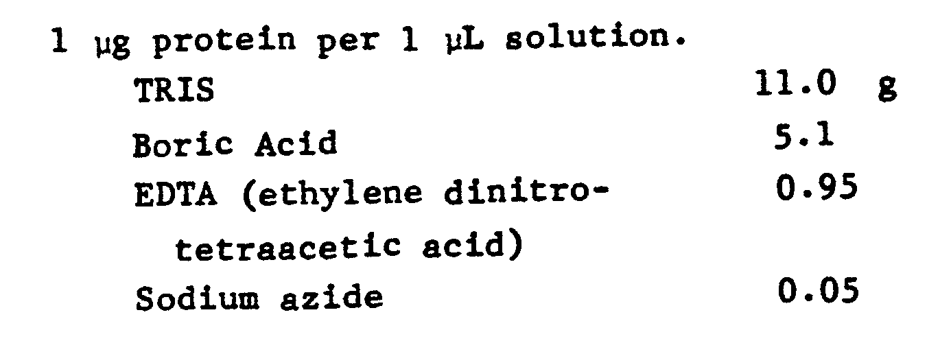
- The above was dissolved in 1 liter of water and the final pH adjusted to 8.3 with NaOH. This solution was used as the buffer in the electrophoresis procedure.
- A non SDS polyacrylamide gradient gel (sold by Separation Sciences) was used. This gel contains the solution described above. The albumin solution was placed in wells cut into the gel and electrophoresed for 5 hours at a constant voltage of 200 V. The gel was then removed and fixed in methanol and water (1:1) for 1 hour.
- The gel was nucleated and developed as described in Examples 3 and 9.
- After 20 minute development, protein bands at the 100 ng level could easily be detected. A control stained with Coomassie Blue gave only a faint image at the 1000 ng level.
The results were compared with those obtained by dye visualization. β-Galactosidase has two major electrophoretic bands. The minimum amount at 1 cm wide bands at which these bands can be observed using the dye stain visualization method (Stock Solutions 8, 9 and 10) where the procedure consisted of:
was approximately 1 µg. The minimum protein concentration that could be detected with the nickel development procedure was below 0.1 µg.
Claims (11)
- A method for determining protein, nucleic acid or polypeptide macromolecules in polyacrylamide gels, said method comprising the steps ofa) forming a latent stain image in the gel andb) developing the latent stain image by treating the gel with a developer solution characterized in that thelatent stain image is formed by nucleating the macromolecules in the gel with a palladium tetramine salt and the latent stain image is developed by treating the gel with a physical developer solution comprising dimethylamine borane and a transition metal salt, tetrazolium salt, or triazolium salt.
- The method of Claim 1 wherein the physical developer solution comprises an antifoggant.
- The method of Claim 2 wherein the antifoggant is ethyl cysteine .HCl.
- The method according to any of the preceding claims wherein the macromolecules in the gels have been subjected to electrophoresis prior to forming the latent stain.
- The method according to any of the preceding claims wherein the macromolecules are proteins.
- The method according to any of the preceding claims wherein the macromolecules are sensitized with sodium dodecyl sulfate prior to forming the gel.
- The method according to any of the preceding claims wherein the physical developing solution comprises a complexing agent.
- The method of Claim 7 wherein the complexing agent is gluconic acid or an ester thereof.
- The method according to any of the preceding claims wherein the physical developing solution comprises sodium dodecyl sulfate.
- A kit for the determination of protein, nucleic acid or polypeptide macromolecules in polyacrylamide gels comprising a palladium tetramine salt, dimethylamine borane, an antifoggant and a transition metal salt, tetrazolium salt, or triazolium salt.
- The kit of claim 10 wherein the palladium tetramine salt is a chloride salt and is in a first solution and the dimethylamine borane is in a second solution.
Applications Claiming Priority (2)
| Application Number | Priority Date | Filing Date | Title |
|---|---|---|---|
| US06/495,216 US4552848A (en) | 1983-05-16 | 1983-05-16 | Macromolecule determination by physical development |
| US495216 | 1983-05-16 |
Publications (3)
| Publication Number | Publication Date |
|---|---|
| EP0126617A2 EP0126617A2 (en) | 1984-11-28 |
| EP0126617A3 EP0126617A3 (en) | 1988-01-07 |
| EP0126617B1 true EP0126617B1 (en) | 1991-08-21 |
Family
ID=23967745
Family Applications (1)
| Application Number | Title | Priority Date | Filing Date |
|---|---|---|---|
| EP84303296A Expired EP0126617B1 (en) | 1983-05-16 | 1984-05-16 | Macromolecule determination by phsysical development |
Country Status (5)
| Country | Link |
|---|---|
| US (1) | US4552848A (en) |
| EP (1) | EP0126617B1 (en) |
| JP (1) | JPS59221665A (en) |
| CA (1) | CA1192820A (en) |
| DE (1) | DE3484941D1 (en) |
Families Citing this family (8)
| Publication number | Priority date | Publication date | Assignee | Title |
|---|---|---|---|---|
| US4672043A (en) * | 1985-02-19 | 1987-06-09 | Eastman Kodak Company | Stabilization of developed electrophoregrams |
| JPS62228168A (en) * | 1986-03-10 | 1987-10-07 | Agency Of Ind Science & Technol | High sensitivity detection of protein |
| JPS63206658A (en) * | 1987-02-23 | 1988-08-25 | Agency Of Ind Science & Technol | High sensitivity detection of protein |
| SE465018B (en) * | 1989-11-27 | 1991-07-15 | Alfa Laval Separation Ab | Centrifugal separator with two end walls and a perimeter wall |
| US5126275A (en) * | 1990-09-19 | 1992-06-30 | Miles Inc. | Analytical method using tetrazolium salt indicators having a reflectance plateau |
| EP0684468A3 (en) * | 1994-05-27 | 1997-03-26 | Eastman Kodak Co | Fog-free electrophoresis device. |
| US5595878A (en) * | 1995-06-02 | 1997-01-21 | Boron Biologicals, Inc. | Detection of biopolymers and biooligomers with boron hydride labels |
| US12566174B2 (en) | 2017-05-11 | 2026-03-03 | The Research Foundation For The State University Of New York | Method and kit for determining the presence of monosodium urate crystals in joint synovial fluid |
Family Cites Families (8)
| Publication number | Priority date | Publication date | Assignee | Title |
|---|---|---|---|---|
| US3390998A (en) * | 1965-10-04 | 1968-07-02 | Eastman Kodak Co | Stabilized physical developers |
| US3650748A (en) * | 1968-11-22 | 1972-03-21 | Eastman Kodak Co | Photographic reproduction using novel physical developers |
| DE1962744A1 (en) * | 1969-06-18 | 1971-04-15 | Wolfen Filmfab Veb | Process for stabilizing and keeping photographic materials clear |
| JPS529373B2 (en) * | 1972-09-05 | 1977-03-15 | ||
| US4307168A (en) * | 1977-05-05 | 1981-12-22 | Eastman Kodak Company | Amplification of developed electrographic image patterns |
| US4144143A (en) * | 1977-10-25 | 1979-03-13 | Virginia Commonwealth University | Electrolytic production of stable methyl viologen film |
| US4405720A (en) * | 1981-03-04 | 1983-09-20 | The United States Of America As Represented By The Department Of Health And Human Services | Silver stains for protein in gels |
| US4434234A (en) * | 1981-04-02 | 1984-02-28 | The Upjohn Company | Method and kit for silver staining substances supported in matrix |
-
1983
- 1983-05-16 US US06/495,216 patent/US4552848A/en not_active Expired - Fee Related
- 1983-07-04 CA CA000431761A patent/CA1192820A/en not_active Expired
-
1984
- 1984-05-14 JP JP59096306A patent/JPS59221665A/en active Pending
- 1984-05-16 EP EP84303296A patent/EP0126617B1/en not_active Expired
- 1984-05-16 DE DE8484303296T patent/DE3484941D1/en not_active Expired - Fee Related
Also Published As
| Publication number | Publication date |
|---|---|
| CA1192820A (en) | 1985-09-03 |
| US4552848A (en) | 1985-11-12 |
| EP0126617A3 (en) | 1988-01-07 |
| JPS59221665A (en) | 1984-12-13 |
| DE3484941D1 (en) | 1991-09-26 |
| EP0126617A2 (en) | 1984-11-28 |
Similar Documents
| Publication | Publication Date | Title |
|---|---|---|
| Jimenez et al. | Measurement of fibrosis in needle liver biopsies: evaluation of a colorimetric method | |
| Rabilloud et al. | Silver-staining of proteins in polyacrylamide gels: a general overview | |
| EP0073247B1 (en) | Silver stains for protein in gels | |
| JPS61502352A (en) | Rapid imaging method for gel electrophoresis | |
| Gratzner et al. | Deoxyribonucleic acid replication in single cells and chromosomes by immunologic techniques. | |
| US4552848A (en) | Macromolecule determination by physical development | |
| CA1242626A (en) | Method for silver staining proteins and nucleic acids | |
| WO1999002960A1 (en) | Composition and method for staining cellular dna | |
| Costello et al. | Characterization of humanized anti-TAC monoclonal antibody by traditional separation techniques and capillary electrophoresis | |
| Naimski et al. | Quantitative fluorescenct analysis of different conformational forms of DNA bound to the dye, 4′, 6-diamidine-2-phenylindole, and sepasated by gel electrophoresis | |
| Robinson et al. | Estimation of protamine and insulin in protamine zinc insulin | |
| JP4893964B2 (en) | Novel compound, reagent for analysis of peptide or protein containing the compound, and analysis method using the analysis reagent | |
| US5292665A (en) | Catalyst for preparing polyacrylamide gel which improves the detection of biomaterials by silver staining | |
| Lodish et al. | Section 3.5, Purifying, detecting, and characterizing proteins | |
| EP0192463B1 (en) | Stabilization of developed electrophoregrams | |
| JP4756422B2 (en) | Protein separation method and staining method, and protein staining solution and protein staining kit used in these methods | |
| Braun et al. | Resolution of immunoglobulin patterns by analytical isoelectric focusing | |
| Österberg et al. | Copper (II) induced polymerization of human albumin, and its depolymerization by diglycyl-L-histidine: a pH static and ultracentrifugation study | |
| JP5213967B2 (en) | Novel ClearNative Electrophoresis Using Aromatic Sulfonic Acid Compound | |
| JPH0634602A (en) | Gel electrophoresis method | |
| JPH0699377B2 (en) | Polyacrylamide gels for improved detection of proteins | |
| Trbojević-Čepe et al. | Detection of IgG oligoclonal bands in unconcentrated CSF by isoelectric focusing in ultrathin polyacrylamide gel, direct antiserum immunofixation and silver nitrate staining | |
| SU1325057A1 (en) | Method of producing dye solution for marking biological fluid | |
| US11186719B2 (en) | Hemicyanine dyes | |
| Byfield et al. | Radioaffinity assay, a new approach to binding assays applied to the measurement of serum albumin |
Legal Events
| Date | Code | Title | Description |
|---|---|---|---|
| PUAI | Public reference made under article 153(3) epc to a published international application that has entered the european phase |
Free format text: ORIGINAL CODE: 0009012 |
|
| AK | Designated contracting states |
Designated state(s): CH DE FR GB IT LI |
|
| PUAL | Search report despatched |
Free format text: ORIGINAL CODE: 0009013 |
|
| AK | Designated contracting states |
Kind code of ref document: A3 Designated state(s): CH DE FR GB IT LI |
|
| 17P | Request for examination filed |
Effective date: 19880621 |
|
| 17Q | First examination report despatched |
Effective date: 19900312 |
|
| GRAA | (expected) grant |
Free format text: ORIGINAL CODE: 0009210 |
|
| ITF | It: translation for a ep patent filed | ||
| AK | Designated contracting states |
Kind code of ref document: B1 Designated state(s): CH DE FR GB IT LI |
|
| REF | Corresponds to: |
Ref document number: 3484941 Country of ref document: DE Date of ref document: 19910926 |
|
| ET | Fr: translation filed | ||
| PG25 | Lapsed in a contracting state [announced via postgrant information from national office to epo] |
Ref country code: GB Effective date: 19920516 |
|
| PG25 | Lapsed in a contracting state [announced via postgrant information from national office to epo] |
Ref country code: LI Effective date: 19920531 Ref country code: CH Effective date: 19920531 |
|
| PLBE | No opposition filed within time limit |
Free format text: ORIGINAL CODE: 0009261 |
|
| STAA | Information on the status of an ep patent application or granted ep patent |
Free format text: STATUS: NO OPPOSITION FILED WITHIN TIME LIMIT |
|
| 26N | No opposition filed | ||
| GBPC | Gb: european patent ceased through non-payment of renewal fee |
Effective date: 19920516 |
|
| PG25 | Lapsed in a contracting state [announced via postgrant information from national office to epo] |
Ref country code: FR Effective date: 19930129 |
|
| REG | Reference to a national code |
Ref country code: CH Ref legal event code: PL |
|
| PG25 | Lapsed in a contracting state [announced via postgrant information from national office to epo] |
Ref country code: DE Effective date: 19930202 |
|
| REG | Reference to a national code |
Ref country code: FR Ref legal event code: ST |