EP0112378B1 - Electro-optical device for monitoring instantaneous singlet oxygen concentration produced during the treatment of cancer by means of photochemotherapy - Google Patents
Electro-optical device for monitoring instantaneous singlet oxygen concentration produced during the treatment of cancer by means of photochemotherapy Download PDFInfo
- Publication number
- EP0112378B1 EP0112378B1 EP83902333A EP83902333A EP0112378B1 EP 0112378 B1 EP0112378 B1 EP 0112378B1 EP 83902333 A EP83902333 A EP 83902333A EP 83902333 A EP83902333 A EP 83902333A EP 0112378 B1 EP0112378 B1 EP 0112378B1
- Authority
- EP
- European Patent Office
- Prior art keywords
- singlet oxygen
- photochemotherapy
- light
- optical detection
- optically coupled
- Prior art date
- Legal status (The legal status is an assumption and is not a legal conclusion. Google has not performed a legal analysis and makes no representation as to the accuracy of the status listed.)
- Expired
Links
- MYMOFIZGZYHOMD-UHFFFAOYSA-N Dioxygen Chemical compound O=O MYMOFIZGZYHOMD-UHFFFAOYSA-N 0.000 title claims abstract description 51
- 238000012544 monitoring process Methods 0.000 title claims abstract description 10
- 206010028980 Neoplasm Diseases 0.000 title claims description 39
- 201000011510 cancer Diseases 0.000 title claims description 4
- 230000001225 therapeutic effect Effects 0.000 claims abstract description 41
- 230000003287 optical effect Effects 0.000 claims abstract description 21
- 238000001514 detection method Methods 0.000 claims abstract description 14
- 238000003199 nucleic acid amplification method Methods 0.000 claims abstract description 9
- 230000003321 amplification Effects 0.000 claims abstract description 8
- 230000005855 radiation Effects 0.000 claims description 12
- 239000003504 photosensitizing agent Substances 0.000 claims description 5
- 230000002165 photosensitisation Effects 0.000 claims description 4
- 238000001914 filtration Methods 0.000 claims description 3
- 238000012545 processing Methods 0.000 claims description 3
- 229910000530 Gallium indium arsenide Inorganic materials 0.000 claims description 2
- 230000009849 deactivation Effects 0.000 claims description 2
- 238000000034 method Methods 0.000 abstract description 18
- 238000010791 quenching Methods 0.000 abstract description 5
- 230000000171 quenching effect Effects 0.000 abstract description 5
- 239000000975 dye Substances 0.000 description 20
- UJKPHYRXOLRVJJ-MLSVHJFASA-N CC(O)C1=C(C)/C2=C/C3=N/C(=C\C4=C(CCC(O)=O)C(C)=C(N4)/C=C4\N=C(\C=C\1/N\2)C(C)=C4C(C)O)/C(CCC(O)=O)=C3C Chemical compound CC(O)C1=C(C)/C2=C/C3=N/C(=C\C4=C(CCC(O)=O)C(C)=C(N4)/C=C4\N=C(\C=C\1/N\2)C(C)=C4C(C)O)/C(CCC(O)=O)=C3C UJKPHYRXOLRVJJ-MLSVHJFASA-N 0.000 description 14
- 229960003569 hematoporphyrin Drugs 0.000 description 14
- QVGXLLKOCUKJST-UHFFFAOYSA-N atomic oxygen Chemical compound [O] QVGXLLKOCUKJST-UHFFFAOYSA-N 0.000 description 12
- 239000001301 oxygen Substances 0.000 description 12
- 229910052760 oxygen Inorganic materials 0.000 description 12
- 230000008569 process Effects 0.000 description 12
- 230000003211 malignant effect Effects 0.000 description 7
- 206010029098 Neoplasm skin Diseases 0.000 description 5
- 208000000453 Skin Neoplasms Diseases 0.000 description 5
- 208000024519 eye neoplasm Diseases 0.000 description 5
- 238000002839 fiber optic waveguide Methods 0.000 description 5
- 230000004044 response Effects 0.000 description 5
- 238000012546 transfer Methods 0.000 description 4
- CSCPPACGZOOCGX-UHFFFAOYSA-N Acetone Chemical compound CC(C)=O CSCPPACGZOOCGX-UHFFFAOYSA-N 0.000 description 3
- 230000009471 action Effects 0.000 description 3
- 230000006378 damage Effects 0.000 description 3
- 230000005283 ground state Effects 0.000 description 3
- 230000017074 necrotic cell death Effects 0.000 description 3
- 230000000717 retained effect Effects 0.000 description 3
- 239000000523 sample Substances 0.000 description 3
- 239000000243 solution Substances 0.000 description 3
- 230000003595 spectral effect Effects 0.000 description 3
- IJGRMHOSHXDMSA-UHFFFAOYSA-N Atomic nitrogen Chemical compound N#N IJGRMHOSHXDMSA-UHFFFAOYSA-N 0.000 description 2
- 206010034972 Photosensitivity reaction Diseases 0.000 description 2
- 210000000170 cell membrane Anatomy 0.000 description 2
- 229940127089 cytotoxic agent Drugs 0.000 description 2
- 239000002254 cytotoxic agent Substances 0.000 description 2
- 231100000599 cytotoxic agent Toxicity 0.000 description 2
- 230000000694 effects Effects 0.000 description 2
- 230000005284 excitation Effects 0.000 description 2
- 229910052732 germanium Inorganic materials 0.000 description 2
- GNPVGFCGXDBREM-UHFFFAOYSA-N germanium atom Chemical compound [Ge] GNPVGFCGXDBREM-UHFFFAOYSA-N 0.000 description 2
- 230000002209 hydrophobic effect Effects 0.000 description 2
- 238000002347 injection Methods 0.000 description 2
- 239000007924 injection Substances 0.000 description 2
- 238000004020 luminiscence type Methods 0.000 description 2
- 238000004519 manufacturing process Methods 0.000 description 2
- 239000012528 membrane Substances 0.000 description 2
- 239000002904 solvent Substances 0.000 description 2
- 230000002123 temporal effect Effects 0.000 description 2
- 238000002560 therapeutic procedure Methods 0.000 description 2
- 230000001052 transient effect Effects 0.000 description 2
- 230000007704 transition Effects 0.000 description 2
- RBTBFTRPCNLSDE-UHFFFAOYSA-N 3,7-bis(dimethylamino)phenothiazin-5-ium Chemical compound C1=CC(N(C)C)=CC2=[S+]C3=CC(N(C)C)=CC=C3N=C21 RBTBFTRPCNLSDE-UHFFFAOYSA-N 0.000 description 1
- IICCLYANAQEHCI-UHFFFAOYSA-N 4,5,6,7-tetrachloro-3',6'-dihydroxy-2',4',5',7'-tetraiodospiro[2-benzofuran-3,9'-xanthene]-1-one Chemical compound O1C(=O)C(C(=C(Cl)C(Cl)=C2Cl)Cl)=C2C21C1=CC(I)=C(O)C(I)=C1OC1=C(I)C(O)=C(I)C=C21 IICCLYANAQEHCI-UHFFFAOYSA-N 0.000 description 1
- GYHNNYVSQQEPJS-UHFFFAOYSA-N Gallium Chemical compound [Ga] GYHNNYVSQQEPJS-UHFFFAOYSA-N 0.000 description 1
- 206010058467 Lung neoplasm malignant Diseases 0.000 description 1
- KXNLCSXBJCPWGL-UHFFFAOYSA-N [Ga].[As].[In] Chemical compound [Ga].[As].[In] KXNLCSXBJCPWGL-UHFFFAOYSA-N 0.000 description 1
- 238000009825 accumulation Methods 0.000 description 1
- 229910052785 arsenic Inorganic materials 0.000 description 1
- RQNWIZPPADIBDY-UHFFFAOYSA-N arsenic atom Chemical compound [As] RQNWIZPPADIBDY-UHFFFAOYSA-N 0.000 description 1
- 230000008901 benefit Effects 0.000 description 1
- 230000005540 biological transmission Effects 0.000 description 1
- 210000000621 bronchi Anatomy 0.000 description 1
- 238000013276 bronchoscopy Methods 0.000 description 1
- 210000003679 cervix uteri Anatomy 0.000 description 1
- 210000004087 cornea Anatomy 0.000 description 1
- 230000008878 coupling Effects 0.000 description 1
- 238000010168 coupling process Methods 0.000 description 1
- 238000005859 coupling reaction Methods 0.000 description 1
- CSCPPACGZOOCGX-WFGJKAKNSA-N deuterated acetone Substances [2H]C([2H])([2H])C(=O)C([2H])([2H])[2H] CSCPPACGZOOCGX-WFGJKAKNSA-N 0.000 description 1
- 238000012631 diagnostic technique Methods 0.000 description 1
- 229910001882 dioxygen Inorganic materials 0.000 description 1
- -1 eosin-y Chemical compound 0.000 description 1
- GNBHRKFJIUUOQI-UHFFFAOYSA-N fluorescein Chemical compound O1C(=O)C2=CC=CC=C2C21C1=CC=C(O)C=C1OC1=CC(O)=CC=C21 GNBHRKFJIUUOQI-UHFFFAOYSA-N 0.000 description 1
- 229910052733 gallium Inorganic materials 0.000 description 1
- 229910052738 indium Inorganic materials 0.000 description 1
- APFVFJFRJDLVQX-UHFFFAOYSA-N indium atom Chemical compound [In] APFVFJFRJDLVQX-UHFFFAOYSA-N 0.000 description 1
- 230000001939 inductive effect Effects 0.000 description 1
- 230000003993 interaction Effects 0.000 description 1
- 238000010253 intravenous injection Methods 0.000 description 1
- 238000011835 investigation Methods 0.000 description 1
- 229940056932 lead sulfide Drugs 0.000 description 1
- 229910052981 lead sulfide Inorganic materials 0.000 description 1
- 150000002632 lipids Chemical class 0.000 description 1
- 239000007788 liquid Substances 0.000 description 1
- 239000006193 liquid solution Substances 0.000 description 1
- 230000004807 localization Effects 0.000 description 1
- 201000005202 lung cancer Diseases 0.000 description 1
- 208000020816 lung neoplasm Diseases 0.000 description 1
- 208000037841 lung tumor Diseases 0.000 description 1
- 230000014759 maintenance of location Effects 0.000 description 1
- 230000007246 mechanism Effects 0.000 description 1
- 229960000907 methylthioninium chloride Drugs 0.000 description 1
- 238000012986 modification Methods 0.000 description 1
- 230000004048 modification Effects 0.000 description 1
- 229910052757 nitrogen Inorganic materials 0.000 description 1
- 231100000252 nontoxic Toxicity 0.000 description 1
- 230000003000 nontoxic effect Effects 0.000 description 1
- 230000037311 normal skin Effects 0.000 description 1
- 230000003647 oxidation Effects 0.000 description 1
- 238000007254 oxidation reaction Methods 0.000 description 1
- 230000035515 penetration Effects 0.000 description 1
- 230000000886 photobiology Effects 0.000 description 1
- 230000004962 physiological condition Effects 0.000 description 1
- 238000002360 preparation method Methods 0.000 description 1
- 238000011160 research Methods 0.000 description 1
- 229930187593 rose bengal Natural products 0.000 description 1
- 229940081623 rose bengal Drugs 0.000 description 1
- STRXNPAVPKGJQR-UHFFFAOYSA-N rose bengal A Natural products O1C(=O)C(C(=CC=C2Cl)Cl)=C2C21C1=CC(I)=C(O)C(I)=C1OC1=C(I)C(O)=C(I)C=C21 STRXNPAVPKGJQR-UHFFFAOYSA-N 0.000 description 1
- 230000001235 sensitizing effect Effects 0.000 description 1
- 241000894007 species Species 0.000 description 1
- 238000001228 spectrum Methods 0.000 description 1
- 230000000638 stimulation Effects 0.000 description 1
- 210000003932 urinary bladder Anatomy 0.000 description 1
Images

Classifications
-
- A—HUMAN NECESSITIES
- A61—MEDICAL OR VETERINARY SCIENCE; HYGIENE
- A61B—DIAGNOSIS; SURGERY; IDENTIFICATION
- A61B5/00—Measuring for diagnostic purposes; Identification of persons
- A61B5/0059—Measuring for diagnostic purposes; Identification of persons using light, e.g. diagnosis by transillumination, diascopy, fluorescence
-
- A—HUMAN NECESSITIES
- A61—MEDICAL OR VETERINARY SCIENCE; HYGIENE
- A61B—DIAGNOSIS; SURGERY; IDENTIFICATION
- A61B90/00—Instruments, implements or accessories specially adapted for surgery or diagnosis and not covered by any of the groups A61B1/00 - A61B50/00, e.g. for luxation treatment or for protecting wound edges
- A61B90/06—Measuring instruments not otherwise provided for
-
- A—HUMAN NECESSITIES
- A61—MEDICAL OR VETERINARY SCIENCE; HYGIENE
- A61F—FILTERS IMPLANTABLE INTO BLOOD VESSELS; PROSTHESES; DEVICES PROVIDING PATENCY TO, OR PREVENTING COLLAPSING OF, TUBULAR STRUCTURES OF THE BODY, e.g. STENTS; ORTHOPAEDIC, NURSING OR CONTRACEPTIVE DEVICES; FOMENTATION; TREATMENT OR PROTECTION OF EYES OR EARS; BANDAGES, DRESSINGS OR ABSORBENT PADS; FIRST-AID KITS
- A61F9/00—Methods or devices for treatment of the eyes; Devices for putting in contact-lenses; Devices to correct squinting; Apparatus to guide the blind; Protective devices for the eyes, carried on the body or in the hand
- A61F9/007—Methods or devices for eye surgery
- A61F9/008—Methods or devices for eye surgery using laser
-
- A—HUMAN NECESSITIES
- A61—MEDICAL OR VETERINARY SCIENCE; HYGIENE
- A61N—ELECTROTHERAPY; MAGNETOTHERAPY; RADIATION THERAPY; ULTRASOUND THERAPY
- A61N5/00—Radiation therapy
- A61N5/06—Radiation therapy using light
- A61N5/0613—Apparatus adapted for a specific treatment
- A61N5/062—Photodynamic therapy, i.e. excitation of an agent
-
- A—HUMAN NECESSITIES
- A61—MEDICAL OR VETERINARY SCIENCE; HYGIENE
- A61B—DIAGNOSIS; SURGERY; IDENTIFICATION
- A61B17/00—Surgical instruments, devices or methods
- A61B2017/00017—Electrical control of surgical instruments
- A61B2017/00022—Sensing or detecting at the treatment site
- A61B2017/00057—Light
-
- A—HUMAN NECESSITIES
- A61—MEDICAL OR VETERINARY SCIENCE; HYGIENE
- A61B—DIAGNOSIS; SURGERY; IDENTIFICATION
- A61B18/00—Surgical instruments, devices or methods for transferring non-mechanical forms of energy to or from the body
- A61B18/18—Surgical instruments, devices or methods for transferring non-mechanical forms of energy to or from the body by applying electromagnetic radiation, e.g. microwaves
- A61B18/20—Surgical instruments, devices or methods for transferring non-mechanical forms of energy to or from the body by applying electromagnetic radiation, e.g. microwaves using laser
- A61B2018/2035—Beam shaping or redirecting; Optical components therefor
- A61B2018/2045—Choppers
-
- A—HUMAN NECESSITIES
- A61—MEDICAL OR VETERINARY SCIENCE; HYGIENE
- A61F—FILTERS IMPLANTABLE INTO BLOOD VESSELS; PROSTHESES; DEVICES PROVIDING PATENCY TO, OR PREVENTING COLLAPSING OF, TUBULAR STRUCTURES OF THE BODY, e.g. STENTS; ORTHOPAEDIC, NURSING OR CONTRACEPTIVE DEVICES; FOMENTATION; TREATMENT OR PROTECTION OF EYES OR EARS; BANDAGES, DRESSINGS OR ABSORBENT PADS; FIRST-AID KITS
- A61F9/00—Methods or devices for treatment of the eyes; Devices for putting in contact-lenses; Devices to correct squinting; Apparatus to guide the blind; Protective devices for the eyes, carried on the body or in the hand
- A61F9/007—Methods or devices for eye surgery
- A61F9/008—Methods or devices for eye surgery using laser
- A61F2009/00844—Feedback systems
-
- A—HUMAN NECESSITIES
- A61—MEDICAL OR VETERINARY SCIENCE; HYGIENE
- A61F—FILTERS IMPLANTABLE INTO BLOOD VESSELS; PROSTHESES; DEVICES PROVIDING PATENCY TO, OR PREVENTING COLLAPSING OF, TUBULAR STRUCTURES OF THE BODY, e.g. STENTS; ORTHOPAEDIC, NURSING OR CONTRACEPTIVE DEVICES; FOMENTATION; TREATMENT OR PROTECTION OF EYES OR EARS; BANDAGES, DRESSINGS OR ABSORBENT PADS; FIRST-AID KITS
- A61F9/00—Methods or devices for treatment of the eyes; Devices for putting in contact-lenses; Devices to correct squinting; Apparatus to guide the blind; Protective devices for the eyes, carried on the body or in the hand
- A61F9/007—Methods or devices for eye surgery
- A61F9/008—Methods or devices for eye surgery using laser
- A61F9/009—Auxiliary devices making contact with the eyeball and coupling in laser light, e.g. goniolenses
-
- A—HUMAN NECESSITIES
- A61—MEDICAL OR VETERINARY SCIENCE; HYGIENE
- A61N—ELECTROTHERAPY; MAGNETOTHERAPY; RADIATION THERAPY; ULTRASOUND THERAPY
- A61N5/00—Radiation therapy
- A61N5/06—Radiation therapy using light
- A61N2005/0635—Radiation therapy using light characterised by the body area to be irradiated
- A61N2005/0643—Applicators, probes irradiating specific body areas in close proximity
- A61N2005/0644—Handheld applicators
Definitions
- This invention relates to an apparatus and method for monitoring the instantaneous concentration of singlet oxygen produced during photochemotherapy and thereby provides a tool to assess the effectiveness of the photochemotherapy treatment and prescribe a correct dosage of therapeutic photoradiation.
- hematoporphyrin preparations When certain hematoporphyrin preparations are injected intravenously into the human body, they are selectively retained by cancerous tissue. Thus, two or three days after injection, significantly higher levels of hematoporphyrin accumulate in malignant tissue.
- the selective retention of hematoporphyrin by cancerous tissue was first used clinically as a diagnostic tool. In the presence of ultraviolet or shortwave length visible light, such tissue will exhibit a bright red light fluorescence while normal tissue appears light pink.
- a discussion of clinical investigations using this diagnostic technique can be found in an article entitled "Hematoporphyrin Diacetate: A Probe to Distinguish Malignant from Normal Tissue by Selective Fluorescence" by R. W. Hen- derspn, G. S. Christie, P. S.
- photochemotherapy involves the injection of a hematoporphyrin dye intravenously into the patient.'After the passage of several days, usually three, the dye is retained in significant amounts by cancerous tissue, however being eliminated by healthy tissue. The tumor is then exposed to a therapeutic light and this light energy causes the photodynamic dye to be excited to a high energy state.
- the dye transfers energy to oxygen molecules present in the tissue and raises them from the ground triplet to the first excited electronic singlet state, '0 2 .
- the singlet oxygen, '0 2 attacks and functionally destroys the cell membranes ultimately inducing necrosis and destroying the cancerous tissue.
- the current inventors recognize that adequate treatment is questionable unless one knows directly the rate at which singlet oxygen is generated in the malignant tissue.
- the inventors recognize that the generation of singlet oxygen involves several key factors: (1) the therapeutic light source must be of the proper intensity and wavelength sufficient to elevate the photodynamic dye to an excited singlet state; (2) there must be a sufficient concentration of oxygen localized within the tumor both initially and during the period of irradiation to be excited by energy transfer from the dye; (3) the local dye concentration may vary considerably, depending on the hydrophobic or hydrophilic nature of the immediate environment; and (4) the rate of quenching Of '0 2 may vary substantially with the existing medium, being generally more rapid in an aqueous than in a lipid environment.
- the singlet oxygen emission was first described in an article by A. A. Krasnovsky, Jr.
- the present inventors have developed a method and apparatus for measuring singlet oxygen generated during photochemotherapy by using an optical sensor.
- This method and apparatus has applications for various types of tumors: for skin tumors the optical sensor can be placed near the skin surface to receive the 1.27 micron radiation emitted from the tumor; for deep seated intracorporeal tumors a fiberoptical probe is inserted into the tumor mass and conveys the radiation to the optical sensor; and, for eye tumors a fiberoptic/contact lens arrangement can be used to collect the 1.27 micron radiation and convey it to the optical sensor.
- the optical sensor generally consists of a photodiode which is preceded by an infrared band-pass filter to provide the necessary rejection.
- the photodiode most suited for this application is an epitaxial unit composed of indium, gallium and arsenic (InGaAs). These detectors have a much better time response characteristic than germanium and since they are relatively insensitive to excitation below 9000A they can provide significant discrimination against the visible exciting light.
- InGaAs device both in regard to temporal discrimination required in separating the short duration exciting light from the more slowly varying '0 2 emission and also in spectral discrimination establishes it as the preferable detector for this application.
- the photodiode output is fed directly to either a lock-in amplifier or first amplified and then fed to a box-car integrator.
- the particular choice depends on temporal characteristics of the incident therapeutic light.
- the signal is fed to an indicator to provide an instantaneous reading of the aver-. age singlet oxygen level within the tumor mass.
- the physician or operator can adjust the therapeutic irradiation conditions (i.e., intensity, wavelength, angle of incidence, time of exposure, etc.) to provide a singlet oxygen level as necessary to satisfy predetermined clinical conditions.
- the invented method and apparatus thus provides a means for remotely monitoring the total singlet oxygen production, a production rate which may vary substantially with local physiological conditions.
- Figure 1 illustrates schematically the overall system kinetics.
- First the photodynamic dye is excited from the ground state, So, to the first excited electronic state, S 1 , by means of a therapeutic light source, such as pulsed laser radiation at 5320 angstroms (green light).
- the dye singlet state S is then transformed via an intramolecular coupling to the lowest-lying dye triplet state, To, as indicated in the figure.
- Subsequent deactivation of this triplet state to the ground singlet state, So, in the absence of oxygen, is forbidden by the spin selection rules, resulting in lifetimes of this particular species under anaerobic (oxygen- excluded) conditions as long as one millisecond.
- the quantity -E is the emission frequency, and corresponds to a wavelength of 1.27 microns.
- the specific site of singlet oxygen attack has been identified as the cell membrane. Significant differences in hematoporphyrin accumulation and membrane damage have been found to occur, depending on the hydrophobic or hydrophilic nature of the specific membrane site and compensating solubility of the photosensitizer. It will be noted that several photosensitizing dyes, such as rose bengal, eosin-y, fluorescein, methylene blue, other than hematoporphyrin, may be used to produce the above-described photodynamic action. Hematoporphyrin, however, has the advantage of being relatively non-toxic and has also evidenced itself to be resistant to oxidation, thus quite stable.
- the problem with the prior art is that the correct intensity and duration of the therapeutic light dosage can't be determined.
- the physician must guess at the correct therapeutic light dosage. If the dosage is too high, normal tissue will experience necrosis; and, if the dosage is insufficient, the tumor will not be destroyed. Since the physician has no way of knowing the rate at which oxygen is being delivered to the tissue or the magnitude of the local dye concentration, it is impossible to predict the effect of a given therapeutic light dosage. If the light dosage is too intense, local oxygen may be depleted more rapidly than it is provided to the tissue and no-additional singlet oxygen will be generated for any increase in intensity or duration of the light dosage. The only adequate way to prescribe the therapeutic light dosage therefore is by direct monitoring of the level of singlet oxygen produced. As has been mentioned, remote optical detection of the singlet oxygen level is possible because'of the existence of a collisionally induced infrared emission at a wavelength of 1.27 microns as indicated in Equation 2.
- FIGS 2 and 3 illustrate, in block diagrammatic form, the invented apparatus used to monitor singlet oxygen concentration during the treatment of skin tumors or-tumors nearthe skin surface.
- the apparatus generally contains: an optical detection means 10 which collects light emitting from the tumor and detects the intensity of such light at the 1.27 micron wavelength band; an amplification means 12 which amplifies and processes electrical signals generated by the optical detection means 10; and, an output indicator 14 which indicates the instantaneous singlet oxygen concentration occurring during photochemotherapy treatment.
- therapeutic light pulsed 16 or substantially continuous 18
- the tumor has been previously treated and has absorbed a photosensitizing dye, such as hematoporphyrin.
- the therapeutic photoradiation produces singlet oxygen, the active cytotoxic agent, which emits radiation in a relatively narrow spectral band centered at a wavelength of 1.27 microns.
- the optical detection means- 10 collects and detects the 1.27 micron emission, and produces an electrical signal which is amplified by the amplification means 12.
- the output indicator 14 connects to the amplification means 12 and displays the level of instantaneous singlet oxygen concentration. The instantaneous concentration level could be used to further determine the maximum level of singlet oxygen produced or the quenching rate.
- the instantaneous concentration level couid be used to adjust the dosage of the therapeutic radiation, i.e., indicate whether the intensity, duration, frequency or modulation of the therapeutic radiation should be modified for more effective photochemotherapy of the particular tumor or a particular part of the tumor.
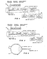
- Figure 2 illustrates the invented apparatus when a pulsed or chopped therapeutic light 16 is used.
- the apparatus consists of: a lens arrangement 22 for collecting and concentrating light generated in the tumor; a band pass filter 24 which filters unwanted light and passes light in the 1.27 micron wavelength band; a photodetector 26 which. detects light passed by the filter 24; a preamplifier 28 and a boxcar integrator (or transient recorder combined with a signal averager) 30 to amplify and process the electrical signal produced by the photodetector 26; and, an output indicator 14 which displays the instantaneous singlet oxygen concentration and/or quenching rate.
- a lens arrangement 22 for collecting and concentrating light generated in the tumor
- a band pass filter 24 which filters unwanted light and passes light in the 1.27 micron wavelength band
- a photodetector 26 which. detects light passed by the filter 24
- a preamplifier 28 and a boxcar integrator (or transient recorder combined with a signal averager) 30 to amplif
- the photodetector 26 must be able to detect weak emissions at a wavelength of 1.27 microns.
- the spectral response of the diode photodetector combined with the bandpass filter 24 must be such that the therapeutic light, which appears in the visible part of the spectrum, and the red fluorescent light, which appears when the dye undergoes a radiative transition from its excited singlet to the ground singlet state, are both filtered out.
- the photodetector 26 must also be fast enough to be responsive to time varying intensity of the light emission.
- the inventors prefer an indium-gallium- arsenide (InGaAs) epitaxial device because it is selectively responsive to infrared emission in the desired wavelength range with minimal filtration requirements and because of its rapid time response. This type of device is used extensively in commercial fiberoptic transmission systems.
- Figure 3 illustrates the invented apparatus when a substantially continuous intensity of therapeutic light 18 is used.
- the apparatus consists of: a lens arrangement 22 for collecting and concentrating light generated in the tumor; a mechanical or electro-optical chopper 32 which is inserted for signal to noise enhancement; a band pass filter 24 which filters unwanted light frequencies and passes light in the 1.27 micron wavelength band; a photodetector 26 which detects light passed by the filter 24; a lock-in amplifier 34 which amplifies and processes the electrical signal generated by the photodetector 26; and, an output indicator 14 which displays the instantaneous level of singlet oxygen concentration.
- the photodetector 26 may be an InGaAs diode.
- FIGS 4 and 5 illustrates, in a block diagrammatic form, the invented apparatus used to monitor singlet oxygen concentration during the treatment of deep seated intracorporeal tumors.
- the apparatus generally contains: an optical detection means 10 which collects and detects light in the 1.27 microns wavelength band emitting from deep seated tumors; an-amplification means 12 to amplify and process the signal produced by the optical detecting means 10; and, an indicator means 14, which indicates the instantaneous singlet oxygen concentration.
- FIG. 4 illustrates the invented apparatus when a pulsed therapeutic light 16 is used.
- the apparatus consists of: a fiberoptic waveguide 36 which is inserted into the tumor mass 38 through a hollow hypodermic needle 40 to collect light emitted within the tumor; a bandpass filter 24 which filters unwanted light frequencies and passes light in the 1.27 micron wavelength band; a photodiode 26 which detects light passed by the filter 24 and may have characteristics which also filters out unwanted light frequencies; a preamplifier 28 which amplifies electrical signals generated by the photodetector 26; a boxcar integrator (or transient recorder combined with a signal averager) 30, to further process the signal; and, an outut indicator 14 which displays the instantaneous level of slight oxygen concentration and or quenching rate.
- the photodetector 26 may be a InGaAs dio
- FIG. 5 illustrates the embodiment when a substantially constant intensity therapeutic light source 28 is used.
- the apparatus consists of: a fiberoptic waveguide 36 which is inserted into the tumor mass 38 through a hollow hypodermic needle 40 to collect light emitted within the tumor; a mechanical or electro-optical chopper 42 is inserted along the fiberoptic waveguide 36; a bandpass filter 24 which filters unwanted light frequencies, and passes light in the 1.27 microns band; a photodetector 26 which detects light passed by the filter 24 and has characteristics which further filters out unwanted light frequencies; a lock-in amplifier 34 which amplifies and processes the electrical signal generated by the photodetector 26; and, an output indicator 14 which displays the instantaneous level of signal oxygen concentration.
- the photodetector 26 may be a InGaAs diode and the chopper 42 may be an optoacoustic modulator.
- the fiberoptic waveguide 36 can be inserted into the cancerous tissue via a hollow hypodermic needle 40.
- the therapeutic light source also enter the cancerous tumor through a fiberoptic link.
- the fiberoptic carrying the therapeutic light and the fiberoptic probe link could be coupled and inserted simultaneously into the cancerous tumor.
- a hypodermic needle or an endoscope would be appropriate for this application.
- the hypodermic needle 40 would permit penetration of the fiberoptic waveguide 36 into the area of the tumor mass to monitor the singlet oxygen generation. Since light at a wavelength of 1.27 microns penetrates more readily through living tissue than does visible light, the exact placement of the fiberoptic probe in the tumor mass is not critical.
- the intensity, frequency, and modulation of the therapeutic light can be adjusted in response to the singlet oxygen level. In this manner the invented apparatus provides a practical way to measure the effect of the incident therapeutic light dosage and makes quantitative photochemotherapy possible as a means of clinical cancer treatment.
- the fiberoptic link 36 shown in Figures 4 and 5 can be optically coupled to a contact lens rather than inserted into the tumor area.
- the contact lens arrangement, shown in Figure 6, generally contains a contact lens 44 which is placed over the cornea and optically coupled to a fiberoptic link 36.
- the contact lens 44 collects the 1.27 micron emission generated by singlet oxygen during photoradiation of eye tumors.
- a single fiberoptic link coupled to the contact lens can transmit therapeutic light into the eye and collect the 1.27 micron emission coming from the eye; or a bundle of fiberoptic links can be optimally coupled to the contact lens and one of the links can collect light emitted from the eye tumor.
Landscapes
- Health & Medical Sciences (AREA)
- Life Sciences & Earth Sciences (AREA)
- Engineering & Computer Science (AREA)
- Biomedical Technology (AREA)
- Surgery (AREA)
- Veterinary Medicine (AREA)
- Public Health (AREA)
- General Health & Medical Sciences (AREA)
- Animal Behavior & Ethology (AREA)
- Heart & Thoracic Surgery (AREA)
- Pathology (AREA)
- Nuclear Medicine, Radiotherapy & Molecular Imaging (AREA)
- Ophthalmology & Optometry (AREA)
- Molecular Biology (AREA)
- Biophysics (AREA)
- Medical Informatics (AREA)
- Physics & Mathematics (AREA)
- Vascular Medicine (AREA)
- Optics & Photonics (AREA)
- Radiology & Medical Imaging (AREA)
- Oral & Maxillofacial Surgery (AREA)
- Radiation-Therapy Devices (AREA)
- Investigating, Analyzing Materials By Fluorescence Or Luminescence (AREA)
- Investigating Or Analysing Biological Materials (AREA)
- Surgical Instruments (AREA)
- Endoscopes (AREA)
- Investigating Or Analysing Materials By Optical Means (AREA)
- Other Investigation Or Analysis Of Materials By Electrical Means (AREA)
Abstract
Description
- This invention relates to an apparatus and method for monitoring the instantaneous concentration of singlet oxygen produced during photochemotherapy and thereby provides a tool to assess the effectiveness of the photochemotherapy treatment and prescribe a correct dosage of therapeutic photoradiation.
- When certain hematoporphyrin preparations are injected intravenously into the human body, they are selectively retained by cancerous tissue. Thus, two or three days after injection, significantly higher levels of hematoporphyrin accumulate in malignant tissue. The selective retention of hematoporphyrin by cancerous tissue was first used clinically as a diagnostic tool. In the presence of ultraviolet or shortwave length visible light, such tissue will exhibit a bright red light fluorescence while normal tissue appears light pink. A discussion of clinical investigations using this diagnostic technique can be found in an article entitled "Hematoporphyrin Diacetate: A Probe to Distinguish Malignant from Normal Tissue by Selective Fluorescence" by R. W. Hen- derspn, G. S. Christie, P. S. Clezy and J. Lineham, Brit. J. Exp. Pathol., Vol. 61, pages 325-350 (1980). Another reference by D. R. Doiron and A. E. Profio entitled "Laser Fluorescence Bronchos- copy for Early Lung Cancer Localization" published in Lasers in Photomedicine and Photobiology (1980) teaches the use of a laser fluorescence bronchoscope to detect and localize small lung tumors by observing this red fluorescence.
- An additional clinical application has recently been found in the treatment and destruction of malignant tissue, i.e., photochemotherapy or photoradiation therapy. The process by which biological damage occurs, as the result of optical excitation of a hematoporphyrin dye in the presence of oxygen, is generally referred to as "photodynamic action". As indicated above, photochemotherapy involves the injection of a hematoporphyrin dye intravenously into the patient.'After the passage of several days, usually three, the dye is retained in significant amounts by cancerous tissue, however being eliminated by healthy tissue. The tumor is then exposed to a therapeutic light and this light energy causes the photodynamic dye to be excited to a high energy state. Through a direct intramolecular process, the dye transfers energy to oxygen molecules present in the tissue and raises them from the ground triplet to the first excited electronic singlet state, '02. The singlet oxygen, '02, attacks and functionally destroys the cell membranes ultimately inducing necrosis and destroying the cancerous tissue.
- In an article by Thomas J. Dougherty et al entitled "Photoradiation Therapy for the Treatment of Malignant Tumors" published in Cancer Research, Vol. 38, pages 2628-2635 (1978), problems associated with prescribing the correct therapeutic light dosages are discussed. If the dosage is too weak, the tumor response will be partial or incomplete. If the irradiation time is too long or intensity too high, normal skin or tissue will experience necrosis. The aforementioned article points out the difficulty of determining the correct therapeutic dosage of light. This problem has been proven to be quite significant and currently is one of the major hurdles that must be overcome before photochemotherapy can be used to treat deep-seated large tumors.
- The current inventors recognize that adequate treatment is questionable unless one knows directly the rate at which singlet oxygen is generated in the malignant tissue. The inventors recognize that the generation of singlet oxygen involves several key factors: (1) the therapeutic light source must be of the proper intensity and wavelength sufficient to elevate the photodynamic dye to an excited singlet state; (2) there must be a sufficient concentration of oxygen localized within the tumor both initially and during the period of irradiation to be excited by energy transfer from the dye; (3) the local dye concentration may vary considerably, depending on the hydrophobic or hydrophilic nature of the immediate environment; and (4) the rate of quenching Of '02 may vary substantially with the existing medium, being generally more rapid in an aqueous than in a lipid environment. Therefore, merely measuring the incident light intensity and time of exposure will not supply adequate information regarding the generation of singlet oxygen within the tumor tissue. The inventors, therefore, recognize the need to determine directly the amount of singlet oxygen which is being generated during the light excited hematoporphyrin process.
- The inventors recognize that direct monitoring is possible because in the interaction of single oxygen molecules, with the surrounding medium, the singlet oxygen molecules undergo a radiative transition to its ground triplet state emitting light at a wavelength of 1.27 micros, e.g., J. G. Parker and W. D. Stanbro, "Optical Determination of the Collisional Lifetime of Singlet Molecular Oxygen [O2 1Δg] in Acetone and Deuterated Acetone", J. Am. Chem. Soc., Vol. 104, pp. 2067-2069 (1982). The singlet oxygen emission was first described in an article by A. A. Krasnovsky, Jr. entitled "Photosensitized Luminescence of Singlet Oxygen in Solution" reprinted in Biophysics (GB), Vol. 21, page 770 (1976). Krasnovsky is generally recognized as the first to identify and optically detect the singlet oxygen emission at 1.27 microns in solution. Krasnovsky used a cryogenically cooled photomultiplier as the means for detecting this emission in a CC14 solvent. Additional work has been done for various other solvents, e.g., A. H. Khan and M. Kasha, "Direct Spectroscopic Observation of Singlet Oxygen Emission at 1268 nm Excited by Sensitizing Dyes of Biological Interest in Liquid Solution", Proc. Natl. Acad. Sci., Vol. 76(12), page 6047 (1979); and K. I. Salokhiddinov, B. N. Dzhagarov, I. M. Byteva, and G. P. Gurinovich, "Photosensitized Luminescence of Singlet Oxygen in Solutions at 159 nm", Chem. Phys. Lett., Vol. 76(1), page 85 (1980). To detect the 1.27 microns emission Khan et al used a near-infrared spectrophotometer that employed a thermoelectrically cooled lead sulfide detector and Salokhiddinov et al used a liquid nitrogen cooled germanium photodiode.
- The present inventors have developed a method and apparatus for measuring singlet oxygen generated during photochemotherapy by using an optical sensor. This method and apparatus has applications for various types of tumors: for skin tumors the optical sensor can be placed near the skin surface to receive the 1.27 micron radiation emitted from the tumor; for deep seated intracorporeal tumors a fiberoptical probe is inserted into the tumor mass and conveys the radiation to the optical sensor; and, for eye tumors a fiberoptic/contact lens arrangement can be used to collect the 1.27 micron radiation and convey it to the optical sensor. The optical sensor generally consists of a photodiode which is preceded by an infrared band-pass filter to provide the necessary rejection. The photodiode most suited for this application is an epitaxial unit composed of indium, gallium and arsenic (InGaAs). These detectors have a much better time response characteristic than germanium and since they are relatively insensitive to excitation below 9000A they can provide significant discrimination against the visible exciting light. The superiority of the InGaAs device both in regard to temporal discrimination required in separating the short duration exciting light from the more slowly varying '02 emission and also in spectral discrimination establishes it as the preferable detector for this application.
- To provide the necessary signal-to-noise enhancement, the photodiode output is fed directly to either a lock-in amplifier or first amplified and then fed to a box-car integrator. The particular choice depends on temporal characteristics of the incident therapeutic light. After processing, the signal is fed to an indicator to provide an instantaneous reading of the aver-. age singlet oxygen level within the tumor mass. By monitoring the singlet oxygen level, the physician or operator can adjust the therapeutic irradiation conditions (i.e., intensity, wavelength, angle of incidence, time of exposure, etc.) to provide a singlet oxygen level as necessary to satisfy predetermined clinical conditions. The invented method and apparatus thus provides a means for remotely monitoring the total singlet oxygen production, a production rate which may vary substantially with local physiological conditions.
-
- Figure 1 is a graph illustrating the overall system kinetics showing therapeutic light stimulation of photosensitizing dye and the resulting generation of singlet oxygen.
- Figure 2 illustrates, in block diagrammatic form, the invented apparatus used to monitor singlet oxygen generated in skin tumors when pulsed therapeutic light is used.
- Figure 3 illustrates, in block diagrammatic form, the invented apparatus used to monitor singlet oxygen generation in skin tumors when substantially continuous therapeutic light is used.
- Figure 4 illustrates, in block diagrammatic form, the invented apparatus used to monitor singlet oxygen generation in deep seated tumors when pulsed therapeutic light is used.
- Figure 5 illustrates, in block diagrammatic form, the invented apparatus used to monitor singlet oxygen generation in deep seated tumors when substantially continuous therapeutic light is used.
- Figure 6 illustrates a contact lens/fiberoptic link arrangement for monitoring singlet oxygen generation in eye tumors.
- The mechanism by which laser-induced photodynamic action destroys malignant tissue appears to involve a two-step process. Figure 1 illustrates schematically the overall system kinetics. First the photodynamic dye is excited from the ground state, So, to the first excited electronic state, S1, by means of a therapeutic light source, such as pulsed laser radiation at 5320 angstroms (green light). The dye singlet state S, is then transformed via an intramolecular coupling to the lowest-lying dye triplet state, To, as indicated in the figure. Subsequent deactivation of this triplet state to the ground singlet state, So, in the absence of oxygen, is forbidden by the spin selection rules, resulting in lifetimes of this particular species under anaerobic (oxygen- excluded) conditions as long as one millisecond. However, in the presence of oxygen localized in the tissue, the dye triplet, To, will deactivate rapidly (0.3 to 3.0 psec) to the dye singlet ground state, So, in a spin-conserving intramolecular process in which a sizeable fraction of the energy in the dye triplet is transferred to the triplet electronic state of oxygen, 302, raising the oxygen to the first excited electronic singlet state, 102, approximately 1 electron-volt above the ground state. This spin-conserving energy transfer is possible only because the ground electronic state of molecular oxygen happens to be a triplet which is not true for most stable molecules. This energy transfer, which is shown un Figure 1, can be represented as follows:
-

- When singlet oxygen, '02, interacts with molecules contained in the surrounding medium M, a relatively weak collision-induced emission occu rs:

- The quantity -E is the emission frequency, and corresponds to a wavelength of 1.27 microns. By optically detecting and monitoring this emission it is possible to determine the instantaneous rate at which the active cytotoxic agent, singlet oxygen, is being generated and is attacking the malignant - tissue.
- The specific site of singlet oxygen attack has been identified as the cell membrane. Significant differences in hematoporphyrin accumulation and membrane damage have been found to occur, depending on the hydrophobic or hydrophilic nature of the specific membrane site and compensating solubility of the photosensitizer. It will be noted that several photosensitizing dyes, such as rose bengal, eosin-y, fluorescein, methylene blue, other than hematoporphyrin, may be used to produce the above-described photodynamic action. Hematoporphyrin, however, has the advantage of being relatively non-toxic and has also evidenced itself to be resistant to oxidation, thus quite stable.
- Current photochemotherapy treatment involves intravenous injection of the patient with hematoporphyrin. After two to three days the hematoporphyrin has been selectively retained by the tumor tissue. Photoradiation is then used to excite the hematoporphyrin to its singlet state and, by the process illustrated in Equation 1, produces singlet oxygen within the malignant tissue. The therapeutic light may penetrate directly through the patient's skin into the tumor area if the tumor is located relatively close to the surface. For deep-seated or large tumors, a system employing fiberoptics may be used which allows delivery of the light to any desired depth within the tumor. The therapeutic light can also be delivered by various types of endoscopes to the cervix, bronchus, bladder, etc.
- However, as mentioned previously, the problem with the prior art is that the correct intensity and duration of the therapeutic light dosage can't be determined. The physician must guess at the correct therapeutic light dosage. If the dosage is too high, normal tissue will experience necrosis; and, if the dosage is insufficient, the tumor will not be destroyed. Since the physician has no way of knowing the rate at which oxygen is being delivered to the tissue or the magnitude of the local dye concentration, it is impossible to predict the effect of a given therapeutic light dosage. If the light dosage is too intense, local oxygen may be depleted more rapidly than it is provided to the tissue and no-additional singlet oxygen will be generated for any increase in intensity or duration of the light dosage. The only adequate way to prescribe the therapeutic light dosage therefore is by direct monitoring of the level of singlet oxygen produced. As has been mentioned, remote optical detection of the singlet oxygen level is possible because'of the existence of a collisionally induced infrared emission at a wavelength of 1.27 microns as indicated in Equation 2.
- Figures 2 and 3 illustrate, in block diagrammatic form, the invented apparatus used to monitor singlet oxygen concentration during the treatment of skin tumors or-tumors nearthe skin surface. The apparatus generally contains: an optical detection means 10 which collects light emitting from the tumor and detects the intensity of such light at the 1.27 micron wavelength band; an amplification means 12 which amplifies and processes electrical signals generated by the optical detection means 10; and, an
output indicator 14 which indicates the instantaneous singlet oxygen concentration occurring during photochemotherapy treatment. - In operation, therapeutic light (pulsed 16 or substantially continuous 18) is used to irradiate the
tumor site 20. The tumor has been previously treated and has absorbed a photosensitizing dye, such as hematoporphyrin. The therapeutic photoradiation produces singlet oxygen, the active cytotoxic agent, which emits radiation in a relatively narrow spectral band centered at a wavelength of 1.27 microns. The optical detection means- 10 collects and detects the 1.27 micron emission, and produces an electrical signal which is amplified by the amplification means 12. Theoutput indicator 14 connects to the amplification means 12 and displays the level of instantaneous singlet oxygen concentration. The instantaneous concentration level could be used to further determine the maximum level of singlet oxygen produced or the quenching rate. The instantaneous concentration level couid be used to adjust the dosage of the therapeutic radiation, i.e., indicate whether the intensity, duration, frequency or modulation of the therapeutic radiation should be modified for more effective photochemotherapy of the particular tumor or a particular part of the tumor. - Figure 2 illustrates the invented apparatus when a pulsed or chopped
therapeutic light 16 is used. The apparatus consists of: alens arrangement 22 for collecting and concentrating light generated in the tumor; aband pass filter 24 which filters unwanted light and passes light in the 1.27 micron wavelength band; aphotodetector 26 which. detects light passed by thefilter 24; apreamplifier 28 and a boxcar integrator (or transient recorder combined with a signal averager) 30 to amplify and process the electrical signal produced by thephotodetector 26; and, anoutput indicator 14 which displays the instantaneous singlet oxygen concentration and/or quenching rate. - The
photodetector 26 must be able to detect weak emissions at a wavelength of 1.27 microns. The spectral response of the diode photodetector combined with thebandpass filter 24 must be such that the therapeutic light, which appears in the visible part of the spectrum, and the red fluorescent light, which appears when the dye undergoes a radiative transition from its excited singlet to the ground singlet state, are both filtered out. Thephotodetector 26 must also be fast enough to be responsive to time varying intensity of the light emission. The inventors prefer an indium-gallium- arsenide (InGaAs) epitaxial device because it is selectively responsive to infrared emission in the desired wavelength range with minimal filtration requirements and because of its rapid time response. This type of device is used extensively in commercial fiberoptic transmission systems. - Figure 3 illustrates the invented apparatus when a substantially continuous intensity of
therapeutic light 18 is used. The apparatus consists of: alens arrangement 22 for collecting and concentrating light generated in the tumor; a mechanical or electro-optical chopper 32 which is inserted for signal to noise enhancement; aband pass filter 24 which filters unwanted light frequencies and passes light in the 1.27 micron wavelength band; aphotodetector 26 which detects light passed by thefilter 24; a lock-inamplifier 34 which amplifies and processes the electrical signal generated by thephotodetector 26; and, anoutput indicator 14 which displays the instantaneous level of singlet oxygen concentration. As discussed above, thephotodetector 26, may be an InGaAs diode. - Figures 4 and 5 illustrates, in a block diagrammatic form, the invented apparatus used to monitor singlet oxygen concentration during the treatment of deep seated intracorporeal tumors. The apparatus generally contains: an optical detection means 10 which collects and detects light in the 1.27 microns wavelength band emitting from deep seated tumors; an-amplification means 12 to amplify and process the signal produced by the optical detecting
means 10; and, an indicator means 14, which indicates the instantaneous singlet oxygen concentration. - If the
therapeutic light 16 is pulsed, or if a substantially continuous therapeutic light is optically chopped, the resulting generation and decay of the singlet oxygen molecules will also result in a pulsed emission. Figure 4 illustrates the invented apparatus when a pulsedtherapeutic light 16 is used. The apparatus consists of: afiberoptic waveguide 36 which is inserted into the tumor mass 38 through a hollowhypodermic needle 40 to collect light emitted within the tumor; abandpass filter 24 which filters unwanted light frequencies and passes light in the 1.27 micron wavelength band; aphotodiode 26 which detects light passed by thefilter 24 and may have characteristics which also filters out unwanted light frequencies; apreamplifier 28 which amplifies electrical signals generated by thephotodetector 26; a boxcar integrator (or transient recorder combined with a signal averager) 30, to further process the signal; and, anoutut indicator 14 which displays the instantaneous level of slight oxygen concentration and or quenching rate. As discussed above thephotodetector 26 may be a InGaAs diode. - Figure 5 illustrates the embodiment when a substantially constant intensity therapeutic
light source 28 is used. The apparatus consists of: afiberoptic waveguide 36 which is inserted into the tumor mass 38 through a hollowhypodermic needle 40 to collect light emitted within the tumor; a mechanical or electro-optical chopper 42 is inserted along thefiberoptic waveguide 36; abandpass filter 24 which filters unwanted light frequencies, and passes light in the 1.27 microns band; aphotodetector 26 which detects light passed by thefilter 24 and has characteristics which further filters out unwanted light frequencies; a lock-inamplifier 34 which amplifies and processes the electrical signal generated by thephotodetector 26; and, anoutput indicator 14 which displays the instantaneous level of signal oxygen concentration. Thephotodetector 26 may be a InGaAs diode and the chopper 42 may be an optoacoustic modulator. - The
fiberoptic waveguide 36 can be inserted into the cancerous tissue via a hollowhypodermic needle 40. In another embodiment, it might be preferable to have the therapeutic light source also enter the cancerous tumor through a fiberoptic link. In this event the fiberoptic carrying the therapeutic light and the fiberoptic probe link could be coupled and inserted simultaneously into the cancerous tumor. A hypodermic needle or an endoscope would be appropriate for this application. - In operation, the
hypodermic needle 40 would permit penetration of thefiberoptic waveguide 36 into the area of the tumor mass to monitor the singlet oxygen generation. Since light at a wavelength of 1.27 microns penetrates more readily through living tissue than does visible light, the exact placement of the fiberoptic probe in the tumor mass is not critical. The intensity, frequency, and modulation of the therapeutic light (pulsed 16 or substantially continuous 18) can be adjusted in response to the singlet oxygen level. In this manner the invented apparatus provides a practical way to measure the effect of the incident therapeutic light dosage and makes quantitative photochemotherapy possible as a means of clinical cancer treatment. - In the treatment of eye tumors, the
fiberoptic link 36 shown in Figures 4 and 5 can be optically coupled to a contact lens rather than inserted into the tumor area. The contact lens arrangement, shown in Figure 6, generally contains acontact lens 44 which is placed over the cornea and optically coupled to afiberoptic link 36. Thecontact lens 44 collects the 1.27 micron emission generated by singlet oxygen during photoradiation of eye tumors. A single fiberoptic link coupled to the contact lens can transmit therapeutic light into the eye and collect the 1.27 micron emission coming from the eye; or a bundle of fiberoptic links can be optimally coupled to the contact lens and one of the links can collect light emitted from the eye tumor. - Obviously many modifications and variations of the present invention are possible in light of the above teachings. It is therefore to be understood that within the scope of the appended claims the invention may be practiced otherwise than as specifically described.
Claims (12)
Applications Claiming Priority (2)
| Application Number | Priority Date | Filing Date | Title |
|---|---|---|---|
| US39318882A | 1982-06-28 | 1982-06-28 | |
| US393188 | 1982-06-28 |
Publications (3)
| Publication Number | Publication Date |
|---|---|
| EP0112378A1 EP0112378A1 (en) | 1984-07-04 |
| EP0112378A4 EP0112378A4 (en) | 1985-12-30 |
| EP0112378B1 true EP0112378B1 (en) | 1989-01-11 |
Family
ID=23553643
Family Applications (1)
| Application Number | Title | Priority Date | Filing Date |
|---|---|---|---|
| EP83902333A Expired EP0112378B1 (en) | 1982-06-28 | 1983-06-02 | Electro-optical device for monitoring instantaneous singlet oxygen concentration produced during the treatment of cancer by means of photochemotherapy |
Country Status (6)
| Country | Link |
|---|---|
| EP (1) | EP0112378B1 (en) |
| JP (1) | JPS59501299A (en) |
| AU (1) | AU562556B2 (en) |
| DE (1) | DE3378889D1 (en) |
| NO (1) | NO162002C (en) |
| WO (1) | WO1984000101A1 (en) |
Cited By (1)
| Publication number | Priority date | Publication date | Assignee | Title |
|---|---|---|---|---|
| DE19513643A1 (en) * | 1995-04-04 | 1996-10-10 | Ludolpf A C Dr Med | Testing of biochemical and electrophysiological parameters esp. of human body |
Families Citing this family (16)
| Publication number | Priority date | Publication date | Assignee | Title |
|---|---|---|---|---|
| US4592361A (en) * | 1982-06-28 | 1986-06-03 | The Johns Hopkins University | Electro-optical device and method for monitoring instantaneous singlet oxygen concentration produced during photoradiation using pulsed excitation and time domain signal processing |
| US4576173A (en) * | 1982-06-28 | 1986-03-18 | The Johns Hopkins University | Electro-optical device and method for monitoring instanteous singlet oxygen concentration produced during photoradiation using a CW excitation source |
| DE3319203C2 (en) * | 1983-05-27 | 1986-03-27 | Fa. Carl Zeiss, 7920 Heidenheim | Device for dose measurement during photocoagulation |
| GB8403976D0 (en) * | 1984-02-15 | 1984-03-21 | British Steel Corp | Analysis of materials |
| WO1987007131A1 (en) * | 1986-05-21 | 1987-12-03 | The Johns Hopkins University | Two-band optical comparator for use with chopped cw singlet oxygen monitor |
| DE4111159A1 (en) * | 1991-04-06 | 1992-10-08 | Basf Ag | POLYKETOMETHINE DYES |
| US5279298A (en) * | 1992-11-20 | 1994-01-18 | The Johns Hopkins University | Method and apparatus to identify and treat neovascular membranes in the eye |
| US5445608A (en) * | 1993-08-16 | 1995-08-29 | James C. Chen | Method and apparatus for providing light-activated therapy |
| US5643334A (en) * | 1995-02-07 | 1997-07-01 | Esc Medical Systems Ltd. | Method and apparatus for the diagnostic and composite pulsed heating and photodynamic therapy treatment |
| WO1997037622A1 (en) * | 1996-04-08 | 1997-10-16 | The University Of Southern California | Method and apparatus for using laser-induced fluorescence during photoretractive keratectomy |
| DE10015480A1 (en) * | 2000-03-29 | 2001-10-11 | M U T Gmbh | Device for conducting optical in vivo measurements on surfaces of living beings has shutter/light waveguide unit, no thermal bridge between stimulation point, detector |
| JP2007526013A (en) | 2003-06-16 | 2007-09-13 | ソルクス インコーポレイテッド | Shunt device for treating glaucoma |
| CN100496640C (en) * | 2003-06-20 | 2009-06-10 | 学校法人庆应义塾 | Photodynamic therapy device, control method for photodynamic therapy device, and photodynamic therapy method |
| EP1715799A4 (en) | 2004-01-22 | 2010-06-02 | Solx Inc | Glaucoma treatment method |
| US20070299485A1 (en) * | 2004-11-02 | 2007-12-27 | Keio University | Photodynamic Therapy Apparatus |
| JP6859556B1 (en) * | 2020-03-13 | 2021-04-14 | 合同会社酸素レーザー研究所 | Cancer treatment device |
Family Cites Families (6)
| Publication number | Priority date | Publication date | Assignee | Title |
|---|---|---|---|---|
| US3123066A (en) * | 1964-03-03 | brumley | ||
| US3136310A (en) * | 1960-01-18 | 1964-06-09 | Bausch & Lomb | Optical catheter |
| US3958560A (en) * | 1974-11-25 | 1976-05-25 | Wayne Front March | Non-invasive automatic glucose sensor system |
| JPS5913697B2 (en) * | 1977-04-01 | 1984-03-31 | オリンパス光学工業株式会社 | Cancer diagnostic equipment using a fluorescent polarization photometric microscope |
| DE2963757D1 (en) * | 1978-01-03 | 1982-11-11 | Shapiro H M | Apparatus for non-invasive detection of zinc protoporphyrin in erythrocytes |
| US4336809A (en) * | 1980-03-17 | 1982-06-29 | Burleigh Instruments, Inc. | Human and animal tissue photoradiation system and method |
-
1983
- 1983-06-02 EP EP83902333A patent/EP0112378B1/en not_active Expired
- 1983-06-02 AU AU17711/83A patent/AU562556B2/en not_active Ceased
- 1983-06-02 JP JP58502293A patent/JPS59501299A/en active Pending
- 1983-06-02 WO PCT/US1983/000851 patent/WO1984000101A1/en not_active Ceased
- 1983-06-02 DE DE8383902333T patent/DE3378889D1/en not_active Expired
-
1984
- 1984-02-21 NO NO84840642A patent/NO162002C/en unknown
Cited By (1)
| Publication number | Priority date | Publication date | Assignee | Title |
|---|---|---|---|---|
| DE19513643A1 (en) * | 1995-04-04 | 1996-10-10 | Ludolpf A C Dr Med | Testing of biochemical and electrophysiological parameters esp. of human body |
Also Published As
| Publication number | Publication date |
|---|---|
| AU1771183A (en) | 1984-01-26 |
| JPS59501299A (en) | 1984-07-26 |
| WO1984000101A1 (en) | 1984-01-19 |
| NO162002C (en) | 1989-10-25 |
| EP0112378A4 (en) | 1985-12-30 |
| DE3378889D1 (en) | 1989-02-16 |
| NO162002B (en) | 1989-07-17 |
| EP0112378A1 (en) | 1984-07-04 |
| AU562556B2 (en) | 1987-06-11 |
| NO840642L (en) | 1984-02-21 |
Similar Documents
| Publication | Publication Date | Title |
|---|---|---|
| AU562556B2 (en) | Electro-optical device for monitoring instantaneous singlet oxygen concentration produced during the treatment of cancer by means of photochemotherapy | |
| EP0187853B1 (en) | Electro-optical device and method for monitoring singlet oxygen produced photoradiation using pulsed excitation and time domain signal processing | |
| US4576173A (en) | Electro-optical device and method for monitoring instanteous singlet oxygen concentration produced during photoradiation using a CW excitation source | |
| US11717166B2 (en) | Method and apparatus for the non-invasive measurement of tissue function and metabolism by determination of steady-state fluorescence anisotropy | |
| Loschenov et al. | Photodynamic therapy and fluorescence diagnostics | |
| Andersson‐Engels et al. | Fluorescence imaging and point measurements of tissue: Applications to the demarcation of malignant tumors and atherosclerotic lesions from normal tissue | |
| WO1984004665A1 (en) | Localization of cancerous tissue by monitoring infrared fluorescence emitted by intravenously injected porphyrin in tumor-specific markers excited by long wavelength light | |
| US5533508A (en) | Vivo dosimeter for photodynamic therapy | |
| JP3773921B2 (en) | Method for measuring tissue properties by fluorescence | |
| US5435307A (en) | Surface fluorescent monitor | |
| US6083487A (en) | Methylene blue and toluidene blue mediated fluorescence diagnosis of cancer | |
| US20090198114A1 (en) | Apparatus and method for elucidating reaction dynamics of photoreactive compounds from optical signals affected by an external magnetic field | |
| WO2002061405A2 (en) | Method and hand-held device for fluorescence detection | |
| WO1993013403A1 (en) | FLUORESCENCE DIAGNOSTICS OF CANCER USING δ-AMINO LEVULINIC ACID | |
| Profio et al. | Fluorescence of hematoporphyrin-derivative for detection and characterization of tumors | |
| D'hallewin et al. | In vivo fluorescence detection of human bladder carcinoma without sensitizing agents | |
| Colasanti et al. | MS‐2 fibrosarcoma characterization by laser induced autofluorescence | |
| Loschenov et al. | Working out the early diagnostics and controls for the cancer treatment method with the use of photosensitizer of modeling action | |
| Pfefer et al. | Long‐term Effects of Photodynamic Therapy on Fluorescence Spectroscopy in the Human Esophagus¶ | |
| Lee et al. | In vivo PDT dosimetry: singlet oxygen emission and photosensitizer fluorescence | |
| Stringer et al. | In vivo monitoring of photosensitizer fluorescence during photodynamic therapy | |
| Profio | Measuring the absorbed dose in photodynamic therapy | |
| Paspa et al. | Fluorescence fiber optic probe with distance correction for endoscopic use | |
| Le Penven et al. | Methods for in vivo haematoporphyrin derivative quantification: a review | |
| Hayata et al. | FLUORESCENCE PHOTODIAGNOSIS IN TUMORS USING LASERS |
Legal Events
| Date | Code | Title | Description |
|---|---|---|---|
| PUAI | Public reference made under article 153(3) epc to a published international application that has entered the european phase |
Free format text: ORIGINAL CODE: 0009012 |
|
| AK | Designated contracting states |
Designated state(s): DE FR GB SE |
|
| 17P | Request for examination filed |
Effective date: 19840613 |
|
| 17Q | First examination report despatched |
Effective date: 19870914 |
|
| GRAA | (expected) grant |
Free format text: ORIGINAL CODE: 0009210 |
|
| AK | Designated contracting states |
Kind code of ref document: B1 Designated state(s): DE FR GB SE |
|
| REF | Corresponds to: |
Ref document number: 3378889 Country of ref document: DE Date of ref document: 19890216 |
|
| ET | Fr: translation filed | ||
| PLBE | No opposition filed within time limit |
Free format text: ORIGINAL CODE: 0009261 |
|
| STAA | Information on the status of an ep patent application or granted ep patent |
Free format text: STATUS: NO OPPOSITION FILED WITHIN TIME LIMIT |
|
| 26N | No opposition filed | ||
| PGFP | Annual fee paid to national office [announced via postgrant information from national office to epo] |
Ref country code: GB Payment date: 19900522 Year of fee payment: 8 |
|
| PGFP | Annual fee paid to national office [announced via postgrant information from national office to epo] |
Ref country code: FR Payment date: 19900613 Year of fee payment: 8 |
|
| PGFP | Annual fee paid to national office [announced via postgrant information from national office to epo] |
Ref country code: SE Payment date: 19900615 Year of fee payment: 8 |
|
| PGFP | Annual fee paid to national office [announced via postgrant information from national office to epo] |
Ref country code: DE Payment date: 19900629 Year of fee payment: 8 |
|
| PG25 | Lapsed in a contracting state [announced via postgrant information from national office to epo] |
Ref country code: GB Effective date: 19910602 |
|
| PG25 | Lapsed in a contracting state [announced via postgrant information from national office to epo] |
Ref country code: SE Effective date: 19910603 |
|
| GBPC | Gb: european patent ceased through non-payment of renewal fee | ||
| PG25 | Lapsed in a contracting state [announced via postgrant information from national office to epo] |
Ref country code: FR Effective date: 19920228 |
|
| PG25 | Lapsed in a contracting state [announced via postgrant information from national office to epo] |
Ref country code: DE Effective date: 19920401 |
|
| REG | Reference to a national code |
Ref country code: FR Ref legal event code: ST |
|
| EUG | Se: european patent has lapsed |
Ref document number: 83902333.0 Effective date: 19920109 |