EP0105221B1 - Process for the determination of interferon and kit for the assay of interferon - Google Patents
Process for the determination of interferon and kit for the assay of interferon Download PDFInfo
- Publication number
- EP0105221B1 EP0105221B1 EP83108837A EP83108837A EP0105221B1 EP 0105221 B1 EP0105221 B1 EP 0105221B1 EP 83108837 A EP83108837 A EP 83108837A EP 83108837 A EP83108837 A EP 83108837A EP 0105221 B1 EP0105221 B1 EP 0105221B1
- Authority
- EP
- European Patent Office
- Prior art keywords
- virus
- ifn
- cells
- vsv
- protein
- Prior art date
- Legal status (The legal status is an assumption and is not a legal conclusion. Google has not performed a legal analysis and makes no representation as to the accuracy of the status listed.)
- Expired
Links
- 108010050904 Interferons Proteins 0.000 title claims description 67
- 102000014150 Interferons Human genes 0.000 title claims description 67
- 229940079322 interferon Drugs 0.000 title claims description 66
- 238000003556 assay Methods 0.000 title claims description 28
- 238000000034 method Methods 0.000 title claims description 21
- 241000700605 Viruses Species 0.000 claims description 76
- 102000004169 proteins and genes Human genes 0.000 claims description 57
- 108090000623 proteins and genes Proteins 0.000 claims description 57
- 241000711975 Vesicular stomatitis virus Species 0.000 claims description 29
- 239000003599 detergent Substances 0.000 claims description 14
- 238000002965 ELISA Methods 0.000 claims description 13
- 238000003127 radioimmunoassay Methods 0.000 claims description 12
- 210000002966 serum Anatomy 0.000 claims description 10
- 102000004190 Enzymes Human genes 0.000 claims description 8
- 108090000790 Enzymes Proteins 0.000 claims description 8
- 239000000872 buffer Substances 0.000 claims description 7
- 239000000758 substrate Substances 0.000 claims description 5
- 238000004113 cell culture Methods 0.000 claims description 4
- 239000012139 lysis buffer Substances 0.000 claims description 3
- 238000003776 cleavage reaction Methods 0.000 claims description 2
- 229920000136 polysorbate Polymers 0.000 claims description 2
- 230000007017 scission Effects 0.000 claims description 2
- 230000002155 anti-virotic effect Effects 0.000 claims 3
- 210000004027 cell Anatomy 0.000 description 57
- 230000000694 effects Effects 0.000 description 24
- 238000003018 immunoassay Methods 0.000 description 23
- 108010067390 Viral Proteins Proteins 0.000 description 16
- 239000004800 polyvinyl chloride Substances 0.000 description 16
- 229920000915 polyvinyl chloride Polymers 0.000 description 15
- 230000000840 anti-viral effect Effects 0.000 description 14
- 239000001963 growth medium Substances 0.000 description 14
- 230000000120 cytopathologic effect Effects 0.000 description 13
- 239000000523 sample Substances 0.000 description 10
- 208000015181 infectious disease Diseases 0.000 description 9
- 230000009467 reduction Effects 0.000 description 9
- 210000002845 virion Anatomy 0.000 description 9
- 230000003612 virological effect Effects 0.000 description 9
- KXGVEGMKQFWNSR-LLQZFEROSA-N deoxycholic acid Chemical compound C([C@H]1CC2)[C@H](O)CC[C@]1(C)[C@@H]1[C@@H]2[C@@H]2CC[C@H]([C@@H](CCC(O)=O)C)[C@@]2(C)[C@@H](O)C1 KXGVEGMKQFWNSR-LLQZFEROSA-N 0.000 description 8
- 238000011534 incubation Methods 0.000 description 8
- 230000002458 infectious effect Effects 0.000 description 6
- 238000011002 quantification Methods 0.000 description 6
- 239000000243 solution Substances 0.000 description 6
- 229940009976 deoxycholate Drugs 0.000 description 5
- CDBYLPFSWZWCQE-UHFFFAOYSA-L sodium carbonate Substances [Na+].[Na+].[O-]C([O-])=O CDBYLPFSWZWCQE-UHFFFAOYSA-L 0.000 description 5
- 238000004448 titration Methods 0.000 description 5
- 102000002260 Alkaline Phosphatase Human genes 0.000 description 4
- 108020004774 Alkaline Phosphatase Proteins 0.000 description 4
- 210000002950 fibroblast Anatomy 0.000 description 4
- 230000005764 inhibitory process Effects 0.000 description 4
- PGSADBUBUOPOJS-UHFFFAOYSA-N neutral red Chemical compound Cl.C1=C(C)C(N)=CC2=NC3=CC(N(C)C)=CC=C3N=C21 PGSADBUBUOPOJS-UHFFFAOYSA-N 0.000 description 4
- 239000002953 phosphate buffered saline Substances 0.000 description 4
- 229910000029 sodium carbonate Inorganic materials 0.000 description 4
- WSFSSNUMVMOOMR-UHFFFAOYSA-N Formaldehyde Chemical compound O=C WSFSSNUMVMOOMR-UHFFFAOYSA-N 0.000 description 3
- 102100026720 Interferon beta Human genes 0.000 description 3
- 108090000467 Interferon-beta Proteins 0.000 description 3
- 229920001213 Polysorbate 20 Polymers 0.000 description 3
- 239000000427 antigen Substances 0.000 description 3
- 102000036639 antigens Human genes 0.000 description 3
- 108091007433 antigens Proteins 0.000 description 3
- 210000005260 human cell Anatomy 0.000 description 3
- 239000000256 polyoxyethylene sorbitan monolaurate Substances 0.000 description 3
- 235000010486 polyoxyethylene sorbitan monolaurate Nutrition 0.000 description 3
- 239000007790 solid phase Substances 0.000 description 3
- 241000283690 Bos taurus Species 0.000 description 2
- 241000282693 Cercopithecidae Species 0.000 description 2
- 108091006027 G proteins Proteins 0.000 description 2
- 108091000058 GTP-Binding Proteins 0.000 description 2
- 101710085938 Matrix protein Proteins 0.000 description 2
- 101710127721 Membrane protein Proteins 0.000 description 2
- 241000283973 Oryctolagus cuniculus Species 0.000 description 2
- 239000002671 adjuvant Substances 0.000 description 2
- 230000030833 cell death Effects 0.000 description 2
- 230000010261 cell growth Effects 0.000 description 2
- 239000013592 cell lysate Substances 0.000 description 2
- 239000003795 chemical substances by application Substances 0.000 description 2
- 230000006378 damage Effects 0.000 description 2
- 229960003964 deoxycholic acid Drugs 0.000 description 2
- 238000010790 dilution Methods 0.000 description 2
- 239000012895 dilution Substances 0.000 description 2
- LOKCTEFSRHRXRJ-UHFFFAOYSA-I dipotassium trisodium dihydrogen phosphate hydrogen phosphate dichloride Chemical compound P(=O)(O)(O)[O-].[K+].P(=O)(O)([O-])[O-].[Na+].[Na+].[Cl-].[K+].[Cl-].[Na+] LOKCTEFSRHRXRJ-UHFFFAOYSA-I 0.000 description 2
- 231100000673 dose–response relationship Toxicity 0.000 description 2
- 210000003953 foreskin Anatomy 0.000 description 2
- 230000012010 growth Effects 0.000 description 2
- 238000005259 measurement Methods 0.000 description 2
- 239000002609 medium Substances 0.000 description 2
- 230000004044 response Effects 0.000 description 2
- 230000035945 sensitivity Effects 0.000 description 2
- 238000001179 sorption measurement Methods 0.000 description 2
- KYRUKRFVOACELK-UHFFFAOYSA-N (2,5-dioxopyrrolidin-1-yl) 3-(4-hydroxyphenyl)propanoate Chemical compound C1=CC(O)=CC=C1CCC(=O)ON1C(=O)CCC1=O KYRUKRFVOACELK-UHFFFAOYSA-N 0.000 description 1
- TYQSHWCKLANOPX-UHFFFAOYSA-N 2,3-dihydroxybutanedioic acid;propane-1,2,3-triol Chemical compound OCC(O)CO.OC(=O)C(O)C(O)C(O)=O TYQSHWCKLANOPX-UHFFFAOYSA-N 0.000 description 1
- 108010017384 Blood Proteins Proteins 0.000 description 1
- 102000004506 Blood Proteins Human genes 0.000 description 1
- 108091003079 Bovine Serum Albumin Proteins 0.000 description 1
- 241000699800 Cricetinae Species 0.000 description 1
- 239000006144 Dulbecco’s modified Eagle's medium Substances 0.000 description 1
- 102000030782 GTP binding Human genes 0.000 description 1
- 108060003951 Immunoglobulin Proteins 0.000 description 1
- 102100040019 Interferon alpha-1/13 Human genes 0.000 description 1
- 108010052285 Membrane Proteins Proteins 0.000 description 1
- 102000018697 Membrane Proteins Human genes 0.000 description 1
- 241001465754 Metazoa Species 0.000 description 1
- 241000711408 Murine respirovirus Species 0.000 description 1
- 102000004389 Ribonucleoproteins Human genes 0.000 description 1
- 108010081734 Ribonucleoproteins Proteins 0.000 description 1
- 229920005654 Sephadex Polymers 0.000 description 1
- 229930006000 Sucrose Natural products 0.000 description 1
- CZMRCDWAGMRECN-UGDNZRGBSA-N Sucrose Chemical compound O[C@H]1[C@H](O)[C@@H](CO)O[C@@]1(CO)O[C@@H]1[C@H](O)[C@@H](O)[C@H](O)[C@@H](CO)O1 CZMRCDWAGMRECN-UGDNZRGBSA-N 0.000 description 1
- 230000009471 action Effects 0.000 description 1
- 230000001064 anti-interferon Effects 0.000 description 1
- 230000008901 benefit Effects 0.000 description 1
- 230000015572 biosynthetic process Effects 0.000 description 1
- 229940098773 bovine serum albumin Drugs 0.000 description 1
- 244000309466 calf Species 0.000 description 1
- 230000003915 cell function Effects 0.000 description 1
- 230000022534 cell killing Effects 0.000 description 1
- 230000003833 cell viability Effects 0.000 description 1
- 230000001413 cellular effect Effects 0.000 description 1
- 230000008859 change Effects 0.000 description 1
- 239000000470 constituent Substances 0.000 description 1
- 230000002596 correlated effect Effects 0.000 description 1
- 230000009089 cytolysis Effects 0.000 description 1
- 238000001514 detection method Methods 0.000 description 1
- 238000011161 development Methods 0.000 description 1
- 238000000502 dialysis Methods 0.000 description 1
- 238000010494 dissociation reaction Methods 0.000 description 1
- 230000005593 dissociations Effects 0.000 description 1
- 239000000975 dye Substances 0.000 description 1
- 230000002255 enzymatic effect Effects 0.000 description 1
- 230000001747 exhibiting effect Effects 0.000 description 1
- 239000012737 fresh medium Substances 0.000 description 1
- 230000006870 function Effects 0.000 description 1
- 230000035931 haemagglutination Effects 0.000 description 1
- 102000018358 immunoglobulin Human genes 0.000 description 1
- 229940047124 interferons Drugs 0.000 description 1
- 108091005979 iodinated proteins Proteins 0.000 description 1
- 210000003734 kidney Anatomy 0.000 description 1
- 239000006166 lysate Substances 0.000 description 1
- 230000002934 lysing effect Effects 0.000 description 1
- 238000004519 manufacturing process Methods 0.000 description 1
- 239000000463 material Substances 0.000 description 1
- 239000012528 membrane Substances 0.000 description 1
- 239000000203 mixture Substances 0.000 description 1
- 230000000877 morphologic effect Effects 0.000 description 1
- 238000012545 processing Methods 0.000 description 1
- 239000001044 red dye Substances 0.000 description 1
- 238000009738 saturating Methods 0.000 description 1
- 238000011896 sensitive detection Methods 0.000 description 1
- 238000013207 serial dilution Methods 0.000 description 1
- FHHPUSMSKHSNKW-SMOYURAASA-M sodium deoxycholate Chemical compound [Na+].C([C@H]1CC2)[C@H](O)CC[C@]1(C)[C@@H]1[C@@H]2[C@@H]2CC[C@H]([C@@H](CCC([O-])=O)C)[C@@]2(C)[C@@H](O)C1 FHHPUSMSKHSNKW-SMOYURAASA-M 0.000 description 1
- 239000005720 sucrose Substances 0.000 description 1
- 239000000725 suspension Substances 0.000 description 1
- 238000012360 testing method Methods 0.000 description 1
- 238000012546 transfer Methods 0.000 description 1
- 210000000605 viral structure Anatomy 0.000 description 1
Images



Classifications
-
- C—CHEMISTRY; METALLURGY
- C12—BIOCHEMISTRY; BEER; SPIRITS; WINE; VINEGAR; MICROBIOLOGY; ENZYMOLOGY; MUTATION OR GENETIC ENGINEERING
- C12Q—MEASURING OR TESTING PROCESSES INVOLVING ENZYMES, NUCLEIC ACIDS OR MICROORGANISMS; COMPOSITIONS OR TEST PAPERS THEREFOR; PROCESSES OF PREPARING SUCH COMPOSITIONS; CONDITION-RESPONSIVE CONTROL IN MICROBIOLOGICAL OR ENZYMOLOGICAL PROCESSES
- C12Q1/00—Measuring or testing processes involving enzymes, nucleic acids or microorganisms; Compositions therefor; Processes of preparing such compositions
- C12Q1/70—Measuring or testing processes involving enzymes, nucleic acids or microorganisms; Compositions therefor; Processes of preparing such compositions involving virus or bacteriophage
-
- G—PHYSICS
- G01—MEASURING; TESTING
- G01N—INVESTIGATING OR ANALYSING MATERIALS BY DETERMINING THEIR CHEMICAL OR PHYSICAL PROPERTIES
- G01N33/00—Investigating or analysing materials by specific methods not covered by groups G01N1/00 - G01N31/00
- G01N33/48—Biological material, e.g. blood, urine; Haemocytometers
- G01N33/50—Chemical analysis of biological material, e.g. blood, urine; Testing involving biospecific ligand binding methods; Immunological testing
- G01N33/53—Immunoassay; Biospecific binding assay; Materials therefor
- G01N33/569—Immunoassay; Biospecific binding assay; Materials therefor for microorganisms, e.g. protozoa, bacteria, viruses
- G01N33/56983—Viruses
-
- G—PHYSICS
- G01—MEASURING; TESTING
- G01N—INVESTIGATING OR ANALYSING MATERIALS BY DETERMINING THEIR CHEMICAL OR PHYSICAL PROPERTIES
- G01N33/00—Investigating or analysing materials by specific methods not covered by groups G01N1/00 - G01N31/00
- G01N33/48—Biological material, e.g. blood, urine; Haemocytometers
- G01N33/50—Chemical analysis of biological material, e.g. blood, urine; Testing involving biospecific ligand binding methods; Immunological testing
- G01N33/68—Chemical analysis of biological material, e.g. blood, urine; Testing involving biospecific ligand binding methods; Immunological testing involving proteins, peptides or amino acids
- G01N33/6863—Cytokines, i.e. immune system proteins modifying a biological response such as cell growth proliferation or differentiation, e.g. TNF, CNF, GM-CSF, lymphotoxin, MIF or their receptors
- G01N33/6866—Interferon
-
- Y—GENERAL TAGGING OF NEW TECHNOLOGICAL DEVELOPMENTS; GENERAL TAGGING OF CROSS-SECTIONAL TECHNOLOGIES SPANNING OVER SEVERAL SECTIONS OF THE IPC; TECHNICAL SUBJECTS COVERED BY FORMER USPC CROSS-REFERENCE ART COLLECTIONS [XRACs] AND DIGESTS
- Y10—TECHNICAL SUBJECTS COVERED BY FORMER USPC
- Y10S—TECHNICAL SUBJECTS COVERED BY FORMER USPC CROSS-REFERENCE ART COLLECTIONS [XRACs] AND DIGESTS
- Y10S435/00—Chemistry: molecular biology and microbiology
- Y10S435/8215—Microorganisms
- Y10S435/822—Microorganisms using bacteria or actinomycetales
- Y10S435/84—Brevibacterium
-
- Y—GENERAL TAGGING OF NEW TECHNOLOGICAL DEVELOPMENTS; GENERAL TAGGING OF CROSS-SECTIONAL TECHNOLOGIES SPANNING OVER SEVERAL SECTIONS OF THE IPC; TECHNICAL SUBJECTS COVERED BY FORMER USPC CROSS-REFERENCE ART COLLECTIONS [XRACs] AND DIGESTS
- Y10—TECHNICAL SUBJECTS COVERED BY FORMER USPC
- Y10S—TECHNICAL SUBJECTS COVERED BY FORMER USPC CROSS-REFERENCE ART COLLECTIONS [XRACs] AND DIGESTS
- Y10S435/00—Chemistry: molecular biology and microbiology
- Y10S435/975—Kit
-
- Y—GENERAL TAGGING OF NEW TECHNOLOGICAL DEVELOPMENTS; GENERAL TAGGING OF CROSS-SECTIONAL TECHNOLOGIES SPANNING OVER SEVERAL SECTIONS OF THE IPC; TECHNICAL SUBJECTS COVERED BY FORMER USPC CROSS-REFERENCE ART COLLECTIONS [XRACs] AND DIGESTS
- Y10—TECHNICAL SUBJECTS COVERED BY FORMER USPC
- Y10S—TECHNICAL SUBJECTS COVERED BY FORMER USPC CROSS-REFERENCE ART COLLECTIONS [XRACs] AND DIGESTS
- Y10S436/00—Chemistry: analytical and immunological testing
- Y10S436/804—Radioisotope, e.g. radioimmunoassay
-
- Y—GENERAL TAGGING OF NEW TECHNOLOGICAL DEVELOPMENTS; GENERAL TAGGING OF CROSS-SECTIONAL TECHNOLOGIES SPANNING OVER SEVERAL SECTIONS OF THE IPC; TECHNICAL SUBJECTS COVERED BY FORMER USPC CROSS-REFERENCE ART COLLECTIONS [XRACs] AND DIGESTS
- Y10—TECHNICAL SUBJECTS COVERED BY FORMER USPC
- Y10S—TECHNICAL SUBJECTS COVERED BY FORMER USPC CROSS-REFERENCE ART COLLECTIONS [XRACs] AND DIGESTS
- Y10S436/00—Chemistry: analytical and immunological testing
- Y10S436/807—Apparatus included in process claim, e.g. physical support structures
- Y10S436/808—Automated or kit
-
- Y—GENERAL TAGGING OF NEW TECHNOLOGICAL DEVELOPMENTS; GENERAL TAGGING OF CROSS-SECTIONAL TECHNOLOGIES SPANNING OVER SEVERAL SECTIONS OF THE IPC; TECHNICAL SUBJECTS COVERED BY FORMER USPC CROSS-REFERENCE ART COLLECTIONS [XRACs] AND DIGESTS
- Y10—TECHNICAL SUBJECTS COVERED BY FORMER USPC
- Y10S—TECHNICAL SUBJECTS COVERED BY FORMER USPC CROSS-REFERENCE ART COLLECTIONS [XRACs] AND DIGESTS
- Y10S436/00—Chemistry: analytical and immunological testing
- Y10S436/828—Protein A
Definitions
- the present invention relates to an assay for the detection and the quantitative determination of interferon (IFN) by measuring the yield of viral proteins in cells treated with the IFN and infected with a suitable virus, using an immunoassay (either ELISA or RIA, i.e. radioimmunoassay); and a kit for carrying out such an assay.
- IFN interferon
- the interferons are proteins which inhibit, specifically, the growth of viruses in cells.
- the 'antiviral' effect of the IFNs is exhibited by minute (10-'3M) IFN concentration and therefore most assays for IFN are based on the quantitation of this effect.
- virus yield is estimated indirectly by determining the extent of cell destruction by the virus.
- the technique has several severe limitations: not in all types of cells is the viral cytopathic effect clear enough to be easily estimated and many other agents, besides viruses, can increase cell death and can, therefore, interfere with this 'cytopathic effect' assay of IFN.
- the invention relates to an assay for the determination of IFN. It is based on a simple solid-phase immunoassay for viral proteins such as those of Vesicular Stomatitis Virus (VSV), a virus which is widely used as a probe for the antiviral activity of IFN. It has been found that the proteins of this virus bind effectively to suitable supports such as polyvinyl-chloride (PVC) surfaces, even in the presence of a high level of other proteins, such as those found in the growth media used for tissue-culture. The accessibility of viral proteins to such binding can be greatly increased by lysing the virus with a suitable detergent.
- VSV Vesicular Stomatitis Virus
- a simple immunoassay for the viral proteins can therefore be performed by adding a detergent to a virus infected cell culture and applying the detergent-treated solution to PVC microwells.
- the amount of virat proteins which binds to the PVC is directly proportional to their concentration in the culture.
- the bound proteins can be quantitated by incubating the microwells with anti-VSV antibody and then measuring the amount of adsorbed IgG.
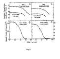
- the decrease of VSV protein in interferon (IFN)-treated cultures is correlated with inhibition of formation of infectious virions; its quantification therefore allows accurate measurement of IFN and of its antiviral effect, see Fig. 3.
- the applicability of the immunoassay for measuring the virus yield is not restricted to cells exhibiting a virus cytopathic effect.
- the decrease of virus protein is obtained at IFN concentrations lower than those that reduce cell killing by the virus.
- the immunoassay therefore provides a more sensitive measure for the IFN effect than that obtained by 'cytopathic effect inhibition' as
- the human cell lines used in this study were: HU obtained from the Hadassah University Hospital, Jerusalem, CCL-6 obtained from the American Type Culture Collection and HeLa obtained from Flow Laboratories.
- FS11 is a strain of human foreskin fibroblasts, established in our laboratory.
- Rabbits were injected intramuscularly with 0.5 mg of purified virions emulsified in complete Freund's adjuvant and 5 months later with a further 2 mg injected by the same route, as well as 0.5 mg u.v.-irradiated virions supplied intravenously. A month later, the animals were again injected intramuscularly with 0.5 mg of purified virions which had been lysed in 0.5% Nonidet@ P40(NP40), fixed in 3% formaldehyde and, after extensive dialysis, mixed with complete Freund's adjuvant. The rabbits were bled a week later. The titre of antibodies in the sera was found to be saturating in the solid-phase immunoassay at a dilution of up to 1:200.
- the virus protein was first solubilized by adding to the culture medium an equal volume of a solution of 0.5% sodium deoxycholate in 0.1 M-sodium carbonate buffer, pH 9.6. Lysis of the cells by the detergent could be accelerated by incubation at 37°C for about 20 min. Fifty pl aliquots of the solubilized protein were then transferred to V-shaped microwells in PVC radioimmunoassay plates (Dy- natech@ 1-220-25) using a 'Titertek'@, multichannel pipette.
- Immunoradiometric assay The PVC plate was incubated for 2 h in a humidified chamber at 37°C. Non-adsorbed protein was rinsed off with a solution of 0.05% Tween-20 in phosphate-buffered saline (Tween-PBS). Anti-VSV antiserum, diluted in Tween-PBS, was then applied in a volume of 20 pl to each well and the plate was incubated again for 2 h at 37°C. After rewashing with Tween-PBS, the plate was incubated for 1 h at 37°C with 125 1- labelled Protein A (20 ⁇ l, 5x 10⁶ cpm per ml of 1% bovine serum albumin in PBS). The plate was then washed with Tween-PBS and the amount of label adsorbed to each well was determined in a y-counter. Control values (usually less than 5%) were subtracted.
- IFN- ⁇ was induced by poly-(rl) poly(rC) in FS11 cells and partially purified on a DEAE-Sephadex@ column.
- IFN-a was induced in Namalwa lymphoblastoid cells by Sendai virus and partially purified by adsorption to immobilize anti-interferon antibody.
- Protein A was purchased and iodinated with Bolton-Hunter reagent or obtained in the labelled form. Tween-20 was obtained commercially.
- Fig. 1B There was a linear correlation between the amount of virus proteins that adsorbed to the plates and their concentration in the tested sample (Fig. 1B).
- This binding formed the basis for a simple immunoassay for the virus protein which involved mixing the virus-containing sample with detergent solution, transferring a sample of the mixture to a PVC plate and then quantifying the virus protein which was bound to the PVC plates by sequential incubation with anti-VSV serum and 125 I-labelled Protein A.
- the serum protein present in rather high amounts in the cell growth medium, could not fully prevent binding of virus proteins to the PVC plates.
- the amount of virus protein formed within 2 h of infection was, in cells of various human (Fig. 2), monkey (Fig. 3), bovine (Fig.
- the immunoassay for measuring the yield of virus protein provides a sensitive and quantitative tool for estimation of the antiviral effect of IFN.
- a 'cytopathic effect inhibition' assay which is now the method most widely applied for measuring the action of IFN; it gives a more direct quantification of the change induced by IFN and it is independent of the morphological response to the virus; in fact, it can even serve for estimating the effect of IFN on viruses which are not cytopathic at all, providing that antibodies against their proteins are available.
- Steps can be taken to permit sensitive detection of very low quantities of antigen either by its enrichment, using plates which have been pre-coated with antibody ('sandwich' technique) or by using a highly labelled sample of antigen (competition radioimmunoassay). Yet the large amounts of VSV proteins found in infected cells, and their effective binding to PVC, allows their quantification by a very simple, direct immunoassay. A major attractive feature of this technique is that the amount of bound antigen is linearly proportional to its concentration in the culture, over a wide range of concentrations. It is therefore not necessary to dilute the samples before the test.
- a kit by which the assay can be applied in any laboratory, which is equipped for tissue culture, will include the following components:
- the kit When using choices a) or c) the kit will also include the substrate for the enzyme, used in the ELISA, and a stopping solution which terminates the cleavage of this substrate by the enzyme.
Landscapes
- Life Sciences & Earth Sciences (AREA)
- Health & Medical Sciences (AREA)
- Engineering & Computer Science (AREA)
- Immunology (AREA)
- Chemical & Material Sciences (AREA)
- Molecular Biology (AREA)
- Urology & Nephrology (AREA)
- Hematology (AREA)
- Biomedical Technology (AREA)
- Proteomics, Peptides & Aminoacids (AREA)
- General Health & Medical Sciences (AREA)
- Microbiology (AREA)
- Virology (AREA)
- Organic Chemistry (AREA)
- Physics & Mathematics (AREA)
- Biotechnology (AREA)
- Biochemistry (AREA)
- Cell Biology (AREA)
- Analytical Chemistry (AREA)
- Zoology (AREA)
- Wood Science & Technology (AREA)
- Food Science & Technology (AREA)
- Medicinal Chemistry (AREA)
- General Physics & Mathematics (AREA)
- Pathology (AREA)
- Genetics & Genomics (AREA)
- Tropical Medicine & Parasitology (AREA)
- General Engineering & Computer Science (AREA)
- Bioinformatics & Cheminformatics (AREA)
- Biophysics (AREA)
- Measuring Or Testing Involving Enzymes Or Micro-Organisms (AREA)
- Preparation Of Compounds By Using Micro-Organisms (AREA)
Description
- The present invention relates to an assay for the detection and the quantitative determination of interferon (IFN) by measuring the yield of viral proteins in cells treated with the IFN and infected with a suitable virus, using an immunoassay (either ELISA or RIA, i.e. radioimmunoassay); and a kit for carrying out such an assay.
- The interferons (lFNs) are proteins which inhibit, specifically, the growth of viruses in cells. The 'antiviral' effect of the IFNs is exhibited by minute (10-'3M) IFN concentration and therefore most assays for IFN are based on the quantitation of this effect.
- Since the direct quantitation of virus yield by the JpJaque assay' is too laborious to be applied as an assay for IFN, a variety of alternative techniques has been developed. In most cases the virus yield is estimated indirectly by determining the extent of cell destruction by the virus.
- The technique has several severe limitations: not in all types of cells is the viral cytopathic effect clear enough to be easily estimated and many other agents, besides viruses, can increase cell death and can, therefore, interfere with this 'cytopathic effect' assay of IFN.
- Other techniques which are used for estimation of the antiviral effect are based on quantitation of various other viral functions including certain viral enzymatic activities and hemag- glutination activity of viruses. These techniques can be used only for those viruses which exhibit the measured activity and are therefore applicable only with limited cell-virus combinations.
- A more general technique which makes possible the direct quantitation of viral components, in an accurate and sensitive manner, is, therefore, necessary for a reliable determination of IFN.
- The invention relates to an assay for the determination of IFN. It is based on a simple solid-phase immunoassay for viral proteins such as those of Vesicular Stomatitis Virus (VSV), a virus which is widely used as a probe for the antiviral activity of IFN. It has been found that the proteins of this virus bind effectively to suitable supports such as polyvinyl-chloride (PVC) surfaces, even in the presence of a high level of other proteins, such as those found in the growth media used for tissue-culture. The accessibility of viral proteins to such binding can be greatly increased by lysing the virus with a suitable detergent. A simple immunoassay for the viral proteins can therefore be performed by adding a detergent to a virus infected cell culture and applying the detergent-treated solution to PVC microwells. The amount of virat proteins which binds to the PVC is directly proportional to their concentration in the culture. The bound proteins can be quantitated by incubating the microwells with anti-VSV antibody and then measuring the amount of adsorbed IgG. The decrease of VSV protein in interferon (IFN)-treated cultures is correlated with inhibition of formation of infectious virions; its quantification therefore allows accurate measurement of IFN and of its antiviral effect, see Fig. 3. The applicability of the immunoassay for measuring the virus yield is not restricted to cells exhibiting a virus cytopathic effect. Moreover, the decrease of virus protein is obtained at IFN concentrations lower than those that reduce cell killing by the virus. The immunoassay therefore provides a more sensitive measure for the IFN effect than that obtained by 'cytopathic effect inhibition' assays.
- The basic steps of the assay of the invention areas follows:
- Step I: Suitable cells, such as cultured fibroblasts are cultivated to confluency in microwells. The sample to be assayed and a sample of known interferon (IFN) concentration are diluted serially and applied to such cell cultures in microwells.
- Step II: After a predetermined incubation time, the cells are infected with a suitable virus at a suitable concentration of same. VSV virus is suitable, and good results were obtained at a concentration of 10 pfu VSV per cell. The cells are further incubated in a suitable growth medium and at the end of the incubation period they are either frozen or lysed with a suitable detergent.
- Step III: The virus is lysed by adding a lysis buffer such as sodium desoxycholate buffer at pH of about 9.6. The lysed samples can be assayed or frozen for subsequent assaying.
- Step IV: Quantitation of the viral protein yield: Samples of the lysed viruses are transferred, to radioimmunoassay or to ELISA microplates. Following incubation for 1-2 h, the plates are then rinsed with Tween@-PBS (0.05% Tween 20 in phosphate buffered saline). The quantitation is then continued, for example in one of the following ways:
- a. The wells are incubated for 1 h with anti-VSV serum, rinsed with Tween-PBS, incubated with 125I-labeled protein A (105 cpm per well) for 1 h and rinsed with Tween-PBS (Fig. 6A); or
- b. The wells are incubated with anti-VSV serum as above, rinsed and incubated either with anti-IgG antibody or with protein A which has been conjugated to alkaline phosphatase and then ELISA is completed as in (Fig. 6B) or
- c. The cells are incubated with purified anti-VSV IgG, conjugated to alkaline phosphatase, in the presence of Tween-PBS, rinsed with Tween-PBS and incubated for 1 hour with colour-producing substrate for the alkaline phosphatase, as in standard ELISA procedures (Fig. 6C). An example for the titration of IFN-y on Wish cells, using procedures A(o) and C(.) is shown in Fig. 5. The IFN concentration causing reduction of the viral protein yield by 50% is, by definition, 1 unit/ vol. Yet, as the IFN activity varies, depending on the type of cell used and its growth conditions, it is preferable to include a sample of known IFN concentration in the assay and to relate the activity of the tested samples to that exhibited by the sample of known concentration.
- This assay can be applied for measuring the effect of IFN on a variety of other viruses using the specific anti-viral antibody. The results obtained are summarized in the enclosed Figures, in which:
- Fig. 1 illustrates the binding of VSV protein to the immunoassay plate,
- Fig. 2 illustrates the titration of IFN effect of VSV protein yield in various human cell lines,
- Fig. 3 illustrates the dose response of the IFN effect of VSV protein yield and on the yield of infectious varions,
- Fig. 4 illustrates the quantitative estimation of the relationship between the effect of IFN on viral cytopathy and its effect on viral yield,
- Fig. 5 compares titration of IFN effect by measuring VSV proteins yield either by radioimmunoassays (0) or by ELISA (t). The first illustrated in Fig. 6A and the second in Fig. 6B.
- Fig. 6 illustrates alternative procedures of immunoassays of VSV proteins.
- The invention is illustrated by way of example with reference to the following detailed example, which is given by way of illustration only.
- Cells. The human cell lines used in this study were: HU obtained from the Hadassah University Hospital, Jerusalem, CCL-6 obtained from the American Type Culture Collection and HeLa obtained from Flow Laboratories. FS11 is a strain of human foreskin fibroblasts, established in our laboratory.
- In addition there were monkey BSC-1 fibroblasts, bovine MDBK cells, baby hamster kidney (BHK) cells and mouse L-929 cells. All cells were grown in Dulbecco's modified Eagle's medium in the presence of 10% foetal calf serum (FCS). VSV and antiserum against its proteins: VSV of the Indiana serotype was re-cloned several times in BSC-1 cells and used at a low passage number. Virions, produced either in BSC-1 cells or in a spinner culture of BHK cells, were purified on a sucrose gradient and on a tartrate-glycerol gradient, as previously described (Wallach & Revel, 1980). Rabbits were injected intramuscularly with 0.5 mg of purified virions emulsified in complete Freund's adjuvant and 5 months later with a further 2 mg injected by the same route, as well as 0.5 mg u.v.-irradiated virions supplied intravenously. A month later, the animals were again injected intramuscularly with 0.5 mg of purified virions which had been lysed in 0.5% Nonidet@ P40(NP40), fixed in 3% formaldehyde and, after extensive dialysis, mixed with complete Freund's adjuvant. The rabbits were bled a week later. The titre of antibodies in the sera was found to be saturating in the solid-phase immunoassay at a dilution of up to 1:200.
- Immunoassay of virus proteins and quantification of the IFN effect
- Treatment with IFN. Cells were seeded at con- centratiQns of 2.5x 10⁵ cells in 18 mm microwells of 3.50x 10⁴ cells in 9 mm microwells. A few hours later, serial dilutions of the solutions to be tested and IFN samples of known concentrations were applied to the cells.
- Infection by the virus. Twelve to twenty-fourth after application of IFN, growth medium was removed from the cells and the virus was applied in growth medium containing 2% FCS, at a multiplicity of 10 p.f.u./cells (50 pl/microwell). Several cell samples were incubated without virus and others with virus but without pretreatment by IFN. Two h after application of the virus, the virus-containing growth medium in the micro wells was replaced by a fresh medium containing 10% FCS (100 pl/microwell) and the cells were incubated for a further 10 to 20 h. The infected cultures were kept frozen before further processing for the assay of the virus proteins. Optimal duration of incubation with the virus could be estimated by observing the development of virus cytopathic effect.
- Transfer of the VSV proteins. The virus protein was first solubilized by adding to the culture medium an equal volume of a solution of 0.5% sodium deoxycholate in 0.1 M-sodium carbonate buffer, pH 9.6. Lysis of the cells by the detergent could be accelerated by incubation at 37°C for about 20 min. Fifty pl aliquots of the solubilized protein were then transferred to V-shaped microwells in PVC radioimmunoassay plates (Dy- natech@ 1-220-25) using a 'Titertek'@, multichannel pipette.
- Immunoradiometric assay. The PVC plate was incubated for 2 h in a humidified chamber at 37°C. Non-adsorbed protein was rinsed off with a solution of 0.05% Tween-20 in phosphate-buffered saline (Tween-PBS). Anti-VSV antiserum, diluted in Tween-PBS, was then applied in a volume of 20 pl to each well and the plate was incubated again for 2 h at 37°C. After rewashing with Tween-PBS, the plate was incubated for 1 h at 37°C with 1251- labelled Protein A (20 µl, 5x 10⁶ cpm per ml of 1% bovine serum albumin in PBS). The plate was then washed with Tween-PBS and the amount of label adsorbed to each well was determined in a y-counter. Control values (usually less than 5%) were subtracted.
- Definition of the unit of antiviral activity. We refer here to a 1 unit of IFN as that amount which, when present in 1 ml, gives 50% reduction in virus yield. The concentrations of IFN necessary to cause a reduction in virus yield are shown in this study to be significantly lower than those causing reduction of virus cytopathic effect. Since the latter parameter is widely used to quantify IFN, concentrations referred to as 1 U/ml in this study might be lower than in many others. Indeed, we found that the currently available international standards for both IFN-a (NIH G 023-901-527) and IFN-β (NIH G-023-902-527) cause a 50% reduction in VSV protein binding at concentrations lower than those defined by there producers as 1 U/ml (at 0.43 U/ml IFN on MDBK cells and at 0.24 U/ml IFN-a on human foreskin fibroblasts).
- Quantification of cytopathic effect by neutral red uptake. The assay was performed as described by Finter (1969) J. Gen. Virology, 5, 414-427. Materials. IFN-β was induced by poly-(rl) poly(rC) in FS11 cells and partially purified on a DEAE-Sephadex@ column. IFN-a was induced in Namalwa lymphoblastoid cells by Sendai virus and partially purified by adsorption to immobilize anti-interferon antibody. Protein A was purchased and iodinated with Bolton-Hunter reagent or obtained in the labelled form. Tween-20 was obtained commercially.
- Low binding of virus protein to PVC plates observed after incubation with cell growth medium which contained virus could be significantly enhanced by adding detergents to the virus suspensions (Fig. 1A). Comparison of several detergents suggested a correlation between an increase in binding to the PVC and the effectiveness of the detergent in dissociating the proteins of VSV. Thus, deoxycholate which causes release of both the G and M proteins of VSV and a disruption of the viral nucleocapsid (Cartwright et al., 1970 a, b, J. of Gen. Virology 7, 19-21 and 7, 267) was significantly more effective than NP40, which is known to release selectively only the G protein (Fig. 1A). Apparently, the effect of detergent on adsorption of the virus proteins to PVC is primarily a reflection of an increase in the availability of these proteins as a result of disruption of the virus membrane and ribonucleoprotein core.
- There was a linear correlation between the amount of virus proteins that adsorbed to the plates and their concentration in the tested sample (Fig. 1B). This binding formed the basis for a simple immunoassay for the virus protein which involved mixing the virus-containing sample with detergent solution, transferring a sample of the mixture to a PVC plate and then quantifying the virus protein which was bound to the PVC plates by sequential incubation with anti-VSV serum and 125I-labelled Protein A. The serum protein, present in rather high amounts in the cell growth medium, could not fully prevent binding of virus proteins to the PVC plates. The amount of virus protein formed within 2 h of infection was, in cells of various human (Fig. 2), monkey (Fig. 3), bovine (Fig. 4), and mouse cell lines (not shown) high enough to be quantified by this immunoassay. While initially this procedure was used for measuring the amount of virus protein in the growth medium of infected cells, we found it later convenient to add deoxycholate directly to the whole infected culture. The detergent lysed the cells and thus permitted measurement of the total amount of virus protein in the culture. The efficiency of binding virus protein to PVC in the presence of cell lysate appears to be lower than in its absence, probably due to competition of some cellular proteins with virus proteins, for binding sites on the PVC (Fig. 1B, compare binding with culture medium to binding with medium plus cells). This competition, however, did not interfere with the assay since even in the presence of cell lysate the binding of virus protein to PVC was linearly proportional to their concentration (Fig. 18).
- As seen in Fig. 2, cells of various lines showed substantially reduced yields of virus protein after treatment with IFN. Comparison of the decrease in virus protein with the decrease in production of infectious virions, as measured by the plaque assay, showed that both responded similarly to IFN. The minimal IFN concentration at which the antiviral effect could be detected was about the same for the decrease of virus proteins and the decrease of infectious virions (Fig. 3). Nevertheless, this comparison shows a clear advantage of the immunoassay over the plaque assay for measuring the antiviral effect at very low concentrations of IFN. The decrease of virus yield by a few percent at such IFN concentration is much simpler to quantify accurately by the immunoassay than by the plaque assay. On the other hand, the immunoassay is clearly not applicable for measuring the antiviral effect of very high concentrations of IFN since it allows the quantification of viral proteins only at virus concentrations higher than about 106 p.f.u./ml (Fig. 3).
- On examining the morphology of the infected cells it was observed that protection from killing by the virus could only be detected with IFN concentrations higher than those causing a reduction in viral protein. To confirm this observation there was quantified the viral cytopathy by measuring the neutral red uptake capacity of the cells. Comparison of the yield of viral protein and the extent of vital cytopathy showed a more effective response of the former parameter to IFN (Fig. 4). At a low concentration of IFN, effective reduction of viral protein with no increase of cell viability could be observed.
- The immunoassay for measuring the yield of virus protein provides a sensitive and quantitative tool for estimation of the antiviral effect of IFN. In several respects it is preferable to a 'cytopathic effect inhibition' assay which is now the method most widely applied for measuring the action of IFN; it gives a more direct quantification of the change induced by IFN and it is independent of the morphological response to the virus; in fact, it can even serve for estimating the effect of IFN on viruses which are not cytopathic at all, providing that antibodies against their proteins are available.
- An interesting phenomenon revealed when comparing the sensitivity of the immunoassay and of the cytopathic effect assay is a difference in the IFN concentration dependence of the reduction in virus yield and in virus cytopathic effect. This difference is probably due to the existence of excess constituents of the virus in the infected cell, beyond the minimal amount necessary to cause destruction. Another factor which might have contributed to this phenomenon is the ability of IFN to induce increased sensitivity of the cells to virus cytopathy. This difference in IFN concentration dependence implies that the immunoassay gives a more sensitive indication of the antiviral effect than that provided by the cytopathic effect inhibition assay. There are a number of different ways whereby solid-phase immunoassay can be performed. Steps can be taken to permit sensitive detection of very low quantities of antigen either by its enrichment, using plates which have been pre-coated with antibody ('sandwich' technique) or by using a highly labelled sample of antigen (competition radioimmunoassay). Yet the large amounts of VSV proteins found in infected cells, and their effective binding to PVC, allows their quantification by a very simple, direct immunoassay. A major attractive feature of this technique is that the amount of bound antigen is linearly proportional to its concentration in the culture, over a wide range of concentrations. It is therefore not necessary to dilute the samples before the test.
- Although there are known many different effects of IFN on cell functions, none is as ubiquit- ously observed and as specific to IFN as the antiviral effect.
- The following are the legends to the Figures:
- Fig. 1. Binding of VSV protein to the immunoassay plate. (a) Effect of detergents. Culture media of VSV-infected BSC-1 cells (D, 0) and non-infected cells (A, 8) were diluted 1:1 in 0.1 M-sodium carbonate buffer pH 9.6, or in that buffer plus deoxycholate (0) or NP-40 (a) to the indicated final detergent concentration and incubated in the microwells of PVC immunoassay plates for 2 h at 37°C. The plates were then washed twice with PBS and twice with Tween-PBS and the amount of bound viral proteins was estimated by sequential incubation with anti-VSV antibody and iodinated Protein A, (b) Dependence on virus concentration. Twelve h after infection of BSC-1 cells (2.5x10⁵ cells in 2 ml growth medium) an equal volume of 0.5% deoxycholate in 0.1 M-sodium carbonate buffer pH 9.6, was added either to the whole culture or just to the culture medium. The lysed samples were mixed in various proportions with samples of a similarly treated but non-infected culture and the extent of binding of the viral protein to the immunoassay plate, at the various dilutions of the virus, was determined as above, ■. Assay with culture medium; •, assay with lysate of the whole culture.
- Fig. 2. Titration of IFN; effect on VSV protein yield in various human cell lines. Cells were treated with human IFN-8 for 24 h and then infected by VSV (10 p.f.u./cell). Twelve hours later, the infected cultures were lysed by deoxycholate and the virus protein content determined by radioimmunoassay. IFN concentration is presented in terms of antiviral activity on the HeLa cells, where 1 unit is defined as the concentration causing reduction of viral protein by 50%.
- Fig. 3. Dose response of the IFN effect on VSV protein yield and on the yield of infectious virions. BSC-1 cells (a, b) were treated with IFN-a and Hela cells (c, d) were treated with IFN-8 for 24 h and then infected for 12 h with VSV. The yield of infectious virions in the medium was determined by plaque assay on L-929 cells (see Fig. 4a), and the yield of viral protein in the whole culture was determined by the radioimmunoassay. The yields of virus protein are presented in (a) and (c) on a logarithmic scale and in (b) and (d) on a linear scale, IFN concentration is presented in terms of antiviral activity in the two types of cell.
- Fig. 4. Quantitative estimation of the relationship between the effect of IFN on viral cytopathy and its effect of virus protein yield. MDBK (a) and FS11 (b) cells were treated with various concentrations of IFN-β for 20 h, infected with VSV and further incubated for 20 h until cytopathic effect had developed in those cells not treated with IFN. The yield of viral protein in the culture and the decrease of neutral red dye uptake by the cell, as an indication of cell death, were then determined in parallel. Viral protein yield (·) is expressed as a percentage of its level in the absence of IFN, and the decrease in neutral red uptake (•) as a percentage of the dye taken up by non-infected culture. IFN concentration is presented as in Fig. 2 and 3. A similar dissociation between the effect of viral cytopathy and on protein yield was observed with IFN-a (not shown).
- Fig. 5. Compares titration of IFN effect by measuring VSV proteins yield either by radioimmunoassays (0) or by ELISA (0). The first illustrated in Fig. 6A and the second in Fig. 6B.
- Fig. 6. Illustrates alternative procedures of immunoassays of VSV proteins.
- A kit by which the assay can be applied in any laboratory, which is equipped for tissue culture, will include the following components:
- 1) A lyophilized sample of virus.
- 2) Concentrated lysis buffer (sodium deoxycholate in sodium carbonate buffer).
- 3) Concentrated Tween-PBS Buffer.
- 4) One of the following alternative agents:
- a) Purified anti-VSV immunoglobulin (or monoclonal antibody against the M Protein of VSV) coupled to alkaline phosphatase or to another enzyme which is in use for ELISA; or
- b) Anti-VSV serum and 125I-labelled Protein A; or
- c) Anti-VSV serum and enzyme coupled anti-IgG serum.
- When using choices a) or c) the kit will also include the substrate for the enzyme, used in the ELISA, and a stopping solution which terminates the cleavage of this substrate by the enzyme.
- 5) Microplates for ELISA (choice a) or c)) of for radioimmunoassay (choice b).
Claims (8)
Applications Claiming Priority (2)
| Application Number | Priority Date | Filing Date | Title |
|---|---|---|---|
| IL66733 | 1982-09-07 | ||
| IL66733A IL66733A (en) | 1982-09-07 | 1982-09-07 | Assay for ifn and kit therefor |
Publications (2)
| Publication Number | Publication Date |
|---|---|
| EP0105221A1 EP0105221A1 (en) | 1984-04-11 |
| EP0105221B1 true EP0105221B1 (en) | 1987-03-25 |
Family
ID=11053747
Family Applications (1)
| Application Number | Title | Priority Date | Filing Date |
|---|---|---|---|
| EP83108837A Expired EP0105221B1 (en) | 1982-09-07 | 1983-09-07 | Process for the determination of interferon and kit for the assay of interferon |
Country Status (8)
| Country | Link |
|---|---|
| US (1) | US4622292A (en) |
| EP (1) | EP0105221B1 (en) |
| JP (1) | JPS5965765A (en) |
| AU (1) | AU572790B2 (en) |
| CA (1) | CA1223811A (en) |
| DE (2) | DE3370543D1 (en) |
| IL (1) | IL66733A (en) |
| ZA (1) | ZA836488B (en) |
Families Citing this family (10)
| Publication number | Priority date | Publication date | Assignee | Title |
|---|---|---|---|---|
| US4675282A (en) * | 1984-07-06 | 1987-06-23 | Damon Biotech, Inc. | Assay for interferon epsilon |
| CA1317860C (en) * | 1987-04-01 | 1993-05-18 | Daniel Louis Kacian | Techniques for preparing specimens for bacterial assays |
| US7007179B2 (en) * | 2001-02-08 | 2006-02-28 | Honeywell International Inc. | Electric load management center |
| US7110562B1 (en) | 2001-08-10 | 2006-09-19 | Hear-Wear Technologies, Llc | BTE/CIC auditory device and modular connector system therefor |
| US7139404B2 (en) | 2001-08-10 | 2006-11-21 | Hear-Wear Technologies, Llc | BTE/CIC auditory device and modular connector system therefor |
| CN103755800B (en) | 2004-02-02 | 2016-02-24 | Ambrx公司 | Modified human interferon polypeptides and its purposes |
| NZ602170A (en) | 2008-02-08 | 2014-03-28 | Ambrx Inc | Modified leptin polypeptides and their uses |
| EP2568289A3 (en) | 2011-09-12 | 2013-04-03 | International AIDS Vaccine Initiative | Immunoselection of recombinant vesicular stomatitis virus expressing hiv-1 proteins by broadly neutralizing antibodies |
| EP2586461A1 (en) | 2011-10-27 | 2013-05-01 | Christopher L. Parks | Viral particles derived from an enveloped virus |
| US9347065B2 (en) | 2012-03-29 | 2016-05-24 | International Aids Vaccine Initiative | Methods to improve vector expression and genetic stability |
Family Cites Families (3)
| Publication number | Priority date | Publication date | Assignee | Title |
|---|---|---|---|---|
| US3910824A (en) * | 1973-10-18 | 1975-10-07 | Searle & Co | Interferon assay |
| DE2941575A1 (en) * | 1978-10-19 | 1980-04-30 | Harvard College | METHOD FOR IDENTIFYING A VIRUS INFECTION AND KIT THEREFOR |
| EP0064063B1 (en) * | 1980-11-07 | 1985-09-11 | Celltech Limited | Assay for interferon |
-
1982
- 1982-09-07 IL IL66733A patent/IL66733A/en not_active IP Right Cessation
-
1983
- 1983-08-22 US US06/525,618 patent/US4622292A/en not_active Expired - Lifetime
- 1983-09-01 ZA ZA836488A patent/ZA836488B/en unknown
- 1983-09-05 AU AU18693/83A patent/AU572790B2/en not_active Expired
- 1983-09-06 CA CA000436076A patent/CA1223811A/en not_active Expired
- 1983-09-07 EP EP83108837A patent/EP0105221B1/en not_active Expired
- 1983-09-07 JP JP58164924A patent/JPS5965765A/en active Granted
- 1983-09-07 DE DE8383108837T patent/DE3370543D1/en not_active Expired
- 1983-09-07 DE DE198383108837T patent/DE105221T1/en active Pending
Non-Patent Citations (1)
| Title |
|---|
| NATIONAL TECHNICAL INFORMATION SERVICE, U.S. Department of Commerce "Immunoassay utilizing I-125-labeled protein A", ref. PB80-981270 * |
Also Published As
| Publication number | Publication date |
|---|---|
| CA1223811A (en) | 1987-07-07 |
| AU572790B2 (en) | 1988-05-19 |
| JPS5965765A (en) | 1984-04-14 |
| US4622292A (en) | 1986-11-11 |
| DE105221T1 (en) | 1985-02-14 |
| IL66733A0 (en) | 1982-12-31 |
| AU1869383A (en) | 1984-03-15 |
| EP0105221A1 (en) | 1984-04-11 |
| IL66733A (en) | 1986-03-31 |
| JPH0331389B2 (en) | 1991-05-02 |
| ZA836488B (en) | 1984-04-25 |
| DE3370543D1 (en) | 1987-04-30 |
Similar Documents
| Publication | Publication Date | Title |
|---|---|---|
| Landegren | Measurement of cell numbers by means of the endogenous enzyme hexosaminidase. Applications to detection of lymphokines and cell surface antigens | |
| Grauballe et al. | Optimized enzyme‐linked immunosorbent assay for detection of human and bovine rotavirus in stools: Comparison with electron‐microscopy, immunoelectro‐osmophoresis, and fluorescent antibody techniques | |
| Fujinami et al. | Alterations in expression of measles virus polypeptides by antibody: molecular events in antibody-induced antigenic modulation. | |
| Stephenson et al. | Feline oncornavirus-associated cell-membrane antigen (FOCMA): distinction between FOCMA and the major virion glycoprotein. | |
| Arstila et al. | A solid-phase radioimmunoassay for IgG and IgM antibodies against measles virus | |
| US4622292A (en) | Assay and kit for determining interferon | |
| KR100730529B1 (en) | Method for detection or determination of hcv core antigens and reagent for detection or determination for use therein | |
| FI110723B (en) | Process for the preparation of immunoglobulin for the treatment of respiratory tract infections | |
| Davey et al. | Laboratory Methods in the Diagnosis and Prognostic Staging of infection with Human Immunodificiency Virus Type 1 | |
| Al‐Fawaz et al. | Hepatitis E virus infection in patients from Saudi Arabia with sickle cell anaemia and β‐thalassemia major: possible transmission by blood transfusion | |
| Mellors et al. | Type C RNA virus-specific antibody in human systemic lupus erythematosus demonstrated by enzymoimmunoassay. | |
| Archambault et al. | Development of an enzyme-linked immunosorbent assay for caprine arthritis-encephalitis virus | |
| McCarron et al. | Hepatitis C antibody detection in dried blood spots | |
| McHugh et al. | The sensitive detection and quantitation of antibody to HVC by using a microsphere‐based immunoassay and flow cytometry | |
| Pronovost et al. | Sensitive chemiluminescent enzyme-linked immunosorbent assay for quantification of human immunoglobulin G and detection of herpes simplex virus | |
| Pringle et al. | Murine pneumonia virus: seroepidemiological evidence of widespread human infection | |
| Chiodi et al. | Measles IgM antibodies in cerebrospinal fluid and serum in subacute sclerosing panencephalitis | |
| Vuorinen et al. | Enzyme immunoassays for detection of IgG and IgM antibodies to parainfluenza types 1, 2 and 3 | |
| Simm et al. | Storage conditions for serum for estimating prostate-specific antigen | |
| Chang et al. | Biotin-labeled antigen sandwich enzyme-linked immunosorbent assay (BLA-S-ELISA) for the detection of Japanese encephalitis antibody in human and a variety of animal sera | |
| EP0234941A2 (en) | Competitive elisa for the detection of antibodies | |
| Hermodsson et al. | Bioimmunoassays (BIAs) of human interferon | |
| van Der Meide et al. | A sensitive two-site enzyme immunoassay for the detection of rat interferon-γ in biological fluids | |
| Hierholzer et al. | Quantitation of antibody to non-hemagglutinating viruses by single radial hemolysis: Serological test for human coronaviruses | |
| KR100454770B1 (en) | How to detect human parvovirus and reagents for it |
Legal Events
| Date | Code | Title | Description |
|---|---|---|---|
| PUAI | Public reference made under article 153(3) epc to a published international application that has entered the european phase |
Free format text: ORIGINAL CODE: 0009012 |
|
| AK | Designated contracting states |
Designated state(s): BE CH DE FR GB IT LI NL SE |
|
| 17P | Request for examination filed |
Effective date: 19840828 |
|
| ITCL | It: translation for ep claims filed |
Representative=s name: FIAMMENGHI FIAMMENGHI RACHELI |
|
| TCNL | Nl: translation of patent claims filed | ||
| EL | Fr: translation of claims filed | ||
| DET | De: translation of patent claims | ||
| ITF | It: translation for a ep patent filed | ||
| GRAA | (expected) grant |
Free format text: ORIGINAL CODE: 0009210 |
|
| AK | Designated contracting states |
Kind code of ref document: B1 Designated state(s): BE CH DE FR GB IT LI NL SE |
|
| REF | Corresponds to: |
Ref document number: 3370543 Country of ref document: DE Date of ref document: 19870430 |
|
| ET | Fr: translation filed | ||
| PLBE | No opposition filed within time limit |
Free format text: ORIGINAL CODE: 0009261 |
|
| STAA | Information on the status of an ep patent application or granted ep patent |
Free format text: STATUS: NO OPPOSITION FILED WITHIN TIME LIMIT |
|
| 26N | No opposition filed | ||
| ITTA | It: last paid annual fee | ||
| EAL | Se: european patent in force in sweden |
Ref document number: 83108837.2 |
|
| REG | Reference to a national code |
Ref country code: GB Ref legal event code: IF02 |
|
| PGFP | Annual fee paid to national office [announced via postgrant information from national office to epo] |
Ref country code: SE Payment date: 20020904 Year of fee payment: 20 Ref country code: GB Payment date: 20020904 Year of fee payment: 20 |
|
| PGFP | Annual fee paid to national office [announced via postgrant information from national office to epo] |
Ref country code: FR Payment date: 20020910 Year of fee payment: 20 |
|
| PGFP | Annual fee paid to national office [announced via postgrant information from national office to epo] |
Ref country code: DE Payment date: 20020911 Year of fee payment: 20 |
|
| PGFP | Annual fee paid to national office [announced via postgrant information from national office to epo] |
Ref country code: CH Payment date: 20020916 Year of fee payment: 20 |
|
| PGFP | Annual fee paid to national office [announced via postgrant information from national office to epo] |
Ref country code: NL Payment date: 20020930 Year of fee payment: 20 |
|
| PGFP | Annual fee paid to national office [announced via postgrant information from national office to epo] |
Ref country code: BE Payment date: 20021129 Year of fee payment: 20 |
|
| PG25 | Lapsed in a contracting state [announced via postgrant information from national office to epo] |
Ref country code: LI Free format text: LAPSE BECAUSE OF EXPIRATION OF PROTECTION Effective date: 20030906 Ref country code: GB Free format text: LAPSE BECAUSE OF EXPIRATION OF PROTECTION Effective date: 20030906 Ref country code: CH Free format text: LAPSE BECAUSE OF EXPIRATION OF PROTECTION Effective date: 20030906 |
|
| PG25 | Lapsed in a contracting state [announced via postgrant information from national office to epo] |
Ref country code: NL Free format text: LAPSE BECAUSE OF EXPIRATION OF PROTECTION Effective date: 20030907 |
|
| BE20 | Be: patent expired |
Owner name: *YEDA RESEARCH AND DEVELOPMENT CY LTD. Effective date: 20030907 |
|
| REG | Reference to a national code |
Ref country code: CH Ref legal event code: PL |
|
| REG | Reference to a national code |
Ref country code: GB Ref legal event code: PE20 |
|
| NLV7 | Nl: ceased due to reaching the maximum lifetime of a patent |
Effective date: 20030907 |
|
| EUG | Se: european patent has lapsed |