EP0016800B1 - Determination of proteolytic enzymes in body fluids and fluorogenic substrates - Google Patents
Determination of proteolytic enzymes in body fluids and fluorogenic substrates Download PDFInfo
- Publication number
- EP0016800B1 EP0016800B1 EP79900842A EP79900842A EP0016800B1 EP 0016800 B1 EP0016800 B1 EP 0016800B1 EP 79900842 A EP79900842 A EP 79900842A EP 79900842 A EP79900842 A EP 79900842A EP 0016800 B1 EP0016800 B1 EP 0016800B1
- Authority
- EP
- European Patent Office
- Prior art keywords
- hydrogen
- wavelength
- substrate
- enzymes
- fluorogenic
- Prior art date
- Legal status (The legal status is an assumption and is not a legal conclusion. Google has not performed a legal analysis and makes no representation as to the accuracy of the status listed.)
- Expired
Links
- 239000007850 fluorescent dye Substances 0.000 title claims abstract description 12
- 210000001124 body fluid Anatomy 0.000 title claims description 11
- 239000010839 body fluid Substances 0.000 title claims description 11
- 108091005804 Peptidases Proteins 0.000 title abstract description 14
- 102000035195 Peptidases Human genes 0.000 title abstract description 12
- 229940024999 proteolytic enzymes for treatment of wounds and ulcers Drugs 0.000 title abstract description 10
- 239000000758 substrate Substances 0.000 claims abstract description 26
- 108090000190 Thrombin Proteins 0.000 claims abstract description 19
- 229960004072 thrombin Drugs 0.000 claims abstract description 18
- 150000001875 compounds Chemical class 0.000 claims abstract description 10
- 229940012957 plasmin Drugs 0.000 claims abstract description 9
- 239000002253 acid Substances 0.000 claims abstract description 8
- 125000003236 benzoyl group Chemical group [H]C1=C([H])C([H])=C(C([H])=C1[H])C(*)=O 0.000 claims abstract description 7
- -1 glutaryl Chemical group 0.000 claims abstract description 7
- 125000003170 phenylsulfonyl group Chemical group C1(=CC=CC=C1)S(=O)(=O)* 0.000 claims abstract description 7
- 150000003839 salts Chemical class 0.000 claims abstract description 7
- GNIDSOFZAKMQAO-SECBINFHSA-N (2r)-3-hydroxy-2-(phenylmethoxycarbonylamino)propanoic acid Chemical compound OC[C@H](C(O)=O)NC(=O)OCC1=CC=CC=C1 GNIDSOFZAKMQAO-SECBINFHSA-N 0.000 claims abstract description 6
- GNIDSOFZAKMQAO-VIFPVBQESA-N (2s)-3-hydroxy-2-(phenylmethoxycarbonylamino)propanoic acid Chemical compound OC[C@@H](C(O)=O)NC(=O)OCC1=CC=CC=C1 GNIDSOFZAKMQAO-VIFPVBQESA-N 0.000 claims abstract description 6
- 108090000631 Trypsin Proteins 0.000 claims abstract description 4
- 102000004142 Trypsin Human genes 0.000 claims abstract description 4
- 239000012588 trypsin Substances 0.000 claims abstract description 4
- 239000012530 fluid Substances 0.000 claims description 16
- 238000000034 method Methods 0.000 claims description 15
- 230000005284 excitation Effects 0.000 claims description 10
- 238000002156 mixing Methods 0.000 claims description 5
- UPSFMJHZUCSEHU-JYGUBCOQSA-N n-[(2s,3r,4r,5s,6r)-2-[(2r,3s,4r,5r,6s)-5-acetamido-4-hydroxy-2-(hydroxymethyl)-6-(4-methyl-2-oxochromen-7-yl)oxyoxan-3-yl]oxy-4,5-dihydroxy-6-(hydroxymethyl)oxan-3-yl]acetamide Chemical compound CC(=O)N[C@@H]1[C@@H](O)[C@H](O)[C@@H](CO)O[C@H]1O[C@H]1[C@H](O)[C@@H](NC(C)=O)[C@H](OC=2C=C3OC(=O)C=C(C)C3=CC=2)O[C@@H]1CO UPSFMJHZUCSEHU-JYGUBCOQSA-N 0.000 claims description 5
- 108090000790 Enzymes Proteins 0.000 abstract description 23
- 102000004190 Enzymes Human genes 0.000 abstract description 23
- 229940088598 enzyme Drugs 0.000 abstract description 23
- 230000000694 effects Effects 0.000 abstract description 14
- 108010088842 Fibrinolysin Proteins 0.000 abstract description 9
- 239000001257 hydrogen Substances 0.000 abstract description 5
- 229910052739 hydrogen Inorganic materials 0.000 abstract description 5
- 125000004435 hydrogen atom Chemical group [H]* 0.000 abstract description 3
- HQABUPZFAYXKJW-UHFFFAOYSA-N butan-1-amine Chemical compound CCCCN HQABUPZFAYXKJW-UHFFFAOYSA-N 0.000 abstract 2
- XBDQKXXYIPTUBI-UHFFFAOYSA-N dimethylselenoniopropionate Natural products CCC(O)=O XBDQKXXYIPTUBI-UHFFFAOYSA-N 0.000 abstract 2
- 102000004225 Cathepsin B Human genes 0.000 abstract 1
- 108090000712 Cathepsin B Proteins 0.000 abstract 1
- 108060005987 Kallikrein Proteins 0.000 abstract 1
- 102000001399 Kallikrein Human genes 0.000 abstract 1
- 108010067372 Pancreatic elastase Proteins 0.000 abstract 1
- 102000016387 Pancreatic elastase Human genes 0.000 abstract 1
- 108090000435 Urokinase-type plasminogen activator Proteins 0.000 abstract 1
- 102000003990 Urokinase-type plasminogen activator Human genes 0.000 abstract 1
- 125000002496 methyl group Chemical group [H]C([H])([H])* 0.000 abstract 1
- 125000001997 phenyl group Chemical group [H]C1=C([H])C([H])=C(*)C([H])=C1[H] 0.000 abstract 1
- 235000019260 propionic acid Nutrition 0.000 abstract 1
- IUVKMZGDUIUOCP-BTNSXGMBSA-N quinbolone Chemical compound O([C@H]1CC[C@H]2[C@H]3[C@@H]([C@]4(C=CC(=O)C=C4CC3)C)CC[C@@]21C)C1=CCCC1 IUVKMZGDUIUOCP-BTNSXGMBSA-N 0.000 abstract 1
- 229960005356 urokinase Drugs 0.000 abstract 1
- 238000003556 assay Methods 0.000 description 22
- 229920000669 heparin Polymers 0.000 description 19
- 210000002381 plasma Anatomy 0.000 description 19
- HTTJABKRGRZYRN-UHFFFAOYSA-N Heparin Chemical compound OC1C(NC(=O)C)C(O)OC(COS(O)(=O)=O)C1OC1C(OS(O)(=O)=O)C(O)C(OC2C(C(OS(O)(=O)=O)C(OC3C(C(O)C(O)C(O3)C(O)=O)OS(O)(=O)=O)C(CO)O2)NS(O)(=O)=O)C(C(O)=O)O1 HTTJABKRGRZYRN-UHFFFAOYSA-N 0.000 description 18
- 229960002897 heparin Drugs 0.000 description 18
- SJRJJKPEHAURKC-UHFFFAOYSA-N N-Methylmorpholine Chemical compound CN1CCOCC1 SJRJJKPEHAURKC-UHFFFAOYSA-N 0.000 description 15
- 239000000243 solution Substances 0.000 description 14
- LFQSCWFLJHTTHZ-UHFFFAOYSA-N Ethanol Chemical compound CCO LFQSCWFLJHTTHZ-UHFFFAOYSA-N 0.000 description 12
- 102000013566 Plasminogen Human genes 0.000 description 11
- 108010051456 Plasminogen Proteins 0.000 description 11
- ZMXDDKWLCZADIW-UHFFFAOYSA-N N,N-Dimethylformamide Chemical compound CN(C)C=O ZMXDDKWLCZADIW-UHFFFAOYSA-N 0.000 description 9
- QTBSBXVTEAMEQO-UHFFFAOYSA-N Acetic acid Chemical compound CC(O)=O QTBSBXVTEAMEQO-UHFFFAOYSA-N 0.000 description 8
- DHMQDGOQFOQNFH-UHFFFAOYSA-N Glycine Chemical compound NCC(O)=O DHMQDGOQFOQNFH-UHFFFAOYSA-N 0.000 description 8
- 239000000203 mixture Substances 0.000 description 8
- KDLHZDBZIXYQEI-UHFFFAOYSA-N Palladium Chemical compound [Pd] KDLHZDBZIXYQEI-UHFFFAOYSA-N 0.000 description 7
- 239000000523 sample Substances 0.000 description 7
- 239000002904 solvent Substances 0.000 description 7
- 238000012360 testing method Methods 0.000 description 7
- 206010053567 Coagulopathies Diseases 0.000 description 6
- 210000004369 blood Anatomy 0.000 description 6
- 239000008280 blood Substances 0.000 description 6
- 230000035602 clotting Effects 0.000 description 6
- 238000006243 chemical reaction Methods 0.000 description 5
- 239000003112 inhibitor Substances 0.000 description 5
- 238000005259 measurement Methods 0.000 description 5
- 108090000765 processed proteins & peptides Proteins 0.000 description 5
- 239000000047 product Substances 0.000 description 5
- HEDRZPFGACZZDS-UHFFFAOYSA-N Chloroform Chemical compound ClC(Cl)Cl HEDRZPFGACZZDS-UHFFFAOYSA-N 0.000 description 4
- 239000004471 Glycine Substances 0.000 description 4
- 108010023197 Streptokinase Proteins 0.000 description 4
- WYURNTSHIVDZCO-UHFFFAOYSA-N Tetrahydrofuran Chemical compound C1CCOC1 WYURNTSHIVDZCO-UHFFFAOYSA-N 0.000 description 4
- 230000023555 blood coagulation Effects 0.000 description 4
- DEKPYXUDJRABNK-UHFFFAOYSA-N dimethyl 5-aminobenzene-1,3-dicarboxylate Chemical group COC(=O)C1=CC(N)=CC(C(=O)OC)=C1 DEKPYXUDJRABNK-UHFFFAOYSA-N 0.000 description 4
- 239000000463 material Substances 0.000 description 4
- 229960005202 streptokinase Drugs 0.000 description 4
- WEQJQNWXCSUVMA-NEPJUHHUSA-N (2s)-1-[(2r)-2-azaniumyl-3-phenylpropanoyl]pyrrolidine-2-carboxylate Chemical compound C([C@@H](N)C(=O)N1[C@@H](CCC1)C(O)=O)C1=CC=CC=C1 WEQJQNWXCSUVMA-NEPJUHHUSA-N 0.000 description 3
- YOETUEMZNOLGDB-UHFFFAOYSA-N 2-methylpropyl carbonochloridate Chemical compound CC(C)COC(Cl)=O YOETUEMZNOLGDB-UHFFFAOYSA-N 0.000 description 3
- XEKOWRVHYACXOJ-UHFFFAOYSA-N Ethyl acetate Chemical compound CCOC(C)=O XEKOWRVHYACXOJ-UHFFFAOYSA-N 0.000 description 3
- PEDCQBHIVMGVHV-UHFFFAOYSA-N Glycerine Chemical compound OCC(O)CO PEDCQBHIVMGVHV-UHFFFAOYSA-N 0.000 description 3
- UFHFLCQGNIYNRP-UHFFFAOYSA-N Hydrogen Chemical compound [H][H] UFHFLCQGNIYNRP-UHFFFAOYSA-N 0.000 description 3
- HEMHJVSKTPXQMS-UHFFFAOYSA-M Sodium hydroxide Chemical compound [OH-].[Na+] HEMHJVSKTPXQMS-UHFFFAOYSA-M 0.000 description 3
- 230000004913 activation Effects 0.000 description 3
- 239000012131 assay buffer Substances 0.000 description 3
- 230000015572 biosynthetic process Effects 0.000 description 3
- 239000000872 buffer Substances 0.000 description 3
- 230000000963 caseinolytic effect Effects 0.000 description 3
- 239000003153 chemical reaction reagent Substances 0.000 description 3
- 235000019441 ethanol Nutrition 0.000 description 3
- 238000001914 filtration Methods 0.000 description 3
- 230000036541 health Effects 0.000 description 3
- 210000004072 lung Anatomy 0.000 description 3
- 108010088904 phenylalanylproline Proteins 0.000 description 3
- 238000011533 pre-incubation Methods 0.000 description 3
- 230000008569 process Effects 0.000 description 3
- 230000002797 proteolythic effect Effects 0.000 description 3
- 230000035945 sensitivity Effects 0.000 description 3
- SFKZPTYRENGBTJ-UHFFFAOYSA-N 4-methoxynaphthalen-2-amine Chemical class C1=CC=C2C(OC)=CC(N)=CC2=C1 SFKZPTYRENGBTJ-UHFFFAOYSA-N 0.000 description 2
- 102000009123 Fibrin Human genes 0.000 description 2
- 108010073385 Fibrin Proteins 0.000 description 2
- BWGVNKXGVNDBDI-UHFFFAOYSA-N Fibrin monomer Chemical compound CNC(=O)CNC(=O)CN BWGVNKXGVNDBDI-UHFFFAOYSA-N 0.000 description 2
- FXHOOIRPVKKKFG-UHFFFAOYSA-N N,N-Dimethylacetamide Chemical compound CN(C)C(C)=O FXHOOIRPVKKKFG-UHFFFAOYSA-N 0.000 description 2
- 239000004365 Protease Substances 0.000 description 2
- 108010094028 Prothrombin Proteins 0.000 description 2
- 102100027378 Prothrombin Human genes 0.000 description 2
- 102100037486 Reverse transcriptase/ribonuclease H Human genes 0.000 description 2
- VYPSYNLAJGMNEJ-UHFFFAOYSA-N Silicium dioxide Chemical compound O=[Si]=O VYPSYNLAJGMNEJ-UHFFFAOYSA-N 0.000 description 2
- 239000007983 Tris buffer Substances 0.000 description 2
- 238000009825 accumulation Methods 0.000 description 2
- 159000000021 acetate salts Chemical class 0.000 description 2
- 239000003146 anticoagulant agent Substances 0.000 description 2
- 229940127219 anticoagulant drug Drugs 0.000 description 2
- 239000000987 azo dye Substances 0.000 description 2
- 235000015278 beef Nutrition 0.000 description 2
- 239000003480 eluent Substances 0.000 description 2
- 238000001704 evaporation Methods 0.000 description 2
- 238000002474 experimental method Methods 0.000 description 2
- 229950003499 fibrin Drugs 0.000 description 2
- 230000000951 immunodiffusion Effects 0.000 description 2
- FEMOMIGRRWSMCU-UHFFFAOYSA-N ninhydrin Chemical compound C1=CC=C2C(=O)C(O)(O)C(=O)C2=C1 FEMOMIGRRWSMCU-UHFFFAOYSA-N 0.000 description 2
- 238000002360 preparation method Methods 0.000 description 2
- 108090000623 proteins and genes Proteins 0.000 description 2
- 102000004169 proteins and genes Human genes 0.000 description 2
- 229940039716 prothrombin Drugs 0.000 description 2
- 238000007430 reference method Methods 0.000 description 2
- 239000000741 silica gel Substances 0.000 description 2
- 229910002027 silica gel Inorganic materials 0.000 description 2
- 239000001509 sodium citrate Substances 0.000 description 2
- 239000007787 solid Substances 0.000 description 2
- 238000002798 spectrophotometry method Methods 0.000 description 2
- YLQBMQCUIZJEEH-UHFFFAOYSA-N tetrahydrofuran Natural products C=1C=COC=1 YLQBMQCUIZJEEH-UHFFFAOYSA-N 0.000 description 2
- LENZDBCJOHFCAS-UHFFFAOYSA-N tris Chemical compound OCC(N)(CO)CO LENZDBCJOHFCAS-UHFFFAOYSA-N 0.000 description 2
- HRXKRNGNAMMEHJ-UHFFFAOYSA-K trisodium citrate Chemical compound [Na+].[Na+].[Na+].[O-]C(=O)CC(O)(CC([O-])=O)C([O-])=O HRXKRNGNAMMEHJ-UHFFFAOYSA-K 0.000 description 2
- 229940038773 trisodium citrate Drugs 0.000 description 2
- 229960001322 trypsin Drugs 0.000 description 2
- PGOHTUIFYSHAQG-LJSDBVFPSA-N (2S)-6-amino-2-[[(2S)-5-amino-2-[[(2S)-2-[[(2S)-2-[[(2S)-2-[[(2S)-4-amino-2-[[(2S)-2-[[(2S)-2-[[(2S)-2-[[(2S)-2-[[(2S)-5-amino-2-[[(2S)-5-amino-2-[[(2S)-2-[[(2S)-2-[[(2S)-2-[[(2S,3R)-2-[[(2S)-5-amino-2-[[(2S)-2-[[(2S)-2-[[(2S,3R)-2-[[(2S)-2-[[(2S)-2-[[(2S)-2-[[(2S)-2-[[(2S)-5-amino-2-[[(2S)-1-[(2S,3R)-2-[[(2S)-2-[[(2S)-2-[[(2R)-2-[[(2S)-2-[[(2S)-2-[[2-[[(2S)-2-[[(2S)-2-[[(2S)-2-[[(2S)-1-[(2S)-2-[[(2S)-2-[[(2S)-2-[[(2S)-2-amino-4-methylsulfanylbutanoyl]amino]-3-(1H-indol-3-yl)propanoyl]amino]-5-carbamimidamidopentanoyl]amino]propanoyl]pyrrolidine-2-carbonyl]amino]-3-methylbutanoyl]amino]-4-methylpentanoyl]amino]-4-methylpentanoyl]amino]acetyl]amino]-3-hydroxypropanoyl]amino]-4-methylpentanoyl]amino]-3-sulfanylpropanoyl]amino]-4-methylsulfanylbutanoyl]amino]-5-carbamimidamidopentanoyl]amino]-3-hydroxybutanoyl]pyrrolidine-2-carbonyl]amino]-5-oxopentanoyl]amino]-3-hydroxypropanoyl]amino]-3-hydroxypropanoyl]amino]-3-(1H-imidazol-5-yl)propanoyl]amino]-4-methylpentanoyl]amino]-3-hydroxybutanoyl]amino]-3-(1H-indol-3-yl)propanoyl]amino]-5-carbamimidamidopentanoyl]amino]-5-oxopentanoyl]amino]-3-hydroxybutanoyl]amino]-3-hydroxypropanoyl]amino]-3-carboxypropanoyl]amino]-3-hydroxypropanoyl]amino]-5-oxopentanoyl]amino]-5-oxopentanoyl]amino]-3-phenylpropanoyl]amino]-5-carbamimidamidopentanoyl]amino]-3-methylbutanoyl]amino]-4-methylpentanoyl]amino]-4-oxobutanoyl]amino]-5-carbamimidamidopentanoyl]amino]-3-(1H-indol-3-yl)propanoyl]amino]-4-carboxybutanoyl]amino]-5-oxopentanoyl]amino]hexanoic acid Chemical compound CSCC[C@H](N)C(=O)N[C@@H](Cc1c[nH]c2ccccc12)C(=O)N[C@@H](CCCNC(N)=N)C(=O)N[C@@H](C)C(=O)N1CCC[C@H]1C(=O)N[C@@H](C(C)C)C(=O)N[C@@H](CC(C)C)C(=O)N[C@@H](CC(C)C)C(=O)NCC(=O)N[C@@H](CO)C(=O)N[C@@H](CC(C)C)C(=O)N[C@@H](CS)C(=O)N[C@@H](CCSC)C(=O)N[C@@H](CCCNC(N)=N)C(=O)N[C@@H]([C@@H](C)O)C(=O)N1CCC[C@H]1C(=O)N[C@@H](CCC(N)=O)C(=O)N[C@@H](CO)C(=O)N[C@@H](CO)C(=O)N[C@@H](Cc1cnc[nH]1)C(=O)N[C@@H](CC(C)C)C(=O)N[C@@H]([C@@H](C)O)C(=O)N[C@@H](Cc1c[nH]c2ccccc12)C(=O)N[C@@H](CCCNC(N)=N)C(=O)N[C@@H](CCC(N)=O)C(=O)N[C@@H]([C@@H](C)O)C(=O)N[C@@H](CO)C(=O)N[C@@H](CC(O)=O)C(=O)N[C@@H](CO)C(=O)N[C@@H](CCC(N)=O)C(=O)N[C@@H](CCC(N)=O)C(=O)N[C@@H](Cc1ccccc1)C(=O)N[C@@H](CCCNC(N)=N)C(=O)N[C@@H](C(C)C)C(=O)N[C@@H](CC(C)C)C(=O)N[C@@H](CC(N)=O)C(=O)N[C@@H](CCCNC(N)=N)C(=O)N[C@@H](Cc1c[nH]c2ccccc12)C(=O)N[C@@H](CCC(O)=O)C(=O)N[C@@H](CCC(N)=O)C(=O)N[C@@H](CCCCN)C(O)=O PGOHTUIFYSHAQG-LJSDBVFPSA-N 0.000 description 1
- RRONHWAVOYADJL-OAHLLOKOSA-N (2r)-3-phenyl-2-(phenylmethoxycarbonylamino)propanoic acid Chemical compound C([C@H](C(=O)O)NC(=O)OCC=1C=CC=CC=1)C1=CC=CC=C1 RRONHWAVOYADJL-OAHLLOKOSA-N 0.000 description 1
- JXGVXCZADZNAMJ-NSHDSACASA-N (2s)-1-phenylmethoxycarbonylpyrrolidine-2-carboxylic acid Chemical compound OC(=O)[C@@H]1CCCN1C(=O)OCC1=CC=CC=C1 JXGVXCZADZNAMJ-NSHDSACASA-N 0.000 description 1
- IEQAICDLOKRSRL-UHFFFAOYSA-N 2-[2-[2-[2-[2-[2-[2-[2-[2-[2-[2-[2-[2-[2-[2-[2-[2-[2-[2-[2-[2-[2-(2-dodecoxyethoxy)ethoxy]ethoxy]ethoxy]ethoxy]ethoxy]ethoxy]ethoxy]ethoxy]ethoxy]ethoxy]ethoxy]ethoxy]ethoxy]ethoxy]ethoxy]ethoxy]ethoxy]ethoxy]ethoxy]ethoxy]ethoxy]ethanol Chemical compound CCCCCCCCCCCCOCCOCCOCCOCCOCCOCCOCCOCCOCCOCCOCCOCCOCCOCCOCCOCCOCCOCCOCCOCCOCCOCCOCCO IEQAICDLOKRSRL-UHFFFAOYSA-N 0.000 description 1
- TYMLOMAKGOJONV-UHFFFAOYSA-N 4-nitroaniline Chemical compound NC1=CC=C([N+]([O-])=O)C=C1 TYMLOMAKGOJONV-UHFFFAOYSA-N 0.000 description 1
- 102000004411 Antithrombin III Human genes 0.000 description 1
- 108090000935 Antithrombin III Proteins 0.000 description 1
- 239000004475 Arginine Substances 0.000 description 1
- 241000283690 Bos taurus Species 0.000 description 1
- 108091003079 Bovine Serum Albumin Proteins 0.000 description 1
- FERIUCNNQQJTOY-UHFFFAOYSA-N Butyric acid Natural products CCCC(O)=O FERIUCNNQQJTOY-UHFFFAOYSA-N 0.000 description 1
- 0 CC(C(NC(C)C(Nc1cc(C(OC)=O)cc(*)c1)=O)=O)NC(C(*)N*)=O Chemical compound CC(C(NC(C)C(Nc1cc(C(OC)=O)cc(*)c1)=O)=O)NC(C(*)N*)=O 0.000 description 1
- 108010014173 Factor X Proteins 0.000 description 1
- 108010049003 Fibrinogen Proteins 0.000 description 1
- 102000008946 Fibrinogen Human genes 0.000 description 1
- 108090001030 Lipoproteins Proteins 0.000 description 1
- 102000004895 Lipoproteins Human genes 0.000 description 1
- KDXKERNSBIXSRK-UHFFFAOYSA-N Lysine Natural products NCCCCC(N)C(O)=O KDXKERNSBIXSRK-UHFFFAOYSA-N 0.000 description 1
- 239000004472 Lysine Substances 0.000 description 1
- 241000124008 Mammalia Species 0.000 description 1
- 241001465754 Metazoa Species 0.000 description 1
- UIIMBOGNXHQVGW-UHFFFAOYSA-M Sodium bicarbonate Chemical class [Na+].OC([O-])=O UIIMBOGNXHQVGW-UHFFFAOYSA-M 0.000 description 1
- 108010086613 Streptodornase and Streptokinase Proteins 0.000 description 1
- 108010000499 Thromboplastin Proteins 0.000 description 1
- 102000002262 Thromboplastin Human genes 0.000 description 1
- 230000002159 abnormal effect Effects 0.000 description 1
- 125000002252 acyl group Chemical group 0.000 description 1
- 230000002411 adverse Effects 0.000 description 1
- 238000004458 analytical method Methods 0.000 description 1
- 210000004102 animal cell Anatomy 0.000 description 1
- 229960005348 antithrombin iii Drugs 0.000 description 1
- 238000013459 approach Methods 0.000 description 1
- 239000007864 aqueous solution Substances 0.000 description 1
- ODKSFYDXXFIFQN-UHFFFAOYSA-N arginine Natural products OC(=O)C(N)CCCNC(N)=N ODKSFYDXXFIFQN-UHFFFAOYSA-N 0.000 description 1
- 125000000637 arginyl group Chemical group N[C@@H](CCCNC(N)=N)C(=O)* 0.000 description 1
- 125000002029 aromatic hydrocarbon group Chemical group 0.000 description 1
- 125000001584 benzyloxycarbonyl group Chemical group C(=O)(OCC1=CC=CC=C1)* 0.000 description 1
- 230000003851 biochemical process Effects 0.000 description 1
- 238000005842 biochemical reaction Methods 0.000 description 1
- 230000000903 blocking effect Effects 0.000 description 1
- 229940098773 bovine serum albumin Drugs 0.000 description 1
- 230000005587 bubbling Effects 0.000 description 1
- 239000007853 buffer solution Substances 0.000 description 1
- 125000003178 carboxy group Chemical group [H]OC(*)=O 0.000 description 1
- 210000004027 cell Anatomy 0.000 description 1
- 238000005119 centrifugation Methods 0.000 description 1
- 235000019365 chlortetracycline Nutrition 0.000 description 1
- 238000003776 cleavage reaction Methods 0.000 description 1
- 239000000470 constituent Substances 0.000 description 1
- 230000002939 deleterious effect Effects 0.000 description 1
- 238000001514 detection method Methods 0.000 description 1
- 239000012954 diazonium Substances 0.000 description 1
- 150000001989 diazonium salts Chemical class 0.000 description 1
- 238000010790 dilution Methods 0.000 description 1
- 239000012895 dilution Substances 0.000 description 1
- 239000012153 distilled water Substances 0.000 description 1
- 231100000673 dose–response relationship Toxicity 0.000 description 1
- 230000008020 evaporation Effects 0.000 description 1
- 239000000284 extract Substances 0.000 description 1
- 229940012952 fibrinogen Drugs 0.000 description 1
- 230000020764 fibrinolysis Effects 0.000 description 1
- 239000012467 final product Substances 0.000 description 1
- 230000008014 freezing Effects 0.000 description 1
- 238000007710 freezing Methods 0.000 description 1
- 230000000762 glandular Effects 0.000 description 1
- 239000011521 glass Substances 0.000 description 1
- ZFGMDIBRIDKWMY-PASTXAENSA-N heparin Chemical compound CC(O)=N[C@@H]1[C@@H](O)[C@H](O)[C@@H](COS(O)(=O)=O)O[C@@H]1O[C@@H]1[C@@H](C(O)=O)O[C@@H](O[C@H]2[C@@H]([C@@H](OS(O)(=O)=O)[C@@H](O[C@@H]3[C@@H](OC(O)[C@H](OS(O)(=O)=O)[C@H]3O)C(O)=O)O[C@@H]2O)CS(O)(=O)=O)[C@H](O)[C@H]1O ZFGMDIBRIDKWMY-PASTXAENSA-N 0.000 description 1
- 238000011534 incubation Methods 0.000 description 1
- 229910001410 inorganic ion Inorganic materials 0.000 description 1
- 125000001288 lysyl group Chemical group 0.000 description 1
- 238000012423 maintenance Methods 0.000 description 1
- 230000007246 mechanism Effects 0.000 description 1
- 238000012544 monitoring process Methods 0.000 description 1
- 230000014508 negative regulation of coagulation Effects 0.000 description 1
- 230000007935 neutral effect Effects 0.000 description 1
- 239000013610 patient sample Substances 0.000 description 1
- 239000002806 plasmin inhibitor Substances 0.000 description 1
- 239000002243 precursor Substances 0.000 description 1
- 125000002924 primary amino group Chemical group [H]N([H])* 0.000 description 1
- 125000006239 protecting group Chemical group 0.000 description 1
- 230000001105 regulatory effect Effects 0.000 description 1
- 230000007017 scission Effects 0.000 description 1
- 238000011896 sensitive detection Methods 0.000 description 1
- 238000003756 stirring Methods 0.000 description 1
- 239000011550 stock solution Substances 0.000 description 1
- 239000000126 substance Substances 0.000 description 1
- 125000001424 substituent group Chemical group 0.000 description 1
- 125000000472 sulfonyl group Chemical group *S(*)(=O)=O 0.000 description 1
- 239000000725 suspension Substances 0.000 description 1
- 238000003786 synthesis reaction Methods 0.000 description 1
- 238000010257 thawing Methods 0.000 description 1
- RTKIYNMVFMVABJ-UHFFFAOYSA-L thimerosal Chemical compound [Na+].CC[Hg]SC1=CC=CC=C1C([O-])=O RTKIYNMVFMVABJ-UHFFFAOYSA-L 0.000 description 1
- 229940033663 thimerosal Drugs 0.000 description 1
- 229960003766 thrombin (human) Drugs 0.000 description 1
- 210000001519 tissue Anatomy 0.000 description 1
- 210000001048 venom Anatomy 0.000 description 1
- 239000002435 venom Substances 0.000 description 1
- 231100000611 venom Toxicity 0.000 description 1
- XLYOFNOQVPJJNP-UHFFFAOYSA-N water Chemical compound O XLYOFNOQVPJJNP-UHFFFAOYSA-N 0.000 description 1
Classifications
-
- C—CHEMISTRY; METALLURGY
- C07—ORGANIC CHEMISTRY
- C07K—PEPTIDES
- C07K5/00—Peptides containing up to four amino acids in a fully defined sequence; Derivatives thereof
- C07K5/04—Peptides containing up to four amino acids in a fully defined sequence; Derivatives thereof containing only normal peptide links
- C07K5/08—Tripeptides
- C07K5/0821—Tripeptides with the first amino acid being heterocyclic, e.g. His, Pro, Trp
- C07K5/0823—Tripeptides with the first amino acid being heterocyclic, e.g. His, Pro, Trp and Pro-amino acid; Derivatives thereof
-
- C—CHEMISTRY; METALLURGY
- C07—ORGANIC CHEMISTRY
- C07K—PEPTIDES
- C07K5/00—Peptides containing up to four amino acids in a fully defined sequence; Derivatives thereof
- C07K5/04—Peptides containing up to four amino acids in a fully defined sequence; Derivatives thereof containing only normal peptide links
- C07K5/08—Tripeptides
- C07K5/0802—Tripeptides with the first amino acid being neutral
- C07K5/0804—Tripeptides with the first amino acid being neutral and aliphatic
-
- C—CHEMISTRY; METALLURGY
- C07—ORGANIC CHEMISTRY
- C07K—PEPTIDES
- C07K5/00—Peptides containing up to four amino acids in a fully defined sequence; Derivatives thereof
- C07K5/04—Peptides containing up to four amino acids in a fully defined sequence; Derivatives thereof containing only normal peptide links
- C07K5/08—Tripeptides
- C07K5/0802—Tripeptides with the first amino acid being neutral
- C07K5/0812—Tripeptides with the first amino acid being neutral and aromatic or cycloaliphatic
-
- C—CHEMISTRY; METALLURGY
- C07—ORGANIC CHEMISTRY
- C07K—PEPTIDES
- C07K5/00—Peptides containing up to four amino acids in a fully defined sequence; Derivatives thereof
- C07K5/04—Peptides containing up to four amino acids in a fully defined sequence; Derivatives thereof containing only normal peptide links
- C07K5/08—Tripeptides
- C07K5/0819—Tripeptides with the first amino acid being acidic
-
- C—CHEMISTRY; METALLURGY
- C07—ORGANIC CHEMISTRY
- C07K—PEPTIDES
- C07K5/00—Peptides containing up to four amino acids in a fully defined sequence; Derivatives thereof
- C07K5/04—Peptides containing up to four amino acids in a fully defined sequence; Derivatives thereof containing only normal peptide links
- C07K5/10—Tetrapeptides
- C07K5/1002—Tetrapeptides with the first amino acid being neutral
- C07K5/1005—Tetrapeptides with the first amino acid being neutral and aliphatic
- C07K5/1013—Tetrapeptides with the first amino acid being neutral and aliphatic the side chain containing O or S as heteroatoms, e.g. Cys, Ser
-
- C—CHEMISTRY; METALLURGY
- C12—BIOCHEMISTRY; BEER; SPIRITS; WINE; VINEGAR; MICROBIOLOGY; ENZYMOLOGY; MUTATION OR GENETIC ENGINEERING
- C12Q—MEASURING OR TESTING PROCESSES INVOLVING ENZYMES, NUCLEIC ACIDS OR MICROORGANISMS; COMPOSITIONS OR TEST PAPERS THEREFOR; PROCESSES OF PREPARING SUCH COMPOSITIONS; CONDITION-RESPONSIVE CONTROL IN MICROBIOLOGICAL OR ENZYMOLOGICAL PROCESSES
- C12Q1/00—Measuring or testing processes involving enzymes, nucleic acids or microorganisms; Compositions therefor; Processes of preparing such compositions
- C12Q1/34—Measuring or testing processes involving enzymes, nucleic acids or microorganisms; Compositions therefor; Processes of preparing such compositions involving hydrolase
- C12Q1/37—Measuring or testing processes involving enzymes, nucleic acids or microorganisms; Compositions therefor; Processes of preparing such compositions involving hydrolase involving peptidase or proteinase
-
- C—CHEMISTRY; METALLURGY
- C12—BIOCHEMISTRY; BEER; SPIRITS; WINE; VINEGAR; MICROBIOLOGY; ENZYMOLOGY; MUTATION OR GENETIC ENGINEERING
- C12Q—MEASURING OR TESTING PROCESSES INVOLVING ENZYMES, NUCLEIC ACIDS OR MICROORGANISMS; COMPOSITIONS OR TEST PAPERS THEREFOR; PROCESSES OF PREPARING SUCH COMPOSITIONS; CONDITION-RESPONSIVE CONTROL IN MICROBIOLOGICAL OR ENZYMOLOGICAL PROCESSES
- C12Q1/00—Measuring or testing processes involving enzymes, nucleic acids or microorganisms; Compositions therefor; Processes of preparing such compositions
- C12Q1/56—Measuring or testing processes involving enzymes, nucleic acids or microorganisms; Compositions therefor; Processes of preparing such compositions involving blood clotting factors, e.g. involving thrombin, thromboplastin, fibrinogen
-
- C—CHEMISTRY; METALLURGY
- C12—BIOCHEMISTRY; BEER; SPIRITS; WINE; VINEGAR; MICROBIOLOGY; ENZYMOLOGY; MUTATION OR GENETIC ENGINEERING
- C12Q—MEASURING OR TESTING PROCESSES INVOLVING ENZYMES, NUCLEIC ACIDS OR MICROORGANISMS; COMPOSITIONS OR TEST PAPERS THEREFOR; PROCESSES OF PREPARING SUCH COMPOSITIONS; CONDITION-RESPONSIVE CONTROL IN MICROBIOLOGICAL OR ENZYMOLOGICAL PROCESSES
- C12Q2337/00—N-linked chromogens for determinations of peptidases and proteinases
- C12Q2337/10—Anilides
-
- C—CHEMISTRY; METALLURGY
- C12—BIOCHEMISTRY; BEER; SPIRITS; WINE; VINEGAR; MICROBIOLOGY; ENZYMOLOGY; MUTATION OR GENETIC ENGINEERING
- C12Q—MEASURING OR TESTING PROCESSES INVOLVING ENZYMES, NUCLEIC ACIDS OR MICROORGANISMS; COMPOSITIONS OR TEST PAPERS THEREFOR; PROCESSES OF PREPARING SUCH COMPOSITIONS; CONDITION-RESPONSIVE CONTROL IN MICROBIOLOGICAL OR ENZYMOLOGICAL PROCESSES
- C12Q2337/00—N-linked chromogens for determinations of peptidases and proteinases
- C12Q2337/30—Naphthyl amides, e.g. beta-NA, 2-NA, 4-methoxy-beta-naphthylamine, i.e. 4MNA
-
- G—PHYSICS
- G01—MEASURING; TESTING
- G01N—INVESTIGATING OR ANALYSING MATERIALS BY DETERMINING THEIR CHEMICAL OR PHYSICAL PROPERTIES
- G01N2333/00—Assays involving biological materials from specific organisms or of a specific nature
- G01N2333/90—Enzymes; Proenzymes
- G01N2333/914—Hydrolases (3)
- G01N2333/948—Hydrolases (3) acting on peptide bonds (3.4)
- G01N2333/968—Plasmin, i.e. fibrinolysin
-
- G—PHYSICS
- G01—MEASURING; TESTING
- G01N—INVESTIGATING OR ANALYSING MATERIALS BY DETERMINING THEIR CHEMICAL OR PHYSICAL PROPERTIES
- G01N2333/00—Assays involving biological materials from specific organisms or of a specific nature
- G01N2333/90—Enzymes; Proenzymes
- G01N2333/914—Hydrolases (3)
- G01N2333/948—Hydrolases (3) acting on peptide bonds (3.4)
- G01N2333/974—Thrombin
-
- G—PHYSICS
- G01—MEASURING; TESTING
- G01N—INVESTIGATING OR ANALYSING MATERIALS BY DETERMINING THEIR CHEMICAL OR PHYSICAL PROPERTIES
- G01N2400/00—Assays, e.g. immunoassays or enzyme assays, involving carbohydrates
- G01N2400/10—Polysaccharides, i.e. having more than five saccharide radicals attached to each other by glycosidic linkages; Derivatives thereof, e.g. ethers, esters
- G01N2400/38—Heteroglycans, i.e. polysaccharides having more than one sugar residue in the main chain in either alternating or less regular sequence, e.g. gluco- or galactomannans, Konjac gum, Locust bean gum or Guar gum
- G01N2400/40—Glycosaminoglycans, i.e. GAG or mucopolysaccharides, e.g. chondroitin sulfate, dermatan sulfate, hyaluronic acid, heparin, heparan sulfate, and related sulfated polysaccharides
-
- Y—GENERAL TAGGING OF NEW TECHNOLOGICAL DEVELOPMENTS; GENERAL TAGGING OF CROSS-SECTIONAL TECHNOLOGIES SPANNING OVER SEVERAL SECTIONS OF THE IPC; TECHNICAL SUBJECTS COVERED BY FORMER USPC CROSS-REFERENCE ART COLLECTIONS [XRACs] AND DIGESTS
- Y10—TECHNICAL SUBJECTS COVERED BY FORMER USPC
- Y10S—TECHNICAL SUBJECTS COVERED BY FORMER USPC CROSS-REFERENCE ART COLLECTIONS [XRACs] AND DIGESTS
- Y10S530/00—Chemistry: natural resins or derivatives; peptides or proteins; lignins or reaction products thereof
- Y10S530/802—Chromogenic or luminescent peptides
-
- Y—GENERAL TAGGING OF NEW TECHNOLOGICAL DEVELOPMENTS; GENERAL TAGGING OF CROSS-SECTIONAL TECHNOLOGIES SPANNING OVER SEVERAL SECTIONS OF THE IPC; TECHNICAL SUBJECTS COVERED BY FORMER USPC CROSS-REFERENCE ART COLLECTIONS [XRACs] AND DIGESTS
- Y10—TECHNICAL SUBJECTS COVERED BY FORMER USPC
- Y10S—TECHNICAL SUBJECTS COVERED BY FORMER USPC CROSS-REFERENCE ART COLLECTIONS [XRACs] AND DIGESTS
- Y10S930/00—Peptide or protein sequence
- Y10S930/01—Peptide or protein sequence
- Y10S930/28—Bound to a nonpeptide drug, nonpeptide label, nonpeptide carrier, or a nonpeptide resin
Definitions
- the present invention relates to the determination of proteolytic enzymes in body fluids.
- proteolytic enzymes along with their cofactors and inhibitors play an important role in the proper functioning of the human body. These enzymes, which catalyze the cleavage of proteins, regulate many delicate and important biochemical processes.
- the clotting of blood represents a complex mechanism of regulated reactions.
- the essential process of blood coagulation is the proteolytic conversion of soluble fibrinogen into insoluble fibrin by the enzyme thrombin. Thrombin arises from its inactive precursor prothrombin through a series of proteolytic reactions involving protein, lipoprotein and inorganic ion cofactors. Inhibitors regulate blood clotting at various steps throughout the complex process.
- thrombin promotes the formation of the clot.
- An insufficient level of thrombin results in inadequate blood clotting activity.
- Elevated thrombin can induce the formation of undesired clots with deleterious consequences.
- the reciprocal situation exists with antithrombin III levels, the inhibitor of thrombin. Elevated levels retard the clotting process while reduced levels may allow excessive clotting to occur.
- Plasmin the primary protease of fibrinolysis, prevents the accumulation of fibrin clots. Excess levels, again, interfere with clotting while an insufficient level may permit the accumulation of undesired clots.
- proteolytic enzymes can be used as a reliable diagnostic tool. Since these compounds exist in extremely small concentrations within the body fluids containing them, the usual chemical procedures are not sensitive enough to detect these enzymes.
- US-A-4 070 245 discloses fluorogenic substrates for quantitative determination of proteolytic enzymes of class E. C. 3. 4. 4. which split peptide chains on the carboxyl side of arginine and lysine in human and mammal body fluids, in animal cell extracts and in glandular venoms of cold-blooded animals, which substrates have the structure: wherein R 1 represents hydrogen or a blocking acyl or sulfonyl group, R 2 represents an aromatic hydrocarbon group which may carry substituents, and X represents an arginyl or lysyl group, -NH-R 2 being a chromogenic or fluorescent group, and which, when subjected to the proteolytic action of said enzymes, yield split products of formula NH 2 -R 2 , the quantities of which are measurable by photometric, spectrophotometric or fluorescence-photometric methods.
- E.C. is the abbreviation for "Enzyme Committee" of the International Union of Biochemistry).
- NL-A-7 607 433 discloses, in particular in claim 1 and page 3, line 1, fluorogenic substrates for proteolytic enzymes.
- NL-A-7711 791 also discloses fluorogenic substrates for proteolytic enzymes.
- fluorophore instead of a chromophore on a substrate molecule allows for the determination of smaller quantities of protease enzymes within body fluids. Attaching this fluorogenic group to particular peptide sequences further increases the sensitivity of the determination as well as enzyme specificity for the substrate.
- a method for determining plasmin in a body fluid characterised by:
- a method for determining thrombin or trypsin in a body fluid characterised by:
- the fluorogenic group in the fluorogenic substrates of the present invention takes the form of 5-amino- isophthalic acid, dimethyl ester group.
- the enzyme to be determined cleaves the fluorogenic group which then displays greater fluorescence when liberated than when attached to the substrate.
- Carbobenzoxy represents a suitable amino protecting group in the fluorogenic substrate.
- other projecting groups may also find use and can include benzoyl, benzenesulfonyl, glutaryl and pyroglutamyl.
- activity of proteolytic enzymes is determined by mixing a solution of the enzyme with the appropriate substrate of the general formula given above.
- the solution when subjected to light of a characteristic wavelength (excitation wavelength) within the range of 320 to 380 nm absorbs some of the light energy, emitting light at a longer wavelength (emission wavelength) within the range of 400 to 480 nm. This emitted fluorescent light is used to determine the enzyme activity.
- the fluorophore, 5-aminoisophthalic acid, dimethyl ester has characteristic excitation and emission wavelengths, the optimum excitation and emission wavelengths in aqueous solutions at neutral pH being respectively 335 nm and 430 nm.
- the substrates given by the formulae above may be used to determine the activity of the enzymes plasmin, thrombin and trypsin.
- plasmin reacts with the following compound: H-D-val-leu-lys-AIE.Thrombin similarly reacts with H-D-phe-pro-arg-AIE.
- Thrombin similarly reacts with H-D-phe-pro-arg-AIE.
- Streptokinase 100,000 units/vial, obtained from Lederle Laboratories, Pearl River, New York, as Varidase was reconstituted in 10.0 ml 0.05 M Tris, 0.1 glycine, pH 7.5, and stored at 20°C. Before use, the streptokinase was thawed and diluted to 2,000 units/ml in the same buffer.
- TFA-D-val-leu-lys (TFA)-AIE molecular weight 777.8 was obtained from Enzyme Systems Products, Inc., P.O. Box 1191, Indianapolis, Indiana. It was dissolved in the assay bufferto a concentration of 0.8 mM/ liter and warmed to 37°C before use. A K m value of 3.1 x 10- 4 M (37°C) was determined within the assay system using the Lineweaver-Burk equation.
- Plasma samples from laboratory workers with normal prothrombin and partial thromboplastin times were collected in plastic tubes containing one volume of 3.8% trisodium citrate for each nine volumes of blood. The specimens were centrifuged at 1,500 to 2,000 g for 10 to 15 minutes to obtain the plasmas which were then stored at -20°C and thawed prior to assay.
- the assays were performed with 10 ⁇ l of plasma sample or diluted reference plasminogen added to 0.5 ml streptokinase solution.
- the reagents were vortex-mixed, incubated for 15 minutes at 37°C to convert the plasminogen to its active for plasmin, and stored at 4°C until assayed. From each of these activation mixtures 200 ⁇ l was transferred to a cuvet containing 2.0 ml of substrate solution prewarmed to 37°C.
- the plasmin activity was determined by kinetic measurement of the released 5-amino-isophthalic acid dimethyl ester over a 2-4 minute interval using a Model 430 spectrofluorometer from G.K. Turner Associates, Palo Alto, California.
- the instrument was equipped with a controlled temperature sample cell holder and Model EV-200 recorder form Health Schlumberger, Benton Harbor, Michigan.
- the excitation and emission wavelength and slit width settings were 335 x 430 nm and 60 x 15 nm, respectively.
- the recorder chart speed was 2.54 cm (1 inch) min.
- the plasmin activity of each sample expressed in CTA units/ml was calculated by comparison of the relative fluorescent rate of the sample with that of the reference plasminogen (4.85 CTA units/ml) as follows: -
- Plasma plasminogen values determined by this fluorescent substrate method were compared with the results from two reference methods.
- M-partigenTM Plasminogen Kits from Behring Diagnostics (American Hoechst Corporation, Somerville, New Jersey 08876) were used to determine plasminogen concentration in mg/dl by the radial immunodiffusion technique.
- a standard caseinolytic assay method was used to measure the plasmin activity generated by streptokinase activation of 'each plasma sample. In the caseinolytic assay method, the relatively large sample volume required acid pretreatment of the plasmas to destroy plasmin inhibitors prior to activation.
- the precision of the fluorescent substrate assay for plasma plasminogen determinations was less than 5% for levels for plasminogen over the normal range.
- Beef lung sodium heparin containing 10,000 U.S.P. units/ml was obtained from the Upjohn Company (Kalamazoo, Michigan 49001). The heparin underwent dilution to 80 units/ml with the general buffer given above. Heparin standards were prepared in the pooled plasma at concentrations of 0.1, 0.2, 0.4 and 0.8 units/mi.
- Lyophilized human thrombin containing 2,500 NIH units/mg were obtained from Calibiochem (San Diego, California 92112). This material was dissolved to 10 units/ml with a buffer solution containing 0.05 M glycine in 0.15 M NaCI having a pH of 5.0 with 2 mM K 2 EDTA and 0.01 % bovine serum albumin. The diluted solution was stored in aliquots at -20°C. Before use, the thrombin was thawed and further diluted with the same solution of 2 NIH units/ml.
- the Perkin Elmer spectrophotometer had an excitation wavelength setting of 335 nm with a slit width of 6 nm and the emission wavelength setting of 430 nm with a slit width of 12 nm.
- Volumes of 200 ⁇ l of 1:40 diluted normal plasma and 5 pl of test plasma or standard heparin in plasma were added to a sample cuvet and warmed to 37°C.
- Figty (50) ⁇ l of thrombin having 0.1 NIH unit was added and the reagents mixed. After preincubation for 60 sec. at 37°C, 2.0 ml of the substrate solution containing 140 mg/l, prewarmed to 37°C was added, and the contents of the cuvet mixed by inversion.
- Unihibited thrombin was determined by kinetic measurement of the released AIE over a 2-4 minute interval. Assay values were calculated according to the following equation with the blank result determined in the absence of heparin:
- Table 2 gives the precision of standard heparin measurements determined according to the above procedure. The within and between-day precision for heparin standards at 0.1 and 0.4 units/ml was better than ⁇ 5%. The reported anticoagulant concentration range of heparin of 0.2 to 0.4 units/ml of plasma lies within the standard range.
- the sensitive detection of thrombin with the fluorescent substrate assay appears to allow its use rather than the factor X. for determining plasma heparin anticoagulant activity. Clotting interference is not encountered since only 1.10 NIH unit of thrombin and 10 ⁇ l of total plasma are employed in each determination.
- the assay precision of better than ⁇ 5% over the heparin anticoagulant range at least equalled or excelled the precision of the standard clotting assays designed for the measurment of heparin. The assay range would appear acceptable for routine monitoring of heparinized patient samples. However, increasing the preincubation or reducing the amount of thrombin used can increase the assay sensitivity as required.
Landscapes
- Chemical & Material Sciences (AREA)
- Organic Chemistry (AREA)
- Life Sciences & Earth Sciences (AREA)
- Health & Medical Sciences (AREA)
- Proteomics, Peptides & Aminoacids (AREA)
- Biochemistry (AREA)
- Biophysics (AREA)
- General Health & Medical Sciences (AREA)
- Genetics & Genomics (AREA)
- Molecular Biology (AREA)
- Medicinal Chemistry (AREA)
- Zoology (AREA)
- Wood Science & Technology (AREA)
- Engineering & Computer Science (AREA)
- Physics & Mathematics (AREA)
- Immunology (AREA)
- Microbiology (AREA)
- Biotechnology (AREA)
- Analytical Chemistry (AREA)
- Bioinformatics & Cheminformatics (AREA)
- General Engineering & Computer Science (AREA)
- Hematology (AREA)
- Neurosurgery (AREA)
- Measuring Or Testing Involving Enzymes Or Micro-Organisms (AREA)
- Immobilizing And Processing Of Enzymes And Microorganisms (AREA)
Abstract
Description
- The present invention relates to the determination of proteolytic enzymes in body fluids.
- Many proteolytic enzymes along with their cofactors and inhibitors play an important role in the proper functioning of the human body. These enzymes, which catalyze the cleavage of proteins, regulate many delicate and important biochemical processes. The clotting of blood represents a complex mechanism of regulated reactions. The essential process of blood coagulation is the proteolytic conversion of soluble fibrinogen into insoluble fibrin by the enzyme thrombin. Thrombin arises from its inactive precursor prothrombin through a series of proteolytic reactions involving protein, lipoprotein and inorganic ion cofactors. Inhibitors regulate blood clotting at various steps throughout the complex process.
- The amounts of these enzymes contained in the body fluids lie within ranges considered "normal". However, the concentrations or activities of these enzymes, their cofactors or inhibitors, on occasion may either exceed or fall below their usual levels. Detecting the abnormal levels of these constituents of the body fluids assumes importance for, inter alia, the following two reasons.
- Firstly, imbalance in the level of a particular enzyme cofactor or inhibitor may directly and adversely affect the chemical reaction it regulates. Thus, in the case of blood clotting, thrombin promotes the formation of the clot. An insufficient level of thrombin results in inadequate blood clotting activity. Elevated thrombin can induce the formation of undesired clots with deleterious consequences. The reciprocal situation exists with antithrombin III levels, the inhibitor of thrombin. Elevated levels retard the clotting process while reduced levels may allow excessive clotting to occur. Plasmin, the primary protease of fibrinolysis, prevents the accumulation of fibrin clots. Excess levels, again, interfere with clotting while an insufficient level may permit the accumulation of undesired clots.
- Secondly, such enzymes arise from biochemical reactions necessary for the maintenance of normal health. The concentration and activity of the enzymes is an indication as to the rate of these entire reactions themselves. Thus, detecting the levels of these enzymes can provide an indirect indication of an improper physical condition.
- Several obstacles must be resolved before the detection of proteolytic enzymes can be used as a reliable diagnostic tool. Since these compounds exist in extremely small concentrations within the body fluids containing them, the usual chemical procedures are not sensitive enough to detect these enzymes.
- An important advance in the quantitative determination of enzymes appears in U.S. Patent 3,862,011 issued to R. E. Smith which discloses peptide derivatives of 4-methoxy-2-naphthylamine. These compounds serve as substrates for proteolytic enzymes in tissue homogenates. The enzymes remove the 4-methoxy-2-naphthylamine group from the peptide substrate. The cleaved group then reacts with a diazonium salt to form an azo dye. The intense color of the azo dye allows a spectrophotometric determination of concentration. The amount of the enzyme present is determined by comparison with a standard curve obtained by running similar tests with known enzyme concentrations.
- U.S. Patent 3,884,896 to G. E. Blomback et al, approaches the measurement of proteases in a manner similar to that described in the Smith patent. However, the Blomback et al. patent utilizes a chromophore, p-nitroaniline, attached to peptide molecules. The enzyme cleaves the chromophore which then undergoes direct spectrophotometric determination.
- US-A-4 070 245 discloses fluorogenic substrates for quantitative determination of proteolytic enzymes of class E. C. 3. 4. 4. which split peptide chains on the carboxyl side of arginine and lysine in human and mammal body fluids, in animal cell extracts and in glandular venoms of cold-blooded animals, which substrates have the structure:

- NL-A-7 607 433 discloses, in particular in claim 1 and page 3, line 1, fluorogenic substrates for proteolytic enzymes.
- NL-A-7711 791 also discloses fluorogenic substrates for proteolytic enzymes..
- Employing a fluorophore instead of a chromophore on a substrate molecule allows for the determination of smaller quantities of protease enzymes within body fluids. Attaching this fluorogenic group to particular peptide sequences further increases the sensitivity of the determination as well as enzyme specificity for the substrate.
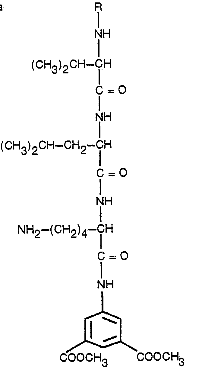
- In accordance with a first aspect of the invention, there is provided a method for determining plasmin in a body fluid, characterised by:
- (A) mixing said fluid with a fluorogenic substrate of the formula
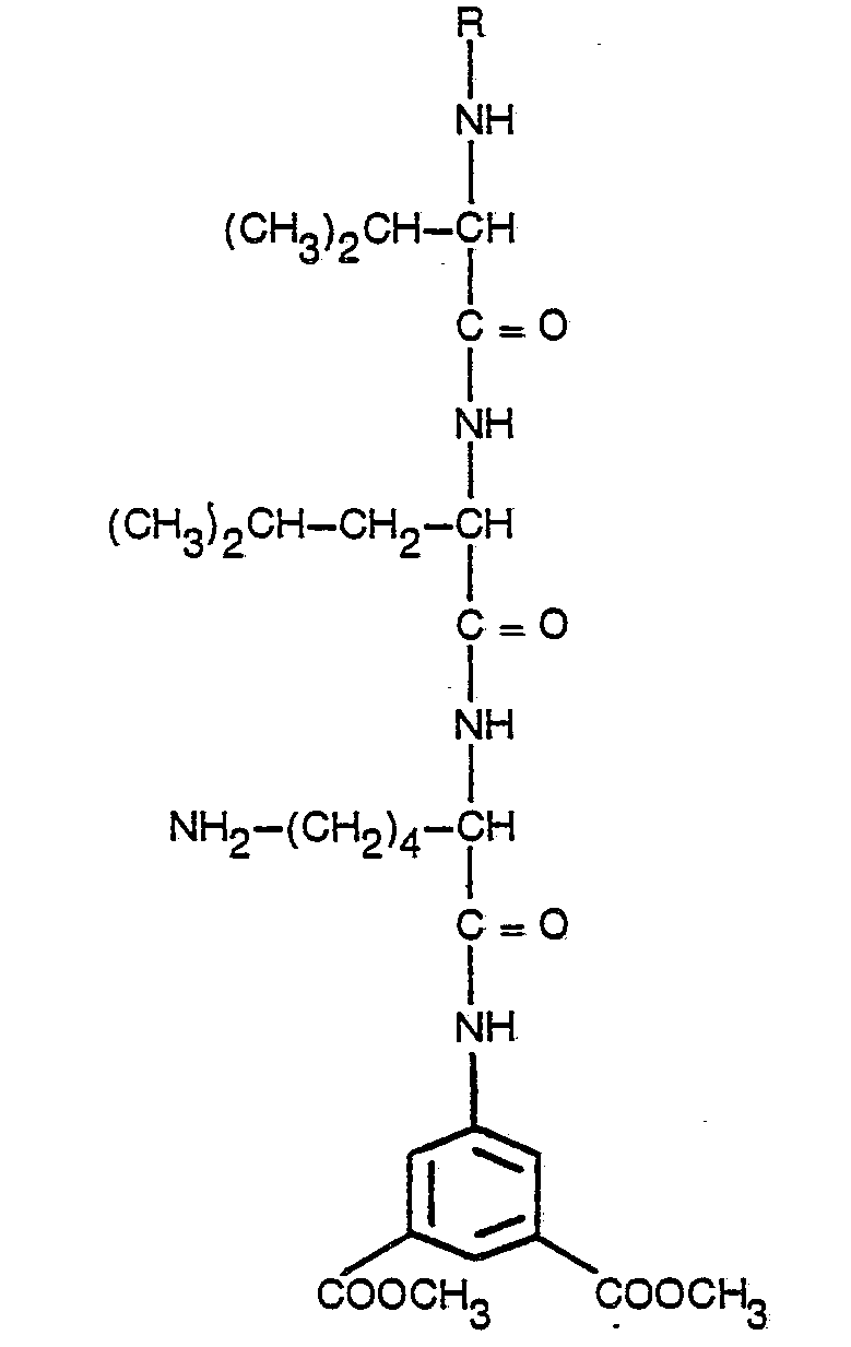
- R is hydrogen-L, hydrogen-D, benzoyl, benzenesulfonyl, glutaryl, pyroglutamyl, carbobenzoxy, D-serine or carbobenzoxy-serine;
- (B) exposing the resulting substrate-containing fluid to excitation light of a first wavelength within the range of 320 to 380 nm; and
- (C) while exposing said substrate-containing fluid to light of said first wavelength, measuring the fluorescence emission from said fluid at a second wavelength within the range of 400 to 480 nm.
- In accordance with a second aspect of the invention, there is provided a method for determining thrombin or trypsin in a body fluid, characterised by:
- (A) mixing said fluid with a fluorogenic substrate of the formula

- R is as stated above;
- (B) exposing the resulting substrate-containing fluid to excitation light of a first wavelength within the range of 320 to 380 nm; and
- (C) while exposing said substrate-containing fluid to light of said first wavelength, measuring the fluoroscence emission from said fluid at a second wavelength within the range of 400 to 480 nm.
- In accordance with a third aspect of the invention, there are provided, as new compounds, the fluorogenic substrates used in the first and second aspects of the invention.
- The fluorogenic group in the fluorogenic substrates of the present invention takes the form of 5-amino- isophthalic acid, dimethyl ester group. The enzyme to be determined cleaves the fluorogenic group which then displays greater fluorescence when liberated than when attached to the substrate.
- Carbobenzoxy represents a suitable amino protecting group in the fluorogenic substrate. However, other projecting groups may also find use and can include benzoyl, benzenesulfonyl, glutaryl and pyroglutamyl.
- In carrying out the methods of the present invention, activity of proteolytic enzymes is determined by mixing a solution of the enzyme with the appropriate substrate of the general formula given above. The solution, when subjected to light of a characteristic wavelength (excitation wavelength) within the range of 320 to 380 nm absorbs some of the light energy, emitting light at a longer wavelength (emission wavelength) within the range of 400 to 480 nm. This emitted fluorescent light is used to determine the enzyme activity.
- The fluorophore, 5-aminoisophthalic acid, dimethyl ester, has characteristic excitation and emission wavelengths, the optimum excitation and emission wavelengths in aqueous solutions at neutral pH being respectively 335 nm and 430 nm.
- The substrates given by the formulae above may be used to determine the activity of the enzymes plasmin, thrombin and trypsin.
- In discussing specific compounds, the following symbols stand for the groups indicated next to them:

- Specifically, plasmin reacts with the following compound: H-D-val-leu-lys-AIE.Thrombin similarly reacts with H-D-phe-pro-arg-AIE.
- Thrombin similarly reacts with H-D-phe-pro-arg-AIE.
- Synthesis of (CH300H) D-phe-pro-arg-(HC1) AIE:
- A suspension of 6 g (1.9 x 10⁻² molar of CBZ-arg in 25 ml dimethyl-formamide (DMF) at -15°C received 0.1 ml n-methylmorpholine and 2.6 ml (1.9 x 10⁻² molar) isobutyl chloroformate. After stirring at -15°C for 45 minutes, this mixture received, in a dropwise fashion over a 10 minute period, a solution of 4 g (1.9 x 10⁻² molar) of 5-aminoisophthalic acid, dimethyl ester in 15 ml DMF. This latter mixture was stirred at -15°C for 1 hour, 0°C for 1 hour, and lastly overnight at ambient temperature. After the removal of the solvent under vacuum, the isolated oil was stirred in saturated sodium bicarbonate and then distilled water to yield a white solid. Seven grams of dry product resulted from this procedure.
- Fifteen (15) ml ethanol and 0.5 ml acetic acid received 500 mg of the solid produced above. To this solution was added 150 mg of 10% palladium on charcoal (Pd/C). Hydrogen gas was then bubbled through this mixture for 16 hours. After filtration, the solvent was removed to yield 440 mg of material which gave a positive ninhydrin test and consisted of arg-AIE (HCI) acetate salt.
- A solution of 442 mg (1.7 x 10⁻³ molar) of CBZ-proline in 3 ml ethyl acetate and 150 ml n-methylmorpholine at -15°, received 275 mg (2.0 x 10⁻³ molar) of isobutyl chloroformate. After eight minutes, this mixture then received a precooled solution containing 694 mg (1.7 x 10⁻³ molar) of the arg-AIE (HCI) acetate salt dissolved in 2 ml dimethylacetamide and 150 µl n-methylmorpholine. This mixture remained at -10°C for 1 hour and then was stirred overnight at ambient temperature. The solvent was removed under vacuum and the resulting material chromatographed over silica gel, using CHCl3/EtOH (7:3) as the eluent to yield 188 mg of product. The product was dissolved in ethyl alcohol and acetic acid to which was added Pd/C (10%). After bubbling hydrogen through it and filtration, removing the solvent yielded pro-arg-(HCI) AIE.
- A solution of 260 mg (8.6 x 10⁻⁴ molar) ov CBZ-D-phenylalanine in 3 ml tetrahydrofuran (THF) and 142 µl of n-methylmorpholine received, at -15°C. 142 mg (1.0 x 10⁻³ molar) of isobutyl chloroformate. To this latter mixture, after minutes, was added a precooled solution of 8.6 x 10⁻⁴ molar of the pro-arg-(HCI) AIE, produced above, in 3 ml dimethylacetamide and 142 µl n-methylmorpholine. This latter mixture remained at -10°C for one-half hour and then at ambient temperature overnight. Evaporating the solvent under vacuum produced an oily material which was then chromatographed over silica gel using CHCl3/ EtOH (8:2) as the eluent. Removing the solvent left an oil
- This oil was dissolved in 3 ml ethyl alcohol which obtained 6 drops of acetic acid. The solution additionally received 30 mg of Pd/C (10%) and had hydrogen bubbled through it for 2 hours. After filtration and evaporation of the solvent, 88 mg of the final product remained which gave a positive ninhydrin test.
- Fluorescent Substrate Assay for Plasminogen Utilizing D-val-leu-lys-AIE:
- Purified plasminogen, approximately 9.7 CTA units/ml in 50% glycerol, was obtained from National Institutes of Health, Bethesda, Maryland. It was diluted 1:2 in the assay buffer, 0.05 M Tris, 0.05 M glycine and 0.01% Brij-35, pH 8.0, and used as the reference plasminogen in the tests.
- Streptokinase, 100,000 units/vial, obtained from Lederle Laboratories, Pearl River, New York, as Varidase was reconstituted in 10.0 ml 0.05 M Tris, 0.1 glycine, pH 7.5, and stored at 20°C. Before use, the streptokinase was thawed and diluted to 2,000 units/ml in the same buffer.
- TFA-D-val-leu-lys (TFA)-AIE, molecular weight 777.8 was obtained from Enzyme Systems Products, Inc., P.O. Box 1191, Indianapolis, Indiana. It was dissolved in the assay bufferto a concentration of 0.8 mM/ liter and warmed to 37°C before use. A Km value of 3.1 x 10⁻⁴M (37°C) was determined within the assay system using the Lineweaver-Burk equation.
- Blood specimens from laboratory workers with normal prothrombin and partial thromboplastin times were collected in plastic tubes containing one volume of 3.8% trisodium citrate for each nine volumes of blood. The specimens were centrifuged at 1,500 to 2,000 g for 10 to 15 minutes to obtain the plasmas which were then stored at -20°C and thawed prior to assay.
- The assays were performed with 10 µl of plasma sample or diluted reference plasminogen added to 0.5 ml streptokinase solution. The reagents were vortex-mixed, incubated for 15 minutes at 37°C to convert the plasminogen to its active for plasmin, and stored at 4°C until assayed. From each of these activation mixtures 200 µl was transferred to a cuvet containing 2.0 ml of substrate solution prewarmed to 37°C. The plasmin activity was determined by kinetic measurement of the released 5-amino-isophthalic acid dimethyl ester over a 2-4 minute interval using a Model 430 spectrofluorometer from G.K. Turner Associates, Palo Alto, California. The instrument was equipped with a controlled temperature sample cell holder and Model EV-200 recorder form Health Schlumberger, Benton Harbor, Michigan. The excitation and emission wavelength and slit width settings were 335 x 430 nm and 60 x 15 nm, respectively. The recorder chart speed was 2.54 cm (1 inch) min. Disposable TPX plastic cuvets, 10 x 8 x 50 nm, were used.
- The plasmin activity of each sample expressed in CTA units/ml was calculated by comparison of the relative fluorescent rate of the sample with that of the reference plasminogen (4.85 CTA units/ml) as follows: -

- Plasma plasminogen values determined by this fluorescent substrate method were compared with the results from two reference methods. M-partigen™ Plasminogen Kits from Behring Diagnostics (American Hoechst Corporation, Somerville, New Jersey 08876) were used to determine plasminogen concentration in mg/dl by the radial immunodiffusion technique. A standard caseinolytic assay method was used to measure the plasmin activity generated by streptokinase activation of 'each plasma sample. In the caseinolytic assay method, the relatively large sample volume required acid pretreatment of the plasmas to destroy plasmin inhibitors prior to activation.
- Good agreement was obtained between the fluoroscent substrate assay and the two reference methods. The correlation coefficients obtained from lease squares analyses of the date were 0.98 for the fluorescent versus caseinolytic assay, and 0.99 for the fluorescent versus radial immunodiffusion assay.
- The precision of the fluorescent substrate assay for plasma plasminogen determinations was less than 5% for levels for plasminogen over the normal range.
- Fluorescent Substrate Assay for Heparin Utilizing D-phe-pro-arg-AIE:
- Five normal blood specimens were collected daily in siliconized glass tubes containing 3.8% trisodium citrate for each 9 volumes of blood. The plasma was obtained by centrifugation of the blood at 1,240 g for 15 minutes. The resulting individual plasma samples were pooled and stored at 4°C until used. Part of the plasma was diluted 1:40 in a general assay buffer for use in all the determinations, including the standard and test plasmas, as an equalizer of AT-III activity. The general assay buffer contained 0.25 M glycine in 0.03 M NaCI with 2 mM K2EDTA and 0.01% thimerosal with the pH adjusted to 8.3 using 5 N NaOH.
- Beef lung sodium heparin containing 10,000 U.S.P. units/ml was obtained from the Upjohn Company (Kalamazoo, Michigan 49001). The heparin underwent dilution to 80 units/ml with the general buffer given above. Heparin standards were prepared in the pooled plasma at concentrations of 0.1, 0.2, 0.4 and 0.8 units/mi.
- The D-phe-pro-arg-AIE, diacetate, having a molecular weight of 728.8, was prepared in absolute ethanol at a concentration of 11.2 mg/ml and stored at 4°C. Before use, the stock solution was diluted in the general buffer to a concentration of 0.19 mM and warmed to 37°C. A Km value of 5.4 x 10⁻⁵M at 37°C was determined within the assay system using the Lineweaver-Burk equation.
- Lyophilized human thrombin containing 2,500 NIH units/mg were obtained from Calibiochem (San Diego, California 92112). This material was dissolved to 10 units/ml with a buffer solution containing 0.05 M glycine in 0.15 M NaCI having a pH of 5.0 with 2 mM K2EDTA and 0.01 % bovine serum albumin. The diluted solution was stored in aliquots at -20°C. Before use, the thrombin was thawed and further diluted with the same solution of 2 NIH units/ml.
- For this experiment, the Perkin Elmer spectrophotometer had an excitation wavelength setting of 335 nm with a slit width of 6 nm and the emission wavelength setting of 430 nm with a slit width of 12 nm. Volumes of 200 µl of 1:40 diluted normal plasma and 5 pl of test plasma or standard heparin in plasma were added to a sample cuvet and warmed to 37°C. Figty (50) µl of thrombin having 0.1 NIH unit was added and the reagents mixed. After preincubation for 60 sec. at 37°C, 2.0 ml of the substrate solution containing 140 mg/l, prewarmed to 37°C was added, and the contents of the cuvet mixed by inversion. Unihibited thrombin was determined by kinetic measurement of the released AIE over a 2-4 minute interval. Assay values were calculated according to the following equation with the blank result determined in the absence of heparin:

- Table 2 gives the precision of standard heparin measurements determined according to the above procedure. The within and between-day precision for heparin standards at 0.1 and 0.4 units/ml was better than ±5%. The reported anticoagulant concentration range of heparin of 0.2 to 0.4 units/ml of plasma lies within the standard range.

- The reduced precision accompanying the measurements of the highest heparin standard, 0.8 units/mi, may result from, assay timing errors. Experiments involving different incubation times showed a greater dependence of this heparin concentration upon this variable.
- Increasing the preincubation time or reducing the thrombin concentration did, however, improve the assay's sensitivity. Freezing heparin standards prepared in plasma with a low platelet count of around 75,900/mm3 at -20°C and subsequently thawing them at 22-28°C produced some loss in the measured heparin activity.
- These data are consistent with the work of others suggesting that plasma standards and test samples may require special treatment to reduce or eliminate platelets if the samples are to be stored frozen before assay.
- Determinations were also made with WHO heparin preparations, bovine mucosal working reagent (No. 63/10) and porcine mucosal standard (Third International Standard No. 65/69). On the basis of their stated activities, there was good agreement between the dose response curves for these two preparations. The commercial beef lung heparin gave a standard curve which proved less sensitive and varied from the WHO standared curves by approximately 20%. These results may be related to the reported discrepancy between lung and mucosal heparin activities.
- The sensitive detection of thrombin with the fluorescent substrate assay appears to allow its use rather than the factor X. for determining plasma heparin anticoagulant activity. Clotting interference is not encountered since only 1.10 NIH unit of thrombin and 10 µl of total plasma are employed in each determination. The assay precision of better than ±5% over the heparin anticoagulant range at least equalled or excelled the precision of the standard clotting assays designed for the measurment of heparin. The assay range would appear acceptable for routine monitoring of heparinized patient samples. However, increasing the preincubation or reducing the amount of thrombin used can increase the assay sensitivity as required.
Claims (5)



Applications Claiming Priority (2)
| Application Number | Priority Date | Filing Date | Title |
|---|---|---|---|
| US930826 | 1978-08-03 | ||
| US05/930,826 US4275153A (en) | 1978-08-03 | 1978-08-03 | Analytical fluorogenic substrates for proteolytic enzymes |
Publications (3)
| Publication Number | Publication Date |
|---|---|
| EP0016800A1 EP0016800A1 (en) | 1980-10-15 |
| EP0016800A4 EP0016800A4 (en) | 1981-01-08 |
| EP0016800B1 true EP0016800B1 (en) | 1986-03-05 |
Family
ID=25459832
Family Applications (1)
| Application Number | Title | Priority Date | Filing Date |
|---|---|---|---|
| EP79900842A Expired EP0016800B1 (en) | 1978-08-03 | 1980-03-11 | Determination of proteolytic enzymes in body fluids and fluorogenic substrates |
Country Status (8)
| Country | Link |
|---|---|
| US (1) | US4275153A (en) |
| EP (1) | EP0016800B1 (en) |
| JP (1) | JPS5729999B2 (en) |
| CA (1) | CA1152494A (en) |
| DE (1) | DE2967581D1 (en) |
| IT (1) | IT1117757B (en) |
| NO (1) | NO792553L (en) |
| WO (1) | WO1980000351A1 (en) |
Families Citing this family (34)
| Publication number | Priority date | Publication date | Assignee | Title |
|---|---|---|---|---|
| DE2936543A1 (en) * | 1979-09-10 | 1981-04-09 | Behringwerke Ag, 3550 Marburg | CHROMOGENIC COMPOUNDS |
| CA1161432A (en) * | 1980-02-12 | 1984-01-31 | Lars G. Svendsen | Tripeptide derivatives and their application in assaying enzymes |
| WO1982000641A1 (en) * | 1980-08-25 | 1982-03-04 | Ab Kabivitrum | Peptide substrates for determination of protease activity |
| JPS5856695A (en) * | 1981-09-28 | 1983-04-04 | Nitto Boseki Co Ltd | Novel substrate for assay of thrombin |
| EP0078764B1 (en) * | 1981-11-02 | 1986-01-02 | Pentapharm A.G. | Process for the quantitative determination of the blood clotting factor xii in human plasma |
| JPS5925699A (en) * | 1982-08-03 | 1984-02-09 | Torii Yakuhin Kk | Determination of enzymatic activity |
| US4473639A (en) * | 1982-09-15 | 1984-09-25 | Miles Laboratories, Inc. | Reagent strip test for antithrombin-III |
| CS231228B1 (en) * | 1982-10-01 | 1984-10-15 | Evzen Kasafirek | Biologically effective tri and tetrapeptide alkylamide derivatives and their processing method |
| DE3244030A1 (en) * | 1982-11-27 | 1984-05-30 | Behringwerke Ag, 3550 Marburg | CHROMOGENIC COMPOUNDS, METHOD FOR THEIR PRODUCTION AND THEIR USE |
| US4621135A (en) * | 1983-12-08 | 1986-11-04 | Yeda Research & Development Company, Ltd. | Novel THF compositions |
| US4801534A (en) * | 1985-05-28 | 1989-01-31 | Coulter Electronics, Inc. | Water soluble zanthylium derivative substrates |
| US4694070A (en) * | 1985-05-28 | 1987-09-15 | Coulter Electronics, Inc. | Water soluble xanthylium derivatives substrates |
| SE461494B (en) * | 1986-03-21 | 1990-02-19 | Thorsman & Co Ab | MACHINE CONTAINS AN ELECTRIC CLUTCH BOX |
| ES2051702T3 (en) * | 1986-04-01 | 1994-07-01 | Wako Pure Chem Ind Ltd | PEPTIDIC DERIVATIVES AND METHOD FOR MEASURING THE ACTIVITY OF PHYSIOLOGICALLY ACTIVE SUBSTANCES USING THEM AS SUBSTRATES. |
| US5023236A (en) * | 1988-04-07 | 1991-06-11 | Corvas, Inc. | Factor VII/VIIA active site inhibitors |
| US5593967A (en) * | 1990-08-31 | 1997-01-14 | Warner-Lambert Company | Cholecystokinin antagonists, their preparation and therapeutic use |
| US5391705A (en) * | 1991-03-15 | 1995-02-21 | Merrell Dow Pharmaceuticals Inc. | Polyfluorinated tripeptide thrombin inhibitors |
| US5534498A (en) * | 1992-01-30 | 1996-07-09 | Corvas International, Inc. | Trypsin inhibitors |
| HUT76346A (en) * | 1994-04-26 | 1997-08-28 | Selectide Corp | Factor xa inhibitors |
| US5849510A (en) * | 1994-04-26 | 1998-12-15 | Selectide Corporation | Factor Xa inhibitors |
| US6759384B1 (en) | 1994-04-26 | 2004-07-06 | Aventis Pharmaceuticals Inc. | Factor Xa inhibitors |
| US5871946A (en) * | 1995-05-18 | 1999-02-16 | Coulter Corporation | Method for determining activity of enzymes in metabolically active whole cells |
| US5733719A (en) * | 1995-05-18 | 1998-03-31 | Coulter Corporation | Method of making an assay compound |
| US5776720A (en) * | 1995-05-18 | 1998-07-07 | Coulter Corporation | Assay reagent |
| US5698411A (en) * | 1995-05-18 | 1997-12-16 | Coulter Corporation | Method for determining activity of enzymes in metabolically active whole cells |
| US6069232A (en) * | 1995-10-02 | 2000-05-30 | Hoechst Marion Roussel, Inc. | Polyfluoroalkyl tryptophan tripeptide thrombin inhibitors |
| GB9525403D0 (en) * | 1995-12-13 | 1996-02-14 | Univ Sunderland | Method for monitoring enzymes |
| US5739354A (en) * | 1996-03-26 | 1998-04-14 | Hoechst Marion Roussel, Inc. | Process for the preparation of N-methyl-D-phenylalanyl-N- 1- 3- (aminoiminomethyl)amino!propyl!-3,3-difluoro-2-oxohexyl!-L-prolinamide |
| DE10116586B4 (en) * | 2001-04-03 | 2005-07-21 | Stief, Thomas, Dr.med. | Test system for the determination of hemostasis activation of biological fluids |
| US8338127B2 (en) * | 2008-01-31 | 2012-12-25 | Anazyme | Testing a mammal for presence, progression or stage of a shock condition |
| WO2010087874A1 (en) * | 2009-01-28 | 2010-08-05 | Anazyme, Llc | Compositions and methods for diagnosis of shock |
| EP2189536A1 (en) * | 2008-11-20 | 2010-05-26 | Nederlandse Organisatie voor toegepast-natuurwetenschappelijk Onderzoek TNO | Rapid FRET based anthrax diagnosis |
| EP2366027B1 (en) * | 2008-11-20 | 2017-01-11 | Nederlandse Organisatie voor toegepast- natuurwetenschappelijk onderzoek TNO | Rapid fret-based diagnosis of bacterial pathogens |
| WO2010091235A2 (en) * | 2009-02-06 | 2010-08-12 | Talecris Biotherapeutics, Inc. | Compositions, kits, and methods for determining plasmin activity |
Family Cites Families (18)
| Publication number | Priority date | Publication date | Assignee | Title |
|---|---|---|---|---|
| US3862011A (en) * | 1971-08-16 | 1975-01-21 | Robert E Smith | Method for quantitatively determining enzyme concentrations in homogenates |
| SE380257B (en) * | 1972-05-02 | 1975-11-03 | Bofors Ab | NEW DIAGNOSTIC OPERATING SUBSTRATES WITH HIGH SPECIFICITY FOR THROMBIN AND OTHER PROTEOLYTIC ENZYMES OF THE PEPTIDYL-PEPTIDE HYDROLASES |
| IL42124A (en) * | 1972-05-02 | 1977-02-28 | Kabi Ab | Substrate for the determination of proteolytic enzymes |
| DE2527932C2 (en) * | 1974-07-02 | 1983-04-21 | Pentapharm AG, 4052 Basel | Acid addition salts of tripeptide derivatives and their use as substrates for the determination of plasma kallikrein |
| US4162941A (en) * | 1974-12-05 | 1979-07-31 | Ab Kabi | Method for determining a proteolytic enzyme |
| SE407058B (en) * | 1974-12-05 | 1979-03-12 | Kabi Ab | NEW CHROMOGENA ENZYME SUBSTRATE FOR SERINE PROTEASES |
| CH622286A5 (en) * | 1975-06-23 | 1981-03-31 | Pentapharm Ag | |
| US4137225A (en) * | 1975-07-11 | 1979-01-30 | Ab Kabi | Novel chromogenic enzyme substrates |
| US4169015A (en) * | 1975-07-11 | 1979-09-25 | Ab Kabi | Novel chromogenic thrombin substrates |
| SE407571B (en) * | 1975-07-11 | 1979-04-02 | Kabi Ab | NEW CHROMOGENA ENZYME SUBSTRATE FOR SERINE PROTEASES |
| US4056519A (en) * | 1976-05-10 | 1977-11-01 | Eli Lilly And Company | Substrate for assay of plasmin |
| CH634662A5 (en) * | 1976-05-28 | 1983-02-15 | Pentapharm Ag | USE OF TRIPEPTIDE DERIVATIVES FOR THE QUANTITATIVE DETERMINATION OF PLASMINOGEN ACTIVATORS. |
| US4167449A (en) * | 1976-07-29 | 1979-09-11 | American Hospital Supply Corporation | Composition and method for determining transferase and protease activity |
| SE437153B (en) * | 1976-12-01 | 1985-02-11 | Kabi Ab | SPECIFIC CHROMOGENA ENZYM SUBSTATE FOR SERINE PROTEASES |
| US4215047A (en) * | 1977-06-06 | 1980-07-29 | Ajinomoto Company Incorporated | 7-(Arginylamino)-4-methylcoumarins |
| CA1087075A (en) * | 1977-08-05 | 1980-10-07 | Robert J. Gargiulo | Composition and method for determining transferase and protease activity |
| US4155916A (en) * | 1977-08-29 | 1979-05-22 | Dolbeare Frank A | Fluorescence method for enzyme analysis which couples aromatic amines with aromatic aldehydes |
| SE7801373L (en) * | 1978-02-07 | 1979-08-08 | Kabi Ab | EASY SPLABLE SUBSTRATE FOR QUANTIFIATION OF PROTEASES |
-
1978
- 1978-08-03 US US05/930,826 patent/US4275153A/en not_active Expired - Lifetime
-
1979
- 1979-06-15 WO PCT/US1979/000421 patent/WO1980000351A1/en not_active Ceased
- 1979-06-15 JP JP50117779A patent/JPS5729999B2/ja not_active Expired
- 1979-06-15 DE DE7979900842T patent/DE2967581D1/en not_active Expired
- 1979-07-05 CA CA000331214A patent/CA1152494A/en not_active Expired
- 1979-07-31 IT IT49905/79A patent/IT1117757B/en active
- 1979-08-02 NO NO792553A patent/NO792553L/en unknown
-
1980
- 1980-03-11 EP EP79900842A patent/EP0016800B1/en not_active Expired
Also Published As
| Publication number | Publication date |
|---|---|
| US4275153A (en) | 1981-06-23 |
| JPS5729999B2 (en) | 1982-06-25 |
| EP0016800A4 (en) | 1981-01-08 |
| NO792553L (en) | 1980-02-05 |
| JPS55500538A (en) | 1980-08-21 |
| IT7949905A0 (en) | 1979-07-31 |
| DE2967581D1 (en) | 1986-04-10 |
| EP0016800A1 (en) | 1980-10-15 |
| WO1980000351A1 (en) | 1980-03-06 |
| IT1117757B (en) | 1986-02-24 |
| CA1152494A (en) | 1983-08-23 |
Similar Documents
| Publication | Publication Date | Title |
|---|---|---|
| US4275153A (en) | Analytical fluorogenic substrates for proteolytic enzymes | |
| US4336186A (en) | Analytical fluorogenic substrates for proteolytic enzymes | |
| EP0046742B1 (en) | Peptide substrates for determination of protease activity | |
| US4784944A (en) | Prothrombin time determining reagent and methods of using and preparing the same | |
| EP0034586B1 (en) | 4-trifluoromethylcoumarin peptide derivatives and their use in proteinase assays | |
| Bergström et al. | Determination of vitamin K sensitive coagulation factors in plasma. Studies on three methods using synthetic chromogenic substrates | |
| DK149333B (en) | Tripeptide AND SALTS OF MINERAL organic acids FOR USE AS SUBSTRATES FOR DETERMINATION OF thrombin and thrombin-like ENZYMES ECARINTHROMBIN, plasmin and plasmin ENZYMES AND proenzymes PROENZYMAKTIVATORER, and enzyme inhibitors PURPOSE In LEGEMSVAESKERFRA PEOPLE AND MAMMALS AND IN animal CELL EXTRACTS | |
| Mitchell et al. | Fluorescent substrate assay for antithrombin III | |
| US6068988A (en) | Detection of microbial metabolites with a 3-indoxyl-myo-inositol-1-phosphate compound | |
| US5922587A (en) | Phospholipid dependent prothrombin activator obtained from snake venom | |
| US4167449A (en) | Composition and method for determining transferase and protease activity | |
| JP3533012B2 (en) | Method for detecting protein C / protein S system failure | |
| CA1292174C (en) | Method for the determination of protease inhibitors | |
| IE840743L (en) | Determination of activated partial thromboplastin time | |
| EP0168738B1 (en) | Single step protease inhibitor assay | |
| US4216142A (en) | Chromogenic substrates for the proteolytic enzymes | |
| EP0455733A1 (en) | Rapid assay for functional human alpha 1?-proteinase inhibitor | |
| Pochron et al. | A fluorescent substrate assay for plasminogen | |
| US4409140A (en) | Substrates and method for determining enzymes | |
| Ratnoff | Studies on the inhibition of ellagic acid-activated Hageman factor (factor XII) and Hageman factor fragments | |
| Lawson et al. | A sensitive fluorescent assay for determining α2-plasmin inhibitor using a synthetic substrate | |
| CA1276391C (en) | Water soluble xanthylium derivative substrates | |
| US4801534A (en) | Water soluble zanthylium derivative substrates | |
| EP0318571B1 (en) | Determination of components active in proteolysis | |
| Eckfeldt et al. | Automated assay for alpha 1-antitrypsin with N-alpha-benzoyl-DL-arginine-p-nitroanilide as trypsin substrate and standardized with p-nitrophenyl-p'-guanidinobenzoate as titrant for trypsin active sites. |
Legal Events
| Date | Code | Title | Description |
|---|---|---|---|
| PUAI | Public reference made under article 153(3) epc to a published international application that has entered the european phase |
Free format text: ORIGINAL CODE: 0009012 |
|
| AK | Designated contracting states |
Designated state(s): CH DE FR GB LU SE |
|
| 17P | Request for examination filed | ||
| GRAA | (expected) grant |
Free format text: ORIGINAL CODE: 0009210 |
|
| AK | Designated contracting states |
Kind code of ref document: B1 Designated state(s): CH DE FR GB LU SE |
|
| REF | Corresponds to: |
Ref document number: 2967581 Country of ref document: DE Date of ref document: 19860410 |
|
| ET | Fr: translation filed | ||
| PG25 | Lapsed in a contracting state [announced via postgrant information from national office to epo] |
Ref country code: LU Free format text: LAPSE BECAUSE OF NON-PAYMENT OF DUE FEES Effective date: 19860630 |
|
| PLBE | No opposition filed within time limit |
Free format text: ORIGINAL CODE: 0009261 |
|
| STAA | Information on the status of an ep patent application or granted ep patent |
Free format text: STATUS: NO OPPOSITION FILED WITHIN TIME LIMIT |
|
| 26N | No opposition filed | ||
| REG | Reference to a national code |
Ref country code: GB Ref legal event code: 732 |
|
| REG | Reference to a national code |
Ref country code: CH Ref legal event code: PUE Owner name: BAXTER TRAVENOL LABORATORIES, INC. |
|
| REG | Reference to a national code |
Ref country code: FR Ref legal event code: TP |
|
| PG25 | Lapsed in a contracting state [announced via postgrant information from national office to epo] |
Ref country code: GB Effective date: 19890615 |
|
| PG25 | Lapsed in a contracting state [announced via postgrant information from national office to epo] |
Ref country code: SE Effective date: 19890616 |
|
| PG25 | Lapsed in a contracting state [announced via postgrant information from national office to epo] |
Ref country code: CH Effective date: 19890630 |
|
| GBPC | Gb: european patent ceased through non-payment of renewal fee | ||
| PG25 | Lapsed in a contracting state [announced via postgrant information from national office to epo] |
Ref country code: FR Free format text: LAPSE BECAUSE OF NON-PAYMENT OF DUE FEES Effective date: 19900228 |
|
| REG | Reference to a national code |
Ref country code: CH Ref legal event code: PL |
|
| PG25 | Lapsed in a contracting state [announced via postgrant information from national office to epo] |
Ref country code: DE Effective date: 19900301 |
|
| REG | Reference to a national code |
Ref country code: FR Ref legal event code: ST |
|
| EUG | Se: european patent has lapsed |
Ref document number: 79900842.0 Effective date: 19900412 |