DE102009027448A1 - Knowledge-based segmentation of weakening regions of the head - Google Patents
Knowledge-based segmentation of weakening regions of the head Download PDFInfo
- Publication number
- DE102009027448A1 DE102009027448A1 DE102009027448A DE102009027448A DE102009027448A1 DE 102009027448 A1 DE102009027448 A1 DE 102009027448A1 DE 102009027448 A DE102009027448 A DE 102009027448A DE 102009027448 A DE102009027448 A DE 102009027448A DE 102009027448 A1 DE102009027448 A1 DE 102009027448A1
- Authority
- DE
- Germany
- Prior art keywords
- class
- area
- voxels
- region
- extracerebral
- Prior art date
- Legal status (The legal status is an assumption and is not a legal conclusion. Google has not performed a legal analysis and makes no representation as to the accuracy of the status listed.)
- Withdrawn
Links
- 230000011218 segmentation Effects 0.000 title claims description 13
- 230000003313 weakening effect Effects 0.000 title description 19
- 238000000034 method Methods 0.000 claims abstract description 53
- 238000002600 positron emission tomography Methods 0.000 claims abstract description 13
- 210000001175 cerebrospinal fluid Anatomy 0.000 claims description 59
- 210000000988 bone and bone Anatomy 0.000 claims description 50
- 210000003128 head Anatomy 0.000 claims description 42
- 210000004556 brain Anatomy 0.000 claims description 39
- 210000001519 tissue Anatomy 0.000 claims description 30
- 210000004884 grey matter Anatomy 0.000 claims description 26
- 210000004885 white matter Anatomy 0.000 claims description 26
- 210000000577 adipose tissue Anatomy 0.000 claims description 25
- 238000002595 magnetic resonance imaging Methods 0.000 claims description 24
- 210000004872 soft tissue Anatomy 0.000 claims description 24
- 210000004027 cell Anatomy 0.000 claims description 22
- 210000003484 anatomy Anatomy 0.000 claims description 19
- 210000001595 mastoid Anatomy 0.000 claims description 16
- 210000003625 skull Anatomy 0.000 claims description 13
- 230000002490 cerebral effect Effects 0.000 claims description 11
- 238000001514 detection method Methods 0.000 claims description 11
- 230000001815 facial effect Effects 0.000 claims description 11
- 230000003628 erosive effect Effects 0.000 claims description 10
- 238000003325 tomography Methods 0.000 claims description 8
- 235000019994 cava Nutrition 0.000 claims description 7
- 210000003928 nasal cavity Anatomy 0.000 claims description 7
- 235000008331 Pinus X rigitaeda Nutrition 0.000 claims description 5
- 235000011613 Pinus brutia Nutrition 0.000 claims description 5
- 241000018646 Pinus brutia Species 0.000 claims description 5
- 238000000926 separation method Methods 0.000 claims description 5
- 210000003718 sphenoid sinus Anatomy 0.000 claims description 5
- 241001465754 Metazoa Species 0.000 claims description 4
- 210000000613 ear canal Anatomy 0.000 claims description 4
- 239000000126 substance Substances 0.000 claims description 4
- 210000001214 frontal sinus Anatomy 0.000 claims description 3
- 238000005259 measurement Methods 0.000 abstract description 18
- 238000012937 correction Methods 0.000 description 15
- 230000005855 radiation Effects 0.000 description 11
- 239000011159 matrix material Substances 0.000 description 10
- 208000018672 Dilatation Diseases 0.000 description 8
- 238000003384 imaging method Methods 0.000 description 8
- 238000000605 extraction Methods 0.000 description 6
- 230000010339 dilation Effects 0.000 description 5
- 230000006870 function Effects 0.000 description 5
- 210000003800 pharynx Anatomy 0.000 description 5
- 238000012549 training Methods 0.000 description 5
- 238000013528 artificial neural network Methods 0.000 description 4
- 210000005013 brain tissue Anatomy 0.000 description 4
- 238000012545 processing Methods 0.000 description 4
- 210000000133 brain stem Anatomy 0.000 description 3
- 210000001638 cerebellum Anatomy 0.000 description 3
- 210000004720 cerebrum Anatomy 0.000 description 3
- 230000004060 metabolic process Effects 0.000 description 3
- 230000000877 morphologic effect Effects 0.000 description 3
- 210000000056 organ Anatomy 0.000 description 3
- 230000008569 process Effects 0.000 description 3
- ZCXUVYAZINUVJD-AHXZWLDOSA-N 2-deoxy-2-((18)F)fluoro-alpha-D-glucose Chemical compound OC[C@H]1O[C@H](O)[C@H]([18F])[C@@H](O)[C@@H]1O ZCXUVYAZINUVJD-AHXZWLDOSA-N 0.000 description 2
- 238000013459 approach Methods 0.000 description 2
- 238000002059 diagnostic imaging Methods 0.000 description 2
- 230000000916 dilatatory effect Effects 0.000 description 2
- 230000000694 effects Effects 0.000 description 2
- 238000011156 evaluation Methods 0.000 description 2
- 238000011835 investigation Methods 0.000 description 2
- 210000003205 muscle Anatomy 0.000 description 2
- 238000001208 nuclear magnetic resonance pulse sequence Methods 0.000 description 2
- 210000001328 optic nerve Anatomy 0.000 description 2
- 230000036961 partial effect Effects 0.000 description 2
- 238000003909 pattern recognition Methods 0.000 description 2
- 230000035790 physiological processes and functions Effects 0.000 description 2
- 229940121896 radiopharmaceutical Drugs 0.000 description 2
- 239000012217 radiopharmaceutical Substances 0.000 description 2
- 230000002799 radiopharmaceutical effect Effects 0.000 description 2
- 230000002829 reductive effect Effects 0.000 description 2
- 230000002123 temporal effect Effects 0.000 description 2
- 230000009466 transformation Effects 0.000 description 2
- BUHVIAUBTBOHAG-FOYDDCNASA-N (2r,3r,4s,5r)-2-[6-[[2-(3,5-dimethoxyphenyl)-2-(2-methylphenyl)ethyl]amino]purin-9-yl]-5-(hydroxymethyl)oxolane-3,4-diol Chemical compound COC1=CC(OC)=CC(C(CNC=2C=3N=CN(C=3N=CN=2)[C@H]2[C@@H]([C@H](O)[C@@H](CO)O2)O)C=2C(=CC=CC=2)C)=C1 BUHVIAUBTBOHAG-FOYDDCNASA-N 0.000 description 1
- UFHFLCQGNIYNRP-UHFFFAOYSA-N Hydrogen Chemical compound [H][H] UFHFLCQGNIYNRP-UHFFFAOYSA-N 0.000 description 1
- 230000006978 adaptation Effects 0.000 description 1
- 230000005540 biological transmission Effects 0.000 description 1
- 230000017531 blood circulation Effects 0.000 description 1
- 210000000746 body region Anatomy 0.000 description 1
- 238000004364 calculation method Methods 0.000 description 1
- 239000003086 colorant Substances 0.000 description 1
- 210000000027 corpus adiposum orbitae Anatomy 0.000 description 1
- 238000011161 development Methods 0.000 description 1
- 230000018109 developmental process Effects 0.000 description 1
- 238000003745 diagnosis Methods 0.000 description 1
- 238000007435 diagnostic evaluation Methods 0.000 description 1
- 238000006073 displacement reaction Methods 0.000 description 1
- 230000005672 electromagnetic field Effects 0.000 description 1
- 229910052739 hydrogen Inorganic materials 0.000 description 1
- 239000001257 hydrogen Substances 0.000 description 1
- 125000004435 hydrogen atom Chemical group [H]* 0.000 description 1
- 238000003709 image segmentation Methods 0.000 description 1
- 230000003993 interaction Effects 0.000 description 1
- 210000000629 knee joint Anatomy 0.000 description 1
- 230000033001 locomotion Effects 0.000 description 1
- 210000004086 maxillary sinus Anatomy 0.000 description 1
- 230000002503 metabolic effect Effects 0.000 description 1
- 238000009206 nuclear medicine Methods 0.000 description 1
- 230000010412 perfusion Effects 0.000 description 1
- 230000002093 peripheral effect Effects 0.000 description 1
- 230000000717 retained effect Effects 0.000 description 1
- 210000004761 scalp Anatomy 0.000 description 1
- 210000003491 skin Anatomy 0.000 description 1
- 239000013589 supplement Substances 0.000 description 1
- 230000007704 transition Effects 0.000 description 1
Images


Classifications
-
- A—HUMAN NECESSITIES
- A61—MEDICAL OR VETERINARY SCIENCE; HYGIENE
- A61B—DIAGNOSIS; SURGERY; IDENTIFICATION
- A61B6/00—Apparatus for radiation diagnosis, e.g. combined with radiation therapy equipment
- A61B6/02—Devices for diagnosis sequentially in different planes; Stereoscopic radiation diagnosis
- A61B6/03—Computerised tomographs
- A61B6/037—Emission tomography
-
- G—PHYSICS
- G06—COMPUTING; CALCULATING OR COUNTING
- G06T—IMAGE DATA PROCESSING OR GENERATION, IN GENERAL
- G06T11/00—2D [Two Dimensional] image generation
- G06T11/003—Reconstruction from projections, e.g. tomography
- G06T11/005—Specific pre-processing for tomographic reconstruction, e.g. calibration, source positioning, rebinning, scatter correction, retrospective gating
-
- G—PHYSICS
- G06—COMPUTING; CALCULATING OR COUNTING
- G06T—IMAGE DATA PROCESSING OR GENERATION, IN GENERAL
- G06T7/00—Image analysis
- G06T7/10—Segmentation; Edge detection
- G06T7/11—Region-based segmentation
-
- G—PHYSICS
- G01—MEASURING; TESTING
- G01R—MEASURING ELECTRIC VARIABLES; MEASURING MAGNETIC VARIABLES
- G01R33/00—Arrangements or instruments for measuring magnetic variables
- G01R33/20—Arrangements or instruments for measuring magnetic variables involving magnetic resonance
- G01R33/44—Arrangements or instruments for measuring magnetic variables involving magnetic resonance using nuclear magnetic resonance [NMR]
- G01R33/48—NMR imaging systems
- G01R33/4808—Multimodal MR, e.g. MR combined with positron emission tomography [PET], MR combined with ultrasound or MR combined with computed tomography [CT]
- G01R33/481—MR combined with positron emission tomography [PET] or single photon emission computed tomography [SPECT]
-
- G—PHYSICS
- G01—MEASURING; TESTING
- G01R—MEASURING ELECTRIC VARIABLES; MEASURING MAGNETIC VARIABLES
- G01R33/00—Arrangements or instruments for measuring magnetic variables
- G01R33/20—Arrangements or instruments for measuring magnetic variables involving magnetic resonance
- G01R33/44—Arrangements or instruments for measuring magnetic variables involving magnetic resonance using nuclear magnetic resonance [NMR]
- G01R33/48—NMR imaging systems
- G01R33/54—Signal processing systems, e.g. using pulse sequences ; Generation or control of pulse sequences; Operator console
- G01R33/56—Image enhancement or correction, e.g. subtraction or averaging techniques, e.g. improvement of signal-to-noise ratio and resolution
- G01R33/5608—Data processing and visualization specially adapted for MR, e.g. for feature analysis and pattern recognition on the basis of measured MR data, segmentation of measured MR data, edge contour detection on the basis of measured MR data, for enhancing measured MR data in terms of signal-to-noise ratio by means of noise filtering or apodization, for enhancing measured MR data in terms of resolution by means for deblurring, windowing, zero filling, or generation of gray-scaled images, colour-coded images or images displaying vectors instead of pixels
-
- G—PHYSICS
- G06—COMPUTING; CALCULATING OR COUNTING
- G06T—IMAGE DATA PROCESSING OR GENERATION, IN GENERAL
- G06T2207/00—Indexing scheme for image analysis or image enhancement
- G06T2207/30—Subject of image; Context of image processing
- G06T2207/30004—Biomedical image processing
- G06T2207/30016—Brain
Abstract
Die vorliegende Erfindung betrifft ein Verfahren und eine Vorrichtung für die Ermittlung von Schwächungsbereichen in einem Körper. Die Erfindung betrifft insbesondere ein Verfahren nebst Vorrichtung zur Durchführung von Magnetresonanz/Positronen-Emissions-Tomografie-Messungen unter Verwendung der ermittelten Schwächungsbereiche.The present invention relates to a method and a device for determining areas of weakness in a body. In particular, the invention relates to a method and device for carrying out magnetic resonance / positron emission tomography measurements using the determined attenuation areas.
Description
Die vorliegende Erfindung betrifft ein Verfahren und eine Vorrichtung für die Ermittlung von Schwächungsbereichen in einem Körper. Die Erfindung betrifft insbesondere ein Verfahren nebst Vorrichtung zur Durchführung von Magnetresonanz/Positronen-Emissions-Tomografie(MR/PET)-Messungen unter Verwendung der ermittelten Schwächungsbereiche.The The present invention relates to a method and an apparatus for the determination of areas of weakness in a body. The invention particularly relates to a method in addition to a device for carrying out magnetic resonance / positron emission tomography (MR / PET) measurements using the determined attenuation ranges.
Die Positronen-Emissions-Tomographie (Abkürzung PET) ist ein bildgebendes Verfahren der Nuklearmedizin, das Schnittbilder von lebenden Organismen erzeugt, indem es die Verteilung einer schwach radioaktiv markierten Substanz im Organismus sichtbar macht und damit biochemische und physiologische Funktionen abbildet.The Positron emission tomography (abbreviation PET) is a imaging technique of nuclear medicine, the sectional images of living organisms generates by distributing a weak makes radiolabelled substance visible in the organism and so that biochemical and physiological functions are depicted.
Zu Beginn einer Untersuchung wird dem zu untersuchenden Lebewesen ein Radiopharmakon verabreicht und zwar Radionuklide, die Positronen emittieren (Betastrahlung). Bei der Wechselwirkung eines Positrons mit einem Elektron im Körper werden zwei hochenergetische Photonen in genau entgegengesetzte Richtungen, also mit dem Winkel 180 Grad zueinander, ausgesandt (Vernichtungsstrahlung). Die PET-Vorrichtung enthält ringförmig um den Patienten angeordnete Detektoren für die Photonen. Registrieren zwei genau gegenüberliegende Detektoren zeitgleich ein Photon, so wird dieses Ereignis einem Zerfall zugeordnet, der auf einer geraden Linie zwischen den beiden Detektoren stattgefunden hat. Aus der sich so ergebenden zeitlichen und räumlichen Verteilung von registrierten Zerfallsereignissen wird auf die räumliche Verteilung des Radiopharmakons im Körperinneren geschlossen und eine Serie von Schnittbildern errechnet. Häufige Anwendung findet die PET bei stoffwechselbezogenen Fragestellungen in der Onkologie, Neurologie sowie Kardiologie.To The beginning of an investigation becomes the living being to be examined Radiopharmaceutical and radionuclides, the positrons emit (beta radiation). In the interaction of a positron with an electron in the body will be two high-energy Photons in exactly opposite directions, ie with the angle 180 degrees to each other, sent out (annihilation radiation). The PET device contains annularly arranged around the patient detectors for the photons. Register two exactly opposite Detectors at the same time a photon, so this event is a Decay, which is on a straight line between the two Detectors has taken place. From the resulting temporal and spatial distribution of registered decay events is based on the spatial distribution of the radiopharmaceutical in the Body interior closed and a series of sectional images calculated. Frequent application finds the PET in metabolic Issues in oncology, neurology and cardiology.
Die Magnetresonanztomographie (Abkürzung MRT) ist ein bildgebendes Verfahren, das in der medizinischen Diagnostik zur Darstellung von Struktur und Funktion der Gewebe und Organe im menschlichen oder tierischen Körper eingesetzt wird. Die Magnetresonanztomographie basiert auf sehr starken Magnetfeldern sowie elektromagnetischen Wechselfeldern im Radiofrequenzbereich, mit denen bestimmte Atomkerne (meistens die Wasserstoffkerne/Protonen) im Körper angeregt werden. Empfangen werden extrem schwache elektromagnetische Felder, die von den angeregten Atomkernen ausgesendet werden. Eine wesentliche Grundlage für den Bildkontrast sind unterschiedliche Relaxationszeiten verschiedener Gewebearten. Daneben trägt auch der unterschiedliche Gehalt an Wasserstoff-Atomen in verschiedenen Geweben (z. B. Muskel, Knochen) zum Bildkontrast bei.The Magnetic resonance imaging (abbreviation MRT) is an imaging Method used in medical diagnostics for the presentation of Structure and function of tissues and organs in human or animal Body is used. Magnetic resonance imaging is based on very strong magnetic fields and electromagnetic alternating fields in the radio frequency range, with which certain atomic nuclei (mostly the hydrogen nuclei / protons) are excited in the body. Receive extremely weak electromagnetic fields, the be sent by the excited atomic nuclei. An essential one Basis for the image contrast are different relaxation times different tissue types. In addition, also carries the different Content of hydrogen atoms in different tissues (eg muscle, Bone) for image contrast.
Aus
den Druckschriften
Eine Positronen-Emissions-Tomographie ermöglicht lediglich eine Messung von Funktionen, zum Beispiel in Bezug auf Durchblutung oder Stoffwechsel. Eine Strukturinformation kann mit Hilfe der Positronen-Emissions-Tomographie nicht erhalten werden. Um ergänzend Strukturinformationen zu erhalten, wird zusätzlich eine MR-Messung und damit eine morphologische Messung durchgeführt. Es wird so u. a. ermittelt, wo welche Gewebebereiche beispielsweise im Fall von einem Gehirn aufzufinden sind, so dass eine Zuordnung zwischen Funktion und Anatomie möglich wird. Mit Hilfe einer MR-Messung können also beispielsweise die Morphologie eines Kniegelenks oder von Organen sichtbar gemacht werden, was mit der Positronen-Emissions-Tomographie nicht möglich ist.A Positron emission tomography allows only one Measurement of functions, for example in relation to blood circulation or Metabolism. Structure information can be obtained using positron emission tomography can not be obtained. To supplement structural information In addition, an MR measurement and thus performed a morphological measurement. It is so u. a. determines where which tissue areas, for example, in the case of To find a brain, so that an association between function and anatomy becomes possible. With the help of an MR measurement can So for example, the morphology of a knee joint or organs be visualized, what about positron emission tomography not possible.
In
der Regel werden heutzutage MRT- und PET-Messungen in zwei verschiedenen
Vorrichtungen durchgeführt, nämlich zum einen
in einem Kernspintomographen zur Durchführung einer Magnetresonanztomographie
und zum anderen in einem Positronen-Emissions-Tomographen. Es gibt
aber auch Vorrichtungen – nachfolgend MR/PET-Vorrichtungen
genannt – (beispielsweise bekannt aus der
Eine im Rahmen von PET- oder MRT-Untersuchungen zu detektierende Strahlung tritt im Inneren eines Organismus auf und wird von außerhalb des Organismus angeordneten Detektoren erfasst. Da die Strahlung auf dem Weg vom Entstehungsort zum Detektor Bereiche des Organismus wie Weichteilgewebe, Knochen sowie luftgefüllte Hohlräume passieren muss, wird diese von diesen Bereichen (nachfolgend auch Schwächungsregionen genannt) geschwächt und damit unvollständig von den Detektoren erfasst. Die verschiedenen Schwächungsregionen schwächen eine Strahlung unterschiedlich stark.A radiation to be detected during PET or MRI examinations occurs inside an organism and is from outside detected detectors detected by the organism. Because the radiation on the way from the place of origin to the detector areas of the organism such as soft tissue, bone and air filled cavities must happen, these of these areas (hereafter also weakening regions called) weakened and thus incomplete of detected by the detectors. The different weakening regions weaken radiation differently.
Problematisch
an einer solchen Schwächung von Strahlung ist, dass hierdurch
Ergebnisse verfälscht werden, wenn der Einfluss einer Schwächung
nicht korrigiert bzw. nicht berücksichtigt wird. Im Fall
einer Positronen-Emissions-Tomographie können beispielsweise
aufgrund von Schwächung abweichend von der Wirklichkeit
im Rahmen einer Messung nur wenige Zerfälle durch die entsprechenden
Detektoren erfasst worden sein. Um in einem solchen Fall die wahre
Zahl an Zerfällen ermitteln und damit eine zutreffende
Information erhalten zu können, ist es erforderlich, Veränderungen
aufgrund von Schwächungen einschätzen und korrigieren
zu können. (siehe
Um eine Schwächung einer detektierten Strahlung korrigieren zu können, ist es erforderlich zu wissen, welche Schwächungsregionen eine Strahlung passieren musste, um zum entsprechenden Detektor zu gelangen. Die dafür erforderlichen Bilddaten, die die dafür benötigten anatomischen Informationen liefern, können wie erwähnt von einer MRT-Messung stammen. Mithilfe dieser Bilddaten können allerdings nicht alle interessierenden Schwächungsregionen bzw. Schwächungsbereiche auf Basis der Intensitätswerte (Grauwerte) unterschieden werden. So ist es nicht möglich, Knochenbereiche und luftgefüllte Hohlräume voneinander zu unterscheiden, da sie im gleichen Grauwertbereich abgebildet werden. Dies wäre aber erforderlich, um zutreffend ermitteln zu können, ob eine Strahlung durch einen Knochen geschwächt wurde oder einen luftgefüllten Hohlraum passiert hat.Around Correct a weakening of a detected radiation In order to know, it is necessary to know which weakening regions a radiation had to pass to the corresponding detector to get. The required image data, the provide needed anatomical information, can, as mentioned, come from an MRI measurement. However, not all of them can be used with this image data Weakening regions or weakening regions of interest distinguished on the basis of the intensity values (gray values) become. So it is not possible to bone areas and air-filled To distinguish cavities from each other, as they are in the same Gray value range are displayed. But that would be necessary to be able to correctly determine whether radiation is transmitted through a bone was weakened or an air-filled one Cavity has happened.
Es ist Aufgabe der vorliegenden Erfindung, Schwächungsbereiche verbessert zu ermitteln, um die MR/PET-Technik weiter entwickeln zu können.It Object of the present invention, weakening areas Improved to determine to further develop the MR / PET technique to be able to.
Zur Lösung der Aufgabe wird ein tierischer oder menschlicher Körper ganz oder teilweise mittels Magnetresonanztomographie erfasst, so dass ein MRT-Bilddatensatz erhalten wird. Die Grauwerte des MRT-Bilddatensatzes werden derart klassifiziert, dass jeder Grauwert einer Gewebeklasse zugeordnet wird. Aus oben genannten Gründen gelingt eine solche erste Zuordnung nur näherungsweise, da nicht alle Schwächungsbereiche aufgrund der Grauwerte eindeutig identifiziert werden können. Daher werden anschließend die auf Basis der Grauwerte klassifizierten Voxel des Bilddatensatzes mit der Anatomie des untersuchten Körpers bzw. des untersuchten Körperteils verglichen, um zu überprüfen, ob das Ergebnis der Klassifizierung plausibel ist. Ergibt der Vergleich, dass das Ergebnis der grauwertbasierten Klassifizierung eines Voxels nicht mit der Anatomie des untersuchten Körpers bzw. des untersuchten Körperteils übereinstimmen kann, so wird das Voxel so umklassifiziert, dass kein Widerspruch mehr zwischen der Einordnung in eine Gewebeklasse und der bekannten Anatomie besteht. Es findet also eine Umklassifizierung in Abhängigkeit vom durchgeführten Vergleich statt.to Solution of the task becomes an animal or human Body in whole or in part by means of magnetic resonance tomography so that an MRI image data set is obtained. The gray values of the MRI image data set are classified such that each Gray value is assigned to a tissue class. From above For reasons such a first assignment succeeds only approximately, because not all areas of weakness due to the gray values can be clearly identified. Therefore, afterwards the voxels of the image dataset classified on the basis of the gray values with the anatomy of the examined body or the examined Body part compared to check whether the result of the classification is plausible. If the comparison shows that the result of the gray value-based classification of a voxel not with the anatomy of the examined body or the examined body part, so the voxel is reclassified to be no longer a contradiction between the classification in a tissue class and the known anatomy consists. So it finds a reclassification in dependence held by the comparison.
Schwächungsbereiche in einem Körper können so verbessert ermittelt werden. Die so verbesserte Kenntnis über die Lage und Ausdehnung der Schwächungsbereiche im untersuchten Körper bzw. im untersuchten Körperteil erlaubt es, verbessert die Schwächung einer Strahlung ermitteln und korrigieren zu können.Weakening areas in a body can thus be detected improved. The so improved Knowledge of the position and extent of the weakened areas in the examined body or in the examined body part makes it possible to be able to determine and correct the weakening of a radiation in an improved manner.
Würde man multispektrale MRT-Bilddaten nutzen, also mehrere Sequenzen, würde man mehrere Parameter (Grauwerte) pro Voxel haben (also einen Vektor), mit dem man klassifizieren würde. Auch in solchen Fällen kann die Erfindung entsprechend angewendet werden.Would to use multispectral MRI image data, ie several sequences, one would have several parameters (gray values) per voxel (ie a vector), with which one would classify. Even in such cases, the invention accordingly be applied.
In einer Ausführungsform der Erfindung wird ein Körper eines Lebewesens mit Hilfe der Positronen-Emissions-Tomographie ganz oder teilweise untersucht und so ein PET-Bilddatensatz erhalten. Außerdem wird das Lebewesen ganz oder teilweise mit Hilfe der Magnetresonanztomographie untersucht und ein MRT-Bilddatensatz erhalten. Wird ein Lebewesen nur teilweise untersucht, so beziehen sich die PET- und die MRT-Untersuchungen auf den gleichen Bereich, so zum Beispiel auf den Kopf des Lebewesens. Bevorzugt finden beide Messungen in einer MR/PET-Vorrichtung statt. Andernfalls sind die beiden Bilddatensätze so aneinander anzupassen (zu registrieren), dass sich die beiden Bilddatensätze auf die gleiche räumliche Orientierung beziehen.In An embodiment of the invention becomes a body of a living being with the help of positron emission tomography examined in whole or in part, thus obtaining a PET image data set. In addition, the living thing is completely or partially with the help of magnetic resonance imaging and an MRI image data set receive. If a living being is only partially examined, then refer PET and MRI exams are in the same area, so for example on the head of the living thing. Both are preferred Measurements take place in an MR / PET device. Otherwise, the to match (register) both image datasets that the two image data sets on the same spatial Get orientation.
Die PET-Bilddaten dienen der Darstellung der Funktion in einem Organ (wie oben schon erwähnt) und diese PET-Bilddaten werden schwächungskorrigiert. Würde man dies nicht tun, so würde die gemessene Funktion (z. B. Stoffwechsel oder Durchblutung) nicht korrekt ermittelt, was wiederum Auswirkung auf die Diagnostik hat (Z. B. Stoffwechsel durch Schwächung zu gering → Unterschätzung → im schlimmsten Fall Fehldiagnose). Mit der vorliegenden Erfindung werden die maßgeblichen Schwächungsregionen aus den MRT-Bilddaten verbessert ermittelt. So gelingt es, PET-Bilddaten verbessert entsprechend zu korrigieren.The PET image data serve to represent the function in an organ (as mentioned above) and these are PET image data attenuation corrected. Would not you do that, so would the measured function (eg metabolism or Perfusion) is not detected correctly, which in turn affects affect the diagnosis has (eg metabolism by weakening too low → underestimation → in the worst Case of misdiagnosis). With the present invention, the relevant Weakened regions from the MRI image data improved determined. This makes it possible to correct PET image data accordingly.
Ein so erhaltener MRT-Bilddatensatz umfasst eine Vielzahl von Voxeln, denen jeweils ein Grauwert zugeordnet ist. Die Grauwerte werden nun wie erwähnt klassifiziert, das heißt, jeder betrachtete Grauwert wird als einem bestimmten Bereich zugehörig (nachfolgend „Gewebeklasse” genannt) interpretiert und entsprechend zugeordnet. Wird beispielsweise ein Kopf untersucht, so werden die Grauwerte eines MRT-Bilddatensatzes als graue Substanz, weiße Substanz, Liquor (Hirnflüssigkeit), Fettgewebe und Hintergrund interpretiert und entsprechend zugeordnet. Für die Durchführung der Klassifikation werden vorzugsweise T1-gewichtete Messergebnisse, die auf Basis von 3D-Sequenzen generiert wurden, verwendet, da diese besonders hochauflösend sind, die wesentlichen Gewebearten kontrastreich darstellen und ein gutes Signal-/Rausch-Verhältnis bei einer für den Patienten akzeptablen Messzeit besitzen. Diese Daten sind damit prädestiniert für die Segmentierung anatomischer Strukturen. Ist eine derart hohe Genauigkeit nicht erforderlich, können auch alternative MR-Sequenzen oder Kombinationen von Bilddaten, die von verschiedenen Sequenzen stammen, eingesetzt werden. Wichtig ist hierbei, dass die zu segmentierenden Bereiche kontrastreich dargestellt werden.One thus obtained MRI image data set comprises a plurality of voxels, each of which is assigned a gray value. The gray values become now classified as mentioned, that is, everyone considered gray value is associated with a particular area (hereafter referred to as "tissue class") and assigned accordingly. For example, if a head is examined, thus, the gray levels of an MRI image data set become gray matter, white matter, cerebrospinal fluid, adipose tissue and background interpreted and assigned accordingly. For the implementation of the classification will be preferred T1-weighted measurement results generated on the basis of 3D sequences were used, since they are particularly high-resolution, represent the main types of tissue in high contrast and a good one Signal-to-noise ratio at one for the patient have acceptable measuring time. These data are thus predestined for the segmentation of anatomical structures. Is a Such high accuracy is not required, too alternative MR sequences or combinations of image data generated by different sequences are used. Important is in this case, that the areas to be segmented represented in high contrast become.
Zur Durchführung der Klassifizierung wird in einer Ausführungsform der Erfindung zunächst ein Bereich bzw. ein Schnitt des untersuchten Lebewesens betrachtet und analysiert, der sämtliche Gewebeklassen umfasst. Im Fall eines Kopfes wird also ein Schnitt durch den Kopf betrachtet, der die Gewebeklassen graue Substanz, weiße Substanz, Liquor (Hirnflüssigkeit), Fettgewebe, Hintergrund bekanntermaßen umfasst. Hierfür geeignet ist beispielsweise ein mittlerer transaxialer Schnitt durch ein menschliches Gehirn. In dieser Schnittebene finden sich die fünf genannten Gewebeklassen, deren Anatomie, und damit auch deren Eigenschaften, wie Homogenität, Größe und räumliche Lage in Relation zueinander bekannt ist. Zunächst beschränkt auf diesen Bereich werden die Grauwerte der Voxel, die zu dem Schnitt gehören, näherungsweise klassifiziert. Es wird also basierend auf der bekannten Anatomie ermittelt, welcher Grauwert welcher Gewebeklasse zuzuordnen ist. Ist das Klassifikationsverfahren auf diese Weise hinreichend trainiert worden, so werden anschließend basierend auf dieser Zuordnung die übrigen Voxel des MRT-Bilddatensatzes auf Basis ihrer Grauwerte klassifiziert.to Carrying out the classification is done in one embodiment The invention first an area or a section of the examined and analyzed all living beings Includes tissue classes. In the case of a head, so is a cut viewed through the head, the tissue classes gray matter, white matter, cerebrospinal fluid, adipose tissue, Background is known. Suitable for this is, for example, a central transaxial section through a human brain. In this section plane you will find the five mentioned tissue classes, their anatomy, and thus their properties, like homogeneity, size and spatial Location in relation to each other is known. Initially limited on this area are the gray values of the voxels that lead to the cut belong, approximately classified. It will determined based on the known anatomy, which gray value which tissue class is to be assigned. Is the classification process have been sufficiently trained in this way, so afterwards based on this assignment, the remaining voxels of the MRI image data set classified on the basis of their gray values.
Als Ergebnis der Gewebeklassifikation wird ein Datensatz 1 eines Klassenbildes erhalten, in dem jedes Voxel einer Gewebeklasse, also im Fall eines Kopfes einer der Klassen graue Substanz (GM), weiße Substanz (WM), Liquor (CSF), Fettgewebe (AT) und Hintergrund (BG) zugeordnet ist.When Result of tissue classification becomes a record 1 of a class image in which each voxel of a tissue class, so in the case of a Head of one of the classes gray matter (GM), white matter (WM), cerebrospinal fluid (CSF), adipose tissue (AT) and background (BG) is.
In einer Ausführungsform der Erfindung werden sämtliche Voxel betrachtet, die als Hintergrund (BG) klassifiziert worden sind und es wird ermittelt, ob ein als Hintergrund klassifiziertes Voxel tatsächlich außerhalb des untersuchten Körpers bzw. Körperbereichs liegt. Ergibt diese Untersuchung, dass ein Voxel innerhalb des untersuchten Körpers liegt, und dennoch aufgrund seines Grauwertes als Hintergrund klassifiziert wurde, so wird dieser geeignet neu klassifiziert und zwar in Abhängigkeit von der Anatomie des untersuchten Körpers bzw. des untersuchten Körperteils. Die Umklassifizierung erfolgt so, dass die Einordnung in eine andere Gewebeklasse sich nicht im Widerspruch zur bekannten Anatomie des untersuchten Körpers bzw. des untersuchten Körperbereichs befindet.In one embodiment of the invention, consider all voxels that have been classified as background (BG), and it is determined whether a voxel classified as background is actually outside the body or body region being examined. If this examination reveals that a voxel lies within the examined body, and yet was classified as a background due to its gray value, it is suitably reclassified depending on the anatomy of the examined body or body part. The reclassification takes place in such a way that the classification into another tissue class does not contradict the known anatomy of the examined body or of the examined the body area.
Ist ein Voxel als Hintergrund (BG) klassifiziert worden, so soll es sich dabei um einen außerhalb des Körpers liegenden Bereich handeln. In einer Ausführungsform der Erfindung werden sämtliche Voxel ermittelt, die außerhalb des betrachteten, bekannten Körpers bzw. außerhalb des betrachteten, bekannten Körperbereichs liegen. Falls sich darunter Voxel befinden, die aufgrund ihres Grauwertes nicht als Hintergrund klassifiziert worden sind, so werden diese neu klassifiziert bzw. umgelabelt und so der Klasse BG zugeordnet.is a voxel has been classified as a background (BG), so it should this is an outside of the body Act area. In one embodiment of the invention all voxels are detected, those outside of the considered, known body or outside of the considered, known body area lie. If There are voxels underneath, which are not due to their gray value are classified as background, they are reclassified or umgelabelt and so assigned to the class BG.
Schritt 1:Step 1:
In einer Ausführungsform der Erfindung wird im Fall der Untersuchung eines Kopfes in jeder Schicht des Datensatzes zunächst der außerhalb des Kopfes liegende Bereich der Gewebeklasse BG beispielsweise durch ein 2D Region-Growing erfasst. Die noch verbleibenden beispielsweise im Region-Growing nicht erfassten BG-Voxel liegen somit innerhalb des Kopfes und werden nach CSF umgelabelt, da sie nicht Hintergrund sein können. Anschließend wird beispielsweise mit einem 3D Region-Growing der Bereich des Kopfes erfasst. Alle nicht zum Kopf gehörenden Voxel werden anschließend der Gewebeklasse BG zugeordnet. Hierdurch werden Fehlklassifikationen im Bereich des Hintergrundes, die z. B. durch Rauschen oder Bewegungsartefakte bei der Patientenmessung bedingt sein können, beseitigt.In An embodiment of the invention will be in the case of the investigation a head in each layer of the record first the out-of-the-head area of the tissue class BG captured for example by a 2D region Growing. The still remaining, for example, in the region Growing unrecognized BG voxels are thus within the head and are renamed to CSF, since they can not be background. Subsequently For example, with a 3D region growing, the area of the Captured head. All non-headed voxels will be subsequently assigned to tissue class BG. This will be Misclassifications in the area of the background, the z. B. by Noise or movement artifacts in the patient measurement conditionally can be eliminated.
Region-Growing ist ein Bildsegmentierungsverfahren. Bei diesem Verfahren werden homogene Bildelemente zu Regionen verschmolzen. Zuerst wird das Bild in initiale Zellen (1×1×1, 2×2×2 oder 4×4×4 Voxel) unterteilt. Beginnend mit der Wahl einer initialen Zelle als Anfangsregion wird diese dann mit den Nachbarzellen verglichen, wobei z. B. die Mittelwerte der Grauwerte der Region und der Nachbarzelle betrachtet werden können. Falls diese ähnlich sind, werden sie verschmolzen. In der vorliegenden Erfindung wird die Klassenzugehörigkeit der Voxel betrachtet, und verschmolzen, wenn die gewünschte Klassenzugehörigkeit vorliegt. Durch den Vergleich mit all seinen Nachbarn wächst die Region weiter. Wenn keine Nachbarn mehr hinzugenommen werden können, ist eine Region gefunden und man wählt eine neue Zelle, welche noch nicht zu einer Region gehört und fängt wieder von vorne an. Der ganze Prozess wird solange wiederholt, bis alle Pixel bzw. Voxel Regionen zugeordnet wurden.Region growing is an image segmentation method. In this procedure will be homogeneous picture elements merged into regions. First, that will be Image in initial cells (1 × 1 × 1, 2 × 2 × 2 or 4 × 4 × 4 voxels). Starting with the The choice of an initial cell as an initial region then becomes with it compared to the neighboring cells, wherein z. For example, the mean values of the gray values the region and the neighboring cell. If these are similar, they are merged. In the The present invention is the class affiliation of Voxels are considered, and merged, if desired Class affiliation exists. By comparing with The region continues to grow for all its neighbors. If no Neighbors can be added, is a region found and you choose a new cell, which is not yet belongs to a region and starts again from the beginning at. The whole process is repeated until all pixels or voxels Regions have been assigned.
Schritt 2:Step 2:
Zur
Trennung der extrazerebralen Region von der zerebralen Region werden
in einer bevorzugten Ausführungsform als erstes die beiden
Augenregionen des Kopfes detektiert und segmentiert, da hier eine ausgeprägte
Verbindung aufgrund des Sehnerven zwischen beiden Regionen besteht.
Diese Regionen liegen im mittleren Schichtbereich des Kopfes lateral/anterior
und sind aufgrund des Corpus adiposum orbitae von Fettgewebe umgeben.
Daher erfolgt die Detektion mittels eines angemessen großen
und geformten, vorzugsweise quadratischen transaxialen Templates
der Gewebeklasse AT. Ein Template ist ein Referenzobjekt (hier einer
bestimmten Form und Klasseneigenschaft), das über einen
Bereich des Bildes bewegt wird. An jeder Position des Templates
werden alle Voxel des Bildes, die vom Template überstrichen
werden, mit dem jeweiligen Voxel des Templates verglichen (also
hier die Klasseneigenschaft). (Das grundsätzliche Prinzip
ist bekannt und geht zum Beispiel aus der Internetseite
Die Bilddaten liegen in einer dreidimensionalen Matrix vor, in der jedes Voxel der Matrix ein Klassenlabel besitzt. Das Template wird im mittleren/vorderen Bereich der dreidimensionalen Bild-Matrix von vorne ausgehend auf der linken und rechten Seite (anatomisch: lateral) solange verschoben, bis auf beiden Seiten jeweils unabhängig voneinander die Region der Klasse AT gefunden ist, bei der die größte Anzahl der Voxel des AT-Templates mit Voxeln der AT-Region überlagert ist. Alle vom Template überlagerten Voxel der Klasse AT auf der rechten und linken Seite werden mit dem Klassenlabel „AUGE” versehen. Der Bereich des Fettgewebes wird anschließend durch 3D Region-Growing komplett erfasst und als AUGE gelabelt. Anschließend wird dieser Bereich in den Bereich WM und dann in den Bereich GM hinein – dilatiert, um so die Augenregion inklusive Sehnerven zu erfassen.The Image data exists in a three-dimensional matrix in which each one Voxel of the Matrix owns a class label. The template is in middle / front area of the three-dimensional image matrix of front on the left and right side (anatomical: lateral) as long as shifted, except on both sides each independently from each other the region of the class AT is found, at which the largest Number of voxels of the AT template superimposed with voxels of the AT region is. All voxels of class AT superimposed by the template on the right and left side are marked with the class label "AUGE". The area of the adipose tissue is then replaced by 3D Region Growing completely recorded and labeled as EYE. Subsequently, will this area into the area WM and then into the area GM - dilated, so as to capture the eye area including optic nerves.
Dilatieren
bedeutet, dass der Bereich an den Rändern erweitert wird
(s. a.
Schritt 3:Step 3:
Im Anschluss daran wird in einer Ausgestaltung des Verfahrens als nächstes die Hirn-Region vom extrazerebralen Bereich durch Erosion getrennt. Erosion ist genau wie die Dilatation eine Basisoperation der morphologischen Bildverarbeitung. Erosion wird mit Hilfe eines Stukturelementes realisiert (z. B. ein 3 × 3 × 3 großes Element). Für jedes Strukturelement wird ein Bezugspunkt definiert, welcher das Platzieren des Elementes an einer bestimmten Voxelposition erlaubt. Die eigentliche Operation besteht aus der voxelweisen Verschiebung des Strukturelementes über das Gesamtbild. Es wird geprüft:Passt das strukturierende Element vollständig in die Menge?in the Connection will be next in one embodiment of the method the brain region separated from the extracerebral area by erosion. Erosion, just like dilatation, is a basic morphological operation Image processing. Erosion is by means of a structural element realized (eg a 3 × 3 × 3 large Element). Each structural element becomes a reference point which defines the placement of the item at a particular Voxel position allowed. The actual operation consists of the voxelwise displacement of the structure element over the Overall picture. It is checked: Fits the structuring element completely in the crowd?
Kann
die Frage mit ja beantwortet werden, so gehört das Voxel
des Bildes, an der Stelle an der sich der Bezugspunkt des Strukturelementes
befindet, zur erodierten Menge (vergleiche
Da das Gehirn von Liquor umgeben ist, wird der Bereich CSF nun durch Erosion dilatiert. Hierzu werden die Voxel der Klassen GM, WM und AT in der Nachbarschaft von CSF erodiert und CSF zugeordnet. CSF wird hierdurch indirekt dilatiert.There the brain is surrounded by cerebrospinal fluid, the CSF area is now going through Erosion dilated. For this the voxels of the classes GM, WM and AT in the neighborhood of CSF is eroded and assigned to CSF. CSF becomes thereby indirectly dilated.
Durch ein anschließendes 3D Region-Growing der Gehirnregion wird in einer Ausführungsform dieser Bereich erfasst und alle übrigen (im extrazerebralen Bereich liegenden) Voxel der Klassen GM, WM und AT nach CSF umgelabelt. Der extrazerebrale Bereich ist nun komplett CSF zugeordnet. Innerhalb der Gehirn-Region werden alle Voxel der Klasse AT der Klasse WM zugeordnet, da es sich um fehlklassifizierte Fettgewebsvoxel handelt, die innerhalb des Gehirns nicht auftreten können. Da im Original-MRT-Bilddatensatz die Grauwerte des Fettgewebes denen der weißen Substanz ähnlich sind, werden AT-Voxel nach WM umgelabelt.By becomes a subsequent 3D Region Growing of the brain region in one embodiment, this area is captured and all others (in the extracerebral area) voxels of the classes GM, WM and AT redirected to CSF. The extracerebral area is now complete Associated with CSF. Within the brain region, all voxels become the Class AT is assigned to class WM because it is misclassified Adipose tissue voxels do not occur within the brain can. Since in the original MRI image data set the gray values of adipose tissue similar to those of white matter AT-voxels are redirected to WM.
Schritt 4:Step 4:
Da die Gehirnregion zur Trennung des extrazerebralen Bereiches durch die Erosion verkleinert wurde, wird dies in einer Ausführungsform der Erfindung nun durch Dilatationsoperationen wieder rückgängig gemacht. Da die weiße Substanz des Gehirns (WM) durch einen dünnen Kortex der grauen Substanz (GM) umgeben ist, durch die Erosion aber dieser GM-Bereich gegebenenfalls an einigen Stellen komplett erodiert wurde, wird zunächst der Bereich der Klasse WM in den Bereich CSF hinein dilatiert. Bei dieser Dilatation werden zudem wieder Voxel, die vor der Erosion der Klasse AT angehörten der Klasse WM zugeordnet. Anschließend wird der Cortex rekonstruiert, indem der Bereich der Klasse GM in den Bereich CSF hinein dilatiert wird. Zum Schluss werden noch alle Voxel der Region AUGE ebenfalls CSF zugeordnet. Das Gehirn ist nun wieder rekonstruiert und der extrazerebrale Bereich komplett der Klasse CSF zugeordnet.There the brain region to separate the extracerebral area The erosion has been reduced, this is in one embodiment The invention now reversed by dilation operations again made. Because the white matter of the brain (WM) by a thin cortex of gray matter (GM) is surrounded by the erosion but this GM area may be in some places is completely eroded, is first the area of Class WM dilated into the CSF area. In this dilatation become again voxels, which belonged before the erosion of the class AT assigned to class WM. Subsequently, the cortex reconstructed by moving the range of class GM into the range CSF is dilated into it. Finally, all the voxels of the region EYE also assigned to CSF. The brain is now reconstructed and the extracerebral area completely assigned to class CSF.
Schritt 5:Step 5:
Nun wird in einer Ausführungsform der Erfindung der Bereich CSF in der extrazerebralen Region reduziert. Hierzu wird in jeder transaxialen Schicht ausgehend von den zum Hintergrund außerhalb des Kopfes (BG) benachbarten CSF-Voxeln die Klasse SB vom Randbereich des Kopfes aus in Richtung Gehirn dilatiert. Hierzu werden CSF-Voxel, die in derselben Schicht ausschließlich von Nachbarvoxeln der Klassen BG, CSF oder SB umgeben sind, in die Klasse SB umgewandelt. Anschließend wird die Nachbarschaft in 3D betrachtet. SB-Voxel in der Nachbarschaft zu GM oder WM werden in CSF-Voxel umgewandelt, da um das Gehirn herum Liquor sein muss. Der extrazerebrale Bereich ist nun SB zugeordnet und der zerebrale Bereich den Klassen GM, WM und CSF.Now In one embodiment of the invention, the area becomes CSF reduced in the extracerebral region. This is done in each transaxial layer starting from the background outside of the head (BG) adjacent CSF voxels the class SB from the edge area of the head from dilated towards the brain. For this purpose, CSF voxels, those in the same layer exclusively from neighboring voxels the classes BG, CSF or SB are surrounded, converted into the class SB. Then the neighborhood is viewed in 3D. SB voxels in the neighborhood to GM or WM are converted into CSF voxels, there must be cerebrospinal fluid around the brain. The extracerebral area is now assigned to SB and the cerebral region to the classes GM, WM and CSF.
Schritt 6:Step 6:
Probleme können im Bereich der Gehörgänge auftreten, da dieser als BG klassifizierte Bereich der Gehörgänge zudem mit dem wirklich außerhalb des Kopfes liegenden Bereich des Hintergrundes verbunden sein kann. Auf der anderen Seite liegen die Gehörgänge in der Nachbarschaft zu den Mastoidzellregionen und können mit diesen ebenfalls als BG klassifizierten, großräumigen Regionen verbunden sein. Dies führt dazu, dass ein großer Bereich innerhalb des Kopfes fälschlicherweise als außerhalb des Kopfes liegend (da BG zugehörig) betrachtet wird. Da der Gehörgang in der Nähe zum wirklichen Hintergrund eine eher kleine Ausdehnung hat, wird dieser Bereich in einer Ausführungsform zunächst geschlossen, indem die Klasse SB in den Bereich BG hinein dilatiert wird. Dadurch werden natürlich auch die Randbereiche des Kopfes in den Hintergrund hinein dilatiert. Durch ein anschließendes 3D Region-Growing im außerhalb des Kopfes liegenden Bereich der Klasse BG kann der Hintergrund erfasst werden. Der durch das Region-Growing nicht erfasste Bereich der BG-Voxel und dilatierten SB-Voxel wird nun nach TEMP umgelabelt. Die Vergrößerung des Kopfbereiches wird rückgängig gemacht, indem der Bereich BG in den Bereich TEMP hinein dilatiert wird. Die verbleibenden TEMP Voxel werden anschließend nach SB umgelabelt und der extrazerebrale Bereich Ist vollständig segmentiert.Problems can arise in the area of the auditory canals, since this region of the ear canals, which is classified as a BG, can also be connected to the region of the background that is really outside the head. On the other hand, the ear canals are in the vicinity of the mastoid cell regions and may be linked to these also classified as BG large-scale regions. As a result, a large area within the head is mistakenly considered to be off-the-head (because of BG). Since the auditory canal has a rather small extent in the vicinity of the actual background, in one embodiment this area is first closed by dilating the class SB into the area BG. As a result, of course, the peripheral areas of the head are dilated into the background. Subsequent 3D Region Growing in the out-of-the-head area of the BG class can capture the background. The not covered by the region Growing Be The BG voxel and dilated SB voxel are now being renamed to TEMP. The enlargement of the head area is reversed by dilating the area BG into the area TEMP. The remaining TEMP voxels are then redirected to SB and the extracerebral region is fully segmented.
Es hat sich herausgestellt, dass besonders gute Ergebnisse erzielt werden, wenn die genannte Reihenfolge der Schritte eingehalten wird. Wird die genannte Reihenfolge nur teilweise eingehalten, so wird zwar kein optimales aber dennoch ein im Vergleich zum Stand der Technik verbessertes Ergebnis erhalten.It has turned out to have particularly good results if the said sequence of steps is followed. If the said order is only partially adhered to, then Although not an optimal but still one compared to the state of Technology received improved result.
Es
wird als Ergebnis ein Datensatz erhalten, in dem die zerebrale Region
in die verschiedenen Hirngewebeklassen unterteilt ist (Klassenbild,
in dem jedes Voxel des zerebralen Bereiches einer der Klassen graue
Substanz (GM), weiße Substanz (WM) und Liquor (CSF) zugeordnet
ist und in dem jedes Voxel des extrazerebralen Kopfbereiches der
Klasse Scalp/Bone (SB) zugeordnet ist und jedes nicht zum Kopf gehörige Voxel
der Klasse Hintergrund (BG) zugeordnet ist.
Nachfolgend wird von einem erhaltenen ersten Datensatz 1 in Bezug auf einen menschlichen Kopf ausgegangen, der aus einer Gewebeklassifikation resultierte. Der Datensatz 1 ist ein Gewebeklassenbild, in dem jedes Voxel einer der Gewebeklassen graue Substanz (GM), weiße Substanz (WM), Liquor (CSF), Fettgewebe (AT) und Hintergrund (BG)) zugeordnet ist. Außerdem wird von einem zweiten Datensatz 2 ausgegangen, der aus einer Trennung von zerebraler und extrazerebraler Region resultierte. Bei dem Datensatz 2 handelt es sich um ein Klassenbild, in dem jedes Voxel des Gehirns einer der Klassen graue Substanz (GM), weiße Substanz (WM) und Liquor (CSF), jedes Voxel des extrazerebralen Kopfbereiches der Klasse (SB) und jedes nicht zum Kopf gehörige Voxel der Klasse Hintergrund zugeordnet ist.following is received from a received first record 1 with respect to a Human head has come out of a tissue classification resulted. The data set 1 is a tissue class image in which each Voxel one of the tissue classes gray matter (GM), white Substance (WM), CSF, adipose tissue (AT) and background (BG) assigned. It also comes from a second record 2, which results from a separation of cerebral and extracerebral Region resulted. Record 2 is a class image, in which each voxel of the brain of one of the classes is gray matter (GM), white matter (WM) and cerebrospinal fluid (CSF), each voxel the extracerebral head area of class (SB) and neither assigned to the head voxel of the class background is.
Es wird anschließend der extrazerebrale Bereich des Kopfes segmentiert, so dass jedes Voxel dieses Bereiches einer der Höhlenbereiche (Klassen MASTOID, SINUS1–SINUS4), dem Knochen (Klasse BONE) oder dem extrazerebralen Weichteilgewebe (KLASSE SOFT) zugeordnet wird:It subsequently the extracerebral area of the head segmented so that each voxel of that area is one of the cave areas (Classes MASTOID, SINUS1-SINUS4), the bone (class BONE) or associated with extracerebral soft tissue (CLASS SOFT):
Region im Shläfenbein [lat. Os temporale]:Region in the Shläfenbein [lat. os temporal]:
- Mastoidzellregion [lat. Processus mastoideus] (Klasse: MASTOID);Mastoid cell region [lat. Mastoid process] (class: MASTOID);
Höhlen im Gesichtsschädel:Caves in the facial skull:
- Stirnhöhle [lat. Sinus frontalis] (Klasse: SINUS1), der Region Nasenhöhle [lat. Cavum nasi], Siebbeinzellen [lat. Cellulae ethmoidales] und Keilbeinhöhle [lat. Sinus sphenoidalis] (Klasse: SINUS2),Frontal sinus [lat. Sinus frontalis] (class: SINUS1), the region nasal cavity [lat. Cavum nasi], ethmoidal cells [Lat. Cellulae ethmoidales] and sphenoid sinus [lat. Sine sphenoidalis] (class: SINUS2),
- Kiefernhöhle [lat. Sinus maxillaris] (Klasse: SINUS3) sowiePine Cave [lat. Maxillary sinus] (class: SINUS3) such as
- Rachen [lat. Pharynx] (Klasse: SINUS4);Throat [lat. Pharynx] (class: SINUS4);
Knochenregionen:Bone regions:
- Knochen des Hirnschädels [lat. Neurocranium] und des Gesichtsschädels [lat. Viscerocranium] (Klasse: BONE)Bones of the brain skull [lat. Neurocranium] and of the facial skull [lat. Viscerocranium] (class: BONE)
Weichteilregion:Soft Region:
- Weichteilgewebe des extrazerebralen Kopfbereiches, also z. B. Muskeln, Fettgewebe, Haut etc. (Klasse: SOFT)Soft tissue of the extracerebral head area, ie z. Muscle, adipose tissue, skin etc. (class: SOFT)
Vorzugsweise wird wie folgt vorgegangen.Preferably the procedure is as follows.
Zunächst wird die Region des Gehirns (Groß- und Kleinhirn, Hirnstamm) wie folgt bearbeitet: Auf Basis von Datensatz 2 werden alle Voxel der Klassen WM und GM zur Klasse WMGM zugeordnet, da die Schwächungseigenschaften dieser Bereiche als gleich betrachtet werden. Alle Voxel der Klasse CSF werden zunächst beibehalten, da diese für die Rekonstruktion der Schädelknochenbereiche wichtig sind. Zum Schluss können die Voxel der Klassen WMGM und CSF aber noch einer einheitlichen Klasse BRAIN zugeordnet werden, die dann einen Schwächungswert erhält.First becomes the region of the brain (cerebrum and cerebellum, brainstem) edited as follows: Based on record 2, all voxels become Classes WM and GM are assigned to class WMGM because of the weakening characteristics these areas are considered equal. All voxels of the class CSF are initially retained as these are for the reconstruction of the cranial bone areas are important. Finally, the voxels of the classes WMGM and CSF but can still be assigned to a single class BRAIN, which then receives an attenuation value.
Die
extrazerebrale Region des Kopfes ist durch alle Voxel der Klasse
SB im Datensatz 2 gegeben. In diesem Bereich wird die Klassenzugehörigkeit
aller Voxel im Datensatz 1 betrachtet. Hohlraumregionen und Knochenregionen
besitzen im Datensatz 1 im Wesentlichen die Klassenzugehörigkeit
BG oder CSF, die extrazerebralen Weichteilregionen besitzen im Wesentlichen
die Klassenzugehörigkeit GM, WM oder AT, die nun zu einer
Klasse SOFT zusammengefasst werden. Unter Verwendung dieser Klassenzugehörigkeiten
wird zur Segmentierung der extrazerebralen Region in schwächungsrelevante
Bereiche wie folgt vorgegangen:
Zunächst werden die
beiden Mastoidzellregionen (MASTOID) detektiert und segmentiert.
Anschließend werden in der Reihenfolge Stirnhöhle
(SINUS1), Nasenhöhle, Siebbeinzellen und Keilbeinhöhle
(SINUS2), Kiefernhöhlen (SINUS3) sowie Rachen (SINUS4)
die Höhlen im Gesichtsschädel detektiert und segmentiert.
Bei der Detektion fließt dabei das anatomische Wissen über
die Lagerelationen dieser Regionen zueinander ein. Unter Segmentierung
ist dabei die Zuordnung jeden Voxels dieses Bereiches zu der entsprechenden
Klasse zu verstehen. Bei der Segmentierung fließt die Klasseneigenschaft
der Voxel in Datensatz 1 ein, entweder zur Klasse CSF oder BG zu
gehören.The extracerebral region of the head is given by all voxels of class SB in dataset 2. In this section, the class affiliation of all voxels in data set 1 is considered. Cavity regions and bone regions essentially have the class membership BG or CSF in data set 1; the extracerebral soft tissue regions essentially have the class membership GM, WM or AT, which are now combined to form a class SOFT. Using these classifications, the segmentation of the extracerebral region into debilitating regions is as follows:
First, the two mastoid cell regions (MASTOID) are detected and segmented. Subsequently, in the frontal sinus (SINUS1), nasal cavity, ethmoidal and sphenoidal sinus (SINUS2), jawbone (SINUS3) and pharyngeal (SINUS4) cavities are detected and segmented in the facial skull. During the detection, the anatomical knowledge about the bearing relations of these regions flows into each other. Segmentation is the assignment of each voxel of that range to the corresponding class. In segmentation, the class property of the voxels in record 1 flows into either class CSF or BG.
Anschließend wird der Bereich der Knochen (BONE) rekonstruiert. Hierbei fließt das anatomische Wissen über die Lagerelationen der Knochenregionen zur Gehirnregion (s. o. CSF umgibt das Gehirn (Groß- und Kleinhirn, Hirnstamm) im Datensatz 2) und zu den bereits detektierten Hohlräumen (Mastoidzellregion, Hohlräume im Gesichtsschädel) zur Detektion mit ein. Unter Segmentierung ist dabei die Zuordnung jeden Voxels dieses Bereiches zu der entsprechenden Klasse zu verstehen. Bei der Segmentierung fließt die Klasseneigenschaft der Voxel in Datensatz 1 ein, entweder zur Klasse CSF oder BG zu gehören sowie die Verbundenheit aller Hirn- und Gesichtsschädelknochen.Subsequently the area of the bones (BONE) is reconstructed. This flows the anatomical knowledge of the bearing relations of the bone regions to the brain region (see above CSF) surrounds the brain (cerebrum and cerebellum, Brain stem) in record 2) and to the already detected cavities (Mastoid cell region, cavities in the facial skull) for detection. Segmentation is the assignment to understand each voxel of that area to the appropriate class. During segmentation, the class property of the Voxels in record 1 to belong either to class CSF or BG and the connectedness of all brain and facial skull bones.
Zum Schluss werden alle verbleibenden Voxel, die nicht einer der Klassen MASTOID, SINUS1–4, BONE oder SOFT zugeordnet sind und gleichzeitig dem extrazerebralen Bereich in Datensatz 2 angehören, der Klasse SOFT zugeordnet. Die Klassen SINUS1–4 können zum Schluss alternativ in eine Klasse SINUS zusammengefasst werden. Ebenso kann die Klasse CSF mit GMWM zu einer Klasse BRAIN verschmolzen werden.To the All remaining voxels that are not one of the classes will be eliminated MASTOID, SINUS1-4, BONE or SOFT are assigned and simultaneously belong to the extracerebral area in dataset 2, the Class SOFT assigned. The classes SINUS1-4 can Finally, alternatively, be summarized in a class SINUS. Similarly, class CSF can be merged with GMWM to form class BRAIN.
Die einzelnen Schritte der Detektion und Segmentierung werden nun noch für die zu extrahierenden Regionen in der Reihenfolge ihrer Extraktion detaillierter beschrieben:The individual steps of detection and segmentation are now still for the regions to be extracted in order of theirs Extraction described in more detail:
Schritt 1:Step 1:
Die im extrazerebralen Bereich (Datensatz 2) gelegenen Bereiche der Klasse BG (Datensatz 1) werden in den Bereich der Klasse CSF (Datensatz 1) hinein dilatiert, sofern die CSF-Voxel im extrazerebralen Bereich (Datensatz 2) liegen, da die BG-Regionen auf jeden Fall zu Hohlräumen oder Knochen gehören und die CSF-Bereiche um die BG-Bereiche herum aufgrund von Partialvolumeneffekten auch noch zu Hohlräumen oder Knochen gehören. CSF-Voxel, die nicht in der Nachbarschaft von BG-Bereichen liegen, gehören dagegen eher nicht zu Hohlräumen oder Knochen und werden daher nun (zunächst) der Klasse SOFT zugeordnet. Anschließend wird der Bereich BG wieder erodiert, um die miteinander verbundenen Regionen zu separieren. Der durch die Erosion entstehende Randbereich wird hierzu zunächst einer temporären Klasse TEMP zugeordnet.The In the extracerebral area (record 2) areas of the Class BG (record 1) are in the range of class CSF (record 1) into dilated, provided the CSF voxels in the extracerebral area (Dataset 2), as the BG regions are definitely cavities or bones belong to the CSF areas around the BG areas around because of partial volume effects even to cavities or Bones belong. CSF voxels that are not in the neighborhood of BG areas, on the other hand, tend to be inappropriate Cavities or bones and are therefore now (initially) assigned to class SOFT. Then the area BG eroded again to separate the interconnected regions. The edge area created by the erosion becomes this first assigned to a temporary class TEMP.
Schritt 2:Step 2:
Nun werden als erste Regionen die beiden Mastoidzellregionen detektiert und segmentiert. Diese Regionen liegen im Kopf lateral/posterior des Gehörganges. Daher erfolgt die Detektion mittels eines der anatomischen Region angemessen großen und geformten (hier quadratischen) transaxialen Templates der Klasse BG. Dieses Template wird im unteren/hinteren Bereich der Matrix (anatomisch; caudal/posterior) von hinten ausgehend auf der linken und rechten Seite (anatomisch: lateral) solange verschoben, bis auf beiden Seiten jeweils unabhängig voneinander die Region der Klasse BG gefunden ist, bei der die größte Anzahl der Voxel des BG-Templates mit Voxeln der BG-Region überlagert ist. Alle vom Template überlagerten Voxel der Klasse BG (Datensatz 1) auf der rechten und linken Seite werden mit dem Klassenlabel MASTOID versehen. Der Bereich MASTOID wird anschließend durch Dilatation in den Bereich BG hinein dilatiert, um die gesamte Mastoidzellregion zu erfassen.Now The two mastoid cell regions are detected as first regions and segmented. These regions are located in the head lateral / posterior of the ear canal. Therefore, the detection by means of a the anatomical region appropriately sized and shaped (here square) transaxial templates of class BG. This Template is placed in the lower / posterior area of the matrix (anatomically; caudal / posterior) from the back on the left and right Side (anatomical: lateral) as long as shifted, except on both sides independently found the region of the class BG is where the largest number of voxels of the BG template superimposed with voxels of the BG region. All overlaid by the template Voxels of class BG (record 1) on the right and left side are provided with the class label MASTOID. The area MASTOID is then by dilation in the area BG into dilated to capture the entire mastoid cell area.
Schritt 3:Step 3:
Als nächste Region wird die Stirnhöhlenregion detektiert und segmentiert. Diese Region liegt im Kopf median/cranial/anterior. Daher erfolgt die Detektion mittels eines der anatomischen Region angemessen großen und geformten (hier rechteckig, schmal und lang in lateraler Richtung) transaxialen Templates der Klasse BG. Dieses Template wird im oberen/vorderen Bereich der Matrix (anatomisch: cranial/anterior) von vorne ausgehend zwischen den Template-Positionen der MASTOID-Templates (also im medianen Bereich) solange verschoben, bis die Region der Klasse BG gefunden ist, bei der die größte Anzahl der Voxel des BG-Templates mit Voxeln der BG-Region überlagert ist. Alle vom Template überlagerten Voxel der Klasse BG (Datensatz 1) werden mit dem Klassenlabel SINUS1 versehen. Der Bereich SINUS1 wird anschließend durch Dilatation in den Bereich BG hinein dilatiert, um die gesamte Sinus frontalis-Region zu erfassen.As the next region, the sinus region is detected and segmented. This region is in the head median / cranial / anterior. Therefore, the detection is done by means of one of the anatomical region reasonably large and shaped (here rectangular, narrow and long in the lateral direction) transaxial templates of the class BG. This template is moved in the upper / anterior area of the matrix (anatomically: cranial / anterior) from the beginning between the template positions of the MASTOID templates (ie in the median area) until the region of the class BG is found, where the largest number of voxels of the BG template is superimposed with voxels of the BG region. All voxels of class BG (data record 1) superimposed by the template are provided with the class label SINUS1. The area SINUS1 is then dilated by dilation into the area BG in order to capture the entire sinus frontalis region.
Schritt 4:Step 4:
Als nächste Region wird der Bereich der Nasenhöhle, Siebbeinzellen und Keilbeinhöhle detektiert und segmentiert. Diese Region wird mit einem einzigen Klassenlabel SINUS2 versehen, da die Übergänge so groß sind, dass eine Trennung kaum möglich ist. Diese Region liegt im Kopf median/anterior und caudal (unterhalb) zur Sinus frontalis-Region. Daher erfolgt die Detektion mittels eines der anatomischen Region angemessen großen und geformten (hier rechteckig, schmal und lang in anterior-posteriorer Richtung) transaxialen Templates der Klasse BG. Dieses Template wird ausgehend von der Template-Position des SINUS1-Templates im mittleren/vorderen Bereich der Matrix von vorne/oben (anterior/cranial) ausgehend solange verschoben, bis die Region der Klasse BG gefunden ist, bei der die größte Anzahl der Voxel des BG-Templates mit Voxeln der BG-Region überlagert ist. Alle vom Template überlagerten Voxel der Klasse BG (Datensatz 1) und SINUS1 werden mit dem Klassenlabel SINUS2 versehen. Der Bereich SINUS2 wird anschließend durch Dilatation (zunächst transaxial 2D, dann 3D) in den Bereich BG und SINUS1 hinein dilatiert, um den gesamten Bereich der Nasenhöhle, Siebbeinzellen und Keilbeinhöhle zu erfassen.When next region becomes the area of the nasal cavity, Ethmoid cells and sphenoid sinus are detected and segmented. This region is provided with a single class label SINUS2, because the transitions are so great that one Separation is hardly possible. This region is in the head median / anterior and caudally (below) to the sinus frontalis region. Therefore, done the detection by means of one of the anatomical region appropriately large and shaped (here rectangular, narrow and long in anterior-posterior Direction) transaxial templates of class BG. This template is based on the template position of the SINUS1 template in the middle / front Area of the matrix starting from the front / top (anterior / cranial) starting as long until the region of class BG is found, at which the largest number of voxels of the BG template with voxels superimposed on the BG region. All overlaid by the template Voxels of class BG (record 1) and SINUS1 are named with the class label SINUS2 provided. The area SINUS2 is then passed through Dilatation (initially transaxial 2D, then 3D) in the area BG and SINUS1 dilated into the entire area of the nasal cavity, Siebenbeinzellen and sphenoid sinus to capture.
Schritt 5:Step 5:
Nun werden als nächste Regionen die beiden Kiefernhöhlen detektiert und segmentiert. Diese Regionen liegen im Kopf median/anterior und caudal (unterhalb) zur Nasenhöhle, Siebbeinzellen und Keilbeinhöhle-Region. Daher erfolgt die Detektion mittels eines der anatomischen Region angemessen großen und geformten (hier fast quadratischen, in anteriorposteriorer Richtung etwas längeren) transaxialen Templates der Klasse BG. Dieses Template wird ausgehend von der Template-Position des SINUS2-Templates im mittleren/vorderen Bereich der Matrix von vorne/oben (anterior/cranial) ausgehend auf der linken und rechten Seite solange verschoben, bis auf beiden Seiten jeweils unabhängig voneinander die Region der Klasse BG gefunden ist, bei der die größte Anzahl der Voxel des BG-Templates mit Voxeln der BG-Region überlagert ist. Alle vom Template überlagerten Voxel der Klasse BG (Datensatz 1) und SINUS2 auf der rechten und linken Seite werden mit dem Klassenlabel SINUS3 versehen. Der Bereich SINUS3 wird anschließend durch Dilatation (zunächst transaxial 2D, dann 3D) in den Bereich BG und SINUS2 hinein dilatiert, um den gesamten Bereich der Kiefernhöhlen zu erfassen.Now become the next regions the two pine caves detected and segmented. These regions are median / anterior in the head and caudal (below) to the nasal cavity, ethmoidal cells and Sphenoid sinus region. Therefore, the detection is done by means of one of the anatomical region appropriately sized and shaped (here almost square, in the anterior-posterior direction something longer) transaxial templates of class BG. This Template is based on the template position of the SINUS2 template in the middle / anterior area of the matrix from the front / top (anterior / cranial) starting on the left and right side until moved until on both sides independently of each other the region the class BG is found in which the largest Number of voxels of the BG template superimposed with voxels of the BG region is. All voxels of class BG superimposed by the template (Record 1) and SINUS2 on the right and left sides provided with the class label SINUS3. The SINUS3 area then becomes by dilatation (first transaxially 2D, then 3D) in the Area BG and SINUS2 dilated into the entire area to capture the pine caves.
Schritt 6:Step 6:
Als nächste Region wird der Bereich des Rachens detektiert und segmentiert. Diese Region liegt im Kopf median/anterior und caudal (unterhalb) zur Nasenhöhle, Siebbeinzellen und Keilbeinhöhle-Region sowie hinter dem Bereich der Kiefernhöhlen. Daher erfolgt die Detektion mittels eines der anatomischen Region angemessen großen und geformten (hier quadratischen) transaxialen Templates der Klasse BG. Dieses Template wird ausgehend von der Template-Position des SINUS2-Templates, hinter (posterior) der mittleren Position der SINUS3-Templates im unteren/vorderen Bereich der Matrix von vorne/oben (anterior/cranial) ausgehend solange verschoben, bis die Region der Klasse BG gefunden ist, bei der die größte Anzahl der Voxel des BG-Templates mit Voxeln der BG-Region überlagert ist. Alle vom Template überlagerten Voxel der Klasse BG (Datensatz 1) werden mit dem Klassenlabel SINUS4 versehen. Der Bereich SINUS4 wird anschließend durch Dilatation in den Bereich BG und SINUS3 hinein dilatiert, um den gesamten Bereich des Rachens zu erfassen.When next region, the area of the pharynx is detected and segmented. This region is in the median / anterior and head caudal (below) to the nasal cavity, ethmoidal cells and sphenoidal region as well as behind the area of the pine caves. Therefore, done the detection by means of one of the anatomical region appropriately large and shaped (here square) transaxial templates of the class BG. This template is based on the template position of the SINUS2 templates, behind (posterior) the middle position of the SINUS3 templates in the lower / front area of the matrix from the front / top (anterior / cranial) starting to move until the region the class BG is found in which the largest Number of voxels of the BG template superimposed with voxels of the BG region is. All voxels of the class BG (record 1) are provided with the class label SINUS4. The area SINUS4 is then by dilation in the area BG and SINUS3 in dilated to the entire area of the pharynx too to capture.
Schritt 7:Step 7:
Da mit Dilatationen gearbeitet wurde, ist noch nicht sichergestellt, dass der gesamte Bereich der im Gesichtsschädel liegenden Höhlen vollständig erfasst ist. Daher werden nun mögliche Lücken zwischen den Regionen geschlossen. Dies sind noch nicht mit einem Label versehene Bereiche der Klasse BG (Datensatz 1) zwischen den Regionen SINUS1–SINUS4. Dies erfolgt durch Dilatation der mittleren Regionen, zunächst SINUS2, dann SINUS3, in den Bereich BG hinein. Anschließend wird die Region SINUS4 nach unten (caudal) durch Dilatation in den Bereich BG (Datensatz 1) hinein erweitert. Dann wird der Bereich SINUS4 innerhalb der transaxialen Orientierung nach TEMP dilatiert, da der Rachen nicht von Gesichtsschädelknochen umgeben ist (s. weiter unten).There worked with dilatations is not yet ensured that the entire area of lying in the facial skull Caves is completely captured. Therefore, now possible gaps between regions closed. These are not yet labeled areas of the class BG (data record 1) between the regions SINUS1-SINUS4. This done by dilation of the middle regions, first SINUS2, then SINUS3, into the area BG. Subsequently, will the region SINUS4 down (caudal) by dilatation in the area BG (data set 1) extended into it. Then the area becomes SINUS4 dilated within the transaxial orientation to TEMP, since the throat is not surrounded by facial skull bones (see below).
Schritt 8:Step 8:
Da nun alle Höhlenbereiche komplett gelabelt sind, werden die noch verbleibenden extracerebralen BG-Voxel zunächst zur Vorbereitung der Knochenrekonstruktion nach TEMP umgelabelt. TEMP-Voxel des extracerebralen Bereiches, die in den Randbereichen zu dem um den Kopf herumliegenden Hintergrund BG (Datensatz 2) liegen, werden nach SOFT umgelabelt, da die Knochen innerhalb des Kopfes liegen und von Weichteilgewebe (SOFT) umgeben sind.There now all cave areas are completely labeled the remaining extracerebral BG voxels initially umgelabelt to prepare the bone reconstruction after TEMP. TEMP voxels of the extracerebral area in the marginal areas lie to the background BG around the head (record 2), are umgelabelt after SOFT, as the bones within the head lie and are surrounded by soft tissue (SOFT).
Schritt 9:Step 9:
Nun erfolgt die Knochenrekonstruktion ausgehend von der Mastoidzellregion und der Stirnhöhlenregion. Die TEMP-Voxel in Nachbarschaft zu MASTOID- und SINUS1-Voxeln werden in BONE umgelabelt. Diese dienen als Seed-Voxel für ein anschließendes Region-Growing in den Bereich TEMP. Hier wird ausgenutzt, dass Hirn- und Gesichtsschädelknochen zusammenhängend sind. Alle vom Region-Growing nicht erfassten TEMP-Voxel werden nun nach SOFT umgelabelt, da sie offensichtlich nicht zum Knochenbereich gehören. Die um die Regionen SINUS2–SINUS4 herumliegenden Bereiche der Klasse BONE sollen nun von der weiteren Knochenrekonstruktion ausgeschlossen werden, da der Knochen hier nicht sehr dick ist und damit nicht erweitert werden soll. Hierzu werden die Regionen SINUS2 nach caudal und SINUS3–SINUS4 in den Bereich BONE hinein dilatiert und der dilatierte Bereich anschließend nach TEMP umgelabelt.Now Bone reconstruction takes place starting from the mastoid cell region and the sinus region. The TEMP voxels in the neighborhood MASTOID and SINUS1 voxels are renamed to BONE. These serve as a seed voxel for a subsequent region-growing in the area TEMP. Here is exploited that brain and facial skull bones are connected. All TEMP voxels not covered by region growing are now umgelabelt to SOFT, since they obviously not to Bone area belong. The around the regions SINUS2-SINUS4 surrounding areas of the class BONE are now intended to be used for further bone reconstruction be excluded because the bone is not very thick here and so that it should not be extended. For this the regions SINUS2 after caudal and SINUS3-SINUS4 into the BONE area dilated and the dilated area afterwards TEMP umgelabelt.
Schritt 10:Step 10:
Nun erfolgt die Rekonstruktion des Knochens zusätzlich ausgehend von dem um das Hirngewebe (Großhirn, Kleinhirn, Hirnstamm) herum liegenden Bereich CSF (Datensatz 1), da das Gehirn im Liquor „schwimmt” und dieser von Knochen umgeben ist. Hierzu wird die CSF-Region (Datensatz 2) in den Bereich SOFT hinein dilatiert, wenn das betrachtete extrazerebrale Voxel in Datensatz 1 der Klasse BG oder CSF angehört. Dieser extrazerebrale CSF-Bereich wird nach TEMP1 umgelabelt und dient als Ausgangspunkt für die Erweiterung des Schädelknochenbereiches. Hierzu wird der Bereich TEMP1 in den Bereich BONE hinein dilatiert.Now the reconstruction of the bone takes place in addition of which around the brain tissue (cerebrum, cerebellum, brainstem) CSF (dataset 1) lying around because the brain "floats" in the cerebrospinal fluid and this is surrounded by bones. For this, the CSF region (dataset 2) into the area SOFT dilated, if considered extracerebral Voxel in record 1 of class BG or CSF. This extracerebral CSF area is umgelabelt after TEMP1 and serves as a starting point for the expansion of the cranial bone area. For this, the area TEMP1 is dilated into the area BONE.
Zusätzlich werden Fehler, die zum Beispiel durch Partialvolumeneffekte entstehen können, korrigiert. Diese Fehler führen dazu, dass es Bereiche um den Liquor des Gehirns herum gibt, die als WM, GM oder AT (Datensatz 1) klassifiziert sind. Um diese Bereiche für die Knochenrekonstruktion mit zu berücksichtigen, wird nun die CSF-Region des Gehirns (Datensatz 2) an solchen Stellen zunächst in den Bereich SOFT hinein dilatiert und die dilatierten Voxel nach TEMP1 umgelabelt. Der TEMP1-Bereich wird dann weiter in den Bereich SOFT hinein dilatiert, wenn das betrachtete extrazerebrale Voxel in Datensatz 1 der Klasse BG oder CSF angehört.additionally become errors, which arise for example by partial volume effects can, corrected. These errors cause that there are areas around the cerebrospinal fluid that, as a WM, GM or AT (record 1) are classified. To these areas for The bone reconstruction will now be taken into account the CSF region of the brain (record 2) in such places initially dilated into the SOFT area and the dilated voxels dilated TEMP1 umgelabelt. The TEMP1 area will then continue into the SOFT area into dilated if the considered extracerebral voxel in record 1 belongs to the class BG or CSF.
Damit ist die Rekonstruktion des Knochens abgeschlossen. Alle TEMP und TEMP1-Voxel werden zum Schluss nach BONE umgelabelt.In order to the reconstruction of the bone is complete. All TEMP and TEMP1 voxels are finally redelivered to BONE.
Als
Ergebnis wird so ein Datensatz erhalten, in dem die unterschiedlich
schwächenden Bereiche des Kopfes segmentiert sind. Bei
dem Datensatz handelt es sich um ein Klassenbild bzw. Gewebeklassenbild,
in dem jedes Voxel des zerebralen Bereiches einer der Klassen Hirngewebe
(GMWM) und Liquor (CSF) zugeordnet ist und in dem jedes Voxel des
extrazerebralen Bereiches des Kopfes einer der Klassen Mastoidzellregion
(MASTOID), Hohlräume im Gesichtsschädel (SINUS1–SINUS4),
Knochen (BONE) und Weichteilgewebe (SOFT) zugeordnet ist. Ein so
erhaltenes Ergebnis wird in der
Die drei Abbildungen a zeigen beispielhaft Schnitte durch Klassenbilder. Hierbei sind alle zerebralen Voxel der Klasse GMWM grün dargestellt, die Voxel der Klasse CSF dunkelgrün. Im extrazerebralen Bereich sind alle Voxel der Klasse SOFT weiß, die Voxel der Klasse BONE fleischfarben dargestellt. Die Voxel der Klasse MASTOID sind lila, die Stirnhöhlenvoxel (KLASSE SINUS1) pink, die Voxel der Klasse SINUS2 rot, die Voxel der Kiefernhöhle (Klasse SINUS3) orange und die Voxel der Klasse SINUS4 sind gelb dargestellt. Die beiden Abbildungen b zeigen beispielhaft Schnitte durch Klassenbilder farbig überlagert auf die grauwertigen Originalschnitte der zugrundeliegenden T1-gewichteten Eingangsbilddaten, um die Qualität der Segmentierungsergebnisse zu verdeutlichen. Der Hintergrund (Klasse BG) ist in den Abbildungen a und b jeweils schwarz dargestellt. Die sechs Abbildungen c zeigen 3D-Darstellungen von drei segmentierten Köpfen. Zu jedem Kopf sind zwei Abbildungen zu sehen. Die jeweils linke Abbildung zeigt die Oberfläche aller segmentierten Höhlen in blau. Die übrigen Bereiche sind zur räumlichen Zuordnung der Höhlenregionen im Kopf in den Farben rot bis gelb und zudem transparent dargestellt. Die jeweils rechte Abbildung zeigt die Oberfläche der segmentierten Schädelknochen in blau. Sowohl die 2D- als auch die 3D-Abbildungen zeigen beispielhaft die sehr gute Qualität der Ergebnisse für verschiedene Eingangsbilddaten.The three figures a show examples of cuts through class images. Here all cerebral voxels of the class GMWM are shown in green, the voxels of the class CSF dark green. In the extracerebral area all voxels of the class SOFT are white, the voxels of the class BONE are flesh-colored. The voxels of the MASTOID class are purple, the sinus voxels (CLASS SINUS1) are pink, the voxels of the class SINUS2 are red, the voxels of the pine cavity (class SINUS3) are orange, and the voxels of the class SINUS4 are yellow. The two illustrations b show, by way of example, sections through class images, superimposed in color on the gray-scale original sections of the underlying T1-weighted input image data, in order to clarify the quality of the segmentation results. The background (class BG) is shown in black in Figures a and b. The six figures c show 3D representations of three segmented heads. There are two pictures for each head. The left picture shows the surface of all segmented caves in blue. The remaining areas are shown in the spatial allocation of the cave regions in the head in the colors red to yellow and also transparent. The right figure shows the surface of the segmented Skull bones in blue. Both the 2D and 3D images exemplify the very good quality of the results for different input image data.
Alternativ kann der Bereich CSF und GMWM noch zu einer Klasse (BRAIN) zusammengefasst werden, da die Schwächungseigenschaften sich kaum unterscheiden.alternative the area CSF and GMWM can still be combined into one class (BRAIN) because the weakening characteristics are hardly different.
Gleiches gilt für die Hohlräume im Gesichtsschädel (SINUS1–SINUS4). Diese können zu einer Region SINUS zusammengefasst werden. Da die Mastoidzellregion aus Luft- und Knochenanteilen besteht, muss diese Region gesondert betrachtet werden.The same applies to the cavities in the facial skull (SINUS1-SINUS4). These can SINUS to a region be summarized. As the mastoid cell region of air and bone fractions exists, this region must be considered separately.
Es hat sich herausgestellt, dass besonders gute Ergebnisse erzielt werden, wenn die genannte Reihenfolge der Schritte eingehalten wird. Wird die genannte Reihenfolge nur teilweise eingehalten, so wird zwar kein optimales aber dennoch ein im Vergleich zum Stand der Technik verbessertes Ergebnis erhalten.It has turned out to have particularly good results if the said sequence of steps is followed. If the said order is only partially adhered to, then Although not an optimal but still one compared to the state of Technology received improved result.
Das erfindungsgemäß bevorzugt eingesetzte MR/PET ist eine Vorrichtung zur Durchführung diagnostischer Untersuchungen. Eine technologische Realisierung eines MR/PET-Tomographiesystems besteht beispielsweise aus einem 3 Tesla-MRT-Gerät, in das ein sogenannter PET-Insert eingebracht ist (zum Beispiel das kommerziell erhältliche Siemens 3T MR/PET TimTrio System). Zur Steuerung des Gerätes, zur Bilddaten-Rekonstruktion und diagnostischen Auswertung der Bilddaten werden Rechnersysteme verwendet.The According to the invention preferably used MR / PET is a device for carrying out diagnostic examinations. A technological realization of an MR / PET tomography system For example, it consists of a 3 Tesla MRI machine, in a so-called PET insert is introduced (for example, the commercially available Siemens 3T MR / PET TimTrio system). For controlling the device, for image data reconstruction and diagnostic evaluation of the image data are computer systems used.
Um das erfindungsgemäße Verfahren durchzuführen, wurde ein Standard-PC eingesetzt.Around to carry out the method according to the invention, a standard PC was used.
Die Berechnungszeit auf einem PC Pentium IV, 3.4 GHz, 2 GB Hauptspeicher beträgt 0.5 sec für die Stichprobenextraktion zum Training des Klassifikationsverfahrens, 12.6 sec für die Klassifikation einer 256er 3D-Matrix, 2 min 47.5 sec für die Trennung von zerebraler und extrazerebraler Region, 36.1 sec für die Segmentierung der extrazerebralen Region in Hohlräume, Knochen und Weichteilgewebe.The Calculation time on a PC Pentium IV, 3.4 GHz, 2 GB main memory is 0.5 sec for the sample extraction for the training of the classification procedure, 12.6 sec the classification of a 256er 3D matrix, 2 min 47.5 sec for the separation of cerebral and extracerebral region, 36.1 sec for the segmentation of the extracerebral region into cavities, Bones and soft tissue.
Bei diesem Verfahren ist keine zusätzliche CT- oder PET-Transmissionsmessung erforderlich, also keine zusätzliche Strahlenbelastung. Es sind keine nichtlinearen Registrierungen erforderlich, die bei Verwendung von Atlanten, z. B. CT-basierten Atlanten, notwendig sind.at This method is not an additional CT or PET transmission measurement required, so no additional radiation exposure. There are no nonlinear registrations required at Use of atlases, eg. As CT-based atlases necessary are.
Bei
dem vorgestellten Verfahren wird die MRT-basierte Morphologiebildgebung
für die PET-Schwächungskorrektur eingesetzt, indem
die unterschiedlich schwächenden Bereiche aus den morphologischen Bilddaten
ermittelt werden. Durch die Zuordnung geeigneter Schwächungskoeffizienten
für 511 keV Strahlung zu jedem Voxel der unterschiedlich
schwächenden Bereiche (
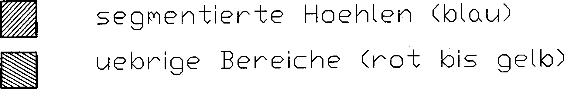
ZITATE ENTHALTEN IN DER BESCHREIBUNGQUOTES INCLUDE IN THE DESCRIPTION
Diese Liste der vom Anmelder aufgeführten Dokumente wurde automatisiert erzeugt und ist ausschließlich zur besseren Information des Lesers aufgenommen. Die Liste ist nicht Bestandteil der deutschen Patent- bzw. Gebrauchsmusteranmeldung. Das DPMA übernimmt keinerlei Haftung für etwaige Fehler oder Auslassungen.This list The documents listed by the applicant have been automated generated and is solely for better information recorded by the reader. The list is not part of the German Patent or utility model application. The DPMA takes over no liability for any errors or omissions.
Zitierte PatentliteraturCited patent literature
- - WO 2008/006451 A [0007] - WO 2008/006451 A [0007]
Zitierte Nicht-PatentliteraturCited non-patent literature
- - „MRI-Based Individual 3D Region-of-Interest Atlases of the Human Brain”, G. Wagenknecht, H.-J. Kaiser, U. Buell, O. Sabri, Methods Inf Med 4/2004, Seiten 383–390 [0005] "MRI-Based Individual 3D Region-of-Interest Atlases of the Human Brain", G. Wagenknecht, H.-J. Kaiser, U. Buell, O. Sabri, Methods Inf Med 4/2004, pages 383-390 [0005]
- - ”Individual 3D region-of-interest atlas of the human brain: automatic training point extraction for neural network based classification of brain tissue types”, G. Wagenknecht, H.-J. Kaiser, T. Obladen, O. Sabri, U. Buell. SPIE Electronic Imaging 2000, San Jose, Proc SPIE 3962: 150–161 [0005] - "Individual 3D region-of-interest atlas of the human brain: automatic training point extraction for neural network based classification of brain tissue types", G. Wagenknecht, H.-J. Kaiser, T. Obladen, O. Sabri, U. Buell. SPIE Electronic Imaging 2000, San Jose, Proc SPIE 3962: 150-161 [0005]
- - ”Individual 3D region-of-interest atlas of the human brain: neural network-based tissue classification with automatic training point extraction”, G. Wagenknecht, H.-J. Kaiser, T. Obladen, O. Sabri, U. Buell, SPIE Medical Imaging 2000, San Diego, Proc SPIE 3979: 306–317 [0005] - "Individual 3D region-of-interest atlas of the human brain: neural network-based tissue classification with automatic training point extraction", G. Wagenknecht, H.-J. Kaiser, T. Obladen, O. Sabri, U. Buell, SPIE Medical Imaging 2000, San Diego, Proc SPIE 3979: 306-317 [0005]
- - R. Standke: Technische Grundlagen der 18F-Fluorodeoxyglukose-Positronen-emissionstomographie-Diagnostik; Acta Medica Austriaca, Blackwell Verlag, 29. Jahrgang, Heft 5 2002, S. 149–155 [0009] - R. Standke: Technical basics of 18F fluorodeoxyglucose positron emission tomography diagnostics; Acta Medica Austriaca, Blackwell Verlag, Volume 29, Issue 5, 2002, pp. 149-155 [0009]
- - http://de.wikipedia.org/wiki/Positronen-Emissions-Tomographie#Korrektur_der_Messdaten [0009] - http://en.wikipedia.org/wiki/Positronen-Emissions-Tomography#Correct_Measurement_Data [0009]
- - C. Burger, G. Goerres, S. Schoenes, PET attenuation coefficients from CT images: experimental evaluation of the transformation of CT into PET 511-keV attenuation coefficients. EJNMMI, 29:922–927, 2002 [0009] - C. Burger, G. Goerres, S. Schoenes, PET attenuation coefficients from CT images: experimental evaluation of the transformation of CT into PET 511-keV attenuation coefficients. EJNMMI, 29: 922-927, 2002 [0009]
- - M. Hofmann, et al, MRI-Based Attenuation Correction for PET/MRI: A Novel Approach Combining Pattern Recognition and Atlas registration. JNM, 49:1875–1883, 2008 [0009] - M. Hofmann, et al., MRI-Based Attenuation Correction for PET / MRI: A Novel Approach Combining Pattern Recognition and Atlas registration. JNM, 49: 1875-1883, 2008 [0009]
- - Tyler DJ, Robson MD, Henkelman RM, Young IR, and Bydder GM Magnetic Resonance Imaging with Ultrashort TE (UTE) Pulse Sequences: Technical Considerations J Magn Reson Imaging (2007) 25:279–289 [0009] Magnetic Resonance Imaging with Ultrashort TE (UTE) Pulse Sequences: Technical Considerations J Magn Reson Imaging (2007) 25: 279-289. [0009]
- - http://en.wikipedia.org/wiki/Template_matching hervor (Stand 18. Juni 2009) [0024] - http://en.wikipedia.org/wiki/Template_matching (as of June 18, 2009) [0024]
- - http://de.wikipedia.org/wiki/Dilatation_(Bildverarbeitung) Stand 18. Juni 2009 [0026] - http://en.wikipedia.org/wiki/Dilatation_(Image Processing) As of June 18, 2009 [0026]
- - http://de.wikipedia.org/wiki/Erosion_(Bildverarbeitung) Stand 18. Juni 2009 [0028] - http://en.wikipedia.org/wiki/Erosion_(Image Processing) As of June 18th, 2009 [0028]
- - „Alternative methods for attenuation correction for PET images in MR-PET scanners”, E. Rota Kops, H. Herzog, IEEE NSS/MIC Conf Record 2007, pp. 4327–4330 [0065] "Alternative methods for attenuation correction for PET images in MR-PET scanners", E. Rota Kops, H. Herzog, IEEE NSS / MIC Conf. Record 2007, pp. 4327-4330 [0065]
- - „Magnetic resonance imaging guided attenuation and scatter corrections in threedimensional brain positron emission tomography”, H. Zaidi, M. L. Montandon, D. O. Slosman, Med. Phys., vol. 30, pp. 937–948, 2003 [0065] "Magnetic resonance imaging guided attenuation and scatter corrections in threedimensional brain positron emission tomography", H. Zaidi, ML Montandon, DO Slosman, Med. Phys., Vol. 30, pp. 937-948, 2003 [0065]
- - „Attenuation Correction in MR-PET Scanners with Segmented T1-weighted MR images”, E. Rota Kops, G. Wagenknecht et al., submitted to IEEE MIC 2009 [0065] "Attenuation Correction in MR-PET Scanners with Segmented T1-weighted MR images", E. Rota Kops, G. Wagenknecht et al., Submitted to IEEE MIC 2009 [0065]
Claims (33)
Priority Applications (6)
| Application Number | Priority Date | Filing Date | Title |
|---|---|---|---|
| DE102009027448A DE102009027448A1 (en) | 2009-07-03 | 2009-07-03 | Knowledge-based segmentation of weakening regions of the head |
| JP2012516698A JP5840125B2 (en) | 2009-07-03 | 2010-06-22 | Segmentation of head-related attenuation-related regions based on knowledge |
| US13/380,291 US8761482B2 (en) | 2009-07-03 | 2010-06-22 | Knowledge-based segmentation of attenuation-relevant regions of the head |
| HUE10724535A HUE035842T2 (en) | 2009-07-03 | 2010-06-22 | Knowledge-based segmentation of weakening-relevant regions of the head |
| PCT/EP2010/058811 WO2011000739A1 (en) | 2009-07-03 | 2010-06-22 | Knowledge-based segmentation of weakening-relevant regions of the head |
| EP10724535.9A EP2449528B1 (en) | 2009-07-03 | 2010-06-22 | Knowledge-based segmentation of weakening-relevant regions of the head |
Applications Claiming Priority (1)
| Application Number | Priority Date | Filing Date | Title |
|---|---|---|---|
| DE102009027448A DE102009027448A1 (en) | 2009-07-03 | 2009-07-03 | Knowledge-based segmentation of weakening regions of the head |
Publications (1)
| Publication Number | Publication Date |
|---|---|
| DE102009027448A1 true DE102009027448A1 (en) | 2011-01-05 |
Family
ID=42313930
Family Applications (1)
| Application Number | Title | Priority Date | Filing Date |
|---|---|---|---|
| DE102009027448A Withdrawn DE102009027448A1 (en) | 2009-07-03 | 2009-07-03 | Knowledge-based segmentation of weakening regions of the head |
Country Status (6)
| Country | Link |
|---|---|
| US (1) | US8761482B2 (en) |
| EP (1) | EP2449528B1 (en) |
| JP (1) | JP5840125B2 (en) |
| DE (1) | DE102009027448A1 (en) |
| HU (1) | HUE035842T2 (en) |
| WO (1) | WO2011000739A1 (en) |
Cited By (1)
| Publication number | Priority date | Publication date | Assignee | Title |
|---|---|---|---|---|
| EP2833163A4 (en) * | 2012-03-28 | 2015-11-25 | Nat Inst Radiolog | Method for generating pet absorption-corrected image from mr image and computer program |
Families Citing this family (14)
| Publication number | Priority date | Publication date | Assignee | Title |
|---|---|---|---|---|
| DE102009027448A1 (en) * | 2009-07-03 | 2011-01-05 | Forschungszentrum Jülich GmbH | Knowledge-based segmentation of weakening regions of the head |
| US10267882B2 (en) * | 2010-10-13 | 2019-04-23 | Toshiba Medical Systems Corporation | MRI T1 image-guided tissue diagnostics |
| WO2014115148A1 (en) | 2013-01-24 | 2014-07-31 | Tylerton International Holdings Inc. | Body structure imaging |
| US9836879B2 (en) * | 2013-04-16 | 2017-12-05 | Autodesk, Inc. | Mesh skinning technique |
| US10052495B2 (en) | 2013-09-08 | 2018-08-21 | Tylerton International Inc. | Detection of reduced-control cardiac zones |
| WO2015085320A1 (en) * | 2013-12-06 | 2015-06-11 | The Johns Hopkins University | Methods and systems for analyzing anatomy from multiple granularity levels |
| US10646183B2 (en) | 2014-01-10 | 2020-05-12 | Tylerton International Inc. | Detection of scar and fibrous cardiac zones |
| US10064589B2 (en) * | 2014-06-27 | 2018-09-04 | General Electric Company | Method, apparatus, and article for pet attenuation correction utilizing MRI |
| CN105491951B (en) | 2014-07-30 | 2020-11-20 | 纳维斯国际有限公司 | Registering nuclear medicine data |
| KR102298253B1 (en) * | 2019-03-05 | 2021-09-06 | 뉴로핏 주식회사 | Method and apparatus for brain image correction using brain structure |
| KR102207824B1 (en) * | 2019-03-05 | 2021-01-26 | 뉴로핏 주식회사 | Method and apparatus for brain image correction using brain structure |
| KR102298249B1 (en) * | 2019-03-05 | 2021-09-06 | 뉴로핏 주식회사 | Method and apparatus for brain image correction using brain structure |
| US11701067B2 (en) * | 2021-09-08 | 2023-07-18 | Siemens Medical Solutions Usa, Inc. | Attenuation correction-based weighting for tomographic inconsistency detection |
| CN116485819B (en) * | 2023-06-21 | 2023-09-01 | 青岛大学附属医院 | Ear-nose-throat examination image segmentation method and system |
Citations (1)
| Publication number | Priority date | Publication date | Assignee | Title |
|---|---|---|---|---|
| WO2008006451A1 (en) | 2006-07-12 | 2008-01-17 | Eberhard-Karls-Universität Tübingen | Method for determining a property map for an object in particular for a living being based on at least one first image in particular a nuclear magnetic resonance image |
Family Cites Families (7)
| Publication number | Priority date | Publication date | Assignee | Title |
|---|---|---|---|---|
| US6430430B1 (en) * | 1999-04-29 | 2002-08-06 | University Of South Florida | Method and system for knowledge guided hyperintensity detection and volumetric measurement |
| US6567684B1 (en) * | 2000-11-08 | 2003-05-20 | Regents Of The University Of Michigan | Imaging system, computer, program product and method for detecting changes in rates of water diffusion in a tissue using magnetic resonance imaging (MRI) |
| WO2006138702A2 (en) * | 2005-06-16 | 2006-12-28 | Russell Michael J | Guided electrical transcranial stimulation (gets) technique |
| US20080135769A1 (en) * | 2006-11-22 | 2008-06-12 | Rosen Bruce R | Attenuation correction of pet image using image data acquired with an mri system |
| EP2184010B1 (en) * | 2007-06-29 | 2019-05-08 | Toshinori Kato | White matter enhancing method |
| WO2009058915A1 (en) * | 2007-10-29 | 2009-05-07 | The Trustees Of The University Of Pennsylvania | Computer assisted diagnosis (cad) of cancer using multi-functional, multi-modal in-vivo magnetic resonance spectroscopy (mrs) and imaging (mri) |
| DE102009027448A1 (en) * | 2009-07-03 | 2011-01-05 | Forschungszentrum Jülich GmbH | Knowledge-based segmentation of weakening regions of the head |
-
2009
- 2009-07-03 DE DE102009027448A patent/DE102009027448A1/en not_active Withdrawn
-
2010
- 2010-06-22 EP EP10724535.9A patent/EP2449528B1/en not_active Not-in-force
- 2010-06-22 JP JP2012516698A patent/JP5840125B2/en not_active Expired - Fee Related
- 2010-06-22 HU HUE10724535A patent/HUE035842T2/en unknown
- 2010-06-22 US US13/380,291 patent/US8761482B2/en not_active Expired - Fee Related
- 2010-06-22 WO PCT/EP2010/058811 patent/WO2011000739A1/en active Application Filing
Patent Citations (1)
| Publication number | Priority date | Publication date | Assignee | Title |
|---|---|---|---|---|
| WO2008006451A1 (en) | 2006-07-12 | 2008-01-17 | Eberhard-Karls-Universität Tübingen | Method for determining a property map for an object in particular for a living being based on at least one first image in particular a nuclear magnetic resonance image |
Non-Patent Citations (14)
| Title |
|---|
| "Alternative methods for attenuation correction for PET images in MR-PET scanners", E. Rota Kops, H. Herzog, IEEE NSS/MIC Conf Record 2007, pp. 4327-4330 |
| "Attenuation Correction in MR-PET Scanners with Segmented T1-weighted MR images", E. Rota Kops, G. Wagenknecht et al., submitted to IEEE MIC 2009 |
| "Individual 3D region-of-interest atlas of the human brain: automatic training point extraction for neural network based classification of brain tissue types", G. Wagenknecht, H.-J. Kaiser, T. Obladen, O. Sabri, U. Buell. SPIE Electronic Imaging 2000, San Jose, Proc SPIE 3962: 150-161 |
| "Individual 3D region-of-interest atlas of the human brain: neural network-based tissue classification with automatic training point extraction", G. Wagenknecht, H.-J. Kaiser, T. Obladen, O. Sabri, U. Buell, SPIE Medical Imaging 2000, San Diego, Proc SPIE 3979: 306-317 |
| "Magnetic resonance imaging guided attenuation and scatter corrections in threedimensional brain positron emission tomography", H. Zaidi, M. L. Montandon, D. O. Slosman, Med. Phys., vol. 30, pp. 937-948, 2003 |
| "MRI-Based Individual 3D Region-of-Interest Atlases of the Human Brain", G. Wagenknecht, H.-J. Kaiser, U. Buell, O. Sabri, Methods Inf Med 4/2004, Seiten 383-390 |
| C. Burger, G. Goerres, S. Schoenes, PET attenuation coefficients from CT images: experimental evaluation of the transformation of CT into PET 511-keV attenuation coefficients. EJNMMI, 29:922-927, 2002 |
| http://de.wikipedia.org/wiki/Dilatation_(Bildverarbeitung) Stand 18. Juni 2009 |
| http://de.wikipedia.org/wiki/Erosion_(Bildverarbeitung) Stand 18. Juni 2009 |
| http://de.wikipedia.org/wiki/Positronen-Emissions-Tomographie#Korrektur_der_Messdaten |
| http://en.wikipedia.org/wiki/Template_matching hervor (Stand 18. Juni 2009) |
| M. Hofmann, et al, MRI-Based Attenuation Correction for PET/MRI: A Novel Approach Combining Pattern Recognition and Atlas registration. JNM, 49:1875-1883, 2008 |
| R. Standke: Technische Grundlagen der 18F-Fluorodeoxyglukose-Positronen-emissionstomographie-Diagnostik; Acta Medica Austriaca, Blackwell Verlag, 29. Jahrgang, Heft 5 2002, S. 149-155 |
| Tyler DJ, Robson MD, Henkelman RM, Young IR, and Bydder GM Magnetic Resonance Imaging with Ultrashort TE (UTE) Pulse Sequences: Technical Considerations J Magn Reson Imaging (2007) 25:279-289 |
Cited By (2)
| Publication number | Priority date | Publication date | Assignee | Title |
|---|---|---|---|---|
| EP2833163A4 (en) * | 2012-03-28 | 2015-11-25 | Nat Inst Radiolog | Method for generating pet absorption-corrected image from mr image and computer program |
| US9342903B2 (en) | 2012-03-28 | 2016-05-17 | National Institute Of Radiological Services | Method for generating image for PET attenuation correction from MR image and computer program |
Also Published As
| Publication number | Publication date |
|---|---|
| US8761482B2 (en) | 2014-06-24 |
| EP2449528A1 (en) | 2012-05-09 |
| HUE035842T2 (en) | 2018-05-28 |
| WO2011000739A1 (en) | 2011-01-06 |
| EP2449528B1 (en) | 2017-11-22 |
| JP2012531229A (en) | 2012-12-10 |
| JP5840125B2 (en) | 2016-01-06 |
| US20120155733A1 (en) | 2012-06-21 |
| WO2011000739A9 (en) | 2011-03-03 |
Similar Documents
| Publication | Publication Date | Title |
|---|---|---|
| EP2449528B1 (en) | Knowledge-based segmentation of weakening-relevant regions of the head | |
| DE102013103832A1 (en) | Damping correction in positron emission tomography using magnetic resonance imaging | |
| DE102010017432A1 (en) | Apparatus and method for isolating an area in an image | |
| DE102009025856A1 (en) | Processing and visualization technology of medical data | |
| DE102010004384B4 (en) | Method for determining information to be based on the calculation of an irradiation plan and combined magnetic resonance PET device | |
| DE102006033383A1 (en) | Method for determining a property map for an object, in particular for a living being, based on at least a first image, in particular nuclear magnetic resonance image | |
| DE112016004907T5 (en) | Virtual CT images from magnetic resonance images | |
| DE102008032996B4 (en) | Method for determining an attenuation map | |
| DE102007018260A1 (en) | Method and apparatus for relative perfusion and / or functionality | |
| DE102004043889B4 (en) | Method for generating a nuclear medical image | |
| DE102008058488A1 (en) | Method and apparatus for processing combined MR emission tomography images | |
| DE102011090047A1 (en) | Control procedure and control system | |
| DE102012208625B4 (en) | Method and system for processing MRI data of the human brain | |
| DE102018214325A1 (en) | Method and provision unit for the provision of a virtual tomographic stroke follow-up examination image | |
| DE102006059383A1 (en) | Method for producing resulting pictures of investigation object, involves detecting multiple magnetic resonance images of investigation object by magnetic resonance photograph mechanism | |
| EP3404436B1 (en) | Functional magnetic resonance imaging | |
| DE102013220018A1 (en) | Method for calculating inspection parameter for determining examination regions e.g. liver, involves determining examination area in second image through projection of segmented examination area or information derived from first image | |
| DE102012222073B4 (en) | Method for evaluating image data sets and combination image recording device | |
| DE102020211945A1 (en) | Method and arrangement for the automatic localization of organ segments in a three-dimensional image | |
| DE102015200850B4 (en) | Method for evaluating medical image data | |
| DE112016005742T5 (en) | Data processing method for computed tomography | |
| DE102019216745A1 (en) | Anonymization of medical image data | |
| DE102017208560A1 (en) | Functional magnetic resonance imaging | |
| DE102019217576B4 (en) | Identifying and providing misalignment image features | |
| DE102013214023A1 (en) | A method for evaluating and comparing temporally successive combined medical imaging examinations and a medical imaging system designed to carry out the method according to the invention |
Legal Events
| Date | Code | Title | Description |
|---|---|---|---|
| OP8 | Request for examination as to paragraph 44 patent law | ||
| R016 | Response to examination communication | ||
| R120 | Application withdrawn or ip right abandoned |