CN101044404A - Methods and apparatus for use in detection and quantitation of various cell types and use of optical bio-disc for performing same - Google Patents
Methods and apparatus for use in detection and quantitation of various cell types and use of optical bio-disc for performing same Download PDFInfo
- Publication number
- CN101044404A CN101044404A CNA2004800104965A CN200480010496A CN101044404A CN 101044404 A CN101044404 A CN 101044404A CN A2004800104965 A CNA2004800104965 A CN A2004800104965A CN 200480010496 A CN200480010496 A CN 200480010496A CN 101044404 A CN101044404 A CN 101044404A
- Authority
- CN
- China
- Prior art keywords
- cell
- target area
- bio
- cell type
- antibody
- Prior art date
- Legal status (The legal status is an assumption and is not a legal conclusion. Google has not performed a legal analysis and makes no representation as to the accuracy of the status listed.)
- Pending
Links
- 238000000034 method Methods 0.000 title claims abstract description 154
- 238000001514 detection method Methods 0.000 title claims description 30
- 230000003287 optical effect Effects 0.000 title abstract description 79
- 210000004027 cell Anatomy 0.000 claims abstract description 376
- 239000011159 matrix material Substances 0.000 claims description 46
- 210000001616 monocyte Anatomy 0.000 claims description 39
- 239000000427 antigen Substances 0.000 claims description 33
- 102000036639 antigens Human genes 0.000 claims description 33
- 108091007433 antigens Proteins 0.000 claims description 33
- 238000012360 testing method Methods 0.000 claims description 32
- 210000005087 mononuclear cell Anatomy 0.000 claims description 29
- 239000012530 fluid Substances 0.000 claims description 27
- 102000004190 Enzymes Human genes 0.000 claims description 24
- 108090000790 Enzymes Proteins 0.000 claims description 24
- 239000003795 chemical substances by application Substances 0.000 claims description 23
- 210000004698 lymphocyte Anatomy 0.000 claims description 22
- 238000011010 flushing procedure Methods 0.000 claims description 20
- 230000008569 process Effects 0.000 claims description 16
- 108010090804 Streptavidin Proteins 0.000 claims description 15
- PCHJSUWPFVWCPO-UHFFFAOYSA-N gold Chemical compound [Au] PCHJSUWPFVWCPO-UHFFFAOYSA-N 0.000 claims description 15
- 239000010931 gold Substances 0.000 claims description 15
- 229910052737 gold Inorganic materials 0.000 claims description 15
- 238000000151 deposition Methods 0.000 claims description 13
- 238000011160 research Methods 0.000 claims description 13
- 239000004793 Polystyrene Substances 0.000 claims description 12
- 229920002223 polystyrene Polymers 0.000 claims description 12
- 206010028980 Neoplasm Diseases 0.000 claims description 11
- 102100024222 B-lymphocyte antigen CD19 Human genes 0.000 claims description 9
- 101000980825 Homo sapiens B-lymphocyte antigen CD19 Proteins 0.000 claims description 9
- 230000005670 electromagnetic radiation Effects 0.000 claims description 9
- 238000002372 labelling Methods 0.000 claims description 9
- YBJHBAHKTGYVGT-ZKWXMUAHSA-N (+)-Biotin Chemical compound N1C(=O)N[C@@H]2[C@H](CCCCC(=O)O)SC[C@@H]21 YBJHBAHKTGYVGT-ZKWXMUAHSA-N 0.000 claims description 8
- 241000700605 Viruses Species 0.000 claims description 8
- 230000000704 physical effect Effects 0.000 claims description 8
- 239000003550 marker Substances 0.000 claims description 7
- 239000004417 polycarbonate Substances 0.000 claims description 7
- 229920000515 polycarbonate Polymers 0.000 claims description 7
- UAIUNKRWKOVEES-UHFFFAOYSA-N 3,3',5,5'-tetramethylbenzidine Chemical group CC1=C(N)C(C)=CC(C=2C=C(C)C(N)=C(C)C=2)=C1 UAIUNKRWKOVEES-UHFFFAOYSA-N 0.000 claims description 6
- HSTOKWSFWGCZMH-UHFFFAOYSA-N 3,3'-diaminobenzidine Chemical compound C1=C(N)C(N)=CC=C1C1=CC=C(N)C(N)=C1 HSTOKWSFWGCZMH-UHFFFAOYSA-N 0.000 claims description 6
- OXEUETBFKVCRNP-UHFFFAOYSA-N 9-ethyl-3-carbazolamine Chemical compound NC1=CC=C2N(CC)C3=CC=CC=C3C2=C1 OXEUETBFKVCRNP-UHFFFAOYSA-N 0.000 claims description 6
- VEXZGXHMUGYJMC-UHFFFAOYSA-M Chloride anion Chemical compound [Cl-] VEXZGXHMUGYJMC-UHFFFAOYSA-M 0.000 claims description 6
- 201000011510 cancer Diseases 0.000 claims description 6
- JPXMTWWFLBLUCD-UHFFFAOYSA-N nitro blue tetrazolium(2+) Chemical compound COC1=CC(C=2C=C(OC)C(=CC=2)[N+]=2N(N=C(N=2)C=2C=CC=CC=2)C=2C=CC(=CC=2)[N+]([O-])=O)=CC=C1[N+]1=NC(C=2C=CC=CC=2)=NN1C1=CC=C([N+]([O-])=O)C=C1 JPXMTWWFLBLUCD-UHFFFAOYSA-N 0.000 claims description 6
- LVSPDZAGCBEQAV-UHFFFAOYSA-N 4-chloronaphthalen-1-ol Chemical compound C1=CC=C2C(O)=CC=C(Cl)C2=C1 LVSPDZAGCBEQAV-UHFFFAOYSA-N 0.000 claims description 5
- 241000894006 Bacteria Species 0.000 claims description 5
- 108010031801 Lipopolysaccharide Receptors Proteins 0.000 claims description 5
- 239000013049 sediment Substances 0.000 claims description 5
- 102000002260 Alkaline Phosphatase Human genes 0.000 claims description 4
- 108020004774 Alkaline Phosphatase Proteins 0.000 claims description 4
- 108010001336 Horseradish Peroxidase Proteins 0.000 claims description 4
- 229960002685 biotin Drugs 0.000 claims description 4
- 235000020958 biotin Nutrition 0.000 claims description 4
- 239000011616 biotin Substances 0.000 claims description 4
- 238000012856 packing Methods 0.000 claims description 4
- IITIZHOBOIBGBW-UHFFFAOYSA-N 3-ethyl-2h-1,3-benzothiazole Chemical compound C1=CC=C2N(CC)CSC2=C1 IITIZHOBOIBGBW-UHFFFAOYSA-N 0.000 claims description 3
- 108010003639 CD56 Antigen Proteins 0.000 claims description 3
- 102000004652 CD56 Antigen Human genes 0.000 claims description 3
- OHDRQQURAXLVGJ-HLVWOLMTSA-N azane;(2e)-3-ethyl-2-[(e)-(3-ethyl-6-sulfo-1,3-benzothiazol-2-ylidene)hydrazinylidene]-1,3-benzothiazole-6-sulfonic acid Chemical compound [NH4+].[NH4+].S/1C2=CC(S([O-])(=O)=O)=CC=C2N(CC)C\1=N/N=C1/SC2=CC(S([O-])(=O)=O)=CC=C2N1CC OHDRQQURAXLVGJ-HLVWOLMTSA-N 0.000 claims description 3
- 238000006911 enzymatic reaction Methods 0.000 claims description 3
- 210000000222 eosinocyte Anatomy 0.000 claims description 3
- 108010087904 neutravidin Proteins 0.000 claims description 3
- 238000004445 quantitative analysis Methods 0.000 claims description 3
- 238000012113 quantitative test Methods 0.000 claims description 3
- 238000002310 reflectometry Methods 0.000 claims description 3
- 108010013709 Leukocyte Common Antigens Proteins 0.000 claims description 2
- 102000017095 Leukocyte Common Antigens Human genes 0.000 claims description 2
- 102100035877 Monocyte differentiation antigen CD14 Human genes 0.000 claims 2
- 101000980463 Treponema pallidum (strain Nichols) Chaperonin GroEL Proteins 0.000 claims 2
- 238000004891 communication Methods 0.000 claims 2
- 210000000056 organ Anatomy 0.000 claims 2
- 102000053028 CD36 Antigens Human genes 0.000 claims 1
- 108010045374 CD36 Antigens Proteins 0.000 claims 1
- 108010092372 Granulocyte-Macrophage Colony-Stimulating Factor Receptors Proteins 0.000 claims 1
- 102000016355 Granulocyte-Macrophage Colony-Stimulating Factor Receptors Human genes 0.000 claims 1
- 239000000020 Nitrocellulose Substances 0.000 claims 1
- 238000002835 absorbance Methods 0.000 claims 1
- 229920001220 nitrocellulos Polymers 0.000 claims 1
- 210000000265 leukocyte Anatomy 0.000 abstract description 21
- 239000000758 substrate Substances 0.000 abstract description 7
- 239000010410 layer Substances 0.000 description 76
- 102100036011 T-cell surface glycoprotein CD4 Human genes 0.000 description 72
- 239000000523 sample Substances 0.000 description 54
- LOKCTEFSRHRXRJ-UHFFFAOYSA-I dipotassium trisodium dihydrogen phosphate hydrogen phosphate dichloride Chemical compound P(=O)(O)(O)[O-].[K+].P(=O)(O)([O-])[O-].[Na+].[Na+].[Cl-].[K+].[Cl-].[Na+] LOKCTEFSRHRXRJ-UHFFFAOYSA-I 0.000 description 46
- 239000002953 phosphate buffered saline Substances 0.000 description 46
- 210000004369 blood Anatomy 0.000 description 44
- 239000008280 blood Substances 0.000 description 43
- 210000001744 T-lymphocyte Anatomy 0.000 description 42
- 238000004458 analytical method Methods 0.000 description 38
- 102100034922 T-cell surface glycoprotein CD8 alpha chain Human genes 0.000 description 34
- KFZMGEQAYNKOFK-UHFFFAOYSA-N Isopropanol Chemical compound CC(C)O KFZMGEQAYNKOFK-UHFFFAOYSA-N 0.000 description 18
- 230000005540 biological transmission Effects 0.000 description 18
- 238000010586 diagram Methods 0.000 description 17
- 238000003556 assay Methods 0.000 description 16
- 238000005516 engineering process Methods 0.000 description 16
- 210000000822 natural killer cell Anatomy 0.000 description 16
- 239000011324 bead Substances 0.000 description 15
- 208000037265 diseases, disorders, signs and symptoms Diseases 0.000 description 15
- 201000010099 disease Diseases 0.000 description 14
- 238000004820 blood count Methods 0.000 description 13
- 208000032839 leukemia Diseases 0.000 description 13
- 239000006228 supernatant Substances 0.000 description 13
- 230000008021 deposition Effects 0.000 description 12
- 239000000463 material Substances 0.000 description 12
- 239000004033 plastic Substances 0.000 description 12
- 239000000126 substance Substances 0.000 description 12
- 101000738771 Homo sapiens Receptor-type tyrosine-protein phosphatase C Proteins 0.000 description 11
- 102100037422 Receptor-type tyrosine-protein phosphatase C Human genes 0.000 description 11
- 239000011248 coating agent Substances 0.000 description 11
- 238000000576 coating method Methods 0.000 description 11
- 238000002156 mixing Methods 0.000 description 11
- 238000012546 transfer Methods 0.000 description 11
- -1 23A compound Chemical class 0.000 description 10
- 102100025237 T-cell surface antigen CD2 Human genes 0.000 description 10
- 150000001875 compounds Chemical class 0.000 description 10
- 238000005520 cutting process Methods 0.000 description 10
- 239000007788 liquid Substances 0.000 description 10
- 210000002381 plasma Anatomy 0.000 description 10
- 239000000243 solution Substances 0.000 description 10
- 238000002474 experimental method Methods 0.000 description 9
- 238000011156 evaluation Methods 0.000 description 8
- 238000002360 preparation method Methods 0.000 description 8
- 238000001035 drying Methods 0.000 description 7
- 230000009977 dual effect Effects 0.000 description 7
- 239000000203 mixture Substances 0.000 description 7
- 238000000926 separation method Methods 0.000 description 7
- XLYOFNOQVPJJNP-UHFFFAOYSA-N water Substances O XLYOFNOQVPJJNP-UHFFFAOYSA-N 0.000 description 7
- KCXVZYZYPLLWCC-UHFFFAOYSA-N EDTA Chemical compound OC(=O)CN(CC(O)=O)CCN(CC(O)=O)CC(O)=O KCXVZYZYPLLWCC-UHFFFAOYSA-N 0.000 description 6
- YXFVVABEGXRONW-UHFFFAOYSA-N Toluene Chemical compound CC1=CC=CC=C1 YXFVVABEGXRONW-UHFFFAOYSA-N 0.000 description 6
- 239000004411 aluminium Substances 0.000 description 6
- 229910052782 aluminium Inorganic materials 0.000 description 6
- XAGFODPZIPBFFR-UHFFFAOYSA-N aluminium Chemical compound [Al] XAGFODPZIPBFFR-UHFFFAOYSA-N 0.000 description 6
- 230000002380 cytological effect Effects 0.000 description 6
- 238000003384 imaging method Methods 0.000 description 6
- 238000003018 immunoassay Methods 0.000 description 6
- 229920002307 Dextran Polymers 0.000 description 5
- 210000001772 blood platelet Anatomy 0.000 description 5
- 238000006243 chemical reaction Methods 0.000 description 5
- 238000013016 damping Methods 0.000 description 5
- 210000003743 erythrocyte Anatomy 0.000 description 5
- 239000010408 film Substances 0.000 description 5
- 210000002443 helper t lymphocyte Anatomy 0.000 description 5
- 238000004519 manufacturing process Methods 0.000 description 5
- 229910052751 metal Inorganic materials 0.000 description 5
- 239000002184 metal Substances 0.000 description 5
- 239000000047 product Substances 0.000 description 5
- 238000003127 radioimmunoassay Methods 0.000 description 5
- 239000010409 thin film Substances 0.000 description 5
- 208000030507 AIDS Diseases 0.000 description 4
- 210000001266 CD8-positive T-lymphocyte Anatomy 0.000 description 4
- 101000581981 Homo sapiens Neural cell adhesion molecule 1 Proteins 0.000 description 4
- 241000725303 Human immunodeficiency virus Species 0.000 description 4
- 102100027347 Neural cell adhesion molecule 1 Human genes 0.000 description 4
- 230000004913 activation Effects 0.000 description 4
- 210000003719 b-lymphocyte Anatomy 0.000 description 4
- 230000009286 beneficial effect Effects 0.000 description 4
- 239000007767 bonding agent Substances 0.000 description 4
- 239000008367 deionised water Substances 0.000 description 4
- 229910021641 deionized water Inorganic materials 0.000 description 4
- 238000013461 design Methods 0.000 description 4
- 238000011161 development Methods 0.000 description 4
- 230000018109 developmental process Effects 0.000 description 4
- 239000000975 dye Substances 0.000 description 4
- 238000000684 flow cytometry Methods 0.000 description 4
- 230000006870 function Effects 0.000 description 4
- 239000011521 glass Substances 0.000 description 4
- 230000036039 immunity Effects 0.000 description 4
- 230000000670 limiting effect Effects 0.000 description 4
- 244000005700 microbiome Species 0.000 description 4
- 230000036961 partial effect Effects 0.000 description 4
- 244000052769 pathogen Species 0.000 description 4
- 230000001717 pathogenic effect Effects 0.000 description 4
- 238000004080 punching Methods 0.000 description 4
- 239000000725 suspension Substances 0.000 description 4
- 210000001519 tissue Anatomy 0.000 description 4
- 102000005482 Lipopolysaccharide Receptors Human genes 0.000 description 3
- 239000012491 analyte Substances 0.000 description 3
- 239000003146 anticoagulant agent Substances 0.000 description 3
- 229940127219 anticoagulant drug Drugs 0.000 description 3
- 210000000612 antigen-presenting cell Anatomy 0.000 description 3
- 238000013459 approach Methods 0.000 description 3
- 230000015572 biosynthetic process Effects 0.000 description 3
- 238000005119 centrifugation Methods 0.000 description 3
- 238000004140 cleaning Methods 0.000 description 3
- 238000011109 contamination Methods 0.000 description 3
- 238000000354 decomposition reaction Methods 0.000 description 3
- 238000003745 diagnosis Methods 0.000 description 3
- 238000001943 fluorescence-activated cell sorting Methods 0.000 description 3
- 208000015181 infectious disease Diseases 0.000 description 3
- 238000012986 modification Methods 0.000 description 3
- 230000004048 modification Effects 0.000 description 3
- 238000012544 monitoring process Methods 0.000 description 3
- 244000045947 parasite Species 0.000 description 3
- 238000012545 processing Methods 0.000 description 3
- 238000005070 sampling Methods 0.000 description 3
- 241000894007 species Species 0.000 description 3
- 102100022005 B-lymphocyte antigen CD20 Human genes 0.000 description 2
- 102000017420 CD3 protein, epsilon/gamma/delta subunit Human genes 0.000 description 2
- 108010041397 CD4 Antigens Proteins 0.000 description 2
- 101100346198 Caenorhabditis elegans mpc-2 gene Proteins 0.000 description 2
- 241000588724 Escherichia coli Species 0.000 description 2
- 102000001554 Hemoglobins Human genes 0.000 description 2
- 108010054147 Hemoglobins Proteins 0.000 description 2
- 101000897405 Homo sapiens B-lymphocyte antigen CD20 Proteins 0.000 description 2
- 101001057504 Homo sapiens Interferon-stimulated gene 20 kDa protein Proteins 0.000 description 2
- 101001055144 Homo sapiens Interleukin-2 receptor subunit alpha Proteins 0.000 description 2
- 101000917858 Homo sapiens Low affinity immunoglobulin gamma Fc region receptor III-A Proteins 0.000 description 2
- 101000917839 Homo sapiens Low affinity immunoglobulin gamma Fc region receptor III-B Proteins 0.000 description 2
- 102100027268 Interferon-stimulated gene 20 kDa protein Human genes 0.000 description 2
- 102100029185 Low affinity immunoglobulin gamma Fc region receptor III-B Human genes 0.000 description 2
- 101710160107 Outer membrane protein A Proteins 0.000 description 2
- 241001494479 Pecora Species 0.000 description 2
- 230000024932 T cell mediated immunity Effects 0.000 description 2
- 239000013543 active substance Substances 0.000 description 2
- 230000000890 antigenic effect Effects 0.000 description 2
- 238000000429 assembly Methods 0.000 description 2
- 230000000712 assembly Effects 0.000 description 2
- 230000027455 binding Effects 0.000 description 2
- 238000012984 biological imaging Methods 0.000 description 2
- 238000007413 biotinylation Methods 0.000 description 2
- 239000002981 blocking agent Substances 0.000 description 2
- 210000001124 body fluid Anatomy 0.000 description 2
- 239000010839 body fluid Substances 0.000 description 2
- 210000000170 cell membrane Anatomy 0.000 description 2
- 230000005859 cell recognition Effects 0.000 description 2
- 210000001175 cerebrospinal fluid Anatomy 0.000 description 2
- 230000008859 change Effects 0.000 description 2
- 239000003153 chemical reaction reagent Substances 0.000 description 2
- 238000003759 clinical diagnosis Methods 0.000 description 2
- 238000004132 cross linking Methods 0.000 description 2
- 230000006378 damage Effects 0.000 description 2
- 230000003247 decreasing effect Effects 0.000 description 2
- 238000002059 diagnostic imaging Methods 0.000 description 2
- 238000010790 dilution Methods 0.000 description 2
- 239000012895 dilution Substances 0.000 description 2
- 239000012153 distilled water Substances 0.000 description 2
- 238000002651 drug therapy Methods 0.000 description 2
- 239000000428 dust Substances 0.000 description 2
- 238000004043 dyeing Methods 0.000 description 2
- 239000011888 foil Substances 0.000 description 2
- 210000003714 granulocyte Anatomy 0.000 description 2
- 230000012447 hatching Effects 0.000 description 2
- 230000036541 health Effects 0.000 description 2
- 230000011132 hemopoiesis Effects 0.000 description 2
- 208000026278 immune system disease Diseases 0.000 description 2
- 238000010166 immunofluorescence Methods 0.000 description 2
- 230000033001 locomotion Effects 0.000 description 2
- 238000001000 micrograph Methods 0.000 description 2
- 230000000877 morphologic effect Effects 0.000 description 2
- 210000003887 myelocyte Anatomy 0.000 description 2
- 239000013642 negative control Substances 0.000 description 2
- 230000009826 neoplastic cell growth Effects 0.000 description 2
- 230000007170 pathology Effects 0.000 description 2
- YNPNZTXNASCQKK-UHFFFAOYSA-N phenanthrene Chemical compound C1=CC=C2C3=CC=CC=C3C=CC2=C1 YNPNZTXNASCQKK-UHFFFAOYSA-N 0.000 description 2
- 230000002265 prevention Effects 0.000 description 2
- 238000003672 processing method Methods 0.000 description 2
- 238000004393 prognosis Methods 0.000 description 2
- 230000002829 reductive effect Effects 0.000 description 2
- 238000005464 sample preparation method Methods 0.000 description 2
- 239000001509 sodium citrate Substances 0.000 description 2
- NLJMYIDDQXHKNR-UHFFFAOYSA-K sodium citrate Chemical compound O.O.[Na+].[Na+].[Na+].[O-]C(=O)CC(O)(CC([O-])=O)C([O-])=O NLJMYIDDQXHKNR-UHFFFAOYSA-K 0.000 description 2
- 239000007787 solid Substances 0.000 description 2
- PLXMOAALOJOTIY-FPTXNFDTSA-N Aesculin Natural products OC[C@@H]1[C@@H](O)[C@H](O)[C@@H](O)[C@H](O)[C@H]1Oc2cc3C=CC(=O)Oc3cc2O PLXMOAALOJOTIY-FPTXNFDTSA-N 0.000 description 1
- QGZKDVFQNNGYKY-UHFFFAOYSA-O Ammonium Chemical compound [NH4+] QGZKDVFQNNGYKY-UHFFFAOYSA-O 0.000 description 1
- 208000019901 Anxiety disease Diseases 0.000 description 1
- 102100038080 B-cell receptor CD22 Human genes 0.000 description 1
- 208000032791 BCR-ABL1 positive chronic myelogenous leukemia Diseases 0.000 description 1
- 206010004173 Basophilia Diseases 0.000 description 1
- 241001260012 Bursa Species 0.000 description 1
- FVYUIUUYNVUJOP-UHFFFAOYSA-N C1=CC=C2C(O)=CCC(Cl)(C#N)C2=C1 Chemical class C1=CC=C2C(O)=CCC(Cl)(C#N)C2=C1 FVYUIUUYNVUJOP-UHFFFAOYSA-N 0.000 description 1
- 108050005493 CD3 protein, epsilon/gamma/delta subunit Proteins 0.000 description 1
- 241000222122 Candida albicans Species 0.000 description 1
- 206010007134 Candida infections Diseases 0.000 description 1
- 208000010833 Chronic myeloid leukaemia Diseases 0.000 description 1
- KRKNYBCHXYNGOX-UHFFFAOYSA-K Citrate Chemical compound [O-]C(=O)CC(O)(CC([O-])=O)C([O-])=O KRKNYBCHXYNGOX-UHFFFAOYSA-K 0.000 description 1
- 102000015612 Complement 3b Receptors Human genes 0.000 description 1
- 108010024114 Complement 3b Receptors Proteins 0.000 description 1
- 206010011831 Cytomegalovirus infection Diseases 0.000 description 1
- 239000003109 Disodium ethylene diamine tetraacetate Substances 0.000 description 1
- 102100021260 Galactosylgalactosylxylosylprotein 3-beta-glucuronosyltransferase 1 Human genes 0.000 description 1
- WQZGKKKJIJFFOK-GASJEMHNSA-N Glucose Natural products OC[C@H]1OC(O)[C@H](O)[C@@H](O)[C@@H]1O WQZGKKKJIJFFOK-GASJEMHNSA-N 0.000 description 1
- 102000006354 HLA-DR Antigens Human genes 0.000 description 1
- 108010058597 HLA-DR Antigens Proteins 0.000 description 1
- HTTJABKRGRZYRN-UHFFFAOYSA-N Heparin Chemical compound OC1C(NC(=O)C)C(O)OC(COS(O)(=O)=O)C1OC1C(OS(O)(=O)=O)C(O)C(OC2C(C(OS(O)(=O)=O)C(OC3C(C(O)C(O)C(O3)C(O)=O)OS(O)(=O)=O)C(CO)O2)NS(O)(=O)=O)C(C(O)=O)O1 HTTJABKRGRZYRN-UHFFFAOYSA-N 0.000 description 1
- 208000009889 Herpes Simplex Diseases 0.000 description 1
- 101000884305 Homo sapiens B-cell receptor CD22 Proteins 0.000 description 1
- 101000894906 Homo sapiens Galactosylgalactosylxylosylprotein 3-beta-glucuronosyltransferase 1 Proteins 0.000 description 1
- 101001046687 Homo sapiens Integrin alpha-E Proteins 0.000 description 1
- 101000878605 Homo sapiens Low affinity immunoglobulin epsilon Fc receptor Proteins 0.000 description 1
- DGAQECJNVWCQMB-PUAWFVPOSA-M Ilexoside XXIX Chemical compound C[C@@H]1CC[C@@]2(CC[C@@]3(C(=CC[C@H]4[C@]3(CC[C@@H]5[C@@]4(CC[C@@H](C5(C)C)OS(=O)(=O)[O-])C)C)[C@@H]2[C@]1(C)O)C)C(=O)O[C@H]6[C@@H]([C@H]([C@@H]([C@H](O6)CO)O)O)O.[Na+] DGAQECJNVWCQMB-PUAWFVPOSA-M 0.000 description 1
- 206010061598 Immunodeficiency Diseases 0.000 description 1
- 108060003951 Immunoglobulin Proteins 0.000 description 1
- 102100022341 Integrin alpha-E Human genes 0.000 description 1
- 102100022297 Integrin alpha-X Human genes 0.000 description 1
- 108010032774 Interleukin-2 Receptor alpha Subunit Proteins 0.000 description 1
- 102000007351 Interleukin-2 Receptor alpha Subunit Human genes 0.000 description 1
- 102000004569 Leukocyte Immunoglobulin-like Receptor B1 Human genes 0.000 description 1
- 108010017736 Leukocyte Immunoglobulin-like Receptor B1 Proteins 0.000 description 1
- 102100038007 Low affinity immunoglobulin epsilon Fc receptor Human genes 0.000 description 1
- 102000004317 Lyases Human genes 0.000 description 1
- 108090000856 Lyases Proteins 0.000 description 1
- 206010027906 Monocytosis Diseases 0.000 description 1
- 208000033761 Myelogenous Chronic BCR-ABL Positive Leukemia Diseases 0.000 description 1
- 102000003729 Neprilysin Human genes 0.000 description 1
- 108090000028 Neprilysin Proteins 0.000 description 1
- 208000001388 Opportunistic Infections Diseases 0.000 description 1
- 241000233872 Pneumocystis carinii Species 0.000 description 1
- 206010035664 Pneumonia Diseases 0.000 description 1
- 239000004743 Polypropylene Substances 0.000 description 1
- 241000220317 Rosa Species 0.000 description 1
- 102100025244 T-cell surface glycoprotein CD5 Human genes 0.000 description 1
- 208000024799 Thyroid disease Diseases 0.000 description 1
- 201000005485 Toxoplasmosis Diseases 0.000 description 1
- 239000007983 Tris buffer Substances 0.000 description 1
- 230000002159 abnormal effect Effects 0.000 description 1
- 238000010521 absorption reaction Methods 0.000 description 1
- 239000002253 acid Substances 0.000 description 1
- 230000003213 activating effect Effects 0.000 description 1
- 230000004520 agglutination Effects 0.000 description 1
- 102000006707 alpha-beta T-Cell Antigen Receptors Human genes 0.000 description 1
- 108010087408 alpha-beta T-Cell Antigen Receptors Proteins 0.000 description 1
- 230000003321 amplification Effects 0.000 description 1
- 230000036506 anxiety Effects 0.000 description 1
- 230000006907 apoptotic process Effects 0.000 description 1
- 210000003651 basophil Anatomy 0.000 description 1
- 230000008901 benefit Effects 0.000 description 1
- 230000000975 bioactive effect Effects 0.000 description 1
- 239000012620 biological material Substances 0.000 description 1
- 230000033228 biological regulation Effects 0.000 description 1
- 239000012472 biological sample Substances 0.000 description 1
- 230000006287 biotinylation Effects 0.000 description 1
- 210000000601 blood cell Anatomy 0.000 description 1
- 238000009582 blood typing Methods 0.000 description 1
- 210000001185 bone marrow Anatomy 0.000 description 1
- 238000011095 buffer preparation Methods 0.000 description 1
- 201000003984 candidiasis Diseases 0.000 description 1
- 238000006555 catalytic reaction Methods 0.000 description 1
- 210000004970 cd4 cell Anatomy 0.000 description 1
- 238000000423 cell based assay Methods 0.000 description 1
- 230000001413 cellular effect Effects 0.000 description 1
- 230000008614 cellular interaction Effects 0.000 description 1
- 210000003850 cellular structure Anatomy 0.000 description 1
- 238000002512 chemotherapy Methods 0.000 description 1
- 230000001427 coherent effect Effects 0.000 description 1
- 239000000470 constituent Substances 0.000 description 1
- RDZGDBJFHDKEDD-UHFFFAOYSA-N dcho Chemical compound C1C2=CC=CCC2=C2OC2=C2CC=CC=C21 RDZGDBJFHDKEDD-UHFFFAOYSA-N 0.000 description 1
- 210000004443 dendritic cell Anatomy 0.000 description 1
- 238000000432 density-gradient centrifugation Methods 0.000 description 1
- 235000019301 disodium ethylene diamine tetraacetate Nutrition 0.000 description 1
- 239000006185 dispersion Substances 0.000 description 1
- 238000011978 dissolution method Methods 0.000 description 1
- 239000003814 drug Substances 0.000 description 1
- 230000000694 effects Effects 0.000 description 1
- 230000008030 elimination Effects 0.000 description 1
- 238000003379 elimination reaction Methods 0.000 description 1
- 230000002255 enzymatic effect Effects 0.000 description 1
- 210000003979 eosinophil Anatomy 0.000 description 1
- 239000007850 fluorescent dye Substances 0.000 description 1
- 238000001215 fluorescent labelling Methods 0.000 description 1
- 238000007710 freezing Methods 0.000 description 1
- 239000000499 gel Substances 0.000 description 1
- 230000002068 genetic effect Effects 0.000 description 1
- 239000008103 glucose Substances 0.000 description 1
- 238000010438 heat treatment Methods 0.000 description 1
- BGOFCVIGEYGEOF-UJPOAAIJSA-N helicin Chemical compound O[C@@H]1[C@@H](O)[C@H](O)[C@@H](CO)O[C@H]1OC1=CC=CC=C1C=O BGOFCVIGEYGEOF-UJPOAAIJSA-N 0.000 description 1
- 229960002897 heparin Drugs 0.000 description 1
- 229920000669 heparin Polymers 0.000 description 1
- 230000004727 humoral immunity Effects 0.000 description 1
- 239000000017 hydrogel Substances 0.000 description 1
- 102000018358 immunoglobulin Human genes 0.000 description 1
- 208000033065 inborn errors of immunity Diseases 0.000 description 1
- 238000011534 incubation Methods 0.000 description 1
- 201000006747 infectious mononucleosis Diseases 0.000 description 1
- 230000001524 infective effect Effects 0.000 description 1
- 230000005764 inhibitory process Effects 0.000 description 1
- 238000002347 injection Methods 0.000 description 1
- 239000007924 injection Substances 0.000 description 1
- 238000007689 inspection Methods 0.000 description 1
- 238000009434 installation Methods 0.000 description 1
- 230000002452 interceptive effect Effects 0.000 description 1
- 230000001678 irradiating effect Effects 0.000 description 1
- 238000002955 isolation Methods 0.000 description 1
- 238000011005 laboratory method Methods 0.000 description 1
- 206010024378 leukocytosis Diseases 0.000 description 1
- 239000003446 ligand Substances 0.000 description 1
- 210000000088 lip Anatomy 0.000 description 1
- 238000001459 lithography Methods 0.000 description 1
- 230000007774 longterm Effects 0.000 description 1
- 230000000527 lymphocytic effect Effects 0.000 description 1
- 210000003563 lymphoid tissue Anatomy 0.000 description 1
- 239000006166 lysate Substances 0.000 description 1
- 210000002540 macrophage Anatomy 0.000 description 1
- 239000006249 magnetic particle Substances 0.000 description 1
- 239000006148 magnetic separator Substances 0.000 description 1
- 238000013507 mapping Methods 0.000 description 1
- 238000005259 measurement Methods 0.000 description 1
- 230000007246 mechanism Effects 0.000 description 1
- 230000001404 mediated effect Effects 0.000 description 1
- 238000007431 microscopic evaluation Methods 0.000 description 1
- 238000000386 microscopy Methods 0.000 description 1
- 239000004005 microsphere Substances 0.000 description 1
- 238000013508 migration Methods 0.000 description 1
- 230000005012 migration Effects 0.000 description 1
- 239000002088 nanocapsule Substances 0.000 description 1
- 239000002105 nanoparticle Substances 0.000 description 1
- 210000004493 neutrocyte Anatomy 0.000 description 1
- 230000009871 nonspecific binding Effects 0.000 description 1
- 238000003199 nucleic acid amplification method Methods 0.000 description 1
- 239000002245 particle Substances 0.000 description 1
- 239000012071 phase Substances 0.000 description 1
- 238000000053 physical method Methods 0.000 description 1
- 210000004180 plasmocyte Anatomy 0.000 description 1
- 229920001155 polypropylene Polymers 0.000 description 1
- 229920000136 polysorbate Polymers 0.000 description 1
- 238000001556 precipitation Methods 0.000 description 1
- 238000004321 preservation Methods 0.000 description 1
- 230000003449 preventive effect Effects 0.000 description 1
- 208000028529 primary immunodeficiency disease Diseases 0.000 description 1
- 230000005180 public health Effects 0.000 description 1
- 208000008128 pulmonary tuberculosis Diseases 0.000 description 1
- 238000000746 purification Methods 0.000 description 1
- 238000004451 qualitative analysis Methods 0.000 description 1
- 238000011002 quantification Methods 0.000 description 1
- 230000003439 radiotherapeutic effect Effects 0.000 description 1
- 238000001959 radiotherapy Methods 0.000 description 1
- 239000002994 raw material Substances 0.000 description 1
- 239000013558 reference substance Substances 0.000 description 1
- 108091008146 restriction endonucleases Proteins 0.000 description 1
- 239000012488 sample solution Substances 0.000 description 1
- 238000012216 screening Methods 0.000 description 1
- 230000028327 secretion Effects 0.000 description 1
- 238000004062 sedimentation Methods 0.000 description 1
- 230000035945 sensitivity Effects 0.000 description 1
- 229910052708 sodium Inorganic materials 0.000 description 1
- 239000011734 sodium Substances 0.000 description 1
- 239000007790 solid phase Substances 0.000 description 1
- 125000006850 spacer group Chemical group 0.000 description 1
- 238000011895 specific detection Methods 0.000 description 1
- 230000003068 static effect Effects 0.000 description 1
- 238000003860 storage Methods 0.000 description 1
- 230000003319 supportive effect Effects 0.000 description 1
- 238000003786 synthesis reaction Methods 0.000 description 1
- 238000002560 therapeutic procedure Methods 0.000 description 1
- 230000004797 therapeutic response Effects 0.000 description 1
- 210000001541 thymus gland Anatomy 0.000 description 1
- 208000021510 thyroid gland disease Diseases 0.000 description 1
- 230000000451 tissue damage Effects 0.000 description 1
- 231100000827 tissue damage Toxicity 0.000 description 1
- 231100000167 toxic agent Toxicity 0.000 description 1
- 239000003440 toxic substance Substances 0.000 description 1
- 238000002834 transmittance Methods 0.000 description 1
- 230000001960 triggered effect Effects 0.000 description 1
- LENZDBCJOHFCAS-UHFFFAOYSA-N tris Chemical compound OCC(N)(CO)CO LENZDBCJOHFCAS-UHFFFAOYSA-N 0.000 description 1
- 241001430294 unidentified retrovirus Species 0.000 description 1
- 235000013311 vegetables Nutrition 0.000 description 1
Images


Classifications
-
- G—PHYSICS
- G01—MEASURING; TESTING
- G01N—INVESTIGATING OR ANALYSING MATERIALS BY DETERMINING THEIR CHEMICAL OR PHYSICAL PROPERTIES
- G01N33/00—Investigating or analysing materials by specific methods not covered by groups G01N1/00 - G01N31/00
- G01N33/48—Biological material, e.g. blood, urine; Haemocytometers
- G01N33/50—Chemical analysis of biological material, e.g. blood, urine; Testing involving biospecific ligand binding methods; Immunological testing
- G01N33/53—Immunoassay; Biospecific binding assay; Materials therefor
- G01N33/569—Immunoassay; Biospecific binding assay; Materials therefor for microorganisms, e.g. protozoa, bacteria, viruses
- G01N33/56966—Animal cells
- G01N33/56972—White blood cells
-
- G01N15/1433—
-
- G—PHYSICS
- G01—MEASURING; TESTING
- G01N—INVESTIGATING OR ANALYSING MATERIALS BY DETERMINING THEIR CHEMICAL OR PHYSICAL PROPERTIES
- G01N33/00—Investigating or analysing materials by specific methods not covered by groups G01N1/00 - G01N31/00
- G01N33/48—Biological material, e.g. blood, urine; Haemocytometers
- G01N33/50—Chemical analysis of biological material, e.g. blood, urine; Testing involving biospecific ligand binding methods; Immunological testing
- G01N33/53—Immunoassay; Biospecific binding assay; Materials therefor
- G01N33/543—Immunoassay; Biospecific binding assay; Materials therefor with an insoluble carrier for immobilising immunochemicals
- G01N33/54366—Apparatus specially adapted for solid-phase testing
-
- G—PHYSICS
- G01—MEASURING; TESTING
- G01N—INVESTIGATING OR ANALYSING MATERIALS BY DETERMINING THEIR CHEMICAL OR PHYSICAL PROPERTIES
- G01N33/00—Investigating or analysing materials by specific methods not covered by groups G01N1/00 - G01N31/00
- G01N33/48—Biological material, e.g. blood, urine; Haemocytometers
- G01N33/50—Chemical analysis of biological material, e.g. blood, urine; Testing involving biospecific ligand binding methods; Immunological testing
- G01N33/53—Immunoassay; Biospecific binding assay; Materials therefor
- G01N33/569—Immunoassay; Biospecific binding assay; Materials therefor for microorganisms, e.g. protozoa, bacteria, viruses
- G01N33/56966—Animal cells
-
- G—PHYSICS
- G01—MEASURING; TESTING
- G01N—INVESTIGATING OR ANALYSING MATERIALS BY DETERMINING THEIR CHEMICAL OR PHYSICAL PROPERTIES
- G01N35/00—Automatic analysis not limited to methods or materials provided for in any single one of groups G01N1/00 - G01N33/00; Handling materials therefor
- G01N35/00029—Automatic analysis not limited to methods or materials provided for in any single one of groups G01N1/00 - G01N33/00; Handling materials therefor provided with flat sample substrates, e.g. slides
- G01N35/00069—Automatic analysis not limited to methods or materials provided for in any single one of groups G01N1/00 - G01N33/00; Handling materials therefor provided with flat sample substrates, e.g. slides whereby the sample substrate is of the bio-disk type, i.e. having the format of an optical disk
-
- G01N15/149—
-
- G—PHYSICS
- G01—MEASURING; TESTING
- G01N—INVESTIGATING OR ANALYSING MATERIALS BY DETERMINING THEIR CHEMICAL OR PHYSICAL PROPERTIES
- G01N15/00—Investigating characteristics of particles; Investigating permeability, pore-volume, or surface-area of porous materials
- G01N15/10—Investigating individual particles
- G01N15/14—Electro-optical investigation, e.g. flow cytometers
- G01N2015/1486—Counting the particles
Abstract
Methods and apparatus for conducting differential cell counts including leukocytes and use of optical bio-discs for performing such cell counts is disclosed. The bio-disc includes a substantially circular substrate having a center and an outer edge, an active layer associated with the substrate; a target zone disposed between the center and the outer edge; and a plurality of capture antibodies bound to the active layer such that the antibodies are immobilized on the active layer in the target zone.
Description
The cross reference of related application
The application requires to obtain U.S. Provisional Application No.60/451, and 587 right of priority, this provisional application are to submit on March 3rd, 2003, and its integral body is attached to here as a reference.
Background of invention
1, invention field
The present invention relates generally to raji cell assay Raji, particularly the raji cell assay Raji that carries out on bio-optical disk.Clearer and more definite is, the present invention relates to comprise the method and apparatus of leukocytic cytological classification counting, and the application of bio-optical disk in carrying out this cell count, below described particular embodiment of carrying out according to best Implementation Modes these are not construed as limiting.
2, the discussion of correlation technique
Big quantity research need separate specific cells with diagnostic work from cell mixture, and analyzes.Particularly this source can be blood, cerebrospinal fluid, marrow, tumour homogenate, lymphoid tissue etc.
In the following up a case by regular visits to of diagnosis, treatment and definite patient health situation, use blood count.Complete blood count (CBC) is a battery of tests, comprises haemoglobin, packed cell volume, mean corpuscular hemoglobin, mean corpuscular hemoglobin, MCV, platelet count and white blood cell count(WBC).Blood count is that the red blood cell of every cubic millimeter of whole blood and leucocyte are counted.
White blood cell count(WBC) (WBC, leucocyte) is a leukocytic sum in the standard blood sample.Normal health philtrum, white blood cell count(WBC) are generally 4000-10800/microlitre (μ l).Can influence these values such as exercise, anxiety and disease.WBC raises can point out infection, leukaemia or tissue damage.If leucocyte drop to 1000/below the microlitre, the danger of take place infecting then increases.
The collected information of leukocyte differential count test surpasses the obtainable information of white blood cell count(WBC) itself.Arneth's count is used to estimate suspicious infection of New Development or heating (even CBC is normal), suspect exist with relevant unusually disease, abnormal white cell counting, doubtful leukaemia, other are unusual, such as Oncocytosis, monocytosis,mononucleosis and basophilia disease.Under severe leukaemia situation, may detect leucocyte or leukocyte differential count (for example after drug therapy) repeatedly.In therapeutic process, for example chemotherapy or radiotherapy, blood count is very important determining whether this treatment elimination cancer cell has also consumed healthy blood cell simultaneously.
Can measure Arneth's count by computerize cell count instrument.This machine is measured tale and five kinds of leukocytic percentages of main type.In normal individual, major part is neutrophil leucocyte (50%), secondly is lymphocyte (20-40%), then is monocyte (2-9%), is a small amount of eosinophilic granulocyte (1-4%) and basophil (0.5-2%) then.
In the lymphocyte kind, also have lymphocyte and hypotype cell.For example, lymphocyte can broadly be divided into T cell (coming from the lymphocyte of thymus gland) and B cell (lymphocyte that is equivalent to the bursa of farbricius), and they mainly are responsible for cell-mediated immunity and humoral immunity respectively.Although morphological feature has been used for the classification of leucocyte inside, only depending on morphology is the many functions that are not enough to differentiate lymphocyte subtype.In order to distinguish the lymphocyte of difference in functionality, developed many technology, comprise rose connection, IFM, the inspection of enzyme group and nearest monoclonal antibody analysis.Distinguish the T cell by surface indicia, surface indicia comprises the two kinds of gC D4 and the CD8 (CD4+T cell and CD8+T cell) on their surfaces.The CD4+ helper cell is relevant with antibody-mediated immunity.They combine with the antigen of B presented by cells.The result is the development that promotes the plasma cell clone of secretion antigen material antibody.The T cell also is essential for cell-mediated immunity.The CD4+ cell combines with the antigen that antigen presenting cell (APC) is presented, and antigen presenting cell for example is engulfing property macrophage and dendritic cell.This then T cell discharges lymphokine, and this lymphokine can be with other cytotaxis to this zone.The result is inflamed, and cell and the molecule of attempting shielding (wall off) and destruction antigenic substance gather.
CD8+, cell toxicant/inhibition type emiocytosis molecule, these molecules destroy the cell of their institute's combinations.If the target cell infective virus, then this function is extremely important, because before this cell can discharge a large amount of new virus that can infect other cells, it is destroyed usually.
HIV and AIDS
The human immunodeficiency virus is a kind of retrovirus, and it has high affinity to the CD4+T cell, so the CD4+T cell is effective target cell of this virus.Aids (AIDS) illustrates the importance of CD4+T cell in immunity vividlyly and tragicly.Human immunodeficiency virus (HIV) combines with the CD4 molecule, invades thus and infects the CD4+T cell.Along with the progress of disease, the quantity of CD4+T cell drops to below about 1000/ microlitre of normal range (μ l).A kind of explanation is that the continual work of patient CD8+T cell is to destroy infected CD4+T cell.
The number of CD4+T cell is lower than 400/ microlitre in blood, and the patient causes immunoreactive ability and sharply descends.Not only the patient is to the pathogen of the invading body super susceptible that becomes, and the microorganism, the especially bacterium that are safe from harm to normally the parasitizing tissue super susceptible that also becomes.Final patient dies from opportunistic infections, such as candidiasis, cytomegalovirus infection, herpes simplex infections, Pneumocystis carinii disease, pneumonia, toxoplasmosis, pulmonary tuberculosis etc.
CD4+T cell and CD8+T cell number and the CD4+T cell/estimation of CD8+T cells ratio are used to estimate immunocompromised host disease patient's immune state.For example, AIDS patient demonstrates the importance of CD4+T cell in immunity.Along with the progress of disease, the CD4+T cell number drops to below the about 1000/μ of the normal range l.Because the CD4+T cell that patient CD8+T cytoclasis infects.The CD4+T cell of uninfection may stand apoptosis.Therefore, the ratio of CD4+T cell and CD8+T cell becomes the diagnostic markers of this progression of disease.CD4+T cellular level of monitoring in the every 3-6 of all the infecteds month (in 600 laboratories of the U.S. every year carry out 4,000 ten thousand times detect) is advised by U.S. public health bureau.
Except CD4 and CD8, also have a lot of other cell surface antigens (for example CD3, CD16, CD19, CD45, CD56), they can be used for identifying lymphocytic hypotype.The ability that detects these cell surface antigens by antibody technique has increased new content for the diagnosis pathology, and various technology can be used for the research of hematopoiesis immunological diseases (for example AIDS, leukaemia and lymthoma) immunophenotype.Conventional skeptophylaxis determination method is such as radioimmunoassay (RIA), enzyme immunoassay (EIA), fluorescence immunoassay (FIA), and whether the existence of their application isotopes, enzyme or fluorescent material mensuration respective analyte.
The leukaemia immunophenotype is identified
Surface marker in the leukaemia helps to differentiate the tumour pedigree that is used to diagnose with judging prognosis.Comprehensive leukaemia phenotypic evaluation is from looking back clinical medical history and morphology, and selects a group mark for each case.In most of case, pedigree can be divided into T cell, B cell or myelocyte, and can diagnose or antidiastole.
The purpose of leukaemia phenotypic evaluation is to differentiate the cell type of neoplasia process.This phenotypic evaluation should be described the overview of cell lineage and ripe level, helps leukaemia or lymphadenomatous classification with this.In addition, this phenotypic evaluation should be beneficial to judges that this cell mass is normally or unusual, and is beneficial to detects cell mass noted earlier in sample, with alleviation, development or the recurrence of monitoring of diseases.
In blood or bone marrow prepare, carry out the leukaemia immunophenotype and identify, but also can use other body fluid or tissue.But can use through RBC dissolution method or the leucocyte that obtains such as the density gradient separation of phenanthrene thypaque sodium.If possible, before processing, check leucocyte tale and classification, and pair cell concentration is done correspondingly adjusting.
The lymthoma immunophenotype is identified
Surface marker in the lymthoma helps to differentiate the tumour pedigree that is used to diagnose with judging prognosis.Comprehensive leukaemia/lymthoma phenotypic evaluation is from looking back clinical medical history and morphology, and selects a group mark for each case.In most of case, pedigree can be divided into T cell, B cell or myelocyte, and diagnoses or antidiastole.
The purpose of lymthoma phenotypic evaluation is to differentiate the cell type of neoplasia process.This phenotypic evaluation should be described the overview of cell lineage and ripe level, helps lymphadenomatous classification with this.In addition, this phenotypic evaluation should be beneficial to judges that this cell mass is normally or unusual, and is beneficial to detects cell mass noted earlier in sample, with alleviation, development or the recurrence of monitoring of diseases.
Check complete blood count (comprising white blood cell count(WBC)).Blood count is the important indicator of this disease to therapeutic response.These countings also are important for grasping drug therapy or radiotherapeutic effect.Normal white blood cell count(WBC) is about the 4000-11000/ cubic millimeter in the blood.If the WBC tale surpasses 11000/mm
3, it is called leukocytosis, and this is a kind of normal reaction of body to infecting.Blood count helps to determine whether medicine works.Usually do the cell counting by the electronic calculator of costliness, FACS scanner for example, carrying out this detection needs professional technique.Whether the models show of every kind of cell type exists lymthoma and lymphadenomatous type.
The monoclonal antibody plate
Although it is just enough to use monochromatic immunofluorescence technique in some cases, many laboratory applications polychrome immunofluorescence techniques.The antibody that routine comprises is CD2, CD3, CD5, CD10, CD11c, CD14, CD19, CD20, CD22, CD23, CD25, CD45, CD103, FMC7, heavy chain, κ chain and λ chain.If clinical or morphological feature prompting " T " or " NK " lymphocyte disease, the associating antibody that will add below then also will checking: CD3/CD4/CD8, CD7/CD5/HLA-DR, CD25/CD2/CD3, CD16/CD56/CD19, CD57/CD8/CD3, TCR alpha-beta/δ-γ/CD3.
The ability that detects the cell related antigen by antibody technique has increased new content for the diagnosis pathology.Various technology can be used for the research of hematopoiesis immunological diseases immunophenotype.But, be used for the global detection method of numerous disease, need further develop the method for immunity of using the antibody antigen reaction, these diseases comprise the disease based on virus, such as aids and T chronic myeloid leukemia, and various cancer.Assay method of the present invention and bio-optical disk system can be used for realizing this immunoassays, and this area professional and technical personnel can clearly realize that this point.
Skeptophylaxis determination method commonly used, such as radioimmunoassay (RIA), enzyme immunoassay (EIA) and fluorescence immunoassay (FIA), they use isotope, enzyme or fluorescent material, whether there are these antibody or antigen and isotope, enzyme or fluorescent material generation idiosyncrasy with what detect corresponding antibodies separately or antigen.But said method exists limitation and shortcoming.RIA needs specific installation, prevention, limited half life period and various other factors.By measuring painted and luminous mensuration, these methods need responsive complicated instrument measuring heat or fluorescence reaction as the method for label for enzyme and fluorescent material, but also need several rinsing steps to remove excessive, not combination, unreacted reagent.In addition, above-mentioned detection method is applied to cell, particularly lymphocyte and cancer cell and similar sample need to improve the technology of efficient production, detection and analysis.
The strong instrument that develops according to the specific fluorescence antibody that is used for cell surface antigen is fluorescence-activated cell sorting technology (FACS).This is very reliable, quick and responsive method.Flow cytometry is the most practical method, and it is automatically and can be quantitative.The most important demand of sample convection type cell analysis is, this sample is in single dispersion suspension liquid, and with the required cell of fluorescence labeling substance markers.But it is the very high experiment of price, and total system needs, and well-trained technician handles with expensive instrument in the clinical analysis laboratory.Monoclonal antibody is as discontinuous probe, and flow cytometry is used for the objective quantitative test of a large amount of cells.
In addition, basic shortcoming is that once analyzed cell can not be used further to replicate analysis or other research, for example microexamination of rare cells.Developed many alternative technology, they are compared with flow cytometry and have superiority, and shortcoming is also arranged, and all have special separately problem.
The surface indicia analysis is important laboratory method, and it is particularly useful in the research of leukaemia, lymthoma and immune deficiency disorder.Based on the micro-permutation technology of antibody yes up-to-date technology, particularly in clinical diagnosis, it is used for the discriminating of sample specific antigen, and these samples comprise blood and tissue specimen.Most of diagnostic requirement of experiment are only measured limited group analyte (such as in cancer, leukaemia, lymthoma, thyroid disease).Therefore, miniaturization technologies only needs blood sample very in a small amount, and the time and the cost of saving lab assistant, and in single experiment, measure all clinical correlation parameters simultaneously, because its cost performance, work efficiency and simplification, these may be very attractive for hospital laboratory and mechanism of clinic.
We have developed a kind of easy, inexpensive system, the analysis, the mensuration and quantitative that are used for cell, especially for haemocyte, also comprise and infect blood and such as the parasite and the pathogen of the other biological liquid of CSF, the alternative prior art of this system is used for Cytometric system and method.Relevant information and signal processing method and software have also been developed, to differentiate various haemocytes, parasite and pathogen.
Compare with existing method and system, we have developed a kind of easy, miniature, hypersensitization, inexpensive cell analysis system.This system applies bio-optical disk, coherent detection assembly, and information and signal processing method and software.
Invention is summed up
Microtechnique is very valuable, particularly in the clinical diagnosis of identification of cell type, parasite, pathogen and other biological material.The Arneth's count that the present invention adopts microtechnique to carry out whole blood on bio-optical disk detects.In addition, the present invention relates to haemocyte imaging, Arneth's count mensuration and correlation process method and software.
Can adopt two kinds of methods to carry out this experiment or mensuration.First method is based on the photoimaging principle of haemocyte in the special ditch on the bio-optical disk.7 microlitre left and right sides whole bloods are injected in the ditch of particular design on the CD.With cell recognition software analysis image, this software can be differentiated the leucocyte hypotype that these are different, and produces Arneth's count.Second method is caught based on specific cell, and it uses the cell specific antibody at specific cell, is the antibody that is directed to lymphocyte (CD4, CD2, CD19), monocyte (CD14), eosinophilic granulocyte (CD15) etc. in this special circumstances.These leucocyte subtype sepcific antibody is installed/adhered to solid surface in bio-optical disk, and this bio-optical disk comprises a kind of flow chamber.
Identify and quantitative specificity in order to improve cell type; the cell that the particulate of available specific antibody coating or globule mark are caught; these specific antibodies or at research cell type, or at not needing in pearl-cell complexes or contamination of cells.The method can be distinguished specificity target cell and the contamination of cells in the capture area.Below in conjunction with accompanying drawing 18-24 globule should be used as in identification of cell described in further detail.
Use the bio-optical disk driven unit to rotate this CD, read and handle the coded message of storing on this CD, and analyze the cell capture area in this bio-optical disk flow chamber.This bio-optical disk driver has the motor that rotates this bio-optical disk, the controller of control CD speed of rotation, the processor of processing CD feedback signal, and the analyzer of analyzing processed signal.Speed of rotation is variable, and all can closely control speed of rotation by rotational speed and rotational time.Also but the applying biological CD writes on information on the bio-optical disk, and what this both can be driven device in the experiment material of flow chamber and target area reads light beam inquiry, and carries out before the analyzed device analysis, also can carry out thereafter.This bio-optical disk can comprise coded message, and it is used to control the rotation of CD; Process information is provided, and this information is to be directed to the cell immunoassay type that will implement especially; And have on the monitor and to show and the relevant result of biological driving.
Usually cytological classification is counted scheme, and particularly Arneth's count scheme is developed to CD, CD-R, DVD or DVD-R form, the revision of these forms, and their alternate formats.Driver read or inquire cells different in the beam detection analytical specimen and pearl-cell complexes, and produce image, the image that the analysis of available cytological classification Counting software is produced.
For finishing the cell counting measuring of these loaded down with trivial details efforts, microscopy or complicated cell counter are essential.The inventive method applying biological CD and assembly thereof.Can produce and analyze the optical image of various cell types free in the analysis room and pearl-cell complexes or the optical image of those cells of catching by the specific antibody method for catching by the cell recognition software program, this program can be differentiated various cell components in blood or other body fluid by the photoscanning characteristic.This method does not need sample is carried out any processing before analysis, for example is that cell dyeing, RBC eliminate and other numerous and diverse schemes.These methods comprise microscopic analysis or cell detection, and it uses a kind of optical-disc reader, and this reader has top detector, floor detection device, event counter or cell counter, below in conjunction with accompanying drawing they is described in detail.
For further accuracy and the degree of accuracy that improves cytological classification method of counting of the present invention, but the different cell mass of mark.For example, these marks can comprise that microsphere, fluorescent-labeled antibody and enzyme put together antibody.For example, pending trial U.S. Patent application No.10/121 when being disclosed in common transfer with the relevant otherwise detailed description of mark of sample and/or indication molecule, in 281, its name is called " Multi-Parameter Assays Including Analysis Discs andMethods Relating Thereto ", it is to submit on April 11st, 2002, and its integral body is attached to here as a reference.
The present invention or its different aspect can be easy to realize in many CDs, mensuration and system, perhaps be adapted to these CDs, mensuration and system, they are disclosed in the patented claim of following common transfer and while pending trial: U.S. Patent application No.09/378,878, name is called " Methodsand Apparatus for Analyzing Operational and Non-operationalData Acquired from Optical Discs ", is filed on August 23rd, 1999; U.S. Provisional Patent Application No.60/150,288, name is called " Methods and Apparatusfor Optical Disc Data Acquisition Using PhysicalSynchronization Markers ", is filed in for 1999 you on Augusts 23; U.S. Patent application No.09/421,870, name is called " Trackable Optical Discs withConcurrently Readable Analyte Material ", is filed on October 26th, 1999; U.S. Patent application No.09/643,106, name is called " Methods and Apparatusfor Optical Disc Data Acquisition Using PhysicalSynchronization Markers ", is filed on August 21st, 2000; U.S. Patent application No.09/999,274, name is called " Optical Biodiscs with ReflectiveLayers ", is filed in November 15 calendar year 2001; U.S. Patent application No.09/988,728, name is called " Methods and Apparatus for Detecting and QuantifyingLymphocytes with Optical Biodiscs ", is filed in November 16 calendar year 2001; U.S. Patent application No.09/988,850, name is called " Methods and Apparatus forBlood Typing with Optical Bio-discs ", is filed in November 19 calendar year 2001; U.S. Patent application No.09/989,684, name is called " Apparatus and Methodsfor Separating Agglutinants and Disperse Particles ", is filed in November 20 calendar year 2001; U.S. Patent application No.09/997,741, name is called " Dual BeadAssays Including Optical Biodiscs and Methods RelatingThereto ", is filed in November 27 calendar year 2001; U.S. Patent application No.09/997,895, name is called " Apparatus and Methods for Separating Components ofParticulate Suspension ", is filed in November 30 calendar year 2001; U.S. Patent application No.10/005,313, name is called " Optical Discs for MeasuringAnalytes ", is filed in Dec 7 calendar year 2001; U.S. Patent application No.10/006,371, name is called " Methods for Detecting Analytes Using Optical Discs andOptical Disc Readers ", is filed in Dec 10 calendar year 2001; U.S. Patent application No.10/006,620, name is called " Multiple Data Layer Optical Discs forDetecting Analytes ", is filed in Dec 10 calendar year 2001; U.S. Patent application No.10/006,619, name is called " Optical Disc Assemblies for PerformingAssays ", is filed in Dec 10 calendar year 2001; U.S. Patent application No.10/020,140, name is called " Detection System For Disk-Based Laboratory and ImprovedOptical Bio-Disc Including Same ", is filed in Dec 14 calendar year 2001; U.S. Patent application No.10/035,836, name is called " Surface Assembly forImmobilizing DNA Capture Probes and Bead-Based Assay IncludingOptical Bio-Discs and Methods Relating Thereto ", is filed in Dec 21 calendar year 2001; U.S. Patent application No.10/038,297, name is called " Dual BeadAssays Including Covalent Linkages for Improved Specificity andRelated Optical Analysis Discs ", is filed on January 4th, 2002; U.S. Patent application No.10/043,688, name is called " Optical Disc Analysis SystemIncluding Related Methods for Biological and Medical Imaging ", is filed on January 10th, 2002; U.S. Provisional Application No.60/348,767, name is called " Optical Disc Analysis System Including Related SignalProcessing Methods and Software ", is filed on January 14th, 2002; U.S. Patent application No.10/086,941, name is called " Methods for DNA ConjugationOnto Solid Phase Including Related Optical Biodiscs and DiscDrive Systems ", is filed on February 26th, 2002; U.S. Patent application No.10/087,549, name is called " Methods for Decreasing Non-Specific Bindingof Beads in Dual Bead Assays Including Related Optical Biodiscsand Disc Drive Systems ", is filed on February 28th, 2002; And U.S. Patent application No.10/099,256, name is called " Dual Bead Assays Using CleavableSpacers and/or Ligation to Improve Specificity and SensitivityIncluding Related Methods and Apparatus ", is filed on March 14th, 2002.
All these application integral body are attached to here as a reference.They repeat here fully, and therefore supportive background technology and open is provided.
The accompanying drawing summary
By following description of the preferred embodiment of the present invention, can know other purpose of the present invention, and its other feature that has and the advantage of bringing thus, these embodiments show in the accompanying drawings, all the time represent identical assembly with identical reference number, wherein:
Accompanying drawing 1 is a pictorial bio-optical disk of the present invention system;
Accompanying drawing 2 is decomposition diagrams of the used reflection bio-optical disk of the present invention;
Accompanying drawing 3 is last planimetric maps of CD shown in the accompanying drawing 2;
Accompanying drawing 4 is skeleton views of CD shown in the accompanying drawing 2, and shows the different layers of this CD by sectional view;
Accompanying drawing 5 is decomposition diagrams of the used transmission bio-optical disk of the present invention;
Accompanying drawing 6 is skeleton views of CD shown in the accompanying drawing 5, and shows the functional aspect of this CD semi-reflective layer by sectional view;
Accompanying drawing 7 is to show the diagram that concerns between the thickness of gold thin film and the transmission;
Accompanying drawing 8 is last planimetric maps of CD shown in the accompanying drawing 5;
Accompanying drawing 9 is skeleton views of CD shown in the accompanying drawing 5, and shows the different layers of this CD by sectional view, and it comprises the semi-reflective layer type shown in the accompanying drawing 6;
Accompanying drawing 10 is perspective block schemes of more detailed description accompanying drawing 1 system;
Accompanying drawing 11 is part cross-sectional view, and this figure is vertical with the radius of the bio-optical disk of reflection shown in 4 with accompanying drawing 2,3, and it shows the mobile ditch that wherein forms;
Accompanying drawing 12 is part cross-sectional view, and this figure is vertical with the radius of the bio-optical disk of transmission shown in 9 with accompanying drawing 5,8, and it shows mobile ditch and the upper detection device that wherein forms;
Accompanying drawing 13 is part profile figure of the bio-optical disk of reflection shown in accompanying drawing 2,3 and 4, and it shows the wobble groove that wherein forms;
Accompanying drawing 14 is part profile figure of transmission bio-optical disk shown in accompanying drawing 5,8 and 9, and it shows wobble groove and the upper detection device that wherein forms;
Accompanying drawing 15 is figures similar to accompanying drawing 11, and its shows the whole thickness of reflective optical disc and initially reflects performance;
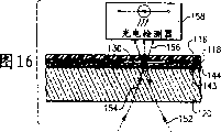
Accompanying drawing 16 is figures similar to accompanying drawing 12, and its shows the whole thickness of transmissive optical disc and initially reflects performance;
Accompanying drawing 17 is a kind of process flow diagrams, and its display application gradient cell isolation method separates leucocyte, and uses methods analyst blood sample of the present invention;
Accompanying drawing 18 is the diagrams with the globule labeled cell;
The diagram of accompanying drawing 19 embodiment of the present invention, its display application globule is to prevent not needing cell to combine on bio-optical disk with agent for capturing;
Accompanying drawing 20A and 20B are the diagrams of another embodiment of the present invention, and it shows the method step of differentiating dissimilar cells in the sample, and this method is used fixing target cell on this bio-optical disk of beads in different specific marker;
Accompanying drawing 21 is to use globule to catch the microorganism of being studied, and detects the diagram whether it exists with bio-optical disk;
Accompanying drawing 22 is to use the diagram that the globule mark does not need cell;
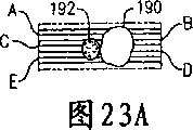
Accompanying drawing 23A is combined in the 1 micron indicator globule in the compound and the diagram of 5 microns cells, and this compound is positioned on the magnetic track of bio-optical disk of the present invention;
Accompanying drawing 23B is the series of features scintigram that is derived from accompanying drawing 23A compound, and it has used the detection signal of CD-ROM driver of the present invention;
Accompanying drawing 24 is the micrographs that do not adhere to globule, unlabeled cells and globule-cell complexes or labeled cell; And
Accompanying drawing 25A and 25B are the diagrams of another embodiment of the present invention, and it shows distinguishes the method step that does not need cell from target cell, and this method is to use the enzyme discriminating not need cell.
The present invention describes
The present invention relates to disk driving system, bio-optical disk and cytological classification and quantitative measurement.More particularly, the present invention relates to be used for the sorting technique of the quantitative various cell masses of biological sample of cell, for example comprise leucocyte, carrying out the application of this cell in quantitatively but also relate to bio-optical disk, below described particular embodiment of carrying out according to best Implementation Modes these are not construed as limiting.Below each of these aspects of the present invention all is described in further detail.
Drive system and associated optical disc
Accompanying drawing 1 is the skeleton view of bio-optical disk 110 of the present invention, and this CD is used to carry out cytological classification counting disclosed herein.With these bio-optical disk 110 demonstrations is CD drive 112 and monitor 114.
Accompanying drawing 2 is decomposition diagrams of the main structural components of 110 1 kinds of embodiments of bio-optical disk.Accompanying drawing 2 is the examples that can be used for bio-optical disk of the present invention 110 echo areas (after this being called " reflective optical disc ").Main structural components comprises cover 116, adherend 118 and matrix 120.Cover 116 comprises one or more inlets 122 and one or more outlet 124.Cover 116 can be formed by polycarbonate, and preferably at its bottom coating reflecting surface 146 (more detailed description in as accompanying drawing 4), can see this bottom from the skeleton view of accompanying drawing 2.In a preferred embodiment, trigger mark 126 is included on the surface in reflection horizon 142 (as more detailed description in the accompanying drawing 4).Trigger mark 126 can be included in the transparent window in all 3 layers of the bio-optical disks, the opacity, perhaps with the reflection or the half reflection district of information coding, it is sent to processor 166 with data, as shown in Figure 10, this processor transfers to interact with the operating function of inquiry or incident beam 152, as accompanying drawing 6 and 10.Second kind of parts shown in the accompanying drawing 2 are adherend 118, wherein have fluid circuit 128 or U type ditch.Punching press or cutting film are removed plastic foil and shape shown in forming forms fluid circuit 128 thus.Each fluid circuit 128 comprises mobile ditch 130 and returns ditch 132.Some fluid circuit 128 shown in the accompanying drawing 2 comprises mixing chamber 134.For example understand two kinds of dissimilar mixing chambers 134.First kind is symmetrical mixing chamber 136, and it is symmetrically formed facing to mobile ditch 130.Second kind is biasing mixing chamber 138.As shown in the figure, this biasing mixing chamber 138 is to form on one side of the ditch 130 that flows.Accompanying drawing 2 described the third parts are matrix 120, and it comprises target area or capture area 140.This matrix 120 preferably is made of polycarbonate, and has coating reflection horizon 142 in the above, sees accompanying drawing 4.By the reflection horizon 142 of shape shown in removing or arbitrarily the reflection horizon of required form form target area 140.Alternately, can form target area 140 by shield technology, this shield technology is included in uses shielding 140 zones, target area before the reflection horizon 142.This reflection horizon 142 can be formed by metal, such as aluminium or gold.
Accompanying drawing 3 is last planimetric maps of accompanying drawing 2 described bio-optical disks 110, and its reflection horizon 142 on cover 116 is shown as transparent, to expose fluid circuit 128, target area 140 and the trigger mark 126 that is positioned at disk inner.
Accompanying drawing 4 is the skeleton views according to echo area type bio-optical disk 110 amplifications of one embodiment of the present invention.This figure comprises the part of its each layer, and cutting them is in order to show the partial cross section figure of various compositions, layer, matrix, coating or film.Accompanying drawing 4 shows that matrix 120 is coated with apposition reflection horizon 142.On reflection horizon 142, be covered with active layer 144.In a preferred embodiment, this active layer 144 can be made of polystyrene.Alternately, but applying polycarbonate, gold, activation glass, modified glass or modified polystyrene, for example polystyrene-altogether-maleic anhydride.Go back the available water gel in addition.Alternately, other described in this embodiment, plastic bonding body 118 applies on active layer 144.The exposure of plastic bonding body 118 shows the U-shaped shape of cutting or punching press, and this shape constitutes fluid circuit 128.Last constituent structure layer is a cover 116 in this bio-optical disk echo area embodiment.Cover 116 comprises the reflecting surface 146 on its bottom.This reflecting surface 146 can be made of metal, such as aluminium or gold.
Accompanying drawing 5 is enlarged perspectives of the main structural components of transmission-type bio-optical disk 110 of the present invention.The main structural components of this transmission-type bio-optical disk 110 comprises 120 layers of cover 116, adherend 118 and matrix equally.This cover 116 comprises one or more inlets 122 and one or more outlet 124.This cover 116 can be made of layer of polycarbonate.Optional trigger mark 126 can be included on thin semi-reflective layer 143 surfaces, and accompanying drawing 6 and 9 has carried out best description to it.Trigger mark 126 can be included in the transparency window in all three layers of the bio-optical disks, the opacity, perhaps coding has the reflection or the half reflection district of information, it is sent to processor 166 with data, see accompanying drawing 10, this processor transfers to interact with the operating function of inquiry light beam 152, and the inquiry light beam is seen accompanying drawing 6 and 10.
Second kind of parts shown in the accompanying drawing 5 are adherends 118, wherein have fluid circuit 128 or U-shaped ditch.By punching press or cutting film, remove plastic foil, and shape shown in forming forms fluid circuit 128 thus.Each fluid circuit 128 comprises mobile ditch 130 and returns ditch 132.Some fluid circuit 128 shown in the accompanying drawing 5 comprises mixing chamber 134.Two kinds of dissimilar mixing chambers 134 have been described.First kind is symmetrical mixing chamber 136, and it forms symmetrically facing to the ditch 130 that flows.Second kind is biasing mixing chamber 138.As shown in the figure, this biasing mixing chamber 138 forms on one side of the ditch 130 that flows.
The third parts shown in the accompanying drawing 5 are matrix 120, and it can comprise target area or capture area 140.Matrix 120 preferably is made of polycarbonate, and has thin semi-reflective layer 143, and this layer applies in the above, sees accompanying drawing 6.The semi-reflective layer 143 relevant with the matrix 120 of CD 110 shown in accompanying drawing 5 and 6 obviously is thinner than the reflection horizon 142 on the matrix 120 of reflective optical disc 110 shown in accompanying drawing 2,3 and 4.Thin semi-reflective layer 143 makes the structural sheet of some transmission of inquiry light beam 152 by transmissive optical disc shown in the accompanying drawing 12.Should can constitute by metal by thin semi-reflective layer 143, such as aluminium or gold.
Accompanying drawing 6 is the matrix 120 of the 110 transmission embodiments of bio-optical disk shown in the accompanying drawing 5 and the skeleton view that semi-reflective layer 143 amplifies.Should can constitute by metal by thin semi-reflective layer 143, such as aluminium or gold.In a preferred embodiment, thin semi-reflective layer 143 thickness of transmissive optical disc shown in the accompanying drawing 5 and 6 are approximately 100-300 , and are no more than 400 .This semi-reflective layer 143 is thin can to make part incident or inquiry light beam 152 penetrate and by semi-reflective layer 143, and is detected by top detector 158, sees accompanying drawing 10, and some light is reflected or returns along the incident light path simultaneously.As follows, table 1 is listed the transmission and reflection characteristic of gold thin film, and it is relevant with the thickness of this film.The thickness of gold thin film layer is during greater than 800 , and it reflects fully.And transmittance is crossed the threshold density of gold thin film and is about 400 .
Table 1
| Gold thin film reflection and transmission (absolute value) | ||||
| Thickness (dust) | Thickness (nm) | Reflection | Transmission coefficient | |
| 0 | 0 | 0.0505 | 0.9495 | |
| 50 | 5 | 0.1683 | 0.7709 | |
| 100 | 10 | 0.3981 | 0.5169 | |
| 150 | 15 | 0.5873 | 0.3264 | |
| 200 | 20 | 0.7142 | 0.2057 | |
| 250 | 25 | 0.7959 | 0.1314 | |
| 300 | 30 | 0.8488 | 0.0851 | |
| 350 | 35 | 0.8836 | 0.0557 | |
| 400 | 40 | 0.9067 | 0.0368 | |
| 450 | 45 | 0.9222 | 0.0244 | |
| 500 | 50 | 0.9328 | 0.0163 | |
| 550 | 55 | 0.9399 | 0.0109 | |
| 600 | 60 | 0.9448 | 0.0073 | |
| 650 | 65 | 0.9482 | 0.0049 | |
| 700 | 70 | 0.9505 | 0.0033 | |
| 750 | 75 | 0.9520 | 0.0022 | |
| 800 | 80 | 0.9531 | 0.0015 | |
Except table 1, accompanying drawing 7 provides thin semi-reflective layer 143 reflections and the diagram of transmission performance inverse relation, and this is based on golden thickness.Reflection and transmission value used in 7 diagrammatic sketch of accompanying drawing are absolute value.
Accompanying drawing 8 is last planimetric maps of transmission-type bio-optical disk 110 shown in accompanying drawing 5 and 6, and its transparency cover part 116 can be exposed fluid ditch, trigger mark 126 and target area 140, and they all are positioned at the inside of this CD.
Accompanying drawing 9 is skeleton views that the bio-optical disk 110 of transmissive optical disc embodiment of the present invention amplifies.This CD 110 is shown as the part of each layer incision, to show the partial cross section figure of each composition, layer, matrix, coating or film.Accompanying drawing 9 shows the form of transmissive optical disc, and it has transparency cover part 116, the semi-reflective layer 143 that approaches on the matrix 120, and trigger mark 126.Trigger mark 126 comprises opaque material, and it places the upper part of lid.Alternately, trigger mark 126 can be made of transparent and unreflecting window, and it is etched on the thin reflection horizon 143 of CD, perhaps can be made of any mark, and this mark can absorb or not reflect the signal from trigger detector 160, sees accompanying drawing 10.Accompanying drawing 9 also shows, target area 140 according to shown in the zone of shape or alternative any required form mark appointment form.Can be in the thin semi-reflective layer 143 enterprising row labels of matrix 120 or matrix 120 bottom portions (under this CD), to show target area 140.Alternately, can form target area 140 by shield technology, this shield technology comprises except target area 140, shields whole thin semi-reflective layer 143.In this embodiment, by marking organizine wire mark is formed target area 140 on thin semi-reflective layer 143.Active layer 144 is applied on this thin semi-reflective layer 143.In a preferred embodiment, this active layer 144 is that thickness is 2% polystyrene layer of 40-200 μ m.Alternately, also can use polycarbonate, gold, activation glass, modified glass or modified polystyrene, such as polystyrene-common-maleic anhydride.In addition, also can use hydrogel.As described in the embodiment, plastic bonding body 118 is applied on active layer 144.The part that this plastic bonding body 118 appears shows cutting or punching press U-shaped shape, and they form fluid circuit 128.The final component structural sheet of bio-optical disk 110 transmission embodiments of the present invention is transparent and unreflecting covers 116, and it comprises inlet 122 and outlet 124.
Accompanying drawing 10 is perspective block schemes, and it describes optical assembly 148, produces the light source 150 of incident beam or inquiry light beam 152, Returning beam 154, and transmitted light beam 156.In the reflection bio-optical disk situation shown in the accompanying drawing 4, Returning beam 154 is that the reflecting surface 146 from these bio-optical disk 110 covers 116 is reflected.In the reflection embodiment of this bio-optical disk 110, with floor detection device 157 detection of reflected light beams 154, and the existence of analytic signal agent whether.On the other hand, in this transmission bio-optical disk form, detect transmitted light beams 156 by top detector 158, but and the existence of analytic signal agent whether.In this transmission embodiment, photodetector can be used as top detector 158.
Accompanying drawing 10 has also shown the hardware trigger structure, and it comprises trigger mark 126 and trigger detector 160 on this CD.All use this hardware trigger structure in reflection bio-optical disk (accompanying drawing 4) and the transmission bio-optical disk (accompanying drawing 9).Only when inquiry light beam 152 was on target area 140 separately, this triggered structure and just allows processor 166 to collect data.And, in transmission bio-optical disk system, but application software trigger also.The software trigger device is used the floor detection device, and when inquiry light beam 152 1 impinged upon on the edge of target area 140 separately, the software trigger device was then collected data with signalisation processor 166.Accompanying drawing 10 has also shown CD-ROM drive motor 162 and controller 164, the rotation of its control bio-optical disk 110.Accompanying drawing 10 has also shown processor 166 and analyzer 168, and they can randomly handle Returning beam 154 and the transmitted light beam 156 relevant with the transmission bio-optical disk.
Accompanying drawing 11 is partial cross sectional view of bio-optical disk 110 reflective optical disc embodiments of the present invention.Accompanying drawing 11 shows matrix 120 and reflection horizon 142.As mentioned above, reflection horizon 142 can be by constituting such as aluminium, gold or other suitable reflecting materials.In this embodiment, the upper surface of matrix 120 is smooth.Accompanying drawing 11 also shows active layer 144, and it applies on reflection horizon 142.As shown in Figure 11, remove one of reflection horizon 142 or part zone and form target area 140, perhaps alternately, before deposited reflection horizon 142, shield desired zone and form this target area 140 in desired location.Accompanying drawing 11 shows that also plastic bonding body 118 applies on active layer 144.Accompanying drawing 11 also shows the reflecting surface 146 of cover 116 and combination thereof.Therefore, when cover 116 being applied on the plastic bonding body 118 that is comprising required cutting shape, can form the ditch 130 that flows.Shown in arrow in the accompanying drawing 11, the light path of incident beam 152 is initial to be guiding matrix 120 below CD 110.Incident beam focuses on a bit of near reflection layer 142 then.Because this focusing occurs in target area 140, there is not reflection horizon 142 herein, so incident continues to pass through active layer 144 along light path, go forward side by side into mobile ditch 130.Incident beam 152 continues upwards to traverse mobile ditch then, is incident at last on the reflecting surface 146.At this point, incident beam 152 returns or reflects along the incident light path, therefore forms Returning beam 154.
The partial cross section figure of accompanying drawing 12 bio-optical disk 110 transmission embodiments of the present invention.Accompanying drawing 12 shows the transmissive optical disc form, the semi-reflective layer 143 that it has transparency cover part 116 and approaches on matrix 120.Accompanying drawing 12 also shows active layer 144, and it applies on thin semi-reflective layer 143.In a preferred embodiment, the semi-reflective layer 143 that transmissive optical disc is thin is made of a kind of metal, and such as aluminium or gold, its thickness is about the 100-300 dust, and is no more than 400 dusts.This thin semi-reflective layer 143 makes a part of incident or the inquiry light beam 152 of light source 150, sees accompanying drawing 10, penetrates and makes progress and pass through this CD, they will be detected by top detector 158, some light reflects back simultaneously, and its light path is identical with incident beam, but direction is opposite.In this arrangement, return or folded light beam 154 from semi-reflective layer 143 reflection.Therefore in this mode, Returning beam 154 does not enter the ditch 130 that flows.Reflected light or Returning beam 154 can be used to be tracked into irradiating light beam 152 according to prerecorded code track, and this code track is formed in the semi-reflective layer 143 or on it, and these will be described in more detail in attached Figure 13 and 14.In the CD embodiment shown in the accompanying drawing 12, the target area 140 of setting is not essential.Form target area 140 by on semi-reflective layer 143 thin on the matrix 120, carrying out direct mark.Can use silk screening or any suitable method is carried out these marks.In alternative embodiment, need not any physical markings determine the target area, the ditch 130 that effectively flows is as limited target area, and its research feature obtains checking.
Accompanying drawing 13 is sectional views of crosscut bio-optical disk 110 reflective optical disc embodiment magnetic tracks of the present invention.This figure carries out rip cutting formation along the radius of CD and mobile ditch.Accompanying drawing 13 comprises matrix 120 and reflection horizon 142.In this embodiment, matrix 120 comprises a series of grooves 170.This groove 170 is a spirality, and it extends to outer rim from the center of this CD.Design groove 170 is for inquiry light beam 152 can be advanced along the spiral groove on this CD 170.Such groove 170 is called " shake groove ".Have the fluctuation or the bottom of wavy sidewall and form groove 170, swell or the part lifted is separated spiral helicine adjacent grooves 170.Reflection horizon 142 is applied on groove 170 in this embodiment, and as shown in the figure, the reflection horizon is actually conformal.Accompanying drawing 13 shows that also active layer 144 applies on reflection horizon 142.As shown in Figure 13, remove one or a part of reflection horizon 142 and form target area 140, perhaps before deposited reflection horizon 142, form target area 140 with regard to shielding desired zone at desired area.Accompanying drawing 13 shows that also plastic bonding body 118 applies on active layer 144.The reflecting surface 146 that accompanying drawing 13 also shows cover 116 and links.Therefore, when cover 116 being applied on the plastic bonding body 118 that is comprising required cutting shape, just form the ditch 130 that flows.
Accompanying drawing 14 is the sectional views by bio-optical disk 110 transmissive optical disc embodiment magnetic tracks of the present invention, as shown in Figure 12.This figure carries out rip cutting formation along the radius of CD and mobile ditch.Accompanying drawing 14 shows matrix 120 and thin semi-reflective layer 143.This thin semi-reflective layer 143 penetrates by this CD the incident of light source 150 or inquiry light beam 152, and is detected by top detector 158, and some light reflects back with the form of Returning beam 154 simultaneously.Determine the thickness of this thin semi-reflective layer 143 by the reflected light of minimum, this reflected light is that optical-disc reader is needed, to keep its tracking power.Matrix 120 in this embodiment comprises a series of grooves 170 as described in the accompanying drawing 13.Groove 170 in this embodiment also is preferably spiral form, and it extends to outer rim near the center of CD.Design groove 170 is for inquiry light beam 152 can be advanced along this spiral.Accompanying drawing 14 has also shown active layer 144, and it applies on thin semi-reflective layer 143.Accompanying drawing 14 shows that also plastic bonding body 118 applies on active layer 144.Accompanying drawing 14 also shows cover 116, and it does not have reflecting surface 146.Therefore, when this lid applies on the plastic bonding body 118 that is comprising required cutting shape, then form the ditch 130 that flows, and allowing a part of incident beam 152 to pass through under the unreflecting situation basically.
Accompanying drawing 15 is similar with accompanying drawing 11, and it shows the gross thickness and the initial refraction performance thereof of reflective optical disc.Accompanying drawing 16 is similar with accompanying drawing 12, and it shows the gross thickness and the initial refraction performance thereof of transmissive optical disc.Can't see groove 170 in the accompanying drawing 15 and 16, because cut along groove 170 in these cross sections.There are narrow mobile ditch 130 in accompanying drawing 15 and 16 demonstrations, and it is vertical with groove 170 in these embodiments.Accompanying drawing 13,14,15 and 16 shows the gross thickness of each reflection and transmissive optical disc.In these features, demonstrate that incident beam 152 is initial to interact with the matrix 120 with refraction performance, matrix 120 changes the light path of incident beams as shown in the figure, makes this light beam 152 focus on the reflection horizon 142 or on the thin semi-reflective layer 143.
Separation Research cell from whole blood
The process flow diagram of accompanying drawing 17 imagerys shows the preparation analysis of blood sample, carries out bunch specifying (CD) marker determination to use above-mentioned bio-optical disk system.In step 1, blood (4-8ml) is directly gathered to 4 or 8ml Becton Dickinson CPT Vacutainer
TMAnd in the anti-coagulants, this anti-coagulants such as EDTA, acid citrate glucose (ACD) or heparin.In the suitable step of another embodiment of the present invention, 3ml blood in the anti-coagulants is superimposed to contains in the test tube 172 that separates gradient 176, this separates gradient 176 such as being Histopaque-1077 (Sigma Diagnostics, St.Louis, the Missouri State).In any case, preferably in 2 hours that gather, use blood sample 174.Under the room temperature, with 1500-1800RCF (2800rpm), with the test tube that separates gradient 176 containing of the blood sample 174 that superposeed 172 centrifugal 25 minutes, this hydro-extractor had horizontal motor and pendulum bucket in the protect against biological hazards hydro-extractor.After centrifugal, remove plasma layer 178 (step 2), on mononuclearcell (MNC) part 180, stay about 2mm blood plasma.Collect MNC layer 180, with phosphate-buffered saline (PBS) flushing.At room temperature centrifugal 10 minutes with 300RCF (1200rpm), remove residual blood platelet, make cell agglomerating.Remove supernatant, MNC group 180 is resuspended among the PBS by beaing test tube gently.Suspending with PBS, (step 3), and dilution make cell count reach 10000-30000/ μ l according to the flow thickness of ditches 130 of bio-optical disk 110 to last cell mass 180 again.Alternately, the applying biological activating agent can separate the T lymphocyte from whole blood, and this biologically active agent can make does not need cell agglutination and precipitation.The limiting examples of biologically active agent is PrepaCyte (BioE, St.Paul, the Minnesota State).PrepaCyte can separate the T lymphocyte by optionally removing granulocyte, blood platelet, monocyte, B cell (reaching 80%), natural killer (NK) cell (reaching 80%) from whole blood.Separation T lymphocyte method is applied to the raji cell assay Raji based on CD, the otherwise more detailed descriptions relevant with these methods, for example be disclosed in the U.S. Provisional Patent Application No.60/382 of common transfer, 327, name is called " Methods for Isolationof Lymphocytes for Use in Differential Cell Counting and Useof Optical Bio-disc for Performing Same ", it is filed on May 22nd, 2002, with its whole being attached to here as a reference.
Cell is differentiated, is detected and be quantitative
Carry out cell by the surface of coating monospecific antibody and catch, all cells that this (almost) causes containing this specific markers is captured.If target is to catch special cell type, t helper cell for example, it may be not enough using single labelled so.Therefore use multiple mark such as the technology of flow cytometry, and the cell that contains all selected markers is counted.T helper cell and monocyte all have CD4 antibody, therefore distinguish them by second kind of mark, and this second kind of mark is such as being CD3 (having only cd4 cell to contain) or CD19 (having only monocyte to contain).This brings a distinctive difficult problem for the application method that monospecific antibody carries out catching on the surface, because possibly can't avoid capturing monocyte.
Although it may be difficult avoiding secondary cell group to combine with the antibody coated surfaces, can carry out mark to them by using the flare with second kind of antibodies, can distinguish them thus.For example, the sort signal agent can be globule or dyestuff, and it absorbs the light of predetermined wavelength.This predetermined wavelength is preferably the wavelength of the incident beam 152 of optical-disc reader 112, or near the wavelength it.Therefore, although two kinds of cell masses all are trapped on the surface of bio-optical disk, can differentiate them by the described mensuration of top accompanying drawing 10 system.
As an example, CD4 antibody is deposited on dish 110 surfaces.Catch CD4+ monocyte and two kinds of subgroups of CD4+T auxiliary cell under its normal condition.But, in the sample preparations process, to introduce IR and absorb globule, it has applied CD19 antibody, causes these globules to be coated on the monocyte like this, because monocyte is the CD19+ cell, and t helper cell is not.Two kinds of effects are arranged like this.The first, by steric hindrance, it has reduced the possibility that monocyte combines with the CD4 capture area.The second, be combined in dish all the other monocytes on the capture area for the IR laser beam, it is darker than CD4+T auxiliary cell, therefore can differentiate between monocyte and t helper cell.
Particularly, when application hardware counting (according to the S Curves Recognition of threshold application) was counted by bio-optical disk of the present invention system, the gained monocyte absorbs should be enough to their S curve amplitude is reduced so far below the threshold value.Equally, if differentiate this monocyte with the IR dyestuff, then they are darker, and can be counted separately.Detailed description about hardware Counting software and S Curves Recognition is disclosed in common transfer pending trial and relevant U.S. Provisional Patent Application No.60/356 simultaneously, 982,60/372,007 and 60/408,227, their name is called " Bio-Disc andBio-Drive Analyser System Including Methods Relating Thereto ", is filed on February 13rd, 2002, on April 11st, 2002 and on September 4th, 2002 respectively.With they whole being attached to here as a reference, even repeat fully.
Can use these methods of double-tagging, be about to recognizable target or material and stick to cell surface, it is above-mentioned in conjunction with possibility to reduce perhaps to apply these cell surfaces, these methods can with the system combined use of the raji cell assay Raji of many types.For example, CD8 antibody capture NK cell, this NK cell of available CD56 mark (in the case, it is non-exclusive); Perhaps can carry out special marking, so that they and lymphocyte are distinguished in the capture area or the target area 140 of coating CD45 antibody to granulocyte.Do not need to use dyestuff, so only the relevant cell of coating: they also can be absorbed to the cell interior structure, such as nucleus non-specificly.If make them stable by fixation, and with known concentration with their premixeds in sample, then these cells can be used as the calibrating device unit on the capture area.
Also can use multiple labelling in principle.For example, a kind of flare can be the dyestuff on the specific antibody, and this antibody combines with mark on the cell.Second kind of flare can be particulate or the globule on the another kind of specific antibody, and this antibody can combine with another kind of mark.If there be second kind of mark, these globules will combine with cell, and stop this cell to combine with the agent for capturing of capture area.If only have first kind of mark, these cell generation combinations so, but can distinguish with the single marking on the cell.
The positive discriminating also is possible.For example, when with optical-disc reader imaging of the present invention, adhere to the cell of IR globule or with the cell of IR dyeing secretly in other cells, these cells can directly be counted.For example, be used for instant Cytometric separate type detector configurations, can use total signal (A+B) and detect, rather than use difference signal (A-B) than dark cell.Relate to the detailed description that is used for instant Cytometric separate type detecting device and S Curves Recognition and be disclosed in the common transfer U.S. Patent application No.10/279 of pending trial simultaneously, 677, its name is called " Segmented Area Detector for Biodrive and Methods RelatingThereto ", be filed on October 24th, 2002, with its whole being attached to here as a reference.To using the cell multiplex mark, differentiate that from the sample that contains dissimilar cells one or more cells describe in further detail below.
Use the particulate discriminating and separate target cell
Use above-mentioned optical disk system, the immunophenotype of determining lymphocyte subgroup by imaging analysis may need secondary gate or parameter with quantitative, to improve the accuracy that cell is differentiated.The present invention relates to application indicators or flare, they are such as being different globule of physical property or particulate, has or do not have functionalized surface, it can be puted together with at least a signal antibody, this signal antibody can be with target cell or is not needed cell-specific to combine, form pearl-cell complexes thus, this compound can be detected by optical-disc reader.This signal antibody can be puted together by crosslinking chemical and indicator or flare.Crosslinking chemical comprises that receptor-ligand binding or bond-affinity agent interact.For example, bond can be streptavidin or neutravidin.For example, affinity agent can be biotin.Alternately, this area professional and technical personnel can know that the globule with modification functionalized surface can be used for the signal antibody covalency is conjugated on the little bead surface, and impels this globule or indicator agent tight adhesion in the research cell.The globule that also may use different physical properties separates and/or the discriminating target cell, and these physical propertys are such as being size, color, texture, reflectivity, absorptance, quality, fluorescence, phosphorescence and/or magnetic.This process can be differentiated target cell better by different features, and this different feature is one type of indicator or the globule that adheres to this cell surface at least, below in conjunction with accompanying drawing 20A and 20B these is described.
Two or more specific cell types have on cell membrane and other subgroups or the identical epitope of cell type, on cell membrane, has T4 antigen such as T cell and monocyte, have CD3 on all mature T cells, and have CD45 on all leucocytes, then make a mistake this moment.Cause like this only depending on and use common capture antibodies, do not come the identification of cell type to encounter difficulties and use mark or sign and do not use the ability of measuring cytomorphology and size, this capture antibodies is such as being anti-CD4 or anti-CD5.
As described below, by globule is used as the tracer-labelling cell, target CD4+T cell and unwanted CD+ monocyte can be distinguished, they are caught with the anti-CD 4 antibodies agent for capturing.By the antigenic epitopes on sign or the mark monocyte, rather than the CD4 epi-position, can reach this purpose.For example, the globule mark monocyte of available adhesion CD14 antibody, this CD14 antibody is specific for monocyte.In case with anti-these monocytes of CD14 globule mark, then available bio-optical disk system carries out imaging analysis, the CD4+ monocyte (Figure 23 as described below) that one or more globules are combined with its CD14 surface antigen distinguishes with the CD4+T cell of no globule, and this is because of not having CD14 antigen at the CD4+T cell surface.Therefore, as shown in Figure 18, can differentiate CD4 T cell and monocyte in MNC or the blood sample.
Accompanying drawing 18 has shown lymphocyte 200 and monocyte 202, all has T4 antigen on their surface.Also have CD14 antigen on the monocyte surface, and lymphocyte does not have.Therefore, carry out mark by using the 204 pairs of monocytes of globule that adhere to signal antibody 206, itself and lymphocyte can be distinguished, signal antibody 206 is an anti-CD 14 antibody herein.This globule is preferred enough greatly, so that can be detected by the inquiry light beam 152 of optical-disc reader 112 (accompanying drawing 10), but less than the cell of institute's mark.This globule diameter preferably is about 0.5-5 μ m.Therefore when indicator compound 208 was placed the cell suspending liquid that contains CD4+ lymphocyte 200 and CD4+ monocyte 202, indicator compound 208 only combined with monocyte 202.When these CD4+ cells are caught on the catch site 140 that is adhering to anti-CD4 capture antibodies, and when analyzing, can differentiate these cells, this is described below in conjunction with accompanying drawing 23A and 23B by gained mark scanning figure with optical-disc reader 112 (accompanying drawing 1).
Accompanying drawing 19 has shown embodiments of the present invention, and this embodiment is described to use the globule separation or remove does not need cell in the blood sample.Use globule and blockade and do not need the antigen of cell surface, stop these cells of blockading to combine, do not need cell or contamination of cells by removing like this with capturing probe on the CD with this.In the embodiment shown in Figure 19, handle sample 210,, contain various types of cells 212 in this sample, comprise CD4+ cell, CD8+ cell and natural killer (NK) cell so that analyze.All these cells 212 all have the CD3 mark on its surface.CD4+ and CD8+ cell are the target cells of being studied 216, and NK cell 214 is not need cell.Use the anti-CD3 of common antibody and catch target cell.The NK cell contains CD56 antigen on its surface, and other cells in the sample do not have.As shown in the figure, antibody being applied globule 208 mixes with sample 210.This globule 208 has applied anti-CD56 antibody.Then the CD56 antigen of globule 208 with NK cell 214 surfaces is combined bow structure or hold the NK cell.The mensuration solution that will contain the NK cell 214 of CD4+, CD8+ and bow structure is then packed in the fluid chamber 130 of described bio-optical disk 110.CD4+ and CD8+ target cell 216 combine with the anti-CD3 agent for capturing 217 of optical disc surface then.The globule on NK cell 214 surfaces is by the CD3 epitope on the NK cell of blockading, and prevention NK cell 214 combines with anti-CD3 capture antibodies 217.Can or centrifugally remove unconjugated NK cell by flushing according to described then.Applying biological optical disk system (accompanying drawing 10) is analyzed the cell caught then, i.e. scanning is by the incident beam 152 (accompanying drawing 10) of target area, this incident beam with catch cell interaction, and analyze Returning beam 154, to determine the relative quantity of target cell in the sample.Alternately, if use the transmission-type CD, analyze the number that this transmitted light beam 156 (accompanying drawing 10) can determine to catch cell so.Bio-optical disk shown in the accompanying drawing 19 comprises top accompanying drawing 2 to 9 described CD compositions, and they comprise flow ditch 130, cover 116, reflecting surface 156, adherend 118, active layer 144, reflection horizon 142 and matrix 120.As mentioned above, also can use transmission-type bio-optical disk (accompanying drawing 8 and 9), wherein remove reflection horizon 146, and layer 142 is half reflections, it can make incident beam 152 by this CD, as described in accompanying drawing 10,12 and 16, uses top detector 158 and can detect and analyze transmitted light beam 156.
In the above in the alternate embodiments of accompanying drawing 19 described methods, will be bigger, heavier and/or magnetic bead sticks to and do not need on the cell.For example, target cell can be the CD4+T cell, is the CD4+ monocyte and do not need cell.Used capture antibodies can be anti-T4 antigen among this embodiment.Because this cell of two types all has T4 antigen at its cell surface, and monocyte has distinctive CD14 surface indicia, so the applied magnetic globule can be removed monocyte from sample, the quality of this magnetic bead is greater than the non magnetic globule of identical size and coating anti-CD 14 antibody.Quality is big more, can remove unwanted globule easily more by centrifugal.Before or after CD that sample is packed into, these globules can be mixed with sample solution.These globules are combined with surface C D14 antigen on the monocyte.Because magnetic-particle is heavier relatively, applying biological CD drive 112 can be separated these monocytes by centrifugal from the research cell, perhaps in conjunction with magnetic separator or magnetooptic disc system, use these monocytes of magnetic resolution of globule.For example, the otherwise detailed description relevant with the magnetooptic disc system is disclosed in the common transfer U.S. Patent application No.10/099 of pending trial simultaneously, 256, name is called " Dual BeadAssays Using Cleavable Spacers and/or Ligation to ImproveSpecificity and Sensitivity Including Related Methods andApparatus ", is filed on March 14th, 2002; U.S. Patent application No.10/099,266, name is called " Use of Restriction Enzymes and Other Chemical Methodsto Decrease Non-Specific Binding in Dual Bead Assays and RelatedBio-Discs; Methods; and System Apparatus for Detecting MedicalTargets ", is filed on March 14th, 2002; With U.S. Patent application No.10/307,263, name is called " Magneto-Optical Bio-Discs and Systems Including RelatedMethods ", is filed on November 27th, 2002, with they whole being attached to here as a reference.
Then accompanying drawing 20A and 20B have shown the non-limiting example of a kind of alternative embodiments of the present invention, and it combines description with accompanying drawing 19.In this embodiment, use globule with different physical properties, by the common surface mark, differentiate the different cell types of catching in one or more target areas or the capture area.Shown in accompanying drawing 20A, use pipettor 218, will contain the sample 210 of studying cell 216 bio-optical disk 110 of packing into.In this embodiment, leucocyte is the research cell, and they can be caught by the anti-CD45 capture antibodies on the optical disc surface 217.Capture antibodies 217 combines with common CD45 leukocyte surface antigen.According to accompanying drawing 20B, use pipettor 218 then, on the same group globule 220 is not packed in the CD 110.Every group of globule has different physical characteristicss and the different antibodies that adheres on it, and preferred available optical-disc reader 112 is differentiated its physical characteristicss.The antibody that adheres to every group of globule has affinity with the specific antigen of studying on the cell, for example one group of transparent bead 221 has CD3, the CD19 of labelled lymphocyte 224 or the antibody of CD56, one group of opaque globule 223 has the antibody that adheres on it with the CD14 of mark monocyte 222, and one group of translucent globule 225 has the antibody of the CD116 of mark eosinophil 226.Give when enough the chien shih globule with they separately target cell combine, will not remove by centrifugal or flushing then in conjunction with globule 227.Use optical-disc reader then,, can carry out quantitatively dissimilar cells according to physical characteristics in conjunction with globule on it.Because use the transmission-type CD in this embodiment, according to described, applying biological optical disk system (accompanying drawing 10) is analyzed globule-cell complexes, and its process is, scanning pass the target area with the interactional incident beam 152 of globule-cell complexes (accompanying drawing 10); Use photodetector and detect transmitted light beam 156; Analyze detected light beam, determine the relative quantity of all kinds target cell in the sample.In above-mentioned analysis, also can use the reflection-type CD.
Alternately, one or more groups globule or indicator can combine with the different surfaces antigen on the same cell type.Then by whether measuring on these cells, and this class cell is carried out quantitatively, and differentiate with the other types cell in conjunction with one or more type indicator.Used multiple parameter because differentiate the single type cell, so increased specificity for detecting like this.CD detects and the relevant otherwise detailed description of method of quantitative dissimilar cells with using, for example be disclosed in the U.S. Provisional Patent Application No.60/382 of common transfer, 944, name is called " Methods and Apparatus for Use in Detection and Quantitationof Cell Populations and Use of Optical Bio-Disc for PerformingSame ", be filed on May 24th, 2002, with its whole being attached to here as a reference.
Also can use particulate or globule sign or tagged tissue cell and such as virus and microorganism such as bacterium so that differentiate, classification and quantitative.The embodiment display application of accompanying drawing 21 is caught globule 204 mark Escherichia coli 234, and this seizure globule 204 has at least a biotin 232 and the capture antibodies 206 of puting together on it.Antigen on 206 pairs of Escherichia coli of antibody has special affinity.As described in Figure, when with these globules when containing colibacillary sample and mix, then form globule-bacterium compound 237.Use streptavidin 236 then, with compound 237 with do not catch on the capture area of CD in conjunction with globule 204.Whether exist according to top described analysis globule-bacterium compound then and measure.
Next accompanying drawing is an accompanying drawing 22, and the globule mark that its display application has antibody does not need cell, this antibody with do not need the antigen on the cell to have affinity.Therefore labels targets cell not, and they are separated or quantitatively with above-mentioned bio-optical disk system.
Accompanying drawing 23A is combined in the 1 micron indicator globule in globule-cell complexes and the diagram of 5 microns cells, and this compound is facing to the A-E magnetic track of bio-optical disk of the present invention.
Following accompanying drawing 23B shows the series of features scintigram of A-E magnetic track, and they are to use the detection signal of CD drive of the present invention and formed by globule and the cell of accompanying drawing 23A.The transmitted light beam 156 that these figure representatives are detected.As shown in the figure, the feature of 1 micron indicator globule 190 is obviously different with the feature of 5 microns cells 192, so that can detect globule-cell complexes, and it and individual cells are distinguished.When the detection Returning beam passed through globule or cell, sweep signal obviously departed from this Returning beam and is called as an incident.The existence that shows globule-cell complexes from these indicators and the more approaching incident of cell whether.As shown in the figure, the scintigram of these indicators and cell is similar each other, shows that they are in a compound.
Alternately, can use other detection methods, with the dissimilar cells in discriminating and the quantitative cell suspending liquid.For example, but indicator globule fluorescence or phosphorescence.Can in fluorescence or phosphorescence type optical-disc reader, carry out the detection of these indicators.For example, other signal detecting methods are described in the common transfer U.S. Patent application No.10/008 of pending trial simultaneously, 156, name is called " Disc Drive System and Methods for Use with Bio-Discs ", be filed in November 9 calendar year 2001, specially it be attached to here as a reference; The U.S. Provisional Application Nos.60/270 that submits to February 20 calendar year 2001,095 and the U.S. Provisional Application Nos.60/292 that submits to May 18 calendar year 2001,108; And U.S. Patent application No.10/043 above-mentioned, 688, name is called " Optical Disc Analysis System IncludingRelated Methods For Biological and Medical Imaging ", is filed on January 10th, 2002.
Accompanying drawing 24 shows the microphoto that does not adhere to globule 238, individual cells 240 and globule-cell complexes 242.Used globule is 4.5 μ m magnetic beads in this experiment, and it has applied anti-CD2 antibody.Therefore have the cell of CD2 antigen with these globule signs or mark, form globule-cell complexes, but not the CD2+ cell still remains individual cells.Below in conjunction with embodiment 8 this experiment is described in detail.
Use not lyase product discriminating target cell
Accompanying drawing 25A and 25B show the diagram of the another kind of embodiment of the inventive method, and this embodiment is a detected sediment of using enzyme reaction, will not need cell and target cell to distinguish.Accompanying drawing 25 shows the enlarged drawing of target area 140 in the fluid circuit 130.For the disclosure, this area professional and technical personnel will clearly realize that, no matter be that reflection-type CD or transmission-type CD all can be used for this analysis.In the embodiment shown in accompanying drawing 25A and the 25B, CD4 capture antibodies 244 is deposited on the target area of bio-optical disk 110 or the capture area 140 (step I).In next step Step II, the sample that will contain mononuclearcell (MNC) is introduced the target area.The cell that contains the CD4 surface antigen then will combine with CD4 capture antibodies 244 in this target area.These cells comprise CD4+T cell and monocyte.Described according to top accompanying drawing 19-21 then, will not rinse out in conjunction with cell, perhaps centrifugally go out the target area.Shown in Step II I, to remove not in conjunction with behind the cell, the solution that will contain enzyme 248 is introduced the target area, and wherein this enzyme and CD14 antibody 246 are puted together.As shown in the figure, because have only monocyte to contain CD14 antigen, only combine with the monocyte that is incorporated into the target area so put together the enzyme of antibody.Wash this target area then, remove not desmoenzyme or indicator, and in step IV, zymolyte introduced this target area.In case this substrate contacts with enzyme, enzyme-substrate reactions 250 then takes place, this reaction produces detectable product 252, shown in step V.This can detect product 252 and be preferably not lysate, and it forms sediment 254 at cell surface, shown in step VI.Use the inquiry beam flying target area of electromagnetic radiation 256 then, and, analyze Returning beam or transmitted light beam, to determine whether the sediment indicator cell line exists according to used optical disc types.Then to sign or labeled cell and sign or unlabeled cells do not carry out quantitatively.Shown in accompanying drawing 25B, this need carry out CD4+T cell and monocytic specific detection, discriminating and quantitatively.The enzyme that can be used for this embodiment includes, but are not limited to horseradish peroxidase (HRP) and alkaline phosphatase (AP).For example, the substrate that can be used in combination with these enzymes can be selected from TMB (3,3 ', 5,5 '-tetramethyl benzidine), DAB (3,3 '-diaminobenzidine), ABTS (2,2 '-azine group-two (3-ethyl benzo thiazole phenanthrolines-6-sulfonic acid)), AEC (3-amino-9-ethyl carbazole), NBT (nitroblue tetrazolium), CN/DAB (4-chloronaphthol/3,3 '-diaminobenzidine, the tetrahydrochysene chloride), (4-CN) 4-chloro-1-naphthols substrate compatible with other, these substrates can make enzyme generation catalytic reaction, and produce the product that can be detected by optical-disc reader.Experiment is described in detail
Though the present invention is described in detail by accompanying drawing, still the present invention is described further below with some embodiment.
Accompanying drawing 17 shows sample preparations, bio-optical disk application and result's process flow diagram is provided.The details of following embodiment, such as single duration, speed of rotation and other details of treatment step, it is described more special all to make a farfetched comparison Figure 17.Yet, the basic step of present embodiment and top described similar.
A. the optical disc production that comprises matrix preparation and chemical substance deposition
In this embodiment, remove all grits with air gun, and cleaning reflective optical disc or transmissive optical disc matrix 120 (being respectively accompanying drawing 2 and 5).Use spin coater, this CD is cleaned twice with isopropyl alcohol.2% polystyrene spin coated on CD, is obtained whole all very thick coating.
Sedimentation chemistry material then.A kind of embodiment comprises three step deposition approach, and it is hatched: streptavidin, hatched 30 minutes; The biotinylated first kind of antibody of b was hatched 60 minutes; And second kind of capture antibodies, hatched 30 minutes.Can produce first kind of antibody in first kind of species (for example sheep), it is aimed at the immunoglobulin (Ig) (for example IgG, IgE, IgM) of second kind of species (for example mouse).Produce second kind of capture antibodies in second kind of species, it is directed to specificity cell surface antigen (for example CD4, CD8).Carry out these steps in the moist chamber at room temperature, and between deposition, use flushing and drying steps.
1 μ l is contained the phosphate-buffered saline of streptavidin, and its ratio is 1mg/ml, is added on each window, and hatches 30 minutes.With distilled water unnecessary streptavidin is rinsed out, and dry CD.In conjunction with biotinylated anti-mouse IgG of equivalent (in PBS, concentration 125 μ g/ml) and activation dextran aldehyde (200 μ g/ml).The anti-mouse IgG of dextran aldehyde (DCHO)-biotinylation is added on each streptavidin of catching window, and in refrigerator, hatched 60 minutes or spend the night.Rinse out unnecessary reagent, this CD of Rotary drying.
B. CD assembling
Assemble this CD, it uses bonding coat, and this bondline thickness for example can be 25,50 or 100 microns (the ditch layers 118 in the accompanying drawing 2 and 5), and has punch-out, such as U-shaped or " e-rad " ditch to produce the fluid ditch; Also use transparency cover 116 (accompanying drawing 5, the transmissive optical disc that is used to have top detector) or have the lid 116 in reflection horizon 142, this reflection horizon is positioned on the capture area (accompanying drawing 2 is used to have the reflective optical disc of floor detection device).
In one embodiment, this CD is forward direction jittering device FDL21:13707 or FDL21:1270 CD-R dish, and it has applied the 300nm gold as the coded message layer.On reflective optical disc, by known lithography technique, on the reflection horizon, etch oval view window, size is 2 * 1mm.In the design of some transmissive optical disc, do not etch independent view window, and whole CD all is available.In this special embodiment, form the ditch layer by Fraylock bonding agent DBL 201 Rev C 3M94661.This lid is transparent CD, and it has 48 sample inlets, and the diameter of each inlet is 0.040 inch, equidistantly is arranged on the radius of 26mm.Use scanning of CD4/CD8 Counting software and reading of data dish, speed is 4X, and sampling rate is 2.67MHz.
C. CD omission
Because what analyze is blood,, guarantee that at first CD neither one chamber in rotary course that original position has a sample is missed so can carry out omission to this CD.Fill each ditch with blocking agent, this blocking agent is such as being StabilGuard and PBS-Tween.Blockade and continue at least 1 hour.CD is with 5000rpm rotation 5 minutes, to carry out omission and to check CD stability.After the omission, CD was placed vacuum chamber 24 hours.Vacuum was placed after 24 hours, CD is placed in the vacuum bag, and is stored in the refrigerator before use always.
D. collection of specimens, prepare and be applied to CD
Relate to the sample disposal step below, they are generally shown among the accompanying drawing 17A.For example use Becton Dickinson CPT Vacutainer, by density gradient centrifugation method purification of individual nucleus (MNC).Directly blood (4-8ml) is gathered to 4 or 8ml contain among the CPT Vacutainer of EDTA.Under the room temperature, in the hydro-extractor of protect against biological hazards, with 1500-1800 * g, with centrifugal 25 minutes of these test tubes, this hydro-extractor had horizontal motor and well-bucket.Preferably in 2 hours that gather, use blood.After centrifugal, remove the blood plasma on the mononuclearcell part, on the MNC layer, only keep about 2mm blood plasma.Collect MNC, and wash with PBS.Under the room temperature with 300 * g with cell centrifugation 10 minutes, make it agglomerating.Remove supernatant, be suspended among the PBS again by patting the little group that test tube will contain MNC.Carry out once flushing again with 300 * g under the room temperature, continue 10 minutes, to remove blood platelet.The final cell mass that suspends again, making cell count is 10000/μ l.18 μ l MNC are introduced one or more analysis rooms or ditch, at room temperature hatched 15 minutes, it is static that this CD keeps.Seal these ditches.CD was rotated 3-4 minute with 3000rpm with CD drive then.Preferably under the sampling rate of 4X speed and 2.67MHz, read this CD with software scans.
If can not handle blood sample immediately, so the CPT test tube is put upside down gently for several times, then the mononuclearcell after for the first time centrifugal can be suspended in the blood plasma again, and it at room temperature can be stored and reach 24 hours.In 24 hours, can collect the cell in the blood plasma according to the method described above, and wash.
E.CD4/CD8 measures form
The determination method of present embodiment is to belong to solid homogeneous phase cell together to catch determination method, and it is used for the fast measuring of absolute number and the CD4+ and the CD8+ lymphocyte ratio of blood sample CD4+ and CD8+T lymphocyte populations.This test is to carry out in being contained in the cell of CD-ROM, the cell number of CD4+, CD8+, CD2+, CD3+ and CD45+ that the specific antibody in district of being captured in these test determination 7 μ l mononuclearcells (MNC) is caught, this monocyte separates from whole blood.The principle of this test basis is that the location cell of specific position on the CD is caught.By catching the position application of chemical substance, on CD, set up several specific cell capture areas, these catch monoclonal antibody or the polyclonal antibody of the position application of chemical substance based on special haemocyte surface antigen.In case (30000/μ l) is full of cell with MNC blood, the cell of expressing CD4, CD8, CD2, CD3 and CD45 antigen then is trapped in the capture area of this CD.The negative control area that also in bar code, adds regulation.
F. dish is gone up and is analyzed
The MNC cell (18 μ l are in PBS) for preparing among the top step D is injected in the CD chamber, and seals the entrance and exit of this chamber.At room temperature this CD was hatched 15 minutes, scan with the 780nm laser in the CD-ROM drive then, and above-mentioned capture area is carried out imaging with top detector.
The software that is coded on the CD can instruct driver to automatically perform following process: (a) centrifugal CD, the excessive not in conjunction with cell of one or more stages removed in rotation, (b) the imaging specificity is caught window, (c) deal with data comprises the specificity of each capture area is caught the ratio (perhaps determining this ratio by program) that CD4/CD8 was counted and produced to cell.
In treatment step, this software reads the image of each capture area, and when running into cell they is carried out mark.For example, measure the number of CD4+ and CD8+ cell after, the ratio of this computed in software CD4+/CD8+ cell, and show the cell absolute number and the CD4+/CD8+ ratio of every microlitre whole blood in CD4+, CD8+, CD3+ and the CD45+ capture area.From CD being inserted CD-ROM drive to obtaining counting and ratio, the time of whole process is about 12 minutes.
G. agents useful for same
Streptavidin (Sigma, cat.# S-4762): add deionized water, form 5mg/ml solution, five equilibrium also is stored in-30 ℃.During use, add the Tris damping fluid, making final concentration is 1mg/ml.
Positive reference substance: CD45 (Sigma, Lot # 038H4892, cat # C7556).Be stored in 2-8 ℃.
Secondary capture antibodies: the anti-mouse IgG of biotinylation (resulting from sheep, Vectorlaboratories, lot # L0602, Catalog # BA-9200) stoste 1.5mg/ml, it prepares in distilled water.0.1M the b-IgG working fluid 125 μ g/ml among the PBS.Be stored in 2-8 ℃.But-30 ℃ of long term storage.
Activation dextran aldehyde (Pierce, lot # 97111761, cat # 1856167).Stoste 5mg/ml among the PBS is stored in 2-8 ℃.
Elementary capture antibodies: CD4 (DAKO, cat # M0716), CD8 (DAKO, cat #M0707), CD2 (DAKO, cat # M720), CD45 (DAKO, cat # M0701), CD14 (DAKO, cat # M825) and CD3 (DAKO, # M7193).Be stored in 2-8 ℃.
Negative control product: mouse IgG1 (DAKO, cat # X0931).Be stored in 2-8 ℃.
Phosphate-buffered saline (PBS), pH7.4 (Life Technologies/GIBCO BRL, cat.# 10010-023) or coordinator.Be stored in room temperature.
Isopropyl alcohol, 90-100%.
H.RBC dissolves scheme
The chloride leach damping fluid
The raw material of 1x chloride leach damping fluid should be stored in 2-8 ℃.It is by 0.155MNH
4Cl, 10mM KHCO
3Form with the 0.1mM disodium ethylene diamine tetraacetate; PH7.3-7.4.Be stored in 2-8 ℃.Be taken under the room temperature again before the use.
Step
1, in per 100 μ l blood, adds 2ml dissolving damping fluid (preferably carrying out this step in the fuming cupboard in protect against biological hazards).
2, whirling motion at room temperature and hatching 15 minutes.
3, use hydro-extractor in the protect against biological hazards fuming cupboard, at room temperature with 500 * g with centrifugal blood 5 minutes.
4, remove supernatant, with the PBS flushing cell that contains 2%FCS or FBS.Centrifuge cell.
5, calculate total white blood cells, making the WBC final concentration is 10000/μ l, to be used for the sample injection.
Embodiment 2
The mononuclearcell separating step
Use Becton ﹠amp; Dickinson Vacutainer CPT (the BD catalogue #362760 of 4ml, BD catalogue # 362761 of 8ml) cell preparation test tube, it contains sodium citrate.According to all protect against biological hazards preventive measure, in the protect against biological hazards fuming cupboard, finish these steps.Step is as follows:
1, blood is directly gathered to 4 or 8ml contain among the CPT Vacutainer of EDTA.If blood sample has been anti-freezing, then at first outwells the EDTA among the Vacutainer, and then the 6-8ml blood sample is poured in the CPT test tube.
2, under the room temperature, in the protect against biological hazards hydro-extractor with 1500-1800 * g with centrifugal 25 minutes of this test tube, this hydro-extractor has horizontal motor and well-bucket.In order to obtain best result, should be in two hours that gather centrifugal blood.And the centrifugal blood that is later than 2 hours can make the MNC decreased number, and RBC pollutes and increases.
3, after centrifugal, keep the blood plasma of about 2mm on the MNC layer, other are removed.Collect, and the mononuclearcell layer that will turn white is transferred in the 15ml taper centrifuge tube.
4,10-15mlPBS is added in the MNC layer,, mix these cells gently by putting upside down centrifuge tube for several times.
5, in the protect against biological hazards hydro-extractor, carried out centrifugal 10 minutes under the room temperature, wash cell with this with 200 * g.
6, remove supernatant.Pat test tube and these cells that suspend again.
7, in 10ml PBS, wash again one time.Under the room temperature centrifugal 10 minutes with 200-300 * g, to remove blood platelet.
8, remove supernatant, little group is resuspended among the 50 μ l PBS.
9, measure cell count in this sample.Carry out CBC,, mix gently, and use hemacytometer and carry out cell count perhaps with 2 μ l cell dilution to 18 μ l trypan blues.The whole cell count that makes sample is 10000/μ l, to analyze.
If 10 can not handle cell immediately, then by the CPT test tube being put upside down gently for several times, and mononuclearcell that will for the first time centrifugal back (top step 2) is suspended in the separated plasma again, stores under the room temperature 24 hours at the most.In 24 hours, collect the cell in the blood plasma, proceed flushing as mentioned above.
Cell number * 100 in total cell count/μ l=25 small square.
Embodiment 3
Use the MNC in the Histopaque-1077 separating whole blood
1ml Histopaque-1077 is placed in the 15ml centrifuge tube, lightly the 1ml whole blood is put thereon.Under the room temperature centrifugal 30 minutes then with 400 * g.(pasture pipette) aspirates potpourri carefully with Pasteur's dropper, and with opaque interfacial migration to centrifuge tube.Then, 10ml PBS is added in the centrifuge tube.Then with 250 * g with this solution centrifugal 10 minutes.Pour out supernatant, cell mass is suspended among the 10.0ml PBS again, and it was rotated 10 minutes with 250 * g.Then little group is suspended among the 10ml PBS again, and is rotated, cell is washed one time again with this with 250 * g.Whole cell mass is suspended among the 0.5ml PBS again.
Embodiment 4
With Dynal globule separation of C D4+ cell
A. material
1, cold PBS/2%FBS, pH7.4
2、PBS/0.5%BSA,pH7.4
3, the positive Dynal Seperating box of CD4
4, Dynal MPC, Dynal mixer, hydro-extractor, polypropylene test tube
B. step
Carry out CBC, and measure the needed globule quantity of each cell (4-10 globule/cell).The cold PBS/2%FBS of 1ml is added in the globule of aequum (1 * 10
7Individual globule/72 μ l), and again suspend.Test tube is placed among the Dynal MPC 30 seconds, removes supernatant.The globule of flushing is suspended into start volume again.The globule of aequum is added in the cell.In being set at 11 Dynal mixer, under 2-8 ℃, hatched 20 minutes.Bow structure cell among the separation Dynal MPC 2 minutes.Remove supernatant.In PBS/2%FBS, these bow structure cells are washed 4 times.This bow structure cell again suspends in 200-400 μ l PBS/2%FBS.Add 10 μ l Detach-a-Bead in per 100 μ l cell suspending liquids.In being set at 11 Dynal mixer, under room temperature, hatched 60 minutes.Globule among the separation Dynal MPC 2 minutes.Shift supernatant and preservation.In 500 μ l PBS/2%FBS, globule is washed 2-3 time, obtain residual cells.The cell that flushing is collected in 400 μ l PBS/0.5%BSA.
Operation CBC, the concentration of mensuration isolated cell.
Embodiment 5
CD preparation and chemical substance deposition (application streptavidin)
A. the optical disc production that comprises matrix preparation and chemical substance deposition
In this embodiment, remove grit, with cleaning transmissive optical disc matrix with air gun.This CD is installed in the spin coater, and it is cleaned twice with stable isopropyl alcohol stream.Then, polystyrene solution is coated on the CD equably, this polystyrene solution is dissolved in 2% polystyrene in 310ml toluene and the 65ml isopropyl alcohol and obtains.
For the streptavidin deposition, in PBS, streptavidin stoste is diluted to 1mg/ml.Needle deposition is deposited on about 1 μ l streptavidin in each capture area of CD by hand.In moist chamber, CD was hatched 30 minutes.Then, with deionized water flush away capture area unnecessary not in conjunction with streptavidin, and with this CD Rotary drying.
For the second antibody deposition, will activate the fresh solution and equivalent Vector IgG (among the PBS, the 125 μ g/ml) combination of dextran aldehyde (among the PBS, 200 μ g/ml).Needle deposition by hand is deposited on about 1 μ l IgG+DCHO compound in each capture area of CD (being positioned over above the streptavidin layer).In moist chamber, CD was hatched 60 minutes.With the unnecessary antibody of deionized water flush away, and with this CD Rotary drying.
For first antibody, in PBS, DAKO CD4 is diluted to 50 μ g/ml, in PBS, DAKO CD8 is diluted to 25 μ g/ml, in PBS, DAKO CD45 is diluted to 145 μ g/ml.The pin type applicator is deposited on the various first antibodies of about 1 μ l above the second antibody of combination by hand.In moist chamber, CD was hatched 30 minutes then.Wash capture area with PBS, unnecessary unconjugated antibody is removed, and with the CD Rotary drying.
B. CD assembling
Used lid CD is transparent CD, and it has the Fraylock bonding agent ditch layer that adheres on it.Stamp out 4 U-shaped ditches on this bonding agent, they become fluid circuit.This lid is placed on the transmissive optical disc matrix, the ditch that flows is in above the capture area.Then, they are pressed 8 times, guarantee that these CDs keep together by CD.
C. CD omission, blockade
Fill each mobile ditch with StableGuard, hatched 1 hour.Between incubation period, in spin coater, CD was rotated 5 minutes with 5000rpm.After the rotation, the CD ditch is carried out omission.Sucking-off StableGuard from these ditches then, and place the vacuum of vacuum chamber to spend the night the CD.Be placed on CD in the vacuum bag second day morning, is stored under 4 ℃.
Embodiment 6
Bio-optical disk CD4/CD8 ratio and FAC CD4/CD8 ratio are relatively
A. prepare clinical blood sample with the CPT test tube
(No.29,30,31,32,33,34 and 35) pours in the single CPT test tube with the clinical EDTA blood sample of 3ml, and the sodium citrate in this test tube is removed.At room temperature with 1500 * g with centrifugal 25 minutes of these test tubes.After centrifugal, take out top blood plasma, i.e. opaque MNC layer in the 0.5cm.To remain opaque MNC layer and move in the clean 15ml test tube, and in each test tube, add 12ml PBS.
Flushing
Then with 250 * g with centrifugal 10 minutes of cell suspending liquid.Take out supernatant, and cell is suspended among the 14ml PBS again.Again with 250 * g with centrifugal 10 minutes of suspending liquid.Take out supernatant, with the 200-175 μ l PBS little group of each cell that suspends again.With hemacytometer these cells are counted, determined the cell concentration of each sample with this.The final concentration of cells of each sample is adjusted to 30000/μ l.
B. bio-optical disk CD4/CD8 ratio and FAC CD4/CD8 ratio are relatively
Use 25um bonding agent ditch, according to preparing CD (No.27a, 27b, 27c, 27d, 27e, 27f and 28) with embodiment 5 similar methods.
Shown in following table 2, each sample is injected in corresponding each CD.After 30 minutes, with 3000rpm with centrifugal 5 minutes of this CD.Take the light micrograph of the cell of catching in the chemistry district then, these cells are counted.Also each clinical samples being carried out FAC analyzes.This result of experiment is presented in the table 2.
Table 2
Bio-optical disk CD4/CD8 ratio and FAC CD4/CD8 ratio are relatively
| Reel number | Mark this shop | Cd cd 4/CD8 | FAC CD4/CD8 |
| 27a | 29 | 2.39 | 2.43 |
| | 30 | 1.47 | 1.67 |
| 27c | 31 | 0.8 | 0.98 |
| 27d | 32 | 1.84 | 2.16 |
| 27e | 33 | 0.96 | 1.14 |
| 27f | 34 | 1.59 | 1.49 |
| 28 | 35 | 1.03 | 1.04 |
Embodiment 7
CD preparation and chemical substance deposition
In this embodiment, remove grit, with cleaning transmissive optical disc matrix with air gun.This CD is installed in the spin coater, and it is cleaned twice with stable isopropyl alcohol stream.Then, polystyrene solution is coated on the CD equably, this polystyrene solution is dissolved in 2% polystyrene in 310ml toluene and the 65ml isopropyl alcohol and obtains.
For the second antibody deposition, will activate the fresh solution and equivalent Vector IgG (among the PBS, the 125 μ g/ml) combination of dextran aldehyde (among the PBS, 200 μ g/ml).Needle deposition is deposited on about 1 μ l IgG+DCHO compound in each capture area of CD by hand.In moist chamber, CD was hatched 60 minutes.With the unnecessary antibody of deionized water flush away, and with this CD Rotary drying.
For first antibody, in PBS, DAKO CD4 is diluted to 50 μ g/ml, in PBS, DAKO CD8 is diluted to 25 μ g/ml, in PBS, DAKO CD45 is diluted to 145 μ g/ml.The pin type applicator is deposited on the various first antibodies of about 1 μ l above the second antibody of absorption by hand.In moist chamber, hatched 30 minutes.With the unnecessary antibody of PBS flush away, and with the CD Rotary drying.
Embodiment 8
Using the globule of coating antibody differentiates target cell and does not need cell
In this embodiment, according to embodiment 3 described preparation transmissive optical discs.
A. use ammonium dissolving buffer preparation clinical samples
20ml 1 * chloride leach damping fluid is added in the 1ml ACD blood sample.This sample of whirling motion was at room temperature hatched it 15 minutes.Then, under the room temperature with 500 * g with these specimen centrifuges 5 minutes.Remove supernatant then, and contain the PBS flushing of 2%BSA with 3ml.With 500 * g these were washed cell centrifugations 5 minutes then.After centrifugal, remove supernatant, these cells are suspended in 1ml again contain among the PBS of 2%BAS.Final concentration of cells is 5500/μ l in this suspending liquid.
B. cell marking and analysis
With the PBS that contains 0.1%BSA the stoste of globule is washed 3 times, prepares the anti-CD2[Dynabeads of Dynal magnetic CD2 (Prod No.111.02) with this] globule.Mixing with cells with preparation in 72 μ l bead suspension and the top A joint.At room temperature cell/bead suspension is hatched 20 minutes then, globule is combined with unwanted CD2+NK cell in the sample.After hatching, this suspending liquid is packed in the CD for preparing above.The CD that at room temperature will contain suspending liquid was then hatched 15 minutes, and the anti-CD4 agent for capturing in CD4+ cell (T-lymphocyte and NK cell) and the CD capture area in the sample is combined.With 1000rpm CD was rotated 5 minutes then, remove not in conjunction with cell.Take the microphoto of capture area then.These microphotos are presented in the accompanying drawing 24.Because have only the NK cell surface to have the CD2 mark, so can in capture area NK cell 242 and T-lymphocyte 240 be distinguished, because as shown in Figure 24, NK cell 242 is by the globule mark, and T-lymphocyte 240 does not have mark.
Sum up at last
The present invention or its different aspect can be easy at many CDs, realize in mensuration and the system, perhaps be adapted to these CDs, measure and system, they are disclosed in the patented claim of following common transfer and while pending trial: U.S. Patent application No.10/099,266, name is called " Use ofRestriction Enzymes and Other Chemical Methods to DecreaseNon-Specific Binding in Dual Bead Assays and Related Bio-Discs; Methods; and System Apparatus for Detecting Medical Targets ", is filed on March 14th, 2002; U.S. Patent application No.10/121,281, name is called " Multi-Parameter Assays Including Analysis Discs and MethodsRelating Thereto ", is filed on April 11st, 2002; U.S. Patent application No.10/150,575, name is called " Variable Sampling Control for RenderingPixelization of Analysis Results in a Bio-Disc Assembly andApparatus Relating Thereto ", is filed on May 16th, 2002; U.S. Patent application No.10/150,702, name is called " Surface Assembly For ImmobilizingDNA Capture Probes in Genetic Assays Using Enzymatic Reactionsto Generate Signals in Optical Bio-Discs and Methods RelatingThereto ", is filed on May 16th, 2002; U.S. Patent application No.10/194,418, name is called " Optical Disc System and Related Detecting and DecodingMethods for Analysis of Microscopic Structures ", is filed on July 12nd, 2002; U.S. Patent application No.10/194,396, name is called " Multi-PurposeOptical Analysis Disc for Conducting Assays and VariousReporting Agents for Use Therewith ", is filed on July 12nd, 2002; U.S. Patent application No.10/199,973, name is called " Transmissive Optical DiscAssemblies for Performing Physical Measurements and MethodsRelating Thereto ", is filed on July 19th, 2002; U.S. Patent application No.10/201,591, name is called " Optical Analysis Disc and Related DriveAssembly for Performing Interactive Centrifugation ", is filed on July 22nd, 2002; U.S. Patent application No.10/205,011, name is called " Methodand Apparatus for Bonded Fluidic Circuit for Optical Bio-Disc ", is filed on July 24th, 2002; U.S. Patent application No.10/205,005, name is called " Magnetic Assisted Detection of Magnetic Beads Using OpticalDisc Drives ", is filed on July 24th, 2002; U.S. Patent application No.10/230,959, name is called " Methods for Qualitative and QuantitativeAnalysis of Cells and Related Optical Bio-Disc Systems ", is filed on August 29th, 2002; U.S. Patent application No.10/233,322, name is called " Capture Layer Assemblies for Cellular Assays IncludingRelated Optical Analysis Discs and Methods ", is filed on August 30th, 2002; U.S. Patent application No.10/236,857, name is called " Nuclear MorphologyBased Identification and Quantification of White Blood CellTypes Using Optical Bio-Disc Systems ", is filed on September 6th, 2002; U.S. Patent application No.10/241,512, name is called " Methods for DifferentialCell Counts Including Related Apparatus and Software forPerforming Same ", is filed on September 11st, 2002; U.S. Patent application No.10/279,677, name is called " Segmented Area Detector for Biodrive andMethods RelatingThereto ", is filed on October 24th, 2002; U.S. Patent application No.10/293,214, name is called " Optical Bio-Discs and FluidicCircuits for Analysis of Cells and Methods Relating Thereto ", is filed on November 13rd, 2002; U.S. Patent application No.10/298,263, name is called " Methods and Apparatus for Blood Typing with OpticalBio-Discs ", is filed on November 15th, 2002; U.S. Patent application No.10/341,326, name is called " Method and Apparatus for Visualizing Data ", is filed on January 13rd, 2003; U.S. Patent application No.10/345,122, name is called " Methods and Apparatus for Extracting Data From an OpticalAnalysis Disc ", is filed on January 14th, 2003; U.S. Patent application No.10/347,155, name is called " Optical Discs Including Equi-Radial and/orSpiral Analysis Zones and Related Disc Drive Systems andMethods ", is filed on January 15th, 2003; U.S. Patent application No.10/347,119, name is called " Bio-Safe Dispenser and Optical Analysis Disc Assembly ", is filed on January 17th, 2003; U.S. Patent application No.10/348,049, name is called " Multi-Purpose Optical Analysis Disc for Conducting Assays andRelated Methods for Attaching Capture Agents ", is filed on January 21st, 2003; U.S. Patent application No.10/348,196, name is called " Processes forManufacturing Optical Analysis Discs with Molded MicrofluidicStructures and Discs Made According Thereto ", is filed on January 21st, 2003; U.S. Patent application No.10/351,604, name is called " Methods forTriggering Through Disc Grooves and Related Optical AnalysisDiscs and System ", is filed on January 23rd, 2003; U.S. Patent application No.10/351,280, name is called " Bio-Safety Features for Optical AnalysisDiscs and Disc System Including Same ", is filed on January 23rd, 2003; U.S. Patent application No.10/351,244, name is called " Manufacturing Processesfor Making Optical Analysis Discs Including SuccessivePatterning Operations and Optical Discs Thereby Manufactured ", is filed on January 24th, 2003; U.S. Patent application No.10/353,777, name is called " Processes for Manufacturing Optical Analysis Discs withMolded Microfluidic Structures and Discs Made AccordingThereto ", is filed on January 27th, 2003; U.S. Patent application No.10/353,839, name is called " Method and Apparatus for Logical Triggering ", is filed on January 28th, 2003; U.S. Patent application No.10/356,666, name is called " MethodsFor Synthesis of Bio-Active Nanoparticles and Nanocapsules ForUse in Optical Bio-Disc Assays and Disc Assembly IncludingSame ", is filed on January 30th, 2003; U.S. Patent application No.10/370,272, name is called " Methods and an Apparatus for Multi-Use Mapping of anOptical Bio-Disc ", be filed on February 19th, 2003 and U.S. Provisional Application No.60/60/479,803, name is called " Fluidic Circuits for Sample PreparationIncluding Bio-Discs and Methods Relating Thereto ", is filed on June 19th, 2003.
With all patents, patented claim and whole being attached to of mentioning in this instructions here as a reference of other announcements, even repeat fully.
Although some preferred implementation is described in detail the present invention, will be appreciated that not to be to limit the present invention in these clear and definite embodiments.For example, except haemocyte, said method can be used for differentiating target cell.Other targets can include, but are not limited to virus, histocyte, bacterium, vegetable cell and microorganism.More precisely, the disclosure has been described and has been implemented best mode of the present invention, and by the disclosure, under the prerequisite of the scope of the invention and spirit, this area professional and technical personnel can carry out some modifications and change to the present invention.Therefore, show scope of the present invention by following claim, rather than with the instructions of front.The all changes, modification and the change that meet this claim equivalent meanings and scope all are considered to meet their scope.
Claims (54)
1, a kind of in dissimilar cells, the detection and the method for quantitative examination cell type, this method may further comprise the steps:
The test specimen that contains target cell is provided, and this target cell has the common surface mark, and it comprises first kind of cell type with first kind of surface indicia and second kind of cell type with second kind of surface indicia;
A kind of bio-optical disk is provided, this bio-optical disk comprises the matrix of essence circle, it has center and outer rim, be in the target area between center and the outer rim, there are a large amount of capture antibodies to be incorporated into the matrix of this essence circle in the target area, so that these a large amount of capture antibodies are fixed in the target area, these a large amount of capture antibodies and described target cell common surface mark have affinity;
Test specimen is deposited on the target area;
Target cell in the test specimen is combined by described common surface mark with described a large amount of capture antibodies, and described target cell comprises first kind of cell type and second kind of cell type;
The flushing target area is to remove not in conjunction with cell;
Indicator is provided, and it has in conjunction with the signal antibody on it, and this signal antibody and first kind of surface indicia have specificity affinity;
Indicator is deposited on the described target area;
Described signal antibody is combined, with first kind of cell type of this mark with first kind of surface indicia of first kind of cell type in the target area;
The flushing target area is to remove unconjugated indicator;
Scan described target area with a branch of electromagnetic radiation, measure number through second kind of cell type cell of first kind of cell type cell of mark and un-marked.
2, the process of claim 1 wherein that described indicator is a kind of globule, it has detectable physical property.
3, the method for claim 2, wherein said detectable physical property is selected from color, size, texture, reflectivity, absorbance, quality, fluorescence, phosphorescent and magnetic.
4, the process of claim 1 wherein that described indicator is an enzyme.
5, the method for claim 4, the step that it also comprises is: a kind of zymolyte is deposited on this target area, this zymolyte can with said enzyme reaction, produce detectable signal.
6, the method for claim 5, wherein said enzyme is selected from horseradish peroxidase and alkaline phosphatase.
7, the method for claim 6, wherein zymolyte is selected from TMB (3,3 ', 5,5 '-tetramethyl benzidine), DAB (3,3 '-diaminobenzidine), ABTS (2,2 '-azine group-two (3-ethyl benzo thiazole phenanthrolines-6-sulfonic acid)), AEC (3-amino-9-ethyl carbazole), NBT (nitroblue tetrazolium), CN/DAB (4-chloronaphthol/3,3 '-diaminobenzidine, the tetrahydrochysene chloride), 4-CN (4-chloro-1-naphthols).
8, the method for claim 7, wherein detectable signal is a sediment, it adheres to described first type of cell.
9, a kind of method of producing bio-optical disk, this bio-optical disk is used for detecting the different cell types of sample, and this method may further comprise the steps:
Matrix with center and outer rim is provided;
Coded message on the Information Level of matrix, coded information can be read to control the rotation of CD by a kind of CD drive assembly;
On the matrix between center and the outer rim, form the target area;
In the target area, use a kind of active layer;
And in the target area, depositing a large amount of capture antibodies, some capture antibodies adheres to active layer at least, therefore is fixed in the target area.
10, the method for claim 9, the step that it also comprises is: form a kind of mobile ditch, it and target area fluid communication.
11, the method for claim 10, the common surface antigen in wherein a large amount of capture antibodies and the dissimilar cell has affinity.
12, the method for claim 11, the step that it also comprises is: form a kind of cell, it communicates with mobile ditch fluid, and this cell has inlet.
13, a kind of applying biological CD detection and the quantitatively method of one or more cell types, this bio-optical disk is produced according to claim 12, and the method may further comprise the steps:
The test specimen that will have dissimilar cells by the inlet described flow chamber of packing into makes test specimen contact with described a large amount of capture antibodies in the target area, and described dissimilar cells have common surface antigen;
A large amount of capture antibodies are combined with common surface antigen, with these dissimilar cell fixation in the target area;
With predetermined speed and time rotating biological CD, from the target area, to remove not in conjunction with cell;
At least one group of signal antibody is provided, and wherein each antibody has adhered to indicator, and this indicator is unique for this group, and a kind of cell type described in every group of signal antibody and the target area in the dissimilar cells has affinity;
By inlet at least one group of signal antibody packed into and to flow in the ditch, make the dissimilar cells contacting in signal antibody and the target area;
At least one group of signal antibody combined, thus mark cell type separately with its cell type separately;
The flushing target area is to remove unconjugated signal antibody;
With a branch of electromagnetic radiation scanning target area;
And analyze the light beam that the target area is returned, measure the number of unlabeled cells thus and through the number of the cell of at least one group of signal antibody institute mark.
14, the method for cell type in a kind of detection and the quantitative test sample, this sample has dissimilar cells, and the method may further comprise the steps:
A kind of bio-optical disk is provided, this CD comprises that essence is circular matrix, it has center and outer rim, between center and outer rim, has the target area, a large amount of capture antibodies are incorporated on the matrix in the target area, so that these a large amount of capture antibodies are fixed on the target area, these a large amount of capture antibodies have affinity for the common antigen between the dissimilar cells;
Test specimen is deposited on the target area;
A large amount of capture antibodies are combined with common antigen, cause described dissimilar cell fixation in the target area;
The flushing target area is to remove not in conjunction with cell;
At least one group of signal antibody is provided, and wherein each antibody has sticked on the unique indicator of this group, and every group of signal antibody has affinity to being fixed on a kind of in the dissimilar cells in the target area;
At least one group of signal antibody combined, with this mark cell type separately with its cell type separately;
The flushing target area is to remove unconjugated signal antibody;
And with a branch of electromagnetic radiation scanning target area, measure the number of unlabeled cells and through the number of the cell of at least one group of signal antibody institute mark with this.
15, a kind ofly detect in the sample a kind of bio-optical disk of cell type in the different cell types, it comprises:
A kind of matrix of essence circle, it has center and outer rim;
A kind of active layer that combines with matrix phase;
Be in the target area between center and the outer rim;
A large amount of capture antibodies, they combine with active layer, and described antibody is fixed on the active layer in the target area.
16, a kind of bio-optical disk according to claim 15, wherein active layer is selected from nitrocellulose, polystyrene, polycarbonate, polystyrene-be total to-maleic anhydride and gold.
17, a kind of bio-optical disk according to claim 16, its mesostroma comprises the coded message of getting in touch with it, this coded message can be read to control the rotation of this bio-optical disk by the disc drives assembly.
18, a kind of bio-optical disk according to claim 17, it also is included in the reflection horizon that forms on the stromal surface.
19, a kind of bio-optical disk according to claim 18, it comprises a kind of and mobile ditch and a kind of and fluid ditch fluid inlet communicating the target area fluid communication.
20, a kind of bio-optical disk according to claim 19, it comprises a kind of enzyme, and wherein when being exposed to zymolyte, this enzyme produces a kind of signal, and this signal can be detected by the incident beam of electromagnetic radiation.
21, a kind of bio-optical disk according to claim 19, the common surface mark on wherein a large amount of capture antibodies pair cells has affinity.
22, a kind of bio-optical disk of application rights requirement 21 differentiates and quantitative methods that to the research cell type this method may further comprise the steps:
The sample that contains research cell type and non-target cell type is provided, and these two kinds of cell types all have said common surface mark, and the non-target cell type also has unique surface indicia;
The marking agent that adheres to antibody is provided, and it has affinity to the unique surperficial label on the non-target cell type;
Marking agent is mixed with this sample;
Marking agent is combined with said unique surface indicia on the non-target cell type, block said common surface mark on the non-target cell type, prevent that the non-target cell type from combining with a large amount of capture antibodies in the target area with this;
With this sample mobile ditch of packing into, this sample is contacted by inlet with capture antibodies in the target area;
Common surface mark on making capture antibodies and studying cell type combines;
Rotate this bio-optical disk to remove unconjugated non-target cell in the target area with predetermined speed and time;
And, measure the number of studying cell type in the target area with a branch of electromagnetic radiation scanning target area.
23, a kind of method according to claim 22, wherein this sample is to comprise lymphocyte and monocytic mononuclearcell.
24, a kind of method according to claim 23, wherein common surface is labeled as T4 antigen.
25, a kind of method according to claim 24, wherein unique surface is labeled as CD14 antigen.
26, the method for a kind of discriminating and quantitative different cell types, this method is used the bio-optical disk of producing according to claim 12, and it may further comprise the steps:
The sample that contains different cell types is provided, these cell types have said common surface antigen, and they comprise first kind of cell type with first kind of surface indicia, have second kind of cell type of second kind of surface indicia and the third cell type with the third surface indicia;
With this sample mobile ditch of packing into, this sample is contacted by inlet with a large amount of capture antibodies in the target area;
A large amount of capture antibodies are combined with common surface antigen, these different cell types are fixed in the target area;
Remove unconjugated cell in the target area with predetermined speed and time rotating biological CD;
The label solution that contains dissimilar indicator is provided, and these dissimilar indicator comprise the first kind of indicator that adheres to first kind of signal antibody, and this signal antibody has affinity to first kind of surface indicia; Adhered to second kind of indicator of second kind of signal antibody, this signal antibody has affinity to second kind of surface indicia; Adhered to the third indicator of the third signal antibody, this signal antibody has affinity to the third surface indicia;
Should indicate by inlet that solution packed in the mobile ditch, and make indicator and be fixed on cells contacting in the target area;
First kind of surface indicia on first kind of cell type combined, with this with first kind of cell type of first kind of indicator note note;
Second kind of surface indicia on second kind of cell type combined, with this with second kind of cell type of second kind of tracer-labelling;
Make that the third surface indicia on the third cell type combines in the third signal antibody and the target area, with this with the third cell type of the third tracer-labelling;
Rotate this bio-optical disk to remove unconjugated indicator in the target area with predetermined speed and time;
And, measure the cell number of the cell number of first kind of tracer-labelling, second kind of tracer-labelling and the cell number of the third tracer-labelling with this with a branch of electromagnetic radiation scanning target area.
27, a kind of method according to claim 26, wherein this sample is a mononuclearcell.
28, a kind of method according to claim 27, wherein common surface antigen is CD45 antigen.
29, a kind of method according to claim 28, wherein a large amount of capture antibodies are anti-CD45 antibody.
30, a kind of method according to claim 29, wherein first kind of cell type is lymphocyte.
31, a kind of method according to claim 30, wherein first kind of surface indicia on first kind of cell type is selected from CD3, CD19 and CD56 antigen.
32, a kind of method according to claim 31, wherein first kind of signal antibody is selected from anti-cd 3 antibodies, anti-CD 19 antibodies and anti-CD56 antibody.
33, a kind of method according to claim 29, wherein second kind of cell type is monocyte.
34, a kind of method according to claim 33, wherein second kind of surface indicia on second kind of cell type is CD14 antigen.
35, a kind of method according to claim 34, wherein second kind of signal antibody is anti-CD 14 antibody.
36, a kind of method according to claim 29, wherein the third cell type is an eosinophilic granulocyte.
37, a kind of method according to claim 36, wherein the third surface indicia on the third cell type is a CD116 antigen.
38, a kind of method according to claim 37, wherein the third signal antibody is anti-CD116 antibody.
39, the method for target cell in a kind of detection and the quantitative test sample, the method may further comprise the steps:
A kind of bio-optical disk is provided, this CD comprises that essence is circular matrix, it has center and outer rim, the active layer that combines with this matrix, between center and outer rim, has the target area, at least a capture antibodies is incorporated into this active layer, and this capture antibodies is fixed on the active layer of target area, and this capture antibodies has affinity to said target cell;
Test specimen is deposited on the target area;
Any target cell in the test specimen all can be combined with said capture antibodies;
The flushing target area is to remove not in conjunction with cell;
A large amount of signal antibodies are provided, and each signal antibody has a kind of affinity agent that combines with bond;
This signal antibody is combined, with these target cells of this mark with any target cell with the antigen that can be discerned by signal antibody;
The flushing target area is to remove unconjugated signal antibody;
Big dosis indicators is provided, and every kind of indicator has a kind of and multiple bond;
All signal antibodies that combine target cell in said indicator and the target area are combined;
The flushing target area is to remove unconjugated indicator;
And with a branch of electromagnetic radiation scanning target area, and analyze Returning beam, measure through the cell of mark and the number of unlabelled cell with this.
40, a kind of method according to claim 39, wherein indicator is a kind of globule, it has the physical property that can be detected by the bio-optical disk reader.
41, a kind of method according to claim 40, wherein physical property is selected from color, size, texture, reflectivity, absorptance, quality, fluorescence, phosphorescent and magnetic.
42, a kind of method according to claim 39, wherein indicator is an enzyme.
43, a kind of method according to claim 42, the step that it also comprises is: at least a zymolyte is deposited on the target area, this zymolyte can with said enzyme reaction to produce detectable signal.
44, a kind of method according to claim 39, wherein affinity agent is a biotin.
45, a kind of method according to claim 42, wherein this enzyme is conjugated with a kind of bond, and this bond can combine with said affinity agent.
46, a kind of method according to claim 45, wherein bond is selected from streptavidin and neutravidin.
47, a kind of method according to claim 42, wherein enzyme is selected from horseradish peroxidase and alkaline phosphatase.
48, a kind of method according to claim 43, wherein zymolyte is selected from TMB (3,3 ', 5,5 '-tetramethyl benzidine), DAB (3,3 '-diaminobenzidine), ABTS (2,2 '-azine group-two (3-ethyl benzo thiazole phenanthrolines-6-sulfonic acid)), AEC (3-amino-9-ethyl carbazole), NBT (nitroblue tetrazolium), CN/DAB (4-chloronaphthol/3,3 '-diaminobenzidine, the tetrahydrochysene chloride) and 4-CN (4-chloro-1-naphthols).
49, a kind of method according to claim 48, wherein signal that can be detected is a sediment, it adheres to target cell.
50, a kind of method according to claim 39, wherein target cell is selected from the cell of leucocyte, cancer cell, bacterium, virus, unicellular organism, tissue and the cell of organ.
51, the method for a kind of detection and quantifying target cell, the method may further comprise the steps:
A kind of bio-optical disk is provided, and this CD comprises that essence is circular matrix, and it has center and outer rim, has the target area between center and outer rim, and a kind of affinity agent is incorporated into the matrix in the target area, and this affinity agent is fixed in the target area;
The sample that contains target cell is provided, and this target cell has surface indicia;
Indicator is mixed with sample, and this indicator has adhered to capture antibodies and bond, and this capture antibodies has affinity to surperficial label, and this bond has affinity to affinity agent;
Capture antibodies is combined with surface indicia on the target cell, use the tracer-labelling target cell with this;
The sample that will contain the labels targets cell is deposited on the target area;
The bond that adheres to indicator is combined with affinity agent, target cell is fixed in the target area with this;
The flushing target area is to remove not in conjunction with cell;
And with a branch of electromagnetic radiation scanning target area, measure number through the cell of tracer-labelling with this.
52, a kind of method according to claim 51, wherein target cell is selected from the cell of leucocyte, cancer cell, bacterium, virus, unicellular organism, tissue and the cell of organ.
53, a kind of method according to claim 52, wherein affinity agent is a biotin.
54, a kind of method according to claim 52, wherein bond is selected from streptavidin and neutravidin.
Applications Claiming Priority (2)
| Application Number | Priority Date | Filing Date | Title |
|---|---|---|---|
| US45158703P | 2003-03-03 | 2003-03-03 | |
| US60/451,587 | 2003-03-03 |
Publications (1)
| Publication Number | Publication Date |
|---|---|
| CN101044404A true CN101044404A (en) | 2007-09-26 |
Family
ID=33489410
Family Applications (1)
| Application Number | Title | Priority Date | Filing Date |
|---|---|---|---|
| CNA2004800104965A Pending CN101044404A (en) | 2003-03-03 | 2004-03-02 | Methods and apparatus for use in detection and quantitation of various cell types and use of optical bio-disc for performing same |
Country Status (7)
| Country | Link |
|---|---|
| US (1) | US20050032126A1 (en) |
| EP (1) | EP1599730A2 (en) |
| JP (1) | JP2007501407A (en) |
| CN (1) | CN101044404A (en) |
| AU (1) | AU2004243690A1 (en) |
| CA (1) | CA2518677A1 (en) |
| WO (1) | WO2004106925A2 (en) |
Cited By (5)
| Publication number | Priority date | Publication date | Assignee | Title |
|---|---|---|---|---|
| WO2013097616A1 (en) * | 2011-12-31 | 2013-07-04 | 深圳迈瑞生物医疗电子股份有限公司 | Method and kit for identifying antibody sensitivity and cloned cell line |
| CN103323448A (en) * | 2013-01-20 | 2013-09-25 | 温州医学院 | Horseradish peroxidase black developer, and preparation method and application thereof |
| CN106769800A (en) * | 2016-11-14 | 2017-05-31 | 天津市康婷生物工程有限公司 | A kind of method of high pass measurement mescenchymal stem cell quantity |
| CN107923913A (en) * | 2015-07-23 | 2018-04-17 | 根蒂安诊断公司 | The method for assessing the cell surface receptor of haemocyte |
| CN108918500A (en) * | 2018-07-14 | 2018-11-30 | 北京航空航天大学青岛研究院 | SERS method for separating based on immunomagnetic beads label |
Families Citing this family (19)
| Publication number | Priority date | Publication date | Assignee | Title |
|---|---|---|---|---|
| US7271913B2 (en) * | 2003-11-24 | 2007-09-18 | General Electric Company | Sensor systems and methods for remote quantification of compounds |
| US7794704B2 (en) | 2004-01-23 | 2010-09-14 | Advanced Cell Technology, Inc. | Methods for producing enriched populations of human retinal pigment epithelium cells for treatment of retinal degeneration |
| WO2005070011A2 (en) * | 2004-01-23 | 2005-08-04 | Advanced Cell Technology, Inc. | Improved modalities for the treatment of degenerative diseases of the retina |
| CN101198866A (en) * | 2005-05-20 | 2008-06-11 | 皇家墨尔本理工大学 | Assay device |
| GB0614297D0 (en) * | 2006-07-19 | 2006-08-30 | Shaw Water Engineering Ltd | Apparatus, system and method for detecting particles |
| US7521200B2 (en) * | 2006-10-05 | 2009-04-21 | Michael Glogauer | Method for non-invasive rinse diagnosis and monitoring of periodontal diseases using colourimetric reagents |
| EP2209888B1 (en) | 2007-10-12 | 2019-12-04 | Astellas Institute for Regenerative Medicine | Improved methods of producing rpe cells and compositions of rpe cells |
| CA2756493C (en) * | 2009-03-24 | 2019-07-02 | Biocept, Inc. | Devices and methods of cell capture and analysis |
| US20120100538A1 (en) | 2009-03-24 | 2012-04-26 | Biocept, Inc. | Devices and methods of cell capture and analysis |
| KR102073730B1 (en) | 2009-11-17 | 2020-02-05 | 아스텔라스 인스티튜트 포 리제너러티브 메디슨 | Methods of producing human rpe cells and pharmaceutical preparations of human rpe cells |
| WO2011063416A2 (en) * | 2009-11-23 | 2011-05-26 | The General Hospital Corporation | Microfluidic devices for the capture of biological sample components |
| HUE054517T2 (en) | 2010-07-23 | 2021-09-28 | Astellas Inst For Regenerative Medicine | Methods for detection of rare subpopulations of cells and highly purified compositions of cells |
| AP3522A (en) * | 2010-11-09 | 2016-01-11 | Gen Hospital Corp | Counting particles using an electrical differential counter |
| JP5958066B2 (en) * | 2011-05-13 | 2016-07-27 | 株式会社Jvcケンウッド | Sample analysis disc |
| KR20210135626A (en) * | 2015-02-10 | 2021-11-15 | 일루미나, 인코포레이티드 | The method and the composition for analyzing the cellular constituent |
| WO2017055324A1 (en) * | 2015-09-29 | 2017-04-06 | INSERM (Institut National de la Santé et de la Recherche Médicale) | Methods for quantifying the population of cells of monocytic origin in a tissue sample |
| WO2017055327A1 (en) * | 2015-09-29 | 2017-04-06 | INSERM (Institut National de la Santé et de la Recherche Médicale) | Methods for quantifying the population of endothelial cells in a tissue sample |
| WO2017055321A1 (en) * | 2015-09-29 | 2017-04-06 | INSERM (Institut National de la Santé et de la Recherche Médicale) | Methods for quantifying the population of fibroblasts in a tissue sample |
| KR102543482B1 (en) * | 2023-03-20 | 2023-06-19 | 주식회사 디앤샤인 | TURNTABLE SYSTEM for FLOW CYTOMETRY of MICROALGAE and METHOD for FLOW CYTOMETRY of MICROALGAE USING the same |
Family Cites Families (71)
| Publication number | Priority date | Publication date | Assignee | Title |
|---|---|---|---|---|
| US3654090A (en) * | 1968-09-24 | 1972-04-04 | Organon | Method for the determination of antigens and antibodies |
| NL154600B (en) * | 1971-02-10 | 1977-09-15 | Organon Nv | METHOD FOR THE DETERMINATION AND DETERMINATION OF SPECIFIC BINDING PROTEINS AND THEIR CORRESPONDING BINDABLE SUBSTANCES. |
| NL154598B (en) * | 1970-11-10 | 1977-09-15 | Organon Nv | PROCEDURE FOR DETERMINING AND DETERMINING LOW MOLECULAR COMPOUNDS AND PROTEINS THAT CAN SPECIFICALLY BIND THESE COMPOUNDS AND TEST PACKAGING. |
| US3817837A (en) * | 1971-05-14 | 1974-06-18 | Syva Corp | Enzyme amplification assay |
| US3939350A (en) * | 1974-04-29 | 1976-02-17 | Board Of Trustees Of The Leland Stanford Junior University | Fluorescent immunoassay employing total reflection for activation |
| US4181650A (en) * | 1975-08-25 | 1980-01-01 | Maier Charles L Jr | Procedure for the assay of pharmacologically immunologically and biochemically active compounds in biological fluids |
| US4233402A (en) * | 1978-04-05 | 1980-11-11 | Syva Company | Reagents and method employing channeling |
| DE2930706A1 (en) * | 1979-07-28 | 1981-02-05 | Medac Klinische Spezialpraep | METHOD FOR DETECTING POTENTIAL-SPECIFIC ANTIBODY |
| US4472509A (en) * | 1982-06-07 | 1984-09-18 | Gansow Otto A | Metal chelate conjugated monoclonal antibodies |
| US4778767A (en) * | 1984-12-17 | 1988-10-18 | Akzo N.V. | Solid phase immunoassay using immunoreagents immobilized on inert synthetic resin surfaces |
| US5262302A (en) * | 1987-03-13 | 1993-11-16 | Coulter Corporation | Method for screening white blood cells |
| US4847205A (en) * | 1987-04-08 | 1989-07-11 | Martin Marietta Energy Systems, Inc. | Device and method for automated separation of a sample of whole blood into aliquots |
| FR2621128B1 (en) * | 1987-09-30 | 1994-05-06 | Sanofi | IMMUNOMETRIC ASSAY KIT AND METHOD FOR WHOLE CELLS |
| FR2645534B1 (en) * | 1989-04-06 | 1991-07-12 | Centre Nat Rech Scient | PROCESS FOR THE PREPARATION OF SULFONYLMETHANES AND DERIVATIVES THEREOF |
| US5120662A (en) * | 1989-05-09 | 1992-06-09 | Abbott Laboratories | Multilayer solid phase immunoassay support and method of use |
| US5087556A (en) * | 1989-05-17 | 1992-02-11 | Actimed Laboratories, Inc. | Method for quantitative analysis of body fluid constituents |
| US5348859A (en) * | 1990-11-23 | 1994-09-20 | Coulter Corporation | Method and apparatus for obtaining an absolute white blood cell subset count and white blood cell multipart differential |
| EP0644587B1 (en) * | 1993-09-01 | 2002-07-24 | Kabushiki Kaisha Toshiba | Semiconductor package and fabrication method |
| GB9418981D0 (en) * | 1994-09-21 | 1994-11-09 | Univ Glasgow | Apparatus and method for carrying out analysis of samples |
| US6327031B1 (en) * | 1998-09-18 | 2001-12-04 | Burstein Technologies, Inc. | Apparatus and semi-reflective optical system for carrying out analysis of samples |
| US5585069A (en) * | 1994-11-10 | 1996-12-17 | David Sarnoff Research Center, Inc. | Partitioned microelectronic and fluidic device array for clinical diagnostics and chemical synthesis |
| US5631166A (en) * | 1995-03-21 | 1997-05-20 | Jewell; Charles R. | Specimen disk for blood analyses |
| US20010055812A1 (en) * | 1995-12-05 | 2001-12-27 | Alec Mian | Devices and method for using centripetal acceleration to drive fluid movement in a microfluidics system with on-board informatics |
| US6709869B2 (en) * | 1995-12-18 | 2004-03-23 | Tecan Trading Ag | Devices and methods for using centripetal acceleration to drive fluid movement in a microfluidics system |
| US20030054376A1 (en) * | 1997-07-07 | 2003-03-20 | Mullis Kary Banks | Dual bead assays using cleavable spacers and/or ligation to improve specificity and sensitivity including related methods and apparatus |
| AU5895898A (en) * | 1996-12-20 | 1998-07-17 | Gamera Bioscience Corporation | An affinity binding-based system for detecting particulates in a fluid |
| EP0983504A2 (en) * | 1997-05-23 | 2000-03-08 | Gamera Bioscience Corporation | Devices and methods for using centripetal acceleration to drive fluid movement in a microfluidics system |
| US5922617A (en) * | 1997-11-12 | 1999-07-13 | Functional Genetics, Inc. | Rapid screening assay methods and devices |
| ATE477850T1 (en) * | 1998-01-12 | 2010-09-15 | Massachusetts Inst Technology | DEVICE FOR PERFORMING MICROTESTS |
| US6342395B1 (en) * | 1998-04-22 | 2002-01-29 | The Regents Of The University Of California | Compact assay system with digital information |
| US6395562B1 (en) * | 1998-04-22 | 2002-05-28 | The Regents Of The University Of California | Diagnostic microarray apparatus |
| CA2338401A1 (en) * | 1998-07-21 | 2000-02-03 | Burstein Laboratories, Inc | Optical disc-based assay devices and methods |
| DK1151300T3 (en) * | 1998-12-23 | 2010-07-12 | Medsaic Pty Ltd | Assay for detection of a binding partner |
| US20020072078A1 (en) * | 1999-04-28 | 2002-06-13 | Development Center For Biotechnology, A Taiwan Corporation | Rapid test for cell surface antigen |
| US6888951B1 (en) * | 1999-08-23 | 2005-05-03 | Nagaoka & Co., Ltd. | Methods and apparatus for analyzing operational and analyte data acquired from optical disc |
| AU2002228872A1 (en) * | 2000-11-09 | 2002-05-21 | Burstein Technologies, Inc. | Disc drive system and methods for use with bio-discs |
| WO2002041004A2 (en) * | 2000-11-16 | 2002-05-23 | Burstein Technologies, Inc. | Optical biodiscs with reflective layers |
| AU2002241602A1 (en) * | 2000-11-16 | 2002-06-11 | Burstein Technologies, Inc. | Methods and apparatus for detecting and quantifying lymphocytes with optical biodiscs |
| US7026131B2 (en) * | 2000-11-17 | 2006-04-11 | Nagaoka & Co., Ltd. | Methods and apparatus for blood typing with optical bio-discs |
| US7087203B2 (en) * | 2000-11-17 | 2006-08-08 | Nagaoka & Co., Ltd. | Methods and apparatus for blood typing with optical bio-disc |
| EP1410045A2 (en) * | 2000-11-22 | 2004-04-21 | Burstein Technologies, Inc. | Apparatus and methods for separating agglutinants and disperse particles |
| US20020172980A1 (en) * | 2000-11-27 | 2002-11-21 | Phan Brigitte Chau | Methods for decreasing non-specific binding of beads in dual bead assays including related optical biodiscs and disc drive systems |
| US20030082568A1 (en) * | 2000-11-27 | 2003-05-01 | Phan Brigitte Chau | Use of restriction enzymes and other chemical methods to decrease non-specific binding in dual bead assays and related bio-discs, methods, and system apparatus for detecting medical targets |
| US20030003464A1 (en) * | 2000-11-27 | 2003-01-02 | Phan Brigitte C. | Dual bead assays including optical biodiscs and methods relating thereto |
| WO2002043866A2 (en) * | 2000-12-01 | 2002-06-06 | Burstein Technologies, Inc. | Apparatus and methods for separating components of particulate suspension |
| WO2002047071A2 (en) * | 2000-12-08 | 2002-06-13 | Burstein Technologies, Inc. | Multiple data layer optical discs for detecting analytes |
| US6760298B2 (en) * | 2000-12-08 | 2004-07-06 | Nagaoka & Co., Ltd. | Multiple data layer optical discs for detecting analytes |
| US7079468B2 (en) * | 2000-12-08 | 2006-07-18 | Burstein Technologies, Inc. | Optical discs for measuring analytes |
| US7054258B2 (en) * | 2000-12-08 | 2006-05-30 | Nagaoka & Co., Ltd. | Optical disc assemblies for performing assays |
| US7091034B2 (en) * | 2000-12-15 | 2006-08-15 | Burstein Technologies, Inc. | Detection system for disk-based laboratory and improved optical bio-disc including same |
| WO2002051537A2 (en) * | 2000-12-22 | 2002-07-04 | Burstein Technologies, Inc. | Optical bio-discs and methods relating thereto |
| AU2002241851A1 (en) * | 2001-01-11 | 2002-07-24 | Burstein Technologies, Inc. | Optical disc analysis system including related methods for biological and medical imaging |
| US20020168663A1 (en) * | 2001-02-27 | 2002-11-14 | Phan Brigitte Chau | Methods for DNA conjugation onto solid phase including related optical biodiscs and disc drive systems |
| WO2003087827A2 (en) * | 2001-04-11 | 2003-10-23 | Burstein Technologies, Inc. | Multi-parameter assays including analysis discs and methods relating thereto |
| US20020171838A1 (en) * | 2001-05-16 | 2002-11-21 | Pal Andrew Attila | Variable sampling control for rendering pixelization of analysis results in a bio-disc assembly and apparatus relating thereto |
| EP1405073B1 (en) * | 2001-07-11 | 2010-03-03 | National Health Laboratory Service | Cell enumeration |
| DE60215353D1 (en) * | 2001-07-19 | 2006-11-23 | Burstein Technologies Inc | TRANSMISSIVE OPTICAL PLATE FOR PHYSICAL MEASUREMENTS |
| CN1556924A (en) * | 2001-07-20 | 2004-12-22 | ̹�� | Optical analysis disc and related drive assembly for performing interactive centrifugation |
| WO2003027723A2 (en) * | 2001-07-24 | 2003-04-03 | Burstein Technologies, Inc. | Method and apparatus for bonded fluidic circuit for optical bio-disc |
| US20040226348A1 (en) * | 2001-07-24 | 2004-11-18 | Phillip Bruce | Magnetic assisted detection of magnetic beads using optical disc drives |
| US20030096324A1 (en) * | 2001-09-12 | 2003-05-22 | Mikhail Matveev | Methods for differential cell counts including related apparatus and software for performing same |
| CA2464476A1 (en) * | 2001-10-24 | 2003-05-01 | Burstein Technologies, Inc. | Optical biological disk analyser |
| WO2003060668A2 (en) * | 2002-01-14 | 2003-07-24 | Burstein Technologies, Inc. | Method and apparatus for visualizing data |
| JP2005516336A (en) * | 2002-01-28 | 2005-06-02 | バースタイン テクノロジーズ,インコーポレイティド | Method and apparatus for logical triggering |
| US20050002827A1 (en) * | 2002-01-29 | 2005-01-06 | Mcintyre Kevin Robert | Optical discs including equi-radial and/or spiral analysis zones and related disc drive systems and methods |
| US20050003459A1 (en) * | 2002-01-30 | 2005-01-06 | Krutzik Siegfried Richard | Multi-purpose optical analysis disc for conducting assays and related methods for attaching capture agents |
| US20050019901A1 (en) * | 2002-01-31 | 2005-01-27 | Evgenia Matveeva | Methods for synthesis of bio-active nanoparticles and nanocapsules for use in optical bio-disc assays and disc assembly including same |
| US20050023765A1 (en) * | 2002-01-31 | 2005-02-03 | Coombs James Howard | Bio-safety features for optical analysis disc and disc system including same |
| US20040241381A1 (en) * | 2002-01-31 | 2004-12-02 | Chen Yihfar | Microfluidic structures with circumferential grooves for bonding adhesives and related optical analysis discs |
| US8321236B2 (en) * | 2002-02-01 | 2012-11-27 | Walgreen Co. | Method and apparatus for prescription processing |
| WO2004113871A2 (en) * | 2003-06-19 | 2004-12-29 | Nagaoka & Co., Ltd. | Fluidic circuits for sample preparation including bio-discs and methods relating thereto |
-
2004
- 2004-03-02 WO PCT/IB2004/002780 patent/WO2004106925A2/en not_active Application Discontinuation
- 2004-03-02 CA CA002518677A patent/CA2518677A1/en not_active Abandoned
- 2004-03-02 AU AU2004243690A patent/AU2004243690A1/en not_active Abandoned
- 2004-03-02 EP EP04769197A patent/EP1599730A2/en not_active Withdrawn
- 2004-03-02 CN CNA2004800104965A patent/CN101044404A/en active Pending
- 2004-03-02 US US10/792,291 patent/US20050032126A1/en not_active Abandoned
- 2004-03-02 JP JP2006530721A patent/JP2007501407A/en active Pending
Cited By (9)
| Publication number | Priority date | Publication date | Assignee | Title |
|---|---|---|---|---|
| WO2013097616A1 (en) * | 2011-12-31 | 2013-07-04 | 深圳迈瑞生物医疗电子股份有限公司 | Method and kit for identifying antibody sensitivity and cloned cell line |
| US9632086B2 (en) | 2011-12-31 | 2017-04-25 | Shenzhen Mindray Bio-Medical Electronics Co., Ltd. | Method and kit for determining-antibody sensitivity and clone cell strain |
| CN103323448A (en) * | 2013-01-20 | 2013-09-25 | 温州医学院 | Horseradish peroxidase black developer, and preparation method and application thereof |
| CN103323448B (en) * | 2013-01-20 | 2018-01-16 | 温州医科大学 | A kind of horseradish peroxidase black developer and preparation method and application |
| CN107923913A (en) * | 2015-07-23 | 2018-04-17 | 根蒂安诊断公司 | The method for assessing the cell surface receptor of haemocyte |
| CN106769800A (en) * | 2016-11-14 | 2017-05-31 | 天津市康婷生物工程有限公司 | A kind of method of high pass measurement mescenchymal stem cell quantity |
| CN106769800B (en) * | 2016-11-14 | 2019-03-22 | 天津市康婷生物工程有限公司 | A kind of method of high pass measurement mescenchymal stem cell quantity |
| CN108918500A (en) * | 2018-07-14 | 2018-11-30 | 北京航空航天大学青岛研究院 | SERS method for separating based on immunomagnetic beads label |
| CN108918500B (en) * | 2018-07-14 | 2020-10-23 | 北京航空航天大学青岛研究院 | SERS sorting method based on immunomagnetic bead labeling |
Also Published As
| Publication number | Publication date |
|---|---|
| WO2004106925A2 (en) | 2004-12-09 |
| EP1599730A2 (en) | 2005-11-30 |
| AU2004243690A1 (en) | 2004-12-09 |
| JP2007501407A (en) | 2007-01-25 |
| WO2004106925A3 (en) | 2005-06-16 |
| CA2518677A1 (en) | 2004-12-09 |
| US20050032126A1 (en) | 2005-02-10 |
Similar Documents
| Publication | Publication Date | Title |
|---|---|---|
| CN101044404A (en) | Methods and apparatus for use in detection and quantitation of various cell types and use of optical bio-disc for performing same | |
| US11703506B2 (en) | Systems and methods for determining a chemical state | |
| US9523682B2 (en) | Methods and systems for detecting an analyte in a sample | |
| JP3623513B2 (en) | Sample preparation methods in scanning capillaries for investigation by immunofluorescence | |
| JP5943521B2 (en) | A method for detecting low-concentration specific cells from high-concentration cell groups and a method for collecting and analyzing the detected cells | |
| US20100248257A1 (en) | Compiled Methods for Analysing and Sorting Samples | |
| US20030129665A1 (en) | Methods for qualitative and quantitative analysis of cells and related optical bio-disc systems | |
| US20060063146A1 (en) | Method for assessment of particles | |
| JP2005509882A (en) | Optical biodisc and fluid circuit for cell analysis and related methods | |
| CN1659439A (en) | Nuclear morphology based identification and quantitation of white blood cell types using optical bio-disc systems | |
| CN1062212A (en) | The method of optically screening microscopic cells and instrument | |
| US10024855B2 (en) | Systems and methods for determining a chemical state | |
| EP2753924A1 (en) | Methods and compositions for cytometric detection of rare target cells in a sample | |
| CN1556918A (en) | Transmissive optical disc assemblies for performing physical measurements and methods relating thereto | |
| CN1034242C (en) | Method and apparatus for screening cells or formed bodies with populations enpressing selected characteristics | |
| JP6017845B2 (en) | Method for detecting cells lacking GPI anchor protein | |
| KR101424720B1 (en) | A Novel Method for Measuring Platelet Activation and Apparatus Using It | |
| CN1894586A (en) | Methods and apparatus for blood typing with optical bio-discs | |
| CN1829914A (en) | Optical bio-discs and fluidic circuits for analysis of cells and methods relating thereto |
Legal Events
| Date | Code | Title | Description |
|---|---|---|---|
| C06 | Publication | ||
| PB01 | Publication | ||
| C10 | Entry into substantive examination | ||
| SE01 | Entry into force of request for substantive examination | ||
| C02 | Deemed withdrawal of patent application after publication (patent law 2001) | ||
| WD01 | Invention patent application deemed withdrawn after publication |
Open date: 20070926 |